Article

In Vitro Evaluation of the Antimicrobial, Antioxidant, and Cytotoxicity Potential Coupled with Molecular Docking Simulation of the Dynamic Fermentation Characteristics of Marine-Derived Bacterium Halomonas saccharevitans

by
Hassan Mohamed
1,2,
Mohamed A. A. Abdrabo
3,
Sahar W. M. Hassan
3,
Hassan A. H. Ibrahim
3,
Mohmed F. Awad
4,
Usama M. Abdul-Raouf
5,* and
Yuanda Song
1,*
1
Colin Ratledge Center for Microbial Lipids, School of Agricultural Engineering and Food Science, Shandong University of Technology, Zibo 255000, China
2
Department of Botany and Microbiology, Faculty of Science, Al-Azhar University, Assiut 71524, Egypt
3
National Institute of Oceanography and Fisheries, Cairo 11865, Egypt
4
Department of Biology, College of Science, Taif University, Taif 21944, Saudi Arabia
5
Department of Botany, Faculty of Science, Aswan University, Aswan 81528, Egypt
*
Authors to whom correspondence should be addressed.
Fermentation 2024, 10(8), 433; https://doi.org/10.3390/fermentation10080433
Submission received: 10 July 2024 / Revised: 11 August 2024 / Accepted: 16 August 2024 / Published: 18 August 2024
(This article belongs to the Section Microbial Metabolism, Physiology & Genetics)

Abstract

:
A variety of metabolites produced by marine microorganisms are gaining high value for their significant biological properties. Therefore, the search for active secondary metabolites from marine bacteria against pathogenic microorganisms has become essential for alternative and effective strategies. In this study, Halomonas saccharevitans, a marine-derived bacterium, was cultured and fermented on a liquid medium. The ethyl acetate (EtOAc) crude extract was then fractioned yielded five fractions to study their biological effect. Two fractions had significantly higher activity, i.e., absolute n-hexane against Staphylococcus aureus and Pseudomonas aeruginosa and absolute methanol (MeOH) against Escherichia coli and Bacillus subtilis, with promising MIC values. The time–kill kinetics assay for the very susceptible bacteria against active fractions was also examined. The antifungal assay of the active fractions had the highest activity against Aspergillus niger and Candida albicans with the examined variable MFC values. The cytotoxic assay against HepG2 cells showed promising activities, resulting in a 78% inhibition of cell viability. Moreover, the antioxidant activities showed reasonable inhibition values at 21.87 ± 0.85% and 98.25 ± 1.45%, compared to the control. Molecular docking revealed a high affinity between major detected compounds with free binding energies. The active fractions were characterized by the presence of diverse chemically esters, phenolics, essential oils, and other organic compounds detected by GC–MS. In conclusion, H. saccharevitans, derived from the Red Sea, might be useful as an alternative source for the possible production of bioactive substances with a variety of biomedical application.

1. Introduction

The marine environment hosts a diversity of microbial species that have adapted to live in hard and rigorous environments. Halophilic microorganisms are considered of interest because of their ability to afford industrially promising substances [1]. Nearly 75% of our planet’s marine ecosystem is the residence of several procaryotic and eukaryotic microbes. Marine organisms are considered a promising source for economically useful compounds in several medicinal, cosmeceutical, and nutraceutical industries [2], and are also a pool of active natural products that do not exist in other habitats. Natural active ingredients from marine microbes have attracted excessive attention as a promising target of novel and active compound entities for pharmaceutical discovery [3]. The unusual marine niches provide a variety of sulfur-containing natural compounds abundant in biological functionality with antibiotic, antitumor, antiviral, and anti-inflammatory properties [4]. In addition, the Red Sea has been recognized as a rich source of microbial diversity, with special metabolites that can be of medicinal and pharmaceutical significance [5,6].
The exploration of new active metabolites presents an intellectual challenge for examining new medicinal products. Although many bacterial strains with potent bioactivities were isolated from the Red Sea, the microbial diversity of marine environments such the Red Sea has remained largely unexplored [7]. New molecular techniques have recently made it possible to easily collect marine samples, and isolate and further classify different aquatic species (e.g., bacteria, fungi, algae, and aquatic invertebrates) [8]. Microbe-based symbiotic relationships exhibit a vital role in securing hosts from various predators by producing active compounds [9]. These molecules are varied in function, displaying antiviral, immunosuppressive, antitumor, antifungal, antiprotozoal, anti-inflammatory, and other functions of medical and biotechnological importance [9,10]. Bacteria continually outperform the weapons used by humans to combat them and show incredible skill at evading the effects of antimicrobial agents [11]. Secondary bacterial essential substances comprise a broad variety of biologically active substances with antibacterial and antioxidant substances [12,13]. The attention on marine bacteria is focused on their capability to synthesize structurally mixed groups of biologically active secondary compounds having high biotechnological potential [14]. Over the last ten years there have been noticeable multiple findings of bacterial symbiont biological molecules which have long been considered marine animal metabolites. The structure and bioactivity of several chemical constituents yielded by halophilic bacteria are unique [15]. Variable salinity, temperature conversions, oxygen concentrations, and hydrostatic pressures are the parameters that determine the high taxonomic variety of marine microflora. These microorganisms make up a consequential part and offer a very abundant bioresource of chemical molecules, and are designed as a potential source of many biologically active substances [16].
Within the genus Halomonas, of the Halomonadaceae family in the Gammaproteobacteria class, Halomonas species have been designated as slight to moderate halophiles [17]. Due to the fact that some bacterial strains develop secondary metabolites when given the nutrients they need, Halomonas sp. exhibits antibacterial activity [9]. A previous study demonstrated a broad pharmacological activity in Halomonas sp. BS4 has antifungal and antitumor activities [18]. The natural active products including antioxidant properties exhibit outstanding roles in preventing the formation of reactive oxygen species (ROS). ROS consist of different pathways and have different pathological impacts, including carcinogenesis, DNA damage, and cell death [19]. Incremental demands for natural bioactive compounds with antioxidant properties have driven the hunt for novel and substitute tools. Microalgae and bacteria are the leading cell factories of useful compounds, including enzymes with antioxidant characteristics (such as catalase and superoxide dismutase) and antioxidant molecules (such as bioactive peptides, exopolysaccharides, and carotenoids) with specific important applications due to their biological properties [2]. The study of emulsifying and antioxidant properties showed an important emulsifying and antioxidant behavior for all potential polymers to be employed as antioxidant and emulsifying agents in the cosmetics, food, and oil industrial sectors [20,21].
The biological activities of marine microorganisms from varied salty niches against different pathogens has increased the attention on biomolecules’ varied applications in the biomedical and pharmaceutical industries. Furthermore, multiple new species of halophilic bacteria have been characterized within the Halomonadaceae family during recent years, particularly within the genus Halomonas [22], which have exhibited multiple biological activities. In addition, many prospective bioactivities of halobacteria are still unexplored [23]. Greater interest is shown in marine microbial associations as a reliable source of new chemical drugs against human pathogenic microorganisms. The purpose of the current study was the fermentation, fractionation, extraction, and characterization of active secondary metabolites from the halophilic bacterium H. saccharevitans strain (H.S-AB2). Furthermore, antimicrobial activity, time–kill kinetics, cytotoxic activity, and antioxidant activities, as well as the in silico analysis of the obtained active fractions, were also investigated.

2. Materials and Methods

2.1. Strain and Cultural Conditions

The target strain was obtained from the marine water in the Suez Gulf, Egypt, and identified by advanced molecular tools using 16S rDNA gene sequencing of Halomonas saccharevitans and authenticated with the accession number KY296312.1. This strain is mainly characterized as a halophilic Gram-negative bacterium that is aerobic, oxidase- and catalase-positive, growing well at high NaCl concentrations. The identified marine bacterium was deposited in the National Institute of Oceanography and Fisheries (NIOF), Alexandria, Egypt, with an institutional number H.S-AB2. The H. saccharevitans was sub-cultured on YMG marine agar medium plates containing (g/L) yeast extract 3.0, glucose 10.0, peptone 5.0, and malt extract 3.0, in 500 mL distilled water and 500 mL aged sea water, and incubated for 2 days at 30 °C. The obtained fresh colonies were cultured and stored on YMG slants for further studies.

2.2. Fermentation and Extraction of H.S-AB2 Crude Extract

Fresh pure colonies from the selected H. saccharevitans strain were used to inoculate a 500 mL Erlenmeyer flask containing 50 mL of yeast malt extract glucose (YMG) and incubated at 37 °C for 24 h. Then, 10% (v/v) from the mixture broth was transferred into 1000 mL Erlenmeyer flasks (5 flasks) containing 250 mL of YMG and incubated at 37 °C for 5 days in an orbital incubator shaker at an agitation speed of 200 rpm. After fermentation, the cultures were centrifuged for 20 min at 4 °C at 18,000 rpm, and the culture supernatants with extracellular mixture were extracted by a liquid–liquid approach using a separatory funnel with an equal amount of EtOAc. The EtOAc layers were condensed by drying out at a temperature of 40–45 °C, and the residues gained were liquified in HPLC-grade methanol as a crude extract [24].

2.3. Vacuum Liquid Chromatography (VLC) Analysis

The crude extract was further analyzed by VLC following the protocol described previously [25]. Using VLC (10 × 15 cm) on a sintered funnel loaded with silica gel (60–120 mesh size, Fisher, Mumbai, India), the resulting crude extract (3.7 g) was extracted using column chromatography (50 × 3 cm). Different solvent systems were eluted, including 100% n-hexane, 50% n-hexane:EtOAc, 100% EtOAc, 50% dichloromethane (DCM):MeOH, 100% DCM, and 100% MeOH. After each fraction was collected, the vacuum was used to create the flow and the column was allowed to run dry, and numbered flasks were employed in the eluted six fractions.

2.4. Thin Layer Chromatography (TLC)

The solvent fractions F1-F6 were analyzed using a silica gel (TLC) sheet 20 × 20 cm, utilizing Merck’s G-60 F25 silica gel (Kenilworth, NJ, USA) as previously described [26]. n-hexane, EtOAc, and MeOH (6:3:1), the solvent system used in the chromatography chamber, were incubated for 20 to 25 min to achieve equilibration. Using a capillary tube, the silica gel displayed spots from the samples, and they were then allowed to air dry. The band separation on TLC was observed at 254 and 366 nm under UV light. The TLC plates were then sprayed with anisaldehyde/H2SO4 or vanilline/H2SO4 reagent, then heated to 110 °C. On the TLC plate, fractions with an equal number of spots and comparable Rf values were combined. after which the antioxidant and antimicrobial properties were examined, and the most active fraction was utilized for further research.

2.5. Evaluating of Biological Activity

2.5.1. Antibacterial Assay

The American Type Culture Collection (ATCC) provided the test organisms that were employed to assess each fraction’s antibacterial properties. The target pathogenic bacteria were Gram-positive (Staphylococcus aureus ATCC 6538P and Bacillus subtilis ATCC 6633) and Gram-negative bacteria (Escherichia coli ATCC 8739 and Pseudomonas aeruginosa ATCC 9027). The antibacterial assay testing was conducted with the help of the disc diffusion method as previously described [27]. Mueller-Hinton broth (MHB) medium containing (g/L) starch 1.5, beef infusion solids 2.0, casein hydrolysate 17.5, 1000 mL of distilled water, and a pH of 7.4 ± 0.2 was used to grow the bacterial species. The medium was then incubated aerobically at 200 rpm shaking overnight at 37 °C. All tested fractions (100 mg/mL, concentration) were prepared and dissolved in 10% DMSO. A sterile syringe filter was used to filter and sterilize each fraction. The sterile discs (Whatman No. 1, 6 mm diameter) were saturated with 10 µg/mL of the target solution. Before being used, the used discs were kept at 4 °C. On solid agar plates, ten microliters of fraction-impregnated discs were placed, and they were incubated for 24 h at 37 °C. A quantity of 10% DMSO (10 μL) was used as the negative control, and an antibiotic tetracycline disc (50 µg/mL) concentration was used as the positive control. Then, the diameter (mm) of the inhibitory zone was estimated.

2.5.2. Determination of the Minimum Inhibitory Concentration (MIC)

A previous protocol of the National Committee for Clinical Laboratory Standards (NCCLS 2003) [28] was carried out to use a broth microdilution susceptibility assay to determine MIC. The stock solution of the relevant active fractions was made, and the serial two-flow dilution was performed. After preparing the bacterial suspension from a 24 h culture plate, which contained roughly 6 × 10⁵ CFU/mL, 100 μL was added to each well for inoculation. For every strain, a sterility control well and a growth control well were also examined. Every microtiter plate was incubated at 37 °C for 24 h. Following the incubation time, each well received 50 μL of a 0.5 mg/mL INT solution as a measure of bacterial growth. The MIC values were assessed visually after the plates were incubated for 30 min at 37 °C. MIC values, which are the lowest concentrations at which observable bacterial growth is absent, were determined by testing each individual fraction three times.

2.5.3. Antifungal Activity

The antifungal assay of the resultant fractions was evaluated using the diffusion method. A number of 6 mm sterile blank discs were impregnated with 20 µL of the bacterial fraction’s test concentration, and after air drying, the discs were aseptically transferred to the appropriate plates. The millimeter (mm) zone of inhibition was used to figure out the activity. Candida albicans and Aspergillus niger pathogenic fungi were tested to evaluate the antifungal assay. Discs of fluconazole (25 µg) were utilized as the reference standard for antifungal activity, while negative control discs were dipped in 10% DMSO. The fungi’s opportunistic pathogenicity and resistance to common drugs were taken into consideration while choosing the test organisms [29]. MFC values were determined by testing all fraction solvents against C. albicans, when Sabouraud dextrose agar (SDA) medium was used in triplicate. The medium consisted of (g/L) glucose 40, peptone 10, and agar 20 in 1000 mL of distilled H2O with a final pH of 5:8 ± 0:2. while potato dextrose agar (PDA) medium was employed for A. niger.

2.5.4. Determination of the Minimum Fungicidal Concentration (MFC)

The MFC of antifungal susceptibility was determined using the broth microdilution method, which was suggested for molds by the Clinical and Laboratory Standards Institute (CLSI) [30]. After a 48 h incubation period with the tested fractions of the target fungal strains, the MFCs were identified by transferring 10 µL of the contents from wells that did not exhibit any visible growth onto Sabouraud dextrose agar plates. After 72 h of incubation, the plates were examined, and MFCs which had the lowest concentrations of the active fractions and eliminated 95% of the inoculum were identified.

2.5.5. MTT Cytotoxicity Assay

A human hepatoblastoma cancer (HepG2) cell line was obtained from Nawah Scientific Inc. (Al-Mokattam, Cairo, Egypt). The cytotoxicity of the active fractions was tested against HepG2 cells using 3-(4,5-Dimethylthiazol-2-yl)-2,5-Diphenyltetrazolium Bromide (MTT) assay. The cell culture medium was Dulbecco’s modified Eagle’s medium (DMEM) supplemented with 100 μg/mL streptomycin, 100 units/mL penicillin, and 10% heat-inactivated fetal bovine serum (FBS). In brief, cells in the exponential growth phase were cultivated overnight at 37 °C and 5% CO2 at a density of 1 × 10⁵ cells/mL on a 96-well plate. The cells were then incubated for 24 h, 48 h, and 72 h, respectively, with varying concentration of the active fractions. The target tested fractions were redissolved in 10% of dimethyl sulfoxide (DMSO) and then filtered through sterile syringe filters (0.22 mm) before applying a 70–80% confluence layer of the cells. After a 24 h incubation period, the cells were cultured in a 100 mL volume containing 10 mL MTT (5 mg/mL) and 80 mL of serum-free DMEM per well, followed by an additional 4 h incubation period at 37 °C in the dark. Next, the medium was switched out for 100 mL of 10% DMSO in order to dissolve the purple formazan crystals. A Bio-Rad Model 680 microplate reader (Bio-Rad Laboratories, Hercules, CA, USA) was used to measure the optical density of the treated and control cells on the 96-well plate at a wavelength of 570 nm. Each experiment was conducted independently at least three times, and each dilution was incubated in five to six duplicates on a 96-well plate.

2.5.6. Antioxidant Assay

The antioxidant activity of the active fractions obtained from the antimicrobial activities produced by H.S-AB2 was determined at the NIOF, Hurghada branch, Egypt, using 2,2-diphenyl-1-picrylhydrazyl (DPPH) for testing of free radical scavenging. Different concentrations (0.25, 0.5, 0.75, 1, 1.25, 1.5, 1.75, and 2 mg/mL) of the active fractions were dissolved in MeOH (100%) at 150 μM and the ascorbic acid (standard) served as a positive control. DPPH (0.002%) in MeOH was employed as a free radical. In a test tube, 2 mL of DPPH solution was mixed with 2 mL of the active fractions and standard separately [31]. After 30 min of dark incubation at ambient temperature, at 517 nm, the optical density (OD) of the tubes was determined. Additionally assessed was the absorbance of the control, DPPH (in the absence of sample), and the measurements were performed in triplicate [31,32].

2.6. Theoretical Validation of Antioxidant Potential

Molecular Docking

The 3D structure of the cytochrome c peroxidase enzyme (PDB code: 2 × 08) was subjected to preparation including hydrogenation and energy minimization for docking through MOE [33]. The analysis of the topological and geometric properties of the protein structure was performed using the CASTp service (https://cfold.bme.uic.edu/castpfold/) accessed on 5 August 2024. This technique allowed for the location, delineation, and measurement of the pockets and cavities in the protein structures. The pocket method and alpha shape concept form the foundation of CASTp in computational geometry. This web-based server calculates the area and volume of each pocket and void using the solvent-accessible surface model (Richard’s surface) and the molecular surface model (Connolly’s surface). The 2D confirmation of top candidates and reference ligand ascorbic acid was retrieved in sdf format from the database PubChem [34], and processed via Protonate3D and the energy minimization platform in MOE software (version 2014.0901), which was then imported to a database for docking. The library of phytochemicals was then docked with cytochrome c peroxidase enzyme via Molecular Operating Environment (MOE) [33]; the Triangular matcher algorithm [35] was employed as the ligand placement method and the London dG scoring function was used to rescore the poses. The 10 highest ranked poses for each complex were minimized through the force field refinement algorithm and, while keeping intact the rigidity of the receptor, the final binding energy was computed through the Generalized Born solvation model. The finest conformation was determined based on S-score and RMSD values. Additionally, for the calculation of binding energies using the MM-GBSA calculation, fastDRH (http://cadd.zju.edu.cn/fastdrh/overview, accessed on 5 August 2024) was used. The FF99SB and GAFF force fields were used for the protein and ligands, respectively. The TIP3P water model was used for solvation.

2.7. Gas Chromatography–Mass Spectrometry (GC-MS) Analysis

Before being injected, the resulting active fraction was filtered through a syringe filter (0.45 μm pore size) after being hydrated with anhydrous sodium sulfate and dissolved in methanol. The chromatographic analysis was performed using a mass spectrometer (Trace GC Ultra-ISQ, Thermo Scientific, Austin, TX, USA), while a 30 m × 0.25 mm × 0.25 µm film thickness TG–5MS capillary column was used to separate the compounds. Starting at 70 °C, the column temperature climbed by 5 °C/min to 280 °C, maintained for two min, and then raised to 300 °C at a rate of 10 °C per min. The temperature of the MS transfer line and injector was held at 300 °C. Helium was used as the carrier gas, and its steady flow rate was 1 mL/min. The AS3000 autosampler connected to a GC in split mode was used to automatically inject a diluted sample of 1 µL after a 3 min solvent delay. Using a mass scan of 40–650 amu, EI mass spectra were produced at an ionization voltage of 70 eV. The retention time and mass spectra records of the Wiley 09 and NIST 11 libraries were used to identify and determine the components of the extract [36].

2.8. Time Kill Kinetics

Utilizing the most sensitive tested bacteria’s MIC values, the time–kill assay was examined. The chosen strain suspension was calibrated using bacterial serial ten-fold dilutions (10−3 and 10−4 from 1.5 × 10⁷ cfu/mL), and antimicrobial activity was assessed using concentrations of MIC values of 1 MIC, 2 MIC, and 3 MIC of the 100% active fractions of n-hexane and MeOH. As a positive control, 40 mg/mL of tetracycline was employed. Each well held 100 µL MHB supplemented with 20 µL of bacterial suspension, and 80 µL of each treatment was added. The wells were then incubated for 24 h at 37 °C. Every sample was taken at 3, 6, 9, 18, and 24 h [37].

2.9. Statistical Analysis

Every experiment was run three times, and Student’s t-test was run after a one-way analysis of variance. The findings are displayed as mean ± SD. OriginPro, version 95 E, was utilized for all computations, and p < 0.05 was employed in statistical analysis to determine significant variations when appropriate [38].

3. Results

3.1. Strain Condition and Activation

The target strain was selected on the basis of its pharmaceuticals, drug delivery, and agriculture significance. Based on the cultural morphology, characteristics, and molecular identification of the H.S-AB2 strain, this strain was previously deposited in the NIOF, Alexandria, Egypt, and obtained for further study after growth verification. The H.S-AB2 strain was sub-cultured from glycerol stock on marine agar plates at 30 °C, with final pH of 7.5 ± 0.2, and NaCl (10%) for 24 h, to obtain active bacteria, which were then kept on slants at 4 °C containing the same culture medium for further investigations.

3.2. Assessment of Each Solvent’s Biological Activity

3.2.1. Antibacterial Activity

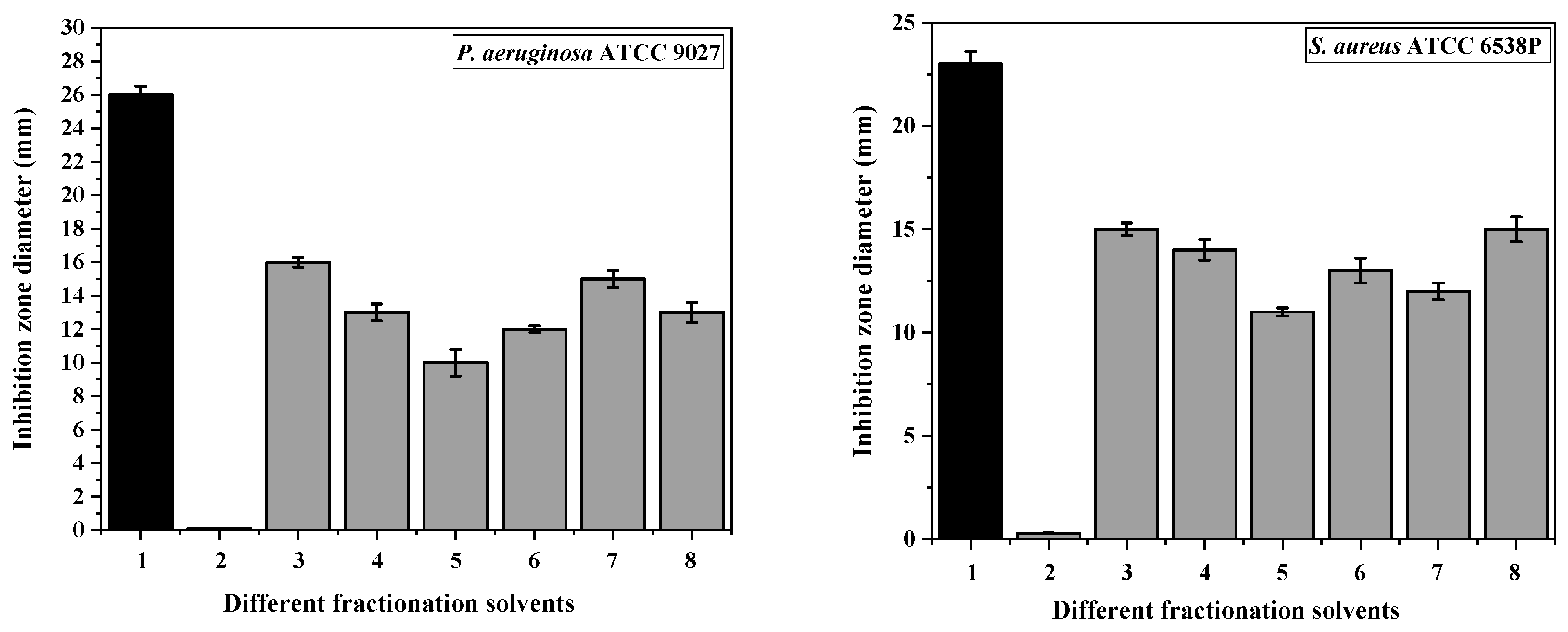
The EtOAc extract’s different fractions from H. saccharevitans were examined to determine their biological activities and MICs against multiple selected bacterial pathogens and compared with the tetracycline reference standard’s activity (Figure 1). Results for the disk diffusion assay using the fractionated extract for the target strain revealed that every fraction inhibited the target microorganisms, for which a significant difference was observed among different fractions, with inhibition zones varying from 9 ± 0.30 mm to 17 ± 1.22 mm. There was a notable variation amongst the various fractions, and they all displayed varying degrees of inhibition. The n-hexane 100% and MeOH 100% fractions were shown to be more effective and displayed noticeably maximum activities against S. aureus and B. subtilis at 17 ± 1.22 mm and 19 ± 1.33 mm, respectively, and Gram-negative bacteria P. aeruginosa and E. coli at 18 ± 1.10 mm and 16 ± 1.20 mm, respectively, which showed significantly higher activity in contrast to an alternative fraction. The susceptibility of EtOAc 100% and DCM 100% fractions against tested microbes was found to be similar. Tetracycline as a positive control was found to be significantly higher than the tested fractions, ranging from 23 ± 0.85 mm to 27 ± 0.55 mm inhibition zones, while DMSO (10%), as a negative control, did not exhibit any activity. Overall, E. coli was the highly susceptible strain against the different tested fractions.

3.2.2. Time–Kill Kinetics Assay

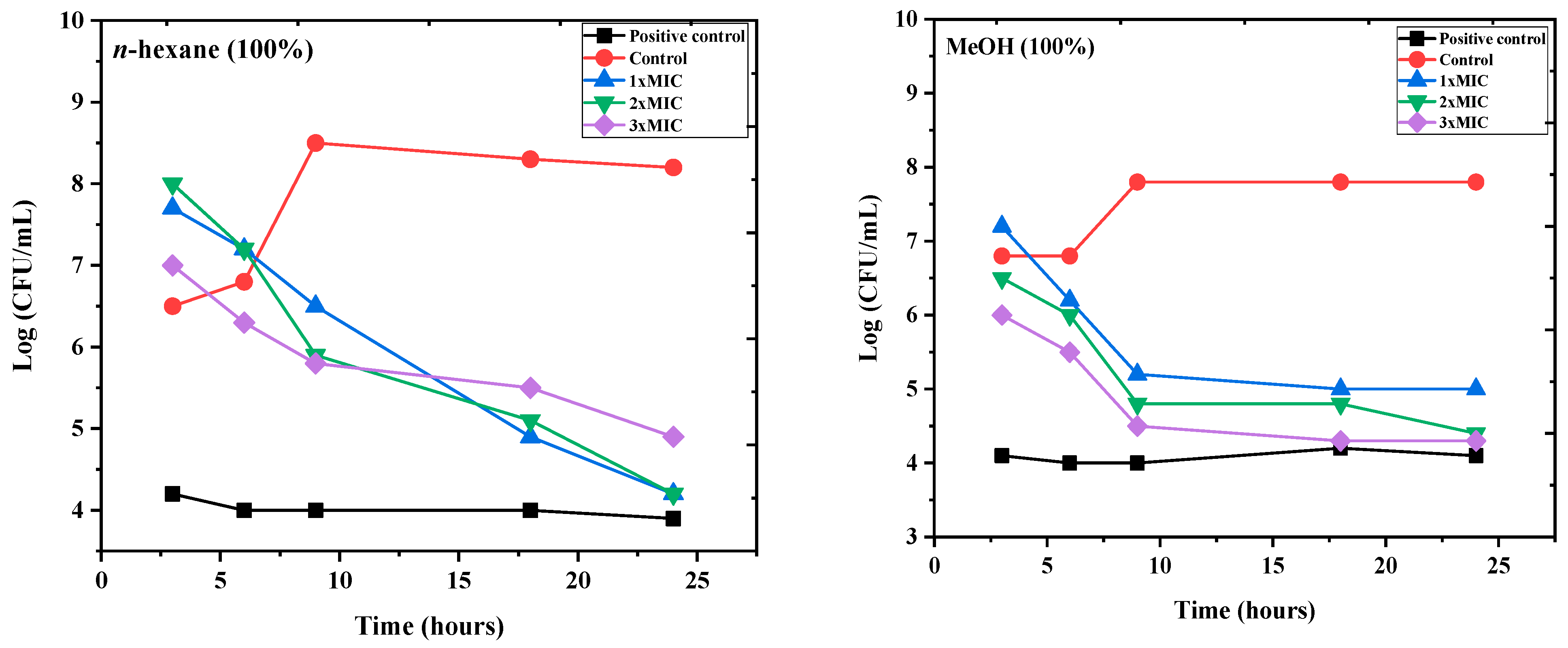
The in vitro time–kill kinetics test is the most applicable process employed for the predictive preclinical modeling framework to assess the antibacterial assay of the studied active substances against several targeted microbial pathogens. The most susceptible bacteria, E. coli, was the subject of a 24 h study using this assay, which revealed MIC values of the investigated active fractions n-hexane 100% and MeOH 100% (4.1 μg/mL and 3.5 μg/mL), 2 MIC (8.2 μg/mL and 7 μg/mL), and 3 MIC (12.3 μg/mL and 10.5 μg/mL), respectively. Figure 2 demonstrates the findings for the incubation times and the logarithmic number of CFU/mL. Comparing the two active fractions with the MIC, two MIC, and control, respectively, each of the three MICs revealed a significant increase in E. coli from 3 to 6 h, which increased steadily to 24 h.

3.2.3. Antifungal Activity

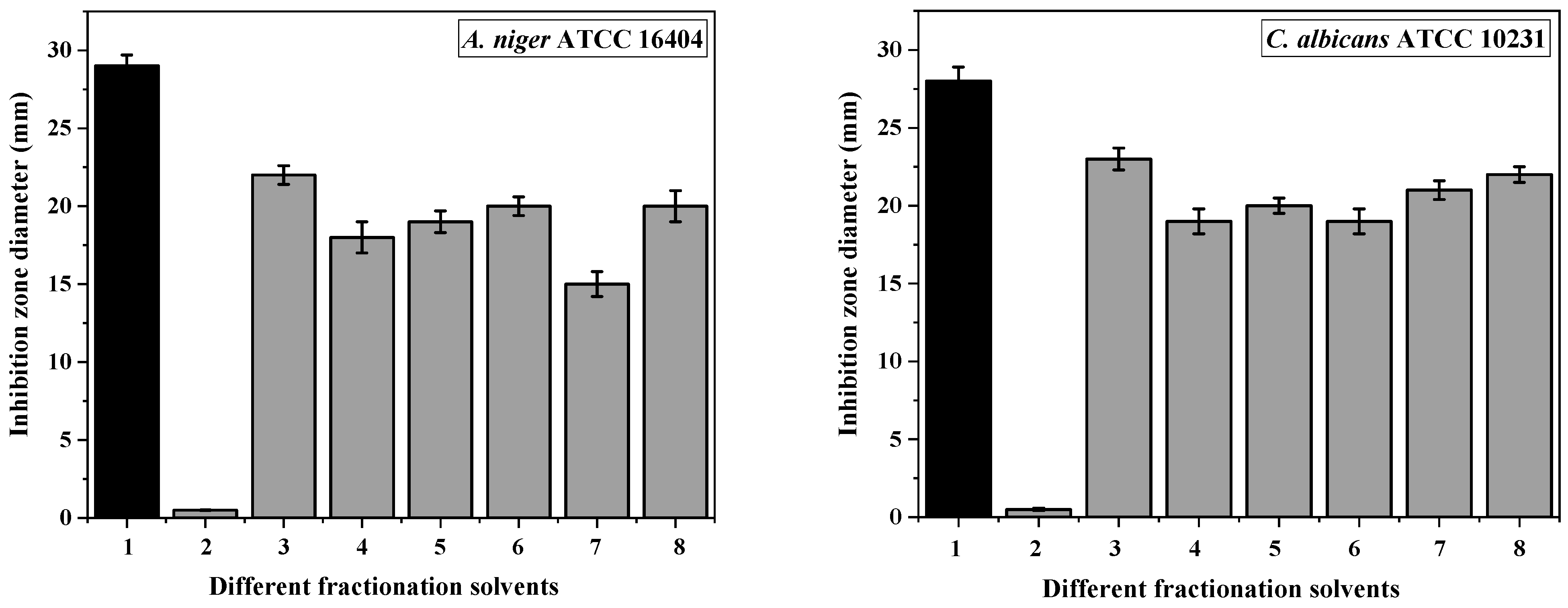
To ascertain the suppression of the growth of the chosen fungal pathogens, the antifungal actions of the various fractions of the bacterial crude extract were also assessed. According to our findings, every fraction exhibited broad-spectrum antifungal efficacy (Figure 3), especially n-hexane 100% and MeOH 100%, which had the highest inhibitory effects against A. niger and C. albicans strains, with inhibition zones ranging from 20 ± 0.85 mm to 23 ± 1.25 mm. The results reveal that the target fungi were susceptible to every fraction that was assessed, exhibiting a substantial degree of sensitivity with varying degrees of inhibition. The n-hexane:EtOAc (50:50) and DCM:MeOH (50:50) fractions showed the minimum effects against A. niger, with zones of inhibition of 18 ± 0.65 mm and 15 ± 0.33 mm, respectively, whereas n-hexane:EtOAc (50:50) and DCM 100% showed the lowest effects, with the same inhibition zones of 19 ± 0.25 mm on C. albicans. As a positive control, the antifungal properties of fluconazole were ascertained at (29 ± 0.30 mm and 28 ± 0.45 mm) in A. niger 16,404 and C. albicans 10231, respectively, (Figure 3). Moreover, the negative control of DMSO (10%) showed no activity on all tested fungal strains. The inhibition zones of the active fractions against tested bacteria and fungi are illustrated on Figure 4.

3.2.4. Determination of the Minimum Inhibitory Concentration (MIC)

The results of the MIC experiments indicate that all tested solvent extracts had broad-spectrum antimicrobial activity (Table 1), especially, n-hexane 100% and MeOH 100%, which exhibited the greatest inhibitory impact on E. coli and A. niger at 4.1 ± 0.024 µg/mL and 3.3 ± 0.01 µg/mL and 3.5 ± 0.02 µg/mL and 4.2 ± 0.18 µg/mL, respectively. Other tested extracts had the least effect on the other tested pathogenic microbes; S. aureus, P. aeruginosa, C. albicans, and B. subtilis had MIC values ranging from 13.2 ± 0.11 µg/mL to 4.5 ± 0.12 µg/mL. Overall, E. coli and A. niger were the strains with the highest sensitivity to the solvent extracts examined. Data display the MIC data, which were determined by averaging three replications (Table 1).

3.2.5. Cytotoxicity Activity

The cytotoxicity of n-hexane 100% and MeOH 100% on HepG2 was assessed using a MTT assay. Responses of HepG2 cells to increased concentrations of active fractions were exponential. HepG2 cells showed a significant decrease in viability with the increase of n-hexane 100% concentrations, while MeOH 100% showed less inhibition. The in vitro dosage (0.5, 1, 5, and 10 mg/mL) and time-dependent effects (24, 48, and 72 h) of the active fractions against HepG2 cells are shown in Figure 5. Incubation of HepG2 cells with 5 and 10 mg/mL of the n-hexane 100% active fraction showed promising cytotoxic activity, and 62%, 70%, and 78% decreases in cell viability were observed after 24, 48, and 72 h of incubation, respectively, as compared to the untreated control cells. Under the same conditions, incubation of HepG2 cells with the same concentration of MeOH 100% active fraction showed weak to moderate cytotoxic activity, with 35%, 45%, and 50% decreases in cell viability at a concentration of 10 mg/mL observed. In addition, 10%, 15%, and 20% inhibition of cells at a concentration of 5 mg/mL was observed after 24, 48, and 72 h of incubation, respectively, while the lowest concentration of 0.5 mg/mL did not show any significant inhibition at the same time points (Figure 5).

3.2.6. Antioxidant Activity

The radical DPPH assay was performed to assess the antioxidant activity of H. saccharevitans active fractions. In the DPPH assay, the radical scavenging activity of the n-hexane and methanolic fractions revealed that the antioxidant capacity increased with increasing levels of phenolic compounds for the target fractions and ascorbic acid. The result indicated that the methanolic extract fraction was more potent than the n-hexane and ascorbic acid scavenging activity (inhibition%), which ranged from 21.87 ± 0.85% and 98.25 ± 1.45% at concentrations of 1 and 8 mg/mL, respectively, and there was a linear relationship between antioxidant capacity and phenolic content, as shown in Table 2.

3.3. Molecular Docking of Identified Compounds

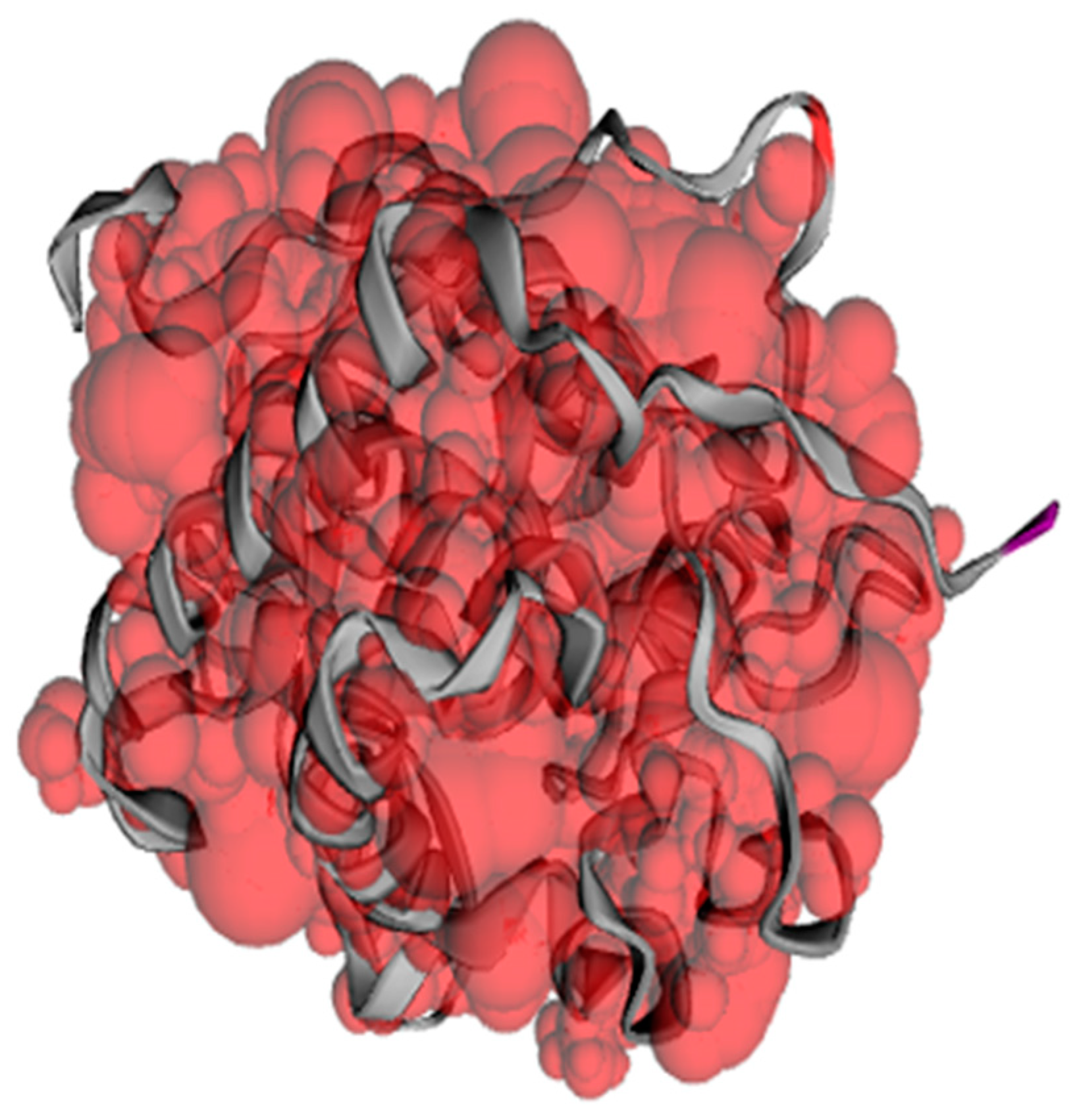
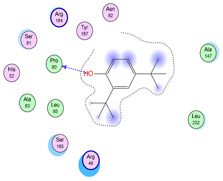
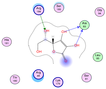
To anticipate the activity of selected compounds as antioxidants, molecular docking was carried out of these top-notch constituents with cytochrome c peroxidase enzyme (PDB code: 2X08) co-crystallized with ascorbic acid used as a reference. The binding pockets in the proteins were predicted via the CASTp server and are displayed in Figure 6, highlighting a surface pocket supported by atoms as identified by CASTpFold. The pocket panels displayed some residues within the pocket that could be targeted for interaction. The statistical parameters, including surface accessible (SA) area, pocket ID (Poc ID), and surface accessible (AS) volume, are represented in Table 3. The top binding pocket of cytochrome c peroxidase had the highest area (SA) of 17,711.840 Å2 and volume (SA) of 9434.812 Å3.
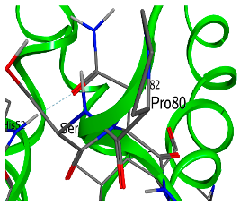
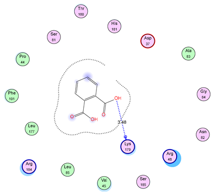
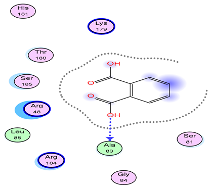
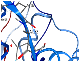
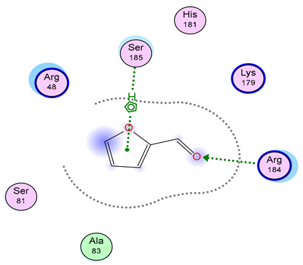
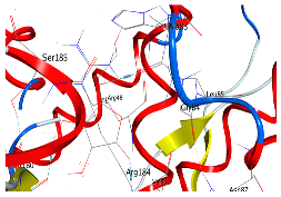
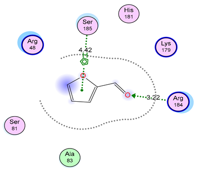
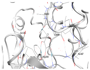
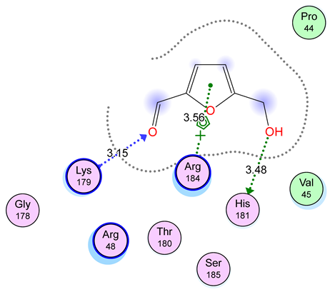
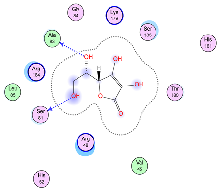
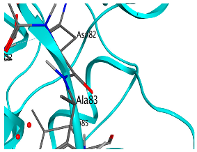
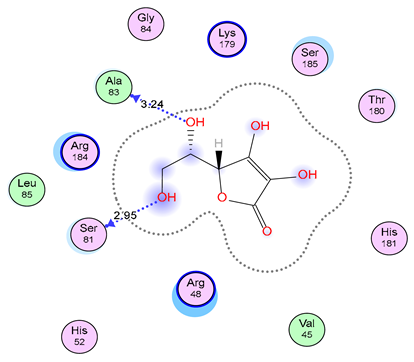
Following docking, the promising compounds displayed favorable scores (1 = −9.2, 2 = −8.5, 3 = −6.3, 4 = −7 kcal/mol) comparable to those of ascorbic acid (−9.5 kcal/mol), as depicted in Table 4. Briefly, the top two phytoconstituents were chosen that included the Phenol, 2,4-bis-(1,1-dimethylethyl) and 1,2-Benzenedicarboxylic acid derived from hexane extract, which exhibited hydrogen bonding interaction with Lys179 and Ala 83, respectively. Additionally, candidates derived from methanolic extract, including 2-Furancarboxaldehyde, were stabilized by a forming pi-hydrogen bond with Ser185 and hydrogen bonding with Arg 184 in the largest pocket of cytochrome c peroxidase. The complex 5-Hydroxymethylfurfural /2X08 exhibited a pi-hydrogen bond via Arg 184 and H-bonding interactions including Lys179 and His-181. Furthermore, the ascorbic acid depicted interaction with residues Ala83 and Ser81 through hydrogen bonding. The docking results showed these compounds docked with higher energy and sound binding interactions to the cytochrome c receptor with a hydrogen bond, which implies a strong interaction of the compounds with the receptor (Table 5).
Furthermore, the current study employed the fastDRH tool to assess the decomposition of energy associated with cytochrome c peroxidase encompassing electrostatic energy (tele), total gas phase energy (TGAS), van der Waals contribution (VDW), non-polar and polar contributions to solvation (TGBSOL), and the ultimate estimated binding free energy (TGBTOT). The cytochrome c peroxidase-Phenol, 2,4-bis-(1,1-dimethylethyl) exhibited the highest binding affinity, with a PB1 score of −0.98 kcal/mol and a GB1 score of −14.08 kcal/mol. The free binding energy (kcal/mol) rescoring profiles between the docked complexes are depicted in Table 6. The MM/PB(GB)SA findings of the cytochrome c peroxidase enzyme analyzed by the fast DRH server are highlighted in Table 7.

3.4. Identification of Potentially Bioactive Fractions by GC–MS

The chemical composition of the active solvent extracts n-hexane 100% and MeOH 100% from the selected strain H. saccharevitans were analyzed using GC–MS, and are shown in Table 8 and Figure 7. Multiple chemical compounds were detected and identified in both solvent extracts, supported by the comparison of the NIST database, where certain peaks resembled the closest compound, in addition to their retention time, molecular weight, and other attributes. It was observed that the most abundant constituents in dominant components of n-hexane 100% extract were phenol, 2,4-bis-(1,1-dimethyl ethyl), 1,2-Benzenedicarboxylic acid, 1-nonadecene, and 1-eicosene. Furthermore, GC–MS analysis of the MeOH 100% fraction of H. saccharevitans identified seven dominant chemical compounds, including 5-Hydroxymethylfurfural, -2-Furancarboxaldehyde, Methyl 2-furoate, oleic acid, and 2-Furancarboxaldehyde, 5-methyl- (Table 8 and Figure 7). All the obtained compounds from the active fractions are known, and the peak area of each compound is directly proportional to its quantity in the extract.

4. Discussion

Halophilic bacteria are among the most prolific microbial producers of bioactive secondary metabolites. In this study, in order to find marine bacteria for the discovery of bioactive metabolites, H. saccharevitans was obtained, grown, and fermented on a broth medium. Subsequently, its EtOAc crude extracts were tested for biological activities with MICs and MFCs against multiple pathogenic microbes. Two fractions, n-hexane and MeOH, were found to be the most active fractions against the target tested organisms and a chemical analysis was then conducted. Marine bacteria have great significance as a novel and promising source of a huge range of biologically active compounds. Some of these marine bacteria live in stressful habitats, under very low temperature, lightless, and high-pressure conditions [1]. Interestingly, many very diverse organisms are able to live in these environments and produce fascinating, structurally complex natural products. Only a small number of microbes have been studied for bioactive metabolites so far, but a large number of active compounds, some of which have distinct structural skeletons, have been identified [39]. Marine pharmacology is a new field that investigates potential therapeutic compounds derived from marine life. Extensive screening of marine substances and their various biological properties has been reported over the previous two decades [40].
Therefore, the search for biologically active molecules has extended to the screening of organisms associated with less explored environments. Marine sources have played a significant role as an origin for lead molecules ascertained for various pharmacological utilization in recent times. It is interesting to note that marine microorganisms occupy a vast expanse of the biosphere, ranging from the shallow water of the shoreline to the vast seaward regions that cover 70% of the biosphere, and continue to be the most important and unknown source of numerous bioactive compounds [1,41]. The explanation for why marine microorganisms are of interest is because they produce a variety of active chemical compounds that are valuable in biotechnology. These bioactive compounds are different in function, exhibiting antiviral, immunosuppressive, antifungal, antibacterial, anti-inflammatory, antiprotozoal, and antitumor properties [42]. Based on their resistance, they are promising biocatalysts for novel or active sustainable bio-industrial processes to address multiple factors, such as poor nutrition, temperature, salt, pH, and contaminants, representing an opportunity for various biotechnological, industrial, and biomedical applications [1]. When compared to other possible biosources, bacteria have been shown to be one of the most prolific biosources of natural compounds, accounting for a remarkably small proportion of molecules that have been identified [43]. Marine bacteria are considered to have biochemical, physiological, and molecular characteristics that are unlike those from their terrestrial equivalents and, consequently, they may produce various compounds [44]. Multiple diseases have few treatment options, and several chemical medications have clear adverse consequences for patients’ health when used in excess. Consequently, more study on marine bacteria is required to investigate potential treatments for these unpleasant bacterial diseases. Using natural products as antimicrobial treatments and for other associated disorders has long been considered a viable therapeutic approach [45].
Halomonas bacterial species in a saline environment can produce various hydrolytic enzymes, extracellular polysaccharides (EPSs), antibiotics, biosurfactants, different pigments, etc. Most of these compounds are being produced in large-scale fermentation for multiple commercial uses [46]. H. stenophila bacterium isolated from a hypersaline environment is reported to produce sulphate exopolysaccharides, which assessed multiple biological activities [21,47]. Our findings supported earlier reports that indicated the highest growth inhibition of B. subtilis and S. aureus bacteria was exhibited by the active chemicals extracted from a fermented marine Halomonas strain, GWS-BW-H8hM. Furthermore, a moderate efficacy of these compounds against C. albicans was observed [48]. In addition, the culture extract of Halomonas EA423 bacterium isolated from marine-associated sponges in the Red Sea exhibited antibacterial activity against multiple human pathogens, Methicillin-resistant S. aureus (MRSA) ATCC 43300, P. aeruginosa ATCC 27853, E. coli ATCC 8739, and Enterococcus faecalis ATCC 29,212 [9]. Furthermore, the EtOAc extracts of the salty H. salifodinae bacteria possess antibacterial activity against various aquatic pathogens, isolated from fish and shrimp [49]. Halomonas elongata strain 153B was determined to inhibit biofilm formations at high concentrations (750 and 1000 μg/mL) of multiple pathogenic bacteria such as S. aureus ATCC6538, P. aeruginosa 11778, E. coli 25922, and C. albicans 10231 [50].
Moreover, the studied strain showed a promising cytotoxic activity against HepG2, and reached decreases of up to 78% in cell viability after 72 h of incubation. Similar to our study, El-Garawani et al. used the MTT assay to investigate the anticancer potential of the Halomonas strain HA1 extract against the HepG2 cell line and found significant cytotoxic potential with a maximal inhibitory concentration (IC50) of 68  ±  1.8 μg/mL [40]. In contrast, the metabolites produced by halophilic archaea Halorhabdus rudnickae isolated from a Polish salt mine showed high resistance against HepG2 cells using the MTT assay with high concentrations [51], which was similar to the studied methanolic active fraction of H. saccharevitans H.S-AB2. It was reported that the polysaccharides produced from H. smyrnensis strain AAD6 exhibited anticancer activity against human cancer cell lines HepG2/C3A. In addition, carotenoid extracts from the haloarchaea Haloplanus vescus strain RO5-8 and Halogeometricum limi strain RO1-6 showed a potent anticancer activity against HepG2 cells in vitro, in a dose-dependent manner [52].
On the other hand, H. elongata demonstrated an excellent antioxidant activity at a 200 µg/mL concentration, reaching up to 67.88%, and was the best radical scavenger when DPPH scavenging was employed, with a powerful antioxidant activity of between 19.67% and 67.88% inhibition [50]. The orange pigment-producing Halomonas spp. were found to have high polyphenol content and demonstrated notable antioxidant qualities in biological and chemical investigations [53]. The production of valuable antioxidant and anti-inflammatory agent compounds from Halomonas neptunia, such as ectoine, has been reported in a previous study [54]. Another investigation showed that the halophilic bacteria Vibrio azureus strain MML1960 exhibited antifungal activity against C. albicans [55]. In agreement with an earlier study, H. aquamarina strain MB598 was isolated from the Khewra Salt Range of Pakistan, and its pigments had a significant antioxidant activity, revealing up to 85% free-radical scavenging activity [56]. Other research showed that different Halomonas taxa obtained from the different saline environments of Algeria’s northeast region also displayed extensive antifungal activities against multiple plant pathogenic fungi [57]. In addition, H. variabilis and H. smyrnensis showed antibacterial properties against E. coli and Sporosarcina pasteuri bacteria [58].
Certain halophilic bacteria can deliver a special chemical compound, such as halocins, which are capable of eliminating numerous other microorganisms, including certain strains that produce pigments with various strong bioactivities, such as antioxidants [59]. Some special proteins and enzymes generated by some halophilic bacteria were evaluated for antibacterial activities against plant diseases from Halomonas and Bacillus spp. [55]. In the biopharmaceutical and biomedical industries, the biological activity of halophilic bacteria from various hypersaline environments against several pathogenic species has drawn a great attention of interest in bioactive chemical applications. Furthermore, multiple prospective bioactivities of halobacteria still remain unexplored [23]. Greater interest should be shown in halo-microbial associations as a powerful biosource of new or active drugs against various resistant microorganisms. The docking results of the major detected compounds, which act as antioxidant agents, showed the lower binding energy score, which conferred better protein–ligand binding stability. MM-PB (GB)SA techniques are widely regarded as a favorable option for the computation of binding energies and the elucidation of appropriate binding configurations [60]. The MM/PBSA approach was utilized to assess the binding energies of the protein complexes incorporating ELE, VDW, GAS, PBSOL, PBTOT, and GBSOL, which supported the outcomes of molecular docking. Furthermore, advances in computational chemistry have made it possible to create some antimicrobial compounds generated from microorganisms that have the potential to be very effective therapeutic agents for managing bacteria that pose a hazard to human health [61]. Similarly, in silico docking studies showed that Pithomyces atroolivaceous mitochondrial F1F0 Adenosine triphosphate synthase enzymes readily interact with phenol, 2,4-bis(1,1-dimethylethyl) [62]. In another study, the molecular docking of 1,2-benzenedicarboxylic acid obtained from Cyamopsis tetragonoloba supercritical extracts demonstrated higher binding energy than that of the tested fatty acids [63].
GC-MS is one of the best, fastest, and most accurate methods for identifying different chemicals [64]. It can be used to identify a wide range of compounds in medicinal plants and microbial extracts, including alcohols, alkaloids, phenolics, long-chain hydrocarbons, organic acids, steroids, esters, and amino acids [65]. Additionally, GC-MS is crucial for adjusting for volatile or semi-volatile bioactive chemicals found in varied extracts, as well as for gaining access to their relevant activities [66]. The active fractions of H. saccharevitans H.S-AB2 were examined by GC-MS chemical analysis to detect the active compounds responsible for its bioactivity. The results showed that the active fractions of the GC–MS analysis profile revealed multiple major well-known compounds such as phenol, 2,4-bis-(1,1-dimethylethyl), 1,2-benzenedicarboxylic acid, 2-furancarboxaldehyde-, and 5-hydroxymethylfurfural. A halophic bacterium Halomonas salifodinae strain MPM-TC was screened, and its antimicrobial secondary metabolites were characterized in vivo using GC-MS analysis [49]. On the other hand, a H. aquamarina strain MB598 was fermented and the obtained bio-pigments were characterized by GC-MS for antimicrobial activities [56]. Halomonas bacterial strain MCTG39a was isolated and chosen for its production of an extracellular emulsion as an antioxidant agent, as confirmed by chromatographic analysis [67].
An earlier study demonstrated that phenol, 2,4-bis(1,1-dimethylethyl), detected by GC-MS as an active compound produced by Kutzneria actinobacteria, had antifungal activity against pathogenic fungi, as confirmed by in silico docking studies [62]. Similar to our findings, 2,4-bis(1,1-dimethylethyl) obtained from thermophilic B. licheniformis isolated from an Algerian hot spring demonstrated bioactivity against two multidrug-resistant bacteria, S. aureus and P. aeruginosa, using a radial diffusion assay at 55 °C [68]. Additionally, the 1,2-benzenedicarboxylic acid and bis(2-Methylpropyl) ester molecule were identified by GC-MS analysis of the Streptomyces cuspidosporus bioactive extract, and compounds were found to have a large peak area and significant biological properties [69]. Other compounds detected by GC-MS, such as 2-furancarboxaldehyde, also showed antibacterial and antifungal activities [70]. This study investigated the antibacterial, antifungal, and antioxidant activities, as well as the bioactive components, of H. saccharevitans active fractions. Taken together, the current findings verified that the antioxidant and antimicrobial activities of the H. saccharevitans H.S-AB2 active fractions might be due to the presence of these compounds, and it is capable of being developed further as biological and pharmacological substances.

5. Conclusions

In the current research, the secondary metabolites of a marine-derived bacteria Halomonas saccharevitans strain H.S-AB2 was extracted and fractioned with the help of chromatographic techniques and further subjected to varied biological activities. The active fractions of the crude extract, an absolute n-hexane and MeOH, showed significant in vitro antimicrobial properties against Gram-positive (S. aureus and Bacillus subtilis) and Gram-negative (P. aeruginosa and E. coli) bacteria, and selected pathogenic fungi A. niger and C. albicans. Moreover, the E. coli time–kill kinetics of a highly sensitive bacterium against active fractions was also examined. In addition, the cytotoxicity of the studied strain against HepG2 cells showed significant activity using the MTT assay. The DPPH scavenging activity confirmed that the active fractions had high excellent antioxidant activity. Furthermore, molecular docking of the investigated major molecules demonstrated high-affinity binding interactions. In GC-MS analysis of the most active fractions, multiple chemical compounds characterized by varied chemically esters, essential oils, phenolics, and other organic compounds were detected in different degrees of existence and concentration. Hence, these findings imply that the predominant components in these bioactive fractions have the potential to be developed as a more secure and potent substitute for the synthetic antioxidants, antimicrobials, and other significant biomedical applications that are now in use. However, future studies are needed to study the relevant key genes involved in biological activities via genetic engineering techniques, to assess the feasibility of producing target active metabolites via marine bacteria and improving the fermentation process. These could be fruitful varied strategies for further improvement of these metabolites and the development of specific therapeutic prospects.

Author Contributions

Conceptualization, H.M., U.M.A.-R. and Y.S.; methodology, H.M. and M.A.A.A.; software, H.M., M.A.A.A., S.W.M.H. and H.A.H.I.; validation, H.M. and M.F.A.; formal analysis, H.M., M.A.A.A. and S.W.M.H.; investigation, H.A.H.I. and M.F.A.; resources, M.A.A.A. and M.F.A.; data curation, H.M. and M.F.A.; writing—original draft preparation, H.M. and M.A.A.A.; writing—review and editing, H.M., M.A.A.A. and Y.S.; supervision, U.M.A.-R. and Y.S.; funding acquisition, Y.S. All authors have read and agreed to the published version of the manuscript.

Funding

This research was funded by Taif University, Saudi Arabia, Project No. (TU-DSPP-2024-186).

Institutional Review Board Statement

Not applicable.

Informed Consent Statement

Not applicable.

Data Availability Statement

The original contributions presented in the study are included in the article; further inquiries can be directed to the corresponding author.

Conflicts of Interest

The authors declare no conflicts of interest.

References

  1. Karthikeyan, A.; Joseph, A.; Nair, B.G. Promising Bioactive Compounds from the Marine Environment and Their Potential Effects on Various Diseases. J. Genet. Eng. Biotechnol. 2022, 20, 14. [Google Scholar] [CrossRef]
  2. Hamidi, M.; Kozani, P.S.; Kozani, P.S.; Pierre, G.; Michaud, P.; Delattre, C. Marine Bacteria versus Microalgae: Who Is the Best for Biotechnological Production of Bioactive Compounds with Antioxidant Properties and Other Biological Applications? Mar. Drugs 2019, 18, 28. [Google Scholar] [CrossRef] [PubMed]
  3. Li, P.; Lu, H.; Zhang, Y.; Zhang, X.; Liu, L.; Wang, M.; Liu, L. The Natural Products Discovered in Marine Sponge-Associated Microorganisms: Structures, Activities, and Mining Strategy. Front. Mar. Sci. 2023, 10, 1191858. [Google Scholar] [CrossRef]
  4. Hai, Y.; Wei, M.-Y.; Wang, C.-Y.; Gu, Y.-C.; Shao, C.-L. The Intriguing Chemistry and Biology of Sulfur-Containing Natural Products from Marine Microorganisms (1987–2020). Mar. Life Sci. Technol. 2021, 3, 488–518. [Google Scholar] [CrossRef] [PubMed]
  5. Ibrahim, A.H.; Attia, E.Z.; Hajjar, D.; Anany, M.A.; Desoukey, S.Y.; Fouad, M.A.; Kamel, M.S.; Wajant, H.; Gulder, T.A.M.; Abdelmohsen, U.R. New Cytotoxic Cyclic Peptide from the Marine Sponge-Associated Nocardiopsis Sp. UR67. Mar. Drugs 2018, 16, 290. [Google Scholar] [CrossRef] [PubMed]
  6. Renn, D.; Shepard, L.; Vancea, A.; Karan, R.; Arold, S.T.; Rueping, M. Novel Enzymes From the Red Sea Brine Pools: Current State and Potential. Front. Microbiol. 2021, 12, 732856. [Google Scholar] [CrossRef]
  7. El-Hossary, E.M.; Abdel-Halim, M.; Ibrahim, E.S.; Pimentel-Elardo, S.M.; Nodwell, J.R.; Handoussa, H.; Abdelwahab, M.F.; Holzgrabe, U.; Abdelmohsen, U.R. Natural Products Repertoire of the Red Sea. Mar. Drugs 2020, 18, 457. [Google Scholar] [CrossRef]
  8. Nadeem, F.; Oves, M.; Qari, H.; Ismail, I. Red Sea Microbial Diversity for Antimicrobial and Anticancer Agents. J. Mol. Biomark. Diagn. 2016, 7, 2. [Google Scholar] [CrossRef]
  9. Bibi, F.; Naseer, M.I.; Azhar, E.I. Assessing the Diversity of Bacterial Communities from Marine Sponges and Their Bioactive Compounds. Saudi J. Biol. Sci. 2021, 28, 2747–2754. [Google Scholar] [CrossRef]
  10. Cör Andrejč, D.; Knez, Ž.; Knez Marevci, M. Antioxidant, Antibacterial, Antitumor, Antifungal, Antiviral, Anti-Inflammatory, and Nevro-Protective Activity of Ganoderma Lucidum: An Overview. Front. Pharmacol. 2022, 13, 934982. [Google Scholar] [CrossRef]
  11. Leekha, S.; Terrell, C.L.; Edson, R.S. General Principles of Antimicrobial Therapy. Mayo Clin. Proc. 2011, 86, 156–167. [Google Scholar] [CrossRef]
  12. Dholakiya, R.N.; Kumar, R.; Mishra, A.; Mody, K.H.; Jha, B. Antibacterial and Antioxidant Activities of Novel Actinobacteria Strain Isolated from Gulf of Khambhat, Gujarat. Front. Microbiol. 2017, 8, 2420. [Google Scholar] [CrossRef]
  13. Al-shaibani, M.M.; Radin Mohamed, R.M.S.; Sidik, N.M.; El Enshasy, H.A.; Al-Gheethi, A.; Noman, E.; Al-Mekhlafi, N.A.; Zin, N.M. Biodiversity of Secondary Metabolites Compounds Isolated from Phylum Actinobacteria and Its Therapeutic Applications. Molecules 2021, 26, 4504. [Google Scholar] [CrossRef]
  14. Andryukov, B.; Mikhailov, V.; Besednova, N. The Biotechnological Potential of Secondary Metabolites from Marine Bacteria. J. Mar. Sci. Eng. 2019, 7, 176. [Google Scholar] [CrossRef]
  15. Petersen, L.-E.; Kellermann, M.Y.; Schupp, P.J. Secondary Metabolites of Marine Microbes: From Natural Products Chemistry to Chemical Ecology. In YOUMARES 9—The Oceans: Our Research, Our Future; Springer International Publishing: Cham, Switzerland, 2020; pp. 159–180. [Google Scholar]
  16. Chen, D.; Qian, X. A Brief History of Bacteria; World Scientific: Singapore, 2018; ISBN 978-981-322-515-2. [Google Scholar]
  17. Hussain, S.A.; Xu, A.; Sommers, C.H.; Sarker, M.I. Draft Genome Sequence of Red-Heat-Causing Halomonas Eurihalina MS1, a Moderately Halophilic Bacterium Isolated from Saline Soil in Alicante, Spain. Microbiol. Resour. Announc. 2020, 9, 1–2. [Google Scholar] [CrossRef] [PubMed]
  18. Donio, M.B.S.; Ronica, F.A.; Viji, V.T.; Velmurugan, S.; Jenifer, J.S.C.A.; Michaelbabu, M.; Dhar, P.; Citarasu, T. Halomonas Sp. BS4, A Biosurfactant Producing Halophilic Bacterium Isolated from Solar Salt Works in India and Their Biomedical Importance. Springerplus 2013, 2, 149. [Google Scholar] [CrossRef]
  19. Juan, C.A.; Pérez de la Lastra, J.M.; Plou, F.J.; Pérez-Lebeña, E. The Chemistry of Reactive Oxygen Species (ROS) Revisited: Outlining Their Role in Biological Macromolecules (DNA, Lipids and Proteins) and Induced Pathologies. Int. J. Mol. Sci. 2021, 22, 4642. [Google Scholar] [CrossRef]
  20. Llamas, I.; Amjres, H.; Mata, J.A.; Quesada, E.; Béjar, V. The Potential Biotechnological Applications of the Exopolysaccharide Produced by the Halophilic Bacterium Halomonas Almeriensis. Molecules 2012, 17, 7103–7120. [Google Scholar] [CrossRef] [PubMed]
  21. Boujida, N.; Palau, M.; Charfi, S.; El Moussaoui, N.; Manresa, A.; Miñana-Galbis, D.; Skali Senhaji, N.; Abrini, J. Isolation and Characterization of Halophilic Bacteria Producing Exopolymers with Emulsifying and Antioxidant Activities. Biocatal. Agric. Biotechnol. 2018, 16, 631–637. [Google Scholar] [CrossRef]
  22. Arahal, D.R.; Ventosa, A. The Family Halomonadaceae. In The Prokaryotes; Springer: New York, NY, USA, 2006; pp. 811–835. [Google Scholar]
  23. Gaffney, E.M.; Simoska, O.; Minteer, S.D. The Use of Electroactive Halophilic Bacteria for Improvements and Advancements in Environmental High Saline Biosensing. Biosensors 2021, 11, 48. [Google Scholar] [CrossRef]
  24. Mohamed, H.; Ebrahim, W.; El-Neketi, M.; Awad, M.F.; Zhang, H.; Zhang, Y.; Song, Y. In Vitro Phytobiological Investigation of Bioactive Secondary Metabolites from the Malus Domestica-Derived Endophytic Fungus Aspergillus Tubingensis Strain AN103. Molecules 2022, 27, 3762. [Google Scholar] [CrossRef]
  25. Al Mousa, A.A.; Mohamed, H.; Hassane, A.M.A.; Abo-Dahab, N.F. Antimicrobial and Cytotoxic Potential of an Endophytic Fungus Alternaria Tenuissima AUMC14342 Isolated from Artemisia judaica L. Growing in Saudi Arabia. J. King Saud. Univ.-Sci. 2021, 33, 101462. [Google Scholar] [CrossRef]
  26. Mohamed, H.; Hassane, A.; Rawway, M.; El-Sayed, M.; Gomaa, A.E.-R.; Abdul-Raouf, U.; Shah, A.M.; Abdelmotaal, H.; Song, Y. Antibacterial and Cytotoxic Potency of Thermophilic Streptomyces Werraensis MI-S.24-3 Isolated from an Egyptian Extreme Environment. Arch. Microbiol. 2021, 203, 4961–4972. [Google Scholar] [CrossRef]
  27. Bauer, A.W.; Kirby, W.M.; Sherris, J.C.; Turck, M. No TitlAntibiotic Susceptibility Testing by a Standardized Single Disk Method E. Am. J. Clin. Pathol. 1966, 45, 493–496. [Google Scholar] [CrossRef] [PubMed]
  28. M07-A9; National Committee for Clinical Laboratory Standards Methods for Dilution Antimicrobial Susceptibility Tests for Bacteria That Grow Aerobically. Clinical and Laboratory Standards Institute: Wayne, PA, USA, 2003.
  29. Fisher, M.C.; Alastruey-Izquierdo, A.; Berman, J.; Bicanic, T.; Bignell, E.M.; Bowyer, P.; Bromley, M.; Brüggemann, R.; Garber, G.; Cornely, O.A.; et al. Tackling the Emerging Threat of Antifungal Resistance to Human Health. Nat. Rev. Microbiol. 2022, 20, 557–571. [Google Scholar] [CrossRef]
  30. M39-A2; Clinical and Laboratory Standards Institute Reference Method for Broth Dilution Antifungal Susceptibility Testing of Filamentous Fungi. Approved Standard—Second Edition; Clinical and Laboratory Standards Institute: Wayne, PA, USA, 2008.
  31. Khalaf, N.A.; Shakya, A.K.; Al-Othman, A.; El-Agbar, Z.; Farah, H. Antioxidant Activity of Some Common Plants. Turkish J. Biol. 2008, 32, 51–55. [Google Scholar]
  32. Ravikumar, Y.S.; Mahadevan, K.M.; Kumaraswamy, M.N.; Vaidya, V.P.; Manjunatha, H.; Kumar, V.; Satyanarayana, N.D. Antioxidant, Cytotoxic and Genotoxic Evaluation of Alcoholic Extract of Polyalthia Cerasoides (Roxb.) Bedd. Environ. Toxicol. Pharmacol. 2008, 26, 142–146. [Google Scholar] [CrossRef] [PubMed]
  33. Vilar, S.; Cozza, G.; Moro, S. Medicinal Chemistry and the Molecular Operating Environment (MOE): Application of QSAR and Molecular Docking to Drug Discovery. Curr. Top. Med. Chem. 2008, 8, 1555–1572. [Google Scholar] [CrossRef]
  34. Kim, S.; Chen, J.; Cheng, T.; Gindulyte, A.; He, J.; He, S.; Li, Q.; Shoemaker, B.A.; Thiessen, P.A.; Yu, B.; et al. PubChem 2019 Update: Improved Access to Chemical Data. Nucleic Acids Res. 2019, 47, D1102–D1109. [Google Scholar] [CrossRef] [PubMed]
  35. Podvinec, M.; Schwede, T.; Peitsch, M.C. Docking for Neglected Diseases as Community Efforts. In Computational Structural Biology; World Scientific: Singapore, 2008; pp. 683–704. [Google Scholar]
  36. Enerijiofi, K.E.; Akapo, F.H.; Erhabor, J.O. GC–MS Analysis and Antibacterial Activities of Moringa Oleifera Leaf Extracts on Selected Clinical Bacterial Isolates. Bull. Natl. Res. Cent. 2021, 45, 179. [Google Scholar] [CrossRef]
  37. Balouiri, M.; Sadiki, M.; Ibnsouda, S.K. Methods for in Vitro Evaluating Antimicrobial Activity: A Review. J. Pharm. Anal. 2016, 6, 71–79. [Google Scholar] [CrossRef]
  38. Mohamed, H.; Awad, M.F.; Shah, A.M.; Sadaqat, B.; Nazir, Y.; Naz, T.; Yang, W.; Song, Y. Coculturing of Mucor Plumbeus and Bacillus Subtilis Bacterium as an Efficient Fermentation Strategy to Enhance Fungal Lipid and Gamma-Linolenic Acid (GLA) Production. Sci. Rep. 2022, 12, 13111. [Google Scholar] [CrossRef]
  39. Debbab, A.; Aly, A.H.; Lin, W.H.; Proksch, P. Bioactive Compounds from Marine Bacteria and Fungi. Microb. Biotechnol. 2010, 3, 544–563. [Google Scholar] [CrossRef] [PubMed]
  40. El-Garawani, I.M.; El-Sabbagh, S.M.; Abbas, N.H.; Ahmed, H.S.; Eissa, O.A.; Abo-Atya, D.M.; Khalifa, S.A.M.; El-Seedi, H.R. A Newly Isolated Strain of Halomonas Sp. (HA1) Exerts Anticancer Potential via Induction of Apoptosis and G2/M Arrest in Hepatocellular Carcinoma (HepG2) Cell Line. Sci. Rep. 2020, 10, 14076. [Google Scholar] [CrossRef] [PubMed]
  41. Ghosh, S.; Sarkar, T.; Pati, S.; Kari, Z.A.; Edinur, H.A.; Chakraborty, R. Novel Bioactive Compounds From Marine Sources as a Tool for Functional Food Development. Front. Mar. Sci. 2022, 9, 832957. [Google Scholar] [CrossRef]
  42. Ameen, F.; AlNadhari, S.; Al-Homaidan, A.A. Marine Microorganisms as an Untapped Source of Bioactive Compounds. Saudi J. Biol. Sci. 2021, 28, 224–231. [Google Scholar] [CrossRef]
  43. Kikuchi, Y.; Kawashima, M.; Iwatsuki, M.; Kimishima, A.; Tsutsumi, H.; Asami, Y.; Inahashi, Y. Comprehensive Analysis of Biosynthetic Gene Clusters in Bacteria and Discovery of Tumebacillus as a Potential Producer of Natural Products. J. Antibiot. 2023, 76, 316–323. [Google Scholar] [CrossRef] [PubMed]
  44. Siddharth, S.; Vittal, R.R. Evaluation of Antimicrobial, Enzyme Inhibitory, Antioxidant and Cytotoxic Activities of Partially Purified Volatile Metabolites of Marine Streptomyces Sp.S2A. Microorganisms 2018, 6, 72. [Google Scholar] [CrossRef]
  45. Suleria, H.A.R.; Gobe, G.; Masci, P.; Osborne, S.A. Marine Bioactive Compounds and Health Promoting Perspectives; Innovation Pathways for Drug Discovery. Trends Food Sci. Technol. 2016, 50, 44–55. [Google Scholar] [CrossRef]
  46. Biswas, J.; Jana, S.K.; Mandal, S. Biotechnological Impacts of Halomonas: A Promising Cell Factory for Industrially Relevant Biomolecules. Biotechnol. Genet. Eng. Rev. 2022, 1–30. [Google Scholar] [CrossRef]
  47. Llamas, I.; Béjar, V.; Martínez-Checa, F.; Martínez-Cánovas, M.J.; Molina, I.; Quesada, E. Halomonas stenophila Sp. Nov., a Halophilic Bacterium That Produces Sulphate Exopolysaccharides with Biological Activity. Int. J. Syst. Evol. Microbiol. 2011, 61, 2508–2514. [Google Scholar] [CrossRef]
  48. Bitzer, J.; Große, T.; Wang, L.; Lang, S.; Beil, W.; Zeeck, A. New Aminophenoxazinones from a Marine Halomonas Sp.: Fermentation, Structure Elucidation, and Biological Activity. J. Antibiot. 2006, 59, 86–92. [Google Scholar] [CrossRef]
  49. Velmurugan, S.; Raman, K.; Thanga Viji, V.; Donio, M.B.S.; Adlin Jenifer, J.; Babu, M.M.; Citarasu, T. Screening and Characterization of Antimicrobial Secondary Metabolites from Halomonas Salifodinae MPM-TC and Its in Vivo Antiviral Influence on Indian White Shrimp Fenneropenaeus Indicus against WSSV Challenge. J. King Saud. Univ.-Sci. 2013, 25, 181–190. [Google Scholar] [CrossRef]
  50. Erdal Altıntaş, Ö.; Toksoy Öner, E.; Çabuk, A.; Aytar Çelik, P. Biosynthesis of Levan by Halomonas Elongata 153B: Optimization for Enhanced Production and Potential Biological Activities for Pharmaceutical Field. J. Polym. Environ. 2023, 31, 1440–1455. [Google Scholar] [CrossRef]
  51. Kowalewicz-Kulbat, M.; Krawczyk, K.T.; Szulc-Kielbik, I.; Rykowski, S.; Denel-Bobrowska, M.; Olejniczak, A.B.; Locht, C.; Klink, M. Cytotoxic Effects of Halophilic Archaea Metabolites on Ovarian Cancer Cell Lines. Microb. Cell Fact. 2023, 22, 197. [Google Scholar] [CrossRef]
  52. Corral, P.; Amoozegar, M.A.; Ventosa, A. Halophiles and Their Biomolecules: Recent Advances and Future Applications in Biomedicine. Mar. Drugs 2019, 18, 33. [Google Scholar] [CrossRef] [PubMed]
  53. Patkar, S.; Shinde, Y.; Chindarkar, P.; Chakraborty, P. Evaluation of Antioxidant Potential of Pigments Extracted from Bacillus Spp. and Halomonas Spp. Isolated from Mangrove Rhizosphere. BioTechnologia 2021, 102, 157–169. [Google Scholar] [CrossRef]
  54. Orhan, F.; Ceyran, E. Valorisation of Cheese Whey for Ectoine Production by Halomonas Neptunia. Int. J. Dairy. Technol. 2024, 77, 146–155. [Google Scholar] [CrossRef]
  55. Santhaseelan, H.; Dinakaran, V.T.; Dahms, H.-U.; Ahamed, J.M.; Murugaiah, S.G.; Krishnan, M.; Hwang, J.-S.; Rathinam, A.J. Recent Antimicrobial Responses of Halophilic Microbes in Clinical Pathogens. Microorganisms 2022, 10, 417. [Google Scholar] [CrossRef]
  56. Fariq, A.; Yasmin, A.; Jamil, M. Production, Characterization and Antimicrobial Activities of Bio-Pigments by Aquisalibacillus Elongatus MB592, Salinicoccus Sesuvii MB597, and Halomonas Aquamarina MB598 Isolated from Khewra Salt Range, Pakistan. Extremophiles 2019, 23, 435–449. [Google Scholar] [CrossRef]
  57. Menasria, T.; Monteoliva-Sánchez, M.; Benammar, L.; Benhadj, M.; Ayachi, A.; Hacène, H.; Gonzalez-Paredes, A.; Aguilera, M. Culturable Halophilic Bacteria Inhabiting Algerian Saline Ecosystems: A Source of Promising Features and Potentialities. World J. Microbiol. Biotechnol. 2019, 35, 132. [Google Scholar] [CrossRef] [PubMed]
  58. Boyadzhieva, I.; Tomova, I.; Radchenkova, N.; Kambourova, M.; Poli, A.; Vasileva-Tonkova, E. Diversity of Heterotrophic Halophilic Bacteria Isolated from Coastal Solar Salterns, Bulgaria and Their Ability to Synthesize Bioactive Molecules with Biotechnological Impact. Microbiology 2018, 87, 519–528. [Google Scholar] [CrossRef]
  59. Oren, A.; Hallsworth, J.E. Microbial Weeds in Hypersaline Habitats: The Enigma of the Weed-like Haloferax Mediterranei. FEMS Microbiol. Lett. 2014, 359, 134–142. [Google Scholar] [CrossRef] [PubMed]
  60. Chen, F.; Sun, H.; Wang, J.; Zhu, F.; Liu, H.; Wang, Z.; Lei, T.; Li, Y.; Hou, T. Assessing the Performance of MM/PBSA and MM/GBSA Methods. 8. Predicting Binding Free Energies and Poses of Protein–RNA Complexes. RNA 2018, 24, 1183–1194. [Google Scholar] [CrossRef] [PubMed]
  61. Vallavan, V.; Krishnasamy, G.; Zin, N.M.; Abdul Latif, M. A Review on Antistaphylococcal Secondary Metabolites from Basidiomycetes. Molecules 2020, 25, 5848. [Google Scholar] [CrossRef] [PubMed]
  62. Devi, T.S.; Vijay, K.; Vidhyavathi, R.M.; Kumar, P.; Govarthanan, M.; Kavitha, T. Antifungal Activity and Molecular Docking of Phenol, 2,4-Bis(1,1-Dimethylethyl) Produced by Plant Growth-Promoting Actinobacterium Kutzneria Sp. Strain TSII from Mangrove Sediments. Arch. Microbiol. 2021, 203, 4051–4064. [Google Scholar] [CrossRef]
  63. Kaushik, S.; Kaushik, S.; Kumar, R.; Dar, L.; Yadav, J.P. In-Vitro and in Silico Activity of Cyamopsis tetragonoloba (Gaur) L. Supercritical Extract against the Dengue-2 Virus. VirusDisease 2020, 31, 470–478. [Google Scholar] [CrossRef]
  64. Konappa, N.; Udayashankar, A.C.; Krishnamurthy, S.; Pradeep, C.K.; Chowdappa, S.; Jogaiah, S. GC–MS Analysis of Phytoconstituents from Amomum Nilgiricum and Molecular Docking Interactions of Bioactive Serverogenin Acetate with Target Proteins. Sci. Rep. 2020, 10, 16438. [Google Scholar] [CrossRef] [PubMed]
  65. Razack, S.; Kumar, K.; Nallamuthu, I.; Naika, M.; Khanum, F. Antioxidant, Biomolecule Oxidation Protective Activities of Nardostachys Jatamansi DC and Its Phytochemical Analysis by RP-HPLC and GC-MS. Antioxidants 2015, 4, 185–203. [Google Scholar] [CrossRef]
  66. Wang, W.; Chen, H.; Zhu, W.; Gong, Z.; Yin, H.; Gao, C.; Zhu, A.; Wang, D. A Two-Staged Adsorption/Thermal Desorption GC/MS Online System for Monitoring Volatile Organic Compounds. Environ. Monit. Assess. 2023, 195, 869. [Google Scholar] [CrossRef]
  67. Gutierrez, T.; Morris, G.; Ellis, D.; Mulloy, B.; Aitken, M.D. Production and Characterisation of a Marine Halomonas Surface-Active Exopolymer. Appl. Microbiol. Biotechnol. 2020, 104, 1063–1076. [Google Scholar] [CrossRef] [PubMed]
  68. Aissaoui, N.; Mahjoubi, M.; Nas, F.; Mghirbi, O.; Arab, M.; Souissi, Y.; Hoceini, A.; Masmoudi, A.S.; Mosbah, A.; Cherif, A.; et al. Antibacterial Potential of 2,4-Di-Tert-Butylphenol and Calixarene-Based Prodrugs from Thermophilic Bacillus Licheniformis Isolated in Algerian Hot Spring. Geomicrobiol. J. 2019, 36, 53–62. [Google Scholar] [CrossRef]
  69. Sholkamy, E.N.; Muthukrishnan, P.; Abdel-Raouf, N.; Nandhini, X.; Ibraheem, I.B.M.; Mostafa, A.A. Antimicrobial and Antinematicidal Metabolites from Streptomyces Cuspidosporus Strain SA4 against Selected Pathogenic Bacteria, Fungi and Nematode. Saudi J. Biol. Sci. 2020, 27, 3208–3220. [Google Scholar] [CrossRef] [PubMed]
  70. Oskoueian, E.; Abdullah, N.; Ahmad, S.; Saad, W.Z.; Omar, A.R.; Ho, Y.W. Bioactive Compounds and Biological Activities of Jatropha curcas L. Kernel Meal Extract. Int. J. Mol. Sci. 2011, 12, 5955–5970. [Google Scholar] [CrossRef] [PubMed]
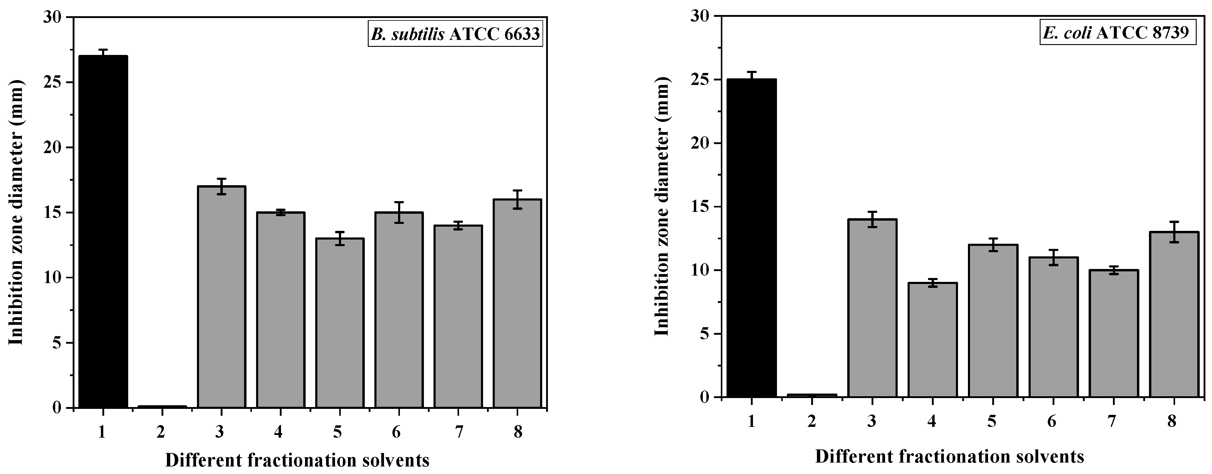
Figure 1. Antibacterial activity using the disc diffusion method. The various fractions of H. saccharevitans were examined for their antibacterial properties against various pathogenic bacteria. 1: tetracycline as a positive control, 2: DMSO (10%), 3: n-hexane (100%), 4: n-hexane: EtOAc (50:50), 5: EtOAc (100%), 6: DCM (100%), 7: DCM:MeOH (50:50), and 8: MeOH (100%). The standard error of the means (n = 3) is shown by error bars.
Fermentation 10 00433 g001aFermentation 10 00433 g001b
Figure 2. The time–kill kinetics of n-hexane 100% and MeOH 100% active fractions obtained from H. saccharevitans H.S-AB2 against E. coli. Positive control cells are treated with 50 µg/mL of tetracycline, whereas red control cells are untreated.
Fermentation 10 00433 g002
Figure 3. Antifungal assay of different solvents against tested fungal pathogens, 1: fluconazole as a positive control, 2: DMSO (10%), 3: n-hexane (100%), 4: n-hexane: EtOAc (50:50), 5: EtOAc (100%), 6: DCM (100%), 7: DCM:MeOH (50:50), and 8: MeOH (100%). The standard error of the means (n = 3) is represented by the error bars.
Fermentation 10 00433 g003
Figure 4. Inhibition zones of the antibacterial and antifungal activities of the active fractions against (A) S. aureus, (B) P. aeruginosa, (C) B. subtilis, (D). E. coli, (E) C. albicans and (F) A. niger. 1: positive control, 2: negative control, 3: MeOH (100%) fraction, and 4: n-hexane (100%) fraction.
Fermentation 10 00433 g004
Figure 5. Time- and dose-dependent inhibitory effect of (A) the n-hexane extract (100%) and (B) the methanolic extract (100%) of H. saccharevitans on HepG2 cells. Error bars are presented as mean ± SD from three independent experiments.
Fermentation 10 00433 g005
Figure 6. A surface pocket supported by atoms in grey as identified by CASTpFold, which depicts antioxidant activity.
Fermentation 10 00433 g006
Figure 7. GC-MS chromatogram of (A) the hexane extract (100%) and (B) the methanolic extract (100%) of H. saccharevitans.
Fermentation 10 00433 g007aFermentation 10 00433 g007b
Table 1. MIC and MFC values of the fractioned EtOAc crude extract of H. saccharevitans against the studied pathogenic microorganisms.
Tested FractionsMIC and MFC Values (μg/mL)
E. coliP. aeruginosaS. aureusB. subtilisA. nigerC. albicans
n-hexane 100%4.1± 0.0245.1 ± 0.115.3 ± 0.164.1 ± 0.203.3 ± 0.016.5 ± 0.25
n-hexane:EtOAc (50:50)13.2 ± 0.118.4 ± 0.115.9 ± 0.195.6 ± 0.156.7 ± 0.3311.4 ± 1.30
EtOAc 100%9.8 ± 0.1311.9 ± 0.147.6 ± 0.217.8 ± 0.335.5 ± 0.1210.6 ± 0.42
DCM 100%8.2 ± 0.158.1 ± 0.226.1 ± 0.296.1 ± 0.294.7 ± 0.3010.2 ± 1.65
DCM:MeOH (50:50)7.5 ± 0.144.5 ± 0.125.1 ± 0.255.5 ± 0.128.8 ± 0.029.8 ± 0.01
MeOH 100%3.5 ± 0.026.5 ± 0.034.3 ± 0.014.1 ± 0.114.2 ± 0.187.6 ± 0.21
Table 2. Inhibition values (%) of standard and active fractions of H. saccharevitans in DPPH radical scavenging assay.
Concentration
(mg/mL)
Scavenging Activity (Inhibition%)Ascorbic Acid
(Inhibition%)
Hexane Extract (100%)Methanolic Extract (100%)
112.32 ± 0.1021.87 ± 0.8510.64 ± 0.11
240.05 ± 0.2247.89 ± 0.9421.56 ± 0.22
354.71 ± 0.3863.92 ± 1.0532.81 ± 0.40
469.8 ± 0.5073.06 ± 1.2040.00 ± 0.55
575.02 ± 0.4880.33 ± 1.1049.78 ± 0.75
681.04 ± 0.5689.28 ± 0.8561.23 ± 0.80
788.1 ± 1.1193.23 ± 1.3378.95 ± 1.12
892.91 ± 1.2598.25 ± 1.4588.23 ± 1.5
Table 3. The CASTp data statistics of the cytochrome c peroxidase protein.
Poc IDArea (SA) Å2Volume (SA) Å3
117,711.8409434.812
28.8304.313
30.9480.373
40.8730.294
Table 4. Pose score results of detected major compounds’ interaction with cytochrome c peroxidase.
Seq.PubChem IDCompoundScore (kcal/mol)RMSD (Å)Receptor Interaction Bond Distance
17311Phenol, 2,4-bis-(1,1-dimethylethyl)−9.21.2Lys179/H-donor3.49
210171,2-Benzenedicarboxylic acid−8.52.4Ala83/H-donor2.88
373622-Furancarboxaldehyde−6.31.5Ser185/pi-H
Arg184/H-acceptor
4.42
3.22
42373325-Hydroxymethylfurfural−70.4Arg184/pi-Hydrogen
Lys179/H-acceptor
His-181/ H-donor
3.56
3.15
3.48
554670067Ascorbic acid−9.51.2Ala83/H-donor
Ser81/H-donor
3.24
2.95
Table 5. Two- and three-dimensional receptor interactions and receptor positioning of the most significant potential candidates compared to the docked ascorbic acid as a reference standard inside the binding pocket of cytochrome c peroxidase. The various colors are the visualization features in MOE used to indicate helix and beta sheets.
Molecule2D3D
Phenol, 2,4-bis-(1,1-dimethylethyl)Fermentation 10 00433 i001Fermentation 10 00433 i002
Fermentation 10 00433 i003
1,2-Benzenedicarboxylic acidFermentation 10 00433 i004Fermentation 10 00433 i005
Fermentation 10 00433 i006
2-FurancarboxaldehydeFermentation 10 00433 i007Fermentation 10 00433 i008
Fermentation 10 00433 i009
5-HydroxymethylfurfuralFermentation 10 00433 i010Fermentation 10 00433 i011
Fermentation 10 00433 i012
Ascorbic acid
(docked)
Fermentation 10 00433 i013Fermentation 10 00433 i014
Fermentation 10 00433 i015
Table 6. Free binding energy (kcal/mol) rescoring profiles between docked enzyme and hit compounds.
Protein CompoundPoseProcedure and Score
1PB1PB3PB4GB1GB2GB5GB6
Cytochrome c peroxidasePhenol, 2,4-bis-(1,1-dimethylethyl) −0.98−9.64−10.98−14.08−13.13−14.05−13.95
1,2-Benzenedicarboxylic acid 4.71−5.01−4.1−7.01−6.06−6.26−7.94
2-Furancarboxaldehyde 1.73−0.19−2.75−5.26−4.01−3.98−1.28
5-Hydroxymethylfurfural −1.49−5.58−6.36−8.34−7.86−8.36−7.97
Ascorbic acid 8.81.672.52−3.15−2.48−2.6−1.84
Table 7. The MM/PB(GB)SA analysis of cytochrome c peroxidase protein.
ELEVDWINTGASPBSUR/GBSURPBCAL/GBPBSOL/GBSOLPBELE/GBELEPBTOT/GBTOT
−15.91−12.340−28.25−11.5918.0627.272.15−0.98
Table 8. The identified major chemical compounds in the absolute hexane and methanolic extracts of H. saccharevitans by GC-MS analysis.
No.CompoundsChemical FormulaMolecular WeightRT (min)Match FactorArea (%)
Hexane extract (100%)
11-NonadeceneC19H3826615.228415.03
2Phenol, 2,4-bis-(1,1-dimethylethyl)C14H22O20616.3097556.33
31-HexadecanolC16H34O24218.348444.36
41-EicoseneC20H4028021.228144.86
5Heptacos-1-eneC27H5437823.848222.62
617-PentatriaconteneC35H7049026.266711.91
71,2-Benzenedicarboxylic acidC24H38O439030.6991614.77
8HexaphenylcyclotrisiloxaneC36H30O3Si359441.217062.10
Methanolic extract (100%)
1-2-FurancarboxaldehydeC5H4O2964.8194012.52
22-Furancarboxaldehyde, 5-methyl-C6H6O21107.349183.10
3Methyl 2-furoateC6H6O312610.049005.50
4Hepta-2,4-dienoic acid, methyl esterC8H12O214011.546761.85
55-HydroxymethylfurfuralC6H6O312614.1893259.44
6Oleic acidC18H34O228225.178453.19
7Methyl 5,13-docosadienoateC23H42O235029.317550.66
Disclaimer/Publisher’s Note: The statements, opinions and data contained in all publications are solely those of the individual author(s) and contributor(s) and not of MDPI and/or the editor(s). MDPI and/or the editor(s) disclaim responsibility for any injury to people or property resulting from any ideas, methods, instructions or products referred to in the content.