Article

In Vitro Probiotic Characterization of Lactiplantibacillus plantarum Strains Isolated from Traditional Fermented Dockounou Paste

by
Natia Joseph Kouadio
1,
Alalet Luc Olivier Zady
1,
Kouassi Aboutou Séverin Kra
1,
Filofteia Camelia Diguță
2,*,
Sébastien Niamke
1 and
Florentina Matei
2,3
1
Laboratory of Biotechnology Agriculture and Valorization of Biological Resource, UFR Biosciences, Felix Houphouet-Boigny University, Abidjan 22 BP 582, Côte d’Ivoire
2
Faculty of Biotechnologies, University of Agronomic Sciences and Veterinary Medicine Bucharest, 59 Marasti Blvd., 011464 Bucharest, Romania
3
Faculty of Food Industry and Tourism, Transilvania University of Brasov, 148 Castelului St., 500014 Brasov, Romania
*
Author to whom correspondence should be addressed.
Fermentation 2024, 10(5), 264; https://doi.org/10.3390/fermentation10050264
Submission received: 25 April 2024 / Revised: 14 May 2024 / Accepted: 17 May 2024 / Published: 19 May 2024
(This article belongs to the Special Issue Applications of Lactic Acid Bacteria in Fermented Foods and Beverages)

Abstract

:
This study aimed to evaluate the probiotic properties of 10 lactic acid bacteria (LAB) isolated from artisanal fermented plantain dockounou paste. A preliminary characterization of the LAB isolates was performed based on phenotypic and several biochemical properties, which was subsequently confirmed through 16S rRNA gene sequencing analysis, indicating that these isolates belonged to the species Lactiplantibacillus plantarum. With regard to safety criteria, the strains exhibited no alpha or beta hemolysis activity. Nevertheless, the majority of LAB strains demonstrated high sensitivity to the antibiotics tested. The results demonstrated that the majority of the strains exhibited remarkably high survival rates under simulated gastrointestinal conditions, such as pH = 1.5 (81.18–98.15%), 0.3% bile salts (68.62–100.89%), 0.4% phenol (40.59–128.24%), as well as 0.1% pepsin and pH = 2.5 (88.54–99.78%). The LAB strains demonstrated elevated levels of cell surface properties, indicative of the presence of a considerable defensive mechanism against pathogens. Intact LAB cells exhibited significant antioxidant abilities (48.18–83.58%). They also demonstrated a pronounced inhibitory effect on the growth of foodborne pathogens. Enzyme pattern analysis revealed that the LAB isolates produced both proteases and cellulases, as well as pectinase and/or amylase activity. The potential of the L. plantarum strains FS43, FS44, and FS48, as indicated by the results obtained from the standard in vitro assays, makes them suitable for further study as potential probiotics.

1. Introduction

Dockounou paste is an Ivorian traditional food made by combining senescent plantain flesh with maize or rice flour and baking or steaming it [1,2]. This food is produced through several processes, including peeling the senescent plantain fruit, scooping out the flesh, combining it with flour, fermenting, packing, frying, and processing it into flour [1,2].
Lactic acid bacteria, including those belonging to the genera Lactococcus, Lactobacillus, and Streptococcus, have been identified as playing a key role in the fermentation of traditional cereal-based foods and beverages worldwide [3,4,5,6]. Fermentation is the most significant of these processes since it improves the texture and organoleptic qualities of the food [4,5,6].
The potential health benefits of indigenous African fermented foods and beverages have not been adequately researched. It is a widely held view that the popularity of traditional fermented foods can be attributed to many factors, including the enhancement of taste, the conservation of food for a longer shelf life, and the improvement of nutritional value and digestive effectiveness. Some African cultural groups espouse the belief that fermented foods can be efficient in the treatment of illnesses, with a particular focus on digestive disorders. The indigenous microbial consortia present in fermented indigenous African foods are responsible for the beneficial properties of fermented foods mentioned above (reviewed by Achi and Asamudo [7]; Malongane and Berejena [8]). In recent years, numerous studies have been conducted with a focus on the isolation of LABs and yeasts from traditional fermented foods to identify potential starter cultures that can also exhibit valuable probiotic properties [3,4,5,6]. LAB species have been identified as the agents responsible for the manufacture of various Ivorian indigenous foods and beverages, such as attiéké, attoupkou, placali (fermented tubers), baca, wômi, doklu (fermented cereal-based foods), adjuevan (fermented fish), bandji and tchapalo (fermented beverages), and dockounou (fermented over-ripe fruits of plantain) [8,9,10,11,12,13,14]. Throughout the fermentation process of dockounou paste, Kouadio et al. [11] found different types of microorganisms predominating, such as yeasts, bacilli, and lactic acid bacteria. L. plantarum (64%), Weissella cibaria (22%), L. fermentum (7%), and Pediococcus acidilactici (7%) were mainly detected during the first 24 h of the production of doklu. L. plantarum (56%) dominated after 48 h of fermentation, and L. fermentum (100%) prevailed at the end of fermentation [12]. Mogmenga et al. [15] successfully isolated Saccharomyces cerevisiae with proven probiotic properties from a traditional fermented beer produced in Burkina Faso, known as Rabilé.
Also, the presence of Lactobacillus spp., Bifidobacterium spp., and S. cerevisiae yeasts has been identified in Romanian fermented beverages (socata and borș) [16,17], Romanian fermented dairy products [18,19,20], and Romanian fermented vegetables [18,21,22,23].
Probiotics are live microorganisms that assist the body in maintaining optimal health when consumed in appropriate quantities [24,25]. Lactic acid bacteria, such as Bifidobacterium and Lactobacillus, isolated from various sources, have been widely investigated using standard in vitro tests for key functional, probiotic, and technological properties [26,27,28,29,30,31,32]. The beneficial effects on health can be attributed to the synthesis of compounds that inhibit the proliferation of pathogens and compete with them for nutrients and adherence sites on the epithelial cells of the gut tract [29,30,31,32]. Probiotics can improve the immune system through macrophage activation, increased levels of immune globulins, increased natural killer cell activity, and/or increased levels of cytokines [30,31,32].
Furthermore, pharmaceutical probiotics are often expensive, and fermented foods and beverages represent a valuable alternative source of probiotics that may be more accessible to those who are unable to afford them, particularly in marginalized communities in Africa.
The objective of the present study was to assess the potential of LAB strains isolated from traditional fermented plantain dockounou paste as a probiotic in vitro. This was achieved by evaluating their functional properties (resistance in simulated gastrointestinal conditions and cell surface properties), probiotic properties (antibacterial, antioxidant, and enzyme activities), and safety properties (molecular identification, antibiotic sensitivity, and hemolysis activity).

2. Materials and Methods

2.1. LAB Strains and Growth Conditions

To evaluate their probiotic properties, 40 bacterial strains were isolated from fermented Dockounou paste [11]. The LAB isolates were preliminarily identified using conventional techniques, including colonial morphology, Gram staining, and biochemical reactions (catalase and oxidase tests). A total of 10 representative LAB isolates were selected for this research and preserved in cryotubes at a temperature of −20 °C in MRS broth, which was additionally supplemented with 20% glycerol. Before use, the stock LAB cultures were inoculated into MRS broth and incubated at 37 °C for 24 h.

2.2. Identification of LAB Strains via 16S rDNA Sequencing

Overnight LAB cultures were harvested through centrifugation at 5000× g for 10 min. The LAB cells were then employed to isolate bacterial DNA using the Zymo Research Quick-DNATM Fungal/Bacterial Miniprep Extraction Kit (Zymo Research, Irvine, CA, USA), following the manufacturer’s instructions. Finally, DNA quantification was conducted using the SpectraMax®® QuickDrop™ (Molecular Devices, San Jose, CA, USA). Polymerase chain reaction (PCR) amplification of the 16S ribosomal DNA (rDNA) region was performed using the primers 27F (5′ -AGAGTTTGATCCTGGCTCAG-3′) and 1492R (5′ -TACGGYTACCTTGTTACGACTT-3′). Amplified DNA fragments were then subjected to sequencing in both directions using the same primers by the Cellular and Molecular Immunological Application (CEMIA) sequencing service (Larissa, Greece). The newly obtained sequences were subjected to comparison with those already deposited in the NCBI database using the Basic Local Alignment Search Tool (BLAST; https://blast.ncbi.nlm.nih.gov/Blast.cgi, latest accessed 4 December 2023) to identify the species level of the LAB strains based on the level of similarity. Furthermore, a phylogenetic tree was constructed using MEGA software version 11.0.13 [33] based on 16S rDNA genes to identify the bacterial species with the highest degree of similarity [34].

2.3. In Vitro Tolerance of LAB Strains to Simulated Gastrointestinal Conditions

2.3.1. Resistance to Low pH

The influence of low pH (1.5) on the growth of LAB strains was explored using the method described by Carr et al. [35]. This involved inoculating fresh bacterial cultures into 6 mL of sterile MRS broth adjusted to pH 1.5 with 1 M HCl and incubating them at 37 °C for 3 h. The number of viable cells was then determined using the plate count agar method, with the results expressed as log CFU/mL. Survival rate (SR) was calculated using the following formula:
SR % = L o g   C F U / m L ( f i n a l e ) L o g   C F U / m L ( i n i t i a l ) × 100 ,
where initial and final mean the viable cells (CFU/mL) at 0 h and after 3 h of incubation.

2.3.2. Resistance to Pepsin and Low pH

The capacity of LAB isolates to tolerate pepsin in acid media was tested following the method proposed by Diguță et al. [36], with few modifications. The simulated gastric juice was formulated by combining 0.128% sodium chloride (NaCl), 0.0239% potassium chloride (KCl), and 0.64% sodium bicarbonate (NaHCO3) (Central Drug House, Mumbai, India) with 0.1% (w/v) pepsin (Himedia, Mumbai, India). The pH was then adjusted to 2.5. Overnight LAB cells were collected via centrifugation at 2000× g for 10 min and washed twice in sterile physiological saline (0.9% NaCl). The cells were then suspended in simulated gastric juice. Samples were taken at 0 and 3 h, serially diluted, and plated on MRS agar plates. Viable cells were quantified using the plate count method. The formula employed to calculate the survival rate in simulated gastric juice is identical to that used to assess acid pH resistance.

2.3.3. Tolerance to Bile Salts

The methodology proposed by Coulibaly et al. [28] was employed to assess the ability of LAB strains to withstand the effects of bile salts. The LAB strains were prepared as previously described. Cell pellets were inoculated in MRS broth supplemented with 0.3% (w/v) bile salts and then incubated for 4 h at 37 °C. Viable cells were quantified using the plate count method, with results expressed as log CFU/mL. The survival rate was performed according to the following formula:
SR % = L o g   C F U / m L ( f i n a l ) L o g   C F U / m L ( i n i t i a l ) × 100 ,
where initial and final mean the viable cells (CFU/mL) at 0 h and after 4 h of incubation.

2.3.4. Phenol Tolerance

The ability of LAB isolates to survive in the presence of a phenol solution was investigated using a methodology described by Xanthopoulos et al. [37]. Overnight LAB cultures were inoculated in MRS broth containing 0.4% phenol and incubated for 24 h at 37 °C. Viable cells were subsequently quantified via the plate count method.

2.4. Safety Assessment

2.4.1. Hemolysis Test

The hemolytic potential of LAB isolates was performed by the methodology described by Yadav et al. [38]. Bacterial strains were inoculated onto blood agar plates (Oxoid, Basingstoke Hampshire, UK) supplemented with 5% (w/v) sheep blood and incubated at a temperature of 37 °C for 48 h. If the area around the bacterial growth was clear, it can be assumed that the bacterium had caused β-hemolysis. Conversely, if the area had turned greenish, this would indicate that the tested bacterium had produced α-hemolysis. It should be noted that γ-hemolysis is the term used to describe the absence of hemolysis or blood cell degradation when a microorganism is present.

2.4.2. Antibiotic Sensitivity

The antibiotic spectrum of the LAB isolates was evaluated using the disc diffusion method with antibiotics aligned with those recommended by the European Food Safety Authority (EFSA). This included chloramphenicol (30 µg/disc); tetracycline (30 µg/disc); erythromycin (10 µg/disc); lincomycin (10 µg/disc); ampicillin ((10 µg/disc); amoxicillin + clavulanic acid (20/10 µg/disc); penicillin (2 µg/disc); nitrofurantoin (300 µg/disc); trimethoprim + sulfamethoxazole (1/19 µg/disc). A 100 μL aliquot of each fresh LAB culture was spread onto MRS agar plates and allowed to dry. Subsequently, antibiotic discs were placed on the inoculated plates and incubated for 48 h at 37 °C. The diameter of the clear zone surrounding each disc was measured in millimeters to determine the antibiotic susceptibility of the isolates. Subsequently, the results were interpreted following the established guidelines set by the Clinical and Laboratory Standards Institute (CLSI) [39]. Isolates exhibiting diameters ≥20 mm were classified as susceptible (S), while those with diameters in the range of 15–20 mm were classified as intermediate (I), and those with diameters <15mm were classified as resistant (R).

2.5. Cell Surface Characteristics

2.5.1. Co-Aggregation Test

The co-aggregation capacities were evaluated using the methodology proposed by Collado et al. [40]. Overnight LAB cultures were harvested via centrifugation at 4000 rpm for 10 min and adjusted to a concentration of 108 CFU/mL using PBS buffer (VWR International, Rosny-sous-Bois, France). The target pathogens employed were Escherichia coli ATCC 8739 and Salmonella enterica serovar Typhimurium ATCC 14028. The indicator pathogens were cultivated in Luria–Bertani broth (Tulip Diagnostics (P) Ltd., Verna, Goa, India) overnight and prepared as described above. The LAB cell suspension (2 mL) was combined with a corresponding volume of pathogenic bacteria suspension. This mixture was vortexed for 10 s to ensure thorough homogenization. The mixture was then incubated at 37 °C for 4 h. The co-aggregation capacity was expressed as follows:
%   co - aggregation = A m i x 0 A m i x A m i x 0 × 100 ,
where Amix0 represents the absorbance of the bacterial mixture at t = 0, while Amix represents the absorbance of the same mixture after 4 h of incubation.

2.5.2. Cell Surface Hydrophobicity

The cell surface hydrophobicity was determined according to the method developed by Rosenberg et al. [41], with a few modifications. For this purpose, bacterial cells in the stationary phase were centrifuged at 10,000× g for 5 min, washed twice with phosphate-buffered saline (PBS: 130 mM sodium chloride, 10 mM sodium phosphate, pH 7.2), and then resuspended in PBS buffer. The absorbance (A0) was measured at 600 nm and standardized to 0.25 ± 0.05. A 3 mL portion of the cell suspension and 1 mL of hydrocarbon (xylene) were mixed and vigorously vortexed for 2 min. Following this, the suspension was incubated at 37 °C for one hour without the application of any shaking to facilitate the separation of the aqueous and organic phases. Subsequently, the aqueous phase was carefully gathered, and the absorbance was quantified at 600 nm (A1). The percentage of bacterial adhesion to the solvent was calculated using the following formula:
%   cell   surface   hydrophobicity = A 1 A 0 × 100 .

2.5.3. Auto Aggregation Test

The capacity of LAB isolates to aggregate spontaneously was evaluated using the methodology delineated by Rosenberg et al. [41]. The overnight culture was collected via centrifugation at 4000 rpm for 10 min at 4 °C, washed twice with PBS and resuspended in PBS buffer. The LAB suspensions were incubated at 37 °C for 24 h. The optical density of the bacterial suspension was measured at 600 nm. The percentage of auto-aggregation was calculated using the following equation:
%   auto - aggregation = 1 A t A 0 × 100 ,
where At is the absorbance after 24 h, and A0 is the absorbance t = 0.

2.6. Study of the Probiotic Properties of Lactic Acid Bacteria

2.6.1. Antibacterial Activity

The antibacterial activity of LAB isolates was tested against four reference pathogens, including Gram-positive bacteria (Listeria monocytogenes ATCC 7644 and Staphylococcus aureus ATCC 33592) and Gram-negative bacteria (Escherichia coli ATCC 8739 and Salmonella enterica serovar Typhimurium ATCC 14028). This was conducted using the agar well diffusion method, as described by Balouiri et al. [42], with minor modifications. LAB isolates were cultivated in MRS broth at 37 °C for 48 h. Subsequently, the cell-free supernatants (CFSs) were obtained from the LAB isolates via centrifugation at 10,000 rpm for 10 min at 4 °C and filtration through sterile 0.22-micron Millipore filters (VWR International, Rosny-sous-Bois, France). An overnight pathogenic culture was prepared by adjusting the optical density at 600 nm (OD600 nm) to 0.2 ± 0.05 units (corresponding to a concentration of approximately 107 to 108 CFU/mL). The next stage was to transfer 1 mL of the pathogenic suspension to a sterile Petri plate (90 mm) and covered with 20 mL of TSA (Scharlab S.L., Barcelona, Spain), which was cooled to 45 °C. The suspension was then homogenized gently until solidified. A sterile tip was employed in an aseptic manner to puncture 6 mm-diameter wells. Thereafter, a volume of 100 µL of the CFSs was added to each well. The plate was incubated at 37 °C for 24 h. The presence of a clear zone of 1 mm or more surrounding each well confirmed positive inhibition, which indicates antibacterial activity.

2.6.2. DPPH Free Radical Scavenging Activity

The DPPH free radical scavenging activity was assessed following the method outlined by Brand-Williams et al. [43]. Specifically, 0.2 mL of a freshly prepared solution of DPPH (0.2 mM) in ethanol was added to 0.8 mL of the sample solution (supernatant or intact cells). The reaction mixture was thoroughly vortexed and then incubated at room temperature in the dark for 30 min. The measurement of absorbance took place at 517 nm against an ethanol-containing blank. The DPPH solution-free sample was used as a positive control. The scavenging capacity is then calculated as follows:
%   Scavenger   effect = A c o n t r o l A s a m p l e A c o n t r o l × 100 .

2.6.3. Plate Screening of LAB Isolates for Hydrolytic Enzymes

The enzymatic patterns were assessed by inoculating LAB strains as spots on Luria agar (Tulip Diagnostics (P) Ltd., Verna, Goa, India) supplemented with different carbon sources such as 2% of soluble starch (amylase), 2% carboxymethylcellulose (cellulase), 2% olive oil (lipase), 2% and pectin (pectinase), as described by Coulibaly et al. [28] and Proca et al. [44] with slight modifications. To detect protease activity, skim milk (0.1% fat) was added in a 1:2 ratio with water (v/v) and 2% agar. The prepared plates were then incubated at 37 °C for 48 h. Amylase activity was revealed using Lugol solution (Tody Laboratories Int. S.R.L., Bucharest, România). A clear zone surrounding the bacterial isolate indicated a positive reaction. Cellulase activity was visualized as a clear halo surrounding bacterial growth by staining with a 0.1% Congo red solution (VWR Bdh Chemicals, Leuven, Belgium) and then washed with 1 M NaCl. A precipitation zone around the colonies was identified as lipase producers. Bacterial strains that developed a clear halo after flooding with Lugol solution indicated the presence of pectinase activity. Bacterial isolates that showed a clear zone of casein degradation were considered positive for protease production.

2.7. Statistical Analysis

The experiments were conducted in triplicate, and the results were expressed as the mean ± standard deviation. Excel 2016 was used for calculations, figures, and box plots. To select the best isolates, principal component analysis (PCA) and ascending hierarchical classification (AHC) were performed using XLStat (Version 2016) software.

3. Results

3.1. Identification of LAB Isolates

Ten distinct colonies were isolated from four dockounou-fermented pastes using classical methods. After growing on the MRS agar surface, the LAB isolates displayed smooth, round, cream-white colonies. Phenotypic characterization revealed rod-shaped cells that were Gram-positive and catalase-negative, as well as oxidase-negative (Table 1).
Sequence homology was demonstrated to range from 98% to 99% with 16S rDNA sequences from reference strains within the NCBI database. This led to the identification of the LAB isolates as belonging to the Lactiplantibacillus plantarum. A phylogenetic tree was created using MEGA (Molecular Evolution Genetic Analysis) software, version Xto display identities and relationships of representative and related standard strains (Figure 1). The partial 16S rDNA sequences from the LAB strains have been submitted to the NCBI database under accession codes PP196396 (Lactiplantibacillus plantarum FS43P4), PP196397 (L. plantarum FS44P4), PP196398 (L. plantarum FS45P4), PP196399 (L. plantarum FS46P4), PP196400 (L. plantarum FS47P4), PP196401 (L. plantarum FS48P4), PP196402 (L. plantarum FS49P4), PP196403 (L. plantarum FS50P4), PP196404 (L. plantarum FS51P4), and PP196405 (L. plantarum FS65P4).

3.2. Safety Criteria of LAB Strains

When assessing the safety of potential probiotic strains in vitro, antibiotic resistance and hemolytic activity are key criteria (refer to Table 2). All LAB strains that were tested were susceptible to the amoxicillin–clavulanic acid complex, nitrofurantoin, chloramphenicol, ampicillin, and sulfonamide–diaminopyrimidine complex. Only strain FS43 showed high susceptibility to the tetracycline class, while strains FS51, FS46, and FS44 showed intermediate susceptibility. Among the macrolide class, only strains FS50, FS47, and FS44 demonstrated resistance to lincomycin. Concerning erythromycin, strains FS50, FS47, and FS46 were susceptible when the diameter was over 20 mm, whereas strains FS65, FS48, FS45, FS44, FS51, and FS49 showed intermediate susceptibility. In the presence of penicillin, only strain FS65 was resistant, while the other strains showed either intermediate or complete sensitivity. The complex of sulphonamide and diaminopyrimidine exhibited intermediate sensitivity only in FS43, whereas the other strains were sensitive to this antibiotic. Additionally, none of the selected strains demonstrated hemolytic activity (refer to Table 2).

3.3. Exploration of Probiotic Properties

3.3.1. Resistance to Simulated Gastrointestinal Tract Conditions

This study found that the survival of LAB strains varied depending on the stress conditions. Tolerance to high acidity levels (pH 1.5) in the stomach is a crucial prerequisite for LAB isolates to function as probiotics. As shown in Table 3, strain FS49 had the lowest survival rate (81.18 ± 1.05%), while strain FS65 had the highest (98.15 ± 0.95%). Furthermore, all LAB strains in this study demonstrated excellent growth at pH 2.5 and 0.1% pepsin, as shown in Table 3. The results indicate that LAB isolates exhibit remarkable resilience to harsh gastric conditions. Upon exposure to 0.4% phenol, the majority of strains (FS43, FS44, FS45, FS46, FS47, FS48, and FS49) demonstrated a survival rate exceeding 70%. Following exposure to 0.3% bile salts for 4 h, all LAB strains tested exhibited excellent survival rates, ranging from 68.62 ± 3.64% (FS46 strain) to 100.89 ± 3.52% (FS44 strain).

3.3.2. Cell Surface Characteristics

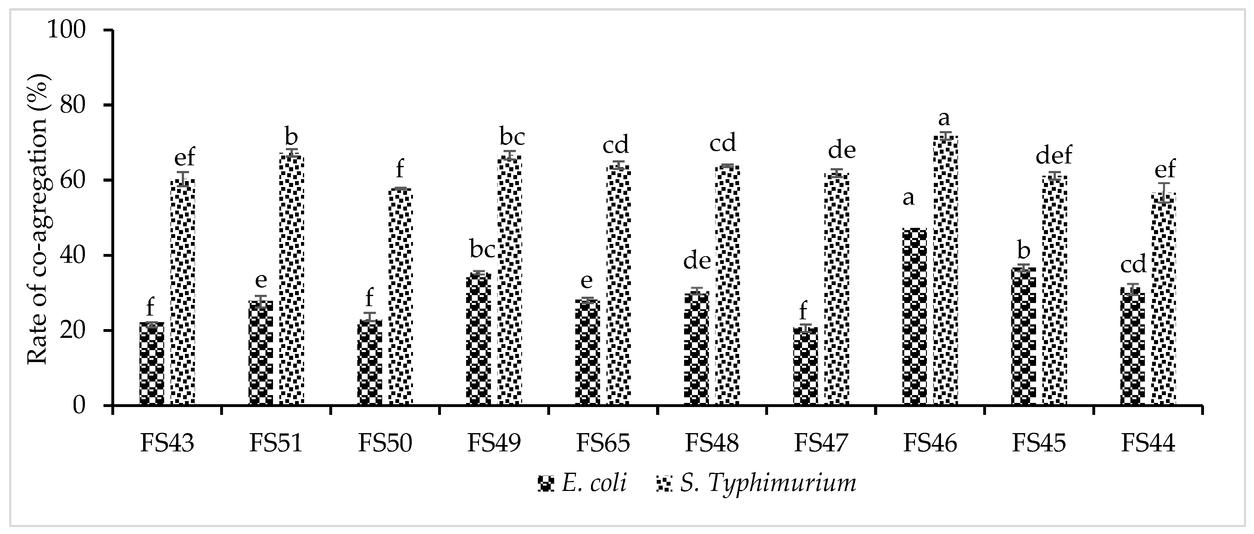
The study of co-aggregation, auto-aggregation, and hydrophobicity offers insights into the adhesion and colonization behavior of LAB strains in the host intestinal tract. The co-aggregation rates of LAB strains with S. Typhimurium were significantly higher than those with E. coli (Figure 2). All LAB strains showed high co-aggregation rates (>55%) against S. Typhimurium. In contrast, E. coli showed the lowest percent co-aggregation with FS47 (20.80 ± 1.22%) and the highest with FS46 (50.89 ± 1.00%). Additionally, the FS46 strain exhibited high co-aggregation with S. Typhimurium (71.78 ± 0.85%) (Figure 2).
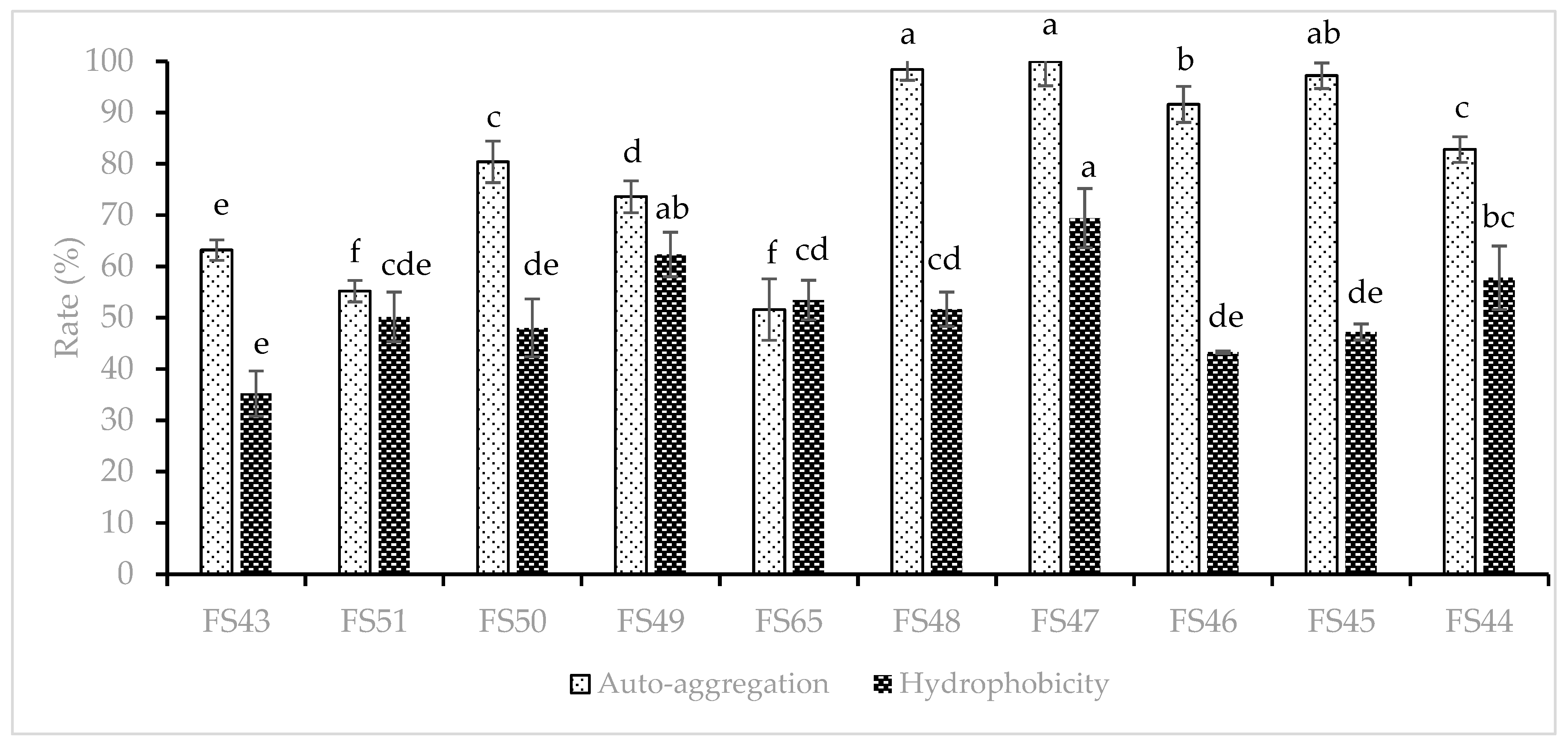
The functional ability of LAB strains to adhere to epithelial cells is closely linked to their auto-aggregation ability and cell surface hydrophobicity (Figure 3). The strains FS47, FS49, and FS44 demonstrated a relatively high hydrophobicity in xylene, exceeding 57%. Conversely, strain FS43 showed minimal hydrophobicity (35.23 ± 4.43%). All LAB strains demonstrated a significant level of auto-aggregation (>50%) after 24 h of incubation. Four LAB strains (FS47, FS48, FS45, and FS46) exhibited exceptionally high auto-aggregation, resulting in the formation of a visible precipitate at the bottom of the tubes.

3.4. Exploration of Probiotic Properties

3.4.1. Antibacterial Spectrum

The antibacterial activity of the CFSs from the LAB strains against four pathogenic species is shown in Table 4. The LAB strains exhibited high antagonist activity against S. aureus ATCC 33592, L. monocytogenes ATCC 7644, and S. enterica serovar Typhimurium ATCC 14028, with inhibition diameters between 18 and 29 mm. Strains FS43, FS65, FS48, FS46, and FS44 demonstrated significant antibacterial activity against E. coli ATCC 8739. However, strains FS51, FS50, FS49, FS47, and FS45 demonstrated moderate antibacterial activity against E. coli, with inhibition diameters between 6 and 10 mm.

3.4.2. Antioxidant activity

The DPPH assay was used to evaluate the antioxidant capacity of both intact LAB cells and their supernatants (see Figure 4). Both the intact LAB cells and supernatants of the FS48 strain showed statistically significant antioxidant activity levels (p < 0.05). Except for the FS44 strain, the antioxidant activity levels detected in the intact cells were significantly higher than those in the free cell supernatants, as shown in Figure 4. Seven strains (FS46, FS45, FS49, FS43, FS65, FS47, and FS48) exhibited DPPH free radical scavenging rates ranging from 51.64% to 83.58%, while the remainder of strains showed rates below 50%. Overall, the CFS from strain FS65 showed the lowest antioxidant activity (13.87 ± 0.44%), while the CFS from strain FS48 showed the highest antioxidant activity (49.09 ± 0.88%).

3.4.3. Enzymatic Activity of LAB Strains

The LAB isolates were inoculated on selective media to detect amylase, cellulase, lipase, pectinase, and protease activities (refer to Table 5). The results showed that all LAB strains tested were positive for protease and cellulase activities. Out of the ten strains tested, only FS43 exhibited positive lipolytic activity. Eight LAB strains demonstrated positive pectinolytic activity. Furthermore, five strains (FS51, FS49, FS65, FS48, and FS45) showed positive amylase activity, while the others did not.

3.5. Principal Component Analysis (PCA)

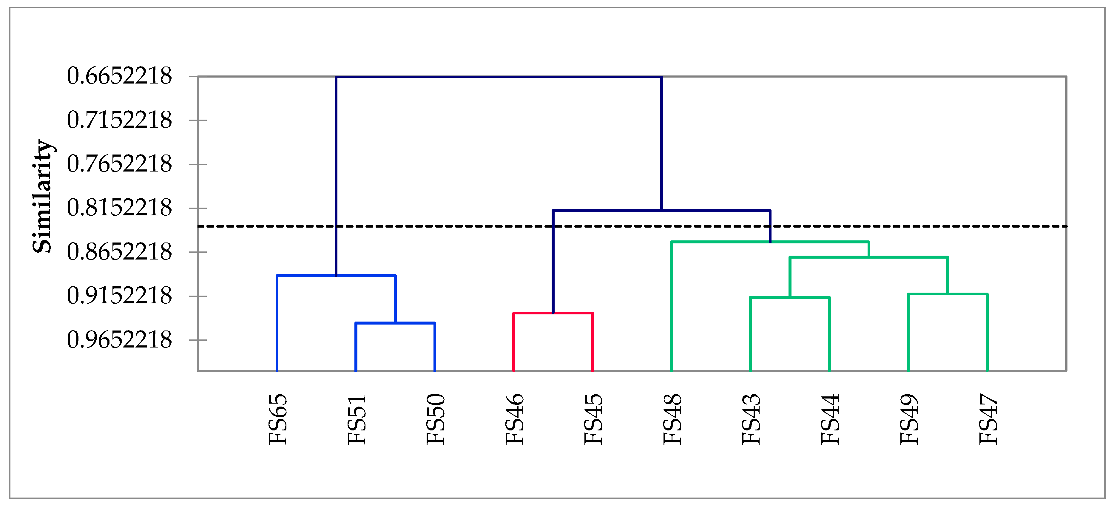
Principal component analysis (PCA) and hierarchical ascending classification (HAC) are powerful methods for synthesizing diverse information to enhance comprehension. The analysis results indicated that the combined influence of the two principal components (F1 and F2) explained 77.51% of the total variation. Of this, 65.03% was attributed to component F1 and 12.48% to component F2 (Figure 5). According to the PCA, based on the probiotic attributes of the LAB isolates, they could be divided into four distinct groups. The first group comprises strains FS43, FS44, and FS48 (positive correlation of both F1 and F2) and exhibited the highest values for hydrophobicity, phenol tolerance, antioxidant activity of the free cell supernatants, and intact cells (Figure 5). These three strains were classified as belonging to the same class based on the hierarchical ascending categorization of similarities (Figure 6). The second group consists of the FS50 and FS51 strains (positive side of F2 and negative side of F1) and exhibited high values for 0.3% bile salt tolerance. The third group comprises strains FS65, FS49, and FS46 (negative of both F1 and F2) and exhibited high values for pH 1.5 tolerance and co-aggregation ability with S. Typhimurium. In the fourth group, the strains FS45 and FS47 (positive side of F1 and negative side of F2, respectively) exhibited high values for pH 2.5 and 0.1% pepsin resistance, co-aggregation with E. coli, and auto-aggregation abilities, as well as a range of other probiotic properties. The results indicated that the strains FS43, FS44, and FS48 demonstrated the greatest probiotic potential.

4. Discussion

Africa has a long history of producing traditional fermented foods in local dietary contexts, including those derived from cassava, maize, millet, tubers, wild legume seeds, and sorghum [6,7,8,9]. Additionally, fermented milk, meat products, and alcoholic beverages have been produced on the continent for centuries [6,7,8,9]. Given that a significant proportion of African foods are fermented by lactic acid bacteria, they can be isolated and studied for their multifunctional ability to ferment foods, impart distinct taste and high nutritional value, and ensure food safety over an extended period. Additionally, they can be studied for their probiotic properties, which could have beneficial health effects. This process also contributes to an enhanced health status in those who consume them, which is of great importance in the African context [8]. The review by Pereira et al. [26] provides a comprehensive overview of the criteria and the methodologies employed for the selection of probiotics, such as their ability to tolerate unfavorable conditions in the human digestive system, adhere to human body cells, exhibit antimicrobial and antioxidant activities, synthesize enzyme, demonstrate antibiotic sensitivity, undergo hemolysis test, and fulfill other pertinent criteria.
This study aimed to assess the probiotic potential of 10 bacterial isolates obtained from the spontaneous fermentation of dockounou paste. The new isolates were preliminarily identified as lactic acid bacteria based on their colonial aspects (shape round or smooth), microscopic observations (bacilli, Gram-positive), and biochemical characteristics (negative oxidase and catalase). All LAB strains were identified as L. plantarum based on the high degree of homology observed between their 16S rDNA gene sequences and those deposited in the NCBI database. Lactobacillus spp. (particularly L. plantarum and L. fermentum) have been predominantly detected in various African fermented foods such as doklu (Ivorian maize dough) [12], Nigerian fermented foods [45,46,47], Maasai traditional fermented milk products [48], ben-saalga (Burkinabé fermented cereal gruel) [49]; bushera (Ugandan fermented cereal beverage) [50], tchapalo produced in Ivory Coast [51].
The paramount concern regarding probiotics must be their safety, which must not harm consumer health. The absence of hemolytic activity indicated that our LAB strains are unable to degrade blood constituents and, therefore, pose no pathological risk. Coulibaly et al. [28] also reported that L. plantarum and two other Pediococcus sp. isolated from the gut of Tilapia showed no hemolytic activity. The EFSA suggests that all microbial strains used as food or feed additives or probiotics should be subjected to testing to determine their sensitivity to different antibiotics [52]. Our findings have revealed that the majority of LAB strains demonstrated high or intermediate susceptibility to at least one antibiotic from the beta-lactam class (which inhibits cell wall synthesis) and the macrolide class (which inhibits protein synthesis). Furthermore, all strains demonstrated sensitivity to chloramphenicol, nitrofurans, and sulfonamide–diaminopyrimidine complex antibiotics. The overuse of antibiotics has resulted in the evolution and prevalence of resistant bacteria. Probiotics isolated from various dietary supplements or foods have demonstrated the ability to resist the effects of antibiotics [53,54,55,56]. Horizontal transfer of antibiotic-resistance genes from these probiotics to pathogenic bacteria residing in the intestinal tract could have significant health implications [54,55].
One of the key considerations when selecting LAB isolates as probiotics is their ability to withstand the challenges of the gastrointestinal tract. In our study, all LAB strains exhibited high survival rates, ranging between 81.18% and 91.38% in an acidic environment with a pH of 1.5. The LAB strains also demonstrated high survival rates, between 88.54% and 99.78%, in the presence of pepsin (0.1%) at a pH of 2.5. Furthermore, they exhibited high survival rates in the presence of bile salts (0.3%), ranging from 68.62 to 100.89%. Various strains of lactic acid bacteria demonstrated high survival rates, indicating their ability to withstand the acidic environment of the stomach (pH 1.5–3.0) and the small intestine (bile salts concentrations of 0.1–0.3%) [28,36,57]. Likewise, Lactobacillus spp. isolated from Shamita and Kocho (traditional Ethiopian beverages and foods) demonstrated that they can survive in extremely acidic conditions (greater than 80%) for 6 h [58]. Two L. plantarum strains, isolated from West African fermented cereals, exhibited comparable levels of acid resistance (pH 2.0) and tolerance to a bile salt concentration of 0.3% [47]. L. plantarum strains, isolated from traditional fermented milk of the Maasai in Kenya, demonstrated acid resistance at pH 2.0, with survival rates varying from 1% to 100% [48]. In a relatively recent study, Matei et al. [59] found that three strains of Pediococcus pentosaceus showed strong tolerance to high concentrations of bile salts (3% and 6%). It is widely accepted that the capacity of bacteria to resist phenol is an important indicator of their general viability. It has been observed that some bacterial species can synthesize harmful metabolites that can be deaminated by gut bacteria, creating toxic compounds with bacteriostatic properties such as phenol. Our results revealed that certain L. plantarum isolates exhibited greater resistance to phenol (0.4%) (73.72–128.24% viability) compared to others. For instance, L. plantarum strains varied in their ability to resist phenol, which had different effects on them [60]. Lactobacillus strains derived from traditional fermented foods, multigrain millet dosa batter, exhibited resistance to 0.2–0.8% phenols, maintaining viability at 53.3%–83.6% [61]. Furthermore, in our study, three L. plantarum isolates demonstrated not only high resistance but also were able to grow in the presence of 0.4% phenol.
The interaction between microorganisms and epithelial cells is influenced by cell wall hydrophobicity [62,63,64]. This parameter can be employed to determine the adhesion capability of lactic acid bacteria to the solvent. High hydrophobicity indicates a strong interaction between LAB isolates and epithelial cells, resulting in enhanced pathogen exclusion. It is generally recommended to select a strain with a hydrophobicity of over 40% as a probiotic. The present study demonstrated that eight LAB strains exhibited hydrophobicity levels ranging from 47.23% to 69.43%. The results of this study are consistent with those of Yasmin et al. [65], who observed that Bifidobacterium strains exhibited high hydrophobicity when exposed to xylene. However, strains of P. pentosaceus and L. plantarum isolated from the gut of Tilapia exhibited a range of cell surface hydrophobicity in the presence of different organic solvents such as chloroform (9.4–87.2%), xylene (3.48–51.10%), and hexane (1.53–16.30%) [28]. This study demonstrated that L. plantarum strains exhibited a high degree of auto-aggregation (>50% at 24 h), indicating that they exhibited an adequate level of adherence, which is in agreement with previously reported results [65]. Also, the co-aggregation rate of LAB isolates was notably high with S. Typhimurium (57.80–71.78%), followed by E. coli (20.80–50.89%). Honey and Keerthi [66] observed that L. plantarum strains exhibited excellent auto-aggregation ability, with values ranging from 99.2% to 99.8%. The highest levels of co-aggregation with S. typhi were also noted, with values between 45.3% and 63.66% [66].
Lactic acid bacteria are capable of suppressing the growth of pathogenic microorganisms through the synthesis of antimicrobial metabolites (e.g., short-chain fatty acids, bacteriocins, hydrogen peroxide (H2O2), and others) and by competing with those for nutrients, as well as by adhering to epithelial cells [67,68]. The current study found that all LAB isolates demonstrated strong inhibitory effects against S. aureus, L. monocytogenes, and S. enterica serovar Typhimurium, with moderate effects against E. coli. These findings are consistent with prior research indicating the antagonist activity of Lactobacillus and Pediococcus strains against a wide range of bacterial pathogens, including those belonging to the Gram-negative and Gram-positive categories, which may contaminate food sources and cause intestinal illnesses [28,36,68,69,70].
Lactic acid bacteria have been extensively studied for their potential as probiotics, given their well-documented health benefits, which appear to be partially attributed to their antioxidant traits [71,72]. In the current study, most of the intact LAB cells exhibited high antioxidant activity, demonstrated by a high percentage reduction in DPPH with an average of 52% to 84%. The present study revealed a significant increase in antioxidant activity in intact cells compared to free-cell supernatants. These results contrast with those reported by Coulibaly et al. [28].
Lactobacilli have been identified as producers of enzyme complexes that enhance the digestibility of food and feed, as well as the activities of digestive enzymes and promote growth performance [73]. The evaluation of probiotic-producing enzymes can be conducted by cultivating candidate strains in culture media supplemented with precursors. These include casein, starch, carboxymethylcellulose, Tween 80 or olive oil, and sodium phytate, which are used to assess the activity of protease, amylase, cellulase, lipases, and phytases, respectively. The results of our study indicated that all LAB isolates possessed proteolytic and cellulolytic activities, with some also exhibiting pectinolytic or amylolytic activities. This result differs from the findings of Coulibaly et al. [28], in which the P. pentosaceus and L. plantarum strains isolated from the gut of Tilapia demonstrated lipase and β-galactosidase activity (with a few exceptions) but lacked amylase, cellulase, and protease activities. L. fermentum URLP18 isolated from C. mrigala demonstrated a high capacity for extracellular enzyme production, including amylase, protease, and lipase [74].
This study’s results indicate that the isolation of lactic acid bacteria from naturally fermented and indigenous products is a topic worthy of further investigation. Nevertheless, strains FS43, FS44, and FS48 were identified as the most promising candidates for further investigation, as they exhibited the greatest potential for beneficial effects, as indicated by principal component analysis (PCA) and hierarchical ascending classification (HAC). Artisanal fermented products are widely produced and consumed in traditional households and can be considered a valuable source of probiotics for improving the health of the Ivorian population.

5. Conclusions

The 10 lactic acid bacteria isolated from fermented plantain dockounou paste have been identified through 16S rDNA sequence analysis as Lactiplantibacillus plantarum. All LAB strains demonstrated high survival rate against pepsin (0.1%) and low pH (1.5 and 2.5) and exhibited high tolerance to bile salts (0.3%) and phenol (0.4%). Furthermore, all strains demonstrated high antioxidant activity and broad antibacterial activity against the main foodborne pathogens. The obtained findings indicated that three of the 10 LAB strains exhibited a high affinity towards xylene (more than 57%). All LAB strains exhibited high auto-aggregation properties, as well as strong degrees of co-aggregation with S. enterica serovar Typhimurium, followed by E. coli. Furthermore, the LAB isolates demonstrated no hemolytic activity and sensitivity to different antibiotic classes. The LAB isolates have been detected as valuable producers of bioactive multi-enzymes. Strains FS43, FS44, and FS48 exhibited multifunctional properties, suggesting that further in vivo research is required to assess their suitability as probiotics or for the development of innovative functional foods.

Author Contributions

Conceptualization, N.J.K., K.A.S.K., S.N. and F.C.D.; methodology, N.J.K., A.L.O.Z., K.A.S.K. and F.C.D.; software, N.J.K., K.A.S.K. and S.N.; validation, N.J.K., F.C.D., and F.M.; formal analysis, K.A.S.K., A.L.O.Z., and F.C.D.; investigation, N.J.K. and F.C.D.; resources, F.C.D. and F.M.; data curation, K.A.S.K., A.L.O.Z., and S.N.; writing—original draft preparation, N.J.K., A.L.O.Z. and S.N.; writing—review and editing, F.C.D. and F.M.; visualization, F.C.D. and F.M.; supervision, S.N., F.C.D. and F.M.; project administration, F.C.D. and F.M.; funding acquisition, N.J.K., K.A.S.K. and F.C.D. All authors have read and agreed to the published version of the manuscript.

Funding

The authors are grateful to AUF (Agence Universitaire de la Francophonie) for the scholarship granted to Natia Jo-seph Kouadio through the Eugen Ionescu program in its 2021 session at the University of Agronomic Sciences and Veterinary Medicine of Bucharest, Romania.

Institutional Review Board Statement

Not applicable.

Informed Consent Statement

Not applicable.

Data Availability Statement

All obtained datasets are included in this publication.

Acknowledgments

Our thanks go to the Ivorian authorities of Felix Houphouet-Boigny University who authorized us to carry out this postdoctoral study in Bucharest, Romania. We thank the technician Badea Florentina from the Laboratory of Microbiology of the Faculty of Biotechnologies for his help, which was of great use to us.

Conflicts of Interest

The authors declare no conflicts of interest.

References

  1. Kra, K.A.S.; Akoa, E.E.F.; Mégnanou, R.M.; Yéboué, K.; Akpa, E.E.; Niamké, S.L. Physicochemical and nutritional characteristics assessments of two different traditional foods prepared with senescent plantain. Afr. J. Food Sci. 2013, 7, 51–55. [Google Scholar]
  2. Akoa, E.E.F.; Kra, K.A.S.; Mégnanou, R.M.; Kouadio, N.J.; Niamké, L.S. Optimization of dockounou manufacturing process parameters. Sustain. Agric. Res. 2014, 3, 67–75. [Google Scholar]
  3. Carboni, A.D.; Martins, G.N.; Gómez-Zavaglia, A.; Castilho, P.C. Lactic acid bacteria in the production of traditional fermented foods and beverages of Latin America. Fermentation 2023, 9, 315. [Google Scholar] [CrossRef]
  4. Petrova, P.; Petrov, K. Lactic acid fermentation of cereals and pseudocereals: Ancient nutritional biotechnologies with modern applications. Nutrients 2020, 12, 1118. [Google Scholar] [CrossRef]
  5. Sionek, B.; Szydłowska, A.; Küçükgöz, K.; Kołożyn-Krajewska, D. Traditional and new microorganisms in lactic acid fermentation of food. Fermentation 2023, 9, 1019. [Google Scholar] [CrossRef]
  6. Kayitesi, E.; Onojakpor, O.; Moyo, S.M. Highlighting the impact of lactic-acid-bacteria-derived flavours or aromas on sensory perception of african fermented cereals. Fermentation 2023, 9, 111. [Google Scholar] [CrossRef]
  7. Achi, O.K.; Asamudo, N.U. Cereal-based fermented foods of Africa as functional foods. Bioact. Mol. Food 2018, 1–32. [Google Scholar]
  8. Malongane, F.; Berejena, T. Exploring the microbiome present in fermented indigenous African foods and their potential impact on human health. J. Agric. Food Res. 2024, 16, 101101. [Google Scholar] [CrossRef]
  9. Assohoun, M.C.N.; Djéni, T.N.; N’Guessan, F.K.; Koussemon, M. Preliminary study on antimicrobial properties of lactic acid bacteria involved in the fermentation of corn dough during doklu processing in Côte D’Ivoire. Food 2012, 6, 65–70. [Google Scholar]
  10. Soro-Yao, A.A.; Kouakou, B.; Koffi-Nevry, R.; Djè, K.M. Microbiology of Ivorian fermented products: A review. Asian J. Agric. Food Sci. 2013, 1, 37–47. [Google Scholar]
  11. Kouadio, N.J.; Goualie, G.B.; Ouattara, G.H.; Kra, K.A.S.; Niamke, L.S. Microorganisms associated with traditional plantain-based food “dockounou” during spontaneous fermentation. Food Environ. Saf. 2014, 13, 276–282. [Google Scholar]
  12. Assohoun-Djeni, N.M.C.; Djeni, N.T.; Messaoudi, S.; Lhomme, E.; Koussemon-Camara, M.; Ouassa, T.; Chobert, J.M.; Onno, B.; Dousset, X. Biodiversity, dynamics and antimicrobial activity of lactic acid bacteria involved in the fermentation of maize flour for doklu production in Côte d’Ivoire. Food Control 2016, 62, 397–404. [Google Scholar] [CrossRef]
  13. Mokoena, M.P.; Mutanda, T.; Olaniran, A.O. Perspectives on the probiotic potential of lactic acid bacteria from African traditional fermented foods and beverages. Food Nutr. Res. 2016, 60, 29630. [Google Scholar] [CrossRef] [PubMed]
  14. Adesulu-Dahunsi, A.T.; Dahunsi, S.O.; Ajayeoba, T.A. Co-occurrence of Lactobacillus species during fermentation of african indigenous foods: Impact on food safety and shelf-life extension. Front. Microbiol. 2022, 13, 684730. [Google Scholar] [CrossRef] [PubMed]
  15. Mogmenga, I.; Somda, M.K.; Ouattara, C.A.T.; Keita, I.; Dabiré, Y.; Diguță, C.F.; Toma, R.C.; Ezeogu, L.I.; Ugwuanyi, J.O.; Ouattara, A.S.; et al. Promising Probiotic Properties of the Yeasts Isolated from Rabilé, a Traditionally Fermented Beer Produced in Burkina Faso. Microorganisms 2023, 11, 802. [Google Scholar] [CrossRef] [PubMed]
  16. Grosu-Tudor, S.; Stefan, I.; Stancu, M.; Cornea, C.; De vuyst, L.; Zamfir, M. Microbial and nutritional characteristics of fermented wheat bran in traditional Romanian borş production. Rom. Biotechnol. Lett. 2018, 24, 440–447. [Google Scholar] [CrossRef]
  17. Constantin, E.A.; Constantinescu-Aruxandei, D.; Matei, F.; Shaposhnikov, S.; Oancea, F. Biochemical and microbiological characterization of traditional Romanian fermented drinks—Socata and borș—A review. AgroLife Sci. J. 2023, 12, 53–61. [Google Scholar] [CrossRef]
  18. Ștefan, I.R.; Grosu-Tudor, S.S.; Zamfir, M.; Cornea, C.P. Screening for S-layer production by some lactobacilli from home-made fermented foods. Sci. Bull. Ser. F Biotechnol. 2016, XX, 167–171. [Google Scholar]
  19. Angelescu, I.R.; Zamfir, M.; Stancu, M.M.; Grosu-Tudor, S.S. Identification and probiotic properties of lactobacilli isolated from two different fermented beverages. Ann. Microbiol. 2019, 69, 1557–1565. [Google Scholar] [CrossRef]
  20. Grosu-Tudor, S.S.; Zamfir, M.; Van der Meulen, R.; De Vuyst, L. Isolation of novel homopolysaccharide-producing lactic acid bacteria from Romanian raw milk and fermented dairy products. Eur. Food Res. Technol. 2013, 237, 609–615. [Google Scholar] [CrossRef]
  21. Petruţ, Ș.M.; Sârbu, I.; Corbu, V.M.; Pelinescu, D.; Iftime, I.; Dimov, T.V. Screening of lactic acid bacteria from spontaneously fermented products of Romania. Rom. Biotechnol. Lett. 2019, 24, 254–260. [Google Scholar] [CrossRef]
  22. Grosu-Tudor, S.S.; Zamfir, M. Functional properties of lactic acid bacteria isolated from Romanian fermented vegetables. Food Biotechnol. 2013, 27, 235–248. [Google Scholar] [CrossRef]
  23. Cornea, C.P.; Israel Roming, F.; Sicuia, O.A.; Voaideș, C.; Zamfir, M.; Grosu-Tudor, S.S. Biosurfactant production by Lactobacillus spp. strains isolated from Romanian traditional fermented food products. Rom. Biotechnol. Lett. 2016, 21, 11312–11320. [Google Scholar]
  24. FAO/WHO. Guidelines for the Evaluation of Probiotics in Food; Food and Agriculture Organization of the United Nations and World Health Organization Working Group Report; FAO/WHO: London, ON, Canada, 2002. [Google Scholar]
  25. Hill, C.; Guarner, F.; Reid, G.; Gibson, G.R.; Merenstein, D.J.; Pot, B.; Morelli, L.; Canani, R.B.; Flint, H.J.; Salminen, S.; et al. Expert consensus document: The International Scientific Association for Probiotics and Prebiotics consensus statement on the scope and appropriate use of the term probiotic. Nat. Rev. Gastroenterol. Hepatol. 2014, 11, 506–514. [Google Scholar] [CrossRef] [PubMed]
  26. De Melo Pereira, G.V.; de Oliveira Coelho, B.; Magalhães Júnior, A.I.; Thomaz-Soccol, V.; Soccol, C.R. How to select a probiotic? A review and update of methods and criteria. Biotechnol. Adv. 2018, 36, 2060–2076. [Google Scholar] [CrossRef] [PubMed]
  27. Chou, L.S.; Weimer, B. Isolation and characterization of acid- and bile-tolerant isolates from strains of Lactobacillus acidophilus. J. Dairy Sci. 1999, 82, 23–31. [Google Scholar] [CrossRef] [PubMed]
  28. Coulibaly, W.H.; Kouadio, N.R.; Camara, F.; Diguță, C.; Matei, F. Functional properties of lactic acid bacteria isolated from Tilapia (Oreochromis niloticus) in Ivory Coast. BMC Microbiol. 2023, 23, 152. [Google Scholar] [CrossRef] [PubMed]
  29. Meena, K.K.; Taneja, N.K.; Jain, D.; Ojha, A.; Kumawat, D.; Mishra, V. In vitro assessment of probiotic and technological properties of lactic acid bacteria isolated from indigenously fermented cereal-based food products. Fermentation 2022, 8, 529. [Google Scholar] [CrossRef]
  30. Ayivi, R.D.; Gyawali, R.; Krastanov, A.; Aljaloud, S.O.; Worku, M.; Tahergorabi, R.; Silva, R.C.D.; Ibrahim, S.A. Lactic Acid Bacteria: Food Safety and Human Health Applications. Dairy 2020, 1, 202–232. [Google Scholar] [CrossRef]
  31. Picard, C.; Fioramonti, J.; Francois, A.; Robinson, T.; Neant, F.; Matuchansky, C. Review article: Bifidobacteria as probiotic agents–physiological effects and clinical benefits. Aliment. Pharmacol. Ther. 2005, 22, 495–512. [Google Scholar] [CrossRef]
  32. Chen, J.; Chen, X.; Ho, C.L. Recent development of probiotic bifidobacteria for treating human diseases. Front. Bioeng. Biotechnol. 2021, 9, 770248. [Google Scholar] [CrossRef] [PubMed]
  33. Kumar, S.; Stecher, G.; Li, M.; Knyaz, C.; Tamura, K. MEGA X: Molecular Evolutionary Genetics Analysis across computing platforms. Mol Biol Evol. 2018, 35, 1547–1549. [Google Scholar] [CrossRef] [PubMed]
  34. Vauterin, L.; Vauterin, P. Computer-aided objective comparison of electrophoresis patterns for grouping and identification of microorganisms. Eur. Microbiol. 1992, 1, 37–41. [Google Scholar]
  35. Carr, F.J.; Chill, D.; Maida, N. The lactic acid bacteria: A literature survey. Crit. Rev. Microbiol. 2002, 28, 281–370. [Google Scholar] [CrossRef] [PubMed]
  36. Diguță, C.F.; Nițoi, G.D.; Matei, F.; Luță, G.; Cornea, C.P. The Biotechnological potential of Pediococcus spp. isolated from Kombucha microbial consortium. Foods 2020, 9, 1780. [Google Scholar] [CrossRef] [PubMed]
  37. Xanthopoulos, V.; Litopoulou-Tzanetaki, E.; Tzanetakis, N. Characterization of Lactobacillus isolates from infant faeces as dietary adjuncts. Food Microbiol. 2000, 17, 205–215. [Google Scholar] [CrossRef]
  38. Yadav, R.; Puniya, A.K.; Shukla, P. Probiotic Properties of Lactobacillus plantarum RYPR1 from an Indigenous Fermented Beverage Raabadi. Front. Microbiol. 2016, 7, 1683. [Google Scholar] [CrossRef] [PubMed]
  39. CLSI. CLSI Supplement M100S. Performance Standards for Antimicrobial Susceptibility Testing, 26th ed.; Clinical and Laboratory Standards Institute: Wayne, PA, USA, 2016. [Google Scholar]
  40. Collado, M.C.; Meriluoto, J.; Salminen, S. Measurement of aggregation properties between probiotics and pathogens: In vitro evaluation of different methods. J. Microbiol. Methods 2007, 71, 71–74. [Google Scholar] [CrossRef] [PubMed]
  41. Rosenberg, M.; Gutnick, D. and Rosenberg, E. Adherence of bacteria to hydrocarbons: A simple method for measuring cell-surface hydrophobicity. FEMS Microbiol. Lett. 1980, 9, 29–33. [Google Scholar] [CrossRef]
  42. Balouiri, M.; Sadiki, M.; Ibnsouda, S.K. Methods for in vitro evaluating antimicrobial activity: A review. J. Pharm. Anal. 2016, 6, 71–79. [Google Scholar] [CrossRef]
  43. Brand-Williams, W.; Cuvelier, M.; Berset, C. Use of a free radical method to evaluate antioxidant activity. LWT 1995, 28, 25–30. [Google Scholar] [CrossRef]
  44. Proca, I.G.; Diguta, C.F.; Jurcoane, S.; Matei, F. Screening of halotolerant bacteria producing hydrolytic enzymes with biotechnological applications. Sci. Bull. Ser. F Biotechnol. 2020, XXIV, 197–204. [Google Scholar]
  45. Sanni, A.; Morlon-Guyot, J.; Guyot, J.P. New efficient amylase-producing strains of Lactobacillus plantarum and L. fermentum isolated from different Nigerian traditional fermented foods. Int. J. Food Microbiol. 2002, 72, 53–62. [Google Scholar] [CrossRef] [PubMed]
  46. Adesulu-Dahunsi, A.T.; Sanni, A.I.; Jeyaram, K.; Banwo, K. Genetic diversity of Lactobacillus plantarum strains from some indigenous fermented foods in Nigeria. LWT 2017, 82, 199–206. [Google Scholar] [CrossRef]
  47. Oguntoyinbo, F.A.; Narbad, A. Multifunctional properties of Lactobacillus plantarum strains isolated from fermented cereal foods. J. Funct. Foods 2015, 17, 621–631. [Google Scholar] [CrossRef]
  48. Mathara, J.M.; Schillinger, U.; Kutima, P.M.; Mbugua, S.K.; Guigas, C.; Franz, C.; Holzapfel, W.H. Functional properties of Lactobacillus plantarum strains isolated from Maasai traditional fermented milk products in Kenya. Curr. Microbiol. 2008, 56, 315–321. [Google Scholar] [CrossRef] [PubMed]
  49. Omar, N.B.; Abriouel, H.; Lucas, R.; Martínez-Cañamero, M.; Guyot, J.-P.; Gálvez, A. Isolation of bacteriocinogenic Lactobacillus plantarum strains from Ben Saalga, a traditional fermented Gruel from Burkina Faso. Int. J. Food Microbiol. 2006, 112, 44–50. [Google Scholar] [CrossRef] [PubMed]
  50. Muyanja, C.; Narvhus, J.A.; Treimo, J.; Langsrud, T. Isolation, characterisation and identification of lactic acid bacteria from bushera: A Ugandan traditional fermented beverage. Int. J. Food Microbiol. 2003, 80, 201–210. [Google Scholar] [CrossRef] [PubMed]
  51. Aka, S.; N’Guessan, K.F.; Nanga, Y.Z.; Loukou, Y.G.; Mazabraud, A.I.; Djè, K.M. Characterization of Lactobacillus species isolated from mash, sour wort and tchapalo produced in Côte d’Ivoire. Food 2010, 4, 49–54. [Google Scholar]
  52. EFSA Panel on Additives and Products or Substances used in Animal Feed (FEEDAP); Rychen, G.; Aquilina, G.; Azimonti, G.; Bampidis, V.; De Lourdes Bastos, M.; Bories, G.; Chesson, A.; Cocconcelli, P.S.; Flachowsky, G.; et al. Guidance on the characterisation of microorganisms used as feed additives or as production organisms. EFSA J. 2018, 16, e05206. [Google Scholar]
  53. Sharma, P.; Tomar, S.K.; Goswami, P.; Sangwan, V.; Singh, R. Antibiotic resistance among commercially available probiotics. Food Res. Int. 2014, 57, 176–195. [Google Scholar] [CrossRef]
  54. Wong, A.; Ngu, D.Y.S.; Dan, L.A.; Ooi, A.; Lim, R.L.H. Detection of antibiotic resistance in probiotics of dietary supplements. Nutr. J. 2015, 14, 95. [Google Scholar] [CrossRef] [PubMed]
  55. Li, T.; Teng, D.; Mao, R.; Hao, Y.; Wang, X.; Wang, J. A critical review of antibiotic resistance in probiotic bacteria. Food Res. Int. 2020, 136, 109571. [Google Scholar] [CrossRef] [PubMed]
  56. Dou, W.; Abdalla, H.B.; Chen, X.; Sun, C.; Chen, X.; Tian, Q.; Wang, J.; Zhou, W.; Chi, W.; Zhou, X.; et al. ProbResist: A database for drug-resistant probiotic bacteria. Database 2022, 2022, baac064. [Google Scholar] [CrossRef] [PubMed]
  57. Lei, V.; Jakobsen, M. Microbiological characterization and probiotic potential of koko and koko sour water, African spontaneously fermented millet porridge and drink. J. Appl. Microbiol. 2004, 96, 384–397. [Google Scholar] [CrossRef] [PubMed]
  58. Negasi, A.; Fassil, A.; Asnake, D. In vitro evaluation of lactic acid bacteria isolated from traditional fermented Shamita and Kocho for their desirable characteristics as probiotics. Afr. J. Biotechnol. 2017, 16, 594–606. [Google Scholar] [CrossRef]
  59. Matei, B.; Salzat, J.; Diguță, C.F.; Cornea, C.P.; Luta, G.; Utoiu, E.R.; Matei, F. Lactic acid bacteria strains isolated from Kombucha with potential probiotic effect. Rom. Biotechnol. Lett. 2018, 23, 13592–13598. [Google Scholar]
  60. Xanthopoulos, V.; Hatzikamari, M.; Adamidis, T.; Tsakalidou, E.; Tzanetakis, N.; Litopoulou-Tzanetaki, E. Heterogeneity of Lactobacillus plantarum isolates from Feta cheese throughout ripening. J. Appl. Microbiol. 2000, 88, 1056–1064. [Google Scholar] [CrossRef]
  61. Divyashree, S.; Ramu, R.; Sreenivasa, M.Y. Evaluation of new candidate probiotic Lactobacillus strains isolated from a traditional fermented food- multigrain-millet dosa batter. Food Biosci. 2024, 57, 103450. [Google Scholar] [CrossRef]
  62. Krausova, G.; Hyrslova, I.; Hynstova, I. In Vitro Evaluation of adhesion capacity, hydrophobicity, and auto-aggregation of newly isolated potential probiotic strains. Fermentation 2019, 5, 100. [Google Scholar] [CrossRef]
  63. Kos, B.; Susković, J.; Vuković, S.; Simpraga, M.; Frece, J.; Matosić, S. Adhesion and Aggregation ability of probiotic strain Lactobacillus acidophilus M92. J. Appl. Microbiol. 2003, 94, 981–987. [Google Scholar] [CrossRef] [PubMed]
  64. García-Cayuela, T.; Korany, A.M.; Bustos, I.; de Cadinanos, L.P.G.; Requena, T.; Peláez, C.; Martínez-Cuesta, M.C. Adhesion abilities of dairy Lactobacillus plantarum strains showing an aggregation phenotype. Food Res. Int. 2014, 57, 44–50. [Google Scholar] [CrossRef]
  65. Yasmin, I.; Saeed, M.; Khan, W.A.; Khaliq, A.; Chughtai, M.F.J.; Iqbal, R.; Tehseen, S.; Naz, S.; Liaqat, A.; Mehmood, T.; et al. In vitro probiotic potential and safety evaluation (hemolytic, cytotoxic activity) of Bifidobacterium strains isolated from raw camel milk. Microorganisms 2020, 8, 354. [Google Scholar] [CrossRef] [PubMed]
  66. Honey, C.C.; Keerthi, T.R. Probiotic potency of Lactobacillus plantarum kx519413 and kx519414 isolated from honey bee gut. FEMS Microbiol. Lett. 2018, 365, fnx285. [Google Scholar]
  67. Mani-López, E.; Arrioja-Bretón, D.; López-Malo, A. The impacts of antimicrobial and antifungal activity of cell-free supernatants from lactic acid bacteria in vitro and foods. Compr. Rev. Food Sci. Food Saf. 2022, 21, 604–641. [Google Scholar] [CrossRef]
  68. Rocchetti, M.T.; Russo, P.; Capozzi, V.; Drider, D.; Spano, G.; Fiocco, D. Bioprospecting antimicrobials from Lactiplantibacillus plantarum: Key factors underlying its probiotic action. Int. J. Mol. Sci. 2021, 22, 12076. [Google Scholar] [CrossRef]
  69. Gómez, N.C.; Ramiro, J.M.P.; Quecan, B.X.V.; de Melo Franco, B.D.G. Use of potential probiotic lactic acid bacteria biofilms for the control of Listeria monocytogenes, Salmonella Typhimurium, and Escherichia coli O157:H7 biofilms formation. Front. Microbiol. 2016, 7, 863. [Google Scholar] [CrossRef]
  70. Arena, M.P.; Silvain, A.; Normanno, G.; Grieco, F.; Drider, D.; Spano, G.; Fiocco, D. Use of Lactobacillus plantarum strains as a bio-control strategy against food-borne pathogenic microorganisms. Front. Microbiol. 2016, 7, 464. [Google Scholar] [CrossRef]
  71. Feng, T.; Wang, J. Oxidative stress tolerance and antioxidant capacity of lactic acid bacteria as probiotic: A systematic review. Gut Microbes 2020, 12, 1801944. [Google Scholar] [CrossRef]
  72. Kullisaar, T.; Zilmer, M.; Mikelsaar, M.; Vihalemm, T.; Annuk, H.; Kairane, C. and Kilk, A. Two antioxidative lactobacilli strains as promising probiotics. Int. J. Food Microbiol. 2002, 72, 215–224. [Google Scholar] [CrossRef]
  73. Maske, B.L.; Pereira, G.V.D.; Vale, A.D.; Neto, D.P.D.; Karp, S.G.; Viesser, J.A.; Lindner, J.D.; Pagnoncelli, M.G.; Soccol, V.T.; Soccol, C.R. A review on enzyme-producing lactobacilli associated with the human digestive process: From metabolism to application. Enzym. Microb. Technol. 2021, 149, 109836. [Google Scholar] [CrossRef] [PubMed]
  74. Govindaraj, K.; Samayanpaulraj, V.; Narayanadoss, V.; Uthandakalaipandian, R. Isolation of lactic acid bacteria from intestine of freshwater fishes and elucidation of probiotic potential for aquaculture application. Probiotics Antimicrob. Proteins 2021, 13, 1598–1610. [Google Scholar] [CrossRef] [PubMed]
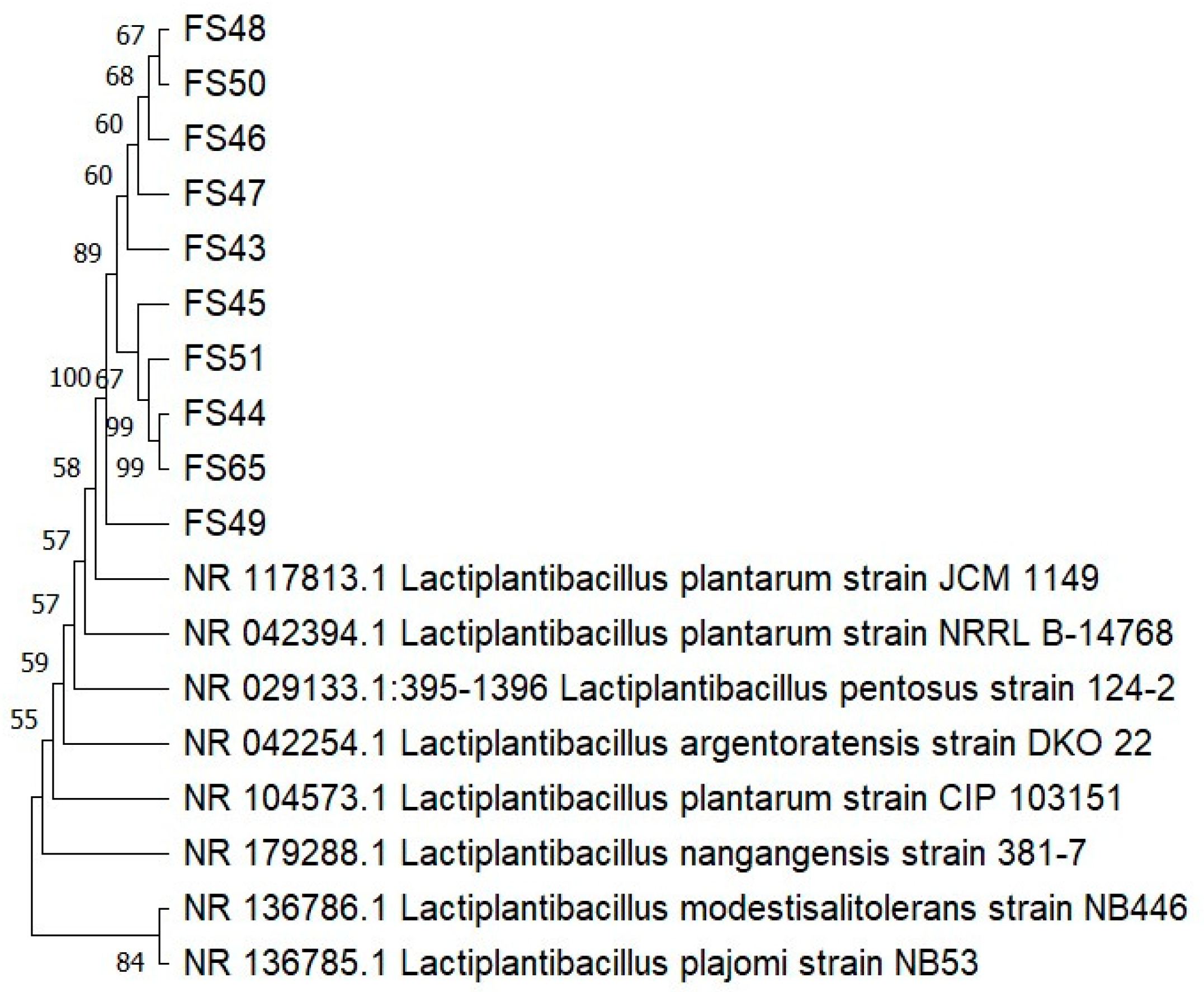
Figure 1. Molecular phylogenetic analysis via maximum parsimony analysis of taxa.
Fermentation 10 00264 g001
Figure 2. Co-aggregation rates of LAB strains with E. coli ATCC 8739 and S. Typhimurium ATCC 14028. Each value was expressed as the mean ± standard deviation (SD). Statistically significant differences (p < 0.05) were indicated by different lowercase letters above the error bars.
Fermentation 10 00264 g002
Figure 3. Auto-aggregation and hydrophobicity rates of LAB strains. Each value was expressed as the mean ± standard deviation (SD). Statistically significant differences (p < 0.05) were indicated by different lowercase letters above the error bars.
Fermentation 10 00264 g003
Figure 4. Antioxidant capacity of free cell supernatants and intact LAB cells evaluated via DPPH assay. Each value was expressed as the mean ± standard deviation (SD). Statistically significant differences (p < 0.05) were indicated by different lowercase letters above the error bars.
Fermentation 10 00264 g004
Figure 5. Principal component analysis of LAB strains according to probiotic properties.
Fermentation 10 00264 g005
Figure 6. Hierarchical ascending classification of LAB strains according to probiotic properties. The dotted line delineates the cutoff point of this specific algorithm, which generates three clusters (indicated by distinct colors) based on probiotic characteristics that exhibit a high degree of similarity.
Fermentation 10 00264 g006
Table 1. Phenotypic characteristics of the LAB strains used in this study.
IsolatesParametersOrigin of Isolate
ShapeGram ReactionCellCatalase TestOxidase Test
FS43Smooth+rod-shape--Rice dockounou paste
FS51Round+rod-shape--Rice dockounou paste
FS50Smooth+rod-shape--Maize dockounou paste
FS49Round+rod-shape--Maize dockounou paste
FS65Smooth+rod-shape--Maize dockounou paste
FS48Smooth+rod-shape--Maize dockounou paste
FS47Round+rod-shape--Millet dockounou paste
FS46Round+rod-shape--Millet dockounou paste
FS45Smooth+rod-shape--Millet dockounou paste
FS44Round+rod-shape--Cassava dockounou paste
Table 2. Antibiotic sensibility and Hemolysis activity of LAB strains.
StrainsFS43FS51FS50FS49FS65FS48FS47FS46FS45FS44
Class/Antibiotics
ChloramphenicolCHL20S24S24S28S24S24S26S25S24S29S
TetracyclinT20S16I14R12R12R14R12R16I14R18I
MacrolidesE12R18I20S19I18I18I21S20S18I16I
L26S24S0R26S20S26S0R28S30S0R
Beta-lactamAM20S24S26S22S20S20S24S22S22S20S
AMC18S20S26S30S30S28S34S32S28S26S
P16I18I18I16I14R20S20S18I20S16I
NitrofuranF22S28S22S26S22S25S28S29S28S24S
Sulfamide + diaminopyrimidineSXT18I20S20S22S20S22S20S20S20S20S
Hemolysis activity γγγγγγγγγγ
CHL: Chloramphenicol (30 µg/disc); T: Tetracycline (30 µg/disc); E: Erythromycin (10 µg/disc); L: Lincomycin (10 µg/disc); AM: Ampicillin ((10 µg/disc); AMC: Amoxicillin + clavulanic acid (20/10 µg/disc); P: Penicillin (2 µg/disc); F: Nitrofurantoin (300 µg/disc); SXT: Trimethoprim + sulfamethoxazole (1/19 µg/disc). S: sensitive; I: intermediate; R: resistant; γ—gamma hemolysis.
Table 3. Resistance of LAB strains to simulated gastrointestinal conditions.
Survival Rate (%)
IsolatespH 1.5Pepsin (0.1%)/pH 2.5Phenol (0.4%)Bile Salts (0.3%)
FS4384.35 ± 0.85 d91.58 ± 3.43 def94.58 ± 3.70 c82.25 ± 0.75 c
FS5183.33 ± 0.91 d89.98 ± 1.81 ef40.59 ± 2.80 g93.09 ± 1.25 b
FS5087.05 ± 1.95 c94.88 ± 2.41 bcd44.12 ± 3.01 fg99.58 ± 2.07 a
FS4981.18 ± 1.05 e91.45 ± 1.78 def85.12 ± 6.60 d79.63 ± 3.90 cd
FS6598.15 ± 0.95 a95.60 ± 0.84 bc48.09 ± 2.80 f77.791 ± 0.40 de
FS4888.30 ± 0.91 c99.78 ± 2.14 a128.24 ± 1.08 a77.49 ± 0.59 de
FS4791.38 ± 0.88 b92.63 ± 1.78 cde112.64 ± 1.60 b75.32 ± 2.15 e
FS4690.74 ± 1.80 b97.96 ± 0.84 ab73.72 ± 0.78 e68.62 ± 3.64 f
FS4585.27 ± 0.57 d92.44 ± 1.88 cde90.22 ± 0.44 cd74.18 ± 1.15 e
FS4485.03 ± 0.98 d88.54 ± 1.26 f109.46 ± 0.51 b100.89 ± 3.52 a
Each value represents the mean of three replicates, accompanied by their respective standard deviations. Values bearing the same lowercase letter (a, b, c, d, e, etc.) are not statistically significantly different at the 5% level within the same column.
Table 4. Antimicrobial activity of LAB strains.
IsolatesE. coli ATCC 8739L. monocytogenes ATCC 7644S. enterica Serovar
Typhimurium ATCC 14028
S. aureus ATCC 33592
FS43++++++++++++
FS51+++++++++++
FS50+++++++++++
FS49+++++++++++
FS65++++++++++++
FS48++++++++++++
FS47+++++++++++
FS46++++++++++++
FS45+++++++++++
FS44++++++++++++
Legend: (-) the absence of a halo formation; (+) the presence of a halo measuring 1–5 mm in diameter; (++) the presence of a halo measuring 5–10 mm in diameter; (+++) the presence of a halo measuring >10 mm in diameter.
Table 5. Enzymatic activities of LAB strains.
ParametersEnzymatic Activities
Strains ProteolyticLipolyticPectinolyticCellulolyticAmylolytic
FS43++++-
FS51+-+++
FS50+-++-
FS49+--++
FS65+-+++
FS48+--++
FS47+-++-
FS46+-++-
FS45+-+++
FS44+-++-
The (+) symbol indicates that the enzymatic activity is positive, while the (-) symbol indicates that the enzymatic activity is negative.
Disclaimer/Publisher’s Note: The statements, opinions and data contained in all publications are solely those of the individual author(s) and contributor(s) and not of MDPI and/or the editor(s). MDPI and/or the editor(s) disclaim responsibility for any injury to people or property resulting from any ideas, methods, instructions or products referred to in the content.