Article

Characterization of Novel Exopolysaccharides from Weissella cibaria and Lactococcus lactis Strains and Their Potential Application as Bio-Hydrocolloid Agents in Emulsion Stability

1
Laboratory of Livestock and Wild Life Institute of Arid Lands (IRA Medenine), Medenine 4119, Tunisia
2
Faculty of Sciences of Gabès, University of Gabès, Gabès 6072, Tunisia
3
Laboratory of Microbial Technology and Ecology (LETMi), National Institute of Applied Sciences and Technology (INSAT), BP 876, Tunis 1080, Tunisia
4
CNRS, LAGEPP UMR 5007, University Lyon, University Claude Bernard Lyon 1, 43 Bd 11 Novembre 1918, 69622 Villeurbanne, France
5
Laboratory of Pastoral Ecosystems and Promotion of Spontaneous Plants and Associated Micro-Organisms, Institute of Arid Lands, University of Gabes, Medenine 4119, Tunisia
*
Authors to whom correspondence should be addressed.
Fermentation 2024, 10(10), 532; https://doi.org/10.3390/fermentation10100532
Submission received: 27 August 2024 / Revised: 26 September 2024 / Accepted: 9 October 2024 / Published: 20 October 2024
(This article belongs to the Section Fermentation for Food and Beverages)

Abstract

The aim of the present study was the isolation of high exopolysaccharide (EPS) producers, Lactic Acid Bacteria (LAB) strains, from three types of milk: goat, sheep, and camel milk. Among 112 LAB isolates tested for their ability to produce EPS on MRS-sucrose agar, only 11 strains were able to produce EPS and only three higher producers’ strains were identified by 16S rRNA gene sequencing as two strains of Lactococcus lactis subsp. lactis (SP255, SP257) isolated from camel milk and one strain of Weissella cibaria (SP213) isolated from goat milk. The physicochemical characterization of the purified EPSs revealed a significant sugar yield, with concentrations ranging from 2.17 to 2.77 g/L, while the protein content remained relatively low (0.03 g/L). The UV-visible spectrum showed high Ultra Violet (UV) absorption at 240–280 nm and the Fourier-Transform Infrared (FTIR) spectra showed the presence of a large number of functional groups, including hydroxyl (-OH), carbonyl (-C=O), and methyl groups (-CH3). The EPS solubility indicated their hydrophilic properties and the investigation of interfacial properties indicated that these EPSs could be used as natural emulsifiers and stabilizers in both acidic and neutral emulsions. Moreover, a new type of emulsion system was developed by the utilization of EPSs in the formation of multilayer interfaces in oil-in-water (O/W) emulsions stabilized by sodium caseinate (CAS). Thus, the impact of an EPS addition on the particle size distribution and electrical charge has been studied. At pH 3, the studied EPSs adhered to the surfaces of caseinate-coated droplets and the stability of O/W emulsions was improved by adding certain concentrations of EPSs. The minimum concentration required to stabilize multilayer emulsions for EPSs SP255, EPS SP257, and EPS SP213 was 1.5, 1.5, and 1.7 g/L, respectively. These findings reveal a new EPS with significant potential for industrial use, particularly as an emulsion stabilizer.

1. Introduction

Over recent decades, the demand for natural food additives has persistently increased due to the health effects of artificial additives [1] and the consumer demands for safe and natural products [2]. The global market size for natural food additives is expected to reach USD 3.95 billion by 2032 from 213.1 million in 2019 and is projected to grow with a high compound annual growth rate (CAGR) of ~64.6% [3].
In response to this demand, exopolysaccharides (EPSs) offer a promising solution to the growing demand for natural food additives. Providing beneficial functionalities, they can be used to improve food quality and the stability of various food products like yogurt and cheese [4].
Indeed, EPSs are high-molecular-weight polymers composed mainly of sugar residues as monosaccharide units and some non-carbohydrate substituents [5], produced by plants, algae, fungi and bacteria. Depending on their chemical composition, EPSs are classified in two types: homopolysaccharides (HoPS), composed of a single type of monosaccharide, and heteropolysaccharides (HeEPS), composed of repeating units of two or more different monosaccharides [4]. The monosaccharide composition includes glucose, mannose, galactose, fructose, ribose, glucosamine, galactosamine, and in some cases, rhamnose [6].
While some EPSs form a biofilm [7] posing hygiene problems, others play a crucial role in the improvement of rheology [8], the texture and functionality of health-related products [5], industry, and cosmetics [9]. These non-toxic and biodegradable polymers also play an important role in environmental applications [10] such as the biodegradation of pollutants and water treatment [11].
Among bacterial exopolysaccharides, EPSs produced by Lactic Acid Bacteria (LAB) are preferred for a wide range of applications, especially in the food industry [9]. Extensive research focuses on LAB-derived EPSs [8] by exploring their structure, physicochemical properties, and bioactivities [12]. These polymers are particularly valuable due to their structural properties and their potential health benefits [13]. The confirmed benefits of these polymers include anti-ulcer [3], anti-oxidant, anti-cholesterol [5], antitumor, and immunostimulant properties [14]. Likewise, numerous studies [2,11] have shown that the structural properties of EPSs from LAB make it possible to optimize the texture, stability, and sensory characteristics of foods. Their ability to emulsify, texturize, sweeten, gel, and retain water helps to improve the quality of the final food products [15]. By exploiting these properties, it is possible to optimize the use of EPSs formed by LAB as a natural alternative to commercial food additives.
In the view of the numerous EPS applications, the main objective of this work was the isolation and structural characterization of EPSs from LAB naturally present in camel, sheep, and goat milk. The rheological, interfacial, and stable multilayered emulsion formation properties of the selected EPSs were also explored.

2. Material and Methods

2.1. Materials

The De Man, Rogosa, and Sharpe (MRS) was purchased from Merck Millipore, Darmstadt, Germany. M17 Agar media were obtained from Biokar Diagnostics, Beauvais (Allonne, France). Sucrose and glucose were purchased from Loba Chemie (Mumbai, India). Sodium caseinate (CAS) powder was obtained from Fisher Scientific (London, UK). Bovine serum albumin (BSA) was purchased from Sigma-Aldrich Chimie (St Quentin Fallavier, France). Acetic acid and analytical-grade imidazole were purchased from Sigma Aldrich Chimie (St Quentin Fallavier, France).

2.2. Sampling and Isolation of LAB

A total of 32 milk samples were collected in the region of Médenine (south of Tunisia) between October 2020 and March 2021 (autumn, spring, and winter), including 20 camel (Camelus dromedarius) milk samples (5 fermented and 15 raw milk), 6 goat (Capra hircus) milk samples (4 fermented and 2 raw milk), and 6 sheep (Ovis aries) milk samples (3 fermented and 3 raw milk). The use of different types of raw and fermented milk from various species (camel, goat, and sheep) allows for the exploration of the diversity of LAB strains and their ability to produce EPSs. This diversity is crucial for assessing the influence of milk composition and species on EPS production and the functional characteristics of LAB in different types of milk. Likewise, conducting studies across different seasons can help identify trends and variations in EPS yield.
One milliliter of each milk sample was homogenized with 9 mL of physiological water solution (0.9% NaCl). Then, serial dilutions were carried out and spread on the surface of MRS and M17. After incubation at 37 °C for 24 h, several colonies of different morphologies were selected and purified by successive streaks on an appropriate medium.
Morphological identification of isolates was conducted by microscopic observation of bacterial cells, Gram staining, and catalase tests. Only Gram-positive, catalase-negative, and non-spore-forming bacteria were considered.

2.3. Screening of EPS-Producing LAB

The potential of different lactic isolates to produce EPS was tested by the method described by Derdak et al. [16] with slight modifications. The isolated strains were inoculated on MRS-agar medium where the glucose was replaced by 50 g/L of sucrose. After incubation at 37 °C for 24 to 48 h, colonies exhibiting a viscous or filamentous appearance were selected as potential EPS producers.
EPS production was detected qualitatively by the formation of viscous colonies on solid medium and quantified by the phenol–sulfuric acid method for sugar determination. Strains producing more than 1.5 g/L of EPS were selected for further analysis

2.4. Identification of the EPS-Producing LAB

After morphological identification, Gram-positive and catalase-negative bacteria were preselected as LAB. Strains exhibiting significant EPS production were then identified using the 16S ribosomal gene to determine their genus and species. This test was selected for its ability to provide rapid and accurate identification.
Extraction of genomic DNA from pure colonies was carried out as previously described by Laguerre et al. [17] using proteinase K. Amplification of the 16S rDNA gene of the selected bacterial strains was carried out using the universal primers 8f′ (AGAGTTTGATCCTGGCTCAG) and 1495r′ (CTACGGCTACCTTGTTACGA) [18].
Amplification parameters are an initial denaturation of 10 min at 94 °C, 30 cycles of 1 min at 94 °C, 1 min at 55 °C, and 1 min at 72 °C, followed by a final extension step of 10 min at 72 °C. Two sequences, each generated by one of the two primers, were thus obtained for the strain studied.
An alignment of the two sequences using the Basic Local Alignment Search Tool (BLAST; www.ncbi.nlm.nih.gov/BLAST, accessed on 14 June 2024) was performed to obtain a single sequence per strain. The species was identified by comparing the sequence to public nucleotide databases from Genbank’s Open-Access Advanced Research program.
The MEGA 11 software with the Maximum Likelihood Composite model was used to perform the phylogenetic analysis.

2.5. EPS Characterization

2.5.1. EPS Quantification and Total Protein Assay

EPS extraction was performed according to Ziadi et al. [19] with a little modification. The studied strains were cultured in a modified liquid MRS medium (50 g/L of sucrose) at 37 °C. After 24 h of incubation, the cultures obtained were centrifuged at 5000 rpm for 10 min at 4 °C. Supernatants were filtered (using 0.45 µm porosity filters) and stored for EPS extraction by ethanol precipitation. The supernatant (50 mL) was treated with 1 M NaCl (final concentration) before being precipitated with 96% (v/v) ethanol overnight. Precipitated EPS were recovered by centrifugation (20 min, 6000 rpm, 4 °C). The resulting pellets were washed with 96% ethanol, recentrifuged (20 min, 6000 rpm, 4 °C), and then resuspended in milliQ water (3 mL). The solutions obtained were frozen at −20 °C and then freeze-dried in a vacuum freeze-drier (Buchi, model: Lyovapor L-200) at −80 °C for 48 h.
The exopolysaccharides obtained by ethanol precipitation were assayed according to the Dubois method [20]. The total protein was studied by colorimetric assay based on the Bradford method [21] using bovine serum albumin (BSA) at different concentrations for the preparation of the standard curve.
These tests were selected for their accuracy, reproducibility, and their ability to provide essential information on the amount of EPSs produced and their purity. At the end of the screening tests, three strains producing the highest concentrations of exopolysaccharides were selected for further analysis.

2.5.2. UV–Vis Analysis

Analyses by UV-vis spectroscopy were performed with a VWR UV-3100PC spectrophotometer (VWR International, Auckland, New Zealand). The EPS solutions of the SP213, SP255, and SP255 strains were prepared with the imidazole–acetate buffer (5 mmol/L; pH 7). UV absorption spectra were measured from 230 nm to 350 nm. The presence of specific peaks in the UV region allows for a qualitative characterization of EPS, especially in terms of their purity and chemical structure.

2.5.3. FTIR Spectrometer

The chemical structures of EPS SP255, EPS SP257, and EPS SP213 were analyzed using the Agilent Cary 630 FTIR spectrometer (Santa Clara, CA, USA). Samples were prepared by grinding the exopolysaccharides (1 mg) with potassium bromide (20 mg) powder and then pressed into pellets. The FTIR spectra at a wavenumber range of 4000–400 cm−1 were recorded. The absorption bands will help identify the functional groups and glycosidic bonds, providing a detailed description of the chemical structure of the EPS.

2.6. Techno-Functional Properties

2.6.1. Zeta Potential Measurement

The measurement of zeta potential provides valuable information about the surface charge and stability of exopolysaccharides (EPS). Understanding these properties is essential for optimizing their application in various fields, including food technology and pharmaceuticals.
The zeta potential (ζ) of biopolymer solutions diluted (0.5% (w/w)) in imidazole–acetate buffer (5 mM) and adjusted to the suitable pH was determined using a Zetasizer NanoZS90 (Malvern Instruments, Malvern, UK).

2.6.2. Measurement of EPS Solubility

The behavior of the exopolysaccharides (EPS SP255, EPS SP257, and EPS SP213) in different solvents was characterized by determining their solubility. As described by Hamidi et al. [22], a determined amount (0.5, 1, 2, 3, and 4 mg) of EPS powder was blended with 1 mL of each solvent (distilled water, chloroform, and methanol) in 2 mL microtubes and they were vortexed for 1 min at room temperature.

2.6.3. Interfacial Properties

Interfacial rheology of exopolysaccharides (EPSs) is an essential measurement to evaluate their ability to stabilize interfaces between two phases, such as oil and water, in emulsified systems. To conduct this analysis, a dynamic pendant-drop volume tracker tensiometer (Teclis instrument, Civrieux-d’Azergues, France) was used for measuring interfacial tensions and viscoelastic modulus at the oil–water interface at 20 ± 0.1 °C. The aqueous phase was EPSs (1 g/L) adjusted at either pH 7 or pH 3 and the oily phase was sunflower oil.

2.7. Multilayer Emulsion Systems Prepared Using EPS Fractions

Multilayer emulsion systems involve the stabilization of oil-in-water (O/W) emulsions through the sequential adsorption of different biopolymers, such as EPS, at the interface [11]. These systems are used to improve the emulsion stability.

2.7.1. Solution Preparation

A stock buffer solution was prepared by initially dissolving the lyophilized EPS fractions (1% w/v) in an imidazole–acetate buffer (5 mM) at pH 3. Each EPS solution was then stirred for at least 3 h until the powders were completely dissolved.

2.7.2. Emulsion Preparation

An emulsifier solution was prepared by dispersing 2 wt % of sodium caseinate (CAS) in imidazole–acetate (5 mM) buffer and adjusted at pH 3 by using HCl (0.1 M) or NaOH (0.1 M) solutions. The obtained solution was then blended with 10 wt % sunflower oil using an Ultra-Turrax T25 high-speed blender (IKA, Staufen, Germany) operating at 15,000 rpm for 60 s. The secondary O/W emulsions were prepared by mixing the primary emulsion containing 2 wt % (CAS) with aqueous EPS solutions using magnetic stirring for 1 h. The pH of these secondary emulsions was adjusted to 3. The resulting emulsions were diluted with different ratios of EPS solution to form a multilayered emulsion with a final composition of 5 wt % sunflower oil, from 0.25 to 1.5% wt sodium caseinate, and from 0 to 2 wt % of EPSs.

2.7.3. Droplet Size and Zeta Potential Measurements

The average droplet size and zeta ζ potential of EPS/caseinate oil emulsions with different concentrations of emulsifiers, as well as the zeta potential of emulsifier solutions, were measured as described by Liao et al. [23]. The zeta potential was examined using a particle electrophoresis instrument (Zetasizer Nano-ZS90, Malvern Instruments, Worcestershire, UK). At least three measurements of each were taken.
A Zetasizer Nano-ZS90 (Malvern Instruments, Worcestershire, UK) was used to determine the size distribution by dynamic light scattering. The particle size was calculated by the instrument and was reported as the surface weight mean diameter:
D   ( 4 ,   3 ) = ni   ×   di 4 / nidi   3
where ni is the number of droplets of diameter di.

2.8. Statistical Analysis

All experiments were performed at least three times under the same conditions. Average and standard deviations were calculated from these triplicate measures.

3. Results and Discussion

3.1. Isolation and Preliminary Identification of LAB

Two hundred and seventy presumptive LAB strains were isolated from different milk samples; 112 strains were Gram-positive and catalase-negative. A macroscopic examination showed the presence of colonies of different sizes, smooth, whitish, cream, round or lenticular in shape, with a regular contour, and varying in size from 0.5 to 2 mm.
A microscopic observation of the selected strains revealed the abundance of the shell form (82 strains) and rod forms represented the rest of the strains (34). This abundance of cocci shapes in milk has been reported in previous studies [24,25]. Thus, the isolates were initially classified as LAB based on the colony and cell morphology, Gram stain, and catalase test [26,27].

3.2. Screening of EPS-Producing Strains

In order to detect the EPS-producing strains, LAB were cultured on modified MRS where glucose was replaced by sucrose. The results of the screening revealed that 11 strains were able to produce EPSs, namely, SP126, SP215, SP80, SP212, SP213, SP214, SP92, SP238, SP255 SP257, and SP258. The retained strains showed a “stringy” phenotype or the “non-stringy mucoid” phenotype.
Among the studied strains, the EPS producers showed variability in their filament length, with strain SP80 (Table 1) exceeding 20 mm. These results suggest that there are biochemical differences among selected EPS-producing LAB.
The production of these two types of capsular and stringy EPSs has been found in lactic acid bacteria [4,28]. Both types of EPS are capable of increasing the viscosity of the fermented product. Consequently, the utilization of EPS-producing LAB in the dairy industry can be highly advantageous, as their polysaccharide production can enhance the texture and quality of products such as yogurt and cheese [28].
However, the stains producing ropy polysaccharides are preferable to those producing stringy polysaccharides, making them suitable as biothickening agents in the dairy industry [28,29].
In literature, some strains were able to produce both stringy and capsular polysaccharides [4]. This diversity observed in bacterial polysaccharides was influenced by genetic factors and environmental conditions [29].

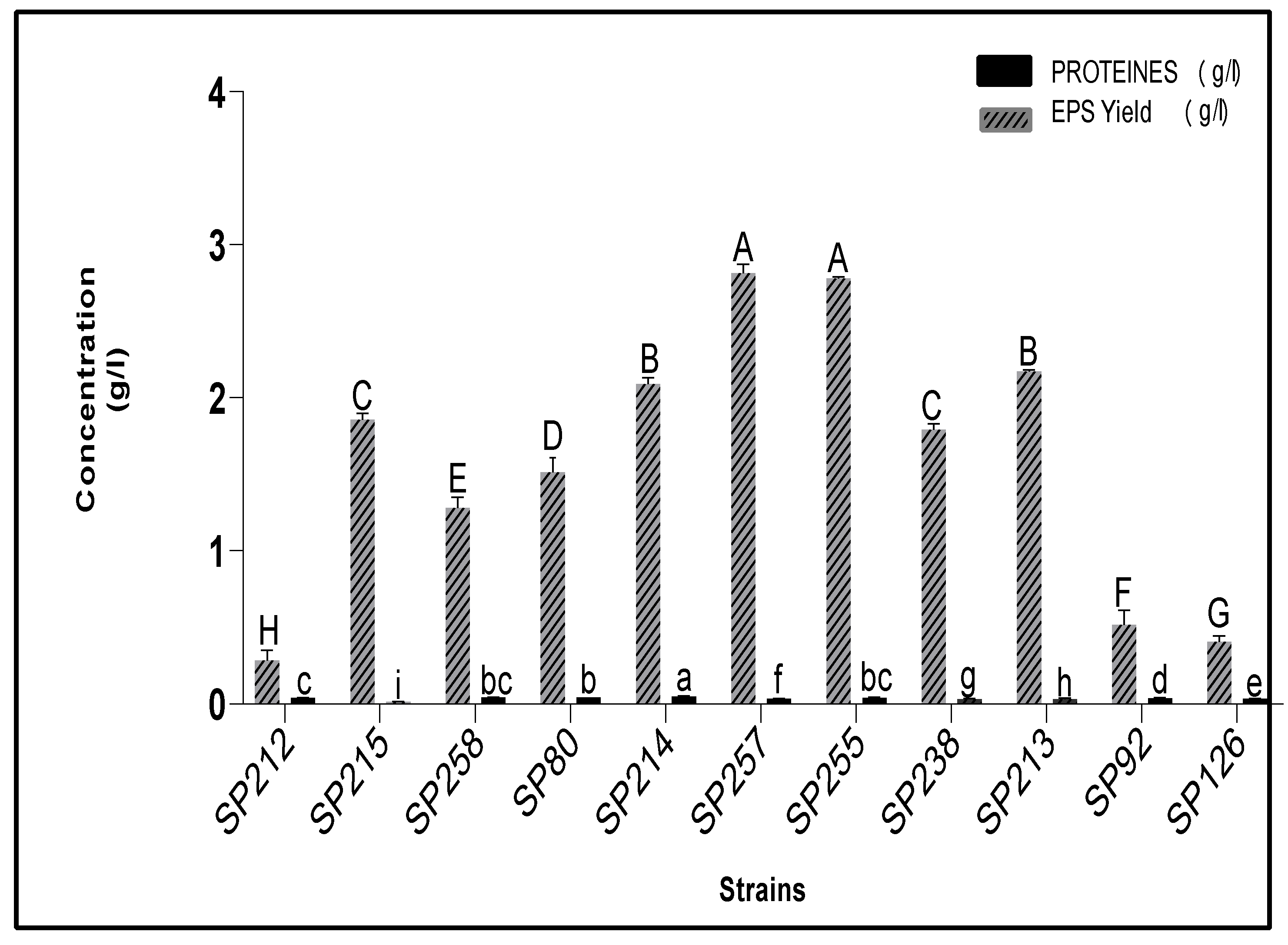
3.3. Estimation of Carbohydrates and Protein Contents of Crude EPS Extracts

The sugar content of the EPS extracts was estimated by the Dubois method [20]. The results are presented in Figure 1 and show that the total sugar contents ranged from 0.287 g/L to 2.778 g/L, indicating that EPS production was strain-dependent. This variability in concentration could be explained by the heterogeneity between the isolated species. Furthermore, the highest (p < 0.005) production of EPSs was shown by the SP255 strain (2.778 g/L) followed by SP257 (2.773 g/L) and SP213 (2.18 g/L). The lowest (p < 0.005) amount was recorded for SP212 (0.287 g/L).
As reported in the literature, there was a variation in the amounts of EPSs produced by LAB, which was influenced by a different parameter including the type of strain used, the carbon source, pH, temperature, inoculum size, and incubation time [29,30,31].
In a previous study, it was found that the strains Lactobacillus rhamnosus RW9595M and Lactobacillus kefiranofaciens WT-2B had the potential to produce high amounts of EPSs, with yields of 2.775 g/L and 2.500 g/L, respectively [32]. These findings were similar to our results for the strains SP255 and SP257.
Despite the importance of EPS amounts, the quantities produced by LAB are generally lower than those produced by other microorganisms [9].
However, the protein content of EPS extracts was low. The highest protein content was found in SP214 (0.0513 g/L) and the lowest protein content was found in SP215 (0.014 g/L). These results revealed that EPS extracts are mainly composed of carbohydrates.
In general, proteins are present in lower quantities in the structure of EPSs produced by microbial isolates compared to carbohydrates [33], which contain various sugars like glucose, galactose, rhamnose, and others in different ratios [10].
According to some authors [34], EPSs of a bacterial origin can contain other components such as lipids, proteins, and nucleic acids, which usually co-precipitate in ethanol. Nevertheless, a low protein content can be an indicator of EPS purity.
These findings demonstrate the ability of three LAB strains to produce high levels of EPS: SP213, SP255, and SP257. Consequently, those strains were retained for further work and the potential of produced EPSs as natural stabilizers was investigated.

3.4. Molecular Identification of SP213, SP255, and SP257 Strains

The three retained strains, SP213, SP255, and SP257, previously selected as the best EPS producers, were identified by sequencing 16S rDNA. The 16S fragments were amplified by two selected primers, resulting in an amplicon with a size of approximately 1500 bp. The obtained 16S rDNA nucleotide sequences were compared to those listed in the GenBank database of the BLAST program. The results demonstrated that both strains SP255 (Gene Bank accession no.PP911458) and SP257 (Gene Bank accession no.PP911459) could be identified as Lactococcus lactis subsp. lactis JCM 5805 and the SP213 (Gene Bank accession no. PP911457) as Weissella cibaria KACC 11862, respectively, with more than 99% of similarity.
The phylogenetic tree presented in Figure 2 shows the relative positions of the exopolysaccharide-producing LAB strains. The strain Weissella cibaria shared a maximum identity of 100% with the Leuconostoc strain. This result suggests a close relationship between Weissella cibaria and Leuconostoc Mesentoroides.
According to previous studies [35,36], Leuconostoc and Weissella have previously been acknowledged as closely related. Both genera are known for their ability to produce lactic acid and other metabolites that contribute to the flavor, texture, and preservation of fermented foods [37,38]. However, the Weissella genus, part of the Leuconostocaceae family, is known to produce significantly high amounts of EPSs compared to other LAB species [4,12].
Similar percentages of relationship homology were obtained for SP255 (Lactococcus lactis PP911458) and SP257 (Lactococcus lactis PP911459). A phylogenetic analysis also revealed that the SP238 strain (Weissella cibaria PP911460) had 60% homology with both the SP213 and SP80 strains (Weissella cibaria PP911457 and Weissella cibaria PP911463).
These findings indicate that most LAB isolates belong to the same phylogenetic lineage. Furthermore, all the isolates demonstrated a remarkable capacity for exopolysaccharides’ production, further supporting their classification as closely related strains. This could be due to a close geographic or ecological link, probably from sharing milk sources [39].

3.5. UV-Visible Analyses

UV-visible spectroscopy is based on the property of chromophore molecules to absorb light radiation of a specific wavelength, providing valuable information about the sample’s composition and concentration [40]. This technique is widely used for various applications, including chemical research, food quality analysis, drug identification, nucleic acid purity checks and quantitation, and bacterial culturing [41,42,43].
In the present study, the absorption spectra of the EPSs extracted from the selected strains are illustrated in Figure 3. For the three EPS extracts, UV spectra showed a strong absorbance peak around 220–290 nm. A similar result was also observed for dextran, a bacterial polysaccharide [44]. This absorbance peak can be attributed to various compounds and molecules present in the analyzed samples, such as carboxyl groups, carbonyl groups, metaldehyde, and uric acid [45].
Additionally, the maximum absorption wavelength varied between 254 nm and 260 nm. The absorption in the interval 240–280 nm could be attributed to the presence of amino acids [46]. The UV absorption below 260 nm is possibly due to the presence of tryptophan and sometimes tyrosine or phenylalanine.
Thus, the structural differences of these exopolysaccharides due to the presence of various functional groups and compounds like tryptophan can significantly impact their physicochemical, biological, and technological properties [47]. Understanding these relationships can enhance the use of EPS in various fields such as the food industry and pharmacology [48].

3.6. Chemical Structure of EPS

The FTIR spectra of the three investigated EPSs obtained in the 4000−420 cm−1 region are illustrated in Figure 4 and showed similar profiles. The presence of several different peaks allowed the identification of the major functional groups present in the structure of EPSs [49].
Indeed, the presence of a broad stretching peak in the region of 3000–3600 cm−1 corresponded to the hydroxyl group of the polysaccharide [50]. These groups lead to strong inter- and intramolecular interactions of the exopolysaccharide chains [11], while a minor band due to the stretching of the methylene group (C–H) vibration appears in the region of 2930 cm–1. These methyl groups are generally found in hexoses or deoxyhexoses [51]. Therefore, the presence of this peak provides valuable information about the composition and structure of the sugars present in EPS extracts [52].
The presence of the two characteristic peaks at 1606 cm−1 and 1409 cm−1 can be due, respectively, to the asymmetric and symmetric stretching vibration of the carboxylic groups (COOH) [53].
The appearance of a peak at about 1231 cm−1 can be related to a C–O stretching vibration with the possible presence of sulfate groups in the EPS extracts [54].
Similarly, the strong band at 1027 cm−1 and the weak absorption peak at 993 cm−1 can be attributed to the C–O stretching vibration of the polysaccharide backbone [55]. Thus, the spectral data in this region (1000–900 cm−1) support obtaining fingerprints related to vibrational modes of the C-O stretching of the polysaccharide and were used to determine the monosaccharide composition [19,56].
Also, the observation of weak peaks at 926 cm−1 can be an indication of the presence of α-anomers of pyranose saccharides with α-(1→6) glucosidic bonds and the peak at 856 cm−1 has been attributed to the presence of α-(1,3)-D-glucan [57,58].
These results suggest that EPSs produced by W. cibaria SP213, L. lactis SP257, and L. lactis SP255 are most likely dextran, which is a type of polysaccharide composed of glucose units linked by α-1,6 glycosidic bonds [59,60]. This α-glucans polymer is the most widely used bacterial polysaccharides with many potential applications in various fields, such as medicine, drug delivery systems, and the cosmetic and food industries [61].
Since the diversity of functional groups gives EPSs their properties [62], the biological activities of our extracts can be exploited.
Indeed, the existence of these functional groups in the EPS backbone leads them to interact with other molecules, such as lipids and proteins [60]. The presence of the hydroxyl groups in exopolysaccharides means they can form hydrogen bonds with water molecules, making them highly soluble in water and contributing to their viscoelastic properties [63]. Likewise, the presence of the methylene group (C–H) makes exopolysaccharides either hydrophobic or water soluble [62]. The occurrence of these functional groups, makes EPSs suitable as emulsifiers and as thickening agents [64]. The carboxyl groups (COO) in EPSs can form ionic bonds with positively charged molecules, such as metal ions [65,66], making them useful in numerous industrial domains, mainly in pharmaceutical applications and in food formulations [67].
In a previous study, the use of EPSs from LAB was shown to have a positive impact on the rheological properties of fermented dairy products [68]. During fermentation, the viscosity of milk gel was influenced by the chemical composition and the EPS structure, including the type of bonds and presence of side chains [69].

3.7. Zeta Potential Measurement (ζ)

The zeta potential (ζ) measurements of the tested EPSs within a pH range of 3 to 8 revealed varying charges, ranging from −14.23 to −24.30 mV for EPS SP255, −12.85 to −19.43 mV for EPS SP255, and −12.8 to −24.76 mV for EPS SP213. The result is shown in Figure 5. As the pH increases, there is a decrease in the zeta potential. It should be noted that the zeta potential is a key index to measure the attraction and repulsion between macromolecules, which could be used to assess the stability [70]. It is also a key parameter in the preparation, utilization, and destruction of colloidal dispersions. Moreover, zeta potential (ζ) is widely used in various fields, including biomedical research and water purification [71]. The negative charge of different EPSs reveals the presence of anionic groups in the polysaccharides backbone.
This negative charge is attributed to the ionization of the carboxyl groups of the EPS [72]. Also, it may be an indication of the presence of amino acids in its protein fraction [73].
Indeed, the presence of charged groups is the best indicator for the stability of dispersion due to their role in improving the solubility of EPSs [74] and prevents electrostatic interactions from producing intermolecular associations [75].
Likewise, the three studied strains exhibited a wide range of zeta potential values, indicating differing anionic characteristics. Thus, the EPSs from strain SP255 measured −24.3 mV at pH 8, while strain SP257 recorded −19.4 mV at the same pH. This variation indicates that the nature of EPSs is significantly influenced by the bacterial strain and its metabolic pathways [76].
Overall, these results suggest that the presence of anionic EPSs can significantly influence the rheological properties of the product by their electrostatic interactions with other existing charged polymers and their ability to adsorb on the surface. This impact can be observed in various industrial products, particularly in new formulations of food and drugs, where the texture and stability of products depend on the rheological properties [77,78].
Furthermore, zeta potential (ζ) measurements could be utilized to evaluate the bioactivities of EPSs. A previous study [79] reported that the negative charge can significantly impact the bioactivities of exopolysaccharides, providing valuable insights into their functional properties and contributing to their application in food and medicine.

3.8. EPS Solubility

The different dried EPS extracts were tested for their solubility in water. The determination of this parameter can give an idea about the hydrophobicity of the polymers [49].
The dried EPSs dissolved completely in water and the EPS solution was a clear and homogenous liquid with no precipitation. This solubility in water suggests that the EPS has hydrophilic properties and contains functional groups (carboxyl groups, sulfate, or phosphate groups) that can form hydrogen bonds with water molecules [80].
However, the same EPS samples remained insoluble in methanol, acetone, and chloroform. This insolubility in organic solvents indicates that the EPS may lack sufficient hydrophobic characteristics to dissolve.
Thus, the solubility characteristics of EPS extracts were in line with previous studies [81,82]. The high water solubility of EPS extracts could be used as an agent that can effectively bind water molecules [49]. This property may be advantageous for certain biomedical or food-related applications [83], such as improving moisture retention in food products and preventing syneresis.

3.9. Interfacial Properties

In general, the effectiveness of an emulsifier is often evaluated by its ability to reduce the interfacial tension between two immiscible phases [84]. In the present study, the measurement of the interfacial properties of EPS extracts at the oil–water interfaces were determined at two different pH values (3 and 7).
For all interfacial tension–time curves (Figure 6), the decrease in interfacial tension γ(t) between the aqueous EPS solution and the oil was an indicator of the EPS extracts’ adsorption on the oil–water interface.
At pH 7, the interfacial tension decreased in the following order: EPS SP257–EPS SP213–EPS SP255. Although, at pH 3 the interfacial tension decreased in the following order: EPS SP213–EPS SP257–EPS SP255. This order implies that among the three samples, EPS SP255 had the lowest (p < 0.005) interfacial tension γ (14.13 mN/m) and EPS SP213 had the highest (p < 0.005) interfacial tension γ value (18.7 mN/m). The notable ability of EPS SP255 to reduce interfacial tension suggests that this ability was superior to other EPS samples.
These results suggest that the presence of protein residues in the studied EPS extracts play a role in the reduction of interfacial properties [85]. In our case, the correlation between the protein content (Figure 1) and interfacial properties shows that EPS SP255 extract exhibits the highest (p < 0.005) protein content among the studied extracts and shows the most significant interfacial reduction. The observed correlation could be attributed to the electrostatic interactions and hydrogen bonding between existing proteins and polysaccharides in EPSs [77].
Other than the adsorption of proteins on the surface, the interfacial tension of EPSs can be influenced by various factors, including molecular configurational changes, realignment, and intermolecular interactions of the molecular chains [84,86].
Our findings also demonstrated that the decrease in interfacial tension due to EPS adsorption varies with the pH. This suggests that the pH of the solution influences the extent of EPS adsorption and its impact on interfacial tension. For example, at pH 7, the interfacial tension γ(t) of EPSs produced by strain SP257 decreased from 22.7 to 14.33 mN/m and from 21.45 to 12.25 mN/m at pH 3 throughout the measurement time. In this case, the EPS appears to be more effective at reducing interfacial tension at a lower pH (pH 3) compared to a higher pH (pH 7). Therefore, this EPS displayed better surface activity in an acidic environment than neutral, whereas for the EPS SP213 and EPS SP255 extracts, the interfacial tension value was lower at pH 7 compared to 3. Thus, the adsorption affinity to the oil–water interface was higher in a neutral environment.
For all strains, understanding how EPSs interact with interfaces and how their adsorption behavior is influenced by pH provide important data to their uses. The interfacial tension reduction property of EPS extracts can have various applications, particularly those related to emulsification [87] where controlling the interfacial properties is essential.
According to our results, all studied EPSs could be used in the formation and stabilization in both neutral and acidic emulsions, which find applications in various industries such as pharmaceuticals, cosmetics, and food.
However, given that the use of EPS SP257 as an emulsifier in acidic emulsions is recommended, it could be employed in acidic emulsions such as yogurt and fermented milk products.
It is worth noting that our study is one of the first to investigate the interfacial properties of native EPS derived exclusively from LAB. The majority of earlier studies are interested in modified or partially purified forms of EPSs. They have also explored the interactions between EPSs and other molecules, particularly proteins [88,89].
The focus on the unmodified form could provide valuable insights into the intrinsic characteristics of the EPS produced by these bacteria. Thus, the results obtained from this study open up a wide range of potential applications for these extracts.

3.10. Formation of Multilayer Emulsions

3.10.1. Zeta Potential Analysis

Improving the stability of oil-in-water (O/W) emulsions is achievable by harnessing the advantageous properties of various emulsifiers, leading to the formation of emulsions where droplets are stabilized by multilayer membranes [90].
To create a stable multilayer emulsion, it is important to select the right combination of emulsifier and biopolymers [91]. As seen previously (Figure 5), the three EPS extracts were negatively charged across the entire studied pH range. For this reason, a combination between the EPS extracts and the commercial protein sodium caseinate (CAS) was used to obtain the colloidal stability of CAS-EPS emulsion systems by electrostatic attractions between anionic groups of EPS chains and positive groups of (CAS) proteins through complexation at an acidic pH [90,92].
Likewise, to demonstrate the presence of distinct interfacial layers in the emulsions co-stabilized by caseinate and exopolysaccharides (EPSs), zeta potential measurements were conducted before and after the addition of EPSs. The results show significant changes in the zeta potential, indicating a shift in the surface charge of the oil droplets. This shift after the addition of EPSs strongly suggests the adsorption of a second stabilizing layer of EPSs onto the caseinate-stabilized droplets, thereby confirming the formation of a multilayered emulsion [85].
In this context, Zeta potential analysis is a crucial method for characterizing the stability and behavior of colloidal dispersions such as a multilayer EPS/caseinate emulsion [93]. ζ-potential measurements can be used to study the ionization behavior of polymers employed and to assess the appropriate concentration of biopolymer coatings around the emulsion droplets [92].
According to Bouyer et al. [94], particles exhibiting a zeta potential beyond +25 mV or below −25 mV typically demonstrate heightened stability owing to their electrostatic stability. Consequently, biopolymer dispersions formed with these particles tend to remain stable. Conversely, lower zeta potential values lead to eventual aggregation within dispersions due to Van der Waals inter-particle attractions.
As shown in Figure 7, the behavior of the ζ-potential of secondary emulsions prepared using different EPS concentrations (0–2 mg/mL) were measured. At pH 3 and without EPSs, the charge of the emulsion droplets was positive and reached approximately 30 mV. The positive charge is attributed to the sodium caseinate (CAS) being below its isoelectric point (IEP) of 4.6, resulting in a net-positive charge on the droplets. When the EPSs were added to the emulsions, the values of the ζ-potentials decreased considerably from positive to negative and reached 0.0 for an EPS/CAS ratio ranging from 0.75 to 1.500 mg/mL for the three emulsions. This change in the ζ-potential could imply the formation of complexes or binding between oppositely charged functional groups present in EPS (-COO) and the protein sodium caseinate (-NH3+).
Indeed, the adsorption of charged polyelectrolytes on oppositely charged surfaces is a well-studied phenomenon in the literature [95,96] and it is influenced by several factors such as the charge fraction, ionic concentration, and particle size [97].

3.10.2. Particle Size Measurements

As shown in Table 2, primary emulsions containing caseinate-coated lipid droplets had a monomodal particle size distribution characterized by a relatively small mean particle diameter (z-average) of approximately 22, 7 ± 0.96 µm at pH 3 and no creaming was observed. Furthermore, the mean particle diameter d (4.3) of the droplets in the secondary emulsions containing different ratios of CAS/EPS measured at saturation (between 0.75 and 1.5 mg/mL) was significantly larger (between 32 and 55 µm) than that of the primary emulsion, but still remained small. Indeed, emulsions prepared with small droplets tend to be more stable due to their surface properties [89].
However, the increase in the mean particle size may be attributed to the adsorption of EPSs to the surface of the CAS-coated oil droplets or due to their ability to bind multiple droplets together, a phenomenon known as bridging flocculation. This phenomenon has been reported in the literature for various other types of anionic polysaccharides added to emulsions stabilized by proteins, like pectin and xanthane [98], fucoidane [95], and alginate [96].
At the same pH conditions, the two emulsions containing CAS/EPS 257 and CAS/EPS 255 at a ratio of approximately 1.5 mg/mL were relatively stable for droplet aggregation, which can be attributed to the increase in steric and electrostatic repulsion. This stability for droplet aggregation is crucial in preventing the emulsions from breaking down or separating. Nevertheless, severe aggregation occurred in the multilayer emulsion prepared from EPS213 at the ratio of 1.7 mg/mL.
Based on previous studies, the characteristics of the emulsifiers or polyelectrolytes as well as the preparation conditions (homogenization method, pH…) play a crucial role in determining the zeta potential, size, and particle size distribution of the emulsion droplets [99]. On the one hand, an excess of emulsifiers can lead to bridging flocculation, where the emulsifier molecules form a network connecting the droplets, causing aggregation and instability [100]. On the other hand, insufficient emulsifiers can result in depletion flocculation, where the emulsifier molecules are depleted from the interface, causing droplets to come together and aggregate [101]. Thus, maintaining a delicate balance between the amount of emulsifier and the preparation conditions is the key to achieve stable and well-performing emulsions with desirable properties.

3.10.3. Influence of EPS Concentration on Emulsion Stability

For all studied EPS extracts, the investigation focused on examining the impact of incorporating an anionic EPS layer on the surface of caseinate (CAS)-coated droplets. At pH 3, a stable multilayer emulsion could be formed at different EPS concentrations. The investigation was expanded to determine the optimum amount of EPSs and caseinate to form the best stability among the examined multilayer emulsions. The potential measurements of the secondary emulsion containing different EPS concentrations (0.05–2.00 g/L, pH 3). As the EPS concentrations increased from 0 to 2 g/L, the ζ-potential values consistently decreased, hitting a constantly negative value. This trend suggested that the EPSs could saturate the surface of the protein-coated droplets surfaces.
Indeed, when anionic EPSs were absent, the droplets in the secondary emulsions exhibited a positive net charge of approximately 30 mV at pH 3. This positive charge was attributed to the cationic nature of the caseinate outer layer within the biopolymer coating. As the concentration of EPSs in the emulsions increased, the electrical charge on the droplets gradually shifted from positive to negative, becoming increasingly more negative.
The zeta potential on the droplets reached zero at an approximately 0.75 g/L EPS/CAS ratio for the EPS-SP255 extract and a 1.5 g/L ratio for the EPS-SP257 and EPS-SP213 extracts, indicating a balance between the anionic and cationic groups. This difference between the three EPS extracts could be explained by the variation in the chemical composition and structure of the different EPSs.
Moreover, when the concentration of anionic EPSs exceeds 1.5 g/L, the negative charge on the droplets stabilizes at a relatively constant level. This suggests that electrostatic interactions caused the negatively charged EPSs to attach to the positively charged protein-CAS coating of the lipid droplets. The fully coated droplets exhibited a zeta potential of approximately −15 mV.
The formation of a complete EPS layer around the droplet suggests a synergistic effect between proteins and exopolysaccharides in stabilizing the emulsion [84]. In addition, as discussed previously, the mean droplet diameter D (4.3) of the emulsions changes when EPSs are introduced into the system. The D (4.3) values of the emulsions increased and then decreased with thhe increasing EPS concentration (0.05–2.00 g/L pH 3).
According to our results, a bridging flocculation was observed at relatively low EPS concentrations (e.g., 0.05 to 0.75 wt % EPS for 3 wt % oil emulsion), which was not high enough to saturate the droplet surfaces. However, this emulsion instability to droplet aggregation could be improved if higher EPS levels were used. The minimum concentration required to stabilize multilayer emulsions for EPS 255 and EPS257 was found to be 1.5 and 1.7 wt %, respectively. For the EPS213 emulsion, when the concentration exceeded saturation, the emulsion flocculated.
Interestingly, these samples observed at pH 3 showed instability against droplet aggregation, but could be improved if higher exopolysaccharide levels were used. Indeed, polysaccharides can stabilize oil droplets by forming a protective coating around them [99], thereby preventing droplet flocculation and coalescence, which leads to phase separation in emulsions [89].

4. Conclusions

In the present study, three novel EPSs, EPS SP213, EPS SP255, and EPS SP257, biosynthesized by Lactococcus lactis strains and Weissella cibaria strains, were characterized. The studied strains produced significant amounts of EPSs, reaching 2.77 g/L.
The reduction in interfacial tension due to EPS adsorption supports their potential use as bio-hydrocolloid agents in the food industry and other biotechnological fields.
Additionally, the influence of the three EPSs on the preparation and stability of multilayer emulsions was demonstrated. The tested EPS extracts were able to improve the emulsion stability to varying extents.
Therefore, the interfacial properties of these EPSs, combined with their ability to create and maintain stable multilayer emulsions, position them as promising alternatives to certain commercial polysaccharide emulsifiers.

Author Contributions

Conceptualization, A.Z., M.Z., A.G. and T.K.; Methodology, A.Z., M.Z., A.G., I.F., I.S. and T.K.; Software, A.Z. and I.S.; Validation, A.Z. and M.Z.; Formal analysis, A.Z.; Resources, A.G., I.S. and M.H.; Writing—original draft, A.Z.; Writing—review & editing, M.Z., A.G., I.F., I.S., M.H. and T.K.; Visualization, M.H.; Supervision, M.Z., A.G. and T.K.; Project administration, T.K. All authors have read and agreed to the published version of the manuscript.

Funding

This research received no external funding.

Institutional Review Board Statement

Not applicable.

Informed Consent Statement

Not applicable.

Data Availability Statement

The original contributions presented in the study are included in the article, further inquiries can be directed to the corresponding author.

Conflicts of Interest

The authors declare no conflict of interest.

Abbreviations

EPSExopolysaccharide
IEPIsoelectric Point
LABLactic Acid Bacteria
CAGRCompound Annual Growth
HoPSHomopolysaccharides
HeEPSHeteropolysaccharides
MRSde Man, Rogosa, and Sharpe
UVUltraviolet
FTIRFourier-Transform Infrared Spectroscopy
CASColony Absorption Spectroscopy
BSABovine Serum Albumin

References

  1. Singh, S. Natural Food Additives Market Size, Share, Trends. Industry Reports. Available online: https://www.marketresearchfuture.com/reports/natural-food-additives (accessed on 24 August 2024).
  2. Ramos, I.M.; Seseña, S.; Poveda, J.M.; Palop, M.L. Screening of Lactic Acid Bacteria Strains to Improve the Properties of Non-fat Set Yogurt by in situ EPS Production. Food Bioprocess Technol. 2023, 16, 2541–2558. [Google Scholar] [CrossRef]
  3. Pourjafar, H.; Ansari, F.; Sadeghi, A.; Samakkhah, S.A.; Jafari, S.M. Functional and health-promoting properties of probiotics’ exopolysaccharides; isolation, characterization, and applications in the food industry. Crit. Rev. Food Sci. Nutr. 2023, 63, 8194–8225. [Google Scholar] [CrossRef]
  4. Fuso, A.; Bancalari, E.; Castellone, V.; Caligiani, A.; Gatti, M.; Bottari, B. Feeding Lactic Acid Bacteria with Different Sugars: Effect on Exopolysaccharides (EPS) Production and Their Molecular Characteristics. Foods 2023, 12, 215. [Google Scholar] [CrossRef] [PubMed]
  5. Singh, S.; Datta, S.; Narayanan, K.B.; Rajnish, K.N. Bacterial exo-polysaccharides in biofilms: Role in antimicrobial resistance and treatments. J. Genet. Eng. Biotechnol. 2021, 19, 140. [Google Scholar] [CrossRef] [PubMed]
  6. Tiwari, O.N.; Sasmal, S.; Kataria, A.K.; Devi, I. Application of microbial extracellular carbohydrate polymeric substances in food and allied industries. 3 Biotech 2020, 10, 221. [Google Scholar] [CrossRef]
  7. Huang, Z.; Lin, F.; Zhu, X.; Zhang, C.; Jiang, M.; Lu, Z. An exopolysaccharide from Lactobacillus plantarum H31 in pickled cabbage inhibits pancreas α-amylase and regulating metabolic markers in HepG2 cells by AMPK/PI3K/Akt pathway. Int. J. Biol. Macromol. 2020, 143, 775–784. [Google Scholar] [CrossRef]
  8. Jurášková, D.; Ribeiro, S.C.; Silva, C.C.G. Exopolysaccharides Produced by Lactic Acid Bacteria: From Biosynthesis to Health-Promoting Properties. Foods 2022, 11, 156. [Google Scholar] [CrossRef]
  9. Yang, S.; Xu, X.; Peng, Q.; Ma, L.; Qiao, Y.; Shi, B. Exopolysaccharides from lactic acid bacteria, as an alternative to antibiotics, on regulation of intestinal health and the immune system. Anim. Nutr. 2023, 13, 78–89. [Google Scholar] [CrossRef]
  10. Abid, Y.; Casillo, A.; Gharsallah, H.; Joulak, I.; Lanzetta, R.; Corsaro, M.M.; Attia, H.; Azabou, S. Production and structural characterization of exopolysaccharides from newly isolated probiotic lactic acid bacteria. Int. J. Biol. Macromol. 2018, 108, 719–728. [Google Scholar] [CrossRef]
  11. Korcz, E.; Varga, L. Exopolysaccharides from lactic acid bacteria: Techno-functional application in the food industry. Trends Food Sci. Technol. 2021, 110, 375–384. [Google Scholar] [CrossRef]
  12. Derdak, R.; Sakoui, S.; Pop, O.L.; Vodnar, D.C.; Elmakssoudi, A.; Errachidi, F.; Suharoschi, R.; Soukri, A.; El Khalfi, B.; AddoumElmakssoudi, B. Screening, optimization and characterization of exopolysaccharides produced by novel strains isolated from Moroccan raw donkey milk. Food Chem. X 2022, 14, 100305. [Google Scholar] [CrossRef] [PubMed]
  13. Laguerre, G.; van Berkum, P.; Amarger, N.; Prévost, D. Genetic diversity of rhizobial symbionts isolated from legume species within the genera Astragalus, Oxytropis, and Onobrychis. Appl. Environ. Microbiol. 1997, 63, 4748–4758. [Google Scholar] [CrossRef] [PubMed]
  14. Oussaief, O.; Jrad, Z.; Sbissi, I.; Nasri, W.; Khorchani, T.; El-Hatmi, H. Technological and probiotic potential of autochthonous lactic acid bacteria from spontaneously fermented dromedary milk. J. Food Process. Preserv. 2020, 44, e14685. [Google Scholar] [CrossRef]
  15. Ziadi, M.; Bouzaiene, T.; M’hir, S.; Zaafouri, K.; Mokhtar, F.; Hamdi, M.; Boisset-Helbert, C. Evaluation of the Efficiency of Ethanol Precipitation and Ultrafiltration on the Purification and Characteristics of Exopolysaccharides Produced by Three Lactic Acid Bacteria. BioMed Res. Int. 2018, 2018, 1–11. [Google Scholar] [CrossRef] [PubMed]
  16. DuBois, M.; Gilles, K.A.; Hamilton, J.K.; Rebers, P.A.; Smith, F. Colorimetric Method for Determination of Sugars and Related Substances. Anal. Chem. 1956, 28, 350–356. [Google Scholar] [CrossRef]
  17. Bradford, M.M. A rapid and sensitive method for the quantitation of microgram quantities of protein utilizing the principle of protein-dye binding. Anal. Biochem. 1976, 72, 248–254. [Google Scholar] [CrossRef]
  18. Hamidi, M.; Okoro, O.V.; Ianiri, G.; Jafari, H.; Rashidi, K.; Ghasemi, S.; Castoria, R.; Palmieri, D.; Delattre, C.; Pierre, G.; et al. Exopolysaccharide from the yeast Papiliotrema terrestris PT22AV for skin wound healing. J. Adv. Res. 2023, 46, 61–74. [Google Scholar] [CrossRef]
  19. Liao, W.; Gharsallaoui, A.; Dumas, E.; Elaissari, A. Understanding of the key factors influencing the properties of emulsions stabilized by sodium caseinate. Compr. Rev. Food Sci. Food Saf. 2022, 21, 5291–5317. [Google Scholar] [CrossRef]
  20. Karakas-Sen, A.; Karakas, E. Isolation, identification and technological properties of lactic acid bacteria from raw cow milk. Biosci. J. 2018, 34, 985–999. [Google Scholar] [CrossRef]
  21. Zantar, S.; El Galiou, O.; Zerrouk, H.M.; Laglaoui, A. Elaboration d’un fromage de chèvre semi-affiné à partir d’une sélection de souches lactiques autochtones isolées du lait de chèvres du Nord du FAO. Options Méditerranéennes Série A 2014, 108, 191–197. [Google Scholar]
  22. Shokryazdan, P.; Liang, J.B.; Abdullah, N.; Jahromi, M.F. Probiotic Potential of Lactic Acid Bacteria Isolated From Mulberry Silage. J. Pure Appl. Microbiol. 2015, 9, 443–452. [Google Scholar]
  23. Stephen, J.M.; Saleh, A.M. Homofermentative Lactobacilli isolated from organic sources exhibit potential ability of lactic acid production. Front. Microbiol. 2023, 14, 1297036. [Google Scholar] [CrossRef] [PubMed]
  24. Patil, P.; Wadehra, A.; Munjal, K.; Behare, P. Isolation of exopolysaccharides producing lactic acid bacteria from dairy products. Asian J. Dairy Food Res. 2015, 34, 280–284. [Google Scholar] [CrossRef]
  25. Taj, R.; Masud, T.; Sohail, A.; Sammi, S.; Naz, R.; Khanal, B.K.S.; Nawaz, M.A. In vitro screening of EPS-producing Streptococcus thermophilus strains for their probiotic potential from Dahi. Food Sci. Nutr. 2022, 10, 2347–2359. [Google Scholar] [CrossRef] [PubMed]
  26. Benhadria, M.K.; Touil, M.A.T.; Meddah, B. Optimization of production of Microbial Exopolysaccharides (EPS) with essential oils from two medicinal plants. J. Appl. Biosci. 2017, 111, 10925. [Google Scholar] [CrossRef]
  27. Almalki, M.A. Exopolysaccharide production by a new Lactobacillus lactis isolated from the fermented milk and its antioxidant properties. J. King Saud Univ. Sci. 2020, 32, 1272–1277. [Google Scholar] [CrossRef]
  28. Şentürk, D.Z.; Dertli, E.; Erten, H.; Şimşek, Ö. Structural and technological characterization of ropy exopolysaccharides produced by Lactobacillus plantarum strains isolated from Tarhana. Food Sci. Biotechnol. 2020, 29, 121–129. [Google Scholar] [CrossRef]
  29. Izadi, P.; Eldyasti, A. Holistic insights into extracellular polymeric substance (EPS) in anammosx bacterial matrix and the potential sustainable biopolymer recovery: A review. Chemosphere 2021, 274, 129703. [Google Scholar] [CrossRef]
  30. Shukla, A.; Mehta, K.; Parmar, J.; Pandya, J.; Saraf, M. Depicting the exemplary knowledge of microbial exopolysaccharides in a nutshell. Eur. Polym. J. 2019, 119, 298–310. [Google Scholar] [CrossRef]
  31. Kavitake, D.; Devi, P.B.; Shetty, P.H. Overview of exopolysaccharides produced by Weissella genus—A review. Int. J. Biol. Macromol. 2020, 164, 2964–2973. [Google Scholar] [CrossRef]
  32. Fusco, V.; Quero, G.M.; Cho, G.S.; Kabisch, J.; Meske, D.; Neve, H.; Bockelmann, W.; Franz, C.M.A.P. The genus Weissella: Taxonomy, ecology and biotechnological potential. Front. Microbiol. 2015, 6, 155. [Google Scholar] [CrossRef] [PubMed]
  33. Fguiri, I.; Ziadi, M.; Atigui, M.; Ayeb, N.; Arroum, S.; Assadi, M.; Khorchani, T. Isolation and characterisation of lactic acid bacteria strains from raw camel milk for potential use in the production of fermented Tunisian dairy products. Int. J. Dairy Technol. 2015, 69, 103–113. [Google Scholar] [CrossRef]
  34. Saleena, L.A.K.; Teo, M.Y.M.; How, Y.H.; In, L.L.A.; Pui, L.P. Immunomodulatory action of Lactococcus lactis. J. Biosci. Bioeng. 2023, 135, 1–9. [Google Scholar] [CrossRef]
  35. Zhang, J.; Yao, Y.; Li, J.; Ju, X.; Wang, L. Impact of exopolysaccharides-producing lactic acid bacteria on the chemical, rheological properties of buckwheat sourdough and the quality of buckwheat bread. Food Chem. 2023, 425, 136369. [Google Scholar] [CrossRef] [PubMed]
  36. Kiraz, D.; Özcan, A.; Yibar, A.; Dertli, E. Genetic diversity and phylogenetic relationships of Streptococcus thermophilus isolates from traditional Turkish yogurt: Multilocus sequence typing (MLST). Arch. Microbiol. 2024, 206, 121. [Google Scholar] [CrossRef]
  37. Power, A.C.; Chapman, J.; Chandra, S.; Cozzolino, D. Ultraviolet-visible spectroscopy for food quality analysis. In Evaluation Technologies for Food Quality; Elsevier: Amsterdam, The Netherlands, 2019; pp. 91–104. [Google Scholar] [CrossRef]
  38. Giglio, C.; Yang, Y.; Kilmartin, P. Analysis of phenolics in New Zealand Pinot noir wines using UV-visible spectroscopy and chemometrics. J. Food Compos. Anal. 2023, 117, 105106. [Google Scholar] [CrossRef]
  39. Dash, H.R.; Shrivastava, P.; Das, S. Quantification of DNA by Using UV–Visible Spectrophotometer. In Principles and Practices of DNA Analysis: A Laboratory Manual for Forensic DNA Typing, in Springer Protocols Handbooks; Springer: New York, NY, USA, 2020; pp. 127–132. [Google Scholar] [CrossRef]
  40. Passos, M.L.; Saraiva, M.L.M. Detection in UV-visible spectrophotometry: Detectors, detection systems, and detection strategies. Measurement 2019, 135, 896–904. [Google Scholar] [CrossRef]
  41. Bhat, V.T.; James, N.R.; Jayakrishnan, A. A photochemical method for immobilization of azidated dextran onto aminated poly(ethylene terephthalate) surfaces. Polym. Int. 2008, 57, 124–132. [Google Scholar] [CrossRef]
  42. Spangenberg, M.; Bryant, J.I.; Gibson, S.J.; Mousley, P.J.; Ramachers, Y.; Bell, G.R. Ultraviolet absorption of contaminants in water. Sci. Rep. 2021, 11, 3682. [Google Scholar] [CrossRef]
  43. Abid, Y.; Azabou, S.; Joulak, I.; Casillo, A.; Lanzetta, R.; Corsaro, M.M.; Gharsallaoui, A.; Attia, H. Potential biotechnological properties of an exopolysaccharide produced by newly isolated Bacillus tequilensis-GM from spontaneously fermented goat milk. LWT 2019, 105, 135–141. [Google Scholar] [CrossRef]
  44. Tripathy, D.B.; Mishra, A.; Clark, J.; Farmer, T. Synthesis, chemistry, physicochemical properties and industrial applications of amino acid surfactants: A review. Comptes Rendus Chim. 2018, 21, 112–130. [Google Scholar] [CrossRef]
  45. Mohammed, A.S.A.; Naveed, M.; Jost, N. Polysaccharides; Classification, Chemical Properties, and Future Perspective Applications in Fields of Pharmacology and Biological Medicine (A Review of Current Applications and Upcoming Potentialities). J. Polym. Environ. 2021, 29, 2359–2371. [Google Scholar] [CrossRef] [PubMed]
  46. Wang, B.; Song, Q.; Zhao, F.; Han, Y.; Zhou, Z. Production optimization, partial characterization and properties of an exopolysaccharide from Lactobacillus sakei L3. Int. J. Biol. Macromol. 2019, 141, 21–28. [Google Scholar] [CrossRef]
  47. Charoenwongpaiboon, T.; Wangpaiboon, K.; Pichyangkura, R.; Nepogodiev, S.A.; Wonganan, P.; Mahalapbutr, P.; Field, R.A. Characterization of a nanoparticulate exopolysaccharide from Leuconostoc holzapfelii KM01 and its potential application in drug encapsulation. Int. J. Biol. Macromol. 2021, 187, 690–698. [Google Scholar] [CrossRef]
  48. Maajid, H.S.; Nurliyani, N.; Widodo, W. Exopolysaccharide production in fermented milk using Lactobacillus casei strains AP and AG. AIMS Microbiol. 2022, 8, 138–152. [Google Scholar] [CrossRef] [PubMed]
  49. Sampaio, I.C.F.; Ferreira, J.d.A.S.; Crugeira, P.J.L.; Oliveira, I.M.d.S.; dos Santos, J.N.; Matos, J.B.T.L.; Pinheiro, A.L.B.; de Almeida, P.F. Oilfield Carbonated Produced Water Recycling Coupled to Exopolysaccharide Transformation by Lelliottia amnigena. Waste Biomass Valorization 2024, 15, 1309–1322. [Google Scholar] [CrossRef]
  50. Yang, L.; Zhao, T.; Wei, H.; Zhang, M.; Zou, Y.; Mao, G.; Wu, X. Carboxymethylation of polysaccharides from Auricularia auricula and their antioxidant activities in vitro. Int. J. Biol. Macromol. 2011, 49, 1124–1130. [Google Scholar] [CrossRef]
  51. Hu, S.-M.; Zhou, J.-M.; Zhou, Q.-Q.; Li, P.; Xie, Y.-Y.; Zhou, T.; Gu, Q. Purification, characterization and biological activities of exopolysaccharides from Lactobacillus rhamnosus ZFM231 isolated from milk. LWT 2021, 147, 111561. [Google Scholar] [CrossRef]
  52. Amiri, S.; Reza, M.; Sowti, K.M.; Alizadeh, M. Optimization of food-grade medium for co-production of bioactive substances by Lactobacillus acidophilus LA-5 for explaining pharmabiotic mechanisms of probiotic. J. Food Sci. Technol. 2021, 58, 1–12. [Google Scholar] [CrossRef]
  53. Kansandee, W.; Moonmangmee, D.; Moonmangmee, S.; Itsaranuwat, P. Characterization and Bifidobacterium sp. growth stimulation of exopolysaccharide produced by Enterococcus faecalis EJRM152 isolated from human breast milk. Carbohydr. Polym. 2019, 206, 102–109. [Google Scholar] [CrossRef]
  54. Naibaho, J.; Butula, N.; Jonuzi, E.; Korzeniowska, M.; Chodaczek, G.; Yang, B. The roles of brewers’ spent grain derivatives in coconut-based yogurt-alternatives: Microstructural characteristic and the evaluation of physico-chemical properties during the storage. Curr. Res. Food Sci. 2022, 5, 1195–1204. [Google Scholar] [CrossRef] [PubMed]
  55. Du, R.; Qiao, X.; Zhao, F.; Song, Q.; Zhou, Q.; Wang, Y.; Pan, L.; Han, Y.; Zhou, Z. Purification, characterization and antioxidant activity of dextran produced by Leuconostoc pseudomesenteroides from homemade wine. Carbohydr. Polym. 2018, 198, 529–536. [Google Scholar] [CrossRef] [PubMed]
  56. Kang, Y.J.; Kim, T.J.; Kim, M.J.; Yoo, J.Y.; Kim, J.H. Isolation of Exopolysaccharide-Producing Lactic Acid Bacteria from Pa-Kimchi and Characterization of Exopolysaccharides. Microbiol. Biotechnol. Lett. 2023, 51, 157–166. [Google Scholar] [CrossRef]
  57. Jiang, G.; Gan, L.; Li, X.; He, J.; Zhang, S.; Chen, J.; Zhang, R.; Xu, Z.; Tian, Y. Characterization of Structural and Physicochemical Properties of an Exopolysaccharide Produced by Enterococcus sp. F2 From Fermented Soya Beans. Front. Microbiol. 2021, 12, 744007. [Google Scholar] [CrossRef]
  58. Delvart, A.; Moreau, C.; Cathala, B. Dextrans and dextran derivatives as polyelectrolytes in layer-by-layer processing materials —A review. Carbohydr. Polym. 2022, 293, 119700. [Google Scholar] [CrossRef]
  59. Boukhelata, N.; Taguett, F.; Kaci, Y. Characterization of an extracellular polysaccharide produced by a Saharan bacterium Paenibacillus tarimensis REG 0201M. Ann. Microbiol. 2018, 69, 93–106. [Google Scholar] [CrossRef]
  60. Lu, X.; Chen, J.; Guo, Z.; Zheng, Y.; Rea, M.C.; Su, H.; Zheng, X.; Zheng, B.; Miao, S. Using polysaccharides for the enhancement of functionality of foods: A review. Trends Food Sci. Technol. 2019, 86, 311–327. [Google Scholar] [CrossRef]
  61. Xiao, R.; Zheng, Y. Overview of microalgal extracellular polymeric substances (EPS) and their applications. Biotechnol. Adv. 2016, 34, 1225–1244. [Google Scholar] [CrossRef]
  62. Naveed, S.; Li, C.; Lu, X.; Chen, S.; Yin, B.; Zhang, C.; Ge, Y. Microalgal extracellular polymeric substances and their interactions with metal(loid)s: A review. Crit. Rev. Environ. Sci. Technol. 2019, 49, 1769–1802. [Google Scholar] [CrossRef]
  63. Joulak, I.; Azabou, S.; Finore, I.; Poli, A.; Nicolaus, B.; Donato, P.D.; Bkhairia, I.; Dumas, E.; Gharsallaoui, A.; Immirzi, B.; et al. Structural characterization and functional properties of novel exopolysaccharide from the extremely halotolerant Halomonas elongata S6. Int. J. Biol. Macromol. 2020, 164, 95–104. [Google Scholar] [CrossRef]
  64. Benalaya, I.; Alves, G.; Lopes, J.; Silva, L.R. A Review of Natural Polysaccharides: Sources, Characteristics, Properties, Food, and Pharmaceutical Applications. Int. J. Mol. Sci. 2024, 25, 1322. [Google Scholar] [CrossRef] [PubMed]
  65. Wang, L.; Tian, H.; Liu, W.; Zheng, H.; Wu, H.; Guan, Y.; Lu, Q.; Lv, Z. Effects of EPS-producing Leuconostoc mesenteroides XR1 on texture, rheological properties, microstructure and volatile flavor of fermented milk. Food Biosci. 2023, 56, 103371. [Google Scholar] [CrossRef]
  66. Berthold-Pluta, A.M.; Pluta, A.S.; Garbowska, M.; Stasiak-Różańska, L. Exopolysaccharide-Producing Lactic Acid Bacteria—Health-Promoting Properties and Application in the Dairy Industry. Postępy Mikrobiol. Adv. Microbiol. 2019, 58, 191–204. [Google Scholar] [CrossRef]
  67. Sabbah, M.; Esposito, M. Insight into Zeta Potential Measurements in Biopolymer Film Preparation. J. Biotechnol. Biomater. 2016, 6, 2–4. [Google Scholar] [CrossRef]
  68. Kamarajan, B.P.; Ananthasubramanian, M. Nanoemulsion-based antimicrobial systems. In Antimicrobial Nanosystems; Elsevier: Amsterdam, The Netherlands, 2023; pp. 61–78. [Google Scholar] [CrossRef]
  69. Barbosa, J.A.; Abdelsadig, M.S.; Conway, B.R.; Merchant, H.A. Using zeta potential to study the ionisation behaviour of polymers employed in modified-release dosage forms and estimating their pKa. Int. J. Pharm. X 2019, 1, 100024. [Google Scholar] [CrossRef] [PubMed]
  70. Gupta, P.; Diwan, B. Bacterial Exopolysaccharide mediated heavy metal removal: A Review on biosynthesis, mechanism and remediation strategies. Biotechnol. Rep. 2017, 13, 58–71. [Google Scholar] [CrossRef]
  71. Gaikwad, V.L.; Choudhari, P.B.; Bhatia, N.M.; Bhatia, M.S. Characterization of pharmaceutical nanocarriers: In vitro and in vivo studies. In Nanomaterials for Drug Delivery and Therapy; Elsevier: Amsterdam, The Netherlands, 2019; pp. 33–58. [Google Scholar] [CrossRef]
  72. Lu, G.W.; Gao, P. Emulsions and Microemulsions for Topical and Transdermal Drug Delivery. In Handbook of Non-Invasive Drug Delivery Systems; Elsevier: Amsterdam, The Netherlands, 2010; pp. 59–94. [Google Scholar] [CrossRef]
  73. Andrew, M.; Jayaraman, G. Molecular Characterization and Biocompatibility of Exopolysaccharide Produced by Moderately Halophilic Bacterium Virgibacillus dokdonensis from the Saltern of Kumta Coast. Polymers 2022, 14, 3986. [Google Scholar] [CrossRef] [PubMed]
  74. Mende, S.; Rohm, H.; Jaros, D. Influence of exopolysaccharides on the structure, texture, stability and sensory properties of yoghurt and related products. Int. Dairy J. 2016, 52, 57–71. [Google Scholar] [CrossRef]
  75. Schmidt, W.; Brouwers, H.J.H.; Kühne, H.-C.; Meng, B. Interactions of polysaccharide stabilising agents with early cement hydration without and in the presence of superplasticizers. Constr. Build. Mater. 2017, 139, 584–593. [Google Scholar] [CrossRef]
  76. Yang, X.; Ren, Y.; Li, L. The relationship between charge intensity and bioactivities/processing characteristics of exopolysaccharides from lactic acid bacteria. LWT 2022, 153, 112345. [Google Scholar] [CrossRef]
  77. Guo, M.Q.; Hu, X.; Wang, C.; Ai, L. Polysaccharides: Structure and Solubility. In Solubility of Polysaccharides; Xu, Z., Ed.; InTech: Rijeka, Croatia, 2017. [Google Scholar] [CrossRef]
  78. Castellane, T.C.L.; Persona, M.R.; Campanharo, J.C.; Lemos, E.G.d.M. Production of exopolysaccharide from rhizobia with potential biotechnological and bioremediation applications. Int. J. Biol. Macromol. 2015, 74, 515–522. [Google Scholar] [CrossRef] [PubMed]
  79. Ilyas, N.; Mumtaz, K.; Akhtar, N.; Yasmin, H.; Sayyed, R.Z.; Khan, W.; El Enshasy, H.A.; Dailin, D.J.; Elsayed, E.A.; Ali, Z. Exopolysaccharides Producing Bacteria for the Amelioration of Drought Stress in Wheat. Sustainability 2020, 12, 8876. [Google Scholar] [CrossRef]
  80. Lobo, R.E.; Gómez, M.I.; de Valdez, G.F.; Torino, M.I. Physicochemical and antioxidant properties of a gastroprotective exopolysaccharide produced by Streptococcus thermophilus CRL1190. Food Hydrocoll. 2019, 96, 625–633. [Google Scholar] [CrossRef]
  81. Wei, Y.; Xie, Y.; Cai, Z.; Guo, Y.; Wu, M.; Wang, P.; Li, R.; Zhang, H. Interfacial and emulsion characterisation of chemically modified polysaccharides through a multiscale approach. J. Colloid Interface Sci. 2020, 580, 480–492. [Google Scholar] [CrossRef]
  82. Liu, J.; Tan, J.; Hua, X.; Jiang, Z.; Wang, M.; Yang, R.; Cao, Y. Interfacial properties of ultrahigh methoxylated pectin. Int. J. Biol. Macromol. 2020, 152, 403–410. [Google Scholar] [CrossRef]
  83. Elmanan, M.; Al-Assaf, S.; Phillips, G.O.; Williams, P.A. Studies on Acacia exudate gums: Part VI. Interfacial rheology of Acacia senegal and Acacia seyal. Food Hydrocoll. 2008, 22, 682–689. [Google Scholar] [CrossRef]
  84. Schroën, K.; de Ruiter, J.; Berton-Carabin, C. The Importance of Interfacial Tension in Emulsification: Connecting Scaling Relations Used in Large Scale Preparation with Microfluidic Measurement Methods. ChemEngineering 2020, 4, 63. [Google Scholar] [CrossRef]
  85. Yang, H.; Wang, S.; Yang, L.; Liu, H. Preparations, application of polysaccharide–protein nanoparticles and their assembly at the oil–water interface. Food Sci. Biotechnol. 2024, 33, 13–22. [Google Scholar] [CrossRef]
  86. Abid, Y.; Azabou, S.; Blecker, C.; Gharsallaoui, A.; Corsaro, M.M.; Besbes, S.; Attia, H. Rheological and emulsifying properties of an exopolysaccharide produced by potential probiotic Leuconostoc citreum-BMS strain. Carbohydr. Polym. 2021, 256, 117523. [Google Scholar] [CrossRef]
  87. Gharsallaoui, A.; Saurel, R.; Chambin, O.; Cases, E.; Voilley, A.; Cayot, P. Utilisation of pectin coating to enhance spray-dry stability of pea protein-stabilised oil-in-water emulsions. Food Chem. 2010, 122, 447–454. [Google Scholar] [CrossRef]
  88. Burgos-Díaz, C.; Rubilar, M.; Morales, E.; Medina, C.; Acevedo, F.; Marqués, A.M.; Shene, C. Naturally occurring protein–polysaccharide complexes from linseed (Linum usitatissimum) as bioemulsifiers. Eur. J. Lipid Sci. Technol. 2016, 118, 165–174. [Google Scholar] [CrossRef]
  89. Liao, W.; Dumas, E.; Elaissari, A.; Gharsallaoui, A. The formation mechanism of multilayer emulsions studied by isothermal titration calorimetry and dynamic light scattering. Food Hydrocoll. 2023, 136, 108275. [Google Scholar] [CrossRef]
  90. Wang, J.; Souihi, S.; Ben Amara, C.; Dumas, E.; Gharsallaoui, A. Influence of low methoxyl pectin on the physicochemical properties of sodium caseinate-stabilized emulsions. J. Food Process Eng. 2018, 41, e12906. [Google Scholar] [CrossRef]
  91. Bouyer, E.; Mekhloufi, G.; Rosilio, V.; Grossiord, J.-L.; Agnely, F. Proteins, polysaccharides, and their complexes used as stabilizers for emulsions: Alternatives to synthetic surfactants in the pharmaceutical field? Int. J. Pharm. 2012, 436, 359–378. [Google Scholar] [CrossRef] [PubMed]
  92. Chang, Y.; McClements, D.J. Interfacial deposition of an anionic polysaccharide (fucoidan) on protein-coated lipid droplets: Impact on the stability of fish oil-in-water emulsions. Food Hydrocoll. 2015, 51, 252–260. [Google Scholar] [CrossRef]
  93. Shi, F.; Chang, Y.; Shen, J.; Chen, G.; Xue, C. A comparative investigation of anionic polysaccharides (sulfated fucan, ι-carrageenan, κ-carrageenan, and alginate) on the fabrication, stability, rheology, and digestion of multilayer emulsion. Food Hydrocoll. 2023, 134, 108081. [Google Scholar] [CrossRef]
  94. McClements, D.J. Theoretical Analysis of Factors Affecting the Formation and Stability of Multilayered Colloidal Dispersions. Langmuir 2005, 21, 9777–9785. [Google Scholar] [CrossRef]
  95. Qiu, C.; Zhao, M.; McClements, D.J. Improving the stability of wheat protein-stabilized emulsions: Effect of pectin and xanthan gum addition. Food Hydrocoll. 2015, 43, 377–387. [Google Scholar] [CrossRef]
  96. Sheng, B.; Li, L.; Zhang, X.; Jiao, W.; Zhao, D.; Wang, X.; Wan, L.; Li, B.; Rong, H. Physicochemical Properties and Chemical Stability of β-Carotene Bilayer Emulsion Coated with Bovine Serum Albumin and Arabic Gum Compared to Monolayer Emulsions. Molecules 2018, 23, 495. [Google Scholar] [CrossRef]
  97. McClements, D.J.; Jafari, S.M. Improving emulsion formation, stability and performance using mixed emulsifiers: A review. Adv. Colloid Interface Sci. 2018, 251, 55–79. [Google Scholar] [CrossRef]
  98. Dickinson, E. Strategies to control and inhibit the flocculation of protein-stabilized oil-in-water emulsions. Food Hydrocoll. 2019, 96, 209–223. [Google Scholar] [CrossRef]
  99. Cho, Y.-H.; Decker, E.A.; McClements, D.J. Competitive Adsorption of Mixed Anionic Polysaccharides at the Surfaces of Protein-Coated Lipid Droplets. Langmuir 2009, 25, 2654–2660. [Google Scholar] [CrossRef] [PubMed]
  100. Surber, G.; Spiegel, T.; Dang, B.P.; Pombo, A.W.; Rohm, H.; Jaros, D. Cream cheese made with exopolysaccharide-producing Lactococcus lactis: Impact of strain and curd homogenization pressure on texture and syneresis. J. Food Eng. 2021, 308, 110664. [Google Scholar] [CrossRef]
  101. Ebrahimi, B.; Rad, A.H.; Ghanbarzadeh, B.; Torbati, M.; Falcone, P.M. The emulsifying and foaming properties of Amuniacum gum (Dorema ammoniacum) in comparison with gum Arabic. Food Sci. Nutr. 2020, 8, 3716–3730. [Google Scholar] [CrossRef] [PubMed]
Figure 1. Total sugars and proteins content of EPS extract. A–H: For each sugar content, difference between strain is significant at p < 0.05, a–i: For each protein content, difference between strain is significant at p < 0.05.
Fermentation 10 00532 g001
Figure 2. Phylogenetic relationships of exopolysaccharide-producing lactic acid bacteria from sheep, goat, and camel milk based on partial 16S rDNA sequences. Escherichia coli NR024570 was used as the out-group.
Fermentation 10 00532 g002
Figure 3. UV-visible absorption spectra of EPS extracts.
Fermentation 10 00532 g003
Figure 4. FTIR spectra of EPSs produced by W. cibaria SP213 (A), Lac. Lactis SP257 (B), and Lac. Lactis SP255 (C).
Fermentation 10 00532 g004
Figure 5. ζ Potential of EPS as a function of pH.
Fermentation 10 00532 g005
Figure 6. Time-dependent changes in interfacial tension of EPSs at pH 3 and pH 7.
Fermentation 10 00532 g006
Figure 7. ζ-potential of multilayer emulsions at pH 3.0 as a function of EPS/Caseinate ratio.
Fermentation 10 00532 g007
Table 1. Main LAB strains able to produce EPSs.
StrainOriginEPS ProductionIdentificationAccession Number
SP80raw camel milk+Weissella cibariaPP911463
SP92raw camel milk+Leuconostoc mesenteroides subsp.-
SP126fermented camel milk+Weissella cibaria-
SP212fermented goat milk+Leuconostoc mesenteroides subsp.PP911461
SP213fermented goat milk+Weissella cibariaPP911457
SP214fermented goat milk+Leuconostoc mesenteroides subsp.PP911462
SP215fermented goat milk+Weissella cibaria-
SP238raw sheep milk+Weissella cibariaPP911460
SP255raw camel milk+Lactococcus lactis subsp. LactisPP911458
SP257raw camel milk+Lactococcus lactis subsp. LactisPP911459
SP258raw camel milk+Leuconostoc mesenteroides subsp.-
The strains SP92, SP126, SP215, and SP258 have not been deposited in the international gene bank yet and, consequently, no accession numbers were attributed.
Table 2. Average particle size D (4.3) for primary and secondary emulsions as function of ratio (w/w).
EmulsionD (4.3) (μm)
Primary emulsion
2 wt % CAS, 10 wt % oil22.70 ± 0.95
Secondary emulsion
0.75 wt % CAS/EPS 25755.90 ± 2.72
1.25 wt % CAS/EPS 25755.20 ± 2.24
1.7 wt % CAS/EPS 25731.24 ± 2.27
0.75 wt % CAS/EPS 25568.16 ± 4.27
1.5 wt % CAS/EPS 25536.30 ± 1.37
1.25 wt % CAS/EPS21347.13 ± 3.25
1.7 wt % CAS/EPS21356.55 ± 1.20
Disclaimer/Publisher’s Note: The statements, opinions and data contained in all publications are solely those of the individual author(s) and contributor(s) and not of MDPI and/or the editor(s). MDPI and/or the editor(s) disclaim responsibility for any injury to people or property resulting from any ideas, methods, instructions or products referred to in the content.

