Review

Biosensors Based on Lipid Modified Graphene Microelectrodes

by
Georgia-Paraskevi Nikoleli
1,
Christina G. Siontorou
2,
Dimitrios P. Nikolelis
3,*,
Spyridoula Bratakou
1,
Stephanos Karapetis
1 and
Nikolaos Tzamtzis
1
1
Laboratory of Inorganic & Analytical Chemistry, School of Chemical Engineering, Dept 1, Chemical Sciences, National Technical University of Athens, 9 Iroon Polytechniou St., GR 15780, Athens, Greece
2
Laboratory of Simulation of Industrial Processes, Department of Industrial Management and Technology, School of Maritime and Industry, University of Piraeus, Piraeus GR 18534, Greece
3
Laboratory of Environmental Chemistry, Department of Chemistry, University of Athens, Panepistimiopolis-Kouponia, GR 15771 Athens, Greece
*
Author to whom correspondence should be addressed.
Submission received: 6 November 2016 / Revised: 20 February 2017 / Accepted: 13 March 2017 / Published: 16 March 2017
(This article belongs to the Special Issue Carbon-Based Sensors)

Abstract

:
Graphene is one of the new materials which has shown a large impact on the electronic industry due to its versatile properties, such as high specific surface area, high electrical conductivity, chemical stability, and large spectrum of electrochemical properties. The graphene material-based electronic industry has provided flexible devices which are inexpensive, simple and low power-consuming sensor tools, therefore opening an outstanding new door in the field of portable electronic devices. All these attractive advantages of graphene give a platform for the development of a new generation of devices in both food and environmental applications. Lipid-based sensors have proven to be a good route to the construction of novel devices with improved characteristics, such as fast response times, increased sensitivity and selectivity, and the possibility of miniaturization for the construction of portable biosensors. Therefore, the incorporation of a lipid substrate on graphene electrodes has provided a route to the construction of a highly sensitive and selective class of biosensors with fast response times and portability of field applications for the rapid detection of toxicants in the environment and food products.

Graphical Abstract

1. Introduction

Graphene nanomaterials have been given tremendous attention recently in the literature for technological applications, owing to their unique physicochemical properties, such as good sensing ability, and excellent mechanical, thermal and electrical properties. The large surface area of graphene enhances the surface loading of desired biomolecules, either through passive adsorption, or by the covalent immobilization of biomolecules. On the other hand, the excellent conductivity and small band gap of graphene are beneficial for the conduction of electrons between the biomolecules and the electrode surface. Graphene is a one-atom-thick planar sheet of carbon atoms, densely packed together into a honeycomb shaped crystal lattice. Graphene looks like atomic-scale chicken wire made up of carbon atoms and their bonds. The carbon-carbon bond length is around 0.142 nm. Graphene is the basic structural element of several carbon allotropes including graphite, carbon nanotubes and fullerenes. Many sheets of graphene stacked together are collectively called graphite. Carbon nanotubes are cylindrical molecules of carbon with novel properties that are potentially useful in a wide variety of applications including nano-electronics, optics and materials applications, among others. They exhibit extraordinary tensile strength, a unique range of electrical properties, and are efficient thermal conductors. Most single-walled nanotubes (SWNT) are close to 1 nanometer in diameter, with a tube length that can be many millions of times longer. The structure of a SWNT can be thought of as a one-atom-thick layer of graphite wrapped into a seamless cylinder. The way that graphene sheet is wrapped is represented by a pair of indices. Multi-walled nanotubes (MWNT) consist of multiple rolled layers (concentric tubes) of graphene. The interlayer distance in multi-walled nanotubes is close to the distance between graphene layers in graphite, approximately 3.3 Å (330 pm). Fullerenes are an allotrope (physical arrangement of atoms) of carbon distinct from both graphite and diamond. They are a cylindrical, spherical or ellipsoid arrangement consisting of dozens of carbon atoms. Fullerenes are a molecular form of carbon distinct from graphite and diamond; they can be spherical, ellipsoid, or cylindrical arrangements of dozens to hundreds of carbon atoms. Complete information of the structure, properties and uses of these nanostructures is given in [1]. Graphene has around two-fold higher effective surface area and has a greater cost-effectiveness than carbon nanotubes. Additionally, it has greater homogenous surface that is responsible for highly uniform and efficient functionalization.
The price of graphene is linked to its quality, and not all applications require superb material quality. For example, graphene oxide powder (graphene functionalized with oxygen and hydrogen) is inexpensive and has been used to make a conductive graphene paper, for DNA analysis, and for other advanced composite and biotechnology applications. Graphene oxide in solution sells for 99 euros per 250 mL from Graphenea. A large part of this expense is the substrate on which graphene is generally produced. By using a process of chemical vapor deposition (CVD), graphene has often been grown as a monolayer (a layer one atom thick) by exposing platinum, nickel or titanium carbide to ethylene or benzene at high temperatures. Recent production methods have lowered these costs by incorporating copper as a substrate, but even this method can still prove expensive. To help drastically reduce these costs, researchers from University of Glasgow came up with the idea of depositing high-quality graphene on the surface of inexpensive copper foils, often used to make the ultra-thin cathodes (negative electrodes) in lithium-ion batteries. As it turns out, the surface of the copper proved to be both completely smooth and a superior substrate on which to form the graphene [2].
The electrochemical responses at microelectrodes can differ greatly from those seen at conventional (i.e., macro) electrodes. Typically, microelectrodes offer higher sensitivity, smaller double-layer capacitance, and lower Ohmic losses that result in higher signal-to-noise ratios. The improved diffusion characteristics of microelectrodes allow measurements in both static and stirred solutions. This characteristic has opened many new possibilities in electrochemistry and may have a positive impact on the commercial realization of chemical sensors, since it allows a sensor to be used as an insertion device. Such sensors may be dipped into the analyte solution without stirring effects (convection), which can cause unwanted fluctuations in the electrode response. To date, the majority of sensors which have reached commercial success have avoided this problem by inhibiting stirring by some means (e.g., droplets of blood in hand-held glucose sensors are held by surface tension); however, this is not possible for all applications, such as the analysis of non-viscous media or the analysis of analytes present in flowing streams. Microelectrodes have several properties which make them attractive as active elements within sensors for the determination of analytes in, for example, flowing water streams. Specifically, microelectrodes exhibit enhanced diffusion in comparison to larger sensors and this leads to enhanced sensitivity—as well as independence from the effects of convectional mass transport and therefore solution flow or other movement. Individual microelectrodes offer, however, very small responses. One approach for overcoming this problem is via the use of many microelectrodes coupled together in the form of an array, to allow a larger cumulative response to be measured, as depicted in Figure 1.
The present chapter describes recent examples in the development of miniaturized electrochemical biosensors by integrating enzyme, antibodies and artificial and natural lipid modified graphene microelectrodes. A detailed overview towards the advancement of graphene-based lipid film biosensors has been reviewed. Latest advances are relating applications of these biosensors to rapidly detect toxicants in foods, environment and bioterrorism weapons. The presented biosensors exhibit good reproducibility, reusability, selectivity, rapid response times, long shelf life, high sensitivity and potentially can be used for rapid applications in the field by even non-skilled personnel. This chapter highlights the significant milestones achieved and elucidates further the emerging future prospects in this area.

1.1. Protein and Peptide-Based Biosensors Based on Nanostructured Materials

Recent biological terrorism threats and outbreaks of microbial pathogens clearly emphasize the need for biosensors that can quickly and accurately identify infectious agents. The majority of rapid biosensors generate detectable signals when a molecular probe in the detector interacts with an analyte of interest. Analytes may be whole bacterial or fungal cells, virus particles, or specific molecules, such as chemicals or protein toxins, produced by the infectious agent. Proteins, peptides and nucleic acids are most commonly used as probes in biosensors because of their versatility in forming various tertiary structures. The interaction between the probe and the analyte can be detected by various sensor platforms, including quartz crystal microbalances, surface acoustical waves, surface plasmon resonance, amperometrics and voltammetry. The field of biosensors is constantly evolving to develop devices that have higher sensitivity and specificity, and are smaller, portable and cost-effective.
Biosensors have become important and practical tools in the field of foodcare, chemical and biological analysis, environmental monitoring, food safety control and homeland security. The performance of biosensors depends on their components, among which the matrix material (i.e., the layer between the recognition layer of biomolecule and transducer) plays a crucial role in defining the stability, sensitivity and shelf-life of a biosensor [3]. Among biosensors, electrochemical ones are of particular interest due to several combined advantages such as low detection limits, short response times, long-term stability, power requirements, low cost, ease of operation and miniaturization capability. A current goal for these types of biosensors is their translation to point-of-care diagnostic devices. Much effort has been put into improving key performance parameters, such as sensitivity, specificity, recognition rates, stability and multiplexing capabilities for parallel recognition, to allow this possibility.
The emergence of nanotechnology has opened new horizons for electrochemical biosensors. It is believed that highly sensitive and selective biosensors can be constructed through the integration of biomolecules and nanomaterial-based sensor platforms. Over the last fifteen years, efforts have focused on the use of nanotechnology to develop nanostructured materials (e.g., graphene, and ZnO nanowires, nanotubes, nanowalls and nanorods) as biomolecule immobilizing matrices/supports to improve electrochemical detection [4]. Nanoscale structures like these offer many unique features and show great promise for faster response and higher sensitivity at the device interface than planar sensor configurations. Their nanometer dimensions, being in the scale of the target analyte, show an increased sensing surface and strong binding properties, thus allowing a higher sensitivity. The interest in developing these nanostructures for biosensing applications has resulted from the development of new synthesis methods and improved characterization techniques, allowing for new functionalities to be created [4].
Because of the interesting graphene advantages among the nanomaterials that have been developed, this chapter describes the increasing application of this nanostruucture material to the fabrication of highly sensitive electrochemical biosensors. Although several strategies have been described for using graphene in such bioaffinity and biocatalytic sensing [5,6] for amplification tagging or modifying electrode transducers, this chapter will focus only on applications as surface modifiers. The broad capabilities of graphene-based (bio)electrodes to the biocatalytic electrochemical detection of numerous important analytes and to other bioelectronic affinity assays will be discussed, along with future prospects and challenges.

1.2. Graphene Nanomaterials Used in Electrochemical (Bio)Sensors Fabrication

Graphene and its derived structures (graphene oxide, graphene platelets, graphene nanoflakes) have become popular materials for fabricating electrode matrixes for sensing and biosensing [7]. Graphene is the mother of all graphitic forms including zero-dimensional fullerenes, one-dimensional carbon nanotubes and three-dimensional graphite [8].
The structure of graphene is defined as a single-layer two-dimensional sp2-hybridized carbon, and is currently, without any doubt, the most intensively-studied material. This single-atom-thick sheet of carbon atoms arrayed in a honeycomb pattern is the world’s thinnest, strongest and stiffest material, as well as being an excellent conductor of both heat and electricity [9]. It is often categorized by the number of stacked layers: single layer, few-layer (2–10 layers) and multi-layer, which is also known as thin graphite. Ideally, for graphene to preserve its distinct properties, its use should be narrowed to single or few-layer morphologies [7].
Considerable attention has been given to graphene as a next generation electronic material derives from its unique electronic, optical, mechanical, thermal, and electrochemical properties [7]. Being a very good low-noise material electronically, graphene can be employed in the achievement of molecular sensing [10].
It is important to note that graphene is attractive for electrochemistry because it is a conductive yet transparent material, with a low cost and low environmental impact, a wide electrochemical potential window, low electrical resistance in comparison to glassy carbon (GC), atomic thickness. Further, it features two well-defined redox peaks linearly aligned with the square root of the scan rate magnitude, suggesting that its redox processes are primarily diffusion controlled. Peak-to-peak values under cyclic voltammetry are low, suggesting rapid electron transfer kinetics, and its apparent electron transfer rate is orders of magnitude higher than that of GC. This rate of electron transfer has been shown to be surface dependent and can be increased significantly by the creation of specific surface functional groups [10]. The high density of edge-plane defect sites on graphene provides multiple electrochemically active sites. Its entire volume is exposed to the surrounding due to its 2D structure, making it very efficient in detecting adsorbed molecules. Graphene-based electrodes also exhibit high enzyme loading due to their high surface area. This, in turn, can facilitate high sensitivity, excellent electron transfer promoting ability for some enzymes, and excellent catalytic behavior towards many biomolecules [10,11]. Graphene-based devices also possess the required biocompatibility to be amenable for in situ biosensing.
The advantages of graphene are as follows: graphene exhibits the advantages of a large surface area (2630 m2·g−1 for single-layer graphene), similar to that of carbon nanotubes (CNTs) [12], and a small size of each individual unit. It also exhibits some other merits like low cost, two external surfaces, facile fabrication and modification and absence of metallic impurities, which may yield unexpected and uncontrolled electrocatalytic effects and toxicological hazards [7,10,11].
The cyclic voltammograms of graphene have been given in the literature [13]. It was found that the shapes of all CV curves were approximately rectangular and exhibited a good symmetry, even at a high scan rate, which demonstrated an excellent capacitive behavior and rate performance. At the low scan rate, the speed of charge and discharge was relatively slow, which allowed the ions in electrolyte to have enough time to diffuse into graphene layers, and therefore, the electrodes exhibited a higher specific capacitance. However, at a high scan rate, the process of charge and discharge was too quick for the full ion diffusion, and the less utilization of electrode surface resulted in a lower specific capacitance. During scanning, no redox peak existed in the CV curves, which indicated that the electrodes were of excellent electrochemical stability.
Graphene, a two-dimensional form of crystalline carbon, either a single layer of carbon atoms forming a honeycomb (hexagonal) lattice or several coupled layers of this honeycomb structure. The word graphene, when used without specifying the form (e.g., bilayer graphene, multilayer graphene), usually refers to single-layer graphene. Graphene is a parent form of all graphitic structures of carbon: graphite, a three-dimensional crystal consisting of relatively weakly coupled graphene layers; nanotubes, which may be represented as scrolls of graphene; and buckyballs, spherical molecules made from graphene with some hexagonal rings replaced by pentagonal rings. Graphene is a crystalline allotrope of carbon with 2-dimensional properties. Its carbon atoms are densely packed in a regular atomic-scale chicken wire (hexagonal) pattern (Figure 2).
Each atom of graphene has four bonds, one σ bond with each of its three neighbors and one π-bond that is oriented out of plane. The atoms are about 1.42 Å apart. Graphene’s hexagonal lattice can be regarded as two interleaving triangular lattices. This perspective was successfully used to calculate the band structure for a single graphite layer using a tight-binding approximation. It has also been reported that the edges of graphene sheets possess a variety of oxygenated species, that can support efficient electrical wiring of the redox centers of several heme-containing metalloproteins to the electrode, and also enhance the adsorption and desorption of molecules [10,11].
Graphene-based nanomaterials can be classified in relation to the method of production. They can be produced by chemical vapor deposition (CVD) growth, by mechanical exfoliation of graphite, or by exfoliation of graphite oxide. Neither CVD-produced graphene nor mechanically-exfoliated graphene contain large quantities of defects or functionalities. Bulk quantities of graphene-based nanomaterials are typically prepared by different methods, such as the thermal exfoliation of graphite oxide which leads to a material called thermally reduced graphene (TRGO) or, for example, sono-assisted exfoliation of graphite oxide to graphene oxide (GO), which can be further reduced chemically or electrochemically. The products are typically referred to as chemically reduced GO (CRGO) or electrochemically reduced GO (ERGO). TRGO contains large amounts of defects and significantly differs from pristine graphene, which has a perfect honeycomb lattice structure. GO has a structure that is not fully planar because the sp2 carbon network is heavily damaged. It contains large amounts of oxygen-containing groups, which can be beneficial to the functionalization through the action of the biomolecules for biorecognition events during biosensing. Reduced forms of GO have a partly-restored sp2 lattice but still hold some fraction of oxygen-containing groups [14]. Therefore, one could have a large graphene “toolbox” to choose the right type of graphene for the right application and transduction mechanism [15]. Most of graphene used in electrochemistry is graphene produced from GO chemical/thermal reduction, which is also called functionalized graphene sheets or chemically reduced GO, and usually has abundant structural defects and functional groups which are advantageous for electrochemical applications. It has been demonstrated that ERGO exhibits much better performance for electrochemical applications than CRGO. Moreover, Chua et al. [16] demonstrated that not all graphene materials are beneficial for the detection at lab-on-chip devices. Their findings could provide valuable insights into the future applicability of graphene materials towards practical applications.
The future development of electrochemical graphene-based nanobiodevices should be based on the better understanding of some electrochemical details (such as the role of the defects and oxygen containing groups at the edges of graphene sheets), the interaction mechanism of biomolecules with graphene surface, and the role of doping heteroatoms in graphene. Furthermore, it is important to remark that novel methods for well-controlled synthesis and processing of graphene should be developed. Although graphene has been synthesized with various strategies, the economical production approach with high yield is still not widely available.

2. Experimental

2.1. Materials and Solutions

Dipalmitoyl phosphatidylcholine (DPPC) was purchased from Sigma Chemical Co., St. Louis, MO, USA. DPPC was used as lipid for the formation of the films supported on a polymer. The functional monomer, methacrylic acid, and the crosslinker, ethylene glycol dimethacrylate, were both supplied by Aldrich (Aldrich-Chemie, Steinheim, Germany). The initiator, 2,2′-azobis-(2-methylpropionitrile) (AIBN), was supplied by Merck KgaA (Darmstadt, Germany). Cholera toxin (CT) was purchased from Calbiochem (La Jolla, CA, USA) and GM1 was obtained from FIDIA Research Laboratory (Abano Terme, Italy). Water was purified by passage through a Milli-Q cartridge filtering system (Milli-Q, Millipore, El Paso, TX, USA) and had minimum resistivity of 18 MΩ cm. All other chemicals were of analytical-reagent grade. The filters and (nominal) pore sizes used were glass microfiber (0.7 and 1.0 µm, Whatmam Scientific Ltd., Kent, UK).

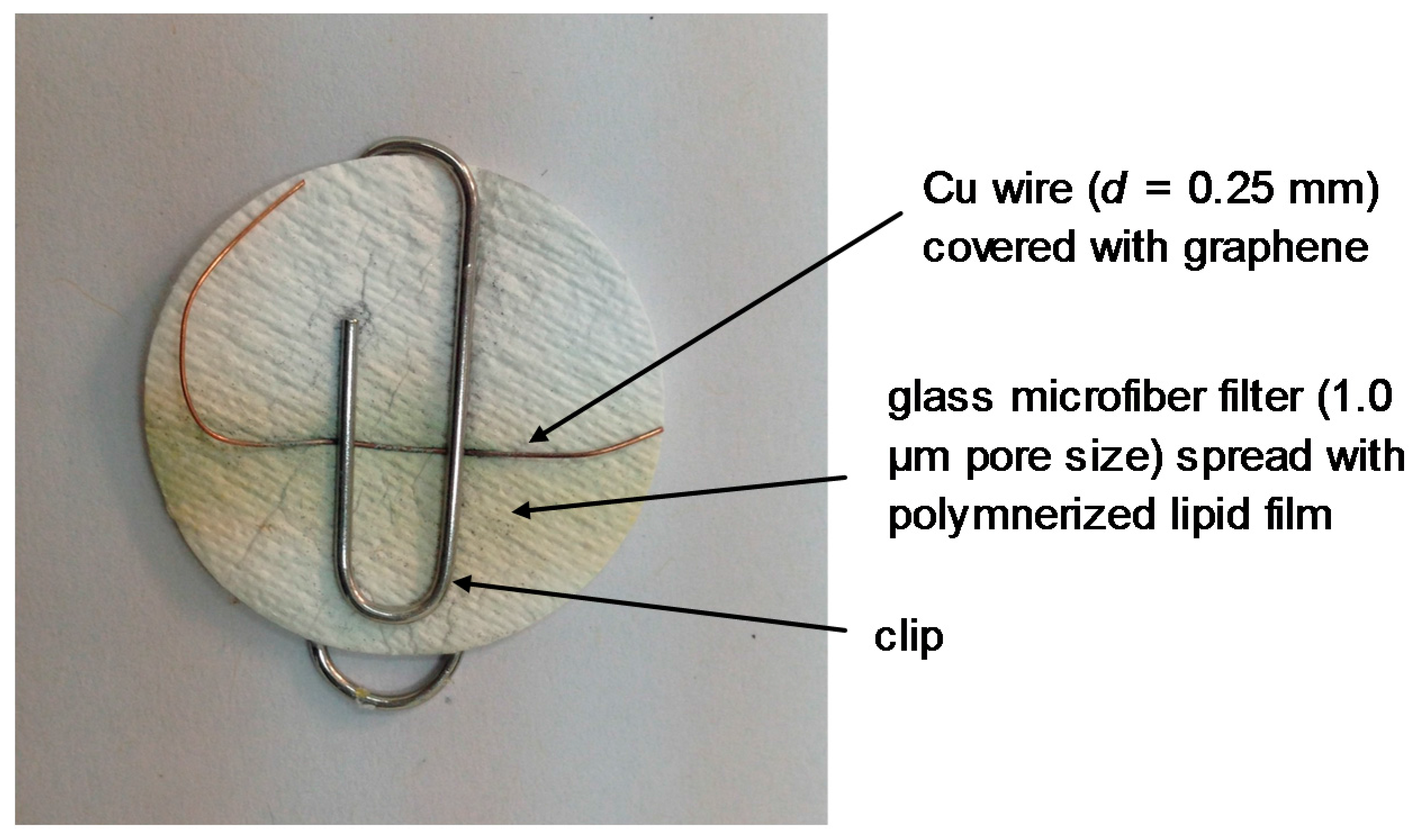
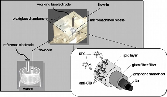
2.2. Fabrication of the Lipid-Based Sensor Electrode with Incorporated Receptor on a Graphene Electrode

The construction of the graphene electrode has been described in detail in one of our previous papers [17]; a 0.4 mg/mL homogeneous graphene dispersion had been obtained in N-methyl-pyrrolidone (NMP) after mild sonication (using a Bandelin SONOREX Digital 10P sonicator, Sigma-Aldrich, Taufkirchen Germany) for 180 h and centrifugation at 700 rpm for 2 h. Stabilized lipid films were prepared by polymerization, as previously described in the literature [18,19]. Shortly, 5 mg of DPPC were added to a mixture of 0.070 mL of methylacrylic acid, 0.8 mL of ethylene glycol dimethacrylate, 8 mg of 2,2′-azobis-(2-methylpropionitrile), 1.0 mL of acetonitrile. The mixture was spurged with nitrogen for about 1 min and sonicated for 30 min. For the preparation of the stabilized lipid films, 0.15 mL of this mixture was spread on the microfilter. The filter with the mixture was then irradiated using the UV deuterium lamp. Polymerization was completed within 4 h. These membranes were stable to store in air for periods of more than two months.
The “receptor” (i.e., enzyme, antibody, artificial and natural) was incorporated in bilayer lipid membranes (BLMs) during polymerization by spreading 10 µL of the “receptor” suspension along with the polymerization mixture. These electrodes can be used once (and then disposed of) or repetitively (after regenerated—see below); when not in use, they are kept at 4 °C.
The preparation of the potentiometric biosensor concluded after the encapsulation of the filter-supported polymerized lipid film onto the copper wire (d = 0.25 mm) containing graphene nanosheets (Figure 3 and Figure 4).

2.3. Electrochemical Measurements

A two electrode system (i.e., the working bioelectrode: stabilized polymeric lipid membrane/graphene, and the reference electrode: standard Ag/AgCl) was used to measure the potentiometric electrochemical response of the fabricated biosensor using a Keithley Electrometer Model 614 (in the voltage mode); the voltage was measured against the reference electrode (Figure 4).
Sensor calibration performed in a stopped-flow mode i.e., 10 or 20 μL aliquots), were injected into the carrier electrolyte in flow; the flow stopped for 5 min (adequate time to record the response) and then re-started for sensor regeneration. The flow rate used was 2.0 mL/min.

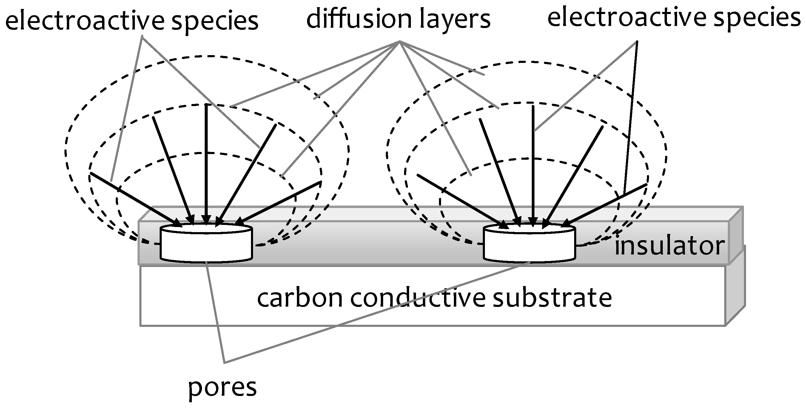
3. Mechanism of Signal Generation

The mechanism of signal generation was previously exploited in order to increase and therefore maximize the sensitivity of the method. In one of our papers, electron scanning microscopy (SEM) experiments were performed in order to investigate the mechanism of signal generation [20]. The results have shown that the structure of the polymerized polyacrylate without the lipid has an amorphous structure, but when the lipid is copolymerized, the structure becomes crystalline. When the receptor (e.g., Anti-STX) is incorporated within the polymer, the structure becomes again amorphous. Finally, when a drop of the analyte (e.g., saxitoxin) is deposited on the filter, the structure again switches to crystals, and this shows the defects in the lipid film that allow ions to diffuse through and result in potential alterations.
The working mechanism for the developed potentiometric cholesterol biosensor using the stabilized polymer lipid membrane on the graphene electrode was also exploited in one of our previous papers [21]. The polymer lipid membrane provided a highly stable microenvironment for the immobilization of the enzyme (cholesterol oxidase), and the polymer lipid membrane has a tendency to carry high concentration of the analyte (cholesterol) molecules, due to their similar chemistry. During the experiment, a large number of cholesterol molecules undergo the oxidation on the surface of highly stabilized polymer lipid membrane containing cholesterol oxidase molecules, as shown in the following chemical reaction [22]:
Cholesterol + O2 → 5-3-ketosteroid + H2O2
The resultant 5-3-ketosteroid is a transient product which spontaneously changes through the isomerization of trans double bond at 5–6 position in steroid cycle by intramolecular change of hydrogen atom at 4–6 β position and consequently a highly stable 4–3-ketosteroid substance is produced, as shown below:
5-3-ketosteroid i s o m e r i s a t i o n 4-3-ketosteroid
The stable output signal is attributed to this mechanism, which creates the charge environment during the oxidation of cholesterol on the surface of stabilized polymer lipid membrane. It is well-known that cholesterol alters the phase structure of the polymeric mixture, which exhibits a strong lipophilic structure, thus attracting cholesterol molecules towards the surface of the stabilized polymeric lipid membrane. This will increase the concentration of cholesterol biomolecules on the surface of working electrode, and a rapid oxidation results in the high sensitivity of the presented biosensor. Moreover, the increased cholesterol concentration on the surface of the stabilized polymeric lipid membrane will alter the phase structure of the lipid film to more packed structure (i.e., the liquid crystalline structure becomes gel-like). Therefore, it has shown increased interactions between cholesterol and the lipid molecule, which also contributes in the generation of stable output voltage signal for the presented cholesterol sensor. In addition, the conjugated stabilized polymeric thin film provides an excellent surface for the biomolecules to access the graphene electrode, having a large surface-area-to-volume ratio, which also results in the generation of the voltage signal.
A number of other articles have also examined the mechanism of signal generation and the physical chemistry of lipid modified graphene and carbon nanotube microelectrodes. SEM images of a SWNT network were presented to provide a schematic illustration of the hybrid device of SWNT-net and lipid bilayer with embedded membrane proteins [23].

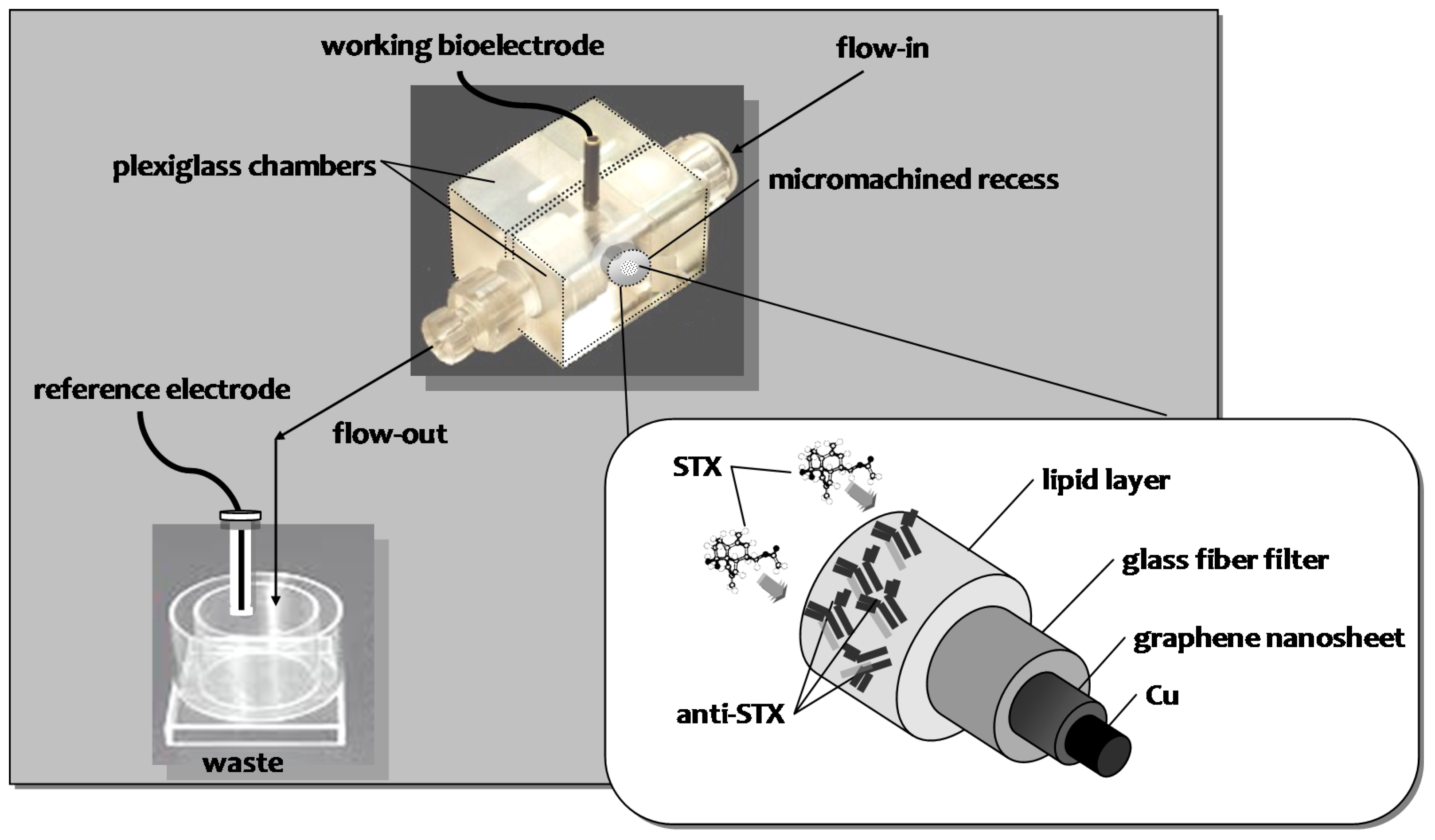
4. Examples of Biosensors Based on Lipid Modified Graphene Microelectrodes

Recent publications related to the use of lipid modified graphene-based microelectrodes are given in the literature. The electronic properties of graphene can be modulated by charged lipid bilayer adsorbing on the surface. Biorecognition events which lead to changes in membrane integrity can be monitored electrically using an electrolyte-gated biomimetic membrane-graphene transistor. It was demonstrated that the bactericidal activity of antimicrobial peptides can be sensed electrically by graphene-based on a complex interplay of biomolecular doping and ionic screening effects [24].
A review was published in the literature on the use of lipid modified graphene-based microelectrodes, providing an overview of the recent advancements in biosensors for tissue engineering applications [25]. Another review presented the electrochemical sensing of dopamine and describes biosensors using graphene microelectrodes and mainly lipid modified [26]. Also, the use graphene microelectrodes for the detection of a wide range of analytes such as microRNA etc. have appeared in the literature recently [27].
A novel system developed by integrating lipid bilayer with single-walled carbon nanotube networks (SWNT-net) based field-effect transistor (FET) and demonstrated that such hybrid nanoelectronic biosensors can specifically and electronically detect the presence and dynamic activities of ionophores (specifically, gramicidin and calcimycin) in their native lipid environment [23]. This technique can potentially be used to examine other membrane proteins (e.g., ligand-gated ion channels, receptors, membrane insertion toxins, and antibacterial peptides).
In a recent published paper [28], lipid monolayer membrane functionalized graphene sheets were prepared using a facile method. Interactions between the graphene and different types of liposomes, including charged and neutral, were also investigated. It was found that the anionic liposomes could spontaneously self-organize into lipid monolayer membranes, partially covering the surface of graphene sheets. The resultant lipid monolayer functionalized graphene nanomaterials exhibited high stability in aqueous solution, and an excellent performance as carrier for loading the anticancer drug, doxorubicin (DOX), with a high loading capacity of 70%. The loaded drug can be released under pH control. About 10% and 14% of the bound DOX was released after 54 h at pH 10.0 and 7.0, respectively, whereas, about 70% of DOX was released after 54 h at pH 5.0.
Graphene nanosheets have been successfully exfoliated onto a thin copper wire and exploited for the miniaturization of a potentiometric urea biosensor [29]. A miniaturized potentiometric urea lipid film-based biosensor on graphene nanosheets was developed. Structural characterization of graphene nanosheets for miniaturization of potentiometric urea lipid film-based biosensors have been studied through atomic force microscopy (AFM) and transmission electron microscopy (TEM) measurements. UV-Vis and Fourrier transform IR (FTIR) spectroscopy have been utilized to study the pre- and post-conjugated surfaces of graphene nanosheets. The presented potentiometric urea biosensor exhibits good reproducibility, reusability, selectivity, rapid response times (∼4 s), long shelf life and high sensitivity of ca. 70 mV/decade over the urea logarithmic concentration range from 1 × 10⁻⁶ M to 1 × 10⁻³ M.
A novel potentiometric cholesterol biosensor has been fabricated through the immobilization of the stabilized polymeric lipid membrane onto graphene electrode [21]. The stabilized polymeric lipid membrane is composed of cholesterol oxidase enzyme and polymerization mixture; which holds paramount influence on the properties of the cholesterol biosensor. The presented biosensor reveals an appreciable reproducibility, good selectivity and high sensing capability with a linear slope curve of ~64 mV per decade. The strong biocompatibility among stabilized polymeric lipid membranes and human biofluids provides the possibility to use for real blood samples and other biological applications.
Proteinaceous moieties are critical elements in most detection systems, including biosensing platforms. Their potential is undoubtedly vast, yet many issues regarding their full exploitation remain unsolved. On the other hand, the biosensor formats with higher marketability probabilities are enzyme in nature and electrochemical in concept. To no surprise, alternative materials for hosting catalysis within an electrode casing have received much attention lately to demonstrate a catalysis-coated device. Graphene is presented as ideal material to modify electrodes and biosensor platforms, especially in protein-based detection. Our group developed electrochemical sensors based on these nanomaterials for the sensitive detection of cholesterol using cholesterol oxidase incorporated in stabilized lipid films [21]. In a broader sense, the not-so-remote prospect of quickly assembling a protein-based flexible biosensing detector to fulfill site-specific requirements is appealing to both university researchers and industry developers.
A miniaturized potentiometric cholera toxin sensor on graphene nanosheets with incorporated lipid films was also developed [30]. Ganglioside GM1, the natural cholera toxin receptor, immobilized on the stabilized lipid films, provided adequate selectivity for detection over a wide range of toxin concentrations, fast response time of ca. 5 min, and detection limit of 1 nM. The proposed sensor is easy to construct and exhibits good reproducibility, reusability, selectivity, long shelf life and high sensitivity of ca. 60 mV/decade of toxin concentration. The method was implemented and validated in lake water samples. This novel ultrathin film technology is currently adapted to the rapid detection of other toxins that could be used in bioterrorism.
A miniaturized potentiometric D-dimer biosensor on graphene nanosheets with incorporated lipid films [31]. The graphene electrode was used for the development of a very selective and sensitive immunosensor for the detection of D-dimer by immobilizing the mouse anti human D-dimer antibody on stabilized lipid films. The immunosensor responded for the wide range of D-dimer concentrations with fast response time of ca. 15 s. The presented potentiometric D-dimer biosensor is easy to construct and exhibits good reproducibility, reusability, selectivity, rapid response times, long shelf life and high sensitivity of ca. 59 mV/decade over the D-dimer logarithmic concentration range from 10−6 to 10−3 μg/L.
A miniaturized potentiometric carbofuran chemical sensor on graphene nanosheets with incorporated lipid films was reported in the literature [32]. The graphene electrode was used for the development of a very selective and sensitive chemical sensor for the detection of carbofuran by immobilizing an artificial selective receptor on stabilized lipid films. The artificial receptor was synthesized by transformation of the hydroxyl groups of resorcin [4] arene receptor into phosphoryl groups. This chemical sensor responded for the wide range of carbofuran concentrations with a fast response time of ca. 20 s. The presented potentiometric carbofuran chemical sensor is easy to construct and exhibits good reproducibility, reusability, selectivity, rapid response times, long shelf life and high sensitivity of ca. 59 mV/decade over the carbofuran logarithmic concentration range from 10−6 to 10−3 M. In the conclusion of this paper, graphene nanosheets were successfully used to prepare a carbofuran chemical sensor that could be used for the rapid determination of this insecticide in fruits and vegetables. The presented potentiometric carbofuran chemical sensor reveals good reproducibility, reusability and selectivity along with high sensitivity of ca. ∼60 mV/decade over a wide logarithmic range of carbofuran concentrations ranging from 1 × 10⁻⁶ to 1×10⁻³ M. Additionally, the sensor exhibits an excellent output stability with a response time of ~20 s and relatively long shelf life were attained. The present method now provides a technique for the rapid detection of carbofuran at the levels of nM concentrations, without interferences from the other constituents, and can be used as complimentary rapid technique to HPLC methods for the rapid detection of carbofuran in fruits and vegetables.
A miniaturized potentiometric naphthalene acetic acid (NAA) sensor on graphene nanosheets with incorporated lipid films was reported recently [33]. Auxin-binding protein 1 receptor immobilized on the stabilized lipid films provided adequate selectivity for detection over a wide range of hormone concentrations, fast response time of ca. 5 min, and detection limit of 10 nM. The proposed sensor is easy to construct and exhibits good reproducibility, reusability, selectivity, long shelf life and high sensitivity of ca. 56 mV/decade of hormone concentration. The reliability of the biosensor was successfully evaluated using a wide range of NAA-spiked fruits and vegetables.
A miniaturized potentiometric saxitoxin sensor on graphene nanosheets with incorporated lipid films and Anti-STX, the natural saxitoxin receptor, immobilized on the stabilized lipid films is described in one of our recent papers [20]. An adequate selectivity for detection over a wide range of toxin concentrations, fast response time of ca. 5–20 min, and detection limit of 1 nM have been achieved. The proposed sensor is easy to construct and exhibits good reproducibility, reusability, selectivity, long shelf life and high sensitivity of ca. 60 mV/decade of toxin concentration. The method was implemented and evaluated in lake water and shellfish samples. This novel ultrathin film technology is currently adapted to the rapid detection of other toxins that could be used in bioterrorism.
Table 1 provides all the above descibed examples of lipid modified graphene-based biosensors for the detection of various analytes.

5. Summary and Conclusions

As a result of the unique physical and chemical properties of graphene, an explosive growth of research work on their biomedical applications can be observed from the literature in the past few years, especially in the areas of biosensors, bioelectronics, drug delivery, cellular imaging, etc.
This chapter has selectively summarized recent approaches in the rapidly developing area of electrochemical biosensors based on graphene nanostructures for the detection of a wide range of analytes. Although this nanomaterial is still in an early stage of material science, its use has taken off rapidly and will surely continue to expand at an accelerated pace. The judicious application of graphene nanostructures has led to the fabrication of novel biosensing devices with enhanced advantages that may address future diagnostic needs or solve environmental or food problems. The broad potential and excellent capabilities offered have been illustrated using numerous examples involving electrochemical sensing and biosensing of food, environmental or clinically relevant analytes.
Graphene nanostructures-based platforms can be employed as immobilization matrixes to construct electrochemical biosensors for the detection of biologically important analytes. These novel bioplatforms allow for the development of many new signal transduction technologies in biosensors, arising from the sub-micrometer dimensions that can be utilized for simple and rapid in vivo analysis, thus providing a new horizon for novel functions with a variety of important applications in medical diagnostics and food analysis, or for the rapid detection of environmental pollutants. Some of these biosensors have demonstrated their excellent performance also in FIA experiments. The use of such platforms will lead to the fabrication of viable commercial electrochemical biosensors and point-of-care systems useful for clinical analysis.
The advantages of lipid modification of graphene-based devices as compared to our previously developed is summarized in the following:
  • The highly suitable microenvironment for the attachment of “receptor” molecules due to the same chemistry of lipid membrane and “receptor” molecules which coexists in living cell membranes.
  • The stabilized lipid membrane has the ability to increase the life time of biosensor compared to previous reported biosensors without lipid membrane on the transducer surface.
  • Due to the stabilized membrane, the sensitivity of presented cholesterol biosensor is improved.
  • The wide range of analyte concentrations that is detected by the proposed biosensor.
  • The biosensor is successfully applied in the real time sample analysis which strongly supported that the present biosensor could be used for the monitoring of an analyte in real samples, however without the stabilized lipid membrane, it is quite difficult to apply the biosensor for the detection of analyte with precise and accurate measurements.
  • The negligible response to the common interfererents demonstrates the good selectivity of the presented biosensor.
  • The detection limit of the present sensors is lower than that previously-described by three fold of a decade in most of the cases.
The ease of fabrication using low cost processes, for the preparation of lipid modified graphene microelectrodes, makes these devices a promising platform for low cost biosensors, one of the challenges currently faced when considering the construction of commercial devices. Although it is still a challenge, enzyme-based bioassays on these nanobioplatforms are also expected to be useful for multianalyte detection, opening up the possibility of fabricating innovative biosensor arrays with desired properties for health care.
The outstanding properties of graphene in combination with lipid chemistry suggest that future interdisciplinary research is likely to lead to a new generation of electrochemical biosensors. Researchers are now focusing on understanding the various biomolecule–transducer interactions using these interesting nanomaterials. Moreover, further characterization of these nanostructured materials is essential to advance the field of electrochemical biosensors and reach the goal of sensitive, fast and inexpensive point-of-care diagnostic devices. The importance of a detailed characterization of these nanomaterials prior to their employment as electrodes cannot be overemphasized, because even small variations in the methods of preparation may lead to nanomaterials with significantly different electrochemical properties.
Moreover, further research is required to improve the reusability of these nanomaterial-based biosensors through the development of advanced techniques, including the simplification of the immobilization method and the enhancement of the components’ stability.
Future efforts will aim also at guiding and tailoring the synthesis of novel materials (e.g., synthetic receptors) for meeting specific electrochemical biosensing applications and needs. These newly-developed bioconjugated nanoarchitectures are expected to display even better properties than current nanobiointerfaces and hence to impart excellent performance onto electrochemical biosensors and to further expand the realm of nanomaterial-based electrochemical biosensors.
The rapid recent progress and growing area of lipid modified graphene nanostructures-based electrochemical biosensors will have a remarkable influence on the development of new biosensing platforms to resolve our future clinical diagnostics and to understand biological processes at a single molecule level. However, there is still much room for scientific research and technological development of graphene nanostructures related theory, materials, synthesis and applications. Many exciting opportunities and challenges thus remain for future bioelectronic sensing applications that will have enormous implications for the benefit of society and human health.

Conflicts of Interest

The authors declare no conflict of interest.

References

  1. Fullerenes and Carbon Nanotubes. Available online: http://www.docbrown.info/page03/nanochem04.htm#Diagrams (accessed on 16 March 2017).
  2. Polat, E.O.; Balci, O.; Kakenov, N.; Uzlu, H.B.; Kocabas, C.; Dahiya, R. Synthesis of large area graphene for high performance in flexible optoelectronic devices. Sci. Rep. 2015. [Google Scholar] [CrossRef] [PubMed] [Green Version]
  3. Arya, S.K.; Saha, S.; Ramirez-Vick, J.E.; Gupta, V.; Bhansali, S.; Singh, S.P. Recent advances in ZnO nanostructures and thin films for biosensor applications: Review. Anal. Chim. Acta 2012, 737, 1–21. [Google Scholar] [CrossRef] [PubMed]
  4. Nikoleli, G.-P.; Tzamtzis, N.; Nikolelis, D.P.; Psaroudakis, N.; Danielsson, B.; Israr, M.Q.; Willander, M. Potentiometric Cholesterol Biosensor Based on ZnO Nanowalls and Stabilized Polymerized Lipid Film. Electroanalysis 2013, 25, 367–372. [Google Scholar]
  5. Campuzano, S.; Wang, J. Nanobioelectroanalysis Based on Carbon/Inorganic Hybrid Nanoarchitectures. Electroanalysis 2011, 23, 1289–1300. [Google Scholar] [CrossRef]
  6. Lei, J.; Ju, H. Signal amplification using functional nanomaterials for biosensing. Chem. Soc. Rev. 2012, 41, 2122–20134. [Google Scholar] [CrossRef] [PubMed]
  7. Ambrosi, A.; Sasaki, T.; Pumera, M. Platelet Graphite Nanofibers for Electrochemical Sensing and Biosensing: The Influence of Graphene Sheet Orientation. Chem. Asian J. 2010, 5, 266–271. [Google Scholar] [CrossRef] [PubMed]
  8. Rao, C.N.R.; Biswas, K.; Subrahmanyam, K.S.; Govindaraj, A. Graphene, the new nanocarbon. Mater. J. Chem. 2009, 19, 2457–2469. [Google Scholar] [CrossRef]
  9. Georgakilas, V.; Otyepka, M.; Bourlinos, A.B.; Chandra, V.; Kim, N.; Kemp, K.C.; Hobza, P.; Zboril, R.; Kim, K.S. Functionalization of Graphene: Covalent and Non-Covalent Approaches, Derivatives and Applications. Chem. Rev. 2012, 112, 6156–6214. [Google Scholar] [CrossRef] [PubMed]
  10. Artiles, M.S.; Rout, C.S.; Fisher, T.S. Graphene-based hybrid materials and devices for biosensing. Adv. Drug Deliv. Rev. 2011, 63, 1352–1360. [Google Scholar] [CrossRef] [PubMed]
  11. Bao, H.; Pan, Y.; Li, L. Recent advances in graphene-based nanomaterials for biomedical applications. Nano Life 2012, 2, 1230001. [Google Scholar] [CrossRef]
  12. Zheng, D.; Vashist, S.K.; Dykas, M.M.; Saha, S.; Al-Rubeaan, K.; Lam, E.; Luong, J.H.T.; Sheu, F.-S. Graphene versus Multi-Walled Carbon Nanotubes for Electrochemical Glucose Biosensing. Materials 2013, 6, 1011–1027. [Google Scholar] [CrossRef]
  13. Gong, Y.; Li, D.; Fu, Q.; Pan, C. Influence of graphene microstructures on electrochemical performance for supercapacitors. Prog. Nat. Sci. Mater. Int. 2015, 25, 379–385. [Google Scholar] [CrossRef]
  14. Israr, M.Q.; ul Hasan, K.; Sadaf, J.R.; Engquist, I.; Nur, O.; Willander, M.; Danielsson, B. Structural Characterization and Biocompatible Applications of Graphene Nanosheets for Miniaturization of Potentiometric Cholesterol Biosensor. J. Biosens. Bioelectron. 2011, 2, 109–114. [Google Scholar] [CrossRef]
  15. Pumera, P. Graphene in biosensing. Mater. Today 2011, 14, 308–315. [Google Scholar] [CrossRef]
  16. Chua, C.K.; Pumera, M. Chemically Modified Graphenes as Detectors in Lab-on-Chip Device. Electroanalysis 2013, 25, 945–950. [Google Scholar] [CrossRef]
  17. Nikolelis, D.P.; Raftopoulou, G.; Nikoleli, G.-P.; Simantiraki, M. Stabilized Lipid Membrane Based Biosensors with Incorporated Enzyme for Repetitive Uses. Electroanalysis 2006, 18, 2467–2474. [Google Scholar] [CrossRef]
  18. Nikolelis, D.P.; Ntanos, N.; Nikoleli, G.-P.; Tampouris, K. Development of an Electrochemical Biosensor for the Rapid Detection of Naphthalene Acetic Acid in Fruits by Using Air Stable Lipid Films with Incorporated Auxin-Binding Protein 1 Receptor. Protein Pept. Lett. 2008, 15, 789–794. [Google Scholar] [CrossRef]
  19. Nikolelis, D.P.; Chaloulakos, T.-I.; Nikoleli, G.-P.; Psaroudakis, N. A portable sensor for the rapid detection of naphthalene acetic acid in fruits and vegetables using stabilized in air lipid films with incorporated auxin-binding protein 1 receptor. Talanta 2008, 77, 786–792. [Google Scholar] [CrossRef]
  20. Bratakou, S.; Nikoleli, G.-P.; Siontorou, C.G.; Nikolelis, D.P.; Karapetis, S.; Tzamtzis, N. Development of an Electrochemical Biosensor for the Rapid Detection of Saxitoxin Based on Air Stable Lipid Films with Incorporated Anti-STX Using Graphene Electrodes. Electroanalysis 2017, in press. [Google Scholar] [CrossRef]
  21. Nikoleli, G.-P.; Ibupoto, Z.H.; Nikolelis, D.P.; Likodimos, V.; Psaroudakis, N.; Tzamtzis, N.; Willander, M.; Hianik, T. Potentiometric cholesterol biosensing application of graphene electrode with stabilized polymeric lipid membrane. Cent. Eur. J. Chem. 2013, 11, 1554–1561. [Google Scholar] [CrossRef]
  22. Charpentier, L.; Murr, I.E. Amperometric determination of cholesterol in serum with use of a renewable surface peroxidase electrode. Anal. Chim. Acta 1995, 318, 89–93. [Google Scholar] [CrossRef]
  23. Huang, Y.; Palkar, P.V.; Li, L.-J.; Zhang, H.; Chen, P. Integrating carbon nanotubes and lipid bilayer for biosensing. Biosens. Bioelectron. 2010, 25, 1834–1837. [Google Scholar] [CrossRef] [PubMed]
  24. Ang, P.K.; Jaiswal, M.; Haley, C.; Lim, Y.X.; Wang, Y.; Sankaran, J.; Li, A.; Lim, C.T.; Wohland, T.; Barbaros, Ö.; et al. A Bioelectronic Platform Using a Graphene-Lipid Bilayer Interface. ACS Nano 2010, 4, 7387–7394. [Google Scholar] [CrossRef] [PubMed]
  25. Hasan, A.; Nurunnabi, M.; Morshed, M.; Paul, A.; Polini, A.; Kuila, T.; Al Hariri, M.; Lee, Y.K.; Jaffa, A.A. Recent Advances in Application of Biosensors in Tissue Engineering. BioMed Res. Int. 2014, 2014. [Google Scholar] [CrossRef] [PubMed]
  26. Jackowska, K.; KrysinskI, P. New trends in the electrochemical sensing of dopamine. Anal. Bioanal. Chem. 2013, 405, 3753–3771. [Google Scholar] [CrossRef] [PubMed]
  27. Erdem, A.; Eksin, F.; Isin, D.; Polat, D. Graphene Oxide Modified Chemically Activated Graphite Electrodes for Detection of microRNA. Electroanalysis 2017. [Google Scholar] [CrossRef]
  28. Liu, J.; Guo, S.; Han, L.; Wang, T.; Hong, W.; Liu, Y.; Wang, E. Synthesis of phospholipid monolayer membrane functionalized graphene for drug delivery. J. Mater. Chem. 2012, 22, 20634–20640. [Google Scholar] [CrossRef]
  29. Nikoleli, G.-P.; Israr, M.Q.; Tzamtzis, N.; Nikolelis, D.P.; Willander, M.; Psaroudakis, N. Structural Characterization of Graphene Nanosheets for Miniaturization of Potentiometric Urea Lipid Film Based Biosensors. Electroanalysis 2012, 24, 1285–1295. [Google Scholar] [CrossRef]
  30. Karapetis, S.; Nikoleli, G.-P.; Siontorou, C.G.; Nikolelis, D.P.; Tzamtzis, N.; Psaroudakis, N. Development of an Electrochemical Biosensor for the Rapid Detection of Cholera Toxin Based on Air Stable Lipid Films with Incorporated Ganglioside GM1 Using Graphene Electrodes. Electroanalysis 2016, 28, 1584–1590. [Google Scholar] [CrossRef]
  31. Nikoleli, G.-P.; Nikolelis, D.P.; Tzamtzis, N.; Psaroudakis, N. A Selective Immunosensor for D-dimer Based on Antibody Immobilized on a Graphene Electrode with Incorporated Lipid Films. Electroanalysis 2014, 26, 1522–1527. [Google Scholar] [CrossRef]
  32. Bratakou, S.; Nikoleli, G.-P.; Siontorou, C.G.; Nikolelis, D.P.; Psaroudakis, N. Development of a Potentiometric Chemical Sensor for the Rapid Detection of Carbofuran Based on Air Stable Lipid Films with Incorporated Calix[4]arene Phosphoryl Receptor Using Graphene Electrodes. Electroanalysis 2015, 27, 2608–2613. [Google Scholar] [CrossRef]
  33. Bratakou, S.; Nikoleli, G.-P.; Siontorou, C.G.; Karapetis, S.; Nikolelis, D.P.; Tzamtzis, N. Electrochemical Biosensor for Naphthalene Acetic Acid in Fruits and Vegetables Based on Lipid Films with Incorporated Auxin-binding Protein Receptor Using Graphene Electrodes. Electroanalysis 2016, 28, 2171–2177. [Google Scholar] [CrossRef]
Figure 1. Schematic of the microelectrode response.
Carbon 03 00009 g001
Figure 2. Graphene is an atomic-scale hexagonal lattice made of carbon atoms.
Carbon 03 00009 g002
Figure 3. Photograph of the filter modified lipid-graphene microelectrode.
Carbon 03 00009 g003
Figure 4. A schematic presentation of the lipid film modified graphene electrochemical biosensor and the bioelectrode edge surface.
Carbon 03 00009 g004
Table 1. Examples of lipid modified graphene-based biosensors for the detection of various analytes.
Analyte“Receptor”Reference
UreaUrease[29]
CholesterolCholesterol oxidase[21]
Cholera toxinGanglioside GM1[30]
D-dimerMouse anti human D-dimer antibody[31]
CarbofuranArtificial receptor for carbofuran[32]
NAAAuxin receptor[33]
SaxitoxinAnti-STX[20]

