Agarose Gel Characterization for the Fabrication of Brain Tissue Phantoms for Infrared Multispectral Vision Systems
 , , ,
, , ,
Abstract
1. Introduction
2. Results and Discussion
2.1. Characterization Results
2.2. Discussions
3. Conclusions
4. Materials and Methods
4.1. Preparation of Agarose Tissue Simile
4.1.1. Preparation of Vegetable Milk Solution
- Heat a quantity L (mL) of distilled water until it reaches a temperature of approximately 70 °C.
- Add a quantity m () of instant milk powder to the hot water. Stir using a magnetic stirrer until complete homogenization is achieved.
4.1.2. Tissue Simile Preparation
- In a glass container, the quantities (g) of agarose and (mL) of distilled water should be combined. The amount of agarose will depend on the desired concentration percentage, . The value is determined utilizing Equation (1).with , where (mL) is the total volume of the phantom, and is the volume of milk.
- On a magnetic stirrer, heat the water with agarose up to the melting temperature, approximately 75 °C, while stirring.
- With the use of a syringe, add a volume (mL) of the prepared milk into the heated and stirred distilled water with agarose, after the desired temperature has been reached. For a minute, continue heating and stirring.
4.1.3. Agarose Phantom Sample Preparation
- Once the preparation has been heated and agitated, remove the sample and allow for it to cool to a temperature of approximately 60 °C.
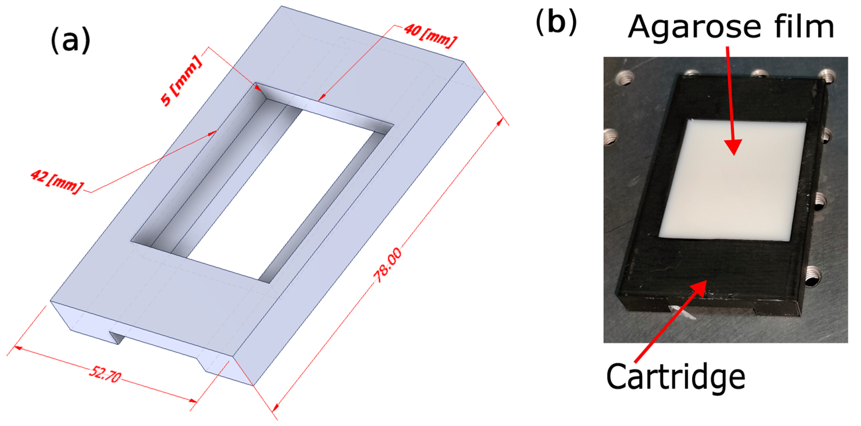
- Pour the sample into a container and allow for it to cool to room temperature (see Figure 8b).
4.2. Optical Characterization of Agarose Tissue Simile
4.2.1. Optical Parameters Estimation
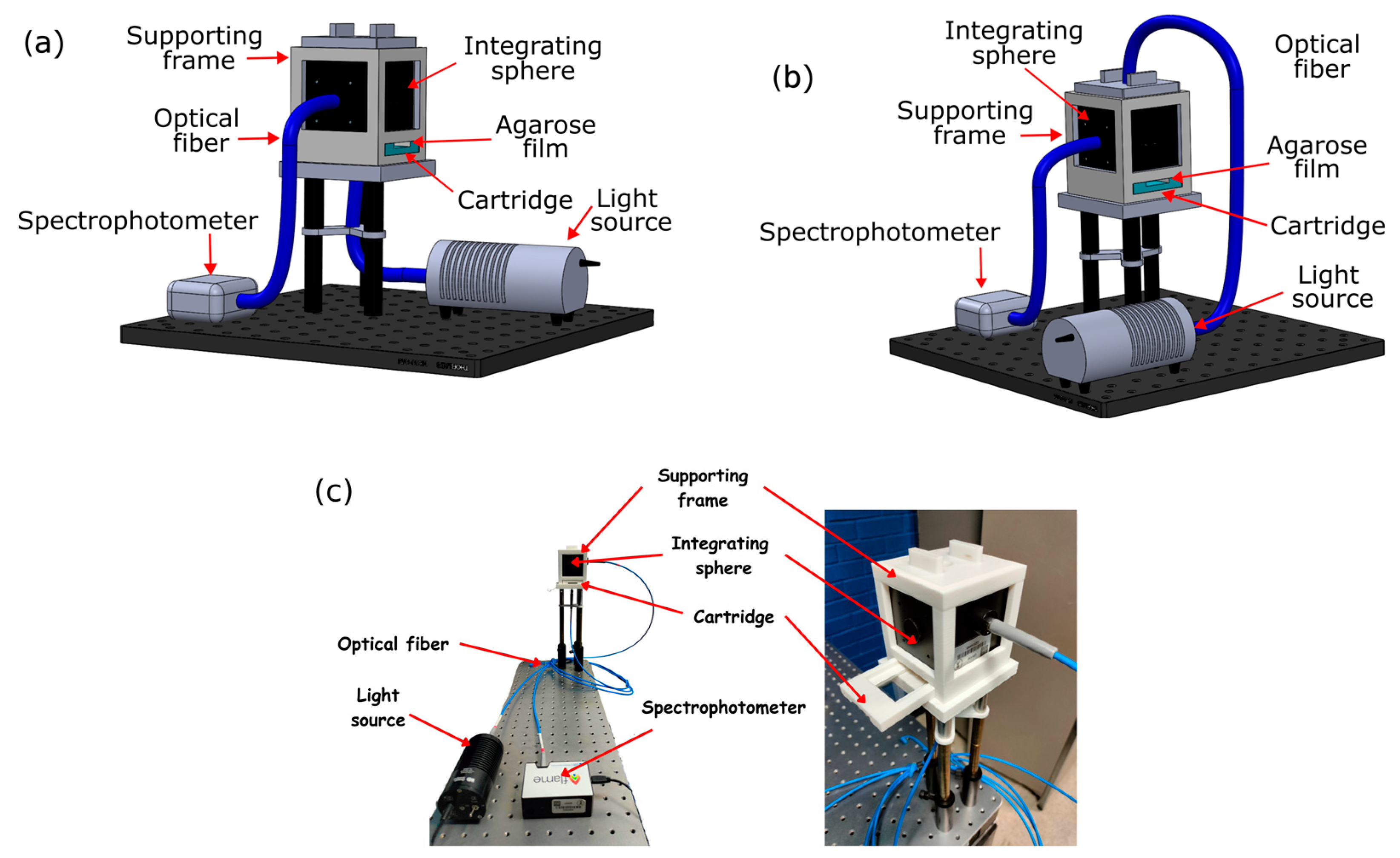
4.2.2. Experimental Set-Up
Author Contributions
Funding
Institutional Review Board Statement
Informed Consent Statement
Data Availability Statement
Acknowledgments
Conflicts of Interest
References
- Sakudo, A. Near-infrared spectroscopy for medical applications: Current status and future perspectives. Clin. Chim. Acta 2016, 455, 181–188. [Google Scholar] [CrossRef] [Scilit] [PubMed]
- Pence, I.; Mahadevan-Jansen, A. Clinical instrumentation and applications of Raman spectroscopy. Chem. Soc. Rev. 2016, 45, 1958–1979. [Google Scholar] [CrossRef] [Scilit] [PubMed]
- Genina, E.A.; Bashkatov, A.N.; Tuchina, D.K.; Dyachenko, P.A.; Navolokin, N.; Shirokov, A.; Khorovodov, A.; Terskov, A.; Klimova, M.; Mamedova, A.; et al. Optical properties of brain tissues at the different stages of glioma development in rats: Pilot study. Biomed. Opt. Express 2019, 10, 5182–5197. [Google Scholar] [CrossRef] [Scilit] [PubMed]
- Wang, H.; Magnain, C.; Sakadžić, S.; Fischl, B.; Boas, D.A. Characterizing the optical properties of human brain tissue with high numerical aperture optical coherence tomography. Biomed. Opt. Express 2017, 8, 5617–5636. [Google Scholar] [CrossRef] [Scilit] [PubMed]
- Rajan, V.; Varghese, B.; Van Leeuwen, T.G.; Steenbergen, W. Review of methodological developments in laser Doppler flowmetry. Lasers Med. Sci. 2009, 24, 269–283. [Google Scholar] [CrossRef] [Scilit] [PubMed]
- Hoshi, Y.; Yamada, Y. Overview of diffuse optical tomography and its clinical applications. J. Biomed. Opt. 2016, 21, 091312. [Google Scholar] [CrossRef] [Scilit] [PubMed]
- Briers, D.; Duncan, D.D.; Hirst, E.; Kirkpatrick, S.J.; Larsson, M.; Steenbergen, W.; Stromberg, T.; Thompson, O.B. Laser speckle contrast imaging: Theoretical and practical limitations. J. Biomed. Opt. 2013, 18, 066018. [Google Scholar] [CrossRef] [Scilit] [PubMed]
- Sandell, J.L.; Zhu, T.C. A review of in-vivo optical properties of human tissues and its impact on PDT. J. Biophotonics 2011, 4, 773–787. [Google Scholar] [CrossRef] [Scilit]
- Chao, S.-L.; Chen, K.-C.; Lin, L.-W.; Wang, T.-L.; Chong, C.-F. Ultrasound Phantoms Made of Gelatin Covered with Hydrocolloid Skin Dressing. J. Emerg. Med. 2013, 45, 240–243. [Google Scholar] [CrossRef] [Scilit]
- Elvira, L.; Durán, C.; Higuti, R.T.; Tiago, M.M.; Ibáñez, A.; Parrilla, M.; Valverde, E.; Jiménez, J.; Bassat, Q. Development and Characterization of Medical Phantoms for Ultrasound Imaging Based on Customizable and Mouldable Polyvinyl Alcohol Cryogel–Based Materials and 3-D Printing: Application to High-Frequency Cranial Ultrasonography in Infants. Ultrasound Med. Biol. 2019, 45, 2226–2241. [Google Scholar] [CrossRef] [Scilit]
- Chen, S.J.-S.; Hellier, P.; Marchal, M.; Gauvrit, J.-Y.; Carpentier, R.; Morandi, X.; Collins, D.L. An anthropomorphic polyvinyl alcohol brain phantom based on Colin27 for use in multimodal imaging. Med. Phys. 2012, 39, 554–561. [Google Scholar] [CrossRef] [Scilit] [PubMed]
- Nomoni, M.; May, J.M.; Kyriacou, P.A. Novel Polydimethylsiloxane (PDMS) Pulsatile Vascular Tissue Phantoms for the In-Vitro Investigation of Light Tissue Interaction in Photoplethysmography. Sensors 2020, 20, 4246. [Google Scholar] [CrossRef] [Scilit] [PubMed]
- Zell, K.; Sperl, J.I.; Vogel, M.W.; Niessner, R.; Haisch, C. Acoustical properties of selected tissue phantom materials for ultrasound imaging. Phys. Med. Biol. 2007, 52, N475–N484. [Google Scholar] [CrossRef] [Scilit] [PubMed]
- Mustari, A.; Nishidate, I.; Wares, M.A.; Maeda, T.; Kawauchi, S.; Sato, S.; Sato, M.; Aizu, Y. Agarose-based Tissue Mimicking Optical Phantoms for Diffuse Reflectance Spectroscopy. JoVE 2018, e57578. [Google Scholar] [CrossRef] [Scilit]
- Azimbagirad, M.; Grillo, F.W.; Hadadian, Y.; Carneiro, A.A.O.; Murta, L.O. Biomimetic phantom with anatomical accuracy for evaluating brain volumetric measurements with magnetic resonance imaging. J. Med. Imaging 2021, 8, 013503. [Google Scholar] [CrossRef] [Scilit] [PubMed]
- Durmuş, H.O.; Karaböce, B.; Seyidov, M.Y. Experimental and Comparative Study of Optical Properties of Different Phantoms by the Kubelka–Munk Function Approach. J. Appl. Spectrosc. 2023, 90, 198–205. [Google Scholar] [CrossRef] [Scilit]
- Thulliez, M.; Bastin, O.; Nonclercq, A.; Delchambre, A.; Reniers, F. Gel models to assess distribution and diffusion of reactive species from cold atmospheric plasma: An overview for plasma medicine applications. J. Phys. D Appl. Phys. 2021, 54, 463001. [Google Scholar] [CrossRef] [Scilit]
- Chen, Z.-J.; Gillies, G.T.; Broaddus, W.C.; Prabhu, S.S.; Fillmore, H.; Mitchell, R.M.; Corwin, F.D.; Fatouros, P.P. A realistic brain tissue phantom for intraparenchymal infusion studies. J. Neurosurg. 2004, 101, 314–322. [Google Scholar] [CrossRef] [Scilit]
- Liu, Y.; Paliwal, S.; Bankiewicz, K.S.; Bringas, J.R.; Heart, G.; Mitragotri, S.; Prausnitz, M.R. Rheometric Studies on Agarose Gel—A Brain Mimic Material. AAPS PharmSciTech 2010, 11, 1005–1017. [Google Scholar] [CrossRef] [Scilit]
- Tejo-Otero, A.; Fenollosa-Artés, F.; Achaerandio, I.; Rey-Vinolas, S.; Buj-Corral, I.; Mateos-Timoneda, M.Á.; Engel, E. Soft-Tissue-Mimicking Using Hydrogels for the Development of Phantoms. Gels 2022, 8, 40. [Google Scholar] [CrossRef] [Scilit]
- Aernouts, B.; Van Beers, R.; Watté, R.; Huybrechts, T.; Jordens, J.; Vermeulen, D.; Van Gerven, T.; Lammertyn, J.; Saeys, W. Effect of ultrasonic homogenization on the Vis/NIR bulk optical properties of milk. Colloids Surf. B Biointerfaces 2015, 126, 510–519. [Google Scholar] [CrossRef] [Scilit] [PubMed]
- Amidi, E.; Yang, G.; Uddin, K.M.S.; Wahidi, R.; Zhu, Q. Low-cost ultrasound and optical gelatin-based phantoms. In Proceedings of the Photons Plus Ultrasound: Imaging and Sensing 2019, San Francisco, CA, USA, 27 February 2019; SPIE: Bellingham, WA, USA, 2019; Volume 10878, pp. 330–336. [Google Scholar]
- Drakos, T.; Antoniou, A.; Evripidou, N.; Alecou, T.; Giannakou, M.; Menikou, G.; Constantinides, G.; Damianou, C. Ultrasonic Attenuation of an Agar, Silicon Dioxide, and Evaporated Milk Gel Phantom. J. Med. Ultrasound 2021, 29, 239–249. [Google Scholar] [CrossRef] [Scilit] [PubMed]
- Dunn, A.K. Laser Speckle Contrast Imaging of Cerebral Blood Flow. Ann. Biomed. Eng. 2012, 40, 367–377. [Google Scholar] [CrossRef] [Scilit] [PubMed]
- Davoodzadeh, N.; Cano-Velázquez, M.S.; Halaney, D.L.; Jonak, C.R.; Binder, D.K.; Aguilar, G. Optical Access to Arteriovenous Cerebral Microcirculation through a Transparent Cranial Implant. Lasers Surg. Med. 2019, 51, 920–932. [Google Scholar] [CrossRef] [Scilit] [PubMed]
- Bachir, W.; Khir, R. Characterization of pasteurized milk in the near infrared range for construction of tissue-mimicking optical phantoms. Opt. Mater. X 2022, 14, 100154. [Google Scholar] [CrossRef] [Scilit]
- Pogue, B.W.; Patterson, M.S. Review of tissue simulating phantoms for optical spectroscopy, imaging and dosimetry. J. Biomed. Opt. 2006, 11, 041102. [Google Scholar] [CrossRef] [Scilit]
- Liu, G.; Huang, K.; Jia, Q.; Liu, S.; Shen, S.; Li, J.; Dong, E.; Lemaillet, P.; Allen, D.W.; Xu, R.X. Fabrication of a multilayer tissue-mimicking phantom with tunable optical properties to simulate vascular oxygenation and perfusion for optical imaging technology. Appl. Opt. 2018, 57, 6772. [Google Scholar] [CrossRef] [Scilit]
- Wagnières, G.; Cheng, S.; Zellweger, M.; Utke, N.; Braichotte, D.; Ballini, J.-P.; Bergh, H.V.D. An optical phantom with tissue-like properties in the visible for use in PDT and fluorescence spectroscopy. Phys. Med. Biol. 1997, 42, 1415–1426. [Google Scholar] [CrossRef] [Scilit]
- Augustin, M.A.; Clarke, P.T.; Craven, H. POWDERED MILK | Characteristics of Milk Powders. In Encyclopedia of Food Sciences and Nutrition, 2nd ed.; Academic Press: Cambridge, MA, USA, 2003; pp. 4703–4711. [Google Scholar] [CrossRef] [Scilit]
- Huang, Y.; Min, S.; Duan, J.; Wu, L.; Li, Q. Identification of additive components in powdered milk by NIR imaging methods. Food Chem. 2014, 145, 278–283. [Google Scholar] [CrossRef] [Scilit]
- Mossoba, M.M.; Yurawecz, M.P.; Delmonte, P.; Kramer, J.K.G. Overview of Infrared Methodologies for trans Fat Determination. J. AOAC Int. 2004, 87, 540–544. [Google Scholar] [CrossRef] [Scilit]
- Bettati, P.; Chalian, M.; Huang, J.; Dormer, J.D.; Shahedi, M.; Fei, B. Augmented reality-assisted biopsy of soft tissue lesions. In Proceedings of the Medical Imaging 2020: Image-Guided Procedures, Robotic Interventions, and Modeling, Houston, TX, USA, 16–19 February 2020; SPIE: Bellingham, WA, USA, 2020; Volume 11315, pp. 222–231. [Google Scholar] [CrossRef] [Scilit]
- Jacques, S.L. Optical properties of biological tissues: A review. Phys. Med. Biol. 2013, 58, R37–R61. [Google Scholar] [CrossRef] [Scilit] [PubMed]
- Yaroslavsky, A.N.; Schulze, P.C.; Yaroslavsky, I.V.; Schober, R.; Ulrich, F.; Schwarzmaier, H.-J. Optical properties of selected native and coagulated human brain tissues in vitro in the visible and near infrared spectral range. Phys. Med. Biol. 2002, 47, 2059–2073. [Google Scholar] [CrossRef] [Scilit] [PubMed]
- Prahl, S. Everything I Think You Should Know about Inverse Adding-Doubling. Available online: https://omlc.org/software/iad/manual.pdf (accessed on 1 August 2023).
- Prahl, S.A.; Van Gemert, M.J.C.; Welch, A.J. Determining the optical properties of turbid media by using the adding–doubling method. Appl. Opt. 1993, 32, 559. [Google Scholar] [CrossRef] [Scilit] [PubMed]
- Madsen, S.J.; Wilson, B.C. Optical Properties of Brain Tissue. In Optical Methods and Instrumentation in Brain Imaging and Therapy; Madsen, S.J., Ed.; Springer: New York, NY, USA, 2013; pp. 1–22. [Google Scholar] [CrossRef] [Scilit]
- Johansson, J.D. Spectroscopic method for determination of the absorption coefficient in brain tissue. J. Biomed. Opt. 2010, 15, 057005. [Google Scholar] [CrossRef] [Scilit]
- Azimipour, M.; Baumgartner, R.; Liu, Y.; Jacques, S.L.; Eliceiri, K.; Pashaie, R. Extraction of optical properties and prediction of light distribution in rat brain tissue. J. Biomed. Opt. 2014, 19, 075001. [Google Scholar] [CrossRef] [Scilit]
- Moffitt, T.; Chen, Y.-C.; Prahl, S.A. Preparation and characterization of polyurethane optical phantoms. J. Biomed. Opt. 2006, 11, 041103. [Google Scholar] [CrossRef] [Scilit]
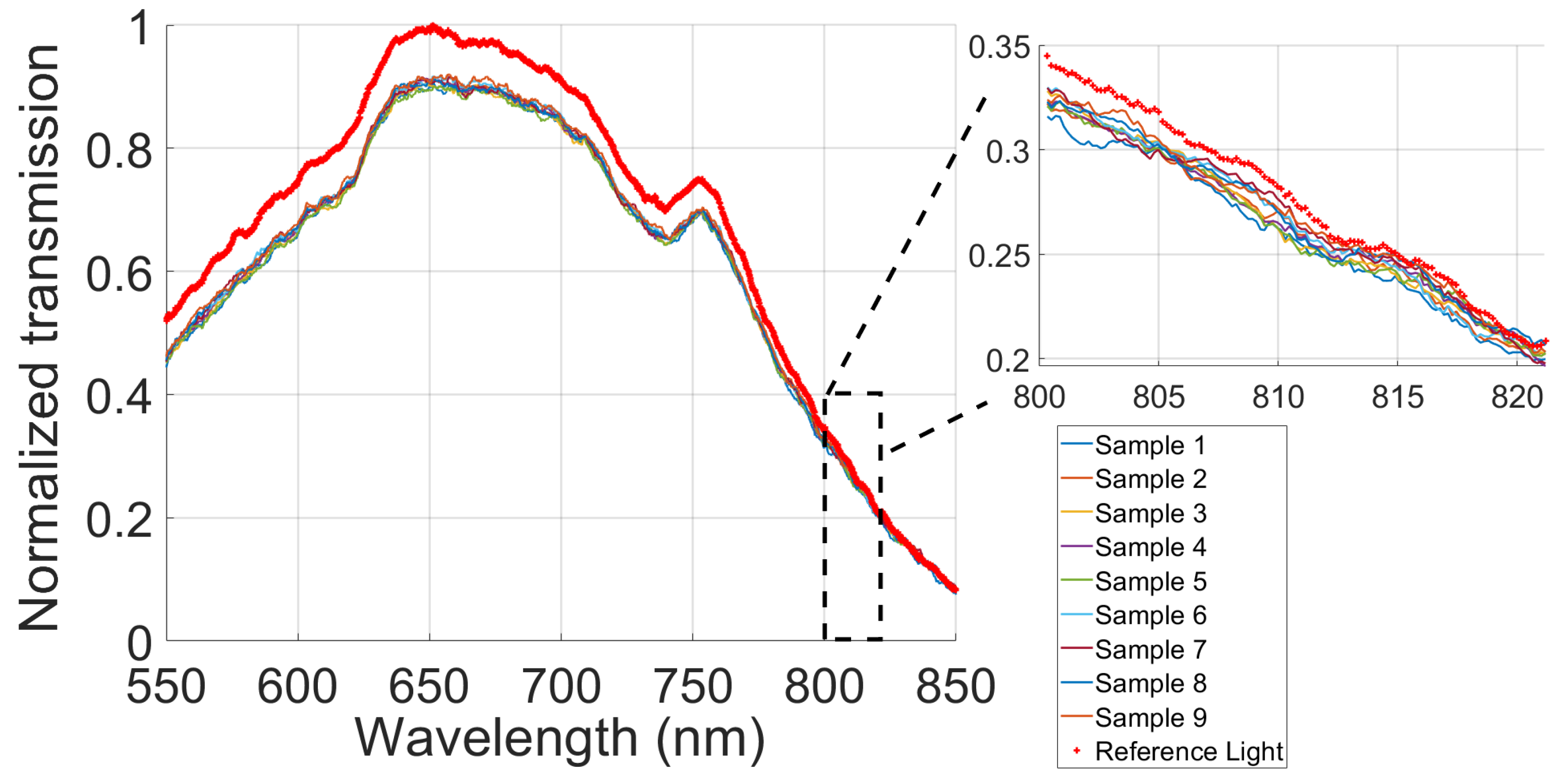


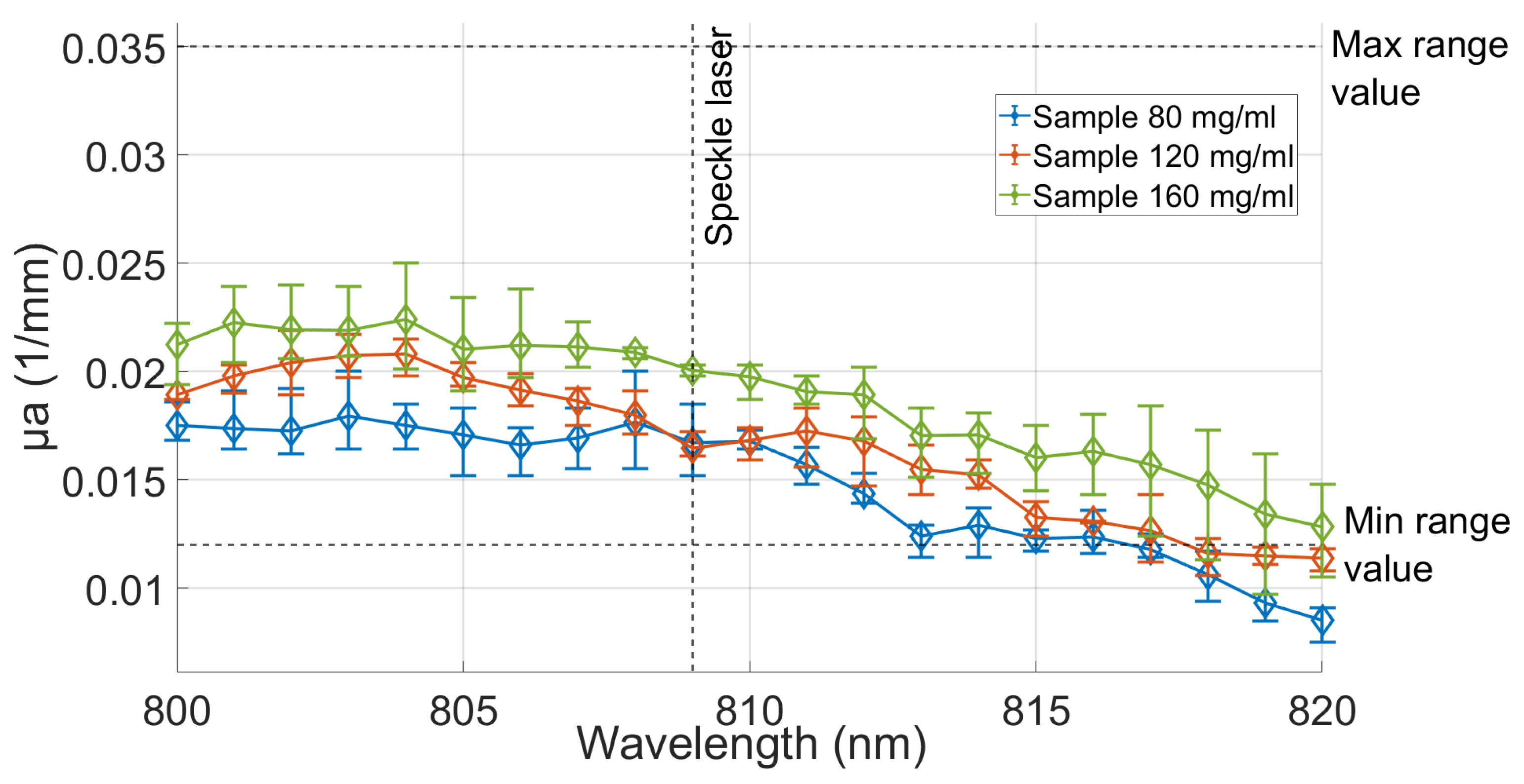
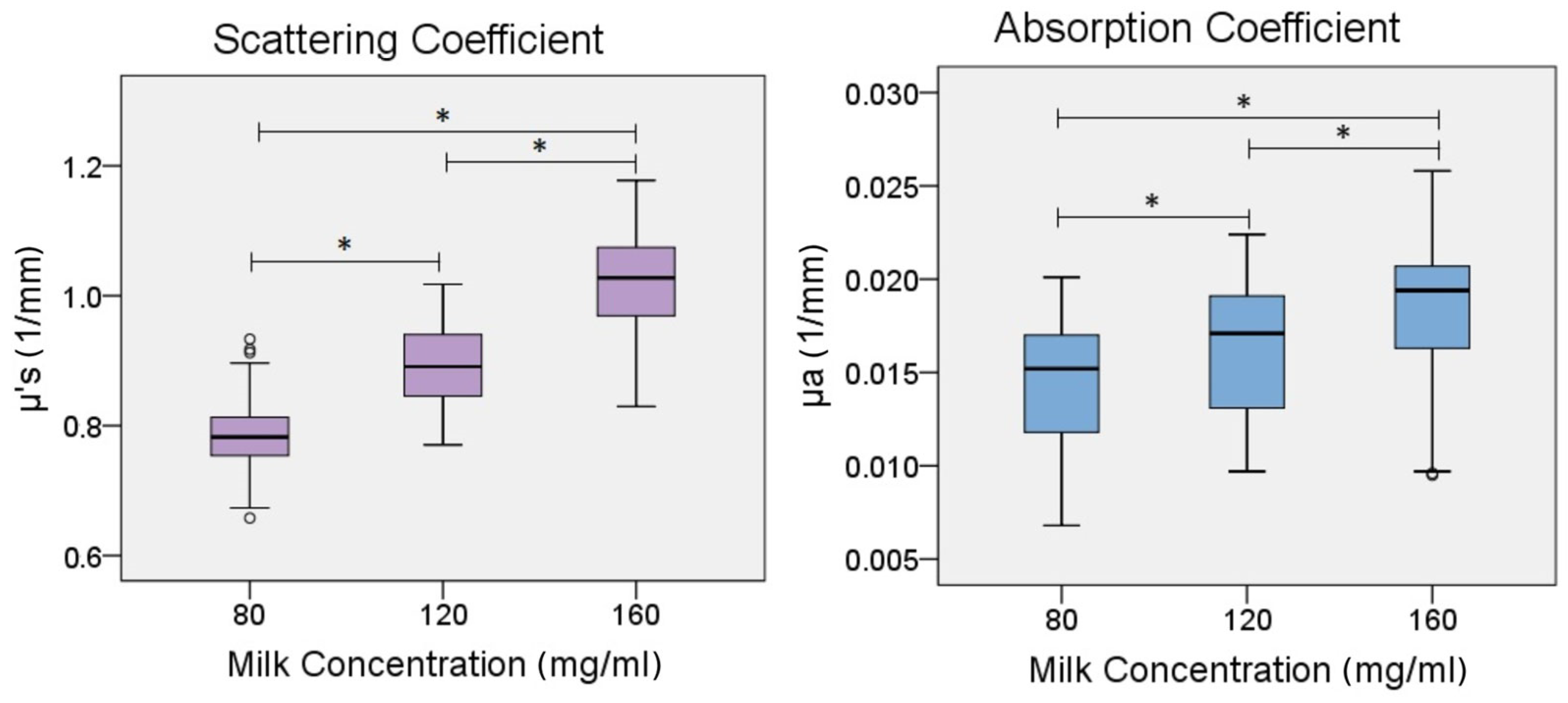
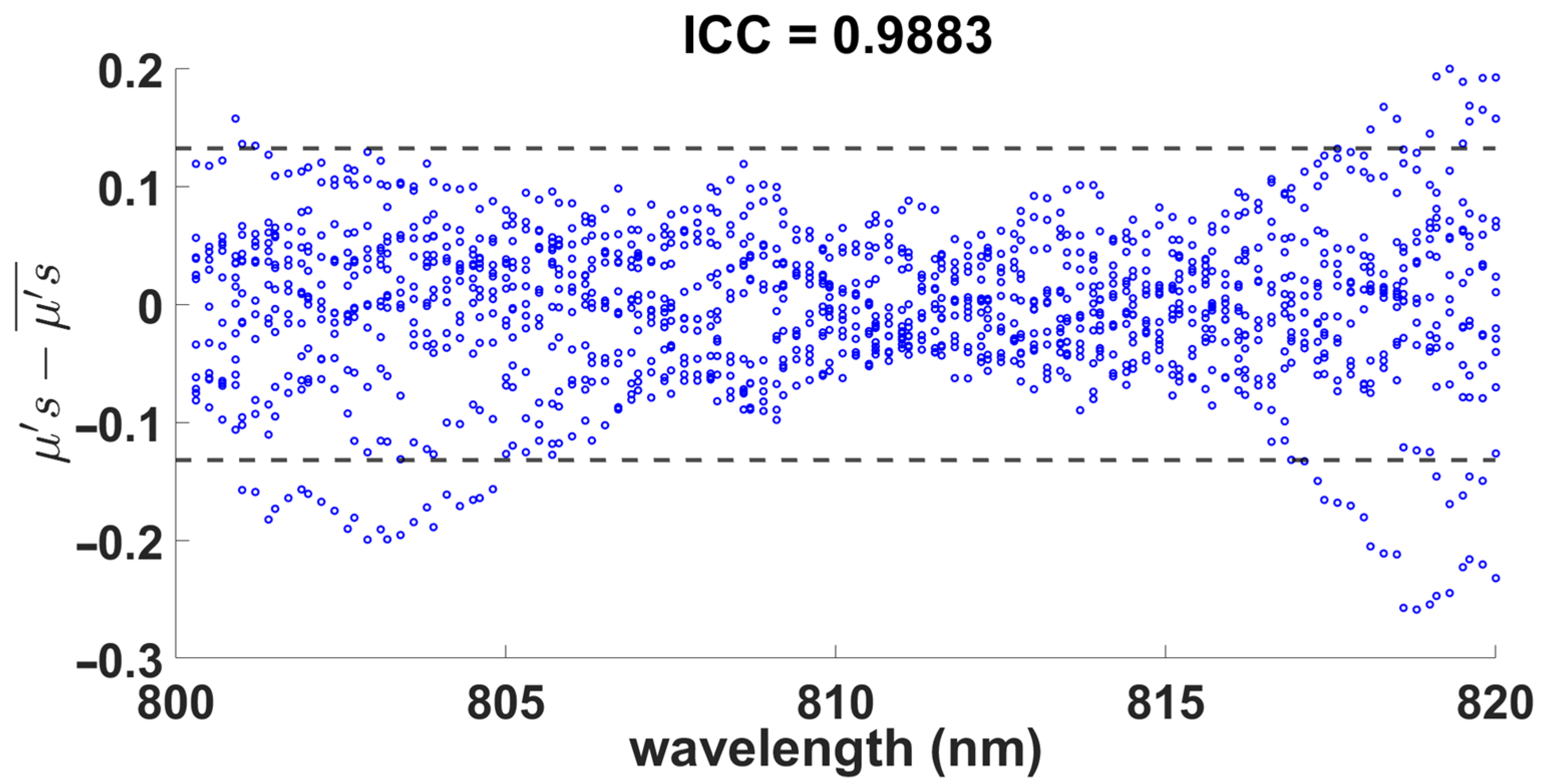



| Coefficients (1/mm) | Milk Concentration (mg/mL) | p-Value | ||
|---|---|---|---|---|
| 80 | 120 | 160 | ||
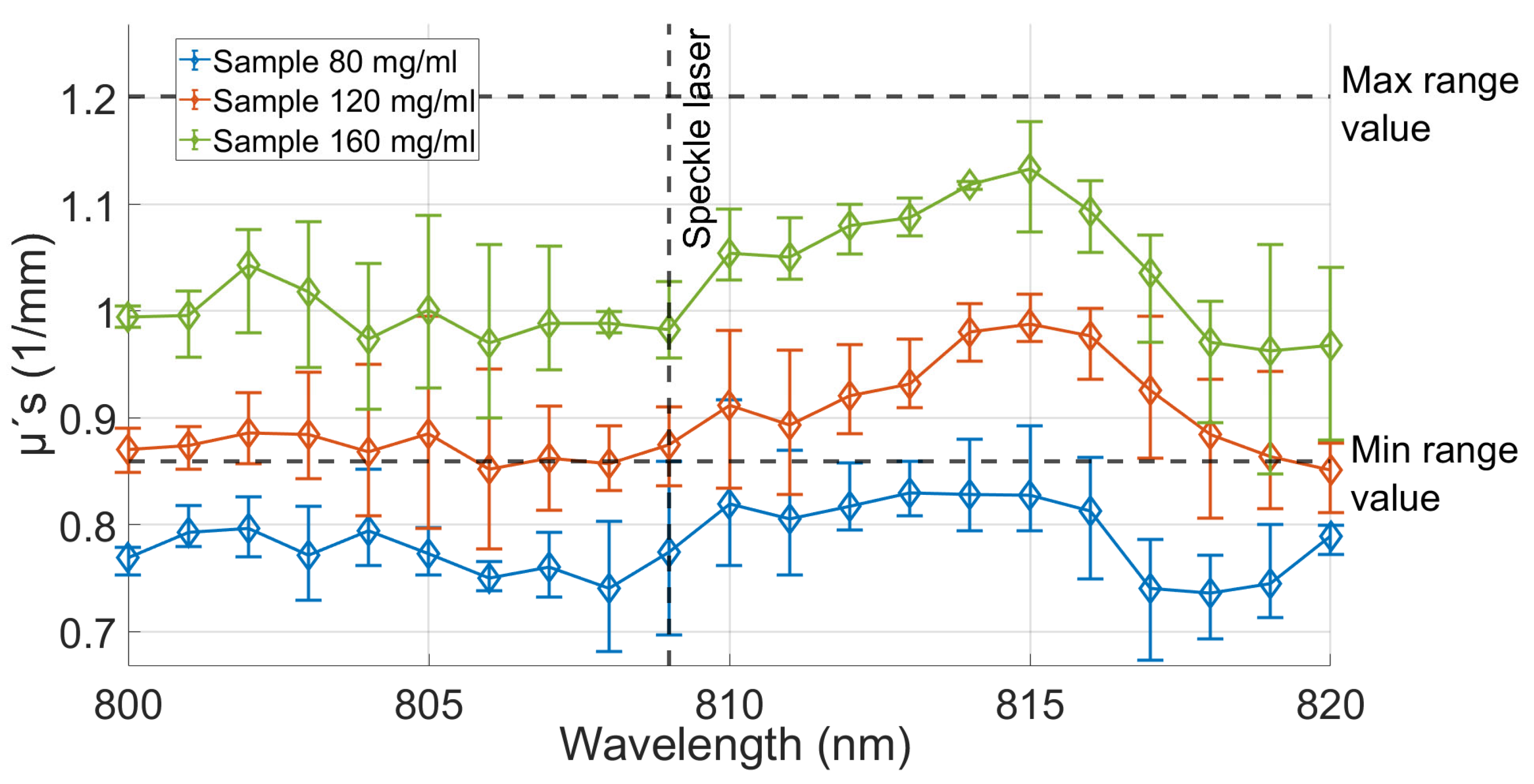
| 0.784 ± 0.048 | 0.894 ± 0.060 | 1.021 ± 0.069 | <0.0001 | |
| 0.014 ± 0.003 | 0.016 ± 0.003 | 0.018 ± 0.004 | <0.0001 | |
Disclaimer/Publisher’s Note: The statements, opinions and data contained in all publications are solely those of the individual author(s) and contributor(s) and not of MDPI and/or the editor(s). MDPI and/or the editor(s) disclaim responsibility for any injury to people or property resulting from any ideas, methods, instructions or products referred to in the content. |
© 2023 by the authors. Licensee MDPI, Basel, Switzerland. This article is an open access article distributed under the terms and conditions of the Creative Commons Attribution (CC BY) license (https://creativecommons.org/licenses/by/4.0/).
Share and Cite
Albor-Ramírez, E.; Reyes-Alberto, M.; Vidal-Flores, L.M.; Gutierrez-Herrera, E.; Padilla-Castañeda, M.A. Agarose Gel Characterization for the Fabrication of Brain Tissue Phantoms for Infrared Multispectral Vision Systems. Gels 2023, 9, 944. https://doi.org/10.3390/gels9120944
Albor-Ramírez E, Reyes-Alberto M, Vidal-Flores LM, Gutierrez-Herrera E, Padilla-Castañeda MA. Agarose Gel Characterization for the Fabrication of Brain Tissue Phantoms for Infrared Multispectral Vision Systems. Gels. 2023; 9(12):944. https://doi.org/10.3390/gels9120944
Chicago/Turabian StyleAlbor-Ramírez, Efraín, Miguel Reyes-Alberto, Luis M. Vidal-Flores, Enoch Gutierrez-Herrera, and Miguel A. Padilla-Castañeda. 2023. "Agarose Gel Characterization for the Fabrication of Brain Tissue Phantoms for Infrared Multispectral Vision Systems" Gels 9, no. 12: 944. https://doi.org/10.3390/gels9120944
APA StyleAlbor-Ramírez, E., Reyes-Alberto, M., Vidal-Flores, L. M., Gutierrez-Herrera, E., & Padilla-Castañeda, M. A. (2023). Agarose Gel Characterization for the Fabrication of Brain Tissue Phantoms for Infrared Multispectral Vision Systems. Gels, 9(12), 944. https://doi.org/10.3390/gels9120944