Article

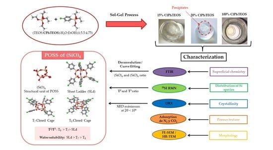
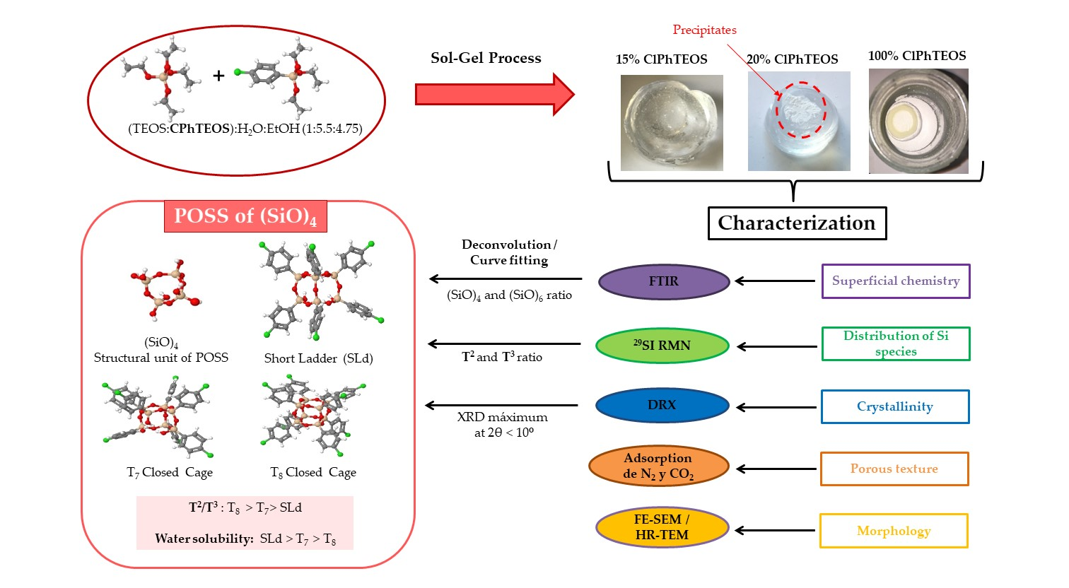
Novel Silica Hybrid Xerogels Prepared by Co-Condensation of TEOS and ClPhTEOS: A Chemical and Morphological Study

by
Guillermo Cruz-Quesada
1,
Maialen Espinal-Viguri
1,*,
María Victoria López-Ramón
2 and
Julián J. Garrido
1,*
1
Institute for Advanced Materials and Mathematics (INAMAT2), Department of Science, Public University of Navarre (UPNA), Campus Arrosadia, 31006 Pamplona, Spain
2
Department of Inorganic and Organic Chemistry, Faculty of Experimental Sciences, University of Jaen, 23071 Jaen, Spain
*
Authors to whom correspondence should be addressed.
Gels 2022, 8(10), 677; https://doi.org/10.3390/gels8100677
Submission received: 29 September 2022 / Revised: 15 October 2022 / Accepted: 17 October 2022 / Published: 20 October 2022

Abstract

:
The search for new materials with improved properties for advanced applications is, nowadays, one of the most relevant and booming fields for scientists due to the environmental and technological needs of our society. Within this demand, hybrid siliceous materials, made out of organic and inorganic species (ORMOSILs), have emerged as an alternative with endless chemical and textural possibilities by incorporating in their structure the properties of inorganic compounds (i.e., mechanical, thermal, and structural stability) in synergy with those of organic compounds (functionality and flexibility), and thus, bestowing the material with unique properties, which allow access to multiple applications. In this work, synthesis using the sol-gel method of a series of new hybrid materials prepared by the co-condensation of tetraethoxysilane (TEOS) and 4-chlorophenyltriethoxysilane (ClPhTEOS) in different molar ratios is described. The aim of the study is not only the preparation of new materials but also their characterization by means of different techniques (FT-IR, 29Si NMR, X-ray Diffraction, and N2/CO2 adsorption, among others) to obtain information on their chemical behavior and porous structure. Understanding how the chemical and textural properties of these materials are modulated with respect to the molar percentage of organic precursor will help to envisage their possible applications: From the most conventional such as catalysis, adsorption, or separation, to the most advanced in nanotechnology such as microelectronics, photoluminescence, non-linear optics, or sensorics.

Graphical Abstract

1. Introduction

Silica hybrid gels are among the so-called organic modified silica (ORMOSILs). Nowadays, these materials are being used for multiple applications such as the adsorption of metals in water [1,2,3], catalyst support [4,5,6], support for luminescent compounds [7,8,9], anti-corrosive or fungicide coatings [10,11,12,13], and biomedicine [14,15,16,17,18,19]. The key behind the tunability of the properties of these materials is the synergetic effect that generates the coexistence, at a nanometric scale, of an inorganic skeleton with flexible and functional organic constituents [20,21]. The hybrid gels are prepared by the sol-gel method, a very versatile synthetic technique that allows different approaches: (i) Anchoring of organic molecules in the porous of the already-formed solid gel [1,2,7,8,9], (ii) grafting of organic moieties on the surface of a silica material [4,22,23], (iii) co-condensation of an organosilane (SiRx(OR’)4−x), with a carbon precursor, to form Si-O-C xerogels [16,18,24,25], or finally, (iv) classic co-condensation of a tetraalkoxysilane (Si(OR)4), usually tetraethoxysilane (TEOS), with one or more organosilanes [8,9,10]. In the latter approach, to obtain materials with the desired properties, it is of paramount importance to control the precursors’ hydrolysis and condensation reactions (Figure 1), due to their susceptibility to the media conditions (pH, solvents, use or not of a catalyst, proportions of H2O/precursor or organosilane/tetra alkoxysilane) [26,27,28,29,30].
The influence of these parameters in the synthesis of silica xerogels has been studied by our research group in recent years [31,32,33,34], with special emphasis on the nature and molar proportion of organosilanes in hybrid xerogels [35,36,37]. By means of different techniques, such as mass spectroscopy, inelastic neutron scattering, and infrared spectra deconvolution, it was confirmed that ordered domains of polyhedral oligomeric silsesquioxanes (POSS) are formed within the silica matrix of the hybrid materials, due to the blocking of the condensation direction in the organic precursor RTEOS (RTEOS = alkyl/chloroalkyl triethoxysilanes) and the intermolecular forces (e.g., hydrophobic or electrostatic interactions) exerted by the organic moieties [38,39,40]. In fact, ordered domains were detected at lower molar percentages of organic precursor when chloroalkyltriethoxysilanes (ClRTEOS) are used instead of their analogous alkoxysilanes [37,40]. The knowledge acquired in those studies has been used to prepare optical fiber sensors (OFS) by dip-coating, using selected gels with specific properties. These sensors have a labile and specific interaction with different adsorbates, such as vapor of water and volatile organic compounds (COVs), allowing their detection and monitoring [41,42,43].
Following this line of research, the aim of the present work is to study the morphological, textural, and chemical properties of silica hybrid xerogels containing a chlorophenyl moiety, and to evaluate their use as coatings for OFS in the future. For this purpose, a new series of transparent hybrid xerogels were prepared by co-condensation, in acidic media, of TEOS with (p-chlorophenyl) triethoxysilane (ClPhTEOS) at different molar percentages. The resulting materials were fully characterized by 29Si NMR (Nuclear Magnetic Resonance), XRD (X-ray Diffraction), FTIR (Fourier-Transform Infrared Spectroscopy), Helium Pycnometry, FE-SEM (Field-Emission Scanning Electron Microscopy), HR-TEM (High-Resolution Transmission Spectroscopy), and the adsorption of N2 (−196 °C) and CO2 (0 °C). In addition, a modified and adjusted deconvolution method of the FT-IR spectra was used to acquire semi-quantitative information about the proportion of (SiO)4 and (SiO)6 rings [40,44], which are related to ordered structures and amorphous silica, respectively. Finally, the results were compared with those of the previously studied xerogels to remark on the influence of the chlorophenyl moiety on the material’s properties.

2. Results and Discussion

2.1. FT-IR

Figure 2 depicts the FT-IR spectra in the range of 1600–400 cm−1 of the hybrid xerogels synthesized at different molar percentages of organic precursor. The spectra in the range of 4000–2750 cm−1 are displayed in Figure S1 of the Supplementary Material.
In Figure 2, the characteristic bands of the amorphous silica matrix can be observed: (i) Rocking of O–Si–O at 455 cm−1 (ρ O–Si–O), (ii) symmetric Si–O–Si stretching vibration at 800 cm−1s Si–O–Si), (iii) Si-O bond stretching vibration of the silanols on the surface at 955 cm−1s Si–OH), (iv) asymmetric Si–O–Si stretching vibration at 1090 cm−1as Si–O–Si), and (v) a wide and intense shoulder from 1350 to 1120 cm−1 associated with various vibrational modes of the Si–O–Si [45]. Additionally, a slight shoulder at 550 cm−1 can be observed, which is associated with the presence of 4-member siloxane rings, (SiO)4 [46]. In the spectra, as the molar percentage of organic precursor gradually increases, a new incipient shoulder becomes more evident at 1140 and 1155 cm−1, and the band at 1080 cm−1 narrows and stands out, similar to an emerging peak. These changes could indicate that the organic precursor is favoring the formation, within the silica matrix, of new structures with vibration modes (νas Si–O–Si) at different specific frequencies [40]. In the spectral region of 400–2750 cm−1 (Figure S1 of the Supplementary Material), it is possible to observe the stretching bands of superficial silanols (ν Si–OH a 3450 cm−1) and those resulting from the interaction of these groups through hydrogen bonds (ν Si–OH–H at 3660 cm−1) [45].
The presence of the chlorophenyl moiety in the hybrid materials can be confirmed by a set of bands observed in the spectra: (i) The stretching bands of the hydrogens of the benzene ring in the range of 3090–3010 cm−1 (ν (C-H), Figure S1), (ii) three C=C bond stretching bands in the spectral range of 1450–1000 cm−1 (1380, 1085 and 1015 cm−1), and finally, (iii) three bands corresponding to the deformation vibrations of the C-H bonds in the aromatic rings (at 815, 760, and 500 cm−1), and a band due to the stretching vibration of the C–Cl bond (at 710 cm−1) [43,47,48]. It should be noted that the spectrum of the xerogel 20ClPh is not shown in Figure 2 because a heterogeneous monolith with two well-differentiated phases was obtained: A non-colored transparent phase above (monolith) and an opaque phase below (precipitate), whose spectra turned out to be very different (Figure S2c in the Supplementary Material). This difference might be because the organic precursor favors the formation of structures with lower solubility in the reaction media, and thus precipitates. To test this hypothesis and obtain information on those structures, a material using only ClPhTEOS was synthesized, resulting in a white, soft, and extremely hydrophobic solid (100ClPh) (Figure S2d in the Supplementary Material). Figure 3 depicts the FT-IR spectra of the reference, 15ClPh, 20ClPh (monolith and precipitate), and 100ClPh, while Table 1 displays the list of bands observed in the spectrum of 100ClPh, the vibrations and structures assigned to those bands, and the literature consulted for the assignment.
The bands due to the vibrations of νas (Si–O–Si) in the spectral range of 1400–1000 cm−1 (Table 1) correspond to simple structures (linear siloxane chains and (SiO)4 or (SiO)6 rings) and more compact and complex structures (i.e., polyhedral oligomers of silsesquioxanes, POSS). Oligomers known as open or closed cages (T7 and T8, respectively) and short ladders (SLd) are among the best known POSS, which are formed by the fusion of two or more four-membered rings ((SiO)4) (Figure 4) [52,53,54,55,56].
The formation of the rings that constitute these structures ((SiO)4) is thermodynamically favored in the oligomerization of siloxanes in acidic media [45,57], in contrast to the six-membered rings ((SiO)6), kinetically favored and typical of amorphous materials [39,58]. To study how the precursor affects the formation of ordered structures, it is necessary to know the proportion of (SiO)4 in the silica matrix. For this purpose, the deconvolution of the FT-IR spectra in the range of 1300–980 cm−1 was performed using the non-linear least-squares method, obtaining the Gaussian–Lorentzian components. The different distances and degrees of torsion of Si–O–Si bonds in (SiO)4 and (SiO)6 rings allow us to distinguish bands belonging to the optical modes of vibration; two in transverse mode between 1100 and 1000 cm−1 (TO4 and TO6) and two in longitudinal mode between 1250 and 1100 cm−1 (LO4 and LO6) [40,44,59,60]. In this work, a modification of this method has been carried out, consisting of the adjustment of additional bands (Table 1) corresponding to (i) C=C and C-H vibrations of the chlorophenyl (1450–700 cm−1); (ii) vibrations of the siloxane groups and Si-O bonds, characteristic of amorphous silica (950 and 800 cm−1, respectively); (iii) vibrations of the (SiO)4 rings that make up the POSS: νring-s and νring-as for the “open” species (T7 and short ladders), and only νring-as for the “closed” species (T8), due to the νring-s vibration mode being forbidden [49]; and (iv) νas (Si–O–Si) vibration of linear siloxanes. As an example, the calculated spectra and the bands generated for the reference xerogel and 100ClPh are displayed in Figure 5. The calculated bands and the fit of 5ClPh, 10ClPh, 15ClPh, and the two phases of 20ClPh are depicted in Figure S3 of the Supplementary Material.
The percentage area of each component and the residual value (difference between the real spectrum and the fit) are exhibited in Table S1 of the Supplementary Material. The percentage of four-membered and six-membered rings was determined by applying the following equations to the Gaussian–Lorentzian bell areas of the TO and LO components:
( SiO ) 6 ,   % = A ( LO 6 ) + A ( TO 6 ) A ( LO 4 ) + A ( TO 4 ) + A ( LO 6 ) + A ( TO 6 ) × 100
( SiO ) 4 ,   % = A ( LO 4 ) + A ( TO 4 ) A ( LO 4 ) + A ( TO 4 ) + A ( LO 6 ) + A ( TO 6 ) × 100
where A(LO)6 is the area of the band at 1190 cm−1, (TO)6 is the area of the band at 1030 cm−1, A(LO)4 is the sum of the areas of the three LO4 bands, 1160, 1135, and 1120 cm−1, and A(TO)4 is the area of the band at 1050 cm−1. Table 2 displays the proportion of rings calculated by applying Equations (1) and (2). It can be observed that increasing the molar percentage of ClPhTEOS in the xerogels favors the formation of (SiO)4 rings (from 46.54% in the reference to 97.35% in 100ClPh).
Another noteworthy observation is the great difference in the percentage of these rings between both phases of 20ClPh: The monolithic phase has a percentage of rings similar to that of the reference 0ClPh, and the phase that precipitates is similar to 100ClPh. This might be explained by taking into account that an increase in ClPhTEOS favors the formation of POSS, which are mainly formed by four-membered rings, and, as their abundance increases, there is a critical point at which these species exceed the molar solubility in the reaction media and segregate as a precipitate [61].

2.2. 29Si Nuclear Magnetic Resonance (NMR)

29Si NMR spectra of the hybrid materials were obtained to determine the relationship between the molar percentage of the precursor and the relative abundance of silicon species in the xerogels. Figure 6a depicts the spectra of the hybrid materials normalized with respect to the signal of the dominant species, Q3, the most intense in all cases.
The signals associated with the less condensed species (Q1 and T1) in Figure 6a are not observed. The dominant species corresponding to the hybrid precursor is the semi-condensed T2, whose intensity is greater than that of the more condensed T3 in all the materials. In addition, Figure 6b displays both the evolution of the relative abundance of Q (Q2 + Q3 + Q4) and T (T2 + T3) species with respect to the molar percentage of ClPhTEOS, as well as that of each species. For example, the Q2 relative abundance increases slightly to stabilize at 11% and Q4 increases up to 5% ClPhTEOS and then decreases at higher molar percentages of organic precursor. Table 3 exhibits the chemical shifts of each 29Si species in the spectra and the integrals of the T species.
There is no significant displacement of the chemical shifts with the increase in ClPhTEOS, indicating that the environment of the silicon atoms does not change substantially. The chemical shifts of the T signals (organic precursor) are less negative than those of the Q signals (TEOS) because the chlorophenyl moiety removes less electronic charge from the silicon atom than oxygen, favoring the Shielding Effect [62,63]. Additionally, a higher positive charge density in the silicon atom favors nucleophilic attacks and therefore condensation [64,65]; however, the more abundant species is the least condensed and not T3, indicating that the inductive effect exerted by the chlorine atom of the chlorophenyl moiety is weaker than its steric effect, preventing total condensation in the materials. The increase in T3 species is related to the presence of POSS in the material, since the silicon atoms that form (SiO)4 rings are mainly condensed species T3, Q3, or Q4 (less condensed structures T7 and SLd also contain T2 and Q2) [49]. In fact, the shifts of T3 species (Table 3) are closer to those observed in T8 structures (−77 ppm) than to those of the aliphatic R-Si-O1.5 species (−66 to −67 ppm) [66,67]. Finally, it is worth mentioning that 100ClPh material only contains T units and, as has been verified in the analysis of its FT-IR spectra, it is composed almost exclusively of (SiO)4 rings.

2.3. X-ray Diffraction (XRD)

Figure 7 depicts the X-ray diffraction patterns of the hybrid materials synthesized at different molar percentages of organic precursor.
All the diffractograms showed a broad diffraction maximum at 2θ~24°, characteristic of the amorphous silica and associated with the distance between the silicon atoms linked by siloxane bridges [68]. This maximum slightly decreases with the increase in the molar percentage of the organic precursor. Interestingly, another maximum can be observed at 2θ < 10° when the molar percentage of the precursor is increased (10ClPh and 15ClPh). This new diffraction maximum is associated, in the literature, with the presence of ordered domains composed of POSS in the form of cages (T7 or T8) or short ladders within the amorphous matrix of the material [60]. The emergence of this maximum as the one at 24° decreases is consistent with the greater local structuration of the materials as we increase the molar percentage of the organic precursor. This behavior was already seen in previously studied ClRTEOS xerogels (R = M, methyl; E, ethyl, or P, propyl), where it was found that the maximum at 2θ < 10° appeared at lower molar percentages than their analogous RTEOS. In these chlorinated series, the minimum molar percentages containing the new maximum were 30, 1, and 10% for ClMTEOS, ClETEOS, and ClPTEOS, respectively [37]. The ClPhTEOS series not only share this minimum percentage with the ClPTEOS but also have more T2 than T3 species, which could indicate that, although the chlorine atom in the precursors favors the condensation and the subsequent formation of POSS, the steric effect of the bulkier organic moieties (both propyl and phenyl) could generate less condensed POSS, such as T7 or SLd. In all the patterns, another low-intensity maximum is observed around 45°, which is associated, according to Bragg’s law (n = 2 in nλ = 2d sin θ), with a replica of the maximum at 2θ~24°, indicating long-range order in the materials. Table 4 displays the angles, intensities, and distances calculated from Bragg’s law for each maximum in the diffractograms of Figure 5.
In 1ClPh and 5ClPh materials, the Si–O–Si bond elongates proportionally to the molar percentage of the precursor (maximum shifts to smaller angles), consistent with what was observed in the 29Si NMR spectra, that is, the increase in T species (from the organic precursor) causes a decrease in the average positive charge density of the silicon atoms, and therefore, the siloxane bonds are less polarized. However, as the molar percentage of the organic precursor increases in 10ClPh and 15ClPh, the bond becomes shorter (the maximum shifts to higher angles). This effect, inverse to the one discussed above, is related to the appearance of a diffraction maximum at 2θ < 10° in 10ClPh and 15ClPh, since this maximum is associated with ordered POSS-type structures, where the siloxane bridges (Si–O–Si) that make up the (SiO)4 rings are more compact than those of the amorphous silica (formed mainly by (SiO)6) [40], thus explaining the decrease in Si–O–Si distances associated with the maximum at 2θ~24°. The calculated distances for the additional maximum at 2θ < 10° (displayed in Table 4) are similar to those associated with the organic moiety in the cage-like structures (1–3 nm) [39,54], and to that of the interplane between short ladders [56,69]. Additionally, the X-ray diffractogram of 100ClPh is depicted in Figure S4 (Supplementary Material). In the diffractogram, a sharp and intense diffraction maximum at 2θ = 6.84° (1.3 nm) can be observed, which confirms that the 20ClPh precipitate contains a large amount of POSS. In a recent study, Nowacka et al. reported an interplane distance of 1.24 nm for ladder-like phenylsilsesquioxane oligomers, suggesting that these are the species formed in 100ClPh [60].

2.4. Helium Pycnometry

Helium pycnometry reveals the skeletal density of the synthesized xerogels. Figure 8 depicts the variation in the skeletal density as a function of the molar percentage of ClPhTEOS.
A decrease in the skeletal density with respect to ClPhTEOS is observed, since the organic precursor blocks one of the hydrolysis and condensation positions, thus reducing the degree of cross-linking with respect to the reference material (100% tetraethoxysilane, TEOS). The skeletal density of 15ClPh is greater than expected, which indicates that the limit of precursor that the system can assimilate has been reached. This new series has lower values of skeletal density than the previously studied ClRTEOS series (R = methyl, M; ethyl, E; propyl, P). For example, for 10ClPh, the skeletal density is 1.68 in contrast to 1.91, 1.85, and 1.80 g cm−1 for ClMTEOS, ClPTEOS, and ClETEOS, respectively [37]. This result is consistent with the information deduced from the 29Si NMR spectra and the XRD diffractograms (ClPhTEOS favors the less condensed POSS, T7 and SLd).

2.5. N2 and CO2 Adsorption Isotherms

N2 and CO2 molecules have a similar size; however, the temperature at which the adsorption takes place is very different, being −196 °C for N2 isotherms and 0 °C for CO2 isotherms. If the pores are very thin, N2 molecules cannot access them due to kinetic restrictions; however, CO2 molecules can. On the other hand, the high saturation pressure of CO2 vapor (3.5 MPa) allows the finer microporosity to be explored in detail, which is covered at very low relative pressures. This fact makes the data provided by both isotherms complementary and makes it possible to differentiate the micropores of less than 0.7 nm and even the finest mesopores. If there are no kinetic constraints, N2 adsorption provides the volume of pores of less than 50 nm, CO2 adsorption of those sized less than 0.7 nm, and the difference of these values would provide the microporosity between 2 and 0.7 nm. This divergence can be explained by the different adsorption mechanisms taking place in both microporous intervals; in the so-called “primary micropore filling”, the ultramicropores are accessed at a very low relative pressure (p/p0 = 0.03) and the adsorbent–adsorbate interactions predominate over those of the adsorbate–adsorbate, whereas, in wider micropores, the adsorbate–adsorbate interactions predominate, favoring a cooperative process of adsorption [70,71].
The N2 adsorption isotherms (at −196 °C) and the CO2 isotherms (at 0 °C) of the hybrid xerogels are shown in Figure 9. The isotherm of the reference material has an open knee, a sign of a wide micropore size distribution, characteristic of type I(b) isotherms. Moreover, its slope is pronounced in the adsorption, and it presents a hysteresis loop in the desorption (H2(a)), which is characteristic of a type IV isotherm typical of mesoporous materials. Therefore, the reference material can be considered micro-mesoporous with a type I(b)-IV(a) mixed isotherm [71]. Except for 1ClPh and 15ClPh materials, the higher the percentage of an organic precursor the smaller the pore volume, obtaining a type I(a) isotherm in all cases, typical of microporous materials with a narrow pore distribution. The isotherm of 1ClPh is a type IV(a) with an H1 hysteresis loop, indicating that, in this case, the organic precursor increases the total volume of pores and mesopores with respect to the reference, an effect that was also observed in analogous hybrid materials previously reported, which is associated with a change in the morphology of the pores, from cone-shaped to inkwell-shaped pores [27,37]. The 15ClPh isotherm is type I(a) but, surprisingly, it adsorbs more N2 than 10ClPh, 7.5ClPh, and ClPh5. This fact is consistent with the value obtained for its skeletal density (Figure 8), which suggests that in this material, the limit of the organic precursor that the xerogel accepts is practically reached and is therefore heterogeneously distributed in the silicon matrix. Table 5 exhibits the textural parameters obtained from the adsorption isotherms.
A decrease in the specific surface area (aBET) with the increase in the molar percentage of ClPhTEOS is observed, except for 15ClPh, which has a larger area than expected. Additionally, the table displays the volume of micropores obtained from the N2 adsorption data (Vmicro(N2)), and of the narrowest micropores (Vmicro(CO2), where φ < 0.7 nm), determined by applying the Dubinin–Raduskevich equation to the CO2 adsorption data. Both volumes decrease with an increase in the molar percentage of ClPhTEOS. The average pore size determined by the Barrett–Joyner–Halenda method (BJH APS) indicates that their mesoporosity narrows with the molar percentage of ClPhTEOS until microporous materials are obtained. In comparison with the ClRTEOS materials with the same percentage of the organic precursor, 10ClPh has a higher surface area and Vmicro(N2) than those of ClPTEOS and ClETEOS, but lower than that of ClMTEOS [37]. This implies, once again, that these materials are less condensed due to the predominance of T2 over T3 species and the bulky nature of the chlorophenyl group. Figure 10 depicts the pore size distribution by applying DFT calculations to the N2 and CO2 isotherm data.
All the materials are microporous, with an internal width close to 1 nm (Figure 10). The distribution shows that the materials do not present a significant volume of mesopores, except for the reference and 1ClPh.

2.6. Microscopy

2.6.1. Field-Emission Scanning Electron Microscopy (FE-SEM)

To determine the influence of the precursor on the texture, micrographs of the materials were acquired using scanning electron spectroscopy (SEM). The micrographs of the whole series are displayed in Figure 11.
The micrograph of the reference material (Figure 11a) exhibits a rough morphology composed of small intersecting globular particles in a chainmail-like surface (110–150 nm). The space between the particles corresponds to the narrow mesoporosity of this material, consistent with the N2 adsorption isotherm. Figure 11b shows how the use of only 1% of the organic precursor in the synthesis of 1ClPh results in a remarkable change: The surface is rougher, and the constituent particles are smaller (30–100 nm), justifying why the isotherm of this material reflects a greater specific surface area and mesoporosity than the reference material (0ClPh). In Figure 11c,d, respectively, ClPh5 and ClPh10 display a progressive smoothing of the surface with the increase in the organic precursor, which is consistent with the loss of mesoporosity and pore volume reduction deducted from their isotherms. A stratification of the surface can also be observed in overlapped compacted sheets to give mainly microporous materials. In ClPh15 (Figure 11e), the layers of compacted sheets, which are related to the stacking of the ladder POSS [56], can be better appreciated.

2.6.2. High Resolution-Transmission Electron Microscopy (HR-TEM)

Figure 12 displays TEM micrographs of the hybrid materials.
The micrograph of the reference material (Figure 12a) exhibits a morphology formed by an agglomerate of large particles of several tens of nm, which leave holes that are related to the mesoporosity of the material. In the following micrograph (Figure 12b, 1ClPh), particles of smaller size are interconnected, but they are still identifiable, unlike in the micrographs of the materials with a higher percentage of ClPhTEOS. To determine the degree of precursor distribution in the material, a mapping of the weight percentage of chlorine in the materials was carried out by X-ray energy dispersion. Table 6 displays the experimental and theoretical weight percentage of chlorine obtained for each material.
The weight percentages of chlorine in Table 6 are the average of the values recorded in different particles of the sample and reflect that the actual content of chlorine in the materials is lower than the theoretical. However, most of the chlorophenyl precursor is found as part of 1ClPh, 5ClPh, and 10ClPh materials, in contrast to 15ClPh, where approximately half of the amount of precursor used for the synthesis is present in the material. This is consistent with what was observed in the rest of the characterization techniques, since the results obtained are characteristic of a material with a lower molar percentage of the organic precursor than the actual amount used for its preparation.

3. Conclusions

A new series of hybrid xerogels have been prepared via the co-condensation of TEOS and ClPhTEOS as the organic precursor at different molar percentages. From the FT-IR spectra of the materials and by applying a deconvolution method, the percentages of (SiO)6 and (SiO)4 rings in the silica matrix of the materials were determined, the former associated with amorphous silica and the latter with ordered structures such as polysilsesquioxanes (POSS). The study determined that increasing the molar percentage of the organic precursor leads to an increase in the proportion of four-membered rings, (SiO)4. The presence of POSS is consistent with the XRD diffractograms, where an additional maximum at 2θ < 10° associated with ordered domains is observed for the materials with the highest ClPhTEOS content. The 29Si NMR spectra indicate that the most abundant silicon species from the organic precursor (T) are the semi-condensed (T2), likely due to the steric and electronic effect exerted by the chlorophenyl groups during the crosslinking of the colloids. Regarding the textural properties, it has been determined that an increase in the organic precursor in the materials results in a loss of mesoporosity and specific surface area, and a narrower pore size distribution. This is closely related to the smoothing of the material’s surface and the decrease in particle size observed in the FE-SEM and HR-TEM when increasing the molar percentage of ClPhTEOS. It is suggested that the layers that the micrographs exhibit are due to the stacking of the less condensed POSS (T7 open cages and short ladders), whereas condensed ones (T8 close cages) containing only hydrophobic chlorophenyl groups in their T3 silicon atoms precipitate and cause the heterogeneity observed in the 20ClPh material. In fact, the aromatic moiety together with the electronic properties of the chlorine atom opens the door to a more specific interaction with volatile aromatic compounds, and thus, for the application of these materials in sensorics for the detection of pollutants in the atmosphere. The study of the chemical and textural properties of these novel hybrid materials is the prelude to their use in different relevant fields. In fact, the aromatic moiety together with the electronic properties of the chlorine atom opens the door to a more specific interaction with volatile aromatic compounds, and thus, to the application of these materials in sensorics for the detection of pollutants in the atmosphere.

4. Materials and Methods

4.1. Materials

The siliceous precursors TEOS (tetraethoxysilane, purity > 99%) and ClPhTEOS ((4-chlorophenyl) triethoxysilane, purity > 97%) were supplied by Sigma-Aldrich (San Luis, MO, USA). Absolute ethanol (Emsure®) and hydrochloric acid (HCl, 37% w/w) were purchased from Merck (Darmstad, Germany) and potassium bromide (FT-IR grade) from Sigma-Aldrich (San Luis, MO, USA). All chemicals were used as received without further purification.

4.2. Synthesis of Silicon Hybrid Xerogels

Hybrid xerogels were prepared following the procedure described in previous works [37,72,73], where the molar ratio of (TEOS + ClPhTEOS): ethanol: water was fixed to 1:4.75:5.5 throughout the whole series, and the amounts of reagent and solvent were adjusted to obtain 20 mL of alcogels. The xerogels were named after the molar percentage used (i.e., 15ClPh for the xerogel with a molar percentage of 15%).
Briefly, TEOS and ClPhTEOS were mixed in a 30 mL vessel (φ 3.5 cm, threaded plastic lid, Schrarlab, Barcelona, Spain). Then, absolute ethanol was added, followed by the dropwise addition of Milli-Q grade water under magnetic stirring to facilitate miscibility. Once the pH of the mixtures remained unchanged (after approximately 10 min), an automatic burette (Tritino mod. 702 SM, Metrohm, Herisau, Switzerland) was used to set the pH at 4.5 (0.05 M HCl), and the mixture was stirred for 10 min to ensure homogenization. The closed vessels were placed in a thermostatized oven at 60 °C (J.P. Selecta S.A, Barcelona, Spain) until gelation (considered the time when the shape of the materials did not change when the vessel was tilted). Subsequently, 5 mL of ethanol was added to cure the alcogel at room temperature for one week. Next, the vessels were opened and covered with parafilmTM, perforated with holes to facilitate solvent evaporation, and dried at room temperature under atmospheric pressure. The monolith was considered dried when no significant variation in its mass was observed. Finally, the xerogels were further dried (90 °C under vacuum) and then ground in an agate mortar.

4.3. Characterization of Silicon Hybrid Xerogels

Various techniques have been applied to characterize the structure of the hybrid materials due to their amorphous nature [74]. FT-IR spectra were acquired using a Jasco spectrometer (mod. 4700, Japan). Two spectra were obtained for each sample: (i) In the range of 4000–2200 cm−1, using 2 mg of the sample to obtain information on the –OH groups and aromatic C–H bonds, and (ii) in the range of 2200–400 cm−1 using 0.6 mg of the sample to avoid saturation of the Si–O–Si asymmetric stretching signal [75]. Tablets were dispersed in KBr and dried overnight. Spectra were recorded using 25 scans and a resolution of 4 cm−1. The absorbance was transformed into Kubelka–Munk units with the internal software of the spectrometer (Spectramanager, SMII FTIR Rev 216A ver2.15A) for the deconvolution of the spectra [44]. The method was modified to generate a maximum of fourteen Gaussian–Lorentzian bands in the 1450–700 cm−1 spectral range, with a maximum of 200 interactions and a fixed baseline.
29Si Cross Polarization Magic-Angle Spinning (CP MAS) solid-state NMR was recorded on a Bruker AV-400 MHz spectrometer (Billerica, MA, USA) operating at 79.5 MHz. Chemical shifts were given in parts per million using TMS as the reference. The spectra were obtained 1H decoupled, with a frequency of rotation of 5 kHz and 800 scans per spectrum. For the 29Si NMR studies, classical notation was employed: T notation for silicon atoms from the organic precursor (bonded to three oxygens capable of forming siloxane bridges), and Q notation for silicon atoms from TEOS (bonded to four oxygens that can yield siloxane bridges). To describe the number of Si–O–Si bridges in each silicon atom, T and Q notations were completed with a superscript i (Ti, i = 0, 1, 2 or 3; Qi, i = 0, 1, 2, 3 or 4) [62].
X-ray diffractograms were obtained using a PANalytical Empyrean XRD instrument (Empyrean, Almelo, The Netherlands) with a copper rotating anode and a graphite monochromator (at 45 kV and 40 mA) to select the CuKα1/2 wavelength at 1.54 nm. Measurements were performed in a stepped scan mode (steps of 0.013°), in the range of 2 ≤ 2θ ≤ 50° at a rate of 0.5 steps s−1 [76].
A helium pycnometer was used to measure the skeletal density (AccuPyc 1330, Micromeritics, Norcross, GA, USA), performing 20 purges and 10 measurements for the initial calibration. The sample was weighed into a 1 cm3 cell and analyzed using 10 purges and 5 measurements.
N2 adsorption isotherms (at −196 °C with an isothermal jacket and immersed in a Dewar with liquid nitrogen) and CO2 adsorption isotherms (at 0 °C in a thermostatized recirculation bath using ethylene glycol as a refrigerant) were determined with a volumetric adsorption system (ASAP2020, Micromeritics, Norcross, GA, USA). First, 150 mg of sample was used for each isotherm, placed into a Pyrex glass tube, and degassed at 150 °C up to a residual vacuum of less than 0.66 Pa. The time needed for the analysis ranged from 14 to 55 h for N2 adsorption and from 2.5 to 9 h for CO2 adsorption. The recorded adsorption data were analyzed with the Microactive software (version 4.06), adjusting the parameters as appropriate for each model. Specific surface areas were calculated using two techniques: (i) The Brunauer–Emmett–Teller (BET) model (aBET), applying the Rouquerol criteria [77] and (ii) the Dubinin–Radushkevich (DR) method, applying a molecular section of 0.17 nm2 to CO2 (aDR) [78]. Pore volumes were defined by their diameter (φ): (i) The volume of micropores (φ ≤ 2 nm) was obtained from the DR method (Vmicro(N2) and Vmicro(CO2)), (ii) the volume of mesopores (2 < φ ≤ 50 nm) was calculated by abstracting the amount of N2 adsorbed at p/p° = 0.3 from that adsorbed at p/p° = 0.8 (Vmeso(N2)), and (iii) the total volume (VTotal(N2)) was considered the amount of N2 adsorbed at p/po = 0.95. Liquid densities of the adsorbates were obtained from the literature (0.808 g cm−3 for N2 and 1.023 g cm−3 for CO2) [70,79]. Porosity distributions were determined according to density-functional theory (DFT) using SAIEUS software and applying the “carbon-N2-77, 2D-NLDFT heterogeneous Surface” model for N2 adsorption and the “carbon-CO2-273, 2D-NLDFT Het Surface, pp max = 10 atm” model for CO2 adsorption. Mean pore sizes were further determined by applying the Barrett–Joyner–Halenda (BJH) method to the desorption curves, using a Kruk–Jaroniec–Sayari correction and thickness curve.
Field-emission scanning electron microscopy (FE-SEM) provides high-resolution images of sample surfaces. Micrographs were obtained with a Carl Zeiss SMT field emission scanning electron microscope (Carl Zeiss SMT, Oberkochen, Germany) at 200 kV.
High-resolution transmittance electron microscopy (HR-TEM) images were obtained at 200 kV with a JEOL 2000FXII microscope (Jeol Ltd., Tokyo, Japan) with a 0.28 nm point-to-point spatial resolution. The instrument was equipped with an Energy Dispersive X-Ray (EDX) INCA 200 spectrometer (Oxford Instruments, Abingdon, UK), which allowed the observation of the atomic distribution of carbon, silicon, and chlorine on the material.

Supplementary Materials

The following supporting information can be downloaded at: https://www.mdpi.com/article/10.3390/gels8100677/s1, Figure S1. FTIR spectra (range 4000–2750 cm−1) of the hybrid materials at different molar percentages and the reference (0ClPh); Figure S2. Photographs of the ClPhTEOS materials: (a) 0Clph, (b) 15Clph, (c) 20ClPh, and (d) 100ClPh; Figure S3. Calculated spectra and Gaussian–Laurentzian bands generated in the curve fitting for: (a) 5ClPh, (b) 10ClPh, (c) 15ClPh, (d) 20ClPh (monolith), and (e) 20ClPh (precipitate); Table S1. Wavelength and percentual area of the generated bands in the curve fitting for each ClPhTEOS material, and the curve fitting parameters; Figure S4. XRD pattern of 100ClPh.

Author Contributions

G.C.-Q.: investigation, writing—original draft. M.E.-V.: methodology, resources, writing—review and editing. M.V.L.-R.: Writing—review and editing. J.J.G.: Conceptualization, supervision, project administration, funding acquisition, writing—review and editing. All authors have read and agreed to the published version of the manuscript.

Funding

This research was funded by “Ministerio de Ciencia e Innovación” (Project ref. PID2020-113558RB-C42).

Institutional Review Board Statement

Not applicable.

Informed Consent Statement

Not applicable.

Data Availability Statement

The data presented in this study are available on request from the corresponding author.

Acknowledgments

The authors gratefully acknowledge the financial support received from “Ministerio de Ciencia e Innovación” from Spain (PID2020-113558RB-C42). G.C.-Q. is thankful to “Ministerio de Universidades” of the Spanish government for the “Formación de Profesorado Universitario (FPU)” grant (FPU18/03467) and M.E.-V. is thankful to “Universidad Pública de Navarra (UPNA)” for the project “Jovenes Investigadores UPNA 2022” (PJUPNA18-2022). The authors would also like to acknowledge the use of the “Centro de Instrumentación Científico-Técnica” at the University of Jaén, UCTAI at the Public University of Navarre and “Servicios Científico-Técnicos” at the University of Oviedo.

Conflicts of Interest

The authors declare no conflict of interest.

References

  1. Hannachi, Y.; Hafidh, A.; Ayed, S. Effectiveness of novel xerogels adsorbents for cadmium uptake from aqueous solution in batch and column modes: Synthesis, characterization, equilibrium, and mechanism analysis. Chem. Eng. Res. Des. 2019, 143, 11–23. [Google Scholar] [CrossRef]
  2. Kobylinska, N.G.; Kessler, V.G.; Seisenbaeva, G.A.; Dudarko, O.A. In Situ Functionalized Mesoporous Silicas for Sustainable Remediation Strategies in Removal of Inorganic Pollutants from Contaminated Environmental Water. ACS Omega 2022, 7, 23576–23590. [Google Scholar] [CrossRef]
  3. Wojciechowska, P.; Cierpiszewski, R. Gelatin—Siloxane Hybrid Monoliths as Novel Heavy Metal Adsorbents. Appl. Sci. 2022, 12, 1258. [Google Scholar] [CrossRef]
  4. Didó, C.A.; Coelho, F.L.; Closs, M.B.; Deon, M.; Horowitz, F.; Bernardi, F.; Schneider, P.H.; Benvenutti, E. V Strategy to isolate ionic gold sites on silica surface: Increasing their efficiency as catalyst for the formation of 1,3-diynes. Appl. Catal. A Gen. 2020, 594, 117444. [Google Scholar] [CrossRef]
  5. Estevez, R.; Luna, D.; Bautista, F.M. Sulfonated organosilica-aluminum phosphates as useful catalysts for acid-catalyzed reactions: Insights into the effect of synthesis parameters on the final catalyst. Catal. Today 2022, 390–391, 12–21. [Google Scholar] [CrossRef]
  6. Liu, M.; Wang, L.; Zhang, L.; Zhao, Y.; Chen, K.; Li, Y. In-Situ Silica Xerogel Assisted Facile Synthesis of Fe-N-C Catalysts with Dense Fe-Nx Active Sites for Efficient Oxygen Reduction. Small 2022, 18, 202104934. [Google Scholar] [CrossRef]
  7. Song, L.; Jia, Y.; Jin, X.; Wang, X.; Chen, M.; Wu, Y. Novel hybrid xerogel with CaF2:Tb3+ nanoparticles covalently embedded into silicon-oxygen network through sol-gel process. J. Lumin. 2022, 250, 119071. [Google Scholar] [CrossRef]
  8. Vilela, R.R.C.; Zanoni, K.P.S.; de Oliveira, M.; de Vicente, F.S.; de Camargo, A.S.S. Structural and photophysical characterization of highly luminescent organosilicate xerogel doped with Ir(III) complex. J. Sol-Gel Sci. Technol. 2022, 102, 236–248. [Google Scholar] [CrossRef]
  9. Santos, J.F.M.; Zanuto, V.S.; Ventura, M.; Bramorski, C.B.; Catunda, T. Quantum yield measurements by thermal lens in highly absorbing samples: The case of highly doped rhodamine B organic/silica xerogels. Phys. Rev. Mater. 2019, 3, 115201. [Google Scholar] [CrossRef]
  10. Alcantara-garcia, A.; Garcia-casas, A.; Jimenez-morales, A. The effect of the organosilane content on the barrier features of sol-gel anticorrosive coatings applied on carbon steel. Prog. Org. Coat. 2020, 139, 105418. [Google Scholar] [CrossRef]
  11. Scotland, K.M.; Shetranjiwalla, S.; Vreugdenhil, A.J. Curable hybrid materials for corrosion protection of steel: Development and application of UV-cured 3-methacryloxypropyltrimethoxysilane-derived coating. J. Coat. Technol. Res. 2020, 17, 977–989. [Google Scholar] [CrossRef]
  12. Rodič, P.; Milošev, I.; Lekka, M.; Andreatta, F.; Fedrizzi, L. Corrosion behaviour and chemical stability of transparent hybrid sol-gel coatings deposited on aluminium in acidic and alkaline solutions. Prog. Org. Coat. 2018, 124, 286–295. [Google Scholar] [CrossRef]
  13. Hao, S.; Hu, C.; Lin, X. Resistance to Growth of Molds for Wood Modified with Hydrophobic Hybrid Silica Gel Containing Copper Amine Complexes. Materials 2021, 14, 577. [Google Scholar] [CrossRef] [PubMed]
  14. Wang, H.; Yue, W.; Zhang, S.; Zhang, Y.; Li, C.; Su, W. Modification of Silica Xerogels with Polydopamine for Lipase B from Candida antarctica Immobilization. Catalysts 2021, 11, 1463. [Google Scholar] [CrossRef]
  15. Hatano, K.; Teraki, M.; Nakajima, D.; Yamatsu, T. Controlled release of molasses melanoidin-like product from hybrid organic–inorganic silica xerogels and its application to the phytoextraction of lead through the Indian mustard. Environ. Sci. Pollut. Res. 2021, 28, 37562–37569. [Google Scholar] [CrossRef] [PubMed]
  16. Pérez-Moreno, A.; Reyes-Peces, M.V.; Vilches-Pérez, J.I.; Fernández-Montesinos, R.; Pinaglia-Tobaruela, G.; Salido, M.; de la Rosa-Fox, N.; Piñero, M. Effect of Washing Treatment on the Textural Properties and Bioactivity of Silica/Chitosan/TCP Xerogels for Bone Regeneration. Int. J. Mol. Sci. 2021, 22, 8321. [Google Scholar] [CrossRef] [PubMed]
  17. Kapusuz, D. Sol-gel derived silica/polyethylene glycol hybrids as potential oligonucleotide vectors. J. Mater. Res. 2019, 34, 3787–3797. [Google Scholar] [CrossRef]
  18. Goel, H.; Santhiya, D. Effect of pH on bio-inspired synthesis of L-Lysine templated bioactive glass hybrid xerogels for tailored textural and rheological properties. Mater. Chem. Phys. 2022, 281, 125828. [Google Scholar] [CrossRef]
  19. Wendels, S.; de Souza Porto, D.; Avérous, L. Synthesis of biobased and hybrid polyurethane xerogels from bacterial polyester for potential biomedical applications. Polymers 2021, 13, 4256. [Google Scholar] [CrossRef]
  20. Judeinstein, P.; Sanchez, C. Hybrid organic-inorganic materials: A land of multidisciplinarity. J. Mater. Chem. 1996, 6, 511–525. [Google Scholar] [CrossRef]
  21. Kaya, G.G.; Deveci, H. Synergistic effects of silica aerogels/xerogels on properties of polymer composites: A review. J. Ind. Eng. Chem. 2020, 89, 13–27. [Google Scholar] [CrossRef]
  22. Kolle, J.M.; Sayari, A. Dry gel grafting of mesoporous silica: Application to amine-based CO2 adsorbents. Microporous Mesoporous Mater. 2022, 343, 112195. [Google Scholar] [CrossRef]
  23. Jahandar, M.; Ziaei-azad, H.; Sayari, A. Unprecedented improvement of the hydrothermal stability of amine-grafted MCM-41 silica for CO2 capture via aluminum incorporation. Chem. Eng. J. 2022, 450, 138393. [Google Scholar] [CrossRef]
  24. Yan, J.; Kuang, M.; Zhu, Y.; Chen, Y.; Zhang, C.; Ma, H.; Zhang, X.; Kong, L. Facile, tunable, and environmental friendly synthesis of silica/resorcinol–formaldehyde hybrid xerogels with ultra-low shrinkage using a cationic polyelectrolyte as a soft template. J. Porous Mater. 2022. [Google Scholar] [CrossRef]
  25. Flores-López, S.L.; Villanueva, S.F.; Rey-Raap, N.; Arenillas, A. Hybrid rf-si xerogels: A cost-effective proposal for insulator materials. Materials 2022, 15, 265. [Google Scholar] [CrossRef] [PubMed]
  26. Ramezani, M.; Vaezi, M.R.; Kazemzadeh, A. The influence of the hydrophobic agent, catalyst, solvent and water content on the wetting properties of the silica films prepared by one-step sol-gel method. Appl. Surf. Sci. 2015, 326, 99–106. [Google Scholar] [CrossRef]
  27. Dudás, Z.; Len, A.; Ianăși, C.; Paladini, G. Structural modifications caused by the increasing MTES amount in hybrid MTES/TEOS-based silica xerogels. Mater. Charact. 2020, 167, 33–36. [Google Scholar] [CrossRef]
  28. Mageste, R.L.; Sampaio, D.; Mayrink, T.F.; Ahmed, H.; Palhares, H.G.; Nunes, E.H.M.; Houmard, M. Influence of the alcoholic solvent and gelation temperature on the structural and water vapor adsorption properties of silica adsorbents synthesized by sol-gel method without catalyst. Microporous Mesoporous Mater. 2022, 342, 112114. [Google Scholar] [CrossRef]
  29. Innocenzi, P. The Sol-Gel Transition, 2nd ed.; Springer: Sassari, Italy, 2019; ISBN 978-3-030-20029-9. [Google Scholar]
  30. Zhao, X.; Wang, Y.; Luo, J.; Wang, P.; Xiao, P.; Jiang, B. The Influence of Water Content on the Growth of the Hybrid-Silica Particles by Sol-Gel Method. Silicon 2020, 13, 3413–3421. [Google Scholar] [CrossRef]
  31. Estella, J.; Echeverría, J.C.; Laguna, M.; Garrido, J.J. Silica xerogels of tailored porosity as support matrix for optical chemical sensors. Simultaneous effect of pH, ethanol:TEOS and water:TEOS molar ratios, and synthesis temperature on gelation time, and textural and structural properties. J. Non-Cryst. Solids 2007, 353, 286–294. [Google Scholar] [CrossRef]
  32. Musgo, J.; Echeverría, J.C.; Estella, J.; Laguna, M.; Garrido, J.J. Ammonia-catalyzed silica xerogels: Simultaneous effects of pH, synthesis temperature, and ethanol:TEOS and water:TEOS molar ratios on textural and structural properties. Microporous Mesoporous Mater. 2009, 118, 280–287. [Google Scholar] [CrossRef]
  33. Echeverría, J.C.; Estella, J.; Barbería, V.; Musgo, J.; Garrido, J.J. Synthesis and characterization of ultramicroporous silica xerogels. J. Non-Cryst. Solids 2010, 356, 378–382. [Google Scholar] [CrossRef]
  34. Echeverría, J.C.; Moriones, P.; Arzamendi, G.; Garrido, J.J.; Gil, M.J.; Cornejo, A.; Martínez-Merino, V. Kinetics of the acid-catalyzed hydrolysis of tetraethoxysilane (TEOS) by 29Si NMR spectroscopy and mathematical modeling. J. Sol-Gel Sci. Technol. 2018, 86, 316–328. [Google Scholar] [CrossRef]
  35. Rios, X.; Moriones, P.; Echeverría, J.C.; Luquín, A.; Laguna, M.; Garrido, J.J. Characterisation of hybrid xerogels synthesised in acid media using methyltriethoxysilane (MTEOS) and tetraethoxysilane (TEOS) as precursors. Adsorption 2011, 17, 583–593. [Google Scholar] [CrossRef]
  36. Moriones, P.; Echeverria, J.C.; Garrido, J.B.P.J.J. Phenyl siloxane hybrid xerogels: Structure and porous texture. Adsorption 2020, 26, 177–188. [Google Scholar] [CrossRef]
  37. Cruz-Quesada, G.; Espinal-Viguri, M.; López-Ramón, M.; Garrido, J. Novel Organochlorinated Xerogels: From Microporous Materials to Ordered Domains. Polymers 2021, 13, 1415. [Google Scholar] [CrossRef]
  38. Rios, X.; Moriones, P.; Echeverría, J.C.; Luquin, A.; Laguna, M.; Garrido, J.J. Ethyl group as matrix modifier and inducer of ordered domains in hybrid xerogels synthesised in acidic media using ethyltriethoxysilane (ETEOS) and tetraethoxysilane (TEOS) as precursors. Mater. Chem. Phys. 2013, 141, 166–174. [Google Scholar] [CrossRef]
  39. Ospino, I.; Luquin, A.; Jiménez-Ruiz, M.; Pérez-Landazábal, J.I.; Recarte, V.; Echeverría, J.C.; Laguna, M.; Urtasun, A.A.; Garrido, J.J. Computational Modeling and Inelastic Neutron Scattering Contributions to the Study of Methyl-silica Xerogels: A Combined Theoretical and Experimental Analysis. J. Phys. Chem. C 2017, 121, 22836–22845. [Google Scholar] [CrossRef]
  40. Cruz-Quesada, G.; Espinal-Viguri, M.; López-Ramón, M.; Garrido, J. Hybrid Xerogels: Study of the Sol-Gel Process and Local Structure by Vibrational Spectroscopy. Polymers 2021, 13, 2082. [Google Scholar] [CrossRef]
  41. Estella, J.; De Vicente, P.; Echeverría, J.C.; Garrido, J.J. A fibre-optic humidity sensor based on a porous silica xerogel film as the sensing element. Sens. Actuators B Chem. 2010, 149, 122–128. [Google Scholar] [CrossRef]
  42. Echeverría, J.C.; Faustini, M.; Garrido, J.J. Effects of the porous texture and surface chemistry of silica xerogels on the sensitivity of fiber-optic sensors toward VOCs. Sens. Actuators B Chem. 2016, 222, 1166–1174. [Google Scholar] [CrossRef]
  43. Echeverría, J.C.; Calleja, I.; Moriones, P.; Garrido, J.J. Fiber optic sensors based on hybrid phenyl-silica xerogel films to detect n-hexane: Determination of the isosteric enthalpy of adsorption. Beilstein J. Nanotechnol. 2017, 8, 475–484. [Google Scholar] [CrossRef]
  44. Fidalgo, A.; Ciriminna, R.; Ilharco, L.M.; Pagliaro, M. Role of the alkyl-alkoxide precursor on the structure and catalytic properties of hybrid sol-gel catalysts. Chem. Mater. 2005, 17, 6686–6694. [Google Scholar] [CrossRef]
  45. Fidalgo, A.; Ilharco, L.M. Chemical Tailoring of Porous Silica Xerogels: Local Structure by Vibrational Spectroscopy. Chem. A Eur. J. 2004, 10, 392–398. [Google Scholar] [CrossRef] [PubMed]
  46. Innocenzi, P. Infrared spectroscopy of sol–gel derived silica-based films: A spectra-microstructure overview. J. Non-Cryst. Solids 2003, 316, 309–319. [Google Scholar] [CrossRef]
  47. Lin, W.; Zheng, J.; Zhuo, J.; Chen, H.; Zhang, X. Characterization of sol-gel ORMOSIL antireflective coatings from phenyltriethoxysilane and tetraethoxysilane: Microstructure control and application. Surf. Coat. Technol. 2018, 345, 177–182. [Google Scholar] [CrossRef]
  48. Launer, P.J.; Arkles, B. Infrared Analysis of Organosilicon Compounds. In Silicon Compd. Silanes Silicones, 3rd ed.; Arkles, B., Larson, G.L., Eds.; Gelest, Inc.: Morrisville, PA; USA, 2013; pp. 175–178. ISBN 978-0-578-12235-9. [Google Scholar]
  49. Park, E.S.; Ro, H.W.; Nguyen, C.V.; Jaffe, R.L.; Yoon, D.Y. Infrared spectroscopy study of microstructures of poly(silsesquioxane)s. Chem. Mater. 2008, 20, 1548–1554. [Google Scholar] [CrossRef]
  50. Guo, M.; Kanezashi, M.; Nagasawa, H.; Yu, L.; Yamamoto, K.; Gunji, T.; Ohshita, J.; Tsuru, T. Tailoring the microstructure and permeation properties of bridged organosilica membranes via control of the bond angles. J. Memb. Sci. 2019, 584, 56–65. [Google Scholar] [CrossRef]
  51. Hayami, R.; Ideno, Y.; Sato, Y.; Tsukagoshi, H.; Yamamoto, K.; Gunji, T. Soluble ethane-bridged silsesquioxane polymer by hydrolysis–condensation of bis(trimethoxysilyl)ethane: Characterization and mixing in organic polymers. J. Polym. Res. 2020, 27, 316. [Google Scholar] [CrossRef]
  52. Marchesi, S.; Carniato, F.; Palin, L.; Boccaleri, E. POSS as building-blocks for the preparation of polysilsesquioxanes through an innovative synthetic approach. Dalt. Trans. 2015, 44, 2042–2046. [Google Scholar] [CrossRef]
  53. Choi, S.S.; Lee, A.S.; Lee, H.S.; Baek, K.Y.; Choi, D.H.; Hwang, S.S. Synthesis and characterization of ladder-like structured polysilsesquioxane with carbazole group. Macromol. Res. 2011, 19, 261–265. [Google Scholar] [CrossRef]
  54. Wu, J.; Mather, P.T. POSS polymers: Physical properties and biomaterials applications. Polym. Rev. 2009, 49, 25–63. [Google Scholar] [CrossRef]
  55. Choi, S.S.; Lee, A.S.; Hwang, S.S.; Baek, K.Y. Structural Control of Fully Condensed Polysilsesquioxanes: Ladder-like vs Cage Structured Polyphenylsilsesquioxanes. Macromolecules 2015, 48, 6063–6070. [Google Scholar] [CrossRef]
  56. Jung, J.; Won, J.; Hwang, S.S. Highly selective composite membranes using ladder-like structured polysilsesquioxane for a non-aqueous redox flow battery. J. Memb. Sci. 2020, 595, 117520. [Google Scholar] [CrossRef]
  57. Depla, A.; Verheyen, E.; Veyfeyken, A.; Van Houteghem, M.; Houthoofd, K.; Van Speybroeck, V.; Waroquier, M.; Kirschhock, C.E.A.; Martens, J.A. UV-Raman and 29Si NMR spectroscopy investigation of the nature of silicate oligomers formed by acid catalyzed hydrolysis and polycondensation of tetramethylorthosilicate. J. Phys. Chem. C 2011, 115, 11077–11088. [Google Scholar] [CrossRef]
  58. Handke, M.; Jastrzȩbski, W. Vibrational spectroscopy of the ring structures in silicates and siloxanes. J. Mol. Struct. 2004, 704, 63–69. [Google Scholar] [CrossRef]
  59. Hagiwara, Y.; Shimojima, A.; Kuroda, K. Alkoxysilylated-Derivatives of Double-Four-Ring Silicate as Novel Building Blocks of Silica-Based Materials. Chem. Mater. 2008, 20, 1147–1153. [Google Scholar] [CrossRef]
  60. Nowacka, M.; Kowalewska, A.; Makowski, T. Structural studies on ladder phenylsilsesquioxane oligomers formed by polycondensation of cyclotetrasiloxanetetraols. Polymer 2016, 87, 81–89. [Google Scholar] [CrossRef]
  61. Blanco, I.; Bottino, F.A.; Abate, L. Influence of n-alkyl substituents on the thermal behaviour of Polyhedral Oligomeric Silsesquioxanes (POSSs) with different cage’s periphery. Thermochim. Acta 2016, 623, 50–57. [Google Scholar] [CrossRef]
  62. Vasil’ev, S.G.; Volkov, V.I.; Tatarinova, E.A.; Muzafarov, A.M. A Solid-State NMR Investigation of MQ Silicone Copolymers. Appl. Magn. Reson. 2013, 44, 1015–1025. [Google Scholar] [CrossRef]
  63. Uhlig, F.; Marsman, H.C. 29Si NMR—Some Practical Aspects. Silicon Compd. Silanes Silicones A Surv. Prop. Chem. 2008, 2, 208–222. [Google Scholar]
  64. Cheng, X.; Chen, D.; Liu, Y. Mechanisms of silicon alkoxide hydrolysis-oligomerization reactions: A DFT investigation. ChemPhysChem 2012, 13, 2392–2404. [Google Scholar] [CrossRef]
  65. Pierre, A.C. Introduction to Sol-Gel Processing; Springer Nature: Berlin, Germany, 2020; ISBN 9780792381211. [Google Scholar]
  66. Laird, M.; Yokoyama, J.; Carcel, C.; Unno, M.; Bartlett, J.R.; Wong Chi Man, M. Sol–gel processing of polyhedral oligomeric silsesquioxanes: Nanohybrid materials incorporating T8 and T10 cages. J. Sol-Gel Sci. Technol. 2020, 95, 760–770. [Google Scholar] [CrossRef]
  67. Wu, X.; Qin, Z.; Zhang, W.; Yang, R. KCl nanoparticles-loaded octaphenylsilsesquioxane as an efficient flame retardant for polycarbonate. React. Funct. Polym. 2022, 177, 105284. [Google Scholar] [CrossRef]
  68. Kamiya, K.; Dohkai, T.; Wada, M.; Hashimoto, T.; Matsuoka, J.; Nasu, H. X-ray diffraction of silica gels made by sol-gel method under different conditions. J. Non-Cryst. Solids 1998, 240, 202–211. [Google Scholar] [CrossRef]
  69. Jo, Y.Y.; Lee, A.S.; Baek, K.Y.; Lee, H.; Hwang, S.S. Multi-crosslinkable self-healing polysilsesquioxanes for the smart recovery of anti-scratch properties. Polymer 2017, 124, 78–87. [Google Scholar] [CrossRef]
  70. Garrido, J.; Linares-solano, A.; Martìn-Martìnez, J.M.; Molina-Sabio, M.; Rodrìguez-Reinoso, F.; Torregrosa, R. Use of N2 vs: CO2 in the Characterization of Activated Carbons. Langmuir 1987, 3, 76–81. [Google Scholar] [CrossRef]
  71. Thommes, M.; Kaneko, K.; Neimark, A.V.; Olivier, J.P.; Rodriguez-Reinoso, F.; Rouquerol, J.; Sing, K.S.W. Physisorption of gases, with special reference to the evaluation of surface area and pore size distribution (IUPAC Technical Report). Pure Appl. Chem. 2015, 87, 1051–1069. [Google Scholar] [CrossRef]
  72. Kowalewska, A.; Fortuniak, W.; Handke, B. New hybrid silsesquioxane materials with sterically hindered carbosilane side groups. J. Organomet. Chem. 2009, 694, 1345–1353. [Google Scholar] [CrossRef]
  73. Moriones, P. Síntesis y Caracterización de Xerogeles Silíceos Híbridos (RTEOS/TEOS; R = P, Ph); Universidad Pública de Navarra: Pamplona, Spain, 2015; Available online: https://academica-e.unavarra.es/handle/2454/20351 (accessed on 13 October 2022).
  74. Kickelbick, G. Hybrid Materials Synthesis, Characterization, and Applications; John Wiley & Sons: New York, NY, USA, 2007; ISBN 9783527312993. [Google Scholar]
  75. Torres-Luna, J.A.; Carriazo, J.G. Porous aluminosilicic solids obtained by thermal-acid modification of a commercial kaolinite-type natural clay. Solid State Sci. 2019, 88, 29–35. [Google Scholar] [CrossRef]
  76. Sakka, S. Handbook of Sol-Gel Science and Technology: Processing, Characterization and Applications. Volume II: Characterization of Sol-Gel Materials and Products, 1st ed.; Sakka, S., Almeida, R.M., Eds.; Kluwer Academic Publishers: Osaka, Japan, 2005; ISBN 1-4020-7967-2. [Google Scholar]
  77. Rouquerol, J.; LLewelyn, P.; Rouquerol, F. Is the BET equation applicable to microporous adsorbents? Stud. Surf. Sci. Catal. 2007, 160, 49–56. [Google Scholar] [CrossRef]
  78. Rouquerol, F.; Rouquerol, J.; Sing, K.S.W.; Llewellyn, P.; Maurin, G. Adsorption by Powders and Porous Solids Principles, Methodology and Applications, 2nd ed.; Academic Press: San Diego, CA, USA, 1999; ISBN 978-0-12-598920-6. [Google Scholar]
  79. García-Martínez, J.; Cazorla-Amorós, D.; Linares-Solano, A. Further evidences of the usefulness of CO2 adsorption to characterise microporous solids. Stud. Surf. Sci. Catal. 2000, 128, 485–494. [Google Scholar] [CrossRef]
Figure 1. Mechanism in acidic media of (a) the first hydrolysis reaction of tetraethoxysilane (R = OEt) or triethoxysilane (R = alkyl or aryl), and (b) the co-condensation reaction of tetraethoxysilane with a triethoxysilane (R = alkyl or aryl).
Gels 08 00677 g001
Figure 2. FT-IR spectra (range 1600–400 cm−1) of the reference (100%TEOS) and the hybrid materials at different molar percentages of organic precursor (ClPhTEOS).
Gels 08 00677 g002
Figure 3. FT-IR spectra (range 1400–900 cm−1) of the reference, 15ClPh, and both phases of 20ClPh and 100ClPh.
Gels 08 00677 g003
Figure 4. From left to right: 4-fold ring, short ladder (SLd), open-cage (T7), and close-cage (T8).
Gels 08 00677 g004
Figure 5. Experimental and calculated spectra of (a) reference and (b) 100ClPh.
Gels 08 00677 g005
Figure 6. (a) Normalized 29Si NMR spectra of the hybrid xerogels. (b) Variation of the relative abundance of the condensed species with respect to the percentage of ClPhTEOS.
Gels 08 00677 g006
Figure 7. XRD diffractograms of the hybrid xerogels (reference, 1ClPh, 5ClPh, 10ClPh, and 15ClPh).
Gels 08 00677 g007
Figure 8. Skeletal density of the hybrid materials with respect to the percentage of ClPhTEOS.
Gels 08 00677 g008
Figure 9. Isotherms of the hybrid xerogels: (a) N2 (−196 °C), and (b) CO2 (0 °C).
Gels 08 00677 g009
Figure 10. Pore size distribution of the materials calculated from (a) N2 isotherms and (b) CO2 isotherms.
Gels 08 00677 g010
Figure 11. FE-SEM micrographs of: (a) 0ClPh, (b) 1ClPh, (c) 5ClPh, (d) 10ClPh, and (e) 15ClPh.
Gels 08 00677 g011
Figure 12. HR-TEM micrographs of (a) 0ClPh, (b) 1ClPh, (c) 5ClPh, (d) 10ClPh, and (e) 15ClPh.
Gels 08 00677 g012
Table 1. List of FT-IR bands of the 100ClPh spectrum and proposed assignation based on the literature.
WavelengthVibrationStructuralReference
(cm−1)AssignationUnit
11380ν (C=C)Si–Ph–Cl[36,48]
21190νas (Si–O–Si), LO mode(SiO)6[40,44]
31160νring-as (Si–O–Si), LO mode(SiO)4; T7[49]
41135νring-as (Si–O–Si), LO mode(SiO)4; T8[40,44,49,50,51]
51120νring-s (Si–O–Si), LO mode(SiO)4; T7, Sld[49]
61085νas (C=C)Si–Ph–Cl[48]
71050νas (Si–O–Si), TO mode(SiO)4, Sld[40,44,49,51]
81030νas (Si–O–Si), TO mode(SiO)6[40,44]
91015νs (C=C)Si–Ph–Cl[48]
101005νas (Si–O–Si)Lineal siloxane[50]
11950ν (Si-O/Si-OH)Si–OH[40,44,46]
12815Τδ,y C–HSi–Ph–Cl[36,48]
13760Φ C–HSi–Ph–Cl[36]
14710C–ClSi–Ph–Cl[36,47]
ν, Stretching vibration; νs, Symmetric stretching vibration, νas, Asymmetric stretching vibration; νring-s, Antiparallel displacements of O atoms on opposite sides of a (SiO)x ring; ν ring-as, Parallel displacements of O atoms on opposite sides of a (SiO)x ring; Τδ,y C-H, Wagging out and inside the plane; Φ, Deformation out and inside the plane; LO, Longitudinal optical vibration mode; TO, Transversal optical vibration mode; (SiO)4, 4-fold ring; (SiO)6, 6-fold ring; T7, Open cage-like silsesquioxane; T8, Cage-like silsesquioxane; SLd, Ladder-like silsesquioxane.
Table 2. Proportion of (SiO)4 and (SiO)6 rings in the hybrid materials.
Hybrid Material LO6LO4TO4TO6(SiO)4(SiO)6
(%)(%)
0ClPh7.028.518.146.546.553.5
5ClPh26.731.838.63.070.429.6
10ClPh12.843.929.813.673.726.3
15ClPh12.343.133.910.777.023.0
20ClPh (Monolith)3.313.430.952.444.355.7
20ClPh (Precipitate)2.621.068.38.189.610.8
100ClPh0.477.220.22.397.42.7
Table 3. Chemical shifts and integral areas of the 29Si NMR spectra of the hybrid materials.
Xerogel29Si NMR (ppm)Band Areas
T2T3Q2Q3Q4TT2T3T3/T2
0ClPhaa−92.1−100.9−109.0aaa-
1ClPhaa−92.3−101.0−109.3aaa-
5ClPh−69.8−78.7−92.3−101.1−109.45.54.70.80.2
10ClPh−69.8−79.1−92.2−101.0−108.512.88.44.40.5
15ClPh−69.8−78.2−92.1−100.9−108.617.611.16.50.6
a Non detected.
Table 4. Bragg angles (2θ), band area (A), and bond distance (d1 and d2 (nm)) calculated from XRD maxima of hybrid xerogels at different molar percentages of organic precursor (ClPhTEOS).
XerogelPeak 2θ < 10°Peak 10° > 2θ < 30°
1 (°)A1d1 (nm)2 (°)A2d2 (nm)
0ClPhaaa24.1164810.369
1ClPhaaa23.9368450.372
5ClPhaaa23.5859210.377
10ClPh3.621122.4324.1457780.369
15ClPh4.119852.1724.5355340.363
a Non detected.
Table 5. Textural parameters of the hybrid xerogels.
XerogelaBETaDR VmicroVmicro VmesoVtotalBJH APS aEc bEc b
(N2)(CO2)(N2)(CO2)(N2)(N2)(N2)(CO2)
(m2 g−1)(cm3 g−1)(nm)(KJ mol−1)
0ClPh6975100.2830.1950.0740.4073.6115.2719.71
1ClPh6564260.2530.1630.2220.5574.3815.5719.97
3.5ClPh5044290.2050.1640.0070.2233.3318.7319.28
5ClPh4934000.2050.1530.0060.2093.4018.1119.77
7.5Clph4313880.1760.1480.0040.1773.2919.4918.92
10ClPh3673630.1510.1390.0030.1472.0516.2419.29
15ClPh4973580.2080.1370.0070.2123.1115.6119.55
a Average Pore Size from desorption loop; b Characteristic energy from Dubinin–Raduskevich.
Table 6. Average weight percentage of chlorine determined theoretically and by EDX of the hybrid materials.
XerogelsWeight Percentage of ChlorineE/T
(wt%)
Theoretical (T)Experimental (E)
1ClPh0.370.290.77
5ClPh1.681.200.72
10ClPh2.972.800.94
15ClPh3.992.280.57
Publisher’s Note: MDPI stays neutral with regard to jurisdictional claims in published maps and institutional affiliations.

