N-Acetylcysteine Applied to Hydrogels: A Comprehensive Systematic Review

 , , , and
Abstract
1. Introduction
2. Application of NAC in Hydrogel
3. Strategic Approaches to Employing NAC in Hydrogels
3.1. N-Acetylcysteine-Modifying Agent for Hydrogel Applications
3.2. N-Acetylcysteine as Crosslinker in Hydrogel Systems
3.3. Hydrogel Formation via N-Acetylcysteine–Metal Complexation
4. Sustained Release of NAC from Hydrogels for Therapeutic Applications
5. Biocompatibility
6. Bioactivity
6.1. Antioxidant Activity and ROS Scavenging
6.2. Antimicrobial and Antibiofilm Activity
6.3. Tissue Regeneration and Wound Healing
7. Conclusions
Supplementary Materials
Author Contributions
Funding
Data Availability Statement
Acknowledgments
Conflicts of Interest
Abbreviations
| (NacMDP)(Ch-SS) | Chitosan modified with N-acetyl-L-cysteine 3-((2acetamido-3-methoxy-3-oxopropyl)dithio) propanoic acid |
| (PE(NAC)4) | Pentaerythritol-based tetrathiol crosslinker (PE(NAC)4) |
| (PEA) diol | Poly (ethylene adipate) diol |
| •OH | Hydroxydyl radicals |
| 4-HNE | 4-hydroxynonenal |
| 5-FU | 5-fluorouracil |
| 8-OHdG | 8-hydroxy-2′-deoxyguanosine |
| AA | Acrylic acid |
| Aam | Oracrylamide |
| AA-PANi-Q | Asiatic acid-poly(aniline)-quercetin |
| AASP | N-acryloyl aspartame |
| ABTS assay | 2,2′-azino-bis(3-ethylbenzothiazoline-6-sulfonic acid) |
| AC16 | Human CMs |
| AgNW | Silver nanowire |
| AI | Artificial Intelligence |
| ALA | 5-aminolevulinic |
| Alg | Alginate |
| Alg-SH | Thiol-functionalized alginate |
| ALT | Alanine aminotransferase |
| AML12 | Alpha Mouse Liver 12 cells |
| APG | Polyethylene glycol modified with 4-formylbenzoic acid |
| APS | Ammonium persulfate |
| AS-HA | Aldehyde-modified hyaluronic acid |
| AST | Aspartate aminotransferase |
| ATP | Adenosine triphosphate |
| BALB/c | Bagg Albino mouse line cells |
| BDDE | 1,4-butane-diol diglycidyl ether |
| bEnd.3 | Mouse brain microvascular endothelial line cells |
| Bfgf | Basic fibroblast growth factor |
| BHI | Brain heart infusion |
| BJ1 | Human normal fibroblast cell line |
| BMSCs | Bone marrow mesenchymal stem cells |
| BSA | Bovine serum albumin |
| C16N-DCA | Charge-reversal antibacterial molecules |
| C57BL/6 | C57 black 6 (inbred mouse strain) |
| CAT | Enzyme catalase |
| CCD-1112SK | Human skin fibroblasts cells |
| CCl4 | Carbon tetrachloride |
| CD31 | Cluster of Differentiation 31 protein |
| CEX | Celecoxib |
| CFU | Colony-forming unit |
| ChMA | Chitosan methacrylate |
| CL-LA-PEG200 | Ε-Caprolactone, Rac-Lactide and Poly(ethylene glycol) copolymer |
| CL-LA-PEG200-PEA | Copolymer of Caprolactone, lactic-acid, poly(ethilene glycol)-poly(ethylene adipate) |
| CMC | Carboxymethyl cellulose |
| CMCS | Carboxymethyl chitosan |
| CMC-SH | Thiol-modified carboxymethyl cellulose |
| CMCS-NAC | NAC-modified carboxymethyl chitosan |
| CMHA-S | Thiol-modified carboxymethyl hyaluronic acid |
| Col | Collagen |
| -COOH | Carboxylic acid group |
| CRL-2522 | Human fibroblast cells |
| CS | Chitosan |
| CS-Cys | Chitosan modified with cysteine |
| CSDS | Catechol-conjugated chitosan modified with thiol group |
| CS-Mal | Maleimide-grafted chitosan |
| CS-NAC | Chitosan modified with NAC |
| CS-NAC-β-lg | Chitosan modified with NAC and β-lactoglobulin |
| CS-SH | Thiol-modified chitosan |
| CS-SNO | Chitosan modified with NAC and NO group |
| CTS-pNAMP-NAC3 | (Chitosan-poly(n-isopropylacrylamide-co-acrylic acid-co-methyl methacrylate-co-pyridyl disulfide ethylmethacrylate)-acetylcysteine) |
| Cys | Cystamine dihydrochloride |
| DA | Dopamine |
| DAC | Disposable Antibacterial Coating |
| Dex-Ma | Maleic acid modified dextran |
| DPPH | 2,2-diphenyl-1-picrylhydrazyl ou 2,2-diphenyl-1-picrylhydrazyl-hydrate |
| DS | Degree of substitution |
| EDC | 1-ethyl-3-(3-dimethylaminopropyl)-carbodiimide hydrochloride |
| EGCG | (-)-Epi-gallocatechin gallate |
| EG-Cu-CA NPs | (-)-epi-gallocatechin gallate (EGCG) -copper ionic-κ-carrageenan (κ-CA) nanoparticles |
| EGDMA | Ethylene glycol dimethacrylate |
| ELISA | Enzyme-Linked Immunosorbent Assay |
| EPLM | Maleimide group modified ε-polylysine |
| EPL-PEG-MAL | Polyethylene glycol-maleimide-modified polypeptide |
| EWPD | Egg white-derived peptides |
| FCS | Fetal calf serum |
| FITC | Fluorescein isothiocyanate isomer |
| FTIR | Fourier transform infrared spectroscopy |
| GA | Glutaraldehyde |
| GC-NAC-MNA | NAC-glycol-chitosan |
| Gel | Gelatin |
| GelMA | Gelatin Methacryloyl |
| Gel-NAC | NAC-modified gelatin (Gel-SH) |
| Gel-Ty | Gelatin modified with tyramine |
| GEM | Gemcitabine |
| GG | Guar gum |
| G-NAC | NAC grafted on gold nanoparticles |
| GNP | Gold nanoparticles |
| GO | Graphene oxide |
| GP | Genipin |
| GSH | Glutathione |
| Gtn-DTPH | Thiol-modified gelatin (Gtn-DTPH) |
| H | 2-hydroxyethyl methacrylate (Gel-HP-NAC) |
| H2S | Hydrogen sulfide |
| HA | Hyaluronic acid |
| HaCat | Human keratinocytes cells |
| HAMA-Cat | Hyaluronic acid methacrylated modified with dopamine |
| HA-NH2 | Hyaluronic acid modified with ethane diamine |
| hASCs | Human adipose-derived stem cells |
| HA-SH | Cysteine-modified sodium hyaluronato |
| hBMSCs | Human buccal pouch stem cells |
| HCA2 | Dermal fibroblasts |
| HCECs | Human corneal epithelial cells |
| HDFs | Human dermal fibroblasts |
| HDI | 1,6-diisocyanatohexane (hexamethylene diisocyanate |
| HEI-OC1 | House Ear Institute-Organ of Corti mouse cells |
| HEMA | 2-hydroxyethyl methacrylate |
| HepG2 | Human hepatocarcinoma cells |
| HGF-1 | Human gingival fibroblasts |
| HO-1 | Heme oxygenase-1 |
| HPLC | High Performance Liquid Chromatography |
| HPMC | Hydroxypropyl methylcellulose |
| Hp-β-CD/CEX | Inclusion complex 2-Hydroxypropyl-β-cyclodextrin/Celecoxib (Hp-β-CD/CEX) |
| HRP | Horseradish peroxidase |
| HS | Human serum |
| HUVECs | Umbilical vein endothelial |
| IC50 | Half-maximal inhibitory concentration |
| ICP-AES | Inductively coupled plasma atomic emission spectroscopy |
| ICR mice | Institute of Cancer Research outbred mouse |
| IFP-MSCs | Infrapatellar fat pad-derived mesenchymal stem cells |
| IL-6 | Interleucina-6 |
| IOSE80 cells | Epithelial cells |
| IRG2959 | 2-hydroxy-4′-(2-hydroxyethoxy)-2-methylpropiophenone |
| KCP | Klebsiella pneumoniae |
| LO2 | Human hepatic cell line |
| L929 | L929 fibroblast |
| LAP | Lithium phenyl-2,4,6-trimethylbenzoylphosphinate |
| Lys | Lysine |
| MBA | N.N-methylenebisacrylamide |
| MBG/SHP | Strontium-substituted mesoporous bioactive glass |
| MBG_Sr_NAC | NAC-loaded strontium-substituted mesoporous bioactive glass |
| MC | methylcellulose |
| MC3T3 cells | Osteoblasts |
| Mcol | Marine collagen |
| MgOL | Magnesium oleate |
| MIC | Minimum inhibitory concentration |
| MMP2 | Matrix metalloproteinase-2 |
| MMP9 | Matrix metalloproteinase-9 |
| MoS2NS | Molybdenum disulfide nanosheets |
| MRSA | Methicillin-resistant Staphylococcus aureus |
| N. R. | Not informed |
| N.P. | Not performed |
| NAC | N-acetylcysteine |
| NAC CPDs | N-acetyl-l-cysteine-derived carbonized polymer dots |
| NAC-Au NCs | NAC-capped gold nanoclusters |
| NAC-CS-PCL | NAC modified chitosan-co-polycaprolactone |
| NaCl | Sodium chloride |
| NaHA | Sodium hyaluronate |
| NaHA-NAC | Hyaluronic acid sodium salt modified by NAC |
| NAPT | Nucleic Acid Purification and Testing |
| NCI-H460 | Human lung cancer cell line |
| NGF | Nerve growth factor |
| NHS | N-hydroxy succinimide system |
| NIH 3 T3 | Mouse embryonic fibroblasts |
| NIPA | N-isopropylacrylamide |
| NIR | Near-infrared irradiation |
| NMR | Nuclear Magnetic Resonance |
| NQO1 | NAD(P)H Quinone Dehydrogenase 1 |
| NRF2 | Nuclear factor erythroid 2-related factor 2 |
| NZW rabbits | New Zealand White rabbits |
| O2•− | Superoxide anions |
| Odex | Partially oxidized dextran |
| OHA | Oxidized hyaluronic acid |
| P | Poly(ethylene glycol) methyl ether methacrylate (Gel-HP-NAC) |
| PA | Polyamide nanofibers (PAs) |
| PAMP | Pro-angiogenic peptide derived from adrenomedullin |
| PBS | Phosphate-buffered solution |
| PCL | Poly (ε-caprolactone) |
| PCNA | Proliferating Cell Nuclear Antigen |
| PD | Polydopamine |
| PDA-PEG | Pyridyl disulfide acrylate (PDA) and poly(ethylene glycol) methyl ether methacrylate (PEGMA) copolymer |
| PEG | Polyethylene glycol |
| PEG-4mal | Poly(ethylene glycol) tetra maleimide |
| PEG-4SH | Poly(ethylene glycol) tetra thiol |
| PEGDA | Polyethylene glycol diacrylate |
| PEGDT | Poly(ethylene glycol) dithiol |
| PEGMA | Poly(ethylene glycol) methyl ether methacrylate |
| PF127 | Poloxamer |
| PHBV | Poly(3-hydroxybutyric acid-co3-hydroxyvaleric acid) |
| PI3K/AKT/mTOR | Phosphoinositide 3-kinase/protein kinase B/mechanistic target of rapamycin signaling pathway |
| PIM(Cn)-Mal | Polyimidazolium-containing maleimide terminal groups |
| PLA | Polylatic acid |
| PLGA | Poly (lactic-co-glycolic acid) |
| PLGA-PDA | Dopamine-modified Poly (lactic-co-glycolic acid) |
| PMMA | Polymethylmethacrylate |
| PMMA | Poly(methyl methacrylate) |
| POSS-PEG-CHO | Benzaldehyde-terminated polyethylene glycol |
| PU | Polyurethane |
| PVA | Poly(vinyl alcohol) |
| PVB | Poly(vinyl butyral) |
| rASCs | Rat adipose stem cells |
| ROS | Reactive oxygen species |
| RP-HPLC | Reverse-phase high-performance liquid chromatography |
| SA | Sodium alginate |
| SA-HA | Aldehyde-modified hyaluronic acid |
| SBF | Simulated body fluid |
| SD rats | Sprague-Dawley rats |
| SDS-PAGE | Sodium dodecyl sulfate polyacrylamide gel electrophoresis |
| SF | Methacrylate silk fibroin |
| SGF | Simulated gastric fluid |
| -SH | Sulfhydryl group |
| SHP | Poly(ether urethane)-based hydrogel |
| SIF | Simulated intestinal fluid |
| SOD | Antioxidant enzymes superoxide dismutase |
| Sr2+ | Strontium ion |
| SWF | Simulated wound fluid |
| TA | Tannic acid |
| TCP | Tilapia collagen peptide |
| THP-1 cells | Human acute monocytic leukemia cell line |
| TK-NH2 | Thioketal linker with amino end groups |
| TNC | NAC-modified poly(N-isopropylacrylamide)-g-chitosan |
| TNF-α | Tumor necrosis factor-alpha |
| TNF-β | Tumor necrosis factor-beta |
| UV-Vis | Ultraviolet-visible |
| VA-086 | 2,2′-azobis[2-methyl-n-(2-hydroxyethyl)propionamide] |
| VEGF | Vascular endothelial growth factor |
| XPS | X-Ray Photoelectron Spectroscopy |
| Zr-MOF-Mn | Zr-organic frame-Mn |
| β-GP | β-glycerol phosphate |
| β-lg | β-lactoblobulin |
| γH2AX | Phosphorylated histone H2AX |
| ε-PL | ε-Poly-L-lysine |
| ε-PL-SATO | ε-poly-L-lysine-S-Aroylthiooximes |
Appendix A

| Ref | Components | Degree of Thiolation | Gelation | Role of NAC | Microbiological and Biocompatibility Models | Effects | Application Intended |
|---|---|---|---|---|---|---|---|
| [74] | NAC–glycol chitosan (GC-NAC-MNA), Pluronic F127, Gemcitabine (GEM) and inclusion complex 2-Hydroxypropyl-β-cyclodextrin/Celecoxib (Hp-β-CD/CEX) | 6.3 mmol/g | Chemical, 37 °C, 30 s | Modifying agent | Freshly excised pig bladders. | Temperature-responsive, good adhesion to bladder tissue, sustained-drug delivery. | Bladder cancer treatment |
| [72] | CS-NAC or chitosan modified with N-acetyl-L-cysteine 3-((2acetamido-3-methoxy-3-oxopropyl)dithio) propanoic acid (NacMDP) (Ch-SS), heparin | 1.9–2.7 μmol/g | Chemical | Modifying agent | 3T3 cells. | Biocompatibility, cell adhesion, and proliferation. | Brain injury treatment and peripheral nerve regeneration |
| [73] | Gellan gum, xanthan gum, propylene glycol, CaCl2, DS, PMMA, nerve growth factor (NGF), NAC, Magnesium oleate (MgOL), poly(3-hydroxybutyric acid-co3-hydroxyvaleric acid) (PHBV) | N.A. | Chemical | Modifying agent | The rat adrenal gland pheochromocytoma PC12 (CPC-12C); Male and female SD rats | Bioactivity, biocompatibility, nerve repair and reduction in muscle mass atrophy Fibers containing NAC improved neurite extension compared to no-NAC fibers. | |
| [75] | CS-NAC, norbornene functionalized chitosan (NorCS), SA, L2959, CaCl2 | 17% | UV, 365 nm, 3 s | Modifying agent, crosslinked. | hBMSCs; Functional assessment of cartilage replacement at bilateral knee joints in mice | Biocompatibility, biodegradability, self-healing, injectability, and cartilage reconstruction. | Cartilage regeneration |
| [76] | Polyurethane (PU), NAC, Alg, Col, GP | 47–80% | Chemical, 6 h | Modifying agent | Rat adipose stem cells (rASCs), chondrogenesis differentiation: static and dynamic compression culture. | Biocompatible, high mechanical resistance, and in vitro expression of SOX-9, Aggrecan in dynamic compression culture. | |
| [51] | Hyaluronic acid sodium salt modified by NAC (NaHA-NAC) | N.A. | Chemical, 10–6.5 h depending on the pH | Modifying agent, crosslinked | N.A. | pH-dependent gelation | Drug delivery system and cell incubation |
| [53] | Poly(ethylene glycol) dithiol (PEGDT), Eosin Y or 2-hydroxy-4′-(2-hydroxyethoxy)-2-methylpropiophenone (IRG2959), NAC Polymers synthesized by the authors: poly(dimethylamino ethyl methacrylate) (P1 or PDMAEMA); poly(2-butenyl-2-oxazoline) (P2); poly(methyl vinyl ether alt-maleic anhydride((P3); Poly(acrylic acid-co-pentenyl acrylate) (P4); carboxymetyl cellulose allyl ester (P5); norbornene-carboxymethyl cellulose (P6); norbornene-functionalized hyaluronic acid (P7) | N.A. | UV-vis, 120–150 s | Crosslinker | HUVECs, dermal fibroblasts (HCA2) (in vitro). | Low cytotoxicity. Polymers with strong electrostatic charge density were more toxic to cells. pH-dependent coupling, most reactions between NAC and polymers containing terminal C=C are more efficient at lower pH. | |
| [55] | Gel, alginate (Alg), NAC, Tiron, AC16 human CMs | N.I. | Physical (CaCl2), 15 min | Loaded into the hydrogel | AC16 human CMs. | NAC’s ROS scavenging effect mitigated the oxidative stress induced by doxorubicin (DOX). | |
| [54] | CS-NAC or chitosan modified with cysteine (CS-Cys), β-lactoglobulin (β-lg), glutaraldehyde (GA), egg white-derived peptides (EWDP), curcumin | N.I. | Chemical, 10 min | Modifying agent | N.A. | The drug release was controlled and stained at a lower pH. | |
| [58] | Insulin, CS-NAC, Bovine serum albumin (BSA) | 210.6–321.4 μmol/g | Chemical, 37 °C | Modifying agent, crosslinked. | NIH 3T3 cells. | Biocompatibility, sustained protein release. Cytotoxicity increased with an increase in the amount of thiol present on the chitosan chains. The cells migrated and adhered to the hydrogel networks. | |
| [52] | Dopamine (DA), Hyaluronic acid (HA), NAC, silk fibroin, 1,4-butanediol diglycidyl ether (BDDE) H2O2 | N.A. | Chemical, 37 °C, 1–1.5 h | Loaded into the hydrogel | L929 cells; Male SD rats. | In vitro, NAC delivery increased when NIR irradiation was applied. In vivo, NAC was delivered to the brain tissue and retained in the nasal cavity for 120 min, and its effect was enhanced by NIR irradiation. | |
| [56] | Maleimide grafted chitosan (CS-Mal), CS-NAC, thiol-modified carboxymethyl cellulose (CMC-SH), BSA, and islet cells | 261 μmol/g (CS-NAC), 664 μmol/g (CMC-SH) | Chemical room temperature (r.t.), 60 s | Modifying agent, crosslinked. | 8 weeks old male SD rats (for islets isolation); 1–8 weeks old C57BL/6J male mice (T1DM in vivo treatment). | Good cell compatibility, excellent hemocompatibility, good glycemic regulation. The chitosan-derivative microgels were stable for 7 days in PBS and lysozyme solutions. CMC-SH scaffolds containing CS-NAC and CS-Mal microgels reduced the expression of TNF-α, iNOS, IL-1β, and IL-6, which are found in the inflammatory response. The encapsulated islet cells showed viability for 7 days. | |
| [94] | Catechol-chitosan (CCS), CS-NAC, FeCl3, NaIO4, doxorubicin (DOX) | 24.5% | Chemical, rt., 45–1874 s | Modifying agent | The human lung cancer cell line NCI-H460. | Sustained drug release, fast gelation, and biocompatibility. | |
| [57] | NAC-modified poly(N-isopropylacrylamide)-g-chitosan (TNC) | 299.39 ± 8.11 μmol/g | Chemical, 37 °C, 10 min | Modifying agent, crosslinked | Infrapatellar fat pad-derived mesenchymal stem cells (IFP-MSCs), NIH-3T3, and osteoblasts (MC3T3-E1). | Non-cytotoxic, fast gelation, body-temperature gelation, injectable, thermosensitive All three cell types displayed progressive growth over 7 days. | |
| [77] | NAC, CS, borax, Polyvinyl alcohol (PVA) | N.A. | Chemical, <1 min | Loaded into the hydrogel | N.I. | Sustained drug delivery | Hearing loss prevention |
| [78] | GelMA, LAP, -acetyl-l-cysteine-derived carbonized polymer dots (NAC CPDs), and manganese porphyrin | N.A. | UV, 405 nm, 10 s | Loaded onto the nanoparticles | HEI-OC1 mouse cochlear HCs; 6–8 weeks old C57BL/6J wild-type male and female mice. | Injectability, high adhesiveness, sustained release, and antioxidant and anti-inflammatory properties. | |
| [59] | NAC-capped gold nanoclusters (NAC-Au NCs), NaHA, Gel, SA | N.A. | Physical | Loaded onto nanoparticles | AML12 and LO2 cells; 6–8 weeks old male C57BL/6 mice. | Antioxidant and anti-inflammatory activities, angiogenesis, and high cell viability. NAC-capped gold nanoclusters, by themselves, displayed cell viability after 12 h, from 50 μg/mL to 500 μg/mL. The hydrogel containing NAC-Au NCs showed a decrease in ROS levels in the liver tissue. In vivo, the composite showed evidence of microvessel formation after three days of implantation. | Liver injury Treatment |
| [61] | CS-NAC, nano-CaCO3, TCP, SA | 9% | Physical | Modifying agent | 28–35 weeks old ale Kunming mice. Liver injury: (10 mL per kg per BW) once a day for 7 days. Brain injury: 56 KingDrink (2 mL per kg per BW) for 8 weeks. | Antioxidant activity, reducing alcoholic liver and brain injury. The activities of ADH and ALDH, alcohol metabolic enzymes, increased significantly when hydrogels were applied in vivo. The hydrogel groups, with and without TCP, displayed an improvement in memory acquisition function in alcohol-injured mice. | |
| [60] | Commercial bioink based on gelatin methacrylate (GelMA), Alg, xanthan gum and LAMININ a5b2y1 (GelXA LAMININK), CaCl2, NAC, Human hepatocarcinoma cells (HepG2 cells) | N.A. | Physical, 5 min | Loaded into the hydrogel | HepG2 cells (encapsulated in the scaffolds). | Anti-inflammatory activity and reduction in hepatotoxicity. The 3D bioprinted hepatic model treated with NAC was able to protect against paracetamol toxicity in vitro and made the spinning cell cultures more susceptible and responsive to drugs than static cultures. | |
| [66] | Chitosan-N-acetyl-L-cysteine nanoparticles containing Bevacizumab (Avastin), Poloxamer (PF127), and hydroxypropyl methylcellulose (HPMC), Sodium tripolyphosphate (TPP) | 32% | Physical, 37 °C, 165 s | Modifying agent | HUVECs; Adult male Wistar rats, blood glucose levels of 250 mg/dL. | Injectability, slow drug release, drug encapsulation, and high cell viability. | Ocular treatment |
| [65] | Chitosan-Poly(n-isopropylacrylamide-co-acrylic acid co-methyl methacrylate-co-pyridyl disulfide ethyl methacrylate) (CTS-pNAMP), NAC, atropine | N.I. | Physical, r. t., 24 h | Crosslinker | Human corneal epithelial cells (HCECs); 35 weeks old female New Zealand White rabbits. | Cell viability was similar to that of the control group after 24 h in vitro and after 4 days in vivo. Good biocompatibility in vitro and in vivo was observed. | |
| [69] | Gold Nano Particles (GNP) or NAC grafted on gold nano particles (G-NAC), Gelatin modified with tyramine (Gel-Ty), Horseradish peroxidase (HRP), and hydrogen peroxide (H2O2) | N.I. | Chemical, 1 min | Loaded onto the nano- particles | Human adipose-derived stem cells (hASCs). | Cell viability and osteodifferentiation. | Osteoregeneration and bone defect treatment |
| [70] | NAC-loaded strontium-substituted mesoporous bioactive glass (MBG_Sr_NAC), Poly(ether urethane)-based hydrogel (SHP) GelMA, chitosan methacrylate (ChMA), LAP, NAC and/or pro-angiogenic peptide derived from adrenomedullin (PAMP) | N.A. N.A. | Physical, 37 °C, 15 min UV, 60 s | Loaded onto the nanoparticles Loaded into the hydrogel | L929 and osteoblast-like SAOS2 cells Pre-osteoblastic MC3T3-E1 cells, bone marrow from an 8 weeks old mouse femur and tibia (in situ); Calvaria bone from a four-day-old mouse (ex vivo) | NAC sustained release, thermosensitive gelation. Strontium-substituted mesoporous bioactive glass exhibits bioactivity properties in vitro. However, hydrogels containing NAC-loaded nanoparticles have not been tested. High biocompatibility, bone mineralization ex vivo, potential osteo differentiation, sustained drug delivery. | |
| [67] | Commercial hydrogel based on HA and polylactic acid (PLA) | N.A. | N.I. | Loaded into the hydrogel | 16 weeks old female New Zeeland White rabbits with a sand-blasted titanium rod (diameter 4 mm, length 25 mm), implanted in the medullary canal of the left tibia, infected with S. aureus. | NA-loaded hydrogels applied to implants showed results similar to those of the no-drug-loaded hydrogel. The concentration used (0.5% w/v NAC) was previously effective in vitro against S. epidermidis and S. aureus. The authors considered that the NAC concentration might have been insufficient for the treatment. | |
| [71] | Commercial collagen membrane (Tissue Guide) and spongy scaffold made from bovine type I atelocollagen (Collaplug), NAC. | N.A. | N.I. | Loaded into the hydrogel | S. aureus 209P and S. pyogenes. Fibroblastic and bone marrow cells from the palatal gingiva of 8 weeks old SD rats. | Anti-inflammatory activity, bacteriostatic activity, oxidative stress reduction, and biocompatibility. Cell viability was evaluated in bacteria and a cell co-culture model. Preloading the hydrogels with NAC prevented the effects of bacterial infection on cell viability, fibroblastic attachment, adhesion, and proliferation. | |
| [103] | NAC, Bovine conditioned medium type I (Cytoplast® RTM), human DFDB (DynaGraft-D™) | N.I. | N.I. | Loaded into the hydrogel | Calvarial osteoblasts from the parietal or the frontal bones of 8 weeks old SD male rats. | Biocompatibility, high cell adhesion, antioxidant and anti-inflammatory activity | |
| [68] | Commercial Disposable Antibacterial Coating (DAC) hydrogel, NAC | N.I. | N.I. | Loaded into the hydrogel | S. aureus, S. epidermidis, E. coli, E. faecalis, A. baumannii, and P. aeruginosa; New Zealand White rabbits’ tibia. | Rapid reabsorption, drug release, shear resistance for chirurgical implantation, bactericidal activity, antibiofilm activity. NAC’s minimum inhibitory concentration (MIC) is lower in hydrogel systems (6.125 mg/mL) than in NAC alone. | |
| [104] | N-isopropylacrylamide (NIPA) oracrylamide (AAm),N.N-methylenebisacrylamide (MBA) and 2,2-dieth DEAP, NAC, and bovine hemoglobin | N.I. | UV | Loaded into the hydrogel | N.A. | Oxygen transportation. NAC was used as a reducing agent that was co-encapsulated with hemoglobin. | Oxygen carrier |
| [79] | GelMA, 2,2′-azobis[2-methyl-n-(2-hydroxyethyl)propionamide] (VA-086), MG63 osteosarcoma cell | N.A. | UV, 440 nm, 1–4 min | Loaded into the hydrogel | MG63 osteosarcoma cell | Antioxidant activity, high cell proliferation, and cell viability. Pre-treatment with 10 mM NAC reduced ROS levels after 2 and 4 min of irradiation. | Periodontal treatment |
| [84] | HAMA, pentaerythritol-based tetrathiol crosslinker (PE(NAC)4) | N.A. | UV, 405 nm, 15–150 s | Crosslinker | HUVECs; C57BL/6 mice | Good biodegradability, biocompatibility, cell growth, precision patterning, and vascular endothelial growth factor (VEGF) release. | Tissue engineering |
| [85] | CS-NAC and e-polylysine polyethylene glycol-maleimide (EPL-PEG-MAL) | 159.4 μmol/g | Chemical, 1 min | Modifying agent, crosslinked | L929 cells. | Non-cytotoxicity, good water uptake, high storage modulus, adhesiveness, and rapid gelation. | |
| [81] | CS-NAC and Poly(ethylene glycol) diacrylate (PEGDA) | 221.0–361.4 umol/g | Chemical, 37 °C, 25–130 min | Modifying agent, crosslinked | HDFs and A549 cells. | Non-cytotoxicity in vitro, good water absorption, good porosity, short time gelation, and degradable in physiological conditions. | |
| [80] | CMHA-S and Gtn-DTPH coating Sephadex G-50 beads | N.I. | Chemical, overnight | Loaded into the hydrogel | Int-407 cells | Cell growth and cytocompatibility. NAC treatment triggered hydrogel dissolution. | |
| [82] | HAMA, hyaluronic acid methacrylated modified with dopamine (HAMA-Cat), LAP and PE(NAC)4) | N.A. | UV, 405 nm, 18–32 s | Crosslinker | NH3T3, L929 cells and HUVECs | Good adhesion, low cytotoxicity, good compressive strength, promotes cell growth, adhesion and differentiation, and printability. | |
| [90] | CS-NAC, PEGDMA, I2959 | 312.6 μmol/g | UV, 30–530 s | Modifying agent | L929 cells | Non-cytotoxicity. | |
| [86] | NAC, silver salts | N.A. | Physical, r. t., 3 h | Complexation agent | Wi-38 human normal embryonic lung cells; S. aureus, A. baumannii, P. aeruginosa and S. intermedius. | Antibacterial and antibiofilm activities. | |
| [17] | GelMA, Gel, 2-hydroxy-2-methylphenylacetone (I2959), NAC | N.A. | UV, 362 nm, 40 min | Loaded into the hydrogel | 6 weeks old male SD rats | Good in vitro coagulation, good adhesion, and blood compatibility. | |
| [83] | CS-NAC, partially oxidized dextran (Odex) | 8.1–40.8% | Chemical, 37 °C, 10 s | Modifying agent | Dermal fibroblasts; mice. | Biocompatibility, good in vivo durability. | |
| [64] | NAC-modified carboxymethyl chitosan (CMCS-NAC), N-acryloyl aspartate (AASP), charge-reversal antibacterial molecules (C16N-DCA), LAP | UV, 5–20 s | Modifying agent | E. coli, S. aureus, H. Pylori. | Self-healing, anti-swelling performance, strong adhesion within 5 s, lasting 24 h, adhesion in pig skin and stomach after 24 h of immersion in PBS, minimizes ulcer contact with the stomach acid and promotes speedy healing, and antibacterial activity attributed to the C16N-DCA molecule and CMCS chain. | Ulcer treatment | |
| [62] | Carboxymethyl cellulose (CMC), Gel, Alg, NAC, CaCl2 | N.A. | Physical | Loaded into the hydrogel | Anticoagulated human blood; NIH3T3 cells; Adult Wistar male rats, pressure ulcer induced. | Sustained drug release, antioxidant activity, anti-inflammatory activity, cytocompatibility, and blood compatibility. Histopathological analysis showed re-epithelialization and regeneration of the dermis, epidermis, sebaceous glands, and hair follicles in the injuries treated with the NAC-containing hydrogel. | |
| [63] | CS-NAC, nano CaCO3, Alg, Tilapia collagen peptide | 9% | Physical | Modifying agent | 4–5 weeks old KM male mice, Red Star Liquor (56% alcohol v/v), 13 mL/kg-BW to induce gastric mucosal injury. | pH-responsive gelation, antioxidant activity, anti-inflammatory activity, biodegradability, and strong mucoadhesion. The hydrogel displayed a significant increase in the levels of protective enzymes (SOD, GSH, and CAT). An increase in the activity of liver enzymes (alcohol dehydrogenase and aldehyde dehydrogenase) was also observed | |
| [16] | NAC, methylcellulose (MC) | N.A. | Physical, 37 °C, 5 min | Loaded into the hydrogel | Human fibroblast cells (CRL-2522); 6 weeks old SD male rats, ulcer chemically induced by 60% acetic acid topical application | Thermo-responsive gelation, cell viability, dose-dependent, ulcer size reduction, anti-inflammation activity, and antioxidant effect. | |
| [15] | Porcine skin gelatin, 2-hydroxyethyl methacrylate (HEMA), poly(ethylene glycol) methyl ether methacrylate (PEGMA), NAC, ethylene glycol dimethacrylate (EGDMA) crosslinker, and ammonium persulfate (APS) thermal initiator. | N.A. | Chemical, 22 °C | Loaded in the hydrogel, it may act as a crosslinker | Human skin fibroblasts cells, CCD-1112SK | Hemocompatibility, ability to promote blood coagulation (hemostasis), anti-inflammatory properties, and induction of accelerated re-epithelialization. | Wound healing |
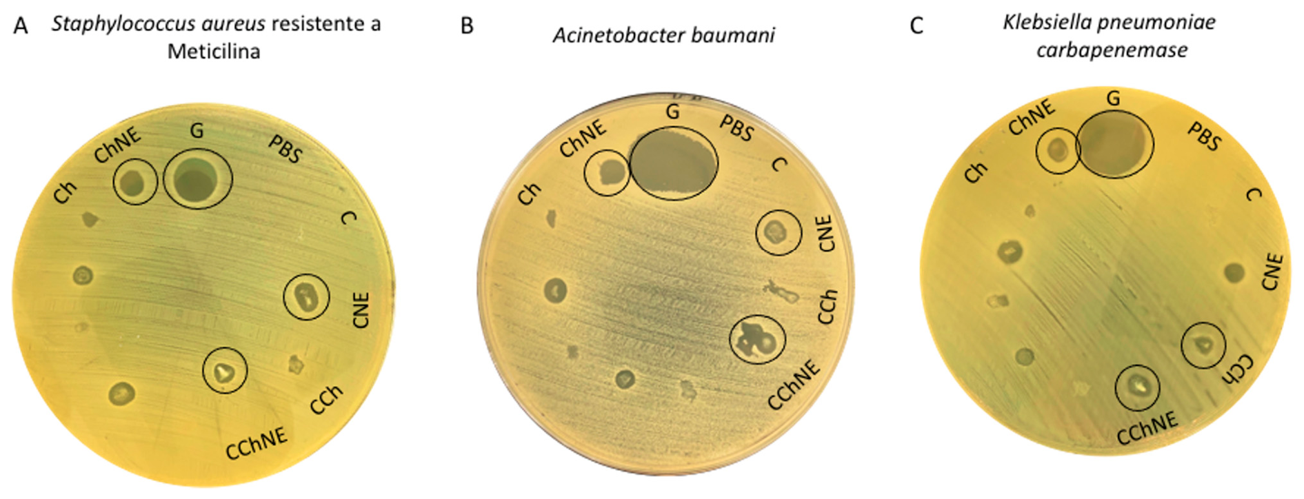
| [42] | Chitosan (CS), collagen (Col), NAC, ε-Poly-L-lysine (ε-PL) | N.I. | Chemical, r. t., 90–120 min | Loaded in the hydrogel, it may act as a crosslinker | Human keratinocytes cells (HaCat); Staphylococcus aureus (S. aureus), Acinetobacter baumani (A. baumani) and Klebsiella pneumoniae carbapenemase (KPC); 7 and 8 weeks old mice. | The conjugation of chitosan and collagen with NAC and ε-PL supports cell viability in vivo and in vitro, reduces inflammatory cells, and induces collagenous expression of MMP-1. | |
| [31] | Chitosan modified with NAC (CS-NAC), genapping (GP), AgNO3 | 292.32 ± 5.98 μmol/g | Chemical r. t., 3 h | Modifying agent, it is crosslinked to silver ions | E. coli and S. aureus; L929 cells. | Increased cell proliferation and good cytocompatibility. Antibacterial effects against E. coli and S. aureus due to Ag-S coordination. | |
| [45] | Acrylic acid (AA), gelatin (Gel), CS-NAC, poly(vinyl butyral) (PVB), Gelatin Methacryloyl (GelMA), α-ketoglutaric acid | N.I. | UV-vis, r. t., 20 min | Modifying agent | Epithelial cells (IOSE80 cells) and rat cardiomyocytes; Sprague-Dawley male rats. | Good adhesion, capacity to adhere strongly to the tissue surface, and easy detachment by the application of a solution. | |
| [46] | Chitosan modified with NAC and NO group (CS-SNO); Hyaluronic acid modified with ethane diamine (HA-NH2); Polyethylene glycol modified with 4-formylbenzoic acid (APG) | 12.85% | Chemical, r. t., 20–50 s | Modifying agent | E. coli and S. epidermidis; NIH 3 T3, HFF-1 and bEnd.3 cells. | Antibacterial activity against E. coli. Promotes cell growth and wound closure at lower concentrations of CS-NO, pH-responsive gelation, and self-healing. | |
| [10] | Aldehyde-modified hyaluronic acid (SA-HA) and thioketal linker with amino end groups (TK-NH2), NAC | N.I. | Chemical, r. t., <10 min | Loaded into the hydrogel | NIH 3 T3 cells; Red blood cells from freshly collected mouse blood; S. aureus and E. coli; ICR mice. | Significant ROS scavenging effect is higher than the components separately. Antibacterial effect against E. coli and S. aureus, good cell viability, and biocompatibility in vitro, with a hemolysis rate lower than 3%. Biocompatibility in vivo. Fast healing in burn wounds, with the highest closure rate, hair growth, attenuated response, and collagen deposition. Self-healing and ROS-responsive. | |
| [30] | CS-NAC, dopamine-modified Poly (lactic-co-glycolic acid) (PLGA) fibers (PLGA-PDA), β-glycerol phosphate (β-GP) | N.I, | Chemical, 37 °C, 24 h | Modifying agent. | Sprague-Dawley (SD) rat bone marrow mesenchymal stem cells (BMSCs, in vitro); 6 week olds (ectopic implantation), and 4- week-old SD rats (in situ transplantation). | Good cell viability and in vitro biocompatibility. Cell migration was higher when fibers were added. Slightly higher proteoglycan expression was observed than that in the control group. Hemolysis rate below 5%, indicating good blood compatibility. In vivo, fibers play a major role in tissue regeneration. Thermosensitive gelation. | |
| [47] | NAC-modified gelatin (Gel-SH), methacrylate silk fibroin (SF), (-)-epi-gallocatechin gallate (EGCG)–copper ionic–κ-carrageenan (κ-CA) nanoparticles (EG−Cu−CA NPs), lithium phenyl-2,4,6-trimethylbenzoylphosphinate (LAP) | 185 mmol/g | UV-vis, 30 s | Modifying agent | E. coli, S. aureus, and Methicillin-resistant S. aureus (MRSA). NIH 3T3 and human umbilical vein endothelial cells (HUVECs); SD rats. | The ROS-scavenging effect was observed even for hydrogel NPs, hemocompatibility, good healing properties induced by the hydrogel matrix alone, and accelerated by NPs. Inflammatory response was observed in the group containing only the matrix hydrogel. | |
| [25] | CS-NAC, benzaldehyde-terminated polyethylene glycol (POSS-PEG-CHO), molybdenum disulfide nanosheets (MoS2 Ns) | 0.32 | Chemical, 37 °C, 5 min | Modifying agent | HUVECs; S. aureus and E. coli; hemorrhagic mouse liver (ex vivo); Diabetic mouse infected with S. aureus. | Fast gelation, self-healing, injectability, biocompatibility, photothermal capacity, antioxidant activity, hemostatic capacity, and in vivo healing capacity. The hydrogel showed excellent antibacterial effect without NIR laser radiation, approximately 96% for both cultures. The effect increased to 100% with NIR laser radiation. The hydrogel and raw materials were virtually free of cytotoxicity, with or without NIR irradiation. | |
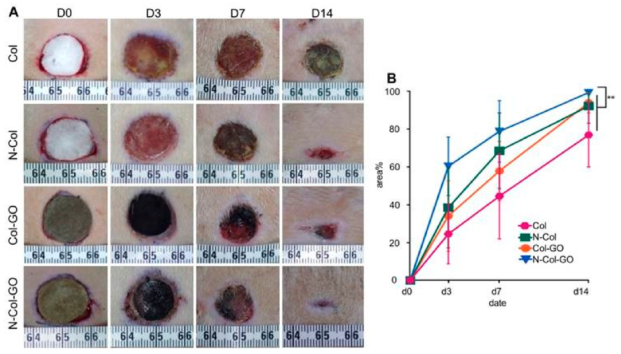
| [26] | Col, graphene oxide (GO), NAC, EDC, n-hydroxy succinimide system (NHS) | N.I. | Chemical, r. t., 24 h | Modifying agent | NIH 3T3 and Hakata; 6 weeks old SD male rats. | Diabetic wound healing and angiogenesis accelerate extracellular matrix synthesis and antioxidant activity. | |
| [34] | Col, EDC and NHS, NAC and polyamide nanofibers (PA) | N.I. | Chemical, r. t., 24 h | Modifying agent | NIH 3T3 cells; 8–10 weeks old male SD rats. | Biocompatible, non-cytotoxic, sustained NAC release, and good wound healing. The NAC-treated groups showed a higher percentage of wound closure, approximately Wound healing 86.17% on day 14. The scaffolds showed a continuous epidermis, high collagen deposition, and regularly arranged new collagen mass. | |
| [32] | Carboxymethyl chitosan (CMCS), oxidized hyaluronic acid (OHA), ε-poly-L-lysine-S-Aroylthiooximes (ε-PL-SATO), NAC | N.A. | Chemical, r. t., <30 s | Loaded in the hydrogel | L929 cells; red blood cells from fresh blood of SD rats (hemolysis) E. coli and S. aureus; SD male rats | fast gelation time, needle injectability, self-healing, H2S release, NAC-dependent, antibacterial activity, biocompatibility, and good wound healing. Hemolysis rate is less than 4%, colorless and transparent. Wound healing was almost complete (99%) after 14 days. | |
| [43] | Collagen sponges (CollaPlug) and Collagen membrane (Bio-Gide), NAC | N.A. | N.I. | Loaded in the hydrogel | Oral mucosal cells from Sprague-Dawley rats (in vitro) | Oral soft-tissue growth. The viability of oral mucosal cells increased with NAC treatment; however, hyperproliferation and collagen deposition were abrogated. Collagen-related gene transcription was downregulated with increasing NAC concentration. The addition of NAC reduced the rate of cell proliferation in a dose-dependent manner. | |
| [44] | CS-NAC, maleic acid modified dextran (Dex-Ma), Basic fibroblast growth factor (bFGF) | N.I. | Chemical, 37 °C, <1 min | Modifying agent | NIH3T3 cells; Male SD rats. | Healing properties, ability to carry cellular growth factors, drug loading, and release. After 9 days of treatment, the group treated with the bFGF-loaded hydrogel showed fewer inflammatory cells than the other three groups, and the formation of new blood vessels and fibroblast proliferation were observed in the granulation tissue of the hydrogel-treated group. | |
| [29] | Carbopol® Ultrez NF 10, NAC, lysine (LYS) | N.A. | N.I. | Loaded into the hydrogel | HaCaT and THP-1 cells. | Biocompatible, drug release, and self-healing. The hydrogel formulation with active ingredients at 20 mM and 100 mM did not show any significant irritant activity, with tissue viability higher than 94% compared to the control. | |
| [36] | Col type I, NAC, GO | N.I. | Chemical | Modifying agent | NIH 3T3 cells; 8–10 weeks old SD male rats. | Biocompatibility, wound healing, drug release. AC reduced the cytotoxicity of GO, downregulated the levels of ROS induced by GO, and promoted the fastest cell migration when aligned with GO. | |
| [93] | ALG, NAC, CaCl2 | N.I. | Physical, 25 °C | Loaded into the hydrogel | MC3T3 cells. | Drug release, wound healing, and re-epithelialization. Reduction in cell death associated with extrusion process. NAC improved cell viability and reduced apoptotic and inflammatory expression. | |
| [87] | NaHA modified with cystamine dihydochloride, PDA-PEG-NAC, PDA-PEG-bFDF | 72.1 μmol/g | Chemical, 5 min | Modifying agent | bFDF; 10–12 weeks old C57BL/6 diabetic mice (blood glucose levels exceeding 16.7 mmoL/mL). | Drug and cell release, anti-inflammatory activity, biocompatibility, wound closure. | |
| [37] | CS-NAC, silver nanowire (AgNW), glass substrate | N.I. | Chemical | Modifying agent, crosslinked | E. coli and S. aureus by the zone of inhibition; Human hepatic cell line L02; 16 months old pregnant rabbits. | Antibacterial effect, biocompatible, and wound healing, low cytotoxicity | |
| [48] | Catechol-conjugated chitosan modified with thiol group (CSDS), Sodium periodate (NaIO4) | 75.21 ± 5.2 μmol/g | Chemical, 30 s | Modifying agent, crosslinked | L929 cells. | Adhesive and biocompatible. | |
| [40] | CS-NAC, maleimide group modified ε-polylysine (EPLM) | 151.8 μmol/g | Chemical, r. t., 15 ± 3 s | Modifying agent, crosslinked | L929 cells. 6 weeks old SD male rats (hemostatic ability in vivo). | Non-cytotoxicity in vitro, rapid gelation, and excellent adhesion. The blood loss from the liver was reduced from 106.7 mg (untreated liver) to 28.3 mg due to the good adhesiveness of the hydrogel. | |
| [33] | Col, NAC, Poly (ε-caprolactone) (PCL) fibers | N.I. | Chemical, r. t., 8 h | Modifying agent | NIH 3 T3 cells; 8–10 weeks old SD male rats. | It is biocompatible and highly porous, promoting cell proliferation, wound healing, angiogenesis, and sustained drug release. | |
| [27] | Poly (ethylene adipate) diol ((PEA) diol), ε-Caprolactone, Rac-Lactide and Poly(ethylene glycol) copolymer (CL-LA-PEG200), CS, 1,6-diisocyanatohexane (hexamethylene diisocyanate (HDI), NAC beads | N.A. | Physical, r. t., 24 h | Loaded in the hydrogel | 10 weeks old diabetic male mice (glucose serum concentration was 483 ± 191.9 mg/dL). | Fast wound closure, re-epithelialization, sustained drug release, and biocompatibility. The 5% NAC hydrogels showed better results for wound closure than those treated with 10% and 20% NAC. | |
| [35] | NAC, silver salts and asiatic acid-poly(aniline)-quercetin (AA-PANi-Q) nanomaterial | N.A. | Chemical (coordination) | Complexation agent | E. coli and S. aureus; L929 cells. | Non-cytotoxicity, antibacterial effect, biocompatibility, controlled drug delivery, photothermal responsiveness, and structural stability. A modest reduction in bacterial colonies was observed with NIR irradiation. The group containing only NAC and silver displayed a more effective bactericidal outcome against S. aureus than against E. coli. Overall, a synergistic effect was observed between the formulation and photothermal activation. | |
| [39] | Sodium alginate (SA), guar gum (GG), NAC | N.A. | Physical | Loaded into the hydrogel | S. aureus, E. coli, P. aeruginosa and Candida albicans (C. albican). | Cell proliferation, antimicrobial, antibiofilm, and antioxidant activities and low cytotoxicity. Free NAC inhibited E. coli biofilm formation by up to 75.80%, whereas the hydrogel (2% v/v SA, 1% v/v GG, 05 mg/mL NAC) inhibited S. aureus biofilm formation by 28.29% even at low doses (12.5 μg/mL). | |
| [38] | PCL, (GO), NAC modified chitosan-co-polycaprolactone (NAC-CS-PCL) | N.I. | Chemical, 24 h | Modifying agent | Human dermal fibroblasts (HDFs); SD male rats. | Angiogenesis, collagen deposition, and biocompatibility. NAC induced slight cell proliferation. In vivo, only the NAC-treated group displayed newly formed blood vessels. | |
| [41] | Polyimidazom containing maleimide terminal groups (PIM(Cn)-Mal), poly(ethylene glycol) tetra thiol (PEG-4SH), poly(ethylene glycol) tetra maleimide (PEG-4mal), thiol-functionalized alginate (Alg-SH), PEG-2Mal-2NAC | N.I. | Chemical, 25 °C, 5 min | Modifying agent | MRSA USA300, CR-AB, PAO1 or CR-PA; N3T3 and HDFs; 8 weeks old male C57BL/6 mice. | Accelerating the healing of diabetic wounds infected by biofilms. Infected wounds were closed in 12 days in vivo. | |
| [50] | Vancomicin, CS-NAC, Dex-Ma | N.I. | Chemical, 37 °C, <1 min | Modifying agent | NIH3T3 cells; S. aureus and E. coli. | Biocompatible, drug administration, antimicrobial The hydrogel group without vancomycin showed antibacterial activity against Gram-negative bacteria (E. coli). | |
| [88] | Polyethylene glycol diacrylate (PEGMA), tannic acid (TA), NAC, and silver nitrate | N.A. | Chemical | Complexation agent | E. coli and S.aureus; NIH3T3 cells. | Injectable, antibacterial activity, antibiofilm activity, sustained drug release for 8 h, scavenger activity, non-cytotoxic. | |
| [89] | Polymethylmethacrylate (PMMA), marine collagen (MCol), NAC | N.I. | Physical, 25 °C | Loaded into the hydrogel | Human normal fibroblast cell line (BJ1); Strains of Sepidermidis, S. aureus, Bacillus cereus, Salmonella paratyphi, E. coli, Klebsiella pneumoniae, Candida glabrata, Candida albicans, and Candida parapsilosis. | Biocompatibility, antioxidant, and antimicrobial properties. |
References
- Ghosh, R.N.; Thomas, J.; Vaidehi, B.R.; Devi, N.G.; Janardanan, A.; Namboothiri, P.K.; Peter, M. An Insight into Synthesis, Properties and Applications of Gelatin Methacryloyl Hydrogel for 3D Bioprinting. Mater. Adv. 2023, 4, 5496–5529. [Google Scholar] [CrossRef] [Scilit]
- Liu, B.; Chen, K. Advances in Hydrogel-Based Drug Delivery Systems. Gels 2024, 10, 262. [Google Scholar] [CrossRef] [Scilit] [PubMed]
- Priya, A.S.; Premanand, R.; Ragupathi, I.; Bhaviripudi, V.R.; Aepuru, R.; Kannan, K.; Shanmugaraj, K. Comprehensive Review of Hydrogel Synthesis, Characterization, and Emerging Applications. J. Compos. Sci. 2024, 8, 457. [Google Scholar] [CrossRef] [Scilit]
- Liang, J.; Peng, X.; Zhou, X.; Zou, J.; Cheng, L. Emerging Applications of Drug Delivery Systems in Oral Infectious Diseases Prevention and Treatment. Molecules 2020, 25, 516. [Google Scholar] [CrossRef] [Scilit] [PubMed]
- Huang, L.; Chen, X.; Yang, X.; Zhang, Y.; Qiu, X. GelMA-based Hydrogel Biomaterial Scaffold: A Versatile Platform for Regenerative Endodontics. J. Biomed. Mater. Res. Part B Appl. Biomater. 2024, 112, e35412. [Google Scholar] [CrossRef] [Scilit] [PubMed]
- Tenório, M.C.D.S.; Graciliano, N.G.; Moura, F.A.; de Oliveira, A.C.M.; Goulart, M.O.F. N-Acetylcysteine (NAC): Impacts on Human Health. Antioxidants 2021, 10, 967. [Google Scholar] [CrossRef] [Scilit] [PubMed]
- Pedre, B.; Barayeu, U.; Ezeriņa, D.; Dick, T.P. The Mechanism of Action of N-Acetylcysteine (NAC): The Emerging Role of H2S and Sulfane Sulfur Species. Pharmacol. Ther. 2021, 228, 107916. [Google Scholar] [CrossRef] [Scilit] [PubMed]
- Comino-Sanz, I.M.; López-Franco, M.D.; Castro, B.; Pancorbo-Hidalgo, P.L. The Role of Antioxidants on Wound Healing: A Review of the Current Evidence. J. Clin. Med. 2021, 10, 3558. [Google Scholar] [CrossRef] [Scilit] [PubMed]
- Fazary, A.E.; Awwad, N.S.; Ibrahium, H.A.; Shati, A.A.; Alfaifi, M.Y.; Ju, Y.-H. Protonation Equilibria of N-Acetylcysteine. ACS Omega 2020, 5, 19598–19605. [Google Scholar] [CrossRef] [Scilit] [PubMed]
- Yi, D.; Wang, T.; Xu, S.; Liu, Y.; Wang, Z.; Li, N.; Wu, Y.; He, Y. Injectable ROS/pH-Responsive Hydrogel for Synergistic Chemo-Photodynamic Tumor Therapy and Enhanced Wound Healing. Adv. Healthc. Mater. 2025, 15, e02349. [Google Scholar] [CrossRef] [Scilit] [PubMed]
- Zheng, H.; Liu, J.; Sun, L.; Meng, Z. The Role of N-Acetylcysteine in Osteogenic Microenvironment for Bone Tissue Engineering. Front. Cell Dev. Biol. 2024, 12, 1435125. [Google Scholar] [CrossRef] [Scilit] [PubMed]
- Camargo, C.H.R.; Gomes, L.C.L.; França, M.C.M.; Bittencourt, T.S.; Valera, M.C.; Camargo, S.E.A.; Bottino, M.C. Incorporating N-Acetylcysteine and Tricalcium Phosphate into Epoxy Resin-Based Sealer Improved Its Biocompatibility and Adhesiveness to Radicular Dentine. Dent. Mater. 2019, 35, 1750–1756. [Google Scholar] [CrossRef] [Scilit] [PubMed]
- Jiao, Y.; Ma, S.; Li, J.; Shan, L.; Yang, Y.; Li, M.; Chen, J. The Influences of N-Acetyl Cysteine (NAC) on the Cytotoxicity and Mechanical Properties of Poly-Methylmethacrylate (PMMA)-Based Dental Resin. PeerJ 2015, 3, e868. [Google Scholar] [CrossRef] [Scilit] [PubMed]
- Yang, Y.; Wang, L.; Huang, Z.; Ge, L.; Shi, J. N-Acetylcysteine as a Novel Methacrylate-Based Resin Cement Component: Effect on Cell Apoptosis and Genotoxicity in Human Gingival Fibroblasts. BMC Oral Health 2024, 24, 222. [Google Scholar] [CrossRef] [Scilit] [PubMed]
- Gomez-Aparicio, L.S.; Bernáldez-Sarabia, J.; Camacho-Villegas, T.A.; Lugo-Fabres, P.H.; Díaz-Martínez, N.E.; Padilla-Camberos, E.; Licea-Navarro, A.; Castro-Ceseña, A.B. Improvement of the Wound Healing Properties of Hydrogels with N-Acetylcysteine through Their Modification with Methacrylate-Containing Polymers. Biomater. Sci. 2021, 9, 726–744. [Google Scholar] [CrossRef] [Scilit] [PubMed]
- Padalhin, A.; Abueva, C.; Ryu, H.S.; Yoo, S.H.; Seo, H.H.; Park, S.Y.; Chung, P.-S.; Woo, S.H. Impact of Thermo-Responsive N-Acetylcysteine Hydrogel on Dermal Wound Healing and Oral Ulcer Regeneration. Int. J. Mol. Sci. 2024, 25, 4835. [Google Scholar] [CrossRef] [Scilit] [PubMed]
- Kong, Y.; Tang, H.; Chai, H.; Zhang, L.; Mao, Z.; Xu, H. Asymmetric Adhesive and Absorbable Fiber Membrane of Methacrylated Gelatin–Gelatin/Acetylcysteine for Advanced Hemostatic Applications. ACS Appl. Polym. Mater. 2025, 7, 8383–8395. [Google Scholar] [CrossRef] [Scilit]
- Galicia-Moreno, M.; Monroy-Ramirez, H.C.; Caloca-Camarena, F.; Arceo-Orozco, S.; Muriel, P.; Sandoval-Rodriguez, A.; García-Bañuelos, J.; García-González, A.; Navarro-Partida, J.; Armendariz-Borunda, J. A New Opportunity for N-Acetylcysteine. An Outline of Its Classic Antioxidant Effects and Its Pharmacological Potential as an Epigenetic Modulator in Liver Diseases Treatment. Naunyn-Schmiedeberg’s Arch. Pharmacol. 2025, 398, 2365–2386. [Google Scholar] [CrossRef] [Scilit] [PubMed]
- Brannan, R.G. Effect of N-Acetyl-Cysteine on Liposomal and Muscle Model Oxidation Induced by Reactive Oxygen, Nitrogen, and Sulfur. Meat Sci. 2011, 88, 733–739. [Google Scholar] [CrossRef] [Scilit] [PubMed]
- European Directorate for the Quality of Medicines. European Pharmacopoeia, 6th ed.; Council of Europe: Strasbourg, France, 2008; ISBN 92-871-6054-6. [Google Scholar]
- Brayfield, A.; Cadart, C. The Complete Drug Reference, 41st ed.; Pharmaceutical Press: London, UK, 2024. [Google Scholar]
- To, T.; Ellis, A.G.; Ching, M.S.; Shilson, A.D.; Kong, D.C.; Garrett, K. Stability of a Formulated N-acetylcysteine Capsule for Prevention of Contrast-Induced Nephropathy. J. Pharm. Pract. Res. 2008, 38, 219–222. [Google Scholar] [CrossRef] [Scilit]
- Qiu, K.; Shi, H.; Chen, M.; Lai, S.; Liew, K.; Wang, Z.; Wei, X. Synergistic PNS-NAC Bioadhesive Microspheres for Enhanced Intestinal Permeability and Oral Bioavailability. Recent Adv. Drug Deliv. Formul. 2026, 20, 0126673878437892260216073431. [Google Scholar] [CrossRef] [Scilit] [PubMed]
- Tsai, M.-S.; Liou, G.-G.; Liao, J.-W.; Lai, P.-Y.; Yang, D.-J.; Wu, S.-H.; Wang, S.-H. N-Acetyl Cysteine Overdose Induced Acute Toxicity and Hepatic Microvesicular Steatosis by Disrupting GSH and Interfering Lipid Metabolisms in Normal Mice. Antioxidants 2024, 13, 832. [Google Scholar] [CrossRef] [Scilit] [PubMed]
- He, Y.; Guo, J.; Li, H.; Jiang, J.; Chen, J.; Yang, G.; Yang, X.; Chen, Z.; Lu, C. MoS2-Functionalized Chitosan Hydrogel with Antibacterial and Antioxidant Functions Promotes Healing of Infected Diabetic Wounds. Mater. Chem. Front. 2024, 8, 3770–3789. [Google Scholar] [CrossRef] [Scilit]
- Qian, B.; Li, J.; Guo, K.; Guo, N.; Zhong, A.; Yang, J.; Wang, J.; Xiao, P.; Sun, J.; Xiong, L. Antioxidant Biocompatible Composite Collagen Dressing for Diabetic Wound Healing in Rat Model. Regen. Biomater. 2021, 8, rbab003. [Google Scholar] [CrossRef] [Scilit] [PubMed]
- Stachura, A.; Sobczak, M.; Kędra, K.; Kopka, M.; Kopka, K.; Włodarski, P.K. The Influence of N-Acetylcysteine-Enriched Hydrogels on Wound Healing in a Murine Model of Type II Diabetes Mellitus. Int. J. Mol. Sci. 2024, 25, 9986. [Google Scholar] [CrossRef] [Scilit] [PubMed]
- Beltrame, J.M.; Guindani, C.; Novy, M.G.; Felipe, K.B.; Sayer, C.; Pedrosa, R.C.; Hermes de Araújo, P.H. Covalently Bonded N-Acetylcysteine-Polyester Loaded in PCL Scaffolds for Enhanced Interactions with Fibroblasts. ACS Appl. Bio Mater. 2021, 4, 1552–1562. [Google Scholar] [CrossRef] [Scilit] [PubMed]
- Brites, G.; Basso, J.; Miranda, M.; Neves, B.M.; Vitorino, C.; Cruz, M.T. Development of a New Hydrogel for the Prevention of Allergic Contact Dermatitis. Int. J. Pharm. 2022, 628, 122265. [Google Scholar] [CrossRef] [Scilit] [PubMed]
- Cai, H.; Yan, J.; Zhao, W.; Ma, S.; Wu, J.; Zhao, Z.; Deng, H. Injectable Interface-Bonded Fiber-Reinforced Thiolated Chitosan Hydrogels for Enhanced Cellular Activities and Cartilage Regeneration. Carbohydr. Polym. 2025, 347, 122643. [Google Scholar] [CrossRef] [Scilit] [PubMed]
- Chen, R.; Hao, Y.; Francesco, S.; Mao, X.; Huang, W.-C. A Chitosan-Based Antibacterial Hydrogel with Injectable and Self-Healing Capabilities. Mar. Life Sci. Technol. 2024, 6, 115–125. [Google Scholar] [CrossRef] [Scilit] [PubMed]
- Gong, L.; Chang, L.; Chen, S.; Wei, X.; Du, H.; Cheng, J.; Chen, X.; Yuan, Z.; Zhao, P.; Geng, M.; et al. Multifunctional Injectable Hydrogel with Self-Supplied H2S Release and Bacterial Inhibition for the Wound Healing with Enhanced Macrophages Polarization via Interfering with PI3K/Akt Pathway. Biomaterials 2025, 318, 123144. [Google Scholar] [CrossRef] [Scilit] [PubMed]
- Hou, J.; Chen, L.; Liu, Z.; Li, J.; Yang, J.; Zhong, A.; Zhou, M.; Sun, Y.; Guo, L.; Yang, Y.; et al. Sustained Release of N-Acetylcysteine by Sandwich Structured Polycaprolactone/Collagen Scaffolds for Wound Healing. J. Biomed. Mater. Res. Part A 2019, 107, 1414–1424. [Google Scholar] [CrossRef] [Scilit] [PubMed]
- Hou, J.; Chen, L.; Zhou, M.; Li, J.; Liu, J.; Fang, H.; Zeng, Y.; Sun, J.; Wang, Z. Multi-Layered Polyamide/Collagen Scaffolds with Topical Sustained Release of N-Acetylcysteine for Promoting Wound Healing. Int. J. Nanomed. 2020, 15, 1349–1361. [Google Scholar] [CrossRef] [Scilit] [PubMed]
- Laxmanan, K.; Shin, H.; Jo, J.; Bose, S.; Ranganathan, P.; Nam, S.Y.; Kang, H.W. Theranostic Polyaniline-Integrated N-Acetyl-l-Cysteine Hydrogel for Synergistic Photothermal Antibacterial Therapy and Enhanced Cell Migration. ACS Appl. Bio Mater. 2025, 8, 8751–8766. [Google Scholar] [CrossRef] [Scilit] [PubMed]
- Li, J.; Zhou, C.; Luo, C.; Qian, B.; Liu, S.; Zeng, Y.; Hou, J.; Deng, B.; Sun, Y.; Yang, J.; et al. N-Acetyl Cysteine-Loaded Graphene Oxide-Collagen Hybrid Membrane for Scarless Wound Healing. Theranostics 2019, 9, 5839–5853. [Google Scholar] [CrossRef] [Scilit] [PubMed]
- Li, J.; Li, L.; Lv, J.; Wang, C.; Liu, Y. Preparation of Thiolated Chitosan/Silver Nanowire Composite Hydrogels with Antimicrobial Activity for Obstetric Wound Care. Mater. Lett. 2020, 280, 128497. [Google Scholar] [CrossRef] [Scilit]
- Liu, J.; Hou, J.; Liu, S.; Li, J.; Zhou, M.; Sun, J.; Wang, R. Graphene Oxide Functionalized Double-Layered Patch with Anti-Adhesion Ability for Abdominal Wall Defects. Int. J. Nanomed. 2021, 16, 3803–3818. [Google Scholar] [CrossRef] [Scilit] [PubMed]
- Mondal, S.; Hazra, A.; Paul, P.; Saha, B.; Roy, S.; Bhowmick, P.; Bhowmick, M. Formulation and Evaluation of N-Acetyl Cysteine Loaded Bi-Polymeric Physically Crosslinked Hydrogel with Antibacterial and Antioxidant Activity for Diabetic Wound Dressing. Int. J. Biol. Macromol. 2024, 279, 135418. [Google Scholar] [CrossRef] [Scilit] [PubMed]
- Nie, W.; Yuan, X.; Zhao, J.; Zhou, Y.; Bao, H. Rapidly in Situ Forming Chitosan/ε-Polylysine Hydrogels for Adhesive Sealants and Hemostatic Materials. Carbohydr. Polym. 2013, 96, 342–348. [Google Scholar] [CrossRef] [Scilit] [PubMed]
- Pranantyo, D.; Yeo, C.K.; Wu, Y.; Fan, C.; Xu, X.; Yip, Y.S.; Vos, M.I.G.; Mahadevegowda, S.H.; Lim, P.L.K.; Yang, L.; et al. Hydrogel Dressings with Intrinsic Antibiofilm and Antioxidative Dual Functionalities Accelerate Infected Diabetic Wound Healing. Nat. Commun. 2024, 15, 954. [Google Scholar] [CrossRef] [Scilit] [PubMed]
- Rodrigues, J.P.; da Costa Silva, J.R.; Ferreira, B.A.; Veloso, L.I.; Quirino, L.S.; Rosa, R.R.; Barbosa, M.C.; Rodrigues, C.M.; Gaspari, P.B.F.; Beletti, M.E.; et al. Development of Collagenous Scaffolds for Wound Healing: Characterization and in Vivo Analysis. J. Mater. Sci. Mater. Med. 2024, 35, 12. [Google Scholar] [CrossRef] [Scilit] [PubMed]
- Sato, N.; Ueno, T.; Kubo, K.; Suzuki, T.; Tsukimura, N.; Att, W.; Yamada, M.; Hori, N.; Maeda, H.; Ogawa, T. N-Acetyl Cysteine (NAC) Inhibits Proliferation, Collagen Gene Transcription, and Redox Stress in Rat Palatal Mucosal Cells. Dent. Mater. 2009, 25, 1532–1540. [Google Scholar] [CrossRef] [Scilit] [PubMed]
- Tang, Y.; Cai, X.; Xiang, Y.; Zhao, Y.; Zhang, X.; Wu, Z. Cross-Linked Antifouling Polysaccharide Hydrogel Coating as Extracellular Matrix Mimics for Wound Healing. J. Mater. Chem. B 2017, 5, 2989–2999. [Google Scholar] [CrossRef] [Scilit] [PubMed]
- Tian, G.; Yang, D.; Liang, C.; Liu, Y.; Chen, J.; Zhao, Q.; Tang, S.; Huang, J.; Xu, P.; Liu, Z.; et al. A Nonswelling Hydrogel with Regenerable High Wet Tissue Adhesion for Bioelectronics. Adv. Mater. 2023, 35, 2212302. [Google Scholar] [CrossRef] [Scilit] [PubMed]
- Yang, Y.; Zhou, Y.; Li, Y.; Guo, L.; Zhou, J.; Chen, J. Injectable and Self-Healing Hydrogel Containing Nitric Oxide Donor for Enhanced Antibacterial Activity. React. Funct. Polym. 2021, 166, 105003. [Google Scholar] [CrossRef] [Scilit]
- Zeng, Z.; Guo, J.; Shen, G.; Guo, C.; Pei, D.; Lu, D.; Geng, Z.; Huang, J.; Yu, S. Antibacterial-Antioxidative Thiolated Gelatin/Methacrylated Silk Fibroin Hydrogels with Nitric Oxide Release Catalyzed by Metal-Polyphenol Nanoparticles for MRSA-Infected Wound Healing. Biomacromolecules 2023, 24, 5116–5131. [Google Scholar] [CrossRef] [Scilit] [PubMed]
- Zeng, Z.; Mo, X. Rapid in Situ Crosslinking of Hydrogel Adhesives Based on Thiol-Grafted Bio-Inspired Catechol-Conjugated Chitosan. J. Biomater. Appl. 2017, 32, 612–621. [Google Scholar] [CrossRef] [Scilit] [PubMed]
- Zertuche-Arias, T.; Alatorre-Meda, M.; Rivero, I.A.; Juárez, P.; Castro-Ceseña, A.B. N-Acetylcysteine and pro-Adrenomedullin Dual-Crosslinked Gelatin–Chitosan Hydrogels with Enhanced Mechanical and Mineralization Performance. RSC Adv. 2025, 15, 22524–22533. [Google Scholar] [CrossRef] [Scilit] [PubMed]
- Zhao, Y.; Zhang, X.; Wang, Y.; Wu, Z.; An, J.; Lu, Z.; Mei, L.; Li, C. In Situ Cross-Linked Polysaccharide Hydrogel as Extracellular Matrix Mimics for Antibiotics Delivery. Carbohydr. Polym. 2014, 105, 63–69. [Google Scholar] [CrossRef] [Scilit] [PubMed]
- Bermejo-Velasco, D.; Azémar, A.; Oommen, O.P.; Hilborn, J.; Varghese, O.P. Modulating Thiol pKa Promotes Disulfide Formation at Physiological pH: An Elegant Strategy To Design Disulfide Cross-Linked Hyaluronic Acid Hydrogels. Biomacromolecules 2019, 20, 1412–1420. [Google Scholar] [CrossRef] [Scilit] [PubMed]
- Chung, T.-W.; Cheng, C.-L.; Liu, Y.-H.; Huang, Y.-C.; Chen, W.-P.; Panda, A.K.; Chen, W.-L. Dopamine-Dependent Functions of Hyaluronic Acid/Dopamine/Silk Fibroin Hydrogels That Highly Enhance N-Acetyl-L-Cysteine (NAC) Delivered from Nasal Cavity to Brain Tissue through a near-Infrared Photothermal Effect on the NAC-Loaded Hydrogels. Biomater. Adv. 2023, 154, 213615. [Google Scholar] [CrossRef] [Scilit] [PubMed]
- Colak, B.; Wu, L.; Cozens, E.J.; Gautrot, J.E. Modulation of Thiol–Ene Coupling by the Molecular Environment of Polymer Backbones for Hydrogel Formation and Cell Encapsulation. ACS Appl. Bio Mater. 2020, 3, 6497–6509. [Google Scholar] [CrossRef] [Scilit] [PubMed]
- Du, Z.; Liu, J.; Zhang, H.; Wu, X.; Zhang, B.; Chen, Y.; Liu, B.; Ding, L.; Xiao, H.; Zhang, T. N-Acetyl-l-Cysteine/L-Cysteine-Functionalized Chitosan−β-Lactoglobulin Self-Assembly Nanoparticles: A Promising Way for Oral Delivery of Hydrophilic and Hydrophobic Bioactive Compounds. J. Agric. Food Chem. 2019, 67, 12511–12519. [Google Scholar] [CrossRef] [Scilit] [PubMed]
- El Khoury, R.; Ramirez, S.P.; Loyola, C.D.; Joddar, B. Demonstration of Doxorubicin’s Cardiotoxicity and Screening Using a 3D Bioprinted Spheroidal Droplet-Based System. RSC Adv. 2023, 13, 8338–8351. [Google Scholar] [CrossRef] [Scilit] [PubMed]
- Li, H.; He, W.; Feng, Q.; Chen, J.; Xu, X.; Lv, C.; Zhu, C.; Dong, H. Engineering Superstable Islets-Laden Chitosan Microgels with Carboxymethyl Cellulose Coating for Long-Term Blood Glucose Regulation in Vivo. Carbohydr. Polym. 2024, 323, 121425. [Google Scholar] [CrossRef] [Scilit] [PubMed]
- Wu, S.-W.; Liu, X.; Miller, A.L.I.; Cheng, Y.-S.; Yeh, M.-L.; Lu, L. Strengthening Injectable Thermo-Sensitive NIPAAm-g-Chitosan Hydrogels Using Chemical Cross-Linking of Disulfide Bonds as Scaffolds for Tissue Engineering. Carbohydr. Polym. 2018, 192, 308–316. [Google Scholar] [CrossRef] [Scilit] [PubMed]
- Wu, Z.M.; Zhang, X.G.; Zheng, C.; Li, C.X.; Zhang, S.M.; Dong, R.N.; Yu, D.M. Disulfide-Crosslinked Chitosan Hydrogel for Cell Viability and Controlled Protein Release. Eur. J. Pharm. Sci. 2009, 37, 198–206. [Google Scholar] [CrossRef] [Scilit] [PubMed]
- Jin, Y.; Zhang, J.; Chen, X.; Li, F.; Xue, T.; Yi, K.; Xu, Y.; Wang, H.; Lao, Y.-H.; Chan, H.F.; et al. 3D Printing Incorporating Gold Nanozymes with Mesenchymal Stem Cell-Derived Hepatic Spheroids for Acute Liver Failure Treatment. Biomaterials 2025, 315, 122895. [Google Scholar] [CrossRef] [Scilit] [PubMed]
- Kang, H.K.; Sarsenova, M.; Kim, D.-H.; Kim, M.S.; Lee, J.Y.; Sung, E.-A.; Kook, M.G.; Kim, N.G.; Choi, S.W.; Ogay, V.; et al. Establishing a 3d in Vitro Hepatic Model Mimicking Physiologically Relevant to in Vivo State. Cells 2021, 10, 1268. [Google Scholar] [CrossRef] [Scilit] [PubMed]
- Lu, S.; Zhang, L.; Hu, Z.; Kong, S.; Zhang, Z.; Li, G. Optimized Preparation of Gastric Acid-Response Sulfhydryl Functionalized Chitosan/Alginate/Tilapia Peptide Hydrogel and Its Protective Effects on Alcohol-Induced Liver and Brain Injury. RSC Adv. 2021, 11, 34544–34557. [Google Scholar] [CrossRef] [Scilit] [PubMed]
- Aldaghi, N.; Kamalabadi-Farahani, M.; Alizadeh, M.; Alizadeh, A.; Salehi, M. Enhancing Pressure Ulcer Healing and Tissue Regeneration by Using N-Acetyl-Cysteine Loaded Carboxymethyl Cellulose/Gelatin/Sodium Alginate Hydrogel. Biomed. Eng. Lett. 2024, 14, 833–845. [Google Scholar] [CrossRef] [Scilit] [PubMed]
- Lu, S.; Kong, S.; Wang, Y.; Hu, Z.; Zhang, L.; Liao, M. Gastric Acid-Response Chitosan/Alginate/Tilapia Collagen Peptide Composite Hydrogel: Protection Effects on Alcohol-Induced Gastric Mucosal Injury. Carbohydr. Polym. 2022, 277, 118816. [Google Scholar] [CrossRef] [Scilit] [PubMed]
- Zhou, Z.; Wang, L.; Yang, D.; Li, Q.; Wang, X.; Nie, J.; Ma, G. Acid-Triggered Charge-Switchable Antibacterial Hydrogel for Accelerated Healing of Gastric Mucosal Wounds. ACS Nano 2025, 19, 17533–17553. [Google Scholar] [CrossRef] [Scilit] [PubMed]
- Ross, M.; Sheardown, L.; Muirhead, B.; Mofford, J.; Tian, J.; Sheardown, H. Mucoadhesive Thermogel Platform for Treating Anterior Ocular Conditions. J. Biomed. Mater. Res. Part A 2023, 111, 1390–1405. [Google Scholar] [CrossRef] [Scilit] [PubMed]
- Taheri, S.L.; Rezazadeh, M.; Hassanzadeh, F.; Akbari, V.; Dehghani, A.; Talebi, A.; Mostafavi, S.A. Preparation, Physicochemical, and Retinal Anti-Angiogenic Evaluation of Poloxamer Hydrogel Containing Dexamethasone/Avastin-Loaded Chitosan-N-Acetyl-L-Cysteine Nanoparticles. Int. J. Biol. Macromol. 2022, 220, 1605–1618. [Google Scholar] [CrossRef] [Scilit] [PubMed]
- Boot, W.; Vogely, H.C.; Nikkels, P.G.; Pouran, B.; van Rijen, M.H.; Ekkelenkamp, M.B.; Hänsch, G.M.; Dhert, W.J.; Gawlitta, D. Prophylaxis of Implant-Related Infections by Local Release of Vancomycin from a Hydrogel in Rabbits. Eur. Cell Mater. 2020, 39, 108–120. [Google Scholar] [CrossRef] [Scilit] [PubMed]
- Drago, L.; Boot, W.; Dimas, K.; Malizos, K.; Hänsch, G.M.; Stuyck, J.; Gawlitta, D.; Romanò, C.L. Does Implant Coating with Antibacterial-Loaded Hydrogel Reduce Bacterial Colonization and Biofilm Formation in Vitro? Clin. Orthop. Relat. Res. 2014, 472, 3311–3323. [Google Scholar] [CrossRef] [Scilit] [PubMed]
- Lee, D.; Heo, D.N.; Nah, H.R.; Lee, S.J.; Ko, W.-K.; Lee, J.S.; Moon, H.-J.; Bang, J.B.; Hwang, Y.-S.; Reis, R.L.; et al. Injectable Hydrogel Composite Containing Modified Gold Nanoparticles: Implication in Bone Tissue Regeneration. Int. J. Nanomed. 2018, 13, 7019–7031. [Google Scholar] [CrossRef] [Scilit] [PubMed]
- Pontremoli, C.; Boffito, M.; Laurano, R.; Iviglia, G.; Torre, E.; Cassinelli, C.; Morra, M.; Ciardelli, G.; Vitale-Brovarone, C.; Fiorilli, S. Mesoporous Bioactive Glasses Incorporated into an Injectable Thermosensitive Hydrogel for Sustained Co-Release of Sr2+ Ions and N-Acetylcysteine. Pharmaceutics 2022, 14, 1890. [Google Scholar] [CrossRef] [Scilit] [PubMed]
- Yamada, M.; Ishihara, K.; Ogawa, T.; Sakurai, K. The Inhibition of Infection by Wound Pathogens on Scaffold in Tissue-Forming Process Using N-Acetyl Cysteine. Biomaterials 2011, 32, 8474–8485. [Google Scholar] [CrossRef] [Scilit] [PubMed][Green Version]
- Le-Vinh, B.; Steinbring, C.; Nguyen Le, N.-M.; Matuszczak, B.; Bernkop-Schnürch, A. S-Protected Thiolated Chitosan versus Thiolated Chitosan as Cell Adhesive Biomaterials for Tissue Engineering. ACS Appl. Mater. Interfaces 2023, 15, 40304–40316. [Google Scholar] [CrossRef] [Scilit] [PubMed]
- Ramburrun, P.; Kumar, P.; Ndobe, E.; Choonara, Y.E. Gellan-Xanthan Hydrogel Conduits with Intraluminal Electrospun Nanofibers as Physical, Chemical and Therapeutic Cues for Peripheral Nerve Repair. Int. J. Mol. Sci. 2021, 22, 11555. [Google Scholar] [CrossRef] [Scilit] [PubMed]
- Ricci, F.; Racaniello, G.F.; Denora, N.; Gentile, L.; Lopalco, A.; Cutrignelli, A.; Franco, M.; Iacobazzi, R.M.; Laquintana, V.; Lopedota, A. Thermoresponsive Mucoadhesive Hydrogel Based on Pluronic F127/Thiolated Glycol Chitosan for Intravesical Administration of Celecoxib/Gemcitabine. J. Drug Deliv. Sci. Technol. 2023, 86, 104687. [Google Scholar] [CrossRef] [Scilit]
- Liu, Y.; Li, S.; Zhu, J.; Fan, L.; Wang, L. Rapid Preparation of Injectable Dual-Network Hydrogels for Biomedical Applications Using UV-Triggered Sulfhydryl Click Reactions. Colloids Surf. B Biointerfaces 2024, 244, 114180. [Google Scholar] [CrossRef] [Scilit] [PubMed]
- Tseng, S.-J.; Huang, S.-T.; Wu, C.-C.; Cheng, C.-H.; Lin, J.-C. Studies of Proliferation and Chondrogenic Differentiation of Rat Adipose Stem Cells Using an Anti-Oxidative Polyurethane Scaffold Combined with Cyclic Compression Culture. Mater. Sci. Eng. C Mater. Biol. Appl. 2020, 112, 110964. [Google Scholar] [CrossRef] [Scilit] [PubMed]
- Ciftci, Z.; Deniz, M.; Yilmaz, I.; Ciftci, H.G.; Sirin, D.Y.; Gultekin, E. In Vitro Analysis of a Novel Controlled Release System Designed for Intratympanic Administration of N-Acetylcysteine: A Preliminary Report. Am. J. Otolaryngol. 2015, 36, 786–793. [Google Scholar] [CrossRef] [Scilit] [PubMed]
- Ge, S.; Sun, A.; Zhou, X.; Niu, P.; Chen, Y.; Bao, X.; Yu, M.; Zhong, Z.; Sun, J.; Li, G. Functionalized Nanozyme Microcapsules Targeting Deafness Prevention via Mitochondrial Homeostasis Remodeling. Adv. Mater. 2025, 37, e2413371. [Google Scholar] [CrossRef] [Scilit] [PubMed]
- Lin, C.-H.; Lin, K.-F.; Mar, K.; Lee, S.-Y.; Lin, Y.-M. Antioxidant N-Acetylcysteine and Glutathione Increase the Viability and Proliferation of MG63 Cells Encapsulated in the Gelatin Methacrylate/VA-086/Blue Light Hydrogel System. Tissue Eng. Part C Methods 2016, 22, 792–800. [Google Scholar] [CrossRef] [Scilit] [PubMed]
- Skardal, A.; Sarker, S.F.; Crabbé, A.; Nickerson, C.A.; Prestwich, G.D. The Generation of 3-D Tissue Models Based on Hyaluronan Hydrogel-Coated Microcarriers within a Rotating Wall Vessel Bioreactor. Biomaterials 2010, 31, 8426–8435. [Google Scholar] [CrossRef] [Scilit] [PubMed]
- Teng, D.; Wu, Z.; Zhang, X.; Wang, Y.; Zheng, C.; Wang, Z.; Li, C. Synthesis and Characterization of in Situ Cross-Linked Hydrogel Based on Self-Assembly of Thiol-Modified Chitosan with PEG Diacrylate Using Michael Type Addition. Polymer 2010, 51, 639–646. [Google Scholar] [CrossRef] [Scilit]
- Yang, F.; Chen, Y.; Zhang, W.; Gu, S.; Liu, Z.; Chen, M.; Chen, L.; Chen, F.; Zhang, H.; Ding, Y.; et al. Tunable and Fast-Cured Hyaluronic Acid Hydrogel Inspired on Catechol Architecture for Enhanced Adhesion Property. Int. J. Biol. Macromol. 2024, 271, 132119. [Google Scholar] [CrossRef] [Scilit] [PubMed]
- Zhang, H.; Qadeer, A.; Chen, W. In Situ Gelable Interpenetrating Double Network Hydrogel Formulated from Binary Components: Thiolated Chitosan and Oxidized Dextran. Biomacromolecules 2011, 12, 1428–1437. [Google Scholar] [CrossRef] [Scilit] [PubMed]
- Zhang, Q.; Wei, X.; Ji, Y.; Yin, L.; Dong, Z.; Chen, F.; Zhong, M.; Shen, J.; Liu, Z.; Chang, L. Adjustable and Ultrafast Light-Cured Hyaluronic Acid Hydrogel: Promoting Biocompatibility and Cell Growth. J. Mater. Chem. B 2020, 8, 5441–5450. [Google Scholar] [CrossRef] [Scilit] [PubMed]
- Zhou, Y.; Nie, W.; Zhao, J.; Yuan, X. Rapidly in Situ Forming Adhesive Hydrogel Based on a PEG-Maleimide Modified Polypeptide through Michael Addition. J. Mater. Sci. Mater. Med. 2013, 24, 2277–2286. [Google Scholar] [CrossRef] [Scilit] [PubMed]
- Vishnevetskii, D.V.; Averkin, D.V.; Efimov, A.A.; Lizunova, A.A.; Shamova, O.V.; Vladimirova, E.V.; Sukhareva, M.S.; Mekhtiev, A.R. L-Cysteine and N-Acetyl-L-Cysteine-Mediated Synthesis of Nanosilver-Based Sols and Hydrogels with Antibacterial and Antibiofilm Properties. J. Mater. Chem. B 2023, 11, 5794–5804. [Google Scholar] [CrossRef] [Scilit] [PubMed]
- Zheng, M.; Song, W.; Huang, P.; Huang, Y.; Lin, H.; Zhang, M.; He, H.; Wu, J. Drug Conjugates Crosslinked Bioresponsive Hydrogel for Combination Therapy of Diabetic Wound. J. Control. Release 2024, 376, 701–716. [Google Scholar] [CrossRef] [Scilit] [PubMed]
- Laxmanan, K.; Yadav, I.; Barani, P.K.; Meena, M.; Kang, H.W.; Dhanka, M. Structurally Interlinked Multi-Crosslinking Bioactive Hydrogel Network with Enhanced Antioxidant, Antibiofilm, and Antibacterial Functionalities. Mater. Today Commun. 2025, 46, 112936. [Google Scholar] [CrossRef] [Scilit]
- Elbadry, A.M.M.; Kamoun, E.A.; Elsabahy, M.; Helmy, M.S.; Kh, M.; EL-Moslamy, S.H.; Eissa, N.G.; Salim, S.A. Enhancing Topical Delivery of N-Acetylcysteine and Collagen via a Novel Electrospun Collagen/PMMA Nanofibrous Mats as Facial Mask Development: Nanofibers Optimization and In Vitro Experiments. J. Drug Deliv. Sci. Technol. 2025, 104, 106566. [Google Scholar] [CrossRef] [Scilit]
- Zeng, Z.; Mo, X.; He, C.; Morsi, Y.; El-Hamshary, H.; El-Newehy, M. An in Situ Forming Tissue Adhesive Based on Poly(Ethylene Glycol)-Dimethacrylate and Thiolated Chitosan through the Michael Reaction. J. Mater. Chem. B 2016, 4, 5585–5592. [Google Scholar] [CrossRef] [Scilit] [PubMed]
- Patroklou, G.; Triantafyllopoulou, E.; Goula, P.-E.; Karali, V.; Chountoulesi, M.; Valsami, G.; Pispas, S.; Pippa, N. pH-Responsive Hydrogels: Recent Advances in Pharmaceutical Applications. Polymers 2025, 17, 1451. [Google Scholar] [CrossRef] [Scilit] [PubMed]
- Rind, L.; Ahmad, M.; Khan, M.I.; Akhtar, J.; Ahmad, U.; Yadav, C.; Owais, M. An Insight on Safety, Efficacy, and Molecular Docking Study Reports of N-Acetylcysteine and Its Compound Formulations. J. Basic Clin. Physiol. Pharmacol. 2022, 33, 223–233. [Google Scholar] [CrossRef] [Scilit] [PubMed]
- Datta, S.; Das, A.; Chowdhury, A.R.; Datta, P. Bioink Formulations to Ameliorate Bioprinting-Induced Loss of Cellular Viability. Biointerphases 2019, 14, 051006. [Google Scholar] [CrossRef] [Scilit] [PubMed]
- Zhang, J.; Tao, X.; Liu, J.; Wei, D.; Ren, Y. Fe3+-Induced Bioinspired Chitosan Hydrogels for the Sustained and Controlled Release of Doxorubicin. RSC Adv. 2016, 6, 47940–47947. [Google Scholar] [CrossRef] [Scilit]
- Guo, S.; DiPietro, L.A. Factors Affecting Wound Healing. J. Dent. Res. 2010, 89, 219–229. [Google Scholar] [CrossRef] [Scilit] [PubMed]
- Zong, Q.; Peng, X.; Ding, Y.; Wu, H.; Lu, C.; Ye, J.; Sun, W.; Zhang, J.; Zhai, Y. Multifunctional Hydrogel Wound Dressing with Rapid On-Demand Degradation Property Based on Aliphatic Polycarbonate and Chitosan. Int. J. Biol. Macromol. 2023, 244, 125138. [Google Scholar] [CrossRef] [Scilit] [PubMed]
- Molina-Espinosa, J.; Pérez-López, I.; Ezomo-Gervilla, P.J.; Ruiz-Villaverde, R. N-acetylcysteine: Reinventing classic pharmacology through skin. Eur. J. Dermatol. 2026, 36, 102–114. [Google Scholar] [CrossRef] [Scilit] [PubMed]
- Li, Z.; Song, P.; Li, G.; Han, Y.; Ren, X.; Bai, L.; Su, J. AI Energized Hydrogel Design, Optimization and Application in Biomedicine. Mater. Today Bio 2024, 25, 101014. [Google Scholar] [CrossRef] [Scilit] [PubMed]
- Negut, I.; Bita, B. Exploring the Potential of Artificial Intelligence for Hydrogel Development—A Short Review. Gels 2023, 9, 845. [Google Scholar] [CrossRef] [Scilit] [PubMed]
- Milanesi, L.; Hunter, C.A.; Tzokova, N.; Waltho, J.P.; Tomas, S. Versatile Low-Molecular-Weight Hydrogelators: Achieving Multiresponsiveness through a Modular Design. Chem. Eur. J. 2011, 17, 9753–9761. [Google Scholar] [CrossRef] [Scilit] [PubMed]
- Dell, A.C.; Wagner, G.; Own, J.; Geibel, J.P. 3D Bioprinting Using Hydrogels: Cell Inks and Tissue Engineering Applications. Pharmaceutics 2022, 14, 2596. [Google Scholar] [CrossRef] [Scilit] [PubMed]
- Page, M.J.; McKenzie, J.E.; Bossuyt, P.M.; Boutron, I.; Hoffmann, T.C.; Mulrow, C.D.; Shamseer, L.; Tetzlaff, J.M.; Akl, E.A.; Brennan, S.E.; et al. The PRISMA 2020 statement: An updated guideline for reporting systematic reviews. BMJ 2021, 372, n71. [Google Scholar] [CrossRef] [Scilit] [PubMed]
- Yamada, M.; Kojima, N.; Att, W.; Minamikawa, H.; Sakurai, K.; Ogawa, T. Improvement in the Osteoblastic Cellular Response to a Commercial Collagen Membrane and Demineralized Freeze-Dried Bone by an Amino Acid Derivative: An in Vitro Study. Clin. Oral Implant. Res. 2011, 22, 165–172. [Google Scholar] [CrossRef] [Scilit] [PubMed]
- Patton, J.N.; Palmer, A.F. Photopolymerization of Bovine Hemoglobin Entrapped Nanoscale Hydrogel Particles within Liposomal Reactors for Use as an Artificial Blood Substitute. Biomacromolecules 2005, 6, 414–424. [Google Scholar] [CrossRef] [Scilit] [PubMed]


| Ref | Therapeutic Substance | Release Assay (Medium, Time, Quantification Technique) | Swelling Degree | EE | Cumulative Release Behavior | Role of NAC/ Application |
|---|---|---|---|---|---|---|
| [65] | Atropine | PBS with physiologic concentrations of lysozyme, 120 h; HPLC | 72–76% in 24 h. (NaCl 1 M) | N.P. | The atropine release was almost complete over 8 h, and stabilized after 24 h on 89% to the CTS-pNAMP-NAC3 hydrogel. | Modifying agent/crosslinking /ocular treatment |
| [66] | Avastin, Dexamethasone | PBS pH 7.4, 216 h (9 d); ELISA (UV-Vis) | N.P. | 68.01 ± 3.24% | The authors compared Avastin loaded in hydrogel (PF127 and HPMC) and encapsulated onto CS-NAC nanoparticles that are incorporated in hydrogel. The release of free Avastin carried on the hydrogel was much faster than the release of Avastin encapsulated. In 24 h, the free Avastin was released almost completely, while around 10% was released from the Avastin encapsulated. 97% of dexamethasone, free in the hydrogel, was released in 18 h. | Modifying agent/ocular treatment |
| [74] | Gemcitabine (GEM) and Celecoxib (CEX) | PBS pH 6.8, 37 °C, HPLC | Not determined | GEM and CEX by continuing release over time from NAC-glycol-chitosan hydrogel, achieving 15% and 90%, respectively over 6 h, without bust effect. Kinetics analysis suggests that hydrogel was not subjected to major swelling or erosion. Due to strong interaction of the gel with pig bladder mucosa, zero percentage of CEX and 7.60% of GEM was permeated. | Modifying agent/mucoadhesive | |
| [64] | C16N+ particles | PBS pH 7.4 and SGF pH 2.0; | Around 160% (PBS pH 7.4) and 120% (SGF pH 2.0), | N.P. | The particle release is slow, with an initial release of 42.63% within 10 h. Maximum release (92.36%) occurred at 36 h from CMCS-NAC-AASP hydrogel. | Modifying agent/gastric mucosa lesion |
| [54] | Egg white-derived peptides (EWPD) and curcumin | SIF and SGF, 58–70 h RP-HPLC-UV and multimode microplate reader. | N.P. | 51–89% (EWPD, pH 2–7.4) 41–57% (curcumin, pH 2–7.4) | A burst release of EWPD and curcumin in the first 2 h in SGF was shown from CS-NAC-β-lg hydrogels, around 10%. The following time was performed in SIF; the release of EWPD was 43% in 10 h. It is followed by a gradual release, reaching 66% at 58 h. Curcumin release was 17% in 10 h and scaled to 51% in 70 h. The release of EWPD and curcumin is inhibited at lower pH. The authors reported that curcumin release by the NAC- or CYS-modified chitosan is slower when compared to the literature of curcumin entrapped in other chitosan-based hydrogels. | Modifying agent/drug delivery |
| [58] | Insulin and Bovine Albumin (BSA) | PBS pH 7.4, 72 h; In vitro, Bradford assay and SDS-PAGE | N.I. | N.P. | An initial burst effect is followed by slower release attributed to protein–chitosan interaction. The disulfide bonds promoted by NAC affected the insulin content released, enhancing from 58.4% to 61.3% in 72 h with the increase in disulfide content, and consequently, crosslinked density. | Modifying agent/crosslinking |
| [52] | NAC | PBS pH 7.4, 6 h; In vitro, UV-Vis spectrophotometry | N.P. | N.P. | 56.5 ± 1.5% (free NIR), 65 ± 0.5% (NIR application) from DA-HA–silk fibroin–BDDE hydrogel. | Loaded into the hydrogel/drug delivery |
| [62] | NAC | SBF, 16 h; UV-Vis spectrophotometry | 226.96% after 12 h. | N.P. | NAC released during the first 6 h was 18.12 ± 1.76%. Subsequently, a sustained release of 97.55 ± 2.45% was observed over the following 16 h, and it did not reach full release from CMC/gelatin/alginate hydrogel. | Loaded into the hydrogel/ulcer treatment |
| [89] | NAC | PBS, 6 h; UV-Vis spectrophotometry | N.R. Around 1700% | N.P. | Cumulative release curve is not presented. The release was evaluated qualitatively from PMMA/Col hydrogel. | Loaded into the hydrogel/topical delivery |
| [39] | NAC | SWF, 24 h; Modified Franz diffusion cells, ex vivo goat abdomen tissue, UV-Vis spectrophotometry | From 105 ± 2.82% to 320 ± 0.70% after 1 h, depending on the formulation. | From 82.5 ± 2.54 to 97.68 ± 0.26 | In vitro release results had different behavior according to the formulation (Alginate/guar gum). The best result (formulation F3, 92.538 ± 3.89%) displayed a sustained release over 8 h. The kinetics of release (non-Fickian) indicate the release is modulated via swelling followed by erosion and diffusion. In vitro permeation assay displayed a nonlinear curve; a rapid release of 17% occurred in 1 h. Over 24 h, the amount of NAC permeated was 103.53 ± 1.80%. Cmax Skin: 86.12 ± 3.123 μg/cm2 (epidermis) and 67.78 ± 3.657 μg/cm2 (dermis); Tmax: 5.00 h (epidermis) and 9.00 h (dermis). | Loaded into the hydrogel/Diabetic wound |
| [15] | NAC | PBS pH 7.4, 145 min; ABTS•+ assay, UV-Vis spectrophotometry | 1200–1400% | N.P. | A fast release of more than 60% of NAC was observed after 15 min, followed by the slowest and nearly complete release of over 145 min from the Gel-HP-NAC hydrogel. | Loaded into the hydrogel/wound healing |
| [77] | NAC | PBS pH 7.4, 16 days; UV-Vis spectrophotometry | N.R. | N.P. | The first detectable release of NAC began 5 h after application, and the release rate remained constant for the first 24 h. After about 5 days (132 h), the release level increased to 60%. By the end of 16 days, the hydrogel had released 86.3% ± 11.4% of the total NAC loaded in CS/PVA hydrogel. | Loaded into the hydrogel/hearing loss prevention |
| [26] | NAC | PBS at 37 °C, HPLC | 600 to 7000% in water, depending on the composition | Crosslinked GO-Col-NAC using EDC/NHS promotes sustained NAC release for 18 days, 51% in 24 h, and 72% cumulative release up to 18 days. | NAC as antioxidant/ diabetic wound dressing | |
| [27] | NAC | Buffer pH 7.4 or 5.5 in 2% (w/v) at 37 °C, HPLC | 334% in 2 h | Hydrogel of chitosan bonded to CL-LA-PEG200-PEA copolymer released 71 to 86% of NAC (24 h) and 100% at 50–55 h, governed by non-Fickian transport model. | NAC as antioxidant/diabetic wound dressing | |
| [33] | NAC | PBS at 37 °C, UV-vis | 370% in water | Scaffold of PCL nanofibers recovered by Col/NAC crosslinked with EDC-NHS present ca 60% (24 h) and 65% of cumulative release after 14 days, classified as sustained release. | NAC as antioxidant/diabetic wound dressing | |
| [87] | NAC and bFGF | PBS and PBS-H2O2 pH 6.5 and 7.4; 72 h; FITC (fluorescence), HPLC | N.R. | N.P. | NAC release in oxidative media (PBS + H2O2) at pH 6.5 was much higher than in PBS, reaching nearly 93.4% in 48 h. The bFGF release was slower; it reached 66.5% after 72 h, without achieving full release. Release was modulated by the S-S bond cleavage of a NaHA-PDA-PEG hydrogel. | Modifying agent/wound healing |
| [29] | NAC and Lysine (LYS) | PBS pH 5.5, 6 h (release), 24 h (permeation); Franz diffusion cells; HPLC | N.P. | N.P. | N.R. The release of NAC and Lys from the Carbopol hydrogel followed the Higuchi model, indicating a controlled and Fickian release behavior. There was a dose depletion of approximately 17% for each molecule. No significant quantities of NAC or Lys were permeated through skin, the active compounds remaining on the skin surface. | Loaded into the hydrogel/wound healing |
| [82] | Nitric oxide from NAC bond on CS | Photometric | 600 to 1100% in PBS, depending on the composition in PBS buffer | N.P. | Crosslinked CS/HA-NH2 anchored with NAC as S-nitrosothiol group bonds the sustained release of NO during 46 h (72 nmol/mg). | Modifying agent, NAC as NO donor/wound dressing |
| [49] | Pro-angiogenic peptide derived from adrenomedullin (PAMP) and NAC | PBS pH 7.4, 14 d; Ninhydrin assay, UV-Vis spectrophotometry | ~1.24 (NAC), ~1.16 (PAMP and NAC), ~1.08 (PAMP), >1.08 (without drug carried out). | N.P. | NAC and PAMP were loaded into GelMA and ChMA hydrogels, separated and combined. NAC-only hydrogel showed an initial burst release within the first 48 h. The hydrogel containing only PAMP showed the lowest amount of drug released. The co-release displayed an intermediary cumulative release at 48 h. In 7 days, the drug release for all samples was around 70–80%. | Loaded into the hydrogel/ osteoregeneration |
| [70] | Sr2+ and NAC | Trizma, 14 d; HPLC and ICP-AES | 4.6% (pH 7.4) after 1 day | N.P. | The NAC release from bioactive glass/poly(ether urethane) hydrogel (MBG/SHP) was much slower when the particles were incorporated in the hydrogel. The NAC released in the first 3 h was around 20%, and 60% after 24 h (5 mM), it took 7 days to reach 90% (8 mM) of NAC released. In the particles, all of NAC content was released in the first hour. 56% of Sr2+ ions were released after 7 days. | Loaded onto the nanoparticles/ osteoregeneration |
| [63] | Tilapia collagen peptide (TCP) | SIF and SGF pH 6.8, 6 h; In vitro, UV-Vis spectrophotometry | N.P. | N.P. | A burst release of TCP was shown in the first hour in SIF and SGF. After 6 h, the release reached 95.20% (SGF) and 98.39% (SIF). | Modifying agent/ protection on mucosa |
| [50] | Vancomycin | PBS pH 7.4; 48 h; UV-Vis spectrophotometry | 250% (on average) in 1, depending on the formulation | N.P. | The cumulative vancomycin-loaded release strongly depends on the formulation. The higher the thiol/maleic acid and the higher the dextran/CS-NAC content, the lower was the drug release. This can be a result of interaction of Vancomycin with dextran due to crosslink density. The release mechanism followed a non-Fickian diffusion to the formulation with the higher and the lowest dextran content with sustained release after 50 h (ca. 50%). | Modifying agent/ multipurpose application |
| [68] | Vancomycin, Gentamicin, Amikacin, Tobramycin, Sodium salicylate, NAC | FCS and HS, 96 h; In vitro, Photometric measurement | N.P. | N.P. | Rapid release from commercial gel, with maximum release between 2–4 h. Complete or nearly complete release to all drugs tested in less than 96 h. | Loaded into the hydrogel/ osteoregeneration |
| Ref | Role of NAC | Antimicrobial Effect/MIC | Antibiofilm Effect |
|---|---|---|---|
| [89] | Loaded into the hydrogel | Zones of inhibition: 34.37 mm (S. aureus), 33.89 mm (C. glabrata), 31.24 mm (K. pneumoniae). | 96.46% reduction (S. epidermidis at 8 h) and 97.18% (C. glabrata at 12 h). |
| [39] | Loaded into the hydrogel | Zones of inhibition: 1.9 cm (S. aureus), 2.16 cm (E. coli), 2.3 cm (C. albicans). Cell permeability at an MIC of 50 µg/mL. | Biofilm inhibition of S. aureus (28.29%) and E. coli (43.62%) at 12.5 µg/mL. |
| [86] | Modifying agent | MIC of 46.9 µM for Gram-positive and 11.7 µM for Gram-negative bacteria (SS-7 system). | Complete biofilm inhibition of A. baumannii and P. aeruginosa (20–40 µM). |
| [25] | Modifying agent | Thiolated chitosan (without laser): 45% (S. aureus) and 23% (E. coli). With NIR laser: 100% elimination. | Not evaluated. |
| [32] | Loaded into the hydrogel | Membrane destroyed by lysis. In infected rats: only 55 CFU/mL of S. aureus remaining on day 3. | Not evaluated. |
| [67] | Loaded into the hydrogel | Ineffective in vivo against tibia infection; bacterial load of 6.6 × 10⁶ CFU/g. | Ineffective in preventing biofilm in vivo. |
| [88] | Loaded into the hydrogel | Zones of inhibition: 28 mm (E. coli) and 29.5 mm (S. aureus) (NAPT formulation). | NAPT strongly inhibited S. aureus biofilm formation as evaluated by crystal violet. |
| [71] | Loaded into the hydrogel | Dose-dependent bacteriostatic action against S. aureus and S. pyogenes in BHI broth for 12 h. | More than 15-fold reduction in S. aureus invasion of gingival fibroblasts. |
| [35] | Loaded into the hydrogel | Under 808 nm NIR irradiation: log10 reduction of 4.28 (E. coli) and 3.19 (S. aureus) by photothermal ablation. | Photothermal eradication of bacterial biofilms. |
| [42] | Modifying agent | Formation of clear zones of inhibition against clinical multidrug-resistant bacteria: MRSA, Acinetobacter baumannii, and KPC. | Prevents the formation of new biofilms by resistant pathogens. |
| [68] | Loaded into the hydrogel | Reduced in vitro MIC up to 4-fold in association with hydrogel (to 6.125 mg/mL against S. epidermidis, S. aureus, etc.). | Significant reduction in mature S. aureus and S. epidermidis biofilm on titanium, polyethylene, and cobalt-chromium from 2–4 h up to 48 h. |
| [31] | Modifying agent | Achieved an elimination rate greater than 99% against E. coli and S. aureus in vitro due to the controlled release of silver ions. | Not evaluated. |
| [10] | Modifying agent | Sustained release of NAC resulted in significant bactericidal activity against E. coli and S. aureus in vitro. | Antimicrobial activity via destruction of the bacterial redox balance inhibits the formation of new biofilms in burns. |
| [64] | Modifying agent | Under simulated acidic stomach pH (pH 3), it underwent charge reversal, eliminating Helicobacter pylori with 98% efficacy in vitro for up to 36 h; potently eliminated E. coli and S. aureus in vitro. | Strongly inhibited the formation and viability of mature E. coli and H. pylori biofilm. |
| [44] | Modifying agent | Zwitterionic hydrogel CS1DM3 prevented bacterial adhesion of S. aureus and E. coli in 12 h assays. | Prevented initial biofilm formation through anti-adhesion (antifouling) properties. |
| Ref | Animal Model | Assay Description | Evaluated Hydrogel | Main In Vivo Result |
|---|---|---|---|---|
| [59] | C57BL/6 mice | Evaluation of therapeutic efficacy in CCl4 and paracetamol-induced acute liver failure models via mesenchymal stem cell-derived hepatic spheroid delivery. | hyaluronic acid/gelatin/sodium alginate scaffold with N-acetylcysteine (NAC)-capped gold nanoclusters | Composite significantly reduced serum ALT/AST levels and improved liver histopathology, effectively treating acute liver failure in mouse models. |
| [64] | Pigs; SD rats | Endoscopic gastric ulcer sealing in pigs and assessment of MRSA-infected full-thickness skin wound healing in a rat model. | Acryl aspartate (AASP) and cysteine-grafted carboxymethyl chitosan (CMCS-NAC) as the base matrix, integrated with gastric acid-responsive charge-reversal antibacterial molecules | Achieved immediate and sustained gastric mucosal adhesion and effectively eradicated MRSA, significantly accelerating the healing of infected wounds. |
| [84] | C57BL/6 mice | Biocompatibility assessment through dorsal subcutaneous implantation, focusing on inflammatory response and local tissue integration over a four-week period. | Methacrylated hyaluronic acid crosslinked with NAC | Showed excellent biocompatibility with mild initial inflammation that resolved completely by week four, demonstrating no adverse effect on organisms. |
| [17] | SD rats | Hemostatic performance evaluation using liver incision and femoral artery transection models to measure clotting time and total blood loss. | GelMA-Gel/NAC | GelMA-Gel/NAC significantly shortened hemostasis time and reduced blood loss compared to commercial gelatin sponges in both rat models. |
| [44] | SD rats (diabetic) | STZ-induced diabetic rat model with full-thickness wounds to evaluate basic fibroblast growth factor release on re-epithelialization and angiogenesis. | Thiolated Chitosan-dextran, using NAC | Accelerated wound contraction and promoted granulation tissue formation and angiogenesis through up-regulation of PCNA and VEGF expression. |
| [52] | SD rats | Investigation of brain-targeted drug delivery through nasal cavity administration, assessing hydrogel retention and potential local tissue toxicity. | Hyaluronic acid/silk fibroin/dopamine/NAC hydrogel | Successfully delivered NAC to brain regions via the nasal cavity without inducing damage to olfactory bulbs or hippocampus tissues. |
| [87] | C57BL/6 mice (diabetic) | Evaluation of diabetic wound healing efficiency using a full-thickness skin defect model with sequential release of NAC and bFGF. | bFGF-HSPP-NAC | Sequential release effectively mitigated inflammation and promoted rapid re-epithelialization, leading to complete wound healing by day seventeen. |
| [56] | C57BL/6J mice (diabetic) | Assessment of long-term blood glucose regulation following transplantation of islet-laden microgels into the epididymal fat pad of diabetic mice. | Combination of Maleimide grafted and thiol (NAC) grafted chitosan | Maintained stable blood glucose levels for over 120 days while protecting transplanted islets from host immune-mediated foreign body reactions. |
| [78] | C57BL/6J mice | Middle ear injection to evaluate prevention of cisplatin-induced hearing loss by maintaining mitochondrial homeostasis and hair cell survival. | GelMA containing encapsulated NAC | Effectively prevented cisplatin-induced hearing loss and hair cell damage by activating the PI3K/AKT/mTOR pathway and remodeling mitochondrial homeostasis. |
| [63] | KM mice | Protective effects against alcohol-induced acute and chronic gastric mucosal injury evaluated via gavage and histopathological scoring of lesions. | CS-NAC/ALG containing tilapia collagen peptide | Reduced gastric injury area and inflammatory markers while significantly improving antioxidant enzyme activities (SOD, CAT, GSH) in the stomach. |
| [73] | SD rats | Functional recovery assessment in a 10 mm sciatic nerve gap model using conduits with physical, chemical, and therapeutic cues. | Gellan–Xanthan conduit | Promoted significant functional nerve regeneration and re-innervation, as evidenced by improved walking track analysis and gastrocnemius muscle mass. |
| [15] | BALB/c mice | Full-thickness skin wound model comparing re-epithelialization rates and collagen deposition quality between experimental hydrogels and commercial dressings. | Methacrylated gelatin–NAC | Accelerated wound closure by 3.37 times compared to untreated groups and promoted regenerative healing with organized collagen and diminished fibrosis. |
| [83] | Mice | Subdermal implantation study to monitor in vivo degradation rates and local tissue inflammatory responses over a 28-day study period. | Thiolated Chitosan–NAC combined with oxidized dextran | Demonstrated high biocompatibility with no signs of edema or necrosis and exhibited gradual, controlled degradation without adverse systemic effects. |
| [10] | BALB/c and ICR mice | Evaluation of synergistic chemo-photodynamic therapy in breast cancer models and anti-inflammatory pro-healing effects in second-degree burn wounds. | Modified Hyaluronic acid, amine crosslinked containing NAC | Achieved enhanced anti-tumor efficacy in 4T1 models and promoted rapid collagen deposition and tissue regeneration in burn wound healing. |
| [30] | SD rats | Assessment of scaffold degradability via subcutaneous pockets and cartilage regeneration in distal femoral condyle defects over four weeks. | Thiol chitosan–Poly(lactic-co-glycolic acid)–Polydopamine | Effectively bridged cartilage defects with newly formed tissue, resulting in significantly higher histological regeneration scores compared to control groups. |
| [25] | Diabetic mice | Infected diabetic wound model to test synergistic antibacterial and antioxidant functions under near-infrared irradiation to promote healing. | Molybdenum-functionalized chitosan, crosslinked with a PEG derivative | Synergistic photothermal and antioxidant therapy successfully eradicated bacteria and promoted rapid healing of infected diabetic skin wounds. |
| [65] | NZW rabbits | Ocular safety and tolerability assessment through instillation into the inferior fornix, evaluating corneal health and anterior chamber inflammation. | poly(n-isopropylacrylamide) crosslinked with chitosan, NAC | Proved safe and well-tolerated, inducing no corneal damage or inflammatory cell infiltration after four days of continuous ocular exposure. |
| [96] | ICR mice (diabetic) | STZ-induced diabetic model with full-thickness wounds to test multifunctional dressings with on-demand degradation and antioxidant properties. | Modified PEG/chitosan crosslinked, NAC | Significantly enhanced the wound healing rate in diabetic mice through synergistic antioxidant and antimicrobial effects compared to saline controls. |
| [32] | SD rats | Hemostatic evaluation via tail amputation and infected wound healing assay involving macrophage polarization and self-supplied H2S release. | Oxidized hyaluronic acid/NAC combined with modified chitosan | Accelerated hemostasis and promoted infected wound healing by enhancing M2 macrophage polarization and increasing CD31-positive blood vessel density. |
| [36] | SD rats | Evaluation of scarless healing in 20 mm full-thickness skin incisions, focusing on collagen organization and wound closure speed. | collagen–GO-loaded NAC | Promoted rapid wound closure and scarless skin regeneration by improving collagen alignment and enhancing the local microenvironment during healing. |
| [61] | Kunming mice | Protection against alcohol-induced acute liver injury and chronic brain injury assessed through serum biochemistry and Morris water maze. | Sulfhydryl functionalized chitosan/alginate/peptide | Mitigated elevation of liver enzymes (ALT/AST) and significantly improved spatial memory and learning ability in alcohol-injured mice. |
| [66] | Wistar rats (diabetic) | Intravitreal injection model for diabetic retinopathy to evaluate anti-angiogenic effects and down-regulation of retinal VEGF expression levels. | Avastin/dexamethasone/chitosan–NAC | Successfully inhibited retinal neovascularization and hemorrhage while significantly down-regulating VEGF expression in diabetic retinopathy rat models. |
| [67] | Rabbits | Established implant-related S. aureus tibia infection model used to evaluate local vancomycin release for infection prophylaxis. | Vancomycin-loaded gel | Proved highly effective for prophylaxis, with vancomycin-loaded groups remaining culture-negative and showing significantly lower infection and inflammation scores. |
| [33] | SD rats | Evaluation of therapeutic effects in an oval full-thickness skin excision model, monitoring wound closure and histological regeneration. | PCL-Col/NAC scaffold | Provided sustained NAC release, achieving 93.59% wound closure by day twelve and significantly increasing the thickness of the epidermal layer. |
Disclaimer/Publisher’s Note: The statements, opinions and data contained in all publications are solely those of the individual author(s) and contributor(s) and not of MDPI and/or the editor(s). MDPI and/or the editor(s) disclaim responsibility for any injury to people or property resulting from any ideas, methods, instructions or products referred to in the content. |
© 2026 by the authors. Licensee MDPI, Basel, Switzerland. This article is an open access article distributed under the terms and conditions of the Creative Commons Attribution (CC BY) license.
Share and Cite
Junckes, E.S.; Munzlinger, P.E.; Dalmolin, C.; Fosca, M.; Meier, M.M.; Rau, J.V. N-Acetylcysteine Applied to Hydrogels: A Comprehensive Systematic Review. Gels 2026, 12, 751. https://doi.org/10.3390/gels12080751
Junckes ES, Munzlinger PE, Dalmolin C, Fosca M, Meier MM, Rau JV. N-Acetylcysteine Applied to Hydrogels: A Comprehensive Systematic Review. Gels. 2026; 12(8):751. https://doi.org/10.3390/gels12080751
Chicago/Turabian StyleJunckes, Ermelinda Silvana, Pâmela Elise Munzlinger, Carla Dalmolin, Marco Fosca, Marcia Margarete Meier, and Julietta V. Rau. 2026. "N-Acetylcysteine Applied to Hydrogels: A Comprehensive Systematic Review" Gels 12, no. 8: 751. https://doi.org/10.3390/gels12080751
APA StyleJunckes, E. S., Munzlinger, P. E., Dalmolin, C., Fosca, M., Meier, M. M., & Rau, J. V. (2026). N-Acetylcysteine Applied to Hydrogels: A Comprehensive Systematic Review. Gels, 12(8), 751. https://doi.org/10.3390/gels12080751