After the Nozzle: Post-Printing Maturation, Failure Modes, and Use-Point Assessment of Cell-Laden Extrusion-Bioprinted Hydrogel Constructs

Abstract
1. Introduction
1.1. Scope and Boundaries
1.2. Positioning and Novelty of the Use-Point Framework
1.3. Literature Identification, Study Selection, and Evidence-Directness Classification
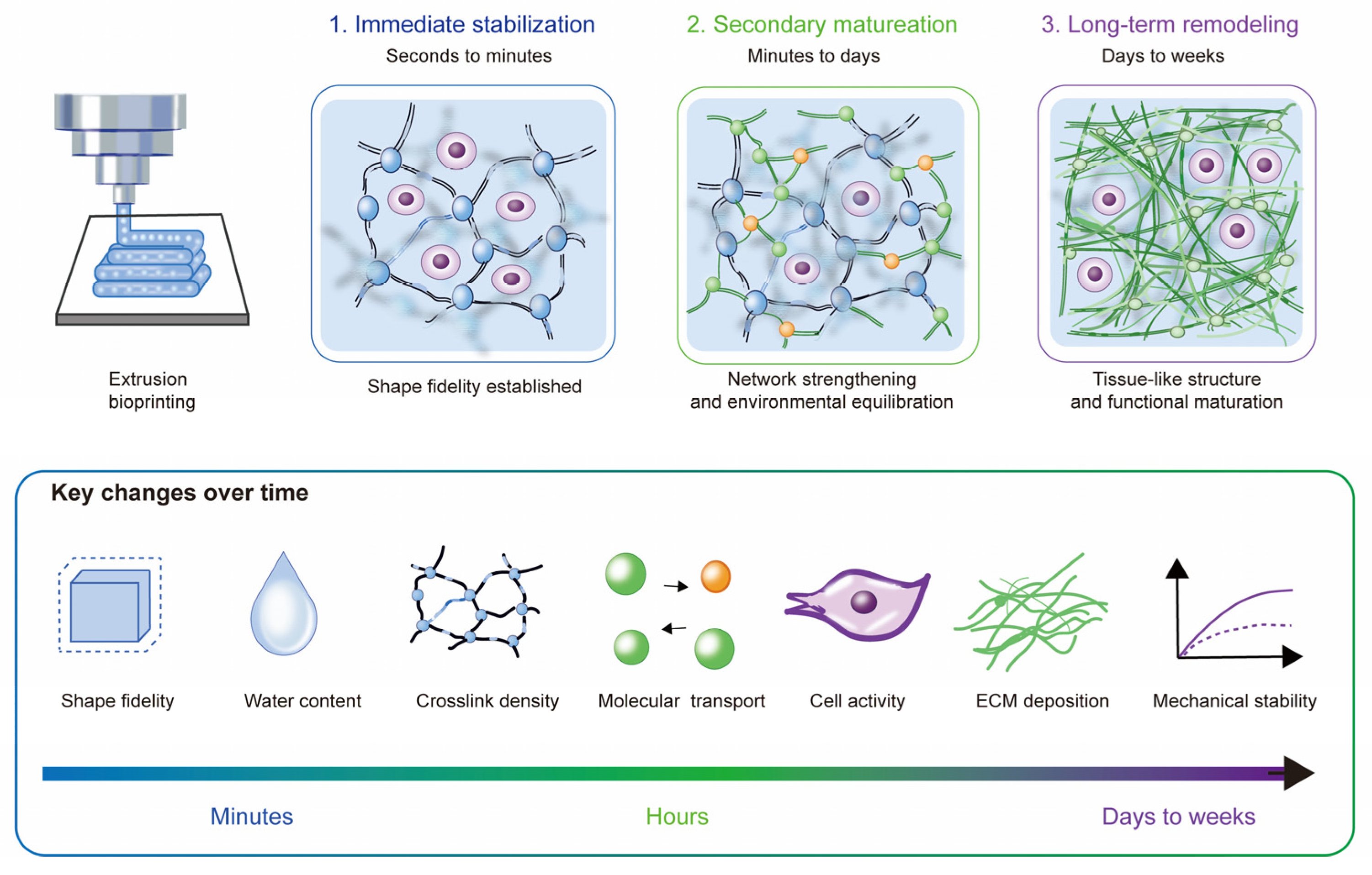
2. Temporal Stages of Post-Printing Maturation
2.1. Immediate Post-Deposition Stabilization
2.2. Secondary Network Formation and Environmental Equilibration
2.3. Long-Term Construct Remodeling
3. Failure Domains Arising from Unbalanced Post-Printing Maturation
3.1. Structural and Dimensional Failure
3.2. Mechanical and Interfacial Failure
3.2.1. Bulk Mechanical Failure
3.2.2. Interlayer or Multimaterial Interface Failure
3.3. Transport and Cell Distribution Failure
3.4. Biofunctional Failure
3.5. Chemical, Biochemical, and Microbiological Safety as a Cross-Cutting Gate
3.6. Coupled and Time-Dependent Failure Trajectories

| Failure Domain or Subdomain | Main Failure Signal | Targeted Response | Principal Trade-Off | Representative Evidence |
|---|---|---|---|---|
| Structural and dimensional | Dimensions, pores, channels, or shape no longer meet the specified requirement | Adjust network composition, crosslinking, ionic exposure, or resistance to cell-mediated contraction | Greater stabilization may restrict transport and tissue remodeling | [22,31,33] (mixed E1–E3) |
| Mechanical and interfacial: bulk mechanical | Loss of stiffness, recovery, creep resistance, fatigue resistance, or load-bearing capacity | Adjust network design, reinforcement, conditioning, or permitted loading | Greater stiffness may reduce stress relaxation, cell spreading, or matrix remodeling | [34,36] (E1) |
| Mechanical and interfacial: interface | Separation, cracking, sliding, or inadequate local load transfer | Adjust the curing sequence, introduce compatible interfacial chemistry, or add mechanical interlocking | Stronger bonding may increase local stiffness, chemical exposure, or stress concentration | [40,41,42] (E3; indirect) |
| Transport and cell distribution | Channel narrowing, increased flow resistance, inadequate oxygenation, regional cell loss, or uneven matrix distribution | Adjust channel geometry, construct thickness, swelling, degradation, or perfusion conditions | Greater porosity or perfusion may reduce mechanical integrity or increase flow-induced stress | [44,45] (mixed E1–E3) |
| Biofunctional | Intended tissue formation, phenotype, fusion, or bioactive delivery is not achieved | Match degradation to tissue formation and adjust cell density, biochemical induction, or bioactive delivery | Slower degradation, stronger reinforcement, or excessive retention may restrict remodeling, fusion, or effective dosing | [28,48,49] (E1) |
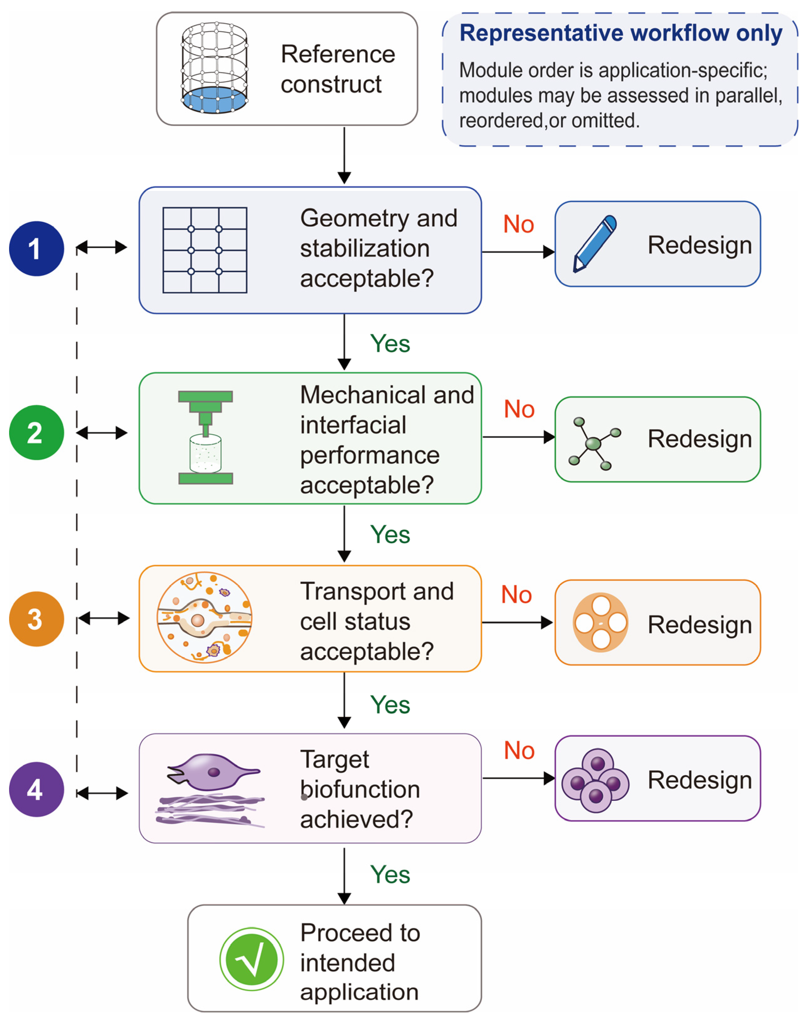
4. Use-Point Assessment with Sequential Use Points and Function-Specific Modules
4.1. Framework Structure
| Use Point | Main Question | Candidate Post-Printing Measurements | Action if Criteria Are Not Met | Representative Evidence |
|---|---|---|---|---|
| After stabilization and transfer | Did stabilization or handling cause a critical change from the reference condition? | Hydrated dimensions, pore or channel retention, handling integrity, interlayer continuity, and regional viability relative to the as-deposited and post-crosslinking reference states | Revise temporary support, crosslinking, environmental exposure, or handling | [16,17] (E1–E2) |
| During or after culture or conditioning | Does the construct remain structurally, mechanically, and biologically suitable during maturation? | Swelling, degradation, contraction, wet-state mechanics, interface integrity, channel retention, spatial viability, and tissue-specific matrix formation relative to the first equilibrated wet-state reference | Address the earliest deviation in Structural and dimensional, Mechanical and interfacial, Transport and cell distribution, or Biofunctional criteria | [28,32,45,48] (E1) |
| Before implantation or application | Does the construct meet its essential requirements after the complete pre-use pathway? | Geometry, wet integrity, load response when relevant, spatial cell status, intended biological function, retained bioactivity, and applicable safety-gate measurements after the complete pre-use pathway | Revise maturation, storage, transport, preparation, or final handling | [39,45,49] (mixed E1–E3) |
Operational Decision Rules
4.2. Application-Specific Use Points and Critical Requirements
| Construct Type or Application | Representative Use Point | Essential Requirements | Main Post-Printing Concern | Representative Evidence |
|---|---|---|---|---|
| Cartilage | End of a defined chondrogenic culture period or before downstream testing | Hydrated geometry, wet-state mechanical response, chondrocyte viability, phenotype maintenance, and cartilage matrix formation | Geometry or viability remains within its predefined limit, but phenotype or cartilage-matrix measures fall outside the predefined target range | [29] (E1) |
| Bone | End of a defined osteogenic culture or conditioning period | Mineral volume and density, construct stiffness, cell viability, osteogenic marker expression, and cellular organization | Mineral volume or density, mechanical response, or osteogenic marker panel remains outside the predefined target range at the use point | [54] (E1) |
| Full-thickness skin model | After the defined air–liquid interface maturation period and before functional testing | Layer organization, epidermal differentiation, ECM organization, barrier integrity, and hypodermis-related gene expression | Layer organization or barrier-function measures remain outside the predefined target range after air–liquid interface maturation | [55] (E1) |
| Perfusable thick construct | After flow initiation and during the defined perfusion period | Channel continuity, internal oxygenation, perfusion continuity, and regional cell viability | Channel geometry remains within tolerance but oxygen or perfusion metrics fall outside the predefined target range | [46] (E1) |
4.3. Illustrative Application to a Cartilage Construct
4.3.1. Early Post-Printing Assessment
4.3.2. Culture and Target Use-Point Assessment
4.3.3. Decision and Redesign
4.4. Validation Status and Scope
5. Outlook for Standardization, Prediction, and Translation
5.1. Standardized Longitudinal Characterization
5.2. Predictive Modeling of Maturation Trajectories
5.3. Translation-Oriented Assessment and Future Specification Strategies
5.4. Limitations of the Current Evidence and Proposed Framework
6. Conclusions
Author Contributions
Funding
Institutional Review Board Statement
Informed Consent Statement
Data Availability Statement
Acknowledgments
Conflicts of Interest
Appendix A
| Reference | Bioink/Crosslinking | Cell Model/Density | Printing/Architecture | Culture or Exposure/Time Points | Post-Printing Measurements | Observed Maturation or Failure Behavior | Evidence Class | Longitudinal/Same Construct | Replicates/Spatial Assessment |
|---|---|---|---|---|---|---|---|---|---|
| [1] | Gelatin–alginate; thermal/ionic | Acellular formulation/printability study | Extrusion; artifact/shape-fidelity geometries | Immediate post-print assessment | Rheology, extrusion, filament/shape fidelity | Defines a printability window; no direct biological maturation trajectory | E3 | No | NR/whole construct |
| [2] | Multiple hydrogel bioinks; formulation dependent | Acellular formulation/shape-fidelity study | Extrusion; standardized shape-fidelity artifacts | Immediate | Filament spreading, collapse, geometry | Quantifies early shape fidelity | E3 | No | NR/whole construct |
| [3] | GelMA-based biomaterial; photocrosslinking | NIH/3T3 fibroblasts; 5 × 10⁶ cells/mL | Direct-write extrusion; 3D tissue geometry | Days 1, 7, 14, and 21 | µCT filament/pore geometry, mechanical properties, degradation, Live/Dead viability | Printed constructs maintained 71–77% viability and consistent mechanical properties over 21 d while geometry and degradation were tracked | E1 | Yes/repeated groups at four time points | n = 3/whole-construct and spatial imaging |
| [4] | Photocurable collagen vs. GelMA; light crosslinking | Cell-laden; density NR | DLP and extrusion comparison | Up to 7 d | Printability, viability, spreading, proliferation, gene expression | Collagen and GelMA differ in printing and cell response | E1 | Yes/repeated groups | NR |
| [5] | Stress-relaxing hydrogels; covalent network variants | Stem cells; density NR | Bulk 3D hydrogel | Days | Stress relaxation, spreading, fate/activity | Relaxation regulates cell behavior independent of initial modulus | E4 | Yes/repeated groups | Replicates reported; bulk |
| [6] | Soft tissue bioinks; crosslinking system-specific | Cell-laden | Extrusion 3D scaffolds | 0–7 d | Nondestructive mechanics, swelling/material loss, cell proliferation | Whole-construct mechanics evolve during culture | E1 | Yes/same construct supported | Reported; whole construct |
| [7] | Gelatin-HA-genipin | High cell density; specific density NR here | Extrusion | Cell assessment on days 1 and 3; formulation disintegration over 48 h | Shape stability, degradation/disintegration, viability | Cell viability increased from day 1 to day 3; one formulation fully dissolved within 48 h at 37 °C while others partially disintegrated | E1 | Yes/repeated groups; disintegration tested separately | NR |
| [8] | Thiol-norbornene gelatin; photopolymerization | NHDF; 2.5 × 10⁶ cells/mL | Extrusion/photocuring | Days 1, 7, and 14 post-printing; proliferation to 21 d | Cure kinetics, mechanics, printability, post-print viability and proliferation | Rapid curing supported high post-print viability and longer-term cell spreading/proliferation; response was dose dependent | E1 | Yes/repeated groups | n = 3/spatial confocal imaging |
| [9] | GelMA; photoinitiator-dependent photocrosslinking | Acellular mechanistic network study | Bulk hydrogel | Seconds–minutes | Reaction kinetics, real-time modulus | Photoinitiator chemistry and oxygen influence network formation | E5 | Real-time repeated | NR |
| [10] | Gelatin-HA dynamic + photocrosslinked double network | BMSCs seeded onto a printed acellular scaffold; not cell-laden during extrusion | 3D extrusion | Days 1, 3, and 5 after seeding | Self-healing, printability, mechanics, post-seeding viability and proliferation | The double network supported recovery and printed stability; biological compatibility was evaluated after scaffold seeding | E3 | Repeated groups after seeding; not a cell-laden printing trajectory | NR |
| [11] | Bioink formulations; crosslinking varies | Acellular/printability focus | Extrusion artifact quantification | Immediate | Rheology and print artifact metrics | Links rheology with early print quality | E3 | No | NR |
| [12] | Hydrogel inks; formulation varies | Acellular/printability focus | Extrusion | Immediate | Optical geometry and modeling | Improves quantitative early quality assessment | E3 | No | Regional optical analysis |
| [13] | Embedded-printing inks/support bath | Cardiac fibroblasts; 2 × 10⁶ cells/mL in the validation experiment | Embedded extrusion | During deposition; cell viability at 24 h | Integrated microscopy, deposited area, positional accuracy, cell distribution and viability validation | In-process defects were quantified relative to design; cell-laden validation confirmed imaging and short-term viability compatibility | E2 | In-process repeated imaging; 24 h cell endpoint | Spatial image analysis |
| [14] | Gelatin–alginate | Breast cancer cells; density/formulations varied | Extrusion 3D hydrogel structures | Up to 11 d | Cell counts/proliferation + cellular automata model | Post-print cell proliferation depends on formulation and initial density | E1 + C | Yes/repeated time points | Reported; spatial/model outputs |
| [16] | GelMA; reversible physical + UV stabilization | Human chondrocytes; density NR | Extrusion cartilage-oriented constructs | Days 1, 7, and 28 | Printability, viability, proliferation, stabilization response | Physical pre-stabilization improves deposition before permanent curing | E1 | Yes/repeated groups | NR |
| [17] | Methylcellulose/GelMA; thermal support + photocrosslink | Human primary osteoblast-laden print used for biological validation; density NR | Extrusion | Cell viability at 48 h; acellular swelling/degradation up to 60 d | Shape integrity, swelling/degradation, printed-cell viability | Temporary methylcellulose support improved deposition, persistent GelMA stabilization maintained the structure, and a cell-laden printed lattice retained high short-term viability | E2 | Cell endpoint plus separate acellular time course | NR |
| [18] | Hyaluronan; enzymatic pre-gel + visible-light crosslink | hMSCs; 1–5 × 10⁶ cells/mL | Extrusion | 24 h and 14 d | Rheology, printability, reinforcement, printed-cell viability | Pre-gel supports extrusion; light exposure reinforces filament | E1 | Yes/repeated groups | NR |
| [19] | GelMA; photo + microbial transglutaminase dual crosslink | Acellular extrusion-printed grids; cell-laden micropatterns made by photolithography | Extrusion printing and photolithography | Post-print treatment; 1 d cell endpoint in micropatterns | Stiffness, swelling, geometry | Secondary crosslinking increases stiffness and geometric stability | E3 | Treatment comparison; no cell-laden extrusion trajectory | NR |
| [20] | HA single/double network; dynamic covalent + permanent network | Cell-laden; density NR | Extrusion | Immediate/short term | Recovery, mechanics, printability, viability | Dynamic bonds support extrusion; second network improves stability | E2 | No | NR |
| [21] | Dynamic hydrogel bioinks; small-molecule bond modulators | Cell-laden; density NR | Extrusion | Immediate/short term | Bond exchange, printability, mechanics, cell compatibility | Small molecules tune dynamic crosslinking and printed stability | E2 | No | NR |
| [22] | Alginate–gelatin; Ca2+ post-print treatment | Acellular | Extrusion scaffolds | Post-print swelling/degradation | Swelling, degradation, geometry | Post-print ionic treatment changes swelling and lifetime | E3 | Time dependent | NR |
| [23] | Alginate with alternative cationic pre-crosslinkers | Chondrocyte/cell-laden cartilaginous tissues; density NR | Extrusion cartilage constructs | Culture time points NR here | Mechanics, degradation, cell response, matrix accumulation | Cation choice alters post-print mechanics and tissue response | E1 | Yes | NR |
| [24] | HA-collagen stress-relaxing hydrogels | Cells in 3D bulk culture; density NR | Bulk hydrogel | Days | Cell spreading, fiber remodeling, focal adhesions | Faster relaxation supports remodeling | E4 | Yes | NR |
| [27] | Collagen inks; physical assembly vs. bioorthogonal covalent crosslink | Cell-laden | Embedded extrusion in microgel bath | Long-term culture | Microstructure, cell spreading, contraction/shape retention | Covalent crosslinking reduces cell-induced contraction | E1 | Yes | Spatial microstructure |
| [28] | Rapidly degrading oxidized-alginate-type bioinks | Cartilage microtissues | Extrusion/biofabrication | Long-term cartilage culture | Degradation, fusion, viability, GAG-rich matrix | Rapid degradation can support microtissue fusion and matrix formation | E1 | Yes | Reported; tissue distribution assessed |
| [29] | Gelatin–alginate; composition/stiffness varied | Cell-loaded cartilage constructs | Extrusion | Long-term culture | Morphology, mechanics, viability, cartilage markers | Optimized composition preserves shape while supporting collagen II | E1 | Yes | NR |
| [30] | GelMA–gelatin-hydroxyapatite | Osteoblast-laden | Extrusion composite hydrogel | Days 1, 7, 14, and 28 | Swelling/degradation, cell viability/proliferation, osteogenic differentiation and mineralization | Hydroxyapatite reduces swelling/degradation and promotes osteogenesis | E1 | Yes/repeated groups | n = 5 for reported culture/degradation measures/whole construct |
| [31] | GelMA–alginate; ionic + covalent crosslink | Sheep adipose-derived stem cells (sADSCs); density NR | Extrusion | Cell viability at 24 h and 7 d; acellular swelling/degradation to 14 d | Viability, proliferation, swelling, degradation, mechanics | Composition/crosslinking tune wet-state properties | E1 | Yes/repeated groups | NR |
| [32] | Bioprinted hydrogel under cell traction | Contractile cells; density NR | Bioprinted construct with force measurement | Culture over time | Nondestructive macroscopic tension/contractility | Cell-generated contraction can be tracked longitudinally | E1 | Yes/same construct compatible | Whole-construct force |
| [33] | Shrink-resistant collagen-HA composite | Cell-laden | 3D bioprinting | Long-term culture | Shrinkage, geometry, cell response | Network design improves dimensional retention against cell contraction | E1 | Yes | Spatial/whole construct |
| [34] | Self-healing tough hydrogel bioink | Cell-laden | Direct extrusion | Days 1, 3, 7, and 14 | Toughness, cyclic/fatigue resistance, cell compatibility | Self-healing network improves antifatigue performance | E1 | Yes/repeated groups | Replicates NR |
| [35] | Elastin-containing bioactive double network | Cell-laden | Extrusion | Days 1 and 7 in vitro; additional regeneration endpoints | Mechanical reinforcement, tissue regeneration outcomes | Double network supports tough elastic constructs | E1 | Yes/repeated groups | NR |
| [36] | Composite hydrogel + human bone allograft particles | hMSCs; approximately 3 × 10⁶ cells/mL | Extrusion | Days 1, 7, 14, 21, and 28 | Stiffness, swelling, viability, migration, ALP and osteogenic markers | Allograft particles increase stiffness and support osteogenesis | E1 | Yes/repeated groups | NR |
| [40] | Dual-network hydrogel + supramolecular gelator | Acellular | 3D extrusion; layer interfaces | Post-processing curing | Interlayer adhesion/tensile performance | Simultaneous curing improves continuity vs. layerwise curing | E3 | No | Interface-level tests |
| [41] | Hydrogel-elastomer chemically coupled interface | Acellular | Multimaterial printing | Post-print mechanical tests | Interfacial adhesion | Chemical coupling strengthens soft-material interfaces | E3 | No | Local interface |
| [42] | Hydrogel-PLA mechanically interlocked interface | Acellular | 3D-printed soft–hard interface | Mechanical testing | Interfacial strength, toughness, failure location | Mechanical interlocking improves strength; failure may remain in hydrogel | E3 | No | Local interface |
| [44] | Perfused hydrogel vascular model | Acellular printed hydrogel subsequently endothelialized with HUVECs | 3D-printed perfusable hydrogel channel | Post-print endothelialization and perfusion exposure | Permeability, endothelial barrier, leakage | Printed channel geometry enabled perfusion, while endothelial barrier function required direct post-seeding measurement | E3 | Repeated perfusion measurements; not cell-laden extrusion | Regional/channel level |
| [45] | Gelatin–alginate–fibrinogen | Fibroblast-laden; centimeter scale | Extrusion + bioreactor | 0–16 d | pH, O2, lactate/Raman, MRI, morphology, perfusion | Pore occlusion, deformation and localized degradation develop during culture | E1 | Yes/same construct imaging | Multimodal regional monitoring |
| [48] | Alginate-based bioinks with tuned degradation | Cartilage cells/microtissues; density NR | Extrusion | Long-term cartilage culture | Degradation, GAG/tissue formation, fusion | Degradation rate must match tissue formation | E1 | Yes | NR |
| [49] | Proteinaceous scaffold with TGF-β1 + FGF-18 delivery | Cell-containing cartilage construct | 3D bioprinting | Cell-laden culture to 21 d; in vivo regeneration endpoints | Growth-factor release, chondrogenesis, cartilage repair | Controlled dual-factor delivery promotes chondrogenesis/regeneration | E1 | Yes/repeated groups and in vivo endpoint | NR |
| [39] | Suspended bioprinting hydrogel | Acellular suspended-printing evidence | Suspended bioprinting | In situ during/after print | Ultrasound shear-wave elasticity | Nondestructive elasticity monitoring is feasible | E3 | Yes/same construct compatible | Whole/local elasticity mapping |
| [54] | Bone-derived cell-laden hydrogel scaffold | Human bone-derived cells; physiological densities compared | Extrusion | Dynamic loading culture | Mineralization rate, stiffness, cell organization | Cell density influences matrix mineralization and stiffness maturation | E1 | Yes | Reported; spatial organization |
| [55] | Full-thickness skin bioink system | Skin cell populations including hypodermal component; density NR | Bioprinted multilayer skin model | Days 5 and 10 of skin differentiation | Layer organization, gene expression, differentiation/barrier-related outcomes | Hypodermis alters maturation-related gene expression | E1 | Yes/repeated groups | Spatial layers |
| [46] | Cell-laden thick constructs with perfusable microchannels | Cell-laden; density NR | 3D-bioprinted thick channel network | Perfusion/culture | Internal oxygen levels, channel network | Direct O2 measurements reveal transport state inside thick constructs | E1 | Yes | Spatial oxygen measurements |
| [56] | Alginate-based in situ crosslinking cartilage system | Primary juvenile bovine MSCs; 20 × 106 cells/mL | Extrusion with in situ crosslinking | Days 0, 3, and 7 for viability/distribution; chondrogenic culture to 56 d | Viability, spatial cell distribution, mechanics/degradation, GAG, collagen and gene expression | In situ crosslinking maintained viability and spatial distribution and supported long-term chondrogenic maturation | E1 | Yes/repeated groups | n ≥ 3/top, middle and bottom spatial assessment |
| [57] | Methacrylated guar gum; photocrosslinking | Bone-marrow MSC-laden; density NR | Extrusion cartilage constructs | Long-term culture | Mass loss, structural stability, GAG, COL2A1 | Higher methacrylation reduces mass loss and supports chondrogenic output | E1 | Yes | NR |
| [58] | Methacrylated flaxseed gum; photocrosslinking | Stem cell-laden; density NR | Extrusion | Up to ~66 d degradation/cartilage formation | Integrity, degradation, chondrogenesis | Material persistence can be matched to cartilage maturation | E1 | Yes | NR |
| [59] | Rheology-modified hydrogel formulation library | Acellular formulation data | Printability/process study | Immediate | Rheological descriptors + machine learning | Rheology features predict printability, not post-print maturation | E3 + C | No | Dataset-level |
| [60] | Biomaterial formulation library | Acellular/process data | Direct ink writing/printability | Immediate | Formulation descriptors + machine learning | Predicts printable formulations, not maturation | E3 + C | No | Dataset-level |
| [61] | Hybrid hydrogel bioink; formulation-dependent | Acellular/process characterization focus | Extrusion-based printing | Immediate | Rheology, printability, ML property prediction | Predicts precursor/printing properties rather than maturation | E3 + C | No | Dataset-level |
| [62] | Cell-laden extrusion construct model | Cell viability/proliferation experimental validation | Extrusion-based constructs | Tissue maturation time course | Nutrient diffusion/consumption, viability, proliferation | Model couples transport with cell growth and matches experimental viability change | E1 + C | Yes | Model + experimental validation |
| [63] | Bioprinted bone contraction model | Cellular traction represented computationally | Bioprinted bone geometry | Post-processing | Contraction, geometry, mechanics, gravity effects | Nonlocal traction predicts deformation and sensitivity to construct properties | C | Modeled time course | Computational |
| [50] | Alginate–gelatin; thermal + CaCl2 crosslinking | MS5 stromal cells; 4 × 10⁵ cells/mL | Extrusion; structures up to 10.3 ± 1.4 mm | 0, 3 h, 24 h, 72 h, 7 d | Live/dead, ATP, morphology; printed vs. unprinted controls | Live/dead and metabolic activity are complementary and may diverge | E1 | Yes; repeated time points | N = 3–4+ depending formulation; whole/sample imaging |
| [52] | GelMA; type I/II photoinitiators | MSC-encapsulated; density NR | Cell-laden hydrogel; light-assisted bioprinting relevance | Up to 1 week | Elastic modulus, ROS, pore size, viability | Higher PI can increase modulus while increasing ROS and reducing viability | E4 | Yes | Replicates reported; bulk/cell-laden |
| [53] | Gelatin/GelMA materials with different endotoxin burden | Macrophage-cancer cell 3D model; density NR | Three-dimensional printed in vitro tumor model | Days 1, 4, and 7 post-bioprinting; therapy exposure | Inflammatory response, cell interaction, therapeutic response | Endotoxin changes immune behavior and apparent treatment effect | E1 | Yes/repeated groups | Three-dimensional model; regional/cellular readouts |
| [15] | Alginate, collagen, and Pluronic inks; formulation-dependent extrusion behavior | Acellular process-control study | Extrusion bioprinting with integrated camera, convolutional neural-network classification, and closed-loop correction; single-line and infill patterns | Real-time during printing; extrusion errors corrected within approximately 10 s | Image-based classification of good, over-, and under-extrusion; real-time monitoring; automated parameter correction | Closed-loop control detected and corrected extrusion errors across inks with different rheological properties, supporting process-state quality control rather than post-printing maturation assessment | E3 | Real-time process monitoring; not a post-printing longitudinal study | Multiple real and synthetic training/testing datasets; process-level image assessment |
| [43] | PVA/alginate/gellan-gum sensor hydrogel with conductive fillers; cell-laden Matrigel/collagen bioink | C2C12 myoblasts; 1 × 10⁶ cells/mL | Multimaterial extrusion bioprinting; centimeter-scale biphasic sensorized muscle construct | Immediately after printing and after 10 d tissue development | Spatial Live/Dead, interface imaging/histology, shape retention, differentiation, mechanoelectrical response | Bioprinted constructs maintained high post-printing viability and a coherent sensor–tissue interface through 10 d of maturation | E1 | Yes; interface followed through 10 d | Three experimental replicates with technical replicates for viability; n = 3 for mechanical/sensing tests |
| [47] | GelMA/gelatin colloidal bioink with oxygen-generating CaO2/MnO2-containing microparticles | Human dermal fibroblasts and hMSCs; density NR | Extrusion-based cell-laden printing; multilayer and tissue-mimetic constructs | Oxygen release to 10 d; cell assessment to 7 d; HIF1-α at 18 h | Oxygen/H2O2 release, rheology, printability, mechanics, degradation, Live/Dead, HIF1-α | Oxygen availability was directly tunable and measurable while maintaining extrusion printability and cytocompatibility | E1 | Yes/repeated groups for oxygen release and biological endpoints | Mostly n = 3; whole-construct and local imaging reported |
| [51] | Methacrylated collagen peptide with short collagen-I microfibers and xanthan gum; UV curing | Human MSCs; density NR | Cell-laden bioprinted fibrous collagen-derived constructs | Culture and differentiation to 28 d | Cell spreading, metabolic activity, extracellular matrix, COL-I, scleraxis, differentiation | Fibrous constructs supported increasing metabolic activity, matrix production, and lineage-related protein expression during 28 d | E1 | Yes; maturation assessed over 28 d | Replicate number NR in abstract; cellular and matrix readouts reported |
| [25] | GelMA/PEO shear-oriented bioink; UV photocrosslinking; carrageenan support bath | C2C12, HUVECs, BMSCs, and MC-3T3 cells; densities NR here | Embedded extrusion; freeform anisotropic, vascular, and muscle-patch structures | Day 1 viability; cell morphology/spreading after 3 and 7 d | Filament geometry, anisotropy, Live/Dead, cytoskeletal orientation, immunofluorescence | Extrusion-induced alignment enabled viable anisotropic constructs and guided cell spreading/orientation | E1 | Repeated groups at 1, 3, and 7 d/same-construct tracking NR | Replicates NR here/spatial fluorescence imaging |
| [26] | Dilute/concentrated PEGDA streams with PAA microgels; photopolymerization | Acellular material-processing study | Millifluidic layer multiplication followed by filament 3D printing | Immediate post-fabrication characterization and programmed actuation | Rheology, internal architecture, swelling, mechanical contrast, and shape transformation | Hierarchically patterned crosslink density produced seamless filaments with programmable mechanical contrast | E3 | No longitudinal biological study | Replicates NR here/multiscale structural assessment |
| [37] | TMSC-laden collagen bioink reinforced with chiral or chevron PCL patterns | Tonsil-derived mesenchymal stem cells; density NR here | Multimaterial 3D printing of hybrid biopatches; porcine mucosal-defect application | Cell assessment on days 1 and 5; mechanical/fatigue testing; in vivo endpoint | Live/Dead, proliferation, contraction, tensile/fatigue behavior, histology, epithelial markers | Pattern geometry controlled anisotropy and reinforcement while maintaining high viability; chiral patches improved mucosal repair | E1 | Repeated in vitro time points and an in vivo endpoint/same-construct tracking NR | n = 4 reported for experiments/whole-patch and histological spatial assessment |
| [38] | Salting-out-tuned photocrosslinkable hydrogel ink | Acellular; synthetic vessel training model | Direct ink writing of multilayer hydrogel structures and synthetic blood vessels | Immediate/post-cure mechanical and surgical-training assessment | Rheology, print width, modulus, tensile/hoop response, leakage, electrocautery, and anastomosis | Salting-out enabled printable hydrogels spanning 0.193–1.072 MPa and mechanically tunable vessel models | E3 | No biological maturation trajectory | n = 150 print-width observations per ink; other replicates as reported/whole construct |
| Process Class | Representative Dominant Time Scale | Key Rate-Controlling Factors | Representative Evidence |
|---|---|---|---|
| Flow recovery and thermoreversible stabilization | Seconds to minutes | Shear recovery, temperature, polymer concentration, reversible physical interactions | [16,17] |
| Ionic or photochemical stabilization | Seconds to hours, with later equilibration possible | Ion diffusion, light dose, photoinitiator chemistry, oxygen, optical path length, construct thickness | [8,9,22,23,52] |
| Enzymatic or dynamic-covalent network evolution | Minutes to days | Enzyme activity, functional-group concentration, bond-exchange kinetics, temperature and medium | [18,19,20,21] |
| Hydration, ion exchange and soluble-fraction equilibration | Minutes to days | Medium composition, ionic strength, geometry, diffusion distance and network density | [22,23,31] |
| Degradation, stress relaxation, cell-driven contraction and ECM deposition | Hours to weeks or longer | Network lability, cell density/activity, tissue formation, loading and culture history | [5,24,27,28,29,30,31,32,33] |
| Database | Complete Search String |
|---|---|
| PubMed | ((bioprint* [Title/Abstract]) OR (“extrusion bioprinting” [Title/Abstract]) OR (“embedded bioprinting” [Title/Abstract]) OR (“coaxial bioprinting” [Title/Abstract])) AND ((bioink* [Title/Abstract]) OR (“cell-laden hydrogel” [Title/Abstract]) OR (“cell laden hydrogel” [Title/Abstract])) AND ((“post-printing” [Title/Abstract]) OR maturation [Title/Abstract] OR culture [Title/Abstract] OR crosslink* [Title/Abstract] OR swelling [Title/Abstract] OR degradation [Title/Abstract] OR contraction [Title/Abstract] OR “stress relaxation” [Title/Abstract] OR mechanic* [Title/Abstract] OR interface* [Title/Abstract] OR transport [Title/Abstract] OR perfusion [Title/Abstract] OR oxygen* [Title/Abstract] OR viability [Title/Abstract] OR phenotype [Title/Abstract] OR differentiation [Title/Abstract] OR matrix [Title/Abstract]) AND (“2016/01/01” [Date—Publication]: “2026/08/09” [Date—Publication]) AND English [Language] |
| Web of Science Core Collection | TS = (bioprint* OR “extrusion bioprinting” OR “embedded bioprinting” OR “coaxial bioprinting”) AND TS = (bioink* OR “cell-laden hydrogel*” OR “cell laden hydrogel*”) AND TS = (“post-print*” OR maturation OR culture OR crosslink* OR swelling OR degradation OR contraction OR “stress relaxation” OR mechanic* OR interface* OR transport OR perfusion OR oxygen* OR viability OR phenotype OR differentiation OR matrix) AND PY = (2016–2026) AND LA = (English) |
| Scopus | TITLE-ABS-KEY (bioprint* OR “extrusion bioprinting” OR “embedded bioprinting” OR “coaxial bioprinting”) AND TITLE-ABS-KEY (bioink* OR “cell-laden hydrogel*” OR “cell laden hydrogel*”) AND TITLE-ABS-KEY (“post-print*” OR maturation OR culture OR crosslink* OR swelling OR degradation OR contraction OR “stress relaxation” OR mechanic* OR interface* OR transport OR perfusion OR oxygen* OR viability OR phenotype OR differentiation OR matrix) AND PUBYEAR > 2015 AND PUBYEAR < 2027 AND (LIMIT-TO (LANGUAGE, “English”)) |
References
- Gao, T.; Gillispie, G.J.; Copus, J.S.; Pr, A.K.; Seol, Y.J.; Atala, A.; Yoo, J.J.; Lee, S.J. Optimization of gelatin-alginate composite bioink printability using rheological parameters: A systematic approach. Biofabrication 2018, 10, 034106. [Google Scholar] [CrossRef] [Scilit] [PubMed]
- Ribeiro, A.; Blokzijl, M.M.; Levato, R.; Visser, C.W.; Castilho, M.; Hennink, W.E.; Vermonden, T.; Malda, J. Assessing bioink shape fidelity to aid material development in 3D bioprinting. Biofabrication 2017, 10, 014102. [Google Scholar] [CrossRef] [Scilit] [PubMed]
- Adib, A.A.; Sheikhi, A.; Shahhosseini, M.; Simeunovic, A.; Wu, S.; Castro, C.E.; Zhao, R.; Khademhosseini, A.; Hoelzle, D.J. Direct-write 3D printing and characterization of a GelMA-based biomaterial for intracorporeal tissue engineering. Biofabrication 2020, 12, 045006. [Google Scholar] [CrossRef] [Scilit] [PubMed]
- Shi, H.; Li, Y.; Xu, K.; Yin, J. Advantages of photo-curable collagen-based cell-laden bioinks compared to methacrylated gelatin (GelMA) in digital light processing (DLP) and extrusion bioprinting. Mater. Today Bio 2023, 23, 100799. [Google Scholar] [CrossRef] [Scilit] [PubMed]
- Chaudhuri, O.; Gu, L.; Klumpers, D.; Darnell, M.; Bencherif, S.A.; Weaver, J.C.; Huebsch, N.; Lee, H.P.; Lippens, E.; Duda, G.N.; et al. Hydrogels with tunable stress relaxation regulate stem cell fate and activity. Nat. Mater. 2016, 15, 326–334. [Google Scholar] [CrossRef] [Scilit] [PubMed]
- Godau, B.; Stefanek, E.; Gharaie, S.S.; Amereh, M.; Pagan, E.; Marvdashti, Z.; Libert-Scott, E.; Ahadian, S.; Akbari, M. Non-destructive mechanical assessment for optimization of 3D bioprinted soft tissue scaffolds. iScience 2022, 25, 104251. [Google Scholar] [CrossRef] [Scilit] [PubMed]
- Khatun, M.R.; Bhattacharyya, A.; Gunbayar, M.; Jung, M.; Noh, I. Study on Bioresponsive Gelatin-Hyaluronic Acid-Genipin Hydrogel for High Cell-Density 3D Bioprinting. Gels 2023, 9, 601. [Google Scholar] [CrossRef] [Scilit] [PubMed]
- Gockler, T.; Haase, S.; Kempter, X.; Pfister, R.; Maciel, B.R.; Grimm, A.; Molitor, T.; Willenbacher, N.; Schepers, U. Tuning Superfast Curing Thiol-Norbornene-Functionalized Gelatin Hydrogels for 3D Bioprinting. Adv. Healthc. Mater. 2021, 10, e2100206. [Google Scholar] [CrossRef] [Scilit] [PubMed]
- Duymaz, D.; Karaoglu, I.C.; Kizilel, S. Effect of Photoinitiation Process on Photo-Crosslinking of Gelatin Methacryloyl Hydrogel Networks. Macromol. Rapid Commun. 2025, 46, e00376. [Google Scholar] [CrossRef] [Scilit] [PubMed]
- Wang, Y.; Chen, Y.; Zheng, J.; Liu, L.; Zhang, Q. Three-Dimensional Printing Self-Healing Dynamic/Photocrosslinking Gelatin-Hyaluronic Acid Double-Network Hydrogel for Tissue Engineering. ACS Omega 2022, 7, 12076–12088. [Google Scholar] [CrossRef] [Scilit] [PubMed]
- Gillispie, G.J.; Copus, J.; Uzun-Per, M.; Yoo, J.J.; Atala, A.; Niazi, M.K.K.; Lee, S.J. The correlation between rheological properties and extrusion-based printability in bioink artifact quantification. Mater. Des. 2023, 233, 112237. [Google Scholar] [CrossRef] [Scilit] [PubMed]
- Lamberger, Z.; Schubert, D.W.; Buechner, M.; Cabezas, N.C.; Schrufer, S.; Murenu, N.; Schaefer, N.; Lang, G. Advanced optical assessment and modeling of extrusion bioprinting. Sci. Rep. 2024, 14, 13972. [Google Scholar] [CrossRef] [Scilit] [PubMed]
- Sergis, V.; Kelly, D.; Pramanick, A.; Britchfield, G.; Mason, K.; Daly, A.C. In-situ quality monitoring during embedded bioprinting using integrated microscopy and classical computer vision. Biofabrication 2025, 17, 025004. [Google Scholar] [CrossRef] [Scilit] [PubMed]
- Mohammadrezaei, D.; Moghimi, N.; Vandvajdi, S.; Powathil, G.; Hamis, S.; Kohandel, M. Predicting and elucidating the post-printing behavior of 3D printed cancer cells in hydrogel structures by integrating in-vitro and in-silico experiments. Sci. Rep. 2023, 13, 1211. [Google Scholar] [CrossRef] [Scilit] [PubMed]
- Kelly, D.; Sergis, V.; Blanco, L.; Mason, K.; Daly, A.C. Autonomous Control of Extrusion Bioprinting Using Convolutional Neural Networks. Adv. Funct. Mater. 2025, 35, 2424553. [Google Scholar] [CrossRef] [Scilit]
- Gu, Y.; Zhang, L.; Du, X.; Fan, Z.; Wang, L.; Sun, W.; Cheng, Y.; Zhu, Y.; Chen, C. Reversible physical crosslinking strategy with optimal temperature for 3D bioprinting of human chondrocyte-laden gelatin methacryloyl bioink. J. Biomater. Appl. 2018, 33, 609–618. [Google Scholar] [CrossRef] [Scilit] [PubMed]
- Rastin, H.; Ormsby, R.T.; Atkins, G.J.; Losic, D. 3D Bioprinting of Methylcellulose/Gelatin-Methacryloyl (MC/GelMA) Bioink with High Shape Integrity. ACS Appl. Bio Mater. 2020, 3, 1815–1826. [Google Scholar] [CrossRef] [Scilit] [PubMed]
- Petta, D.; Armiento, A.R.; Grijpma, D.; Alini, M.; Eglin, D.; D’Este, M. 3D bioprinting of a hyaluronan bioink through enzymatic-and visible light-crosslinking. Biofabrication 2018, 10, 044104. [Google Scholar] [CrossRef] [Scilit] [PubMed]
- Basara, G.; Yue, X.; Zorlutuna, P. Dual Crosslinked Gelatin Methacryloyl Hydrogels for Photolithography and 3D Printing. Gels 2019, 5, 34. [Google Scholar] [CrossRef] [Scilit] [PubMed]
- Wang, L.L.; Highley, C.B.; Yeh, Y.C.; Galarraga, J.H.; Uman, S.; Burdick, J.A. Three-dimensional extrusion bioprinting of single- and double-network hydrogels containing dynamic covalent crosslinks. J. Biomed. Mater. Res. Part A 2018, 106, 865–875. [Google Scholar] [CrossRef] [Scilit] [PubMed]
- Hull, S.M.; Lou, J.; Lindsay, C.D.; Navarro, R.S.; Cai, B.; Brunel, L.G.; Westerfield, A.D.; Xia, Y.; Heilshorn, S.C. 3D bioprinting of dynamic hydrogel bioinks enabled by small molecule modulators. Sci. Adv. 2023, 9, eade7880. [Google Scholar] [CrossRef] [Scilit] [PubMed]
- Kaliampakou, C.; Lagopati, N.; Pavlatou, E.A.; Charitidis, C.A. Alginate-Gelatin Hydrogel Scaffolds; An Optimization of Post-Printing Treatment for Enhanced Degradation and Swelling Behavior. Gels 2023, 9, 857. [Google Scholar] [CrossRef] [Scilit] [PubMed]
- Steiner, O.; St-Pierre, J.-P. Improving alginate printability with alternative cationic pre-crosslinkers: Strontium-based bioinks and their impact on bioprinted cartilaginous tissues. Bioprinting 2026, 55, e00468. [Google Scholar] [CrossRef] [Scilit]
- Lou, J.; Stowers, R.; Nam, S.; Xia, Y.; Chaudhuri, O. Stress relaxing hyaluronic acid-collagen hydrogels promote cell spreading, fiber remodeling, and focal adhesion formation in 3D cell culture. Biomaterials 2018, 154, 213–222. [Google Scholar] [CrossRef] [Scilit] [PubMed]
- Shao, L.; Jiang, J.; Yuan, C.; Zhang, X.; Gu, L.; Wang, X. Omnidirectional anisotropic embedded 3D bioprinting. Mater. Today Bio 2024, 27, 101160. [Google Scholar] [CrossRef] [Scilit] [PubMed]
- Bayles, A.V.; Pleij, T.; Hofmann, M.; Hauf, F.; Tervoort, T.; Vermant, J. Structuring Hydrogel Cross-Link Density Using Hierarchical Filament 3D Printing. ACS Appl. Mater. Interfaces 2022, 14, 15667–15677. [Google Scholar] [CrossRef] [Scilit] [PubMed]
- Brunel, L.G.; Christakopoulos, F.; Kilian, D.; Cai, B.; Hull, S.M.; Myung, D.; Heilshorn, S.C. Embedded 3D Bioprinting of Collagen Inks into Microgel Baths to Control Hydrogel Microstructure and Cell Spreading. Adv. Healthc. Mater. 2024, 13, e2303325. [Google Scholar] [CrossRef] [Scilit] [PubMed]
- Kronemberger, G.S.; Spagnuolo, F.D.; Karam, A.S.; Chattahy, K.; Storey, K.J.; Kelly, D.J. Rapidly Degrading Hydrogels to Support Biofabrication and 3D Bioprinting Using Cartilage Microtissues. ACS Biomater. Sci. Eng. 2024, 10, 6441–6450. [Google Scholar] [CrossRef] [Scilit] [PubMed]
- Liu, G.; Wei, X.W.; Li, J.L.; Zhai, Y.; Zhang, J.R.; Jin, M.; Guan, T.M.; Zhao, D.W. Hydrogel composition and mechanical stiffness of 3D bioprinted cell-loaded scaffolds promote cartilage regeneration. Front. Mater. 2024, 11, 1501505. [Google Scholar] [CrossRef] [Scilit]
- Allen, N.B.; Abar, B.; Johnson, L.; Burbano, J.; Danilkowicz, R.M.; Adams, S.B. 3D-bioprinted GelMA-gelatin-hydroxyapatite osteoblast-laden composite hydrogels for bone tissue engineering. Bioprinting 2022, 26, e00196. [Google Scholar] [CrossRef] [Scilit]
- Aldana, A.A.; Valente, F.; Dilley, R.; Doyle, B. Development of 3D bioprinted GelMA-alginate hydrogels with tunable mechanical properties. Bioprinting 2021, 21, e00105. [Google Scholar] [CrossRef] [Scilit]
- Pragnere, S.; El Kholti, N.; Gudimard, L.; Essayan, L.; Marquette, C.; Petiot, E.; Pailler-Mattei, C. Quantification of cell contractile behavior based on non-destructive macroscopic measurement of tension forces on bioprinted hydrogel. J. Mech. Behav. Biomed. Mater. 2022, 134, 105365. [Google Scholar] [CrossRef] [Scilit] [PubMed]
- Roshanbinfar, K.; Evans, A.D.; Samanta, S.; Kolesnik-Gray, M.; Fiedler, M.; Krstic, V.; Engel, F.B.; Oommen, O.P. Enhancing biofabrication: Shrink-resistant collagen-hyaluronan composite hydrogel for tissue engineering and 3D bioprinting applications. Biomaterials 2025, 318, 123174. [Google Scholar] [CrossRef] [Scilit] [PubMed]
- Liu, Q.; Yang, J.; Wang, Y.; Wu, T.; Liang, Y.; Deng, K.; Luan, G.; Chen, Y.; Huang, Z.; Yue, K. Direct 3D Bioprinting of Tough and Antifatigue Cell-Laden Constructs Enabled by a Self-Healing Hydrogel Bioink. Biomacromolecules 2023, 24, 2549–2562. [Google Scholar] [CrossRef] [Scilit] [PubMed]
- Wang, D.; Zeng, J.S.; Zhu, H.L.; Liu, S.Y.; Jia, L.T.; Liu, W.S.; Wang, Q.; Wang, S.M.; Liu, W.; Zhou, J.Y.; et al. Extrusion bioprinting of elastin-containing bioactive double-network tough hydrogels for complex elastic tissue regeneration. Aggregate 2024, 5, e477. [Google Scholar] [CrossRef] [Scilit]
- Gharacheh, H.; Guvendiren, M. Cell-Laden Composite Hydrogel Bioinks with Human Bone Allograft Particles to Enhance Stem Cell Osteogenesis. Polymers 2022, 14, 3788. [Google Scholar] [CrossRef] [Scilit] [PubMed]
- Ahn, M.; Park, G.T.; Shukla, A.K.; Kwon, B.; Kim, J.H.; Sung, E.S.; Kim, B.S. 3D Bioprinting-Assisted Engineering of Stem Cell-Laden Hybrid Biopatches with Distinct Geometric Patterns Considering the Mechanical Characteristics of Regular and Irregular Connective Tissues. Adv. Healthc. Mater. 2025, 14, e2502763. [Google Scholar] [CrossRef] [Scilit] [PubMed]
- Ko, K.W.; Lee, T.; Jeong, J.H.; Lee, Y.; Kim, I.; Seo, J.; Park, Y.H.; Kim, J.O.; Park, S. Hydrogel Ink for 3D Printing with High and Widely Tunable Mechanical Properties via the Salting-Out Effect. Adv. Mater. Technol. 2024, 9, 2400157. [Google Scholar] [CrossRef] [Scilit]
- Kim, G.; Oh, D.; Kim, D.; Lee, G.; Park, S.H.; Yoon, C.; Nam, S.Y. Suspended bioprinting with in-situ elasticity monitoring using the assessment of shear wave phase velocity. Addit. Manuf. 2025, 99, 104667. [Google Scholar] [CrossRef] [Scilit]
- Zhou, Z.X.; Samperi, M.; Santu, L.; Dizon, G.; Aboarkaba, S.; Limón, D.; Tuck, C.; Pérez-García, L.; Irvine, D.J.; Amabilino, D.B.; et al. An imidazolium-based supramolecular gelator enhancing interlayer adhesion in 3D printed dual network hydrogels. Mater. Des. 2021, 206, 109792. [Google Scholar] [CrossRef] [Scilit]
- Tian, K.; Suo, Z.; Vlassak, J.J. Chemically Coupled Interfacial Adhesion in Multimaterial Printing of Hydrogels and Elastomers. ACS Appl. Mater. Interfaces 2020, 12, 31002–31009. [Google Scholar] [CrossRef] [Scilit] [PubMed]
- Kunkels, L.B.; Saldívar, M.C.; Putra, N.E.; Kruize, C.P.; Panahkhahi, S.; Leeflang, M.A.; Fratila-Apachitei, L.E.; Zadpoor, A.A.; Mirzaali, M.J. High-Performance 3D Printed Mechanically Interlocked Soft-Hard Interfaces of Hydrogels and Polylactide. Adv. Mater. Technol. 2025, 10, 2401081. [Google Scholar] [CrossRef] [Scilit]
- Georgopoulou, A.; Filippi, M.; Stefani, L.; Drescher, F.; Balciunaite, A.; Scherberich, A.; Katzschmann, R.; Clemens, F. Bioprinting of Stable Bionic Interfaces Using Piezoresistive Hydrogel Organoelectronics. Adv. Healthc. Mater. 2024, 13, e2400051. [Google Scholar] [CrossRef] [Scilit] [PubMed]
- Royse, M.K.; Means, A.K.; Calderon, G.A.; Kinstlinger, I.S.; He, Y.; Durante, M.R.; Procopio, A.T.; Veiseh, O.; Xu, J. A 3D printable perfused hydrogel vascular model to assay ultrasound-induced permeability. Biomater. Sci. 2022, 10, 3158–3173. [Google Scholar] [CrossRef] [Scilit] [PubMed]
- Chastagnier, L.; Pragnere, S.; Gimenez, Y.; Loubiere, C.; Essayan, L.; Mazarakis, K.; Schmidberger, T.; Olmos, E.; Lambert, S.A.; Marquette, C.A.; et al. Integration of multi-modal monitoring for dynamic control of large-scale 3D tissue bioreactors. Sci. Rep. 2026, 16, 903. [Google Scholar] [CrossRef] [Scilit] [PubMed]
- Figueiredo, L.; Le Visage, C.; Weiss, P.; Yang, J. Quantifying Oxygen Levels in 3D Bioprinted Cell-Laden Thick Constructs with Perfusable Microchannel Networks. Polymers 2020, 12, 1260. [Google Scholar] [CrossRef] [Scilit] [PubMed]
- Jeong, S.H.; Hiemstra, J.; Blokzijl, P.V.; Damian-Ferrara, R.; Dos Santos, D.M.; da Fonseca, J.H.L.; Kang, M.H.; Kim, J.; Yilmaz-Aykut, D.; Cham-Perez, M.L.L.; et al. An oxygenating colloidal bioink for the engineering of biomimetic tissue constructs. Bio-Des. Manuf. 2024, 7, 240–261. [Google Scholar] [CrossRef] [Scilit] [PubMed]
- Barcelo, X.; Eichholz, K.F.; Garcia, O.; Kelly, D.J. Tuning the Degradation Rate of Alginate-Based Bioinks for Bioprinting Functional Cartilage Tissue. Biomedicines 2022, 10, 1621. [Google Scholar] [CrossRef] [Scilit] [PubMed]
- Shanto, P.C.; Park, S.; Fahad, M.A.A.; Park, M.; Lee, B.T. 3D bio-printed proteinaceous bioactive scaffold loaded with dual growth factor enhanced chondrogenesis and in situ cartilage regeneration. Bioact. Mater. 2025, 46, 365–385. [Google Scholar] [CrossRef] [Scilit] [PubMed]
- Mazzoldi, E.L.; Gaudenzi, G.; Ginestra, P.S.; Ceretti, E.; Giliani, S.C. Evaluating cells metabolic activity of bioinks for bioprinting: The role of cell-laden hydrogels and 3D printing on cell survival. Front. Bioeng. Biotechnol. 2024, 12, 1450838. [Google Scholar] [CrossRef] [Scilit] [PubMed]
- Weng, H.; Decarli, M.C.; Chen, W.; Bernaerts, K.V.; Moroni, L. Engineering bioactive fibrous constructs: Bioprinting stem cell-laden collagen-derived hydrogels with short collagen microfibers. Biomaterials 2026, 329, 123965. [Google Scholar] [CrossRef] [Scilit] [PubMed]
- Dogan, E.; Austin, A.; Pourmostafa, A.; Yogeshwaran, S.; Hosseinabadi, H.G.; Miri, A.K. Design considerations for photoinitiator selection in cell-laden gelatin methacryloyl hydrogels. Biomater. Sci. 2026, 14, 807–816. [Google Scholar] [CrossRef] [Scilit] [PubMed]
- Heinrich, M.A.; Heinrich, L.; Ankone, M.J.K.; Vergauwen, B.; Prakash, J. Endotoxin contamination alters macrophage-cancer cell interaction and therapeutic efficacy in pre-clinical 3D in vitro models. Biomater. Adv. 2023, 144, 213220. [Google Scholar] [CrossRef] [Scilit] [PubMed]
- de Leeuw, A.M.; Graf, R.; Lim, P.J.; Zhang, J.; Schadli, G.N.; Peterhans, S.; Rohrbach, M.; Giunta, C.; Ruger, M.; Rubert, M.; et al. Physiological cell bioprinting density in human bone-derived cell-laden scaffolds enhances matrix mineralization rate and stiffness under dynamic loading. Front. Bioeng. Biotechnol. 2024, 12, 1310289. [Google Scholar] [CrossRef] [Scilit] [PubMed]
- Avelino, T.M.; Harb, S.V.; Adamoski, D.; Oliveira, L.C.M.; Horinouchi, C.D.S.; Azevedo, R.J.; Azoubel, R.A.; Thomaz, V.K.; Batista, F.A.H.; d’Avila, M.A.; et al. Unveiling the impact of hypodermis on gene expression for advancing bioprinted full-thickness 3D skin models. Commun. Biol. 2024, 7, 1437. [Google Scholar] [CrossRef] [Scilit] [PubMed]
- Galarraga, J.H.; Kwon, M.Y.; Burdick, J.A. 3D bioprinting via an in situ crosslinking technique towards engineering cartilage tissue. Sci. Rep. 2019, 9, 19987. [Google Scholar] [CrossRef] [Scilit] [PubMed]
- Shang, Y.; Jiang, Q.; Yang, Y.; Xi, L.; Li, J.; Shen, S.; Chen, Y.; Su, W.; Liang, R. Development of biodegradable methacrylated guar gum 3D bioprinting bioinks for stem cell delivery and cartilage tissue engineering. J. Mater. Sci. Mater. Med. 2026, 37, 50. [Google Scholar] [CrossRef] [Scilit] [PubMed]
- Shu, K.G.; Huang, Z.Q.; Pei, X.M.; Yew, P.Y.M.; Wei, S.S.; Yang, Y.; Lan, Y.; Kai, D.; Zheng, L.; Zhao, J.M. 3D printing of high-strength photo-crosslinking flaxseed gum bioink for cartilage regeneration. Compos. Part B—Eng. 2023, 263, 110864. [Google Scholar] [CrossRef] [Scilit]
- Nadernezhad, A.; Groll, J. Machine Learning Reveals a General Understanding of Printability in Formulations Based on Rheology Additives. Adv. Sci. 2022, 9, e2202638. [Google Scholar] [CrossRef] [Scilit] [PubMed]
- Chen, H.; Liu, Y.; Balabani, S.; Hirayama, R.; Huang, J. Machine Learning in Predicting Printable Biomaterial Formulations for Direct Ink Writing. Research 2023, 6, 0197. [Google Scholar] [CrossRef] [Scilit] [PubMed]
- Sarah, R.; Schimmelpfennig, K.; Rohauer, R.; Lewis, C.L.; Limon, S.M.; Habib, A. Characterization and Machine Learning-Driven Property Prediction of a Novel Hybrid Hydrogel Bioink Considering Extrusion-Based 3D Bioprinting. Gels 2025, 11, 45. [Google Scholar] [CrossRef] [Scilit] [PubMed]
- Gironi, P.; Petraro, L.; Santoni, S.; Dede, L.; Colosimo, B.M. A computational model of cell viability and proliferation of extrusion-based 3D-bioprinted constructs during tissue maturation process. Int. J. Bioprint. 2023, 9, 741. [Google Scholar] [CrossRef] [Scilit] [PubMed]
- Kadkhodai Eliaderani, N.; Katoozian, H.; Abolfathi, N. Analysis and modeling of the contraction phenomenon in the post-processing of bioprinted bone. Mater. Des. 2026, 266, 116044. [Google Scholar] [CrossRef] [Scilit]
- U.S. Food and Drug Administration. Potency Tests for Cellular and Gene Therapy Products: Final Guidance for Industry; Center for Biologics Evaluation and Research, U.S. Food and Drug Administration: Silver Spring, MD, USA, 2011.
- U.S. Food and Drug Administration. Potency Assurance for Cellular and Gene Therapy Products: Draft Guidance for Industry; Center for Biologics Evaluation and Research, U.S. Food and Drug Administration: Silver Spring, MD, USA, 2023.
- European Medicines Agency. Guideline on Quality, Non-Clinical and Clinical Requirements for Investigational Advanced Therapy Medicinal Products in Clinical Trials; EMA/CAT/22473/2025; European Medicines Agency: Amsterdam, The Netherlands, 2025. [Google Scholar]
- ISO/TS 21560:2020; General Requirements of Tissue-Engineered Medical Products. International Organization for Standardization: Geneva, Switzerland, 2020.
- Maddalon, A.; Piergiovanni, M.; Urbán Lopez, P.; Armas, F.; Barrero, J.; Colpo, P.; Kourti, N.; Zimmer, S.; Dalier, C.; Ogbonna, J. Putting Science into Standards—3D Bioprinting: Towards Standards in Biomedicine; Publications Office of the European Union: Luxembourg, 2026. [Google Scholar] [CrossRef]


Disclaimer/Publisher’s Note: The statements, opinions and data contained in all publications are solely those of the individual author(s) and contributor(s) and not of MDPI and/or the editor(s). MDPI and/or the editor(s) disclaim responsibility for any injury to people or property resulting from any ideas, methods, instructions or products referred to in the content. |
© 2026 by the authors. Licensee MDPI, Basel, Switzerland. This article is an open access article distributed under the terms and conditions of the Creative Commons Attribution (CC BY) license.
Share and Cite
Li, Y.; Wen, X.; Li, L.; Li, L.; He, J. After the Nozzle: Post-Printing Maturation, Failure Modes, and Use-Point Assessment of Cell-Laden Extrusion-Bioprinted Hydrogel Constructs. Gels 2026, 12, 742. https://doi.org/10.3390/gels12080742
Li Y, Wen X, Li L, Li L, He J. After the Nozzle: Post-Printing Maturation, Failure Modes, and Use-Point Assessment of Cell-Laden Extrusion-Bioprinted Hydrogel Constructs. Gels. 2026; 12(8):742. https://doi.org/10.3390/gels12080742
Chicago/Turabian StyleLi, Yifan, Xiuyu Wen, Linken Li, Li Li, and Jianghong He. 2026. "After the Nozzle: Post-Printing Maturation, Failure Modes, and Use-Point Assessment of Cell-Laden Extrusion-Bioprinted Hydrogel Constructs" Gels 12, no. 8: 742. https://doi.org/10.3390/gels12080742
APA StyleLi, Y., Wen, X., Li, L., Li, L., & He, J. (2026). After the Nozzle: Post-Printing Maturation, Failure Modes, and Use-Point Assessment of Cell-Laden Extrusion-Bioprinted Hydrogel Constructs. Gels, 12(8), 742. https://doi.org/10.3390/gels12080742