
A Comprehensive Review of Thermosensitive Hydrogels: Mechanism, Optimization Strategies, and Applications

 ,
,
Abstract

1. Introduction
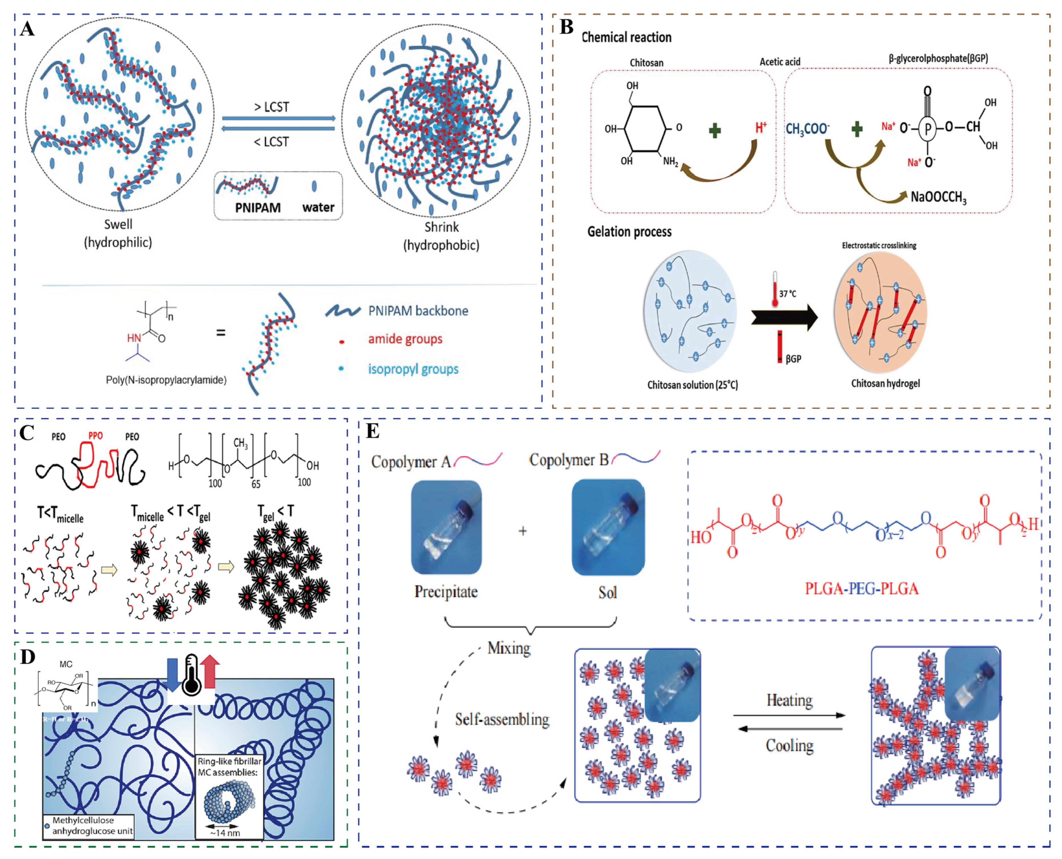
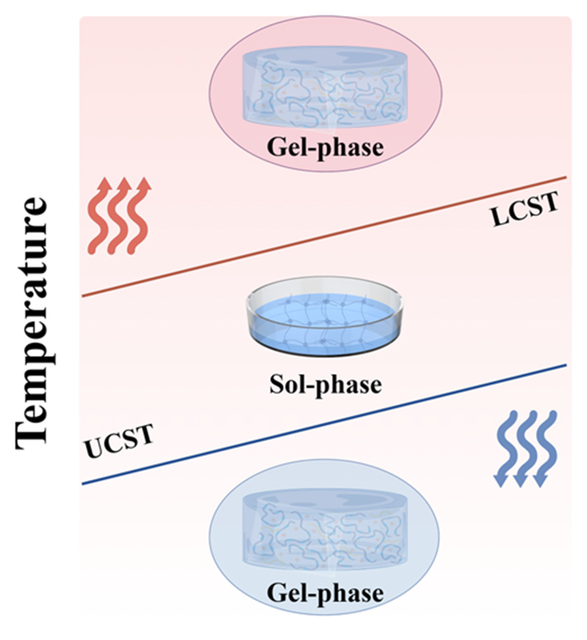
2. Phase Transition and Gelation Mechanisms of Thermosensitive Hydrogel
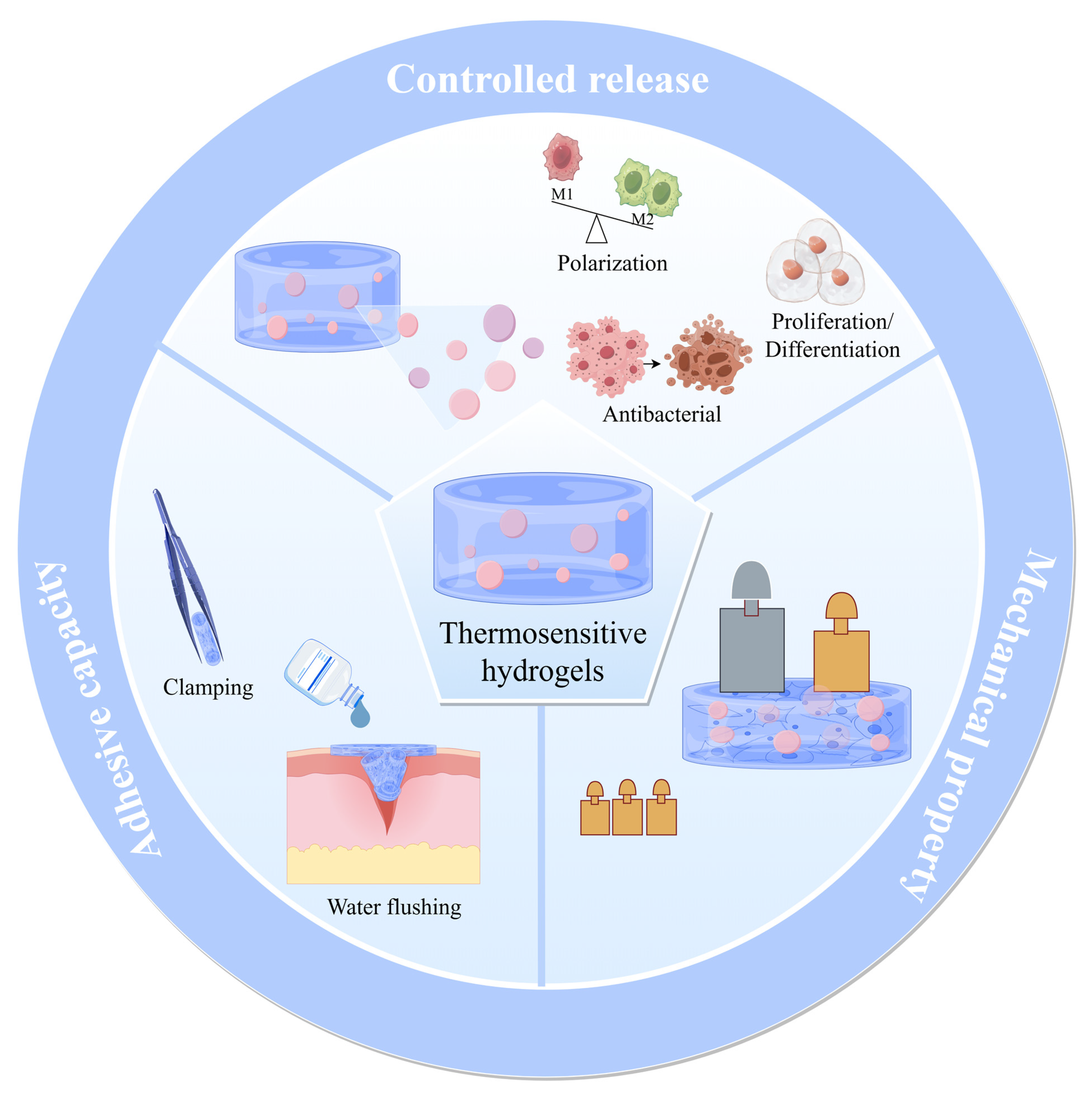
3. Optimization Strategies of Thermosensitive Hydrogels
3.1. Crosslinking Method
3.2. Internal Encapsulation
3.3. Three-Dimensional Printing Technique
3.4. Multiple Stimulus Response
4. Application of Thermosensitive Hydrogel in Tissue Repair and Treatment
4.1. Application of Thermosensitive Hydrogel in Bone Tissues
4.2. Application of Thermosensitive Hydrogels in Cartilage Tissues
4.3. Application of Thermosensitive Hydrogels in Other Tissues
4.3.1. Skin Injuries
4.3.2. Tumor
4.3.3. Tendon–Bone Interface
4.3.4. Muscle
4.3.5. Nerve
5. Challenge and Prospects
6. Conclusions
Author Contributions
Funding
Acknowledgments
Conflicts of Interest
References
- Hong, S.-C.; Yoo, S.-Y.; Kim, H.; Lee, J. Chitosan-Based Multifunctional Platforms for Local Delivery of Therapeutics. Mar. Drugs 2017, 15, 60. [Google Scholar] [CrossRef] [PubMed]
- Beaman, F.D.; Bancroft, L.W.; Peterson, J.J.; Kransdorf, M.J. Bone Graft Materials and Synthetic Substitutes. Radiol. Clin. N. Am. 2006, 44, 451–461. [Google Scholar] [CrossRef] [PubMed]
- Zhu, W.; Li, C.; Yao, M.; Wang, X.; Wang, J.; Zhang, W.; Chen, W.; Lv, H. Advances in Osseointegration of Biomimetic Mineralized Collagen and Inorganic Metal Elements of Natural Bone for Bone Repair. Regen. Biomater. 2023, 10, rbad030. [Google Scholar] [CrossRef] [PubMed]
- Weiser, J.R.; Saltzman, W.M. Controlled Release for Local Delivery of Drugs: Barriers and Models. J. Control. Release 2014, 190, 664–673. [Google Scholar] [CrossRef]
- Li, B.-Y.; Lin, T.-Y.; Lai, Y.-J.; Chiu, T.-H.; Yeh, Y.-C. Engineering Multiresponsive Alginate/PNIPAM/Carbon Nanotube Nanocomposite Hydrogels as On-Demand Drug Delivery Platforms. Small 2025, 21, e2407420. [Google Scholar] [CrossRef]
- Chen, H.; Xu, J.; Jiang, Y.; Sun, J.; Zheng, W.; Qian, H.; Sun, J.; Hu, W. Controlled Delivery of MTX from Thermosensitive Hydrogels Using Au Nanorods for Enhanced Therapeutics of Psoriasis via Improving Stratum Corneum Penetration. Small 2025, 21, e2410566. [Google Scholar] [CrossRef]
- Mi, B.; Mu, J.; Ding, X.; Guo, S.; Hua, X. Responsive Microneedles for Diagnostic and Therapeutic Applications of Ocular Diseases. Small Methods 2025, 2402048. [Google Scholar] [CrossRef]
- Liu, T.; Fu, J.; Zheng, Z.; Chen, M.; Wang, W.; Wu, C.; Quan, G.; Pan, X. Active Microneedle Patch Equipped with Spontaneous Bubble Generation for Enhanced Rheumatoid Arthritis Treatment. Theranostics 2025, 15, 3424–3438. [Google Scholar] [CrossRef]
- Wang, C.; Dong, Z.; Zhang, Q.; Guo, M.; Hu, W.; Dong, S.; Jakkree, T.; Lu, Y.; Du, S. Stimulus-Responsive Nano Lipidosome for Enhancing the Anti-Tumour Effect of a Novel Peptide Dermaseptin-PP. IET Nanobiotechnol. 2023, 17, 352–359. [Google Scholar] [CrossRef]
- Guo, W.; Dong, H.; Wang, X. Emerging Roles of Hydrogel in Promoting Periodontal Tissue Regeneration and Repairing Bone Defect. Front. Bioeng. Biotechnol. 2024, 12, 1380528. [Google Scholar] [CrossRef]
- Zhu, T.; Ni, Y.; Biesold, G.M.; Cheng, Y.; Ge, M.; Li, H.; Huang, J.; Lin, Z.; Lai, Y. Recent Advances in Conductive Hydrogels: Classifications, Properties, and Applications. Chem. Soc. Rev. 2023, 52, 473–509. [Google Scholar] [CrossRef] [PubMed]
- Zhang, Y.; Wu, B.M. Current Advances in Stimuli-Responsive Hydrogels as Smart Drug Delivery Carriers. Gels 2023, 9, 838. [Google Scholar] [CrossRef] [PubMed]
- Zheng, L.; Li, C.; Huang, X.; Lin, X.; Lin, W.; Yang, F.; Chen, T. Thermosensitive Hydrogels for Sustained-Release of Sorafenib and Selenium Nanoparticles for Localized Synergistic Chemoradiotherapy. Biomaterials 2019, 216, 119220. [Google Scholar] [CrossRef] [PubMed]
- Calderón, M.; Quadir, M.A.; Strumia, M.; Haag, R. Functional Dendritic Polymer Architectures as Stimuli-Responsive Nanocarriers. Biochimie 2010, 92, 1242–1251. [Google Scholar] [CrossRef]
- Lin, Z.; Gao, W.; Hu, H.; Ma, K.; He, B.; Dai, W.; Wang, X.; Wang, J.; Zhang, X.; Zhang, Q. Novel Thermo-Sensitive Hydrogel System with Paclitaxel Nanocrystals: High Drug-Loading, Sustained Drug Release and Extended Local Retention Guaranteeing Better Efficacy and Lower Toxicity. J. Control. Release 2014, 174, 161–170. [Google Scholar] [CrossRef]
- Shim, W.S.; Kim, J.-H.; Park, H.; Kim, K.; Chan Kwon, I.; Lee, D.S. Biodegradability and Biocompatibility of a pH- and Thermo-Sensitive Hydrogel Formed from a Sulfonamide-Modified Poly(Epsilon-Caprolactone-Co-Lactide)-Poly(Ethylene Glycol)-Poly(Epsilon-Caprolactone-Co-Lactide) Block Copolymer. Biomaterials 2006, 27, 5178–5185. [Google Scholar] [CrossRef]
- Liu, F.; Seuring, J.; Agarwal, S. A Non-Ionic Thermophilic Hydrogel with Positive Thermosensitivity in Water and Electrolyte Solution. Macromol. Chem. Phys. 2014, 215, 1466–1472. [Google Scholar] [CrossRef]
- Crespy, D.; Rossi, R.M. Temperature-responsive Polymers with LCST in the Physiological Range and Their Applications in Textiles. Polym. Int. 2007, 56, 1461–1468. [Google Scholar] [CrossRef]
- Belal, K.; Stoffelbach, F.; Lyskawa, J.; Fumagalli, M.; Hourdet, D.; Marcellan, A.; Smet, L.D.; de la Rosa, V.R.; Cooke, G.; Hoogenboom, R.; et al. Recognition-Mediated Hydrogel Swelling Controlled by Interaction with a Negative Thermoresponsive LCST Polymer. Angew. Chem. Int. Ed. Engl. 2016, 55, 13974–13978. [Google Scholar] [CrossRef]
- Zhao, H.; An, H.; Xi, B.; Yang, Y.; Qin, J.; Wang, Y.; He, Y.; Wang, X. Self-Healing Hydrogels with Both LCST and UCST through Cross-Linking Induced Thermo-Response. Polymers 2019, 11, 490. [Google Scholar] [CrossRef]
- Klouda, L. Thermoresponsive Hydrogels in Biomedical Applications: A Seven-Year Update. Eur. J. Pharm. Biopharm. 2015, 97, 338–349. [Google Scholar] [CrossRef] [PubMed]
- Fan, R.; Cheng, Y.; Wang, R.; Zhang, T.; Zhang, H.; Li, J.; Song, S.; Zheng, A. Thermosensitive Hydrogels and Advances in Their Application in Disease Therapy. Polymers 2022, 14, 2379. [Google Scholar] [CrossRef] [PubMed]
- Kim, Y.-J.; Matsunaga, Y.T. Thermo-Responsive Polymers and Their Application as Smart Biomaterials. J. Mater. Chem. B 2017, 5, 4307–4321. [Google Scholar] [CrossRef] [PubMed]
- Ruel-Gariépy, E.; Leroux, J.-C. In Situ-Forming Hydrogels—Review of Temperature-Sensitive Systems. Eur. J. Pharm. Biopharm. 2004, 58, 409–426. [Google Scholar] [CrossRef]
- Zhang, X.; Zhuo, R. Synthesis of Temperature-Sensitive Poly(N-Isopropylacrylamide) Hydrogel with Improved Surface Property. J. Colloid Interface Sci. 2000, 223, 311–313. [Google Scholar] [CrossRef]
- Doberenz, F.; Zeng, K.; Willems, C.; Zhang, K.; Groth, T. Thermoresponsive Polymers and Their Biomedical Application in Tissue Engineering—a Review. J. Mater. Chem. B 2020, 8, 607–628. [Google Scholar] [CrossRef]
- Buchecker, T.; Schmid, P.; Grillo, I.; Prévost, S.; Drechsler, M.; Diat, O.; Pfitzner, A.; Bauduin, P. Self-Assembly of Short Chain Poly- N-Isopropylacrylamid Induced by Superchaotropic Keggin Polyoxometalates: From Globules to Sheets. J. Am. Chem. Soc. 2019, 141, 6890–6899. [Google Scholar] [CrossRef]
- Han, X.; Zhang, X.; Zhu, H.; Yin, Q.; Liu, H.; Hu, Y. Effect of Composition of PDMAEMA-b-PAA Block Copolymers on Their pH- and Temperature-Responsive Behaviors. Langmuir 2013, 29, 1024–1034. [Google Scholar] [CrossRef]
- Maeda, Y.; Kubota, T.; Yamauchi, H.; Nakaji, T.; Kitano, H. Hydration Changes of Poly(2-(2-Methoxyethoxy)Ethyl Methacrylate) during Thermosensitive Phase Separation in Water. Langmuir 2007, 23, 11259–11265. [Google Scholar] [CrossRef]
- Lutz, J.-F.; Hoth, A. Preparation of Ideal PEG Analogues with a Tunable Thermosensitivity by Controlled Radical Copolymerization of 2-(2-Methoxyethoxy)Ethyl Methacrylate and Oligo(Ethylene Glycol) Methacrylate. Macromolecules 2006, 39, 893–896. [Google Scholar] [CrossRef]
- Grishkewich, N.; Akhlaghi, S.P.; Zhaoling, Y.; Berry, R.; Tam, K.C. Cellulose Nanocrystal-Poly(Oligo(Ethylene Glycol) Methacrylate) Brushes with Tunable LCSTs. Carbohydr. Polym. 2016, 144, 215–222. [Google Scholar] [CrossRef] [PubMed]
- Schnell, C.N.; Galván, M.V.; Zanuttini, M.A.; Mocchiutti, P. Hydrogels from Xylan/Chitosan Complexes for the Controlled Release of Diclofenac Sodium. Cellulose 2020, 27, 1465–1481. [Google Scholar] [CrossRef]
- Rezaei, N.; Hamidabadi, H.G.; Khosravimelal, S.; Zahiri, M.; Ahovan, Z.A.; Bojnordi, M.N.; Eftekhari, B.S.; Hashemi, A.; Ganji, F.; Darabi, S.; et al. Antimicrobial Peptides-Loaded Smart Chitosan Hydrogel: Release Behavior and Antibacterial Potential against Antibiotic Resistant Clinical Isolates. Int. J. Biol. Macromol. 2020, 164, 855–862. [Google Scholar] [CrossRef]
- Chenite, A. Rheological Characterisation of Thermogelling Chitosan/Glycerol-Phosphate Solutions. Carbohydr. Polym. 2001, 46, 39–47. [Google Scholar] [CrossRef]
- Kushan, E.; Senses, E. Thermoresponsive and Injectable Composite Hydrogels of Cellulose Nanocrystals and Pluronic F127. ACS Appl. Bio Mater. 2021, 4, 3507–3517. [Google Scholar] [CrossRef]
- White, J.M.; Calabrese, M.A. Impact of Small Molecule and Reverse Poloxamer Addition on the Micellization and Gelation Mechanisms of Poloxamer Hydrogels. Colloids Surfaces A Physicochem. Eng. Asp. 2022, 638, 128246. [Google Scholar] [CrossRef]
- Hynninen, V.; Hietala, S.; McKee, J.R.; Murtomäki, L.; Rojas, O.J.; Ikkala, O. Nonappa Inverse Thermoreversible Mechanical Stiffening and Birefringence in a Methylcellulose/Cellulose Nanocrystal Hydrogel. Biomacromolecules 2018, 19, 2795–2804. [Google Scholar] [CrossRef]
- Xu, W.-K.; Tang, J.-Y.; Yuan, Z.; Cai, C.-Y.; Chen, X.-B.; Cui, S.-Q.; Liu, P.; Yu, L.; Cai, K.-Y.; Ding, J.-D. Accelerated Cutaneous Wound Healing Using an Injectable Teicoplanin-Loaded PLGA-PEG-PLGA Thermogel Dressing. Chin. J. Polym. Sci. 2019, 37, 548–559. [Google Scholar] [CrossRef]
- Steinman, N.Y.; Haim-Zada, M.; Goldstein, I.A.; Goldberg, A.H.; Haber, T.; Berlin, J.M.; Domb, A.J. Effect of PLGA Block Molecular Weight on Gelling Temperature of PLGA-PEG-PLGA Thermoresponsive Copolymers. J. Polym. Sci. Part A Polym. Chem. 2019, 57, 35–39. [Google Scholar] [CrossRef]
- Ma, L.; Shi, T.; Zhang, Z.; Liu, X.; Wang, H. Wettability of HPMC/PEG/CS Thermosensitive Porous Hydrogels. Gels 2023, 9, 667. [Google Scholar] [CrossRef]
- Kara, M.; Kocaaga, N.; Akgul, B.; Abamor, E.S.; Erdogmus, A.; Topuzogullari, M.; Acar, S. Micelles of Poly[Oligo(Ethylene Glycol) Methacrylate] as Delivery Vehicles for Zinc Phthalocyanine Photosensitizers. Nanotechnology 2024, 35, 475602. [Google Scholar] [CrossRef] [PubMed]
- Shi, F.-K.; Wang, X.-P.; Guo, R.-H.; Zhong, M.; Xie, X.-M. Highly Stretchable and Super Tough Nanocomposite Physical Hydrogels Facilitated by the Coupling of Intermolecular Hydrogen Bonds and Analogous Chemical Crosslinking of Nanoparticles. J. Mater. Chem. B 2015, 3, 1187–1192. [Google Scholar] [CrossRef] [PubMed]
- Li, X.; Chen, R.; Xu, S.; Liu, H.; Hu, Y. Thermoresponsive Behavior and Rheology of SiO–Hyaluronic Acid/Poly(N-Isopropylacrylamide) (NaHA/PNIPAm) Core–Shell Structured Microparticles. J. Chem. Technol. Biotechnol. 2015, 90, 407–414. [Google Scholar] [CrossRef]
- Yan, X.; Huang, H.; Bakry, A.M.; Wu, W.; Liu, X.; Liu, F. Advances in Enhancing the Mechanical Properties of Biopolymer Hydrogels via Multi-Strategic Approaches. Int. J. Biol. Macromol. 2024, 272, 132583. [Google Scholar] [CrossRef]
- Hao, J.; Weiss, R.A. Mechanical Behavior of Hybrid Hydrogels Composed of a Physical and a Chemical Network. Polymer 2013, 54, 2174–2182. [Google Scholar] [CrossRef]
- Berger, J.; Reist, M.; Mayer, J.M.; Felt, O.; Peppas, N.A.; Gurny, R. Structure and Interactions in Covalently and Ionically Crosslinked Chitosan Hydrogels for Biomedical Applications. Eur. J. Pharm. Biopharm. 2004, 57, 19–34. [Google Scholar] [CrossRef]
- Calder, D.; Fathi, A.; Oveissi, F.; Maleknia, S.; Abrams, T.; Wang, Y.; Maitz, J.; Tsai, K.H.-Y.; Maitz, P.; Chrzanowski, W.; et al. Thermoresponsive and Injectable Hydrogel for Tissue Agnostic Regeneration. Adv. Healthc. Mater. 2022, 11, 2201714. [Google Scholar] [CrossRef]
- Wang, L.; Ding, X.; He, X.; Tian, N.; Ding, P.; Guo, W.; Okoro, O.V.; Sun, Y.; Jiang, G.; Liu, Z.; et al. Fabrication and Properties of Hydrogel Dressings Based on Genipin Crosslinked Chondroitin Sulfate and Chitosan. Polymers 2024, 16, 2876. [Google Scholar] [CrossRef]
- Yang, Y.; Ritchie, A.C.; Everitt, N.M. Comparison of Glutaraldehyde and Procyanidin Cross-Linked Scaffolds for Soft Tissue Engineering. Mater. Sci. Eng. C Mater. Biol. Appl. 2017, 80, 263–273. [Google Scholar] [CrossRef]
- Ahammed, S.; Easdani, M.; Liu, F.; Zhong, F. Encapsulation of Tea Polyphenol in Zein through Complex Coacervation Technique to Control the Release of the Phenolic Compound from Gelatin-Zein Composite Film. Polymers 2023, 15, 2882. [Google Scholar] [CrossRef]
- Deng, Q.-S.; Gao, Y.; Rui, B.-Y.; Li, X.-R.; Liu, P.-L.; Han, Z.-Y.; Wei, Z.-Y.; Zhang, C.-R.; Wang, F.; Dawes, H.; et al. Double-Network Hydrogel Enhanced by SS31-Loaded Mesoporous Polydopamine Nanoparticles: Symphonic Collaboration of near-Infrared Photothermal Antibacterial Effect and Mitochondrial Maintenance for Full-Thickness Wound Healing in Diabetes Mellitus. Bioact. Mater. 2023, 27, 409–428. [Google Scholar] [CrossRef] [PubMed]
- Hakim Khalili, M.; Zhang, R.; Wilson, S.; Goel, S.; Impey, S.A.; Aria, A.I. Additive Manufacturing and Physicomechanical Characteristics of PEGDA Hydrogels: Recent Advances and Perspective for Tissue Engineering. Polymers 2023, 15, 2341. [Google Scholar] [CrossRef] [PubMed]
- Zhou, R.; Zhang, W.; Wu, J.; Huang, J.; Wang, W.; Peng, W.; Chen, J.; Bo, R.; Liu, M.; Li, J. Polyvinyl Alcohol/Chitosan Hydrogel Based on Deep Eutectic Solvent for Promoting Methicillin-Resistant Staphylococcus Aureus-Infected Wound Healing. Int. J. Biol. Macromol. 2025, 297, 139916. [Google Scholar] [CrossRef]
- Wu, Q.; Fu, Y.; Yang, W.; Liu, S. A Temperature/pH Double-Responsive and Physical Double-Crosslinked Hydrogel Based on PLA and Histidine. Gels 2022, 8, 570. [Google Scholar] [CrossRef]
- Nordin, N.; Zairul Azman, Z.A.; Adnan, N.A.; Majid, S.R. On the Dual Crosslinking for Functionality Enhancement of Poly (Acrylamide-Co-Acrylic Acid)/Chitosan-Aluminum (III) Ions and Its Characterization and Sensory Hydrogel Fibers. Int. J. Biol. Macromol. 2024, 274, 133383. [Google Scholar] [CrossRef]
- Zhou, Q.; Huang, C.; Lu, S.; Abushammala, H.; Gao, D.; Xu, P.; Niu, D.; Yang, W.; Ma, P. Skin-Inspired Polysaccharide-Based Hydrogels with Tailored Properties for Information Transmission Application. Int. J. Biol. Macromol. 2025, 306, 141354. [Google Scholar] [CrossRef]
- Wang, Y.; Wang, J.; Du, H.; Zhao, Q.; Wang, S.; Liu, T.; Bi, S.; Zhang, Q.; An, M.; Wen, S. A Dynamically Cross-Linked Catechol-Grafted Chitosan/Gelatin Hydrogel Dressing Synergised with Photothermal Therapy and Baicalin Reduces Wound Infection and Accelerates Wound Healing. Int. J. Biol. Macromol. 2024, 273, 132802. [Google Scholar] [CrossRef]
- Yang, Y.; Ma, Y.; Wu, M.; Wang, X.; Zhao, Y.; Zhong, S.; Gao, Y.; Cui, X. Fe3+-Induced Coordination Cross-Linking Gallic Acid-Carboxymethyl Cellulose Self-Healing Hydrogel. Int. J. Biol. Macromol. 2024, 267, 131626. [Google Scholar] [CrossRef]
- Liu, Y.; Wang, Y.; Fu, Y.; Wang, N.N.; Zhan, S.Q.; Sheng, L.; Yang, H.Y.; Liu, C. Healable and Transparent Ionic Conductive Hydrogels Based on PNATF as Multiple-Signal Sensors. ACS Appl. Polym. Mater. 2025, 7, 2529–2540. [Google Scholar] [CrossRef]
- Tordi, P.; Tamayo, A.; Jeong, Y.; Bonini, M.; Samorì, P. Multiresponsive Ionic Conductive Alginate/Gelatin Organohydrogels with Tunable Functions. Adv. Funct. Mater. 2024, 34, 2410663. [Google Scholar] [CrossRef]
- Torchio, A.; Boffito, M.; Laurano, R.; Cassino, C.; Lavella, M.; Ciardelli, G. Double-Crosslinkable Poly(Urethane)-Based Hydrogels Relying on Supramolecular Interactions and Light-Initiated Polymerization: Promising Tools for Advanced Applications in Drug Delivery. J. Mater. Chem. B 2024, 12, 8389–8407. [Google Scholar] [CrossRef] [PubMed]
- Guo, H.; Ding, Q.; Yang, Y.; Du, C.; Nie, Z. Dual Light-Responsive Shape Transformations of a Nanocomposite Hydrogel Sheet Enabled by in Situ Etching Shaped Plasmonic Nanoparticles. RSC Adv. 2024, 14, 34804–34810. [Google Scholar] [CrossRef] [PubMed]
- Xing, Y.; Zeng, B.; Yang, W. Light Responsive Hydrogels for Controlled Drug Delivery. Front. Bioeng. Biotechnol. 2022, 10, 1075670. [Google Scholar] [CrossRef] [PubMed]
- Qiao, B.; Pang, Q.; Yuan, P.; Luo, Y.; Ma, L. Smart Wound Dressing for Infection Monitoring and NIR-Triggered Antibacterial Treatment. Biomater. Sci. 2020, 8, 1649–1657. [Google Scholar] [CrossRef]
- Zhu, Y.; Zeng, Q.; Zhang, Q.; Li, K.; Shi, X.; Liang, F.; Han, D. Temperature/near-Infrared Light-Responsive Conductive Hydrogels for Controlled Drug Release and Real-Time Monitoring. Nanoscale 2020, 12, 8679–8686. [Google Scholar] [CrossRef]
- Ashrafizadeh, M.; Delfi, M.; Zarrabi, A.; Bigham, A.; Sharifi, E.; Rabiee, N.; Paiva-Santos, A.C.; Kumar, A.P.; Tan, S.C.; Hushmandi, K.; et al. Stimuli-Responsive Liposomal Nanoformulations in Cancer Therapy: Pre-Clinical & Clinical Approaches. J. Control. Release 2022, 351, 50–80. [Google Scholar] [CrossRef]
- Mantha, S.; Pillai, S.; Khayambashi, P.; Upadhyay, A.; Zhang, Y.; Tao, O.; Pham, H.M.; Tran, S.D. Smart Hydrogels in Tissue Engineering and Regenerative Medicine. Materials 2019, 12, 3323. [Google Scholar] [CrossRef]
- Yang, M.; Liu, L. MHC II Gene Knockout in Tissue Engineering May Prevent Immune Rejection of Transplants. Med. Hypotheses 2008, 70, 798–801. [Google Scholar] [CrossRef]
- Ren, X.; Zhao, M.; Lash, B.; Martino, M.M.; Julier, Z. Growth Factor Engineering Strategies for Regenerative Medicine Applications. Front. Bioeng. Biotechnol. 2019, 7, 469. [Google Scholar] [CrossRef]
- Li, C.; Nyaruaba, R.; Zhao, X.; Xue, H.; Li, Y.; Yang, H.; Wei, H. Thermosensitive Hydrogel Wound Dressing Loaded with Bacteriophage Lysin LysP53. Viruses 2022, 14, 1956. [Google Scholar] [CrossRef]
- Koland, M.; Narayanan Vadakkepushpakath, A.; John, A.; Tharamelveliyil Rajendran, A.; Raghunath, I. Thermosensitive In Situ Gels for Joint Disorders: Pharmaceutical Considerations in Intra-Articular Delivery. Gels 2022, 8, 723. [Google Scholar] [CrossRef] [PubMed]
- Kansız, S.; Elçin, Y.M. Advanced Liposome and Polymersome-Based Drug Delivery Systems: Considerations for Physicochemical Properties, Targeting Strategies and Stimuli-Sensitive Approaches. Adv. Colloid Interface Sci. 2023, 317, 102930. [Google Scholar] [CrossRef] [PubMed]
- Cheng, L.; Lin, T.; Khalaf, A.T.; Zhang, Y.; He, H.; Yang, L.; Yan, S.; Zhu, J.; Shi, Z. The Preparation and Application of Calcium Phosphate Biomedical Composites in Filling of Weight-Bearing Bone Defects. Sci. Rep. 2021, 11, 4283. [Google Scholar] [CrossRef] [PubMed]
- Cui, Y.; Wang, J.; Tian, Y.; Fan, Y.; Li, S.; Wang, G.; Peng, C.; Liu, H.; Wu, D. Functionalized Decellularized Bone Matrix Promotes Bone Regeneration by Releasing Osteogenic Peptides. ACS Biomater. Sci. Eng. 2023, 9, 4953–4968. [Google Scholar] [CrossRef]
- Rajabi, N.; Rezaei, A.; Kharaziha, M.; Bakhsheshi-Rad, H.R.; Luo, H.; RamaKrishna, S.; Berto, F. Recent Advances on Bioprinted Gelatin Methacrylate-Based Hydrogels for Tissue Repair. Tissue Eng. Part A 2021, 27, 679–702. [Google Scholar] [CrossRef]
- Khattak, S.F.; Bhatia, S.R.; Roberts, S.C. Pluronic F127 as a Cell Encapsulation Material: Utilization of Membrane-Stabilizing Agents. Tissue Eng. 2005, 11, 974–983. [Google Scholar] [CrossRef]
- Phan, V.H.G.; Murugesan, M.; Huong, H.; Le, T.-T.; Phan, T.-H.; Manivasagan, P.; Mathiyalagan, R.; Jang, E.-S.; Yang, D.C.; Li, Y.; et al. Cellulose Nanocrystals-Incorporated Thermosensitive Hydrogel for Controlled Release, 3D Printing, and Breast Cancer Treatment Applications. ACS Appl. Mater. Interfaces 2022, 14, 42812–42826. [Google Scholar] [CrossRef]
- Hinton, T.J.; Jallerat, Q.; Palchesko, R.N.; Park, J.H.; Grodzicki, M.S.; Shue, H.-J.; Ramadan, M.H.; Hudson, A.R.; Feinberg, A.W. Three-Dimensional Printing of Complex Biological Structures by Freeform Reversible Embedding of Suspended Hydrogels. Sci. Adv. 2015, 1, e1500758. [Google Scholar] [CrossRef]
- Mirdamadi, E.; Tashman, J.W.; Shiwarski, D.J.; Palchesko, R.N.; Feinberg, A.W. FRESH 3D Bioprinting a Full-Size Model of the Human Heart. ACS Biomater. Sci. Eng. 2020, 6, 6453–6459. [Google Scholar] [CrossRef]
- Khaled Wassif, R.; Daihom, B.A.; Maniruzzaman, M. FRESH 3D Printing of Zoledronic Acid-Loaded Chitosan/Alginate/Hydroxyapatite Composite Thermosensitive Hydrogel for Promoting Bone Regeneration. Int. J. Pharm. 2024, 667, 124898. [Google Scholar] [CrossRef]
- Koskela, J.E.; Turunen, S.; Ylä-Outinen, L.; Narkilahti, S.; Kellomäki, M. Two-Photon Microfabrication of Poly(Ethylene Glycol) Diacrylate and a Novel Biodegradable Photopolymer—Comparison of Processability for Biomedical Applications. Polym. Adv. Technol. 2012, 23, 992–1001. [Google Scholar] [CrossRef]
- Fu, H.; Yu, B. 3D Micro/Nano Hydrogel Structures Fabricated by Two-Photon Polymerization for Biomedical Applications. Front. Bioeng. Biotechnol. 2024, 12, 1339450. [Google Scholar] [CrossRef] [PubMed]
- Zhou, Y.; Ye, M.; Zhao, H.; Wang, X. 3D-Printed PNAGA Thermosensitive Hydrogelbased Microrobots: An Effective Cancer Therapy by Temperature-Triggered Drug Release. Int. J. Bioprint. 2023, 9, 709. [Google Scholar] [CrossRef]
- Saravanou, S.F.; Ioannidis, K.; Dimopoulos, A.; Paxinou, A.; Kounelaki, F.; Varsami, S.M.; Tsitsilianis, C.; Papantoniou, I.; Pasparakis, G. Dually Crosslinked Injectable Alginate-Based Graft Copolymer Thermoresponsive Hydrogels as 3D Printing Bioinks for Cell Spheroid Growth and Release. Carbohydr. Polym. 2023, 312, 120790. [Google Scholar] [CrossRef]
- Mansha, S.; Sajjad, A.; Zarbab, A.; Afzal, T.; Kanwal, Z.; Iqbal, M.J.; Raza, M.A.; Ali, S. Development of pH-Responsive, Thermosensitive, Antibacterial, and Anticancer CS/PVA/Graphene Blended Hydrogels for Controlled Drug Delivery. Gels 2024, 10, 205. [Google Scholar] [CrossRef]
- Liu, G.; Hu, D.; Chen, M.; Wang, C.; Wu, L. Multifunctional PNIPAM/Fe3O4-ZnS Hybrid Hollow Spheres: Synthesis, Characterization, and Properties. J. Colloid Interface Sci. 2013, 397, 73–79. [Google Scholar] [CrossRef]
- Kang, W.; Fu, S.; Li, W.; Wu, Y.; Li, H.; Wang, J. Design and Characterization of a ROS-Responsive Antibacterial Composite Hydrogel for Advanced Full-Thickness Wound Healing. Int. J. Biol. Macromol. 2025, 294, 139349. [Google Scholar] [CrossRef]
- Fan, K.; Yang, D.; Zhu, X.; Zheng, L.; Han, Y.; Lin, J.; Xiang, Z.; Guo, Y.; Tao, K.; Li, J.; et al. High-Efficiency Antioxidant ROS-Responsive Thermosensitive Hydrogel Encapsulated Fenofibrate for the Treatment of Corneal Neovascularization. J. Control. Release 2025, 382, 113650. [Google Scholar] [CrossRef]
- Zhang, H.; Yuan, S.; Zheng, B.; Wu, P.; He, X.; Zhao, Y.; Zhong, Z.; Zhang, X.; Guan, J.; Wang, H.; et al. Lubricating and Dual-Responsive Injectable Hydrogels Formulated From ZIF-8 Facilitate Osteoarthritis Treatment by Remodeling the Microenvironment. Small 2025, 21, 2407885. [Google Scholar] [CrossRef]
- Miron, R.J.; Fujioka-Kobayashi, M.; Pikos, M.A.; Nakamura, T.; Imafuji, T.; Zhang, Y.; Shinohara, Y.; Sculean, A.; Shirakata, Y. The Development of Non-Resorbable Bone Allografts: Biological Background and Clinical Perspectives. Periodontology 2000 2024, 94, 161–179. [Google Scholar] [CrossRef]
- Kolios, G.; Moodley, Y. Introduction to Stem Cells and Regenerative Medicine. Respiration 2013, 85, 3–10. [Google Scholar] [CrossRef] [PubMed]
- Guo, X.; Zheng, H.; Guo, Y.; Heng, B.C.; Yang, Y.; Yao, W.; Jiang, S. A Three-Dimensional Actively Spreading Bone Repair Material Based on Cell Spheroids Can Facilitate the Preservation of Tooth Extraction Sockets. Front. Bioeng. Biotechnol. 2023, 11, 1161192. [Google Scholar] [CrossRef] [PubMed]
- Weng, Z.; Zhang, B.; Wu, C.; Yu, F.; Han, B.; Li, B.; Li, L. Therapeutic Roles of Mesenchymal Stem Cell-Derived Extracellular Vesicles in Cancer. J. Hematol. Oncol. 2021, 14, 136. [Google Scholar] [CrossRef]
- Ming, L.; Qu, Y.; Wang, Z.; Dong, L.; Li, Y.; Liu, F.; Wang, Q.; Zhang, D.; Li, Z.; Zhou, Z.; et al. Small Extracellular Vesicles Laden Oxygen-Releasing Thermosensitive Hydrogel for Enhanced Antibacterial Therapy against Anaerobe-Induced Periodontitis Alveolar Bone Defect. ACS Biomater. Sci. Eng. 2024, 10, 932–945. [Google Scholar] [CrossRef]
- Wu, D.; Qin, H.; Wang, Z.; Yu, M.; Liu, Z.; Peng, H.; Liang, L.; Zhang, C.; Wei, X. Bone Mesenchymal Stem Cell-Derived sEV-Encapsulated Thermosensitive Hydrogels Accelerate Osteogenesis and Angiogenesis by Release of Exosomal miR-21. Front. Bioeng. Biotechnol. 2021, 9, 829136. [Google Scholar] [CrossRef]
- Tessmar, J.K.; Göpferich, A.M. Matrices and Scaffolds for Protein Delivery in Tissue Engineering. Adv. Drug Deliv. Rev. 2007, 59, 274–291. [Google Scholar] [CrossRef]
- Samorezov, J.E.; Alsberg, E. Spatial Regulation of Controlled Bioactive Factor Delivery for Bone Tissue Engineering. Adv. Drug Deliv. Rev. 2015, 84, 45–67. [Google Scholar] [CrossRef]
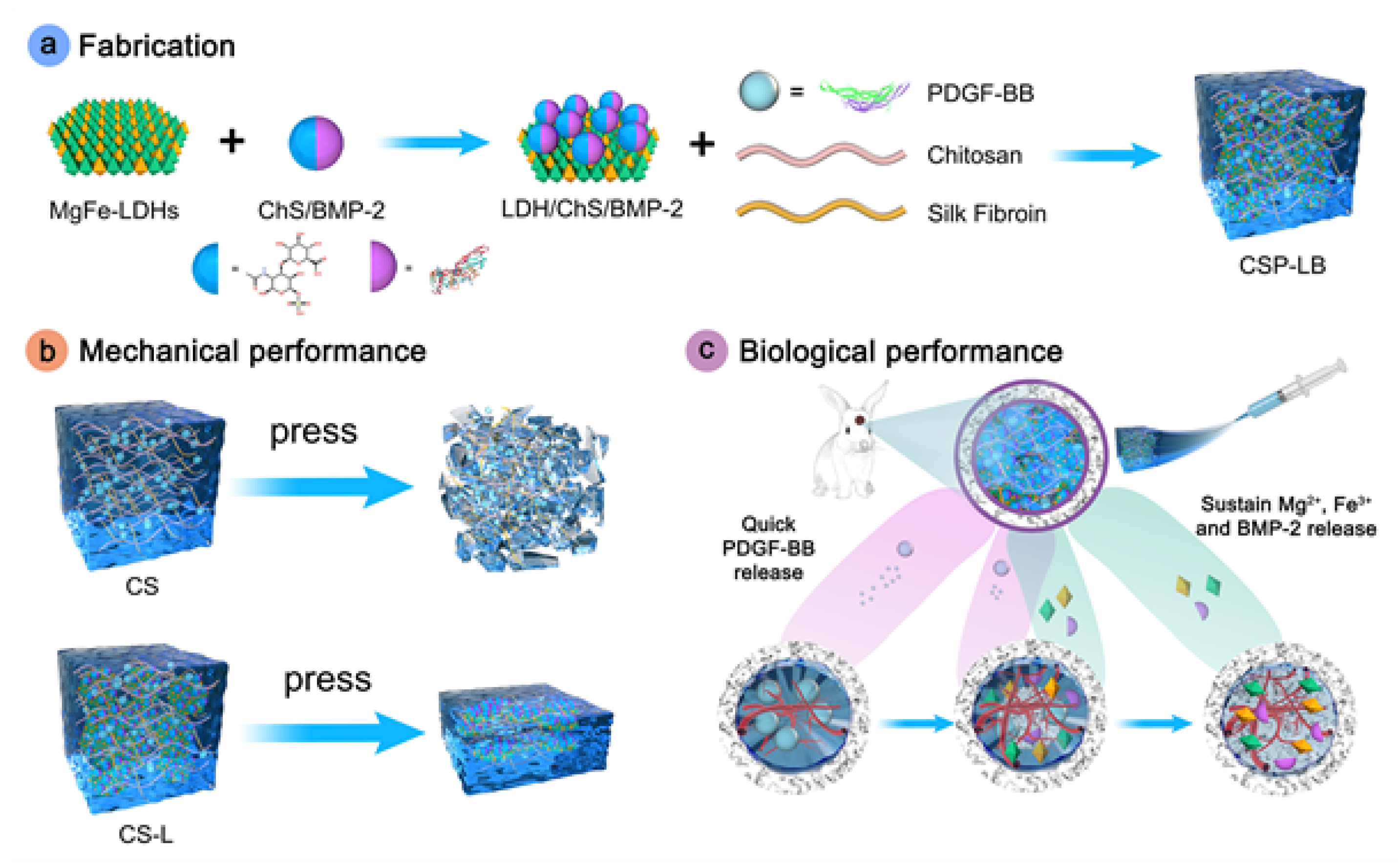
- Lv, Z.; Hu, T.; Bian, Y.; Wang, G.; Wu, Z.; Li, H.; Liu, X.; Yang, S.; Tan, C.; Liang, R.; et al. A MgFe-LDH Nanosheet-Incorporated Smart Thermo-Responsive Hydrogel with Controllable Growth Factor Releasing Capability for Bone Regeneration. Adv. Mater. 2023, 35, e2206545. [Google Scholar] [CrossRef]
- Zhou, X.; Chen, J.; Sun, H.; Wang, F.; Wang, Y.; Zhang, Z.; Teng, W.; Ye, Y.; Huang, D.; Zhang, W.; et al. Spatiotemporal Regulation of Angiogenesis/Osteogenesis Emulating Natural Bone Healing Cascade for Vascularized Bone Formation. J. Nanobiotechnol. 2021, 19, 420. [Google Scholar] [CrossRef]
- Zhang, C.; Yuan, T.-J.; Tan, M.-H.; Xu, X.-H.; Huang, Y.-F.; Peng, L.-H. Smart Graphene-Based Hydrogel Promotes Recruitment and Neural-like Differentiation of Bone Marrow Derived Mesenchymal Stem Cells in Rat Skin. Biomater. Sci. 2021, 9, 2146–2161. [Google Scholar] [CrossRef]
- Chu, C.; Qiu, J.; Zhao, Q.; Xun, X.; Wang, H.; Yuan, R.; Xu, X. Injectable Dual Drug-Loaded Thermosensitive Liposome-Hydrogel Composite Scaffold for Vascularised and Innervated Bone Regeneration. Colloids Surf. B Biointerfaces 2025, 245, 114203. [Google Scholar] [CrossRef] [PubMed]
- Dumortier, G.; Grossiord, J.L.; Agnely, F.; Chaumeil, J.C. A Review of Poloxamer 407 Pharmaceutical and Pharmacological Characteristics. Pharm. Res. 2006, 23, 2709–2728. [Google Scholar] [CrossRef] [PubMed]
- Almoshari, Y.; Ren, R.; Zhang, H.; Jia, Z.; Wei, X.; Chen, N.; Li, G.; Ryu, S.; Lele, S.M.; Reinhardt, R.A.; et al. GSK3 Inhibitor-Loaded Osteotropic Pluronic Hydrogel Effectively Mitigates Periodontal Tissue Damage Associated with Experimental Periodontitis. Biomaterials 2020, 261, 120293. [Google Scholar] [CrossRef] [PubMed]
- Yang, S.; Zhu, Y.; Ji, C.; Zhu, H.; Lao, A.; Zhao, R.; Hu, Y.; Zhou, Y.; Zhou, J.; Lin, K.; et al. A Five-in-One Novel MOF-Modified Injectable Hydrogel with Thermo-Sensitive and Adhesive Properties for Promoting Alveolar Bone Repair in Periodontitis: Antibacterial, Hemostasis, Immune Reprogramming, pro-Osteo-/Angiogenesis and Recruitment. Bioact. Mater. 2024, 41, 239–256. [Google Scholar] [CrossRef]
- Hofbauer, L.C.; Busse, B.; Eastell, R.; Ferrari, S.; Frost, M.; Müller, R.; Burden, A.M.; Rivadeneira, F.; Napoli, N.; Rauner, M. Bone Fragility in Diabetes: Novel Concepts and Clinical Implications. Lancet Diabetes Endocrinol. 2022, 10, 207–220. [Google Scholar] [CrossRef]
- Bhagavan, H.N.; Chopra, R.K. Coenzyme Q10: Absorption, Tissue Uptake, Metabolism and Pharmacokinetics. Free. Radic. Res. 2006, 40, 445–453. [Google Scholar] [CrossRef]
- Ghanem, M.; Heikal, L.; Abdel Fattah, H.; El Ashwah, A.; Fliefel, R. The Effect of Coenzyme Q10/Collagen Hydrogel on Bone Regeneration in Extraction Socket Prior to Implant Placement in Type II Diabetic Patients: A Randomized Controlled Clinical Trial. J. Clin. Med. 2022, 11, 3059. [Google Scholar] [CrossRef]
- Sheng, N.; Xing, F.; Zhang, Q.-Y.; Tan, J.; Nie, R.; Huang, K.; Li, H.-X.; Jiang, Y.-L.; Tan, B.; Xiang, Z.; et al. A Pleiotropic SIS-Based Hydrogel with Immunomodulation via NLRP3 Inflammasome Inhibition for Diabetic Bone Regeneration. Chem. Eng. J. 2024, 480, 147985. [Google Scholar] [CrossRef]
- Moradi, L.; Witek, L.; Vivekanand Nayak, V.; Cabrera Pereira, A.; Kim, E.; Good, J.; Liu, C.-J. Injectable Hydrogel for Sustained Delivery of Progranulin Derivative Atsttrin in Treating Diabetic Fracture Healing. Biomaterials 2023, 301, 122289. [Google Scholar] [CrossRef]
- Gu, Z.; Qiu, C.; Chen, L.; Wang, X. Injectable Thermosensitive Hydrogel Loading Erythropoietin and FK506 Alleviates Gingival Inflammation and Promotes Periodontal Tissue Regeneration. Front. Bioeng. Biotechnol. 2023, 11, 1323554. [Google Scholar] [CrossRef]
- Barik, D.; Shyamal, S.; Das, K.; Jena, S.; Dash, M. Glycoprotein Injectable Hydrogels Promote Accelerated Bone Regeneration through Angiogenesis and Innervation. Adv. Healthc. Mater. 2023, 12, e2301959. [Google Scholar] [CrossRef] [PubMed]
- Zhang, Y.; Wang, T.; Zhang, D.; Li, J.; Yue, X.; Kong, W.; Gu, X.; Jiao, Z.; Yang, C. Thermosensitive Hydrogel Loaded with Concentrated Growth Factors Promote Bone Repair in Segmental Bone Defects. Front. Bioeng. Biotechnol. 2022, 10, 1039117. [Google Scholar] [CrossRef] [PubMed]
- Li, J.; Li, M.; Zhang, C.; Fei, Y.; Wang, Y.; Zhong, Z.; Peng, C.; Li, M.; Gui, S.; Guo, J. Active Targeting Microemulsion-Based Thermosensitive Hydrogel against Periodontitis by Reconstructing Th17/Treg Homeostasis via Regulating ROS-Macrophages Polarization Cascade. Int. J. Pharm. 2024, 659, 124263. [Google Scholar] [CrossRef]
- Deng, J.; Pan, J.; Han, X.; Yu, L.; Chen, J.; Zhang, W.; Zhu, L.; Huang, W.; Liu, S.; You, Z.; et al. PDGFBB-Modified Stem Cells from Apical Papilla and Thermosensitive Hydrogel Scaffolds Induced Bone Regeneration. Chem. Biol. Interact. 2020, 316, 108931. [Google Scholar] [CrossRef]
- Li, J.; Li, L.; Wu, T.; Shi, K.; Bei, Z.; Wang, M.; Chu, B.; Xu, K.; Pan, M.; Li, Y.; et al. An Injectable Thermosensitive Hydrogel Containing Resveratrol and Dexamethasone-Loaded Carbonated Hydroxyapatite Microspheres for the Regeneration of Osteoporotic Bone Defects. Small Methods 2023, 316, 2300843. [Google Scholar] [CrossRef]
- Wang, H.; Chang, X.; Ma, Q.; Sun, B.; Li, H.; Zhou, J.; Hu, Y.; Yang, X.; Li, J.; Chen, X.; et al. Bioinspired Drug-Delivery System Emulating the Natural Bone Healing Cascade for Diabetic Periodontal Bone Regeneration. Bioact. Mater. 2023, 21, 324–339. [Google Scholar] [CrossRef]
- Li, J.; Leung, S.S.Y.; Chung, Y.L.; Chow, S.K.H.; Alt, V.; Rupp, M.; Brochhausen, C.; Chui, C.S.; Ip, M.; Cheung, W.-H.; et al. Hydrogel Delivery of DNase I and Liposomal Vancomycin to Eradicate Fracture-Related Methicillin-Resistant Staphylococcus Aureus Infection and Support Osteoporotic Fracture Healing. Acta Biomater. 2023, 164, 223–239. [Google Scholar] [CrossRef]
- Zhou, H.; He, Z.; Cao, Y.; Chu, L.; Liang, B.; Yu, K.; Deng, Z. An Injectable Magnesium-Loaded Hydrogel Releases Hydrogen to Promote Osteoporotic Bone Repair via ROS Scavenging and Immunomodulation. Theranostics 2024, 14, 3739–3759. [Google Scholar] [CrossRef]
- Heir, S.; Nerhus, T.K.; Røtterud, J.H.; Løken, S.; Ekeland, A.; Engebretsen, L.; Arøen, A. Focal Cartilage Defects in the Knee Impair Quality of Life as Much as Severe Osteoarthritis: A Comparison of Knee Injury and Osteoarthritis Outcome Score in 4 Patient Categories Scheduled for Knee Surgery. Am. J. Sports Med. 2010, 38, 231–237. [Google Scholar] [CrossRef]
- Chen, Z.; Liu, W.; Liu, D.; Xiao, Y.; Chen, H.; Chen, J.; Li, W.; Cai, H.; Li, W.; Cai, B.; et al. Development of Brucine-Loaded Microsphere/Thermally Responsive Hydrogel Combination System for Intra-Articular Administration. J. Control. Release 2012, 162, 628–635. [Google Scholar] [CrossRef]
- Kampen, W.U.; Boddenberg-Pätzold, B.; Fischer, M.; Gabriel, M.; Klett, R.; Konijnenberg, M.; Kresnik, E.; Lellouche, H.; Paycha, F.; Terslev, L.; et al. The EANM Guideline for Radiosynoviorthesis. Eur. J. Nucl. Med. Mol. Imaging 2022, 49, 681–708. [Google Scholar] [CrossRef] [PubMed]
- Liu, P.; Zhou, M.; Luo, Z.; Hao, L.; Hsu, J.C.; Cai, W.; Zhou, W.; Hu, S. A 177Lu-Nucleotide Coordination Polymer-Incorporated Thermosensitive Hydrogel with Anti-Inflammatory and Chondroprotective Capabilities for Osteoarthritis Treatment. Biomaterials 2025, 317, 123098. [Google Scholar] [CrossRef] [PubMed]
- Chang, Y.; Wang, Y.; Liu, J.; Chen, X.; Ma, X.; Hu, Y.; Tian, H.; Wang, X.; Mu, C. Glucosamine-Loaded Injectable Hydrogel Promotes Autophagy and Inhibits Apoptosis after Cartilage Injury. Heliyon 2023, 9, e19879. [Google Scholar] [CrossRef] [PubMed]
- Yuan, X.; Wan, J.; Yang, Y.; Huang, L.; Zhou, C.; Su, J.; Hua, S.; Pu, H.; Zou, Y.; Zhu, H.; et al. Thermosensitive Hydrogel for Cartilage Regeneration via Synergistic Delivery of SDF-1α like Polypeptides and Kartogenin. Carbohydr. Polym. 2023, 304, 120492. [Google Scholar] [CrossRef]
- Weyand, C.M.; Goronzy, J.J. The Immunology of Rheumatoid Arthritis. Nat. Immunol. 2021, 22, 10–18. [Google Scholar] [CrossRef]
- Firestein, G.S. Evolving Concepts of Rheumatoid Arthritis. Nature 2003, 423, 356–361. [Google Scholar] [CrossRef]
- Joosten, L.A.B.; Abdollahi-Roodsaz, S.; Dinarello, C.A.; O’Neill, L.; Netea, M.G. Toll-like Receptors and Chronic Inflammation in Rheumatic Diseases: New Developments. Nat. Rev. Rheumatol. 2016, 12, 344–357. [Google Scholar] [CrossRef]
- Lee, S.; Choi, S.; Kim, M.S. Intra-Articular Hydrogel Formulation Prolongs the in Vivo Stability of Toll-like Receptor Antagonistic Peptides for Rheumatoid Arthritis Treatment. J. Control. Release 2024, 372, 467–481. [Google Scholar] [CrossRef]
- Shaygani, H.; Shamloo, A.; Akbarnataj, K.; Maleki, S. In Vitro and in Vivo Investigation of Chitosan/Silk Fibroin Injectable Interpenetrating Network Hydrogel with Microspheres for Cartilage Regeneration. Int. J. Biol. Macromol. 2024, 270, 132126. [Google Scholar] [CrossRef]
- Li, J.; Jiang, H.; Lv, Z.; Sun, Z.; Cheng, C.; Tan, G.; Wang, M.; Liu, A.; Sun, H.; Guo, H.; et al. Articular Fibrocartilage-Targeted Therapy by Microtubule Stabilization. Sci. Adv. 2022, 8, eabn8420. [Google Scholar] [CrossRef]
- Sang, X.; Zhao, X.; Yan, L.; Jin, X.; Wang, X.; Wang, J.; Yin, Z.; Zhang, Y.; Meng, Z. Thermosensitive Hydrogel Loaded with Primary Chondrocyte-Derived Exosomes Promotes Cartilage Repair by Regulating Macrophage Polarization in Osteoarthritis. Tissue Eng. Regen. Med. 2022, 19, 629–642. [Google Scholar] [CrossRef] [PubMed]
- Zhang, Y.; Wang, X.; Chen, J.; Qian, D.; Gao, P.; Qin, T.; Jiang, T.; Yi, J.; Xu, T.; Huang, Y.; et al. Exosomes Derived from Platelet-Rich Plasma Administration in Site Mediate Cartilage Protection in Subtalar Osteoarthritis. J. Nanobiotechnol. 2022, 20, 56. [Google Scholar] [CrossRef] [PubMed]
- García-Sobrino, R.; Casado-Losada, I.; Caltagirone, C.; García-Crespo, A.; García, C.; Rodríguez-Hernández, J.; Reinecke, H.; Gallardo, A.; Elvira, C.; Martínez-Campos, E. Osteoblastic Cell Sheet Engineering Using P(VCL-HEMA)-Based Thermosensitive Hydrogels Doped with pVCL@Icariin Nanoparticles Obtained with Supercritical CO2-SAS. Pharmaceutics 2024, 16, 1063. [Google Scholar] [CrossRef]
- Jia, S.; Liu, W.; Zhang, M.; Wang, L.; Ren, C.; Feng, C.; Zhang, T.; Lv, H.; Hou, Z.; Zou, W.; et al. Insufficient Mechanical Loading Downregulates Piezo1 in Chondrocytes and Impairs Fracture Healing Through ApoE-Induced Senescence. Adv. Sci. 2024, 11, e2400502. [Google Scholar] [CrossRef]
- Zou, S.; Xu, G.; Zheng, Z.; Chen, T.; Huang, Y. Repair of Osteochondral Defect with Acellular Cartilage Matrix and Thermosensitive Hydrogel Scaffold. Tissue Eng. Part A 2024, 31, 1015–1025. [Google Scholar] [CrossRef]
- García-Couce, J.; Schomann, T.; Chung, C.K.; Que, I.; Jorquera-Cordero, C.; Fuentes, G.; Almirall, A.; Chan, A.; Cruz, L.J. Thermosensitive Injectable Hydrogels for Intra-Articular Delivery of Etanercept for the Treatment of Osteoarthritis. Gels 2022, 8, 488. [Google Scholar] [CrossRef]
- García-Couce, J.; Tomás, M.; Fuentes, G.; Que, I.; Almirall, A.; Cruz, L.J. Chitosan/Pluronic F127 Thermosensitive Hydrogel as an Injectable Dexamethasone Delivery Carrier. Gels 2022, 8, 44. [Google Scholar] [CrossRef]
- Ma, T.; Xu, G.; Gao, T.; Zhao, G.; Huang, G.; Shi, J.; Chen, J.; Song, J.; Xia, J.; Ma, X. Engineered Exosomes with ATF5-Modified mRNA Loaded in Injectable Thermogels Alleviate Osteoarthritis by Targeting the Mitochondrial Unfolded Protein Response. ACS Appl. Mater. Interfaces 2024, 16, 21383–21399. [Google Scholar] [CrossRef]
- Kim, H.S.; Sun, X.; Lee, J.-H.; Kim, H.-W.; Fu, X.; Leong, K.W. Advanced Drug Delivery Systems and Artificial Skin Grafts for Skin Wound Healing. Adv. Drug Deliv. Rev. 2019, 146, 209–239. [Google Scholar] [CrossRef]
- Blebea, N.-M.; Pușcașu, C.; Vlad, R.-A.; Hancu, G. Chitosan-Based Gel Development: Extraction, Gelation Mechanisms, and Biomedical Applications. Gels 2025, 11, 275. [Google Scholar] [CrossRef]
- Lv, X.; Li, H.; Chen, Y.; Wang, Y.; Chi, J.; Wang, S.; Yang, Y.; Han, B.; Jiang, Z. Crocin-1 Laden Thermosensitive Chitosan-Based Hydrogel with Smart Anti-Inflammatory Performance for Severe Full-Thickness Burn Wound Therapeutics. Carbohydr. Polym. 2024, 345, 122603. [Google Scholar] [CrossRef] [PubMed]
- Chen, Y.; Li, Y.; Song, H.; Liu, X.; Zhang, H.; Jiang, J.; Liu, H.; Zhuo, R.; Cheng, G.; Fang, J.; et al. Injectable Nanocomposite Hydrogel for Accelerating Diabetic Wound Healing Through Inflammatory Microenvironment Regulation. Int. J. Nanomed. 2025, 20, 1679–1696. [Google Scholar] [CrossRef] [PubMed]
- Pan, W.; Wu, B.; Nie, C.; Luo, T.; Song, Z.; Lv, J.; Tan, Y.; Liu, C.; Zhong, M.; Liao, T.; et al. NIR-II Responsive Nanohybrids Incorporating Thermosensitive Hydrogel as Sprayable Dressing for Multidrug-Resistant-Bacteria Infected Wound Management. ACS Nano 2023, 17, 11253–11267. [Google Scholar] [CrossRef] [PubMed]
- Sung, H.; Ferlay, J.; Siegel, R.L.; Laversanne, M.; Soerjomataram, I.; Jemal, A.; Bray, F. Global Cancer Statistics 2020: GLOBOCAN Estimates of Incidence and Mortality Worldwide for 36 Cancers in 185 Countries. CA Cancer J. Clin. 2021, 71, 209–249. [Google Scholar] [CrossRef]
- Kwon, M.; Jung, H.; Nam, G.-H.; Kim, I.-S. The Right Timing, Right Combination, Right Sequence, and Right Delivery for Cancer Immunotherapy. J. Control. Release 2021, 331, 321–334. [Google Scholar] [CrossRef]
- Tanga, S.; Aucamp, M.; Ramburrun, P. Injectable Thermoresponsive Hydrogels for Cancer Therapy: Challenges and Prospects. Gels 2023, 9, 418. [Google Scholar] [CrossRef]
- Li, Z.; Xu, W.; Yang, J.; Wang, J.; Wang, J.; Zhu, G.; Li, D.; Ding, J.; Sun, T. A Tumor Microenvironments-Adapted Polypeptide Hydrogel/Nanogel Composite Boosts Antitumor Molecularly Targeted Inhibition and Immunoactivation. Adv. Mater. 2022, 34, e2200449. [Google Scholar] [CrossRef]
- Kang, T.; Cha, G.D.; Park, O.K.; Cho, H.R.; Kim, M.; Lee, J.; Kim, D.; Lee, B.; Chu, J.; Koo, S.; et al. Penetrative and Sustained Drug Delivery Using Injectable Hydrogel Nanocomposites for Postsurgical Brain Tumor Treatment. ACS Nano 2023, 17, 5435–5447. [Google Scholar] [CrossRef]
- Stover, A.M.; Basak, R.; Mueller, D.; Lipman, R.; Teal, R.; Hilton, A.; Giannone, K.; Waheed, M.; Smith, A.B. Minimal Patient-Reported Side Effects for a Chemoablative Gel (UGN-102) Used as Frontline Treatment in Adults with Nonmuscle-Invasive Bladder Cancer. J. Urol. 2022, 208, 580–588. [Google Scholar] [CrossRef]
- Lei, T.; Zhang, T.; Ju, W.; Chen, X.; Heng, B.C.; Shen, W.; Yin, Z. Biomimetic Strategies for Tendon/Ligament-to-Bone Interface Regeneration. Bioact. Mater. 2021, 6, 2491–2510. [Google Scholar] [CrossRef]
- Li, J.; Ke, H.; Lei, X.; Zhang, J.; Wen, Z.; Xiao, Z.; Chen, H.; Yao, J.; Wang, X.; Wei, Z.; et al. Controlled-Release Hydrogel Loaded with Magnesium-Based Nanoflowers Synergize Immunomodulation and Cartilage Regeneration in Tendon-Bone Healing. Bioact. Mater. 2024, 36, 62–82. [Google Scholar] [CrossRef] [PubMed]
- Farber, A.; Eberhardt, R.T. The Current State of Critical Limb Ischemia: A Systematic Review. JAMA Surg. 2016, 151, 1070–1077. [Google Scholar] [CrossRef] [PubMed]
- Zhuang, J.; Zhang, X.; Liu, Q.; Zhu, M.; Huang, X. Targeted Delivery of Nanomedicines for Promoting Vascular Regeneration in Ischemic Diseases. Theranostics 2022, 12, 6223–6241. [Google Scholar] [CrossRef]
- Niu, H.; Gao, N.; Dang, Y.; Guan, Y.; Guan, J. Delivery of VEGF and Delta-like 4 to Synergistically Regenerate Capillaries and Arterioles in Ischemic Limbs. Acta Biomater. 2022, 143, 295–309. [Google Scholar] [CrossRef]
- Ropper, A.E.; Ropper, A.H. Acute Spinal Cord Compression. N. Engl. J. Med. 2017, 376, 1358–1369. [Google Scholar] [CrossRef]
- Albashari, A.A.; He, Y.; Luo, Y.; Duan, X.; Ali, J.; Li, M.; Fu, D.; Xiang, Y.; Peng, Y.; Li, S.; et al. Local Spinal Cord Injury Treatment Using a Dental Pulp Stem Cell Encapsulated H2S Releasing Multifunctional Injectable Hydrogel. Adv. Healthc. Mater. 2024, 13, e2302286. [Google Scholar] [CrossRef]
- Wang, Y.; Liu, J.; Li, P.; Peng, Z.; Zhang, Y.; Liu, Y.; Li, M.; Gong, X.; Liu, D.; Xu, E.; et al. Nasal Delivery of Engineered Exosomes via a Thermo-Sensitive Hydrogel Depot Reprograms Glial Cells for Spinal Cord Repair. Adv. Sci. 2025, e04486. [Google Scholar] [CrossRef]
- Liu, G.; Yang, Y.; Liu, Y.; Li, Y.; Jiang, M.; Li, Y.; Meng, Z.; Zhao, Z.; Liu, Z.; Liu, J.; et al. Injectable and Thermosensitive Hydrogel with Platelet-Rich Plasma for Enhanced Biotherapy of Skin Wound Healing. Adv. Healthc. Mater. 2024, 13, e2303930. [Google Scholar] [CrossRef]
- Zhou, J.; Li, T.; Zhang, M.; Han, B.; Xia, T.; Ni, S.; Liu, Z.; Chen, Z.; Tian, X. Thermosensitive Black Phosphorus Hydrogel Loaded with Silver Sulfadiazine Promotes Skin Wound Healing. J. Nanobiotechnol. 2023, 21, 330. [Google Scholar] [CrossRef]
- Xu, L.; Liu, D.; Yun, H.-L.; Zhang, W.; Ren, L.; Li, W.-W.; Han, C. Effect of Adipose-Derived Stem Cells Exosomes Cross-Linked Chitosan-Aβ-Glycerophosphate Thermosensitive Hydrogel on Deep Burn Wounds. World J. Stem Cells 2025, 17, 102091. [Google Scholar] [CrossRef]
- Liu, X.; Ding, Q.; Liu, W.; Zhang, S.; Wang, N.; Chai, G.; Wang, Y.; Sun, S.; Zheng, R.; Zhao, Y.; et al. A Poloxamer 407/Chitosan-Based Thermosensitive Hydrogel Dressing for Diabetic Wound Healing via Oxygen Production and Dihydromyricetin Release. Int. J. Biol. Macromol. 2024, 263, 130256. [Google Scholar] [CrossRef] [PubMed]
- Wang, Z.; Sun, Y.; Dong, P.; Wang, J.; Wang, L.; Zhao, A.; Qu, G.; Li, H.; Maheshika Gunarathne, K.D.; Zhang, W.; et al. Thermosensitive-Based Synergistic Antibacterial Effects of Novel LL37@ZPF-2 Loaded Poloxamer Hydrogel for Infected Skin Wound Healing. Int. J. Pharm. 2025, 670, 125210. [Google Scholar] [CrossRef] [PubMed]
- Yang, Y.; Ma, Y.; Wang, J.; Zhang, R.; Wu, M.; Zhong, S.; He, W.; Cui, X. An Injectable Thermosensitive Hydrogel with Antibacterial and Antioxidation Properties for Accelerating Wound Healing. ACS Appl. Mater. Interfaces 2024, 16, 46053–46065. [Google Scholar] [CrossRef] [PubMed]
- Xi, L.; Wang, L.; Zhang, M.; He, C.; Yang, X.; Pang, Y.; Chen, H.; Cheng, F. TNF-R1 Cellular Nanovesicles Loaded on the Thermosensitive F-127 Hydrogel Enhance the Repair of Scalded Skin. ACS Biomater. Sci. Eng. 2023, 9, 5843–5854. [Google Scholar] [CrossRef]
- Yao, Y.; Wang, L.; Ding, J.; Pan, X.; Yang, L.; Guo, C.; Wang, Y.; Gruber, R.; Nan, K.; Li, L. Nerve Growth Factor Loaded Hypotonic Eye Drops for Corneal Nerve Repair. J. Control. Release 2025, 380, 71–84. [Google Scholar] [CrossRef]
- Lee, W.; Lee, J.-Y.; Lee, H.S.; Yoo, Y.; Shin, H.; Kim, H.; Min, D.S.; Bae, J.-S.; Seo, Y.-K. Thermosensitive Hydrogel Harboring CD146/IGF-1 Nanoparticles for Skeletal-Muscle Regeneration. ACS Appl. Bio Mater. 2021, 4, 7070–7080. [Google Scholar] [CrossRef]
- Huang, C.; Teng, J.; Liu, W.; Wang, J.; Liu, A. Modulation of Macrophages by a Phillyrin-Loaded Thermosensitive Hydrogel Promotes Skin Wound Healing in Mice. Cytokine 2024, 177, 156556. [Google Scholar] [CrossRef]
- Liu, L.; Yao, S.; Mao, X.; Fang, Z.; Yang, C.; Zhang, Y. Thermosensitive Hydrogel Coupled with Sodium Ascorbyl Phosphate Promotes Human Umbilical Cord-Derived Mesenchymal Stem Cell-Mediated Skin Wound Healing in Mice. Sci. Rep. 2023, 13, 11909. [Google Scholar] [CrossRef]
- Kang, L.; Fang, E.; Gu, M.; Guan, Y.; Wu, D.; Zhang, X.; Yu, W.; Wang, J.; Zeng, Z.; Xu, S.; et al. An Injectable Thermosensitive Pluronic F127 Loaded-Nanohydroxyapatite / Polydopamine for Promoting Sciatic Nerve Repair after Crush Injury. Colloids Surf. B Biointerfaces 2025, 245, 114324. [Google Scholar] [CrossRef]
- Jin, X.; Zhang, W.; Shan, J.; He, J.; Qian, H.; Chen, X.; Wang, X. Thermosensitive Hydrogel Loaded with Nickel-Copper Bimetallic Hollow Nanospheres with SOD and CAT Enzymatic-Like Activity Promotes Acute Wound Healing. ACS Appl. Mater. Interfaces 2022, 14, 50677–50691. [Google Scholar] [CrossRef]
- Luo, L.; Zhu, Q.; Li, Y.; Hu, F.; Yu, J.; Liao, X.; Xing, Z.; He, Y.; Ye, Q. Application of Thermosensitive-Hydrogel Combined with Dental Pulp Stem Cells on the Injured Fallopian Tube Mucosa in an Animal Model. Front. Bioeng. Biotechnol. 2022, 10, 1062646. [Google Scholar] [CrossRef] [PubMed]
- Sangboonruang, S.; Semakul, N.; Manokruang, K.; Khammata, N.; Jantakee, K.; Mai-Ngam, K.; Charoenla, S.; Khamnoi, P.; Saengsawang, K.; Wattananandkul, U.; et al. Multifunctional Poloxamer-Based Thermo-Responsive Hydrogel Loaded with Human Lactoferricin Niosomes: In Vitro Study on Anti-Bacterial Activity, Accelerate Wound Healing, and Anti-Inflammation. Int. J. Pharm. X 2024, 8, 100291. [Google Scholar] [CrossRef] [PubMed]
- Sendon-Lago, J.; Rio, L.G.-D.; Eiro, N.; Diaz-Rodriguez, P.; Avila, L.; Gonzalez, L.O.; Vizoso, F.J.; Perez-Fernandez, R.; Landin, M. Tailored Hydrogels as Delivery Platforms for Conditioned Medium from Mesenchymal Stem Cells in a Model of Acute Colitis in Mice. Pharmaceutics 2021, 13, 1127. [Google Scholar] [CrossRef] [PubMed]
- Li, Y.; Sang, Y.; Yu, W.; Zhang, F.; Wang, X. Antibacterial Actions of Ag Nanoparticles Synthesized from Cimicifuga Dahurica (Turcz.) Maxim. and Their Application in Constructing a Hydrogel Spray for Healing Skin Wounds. Food Chem. 2023, 418, 135981. [Google Scholar] [CrossRef]
- Fan, R.; Sun, W.; Zhang, T.; Wang, R.; Tian, Y.; Zhang, H.; Li, J.; Zheng, A.; Song, S. Paclitaxel-Nanocrystals-Loaded Network Thermosensitive Hydrogel for Localised Postsurgical Recurrent of Breast Cancer after Surgical Resection. Biomed. Pharmacother. 2022, 150, 113017. [Google Scholar] [CrossRef]
- Feng, T.; Wu, H.; Ma, W.; Wang, Z.; Wang, C.; Wang, Y.; Wang, S.; Zhang, M.; Hao, L. An Injectable Thermosensitive Hydrogel with a Self-Assembled Peptide Coupled with an Antimicrobial Peptide for Enhanced Wound Healing. J. Mater. Chem. B 2022, 10, 6143–6157. [Google Scholar] [CrossRef]
- Wang, Y.-Y.; Addisu, K.D.; Gebrie, H.T.; Darge, H.F.; Wu, T.-Y.; Hong, Z.-X.; Tsai, H.-C. Multifunctional Thermosensitive Hydrogel Based on Alginate and P(NIPAM-Co-HEMIN) Composites for Accelerated Diabetic Wound Healing. Int. J. Biol. Macromol. 2023, 241, 124540. [Google Scholar] [CrossRef]
- Zhao, G.; Lu, G.; Fan, H.; Wei, L.; Yu, Q.; Li, M.; Li, H.; Yu, N.; Wang, S.; Lu, M. Herbal Products-Powered Thermosensitive Hydrogel with Phototherapy and Microenvironment Reconstruction for Accelerating Multidrug-Resistant Bacteria-Infected Wound Healing. Adv. Healthc. Mater. 2024, 13, e2400049. [Google Scholar] [CrossRef]
- Zhang, H.; Xu, H.; Luo, H.; Li, X.; Gao, T.; Wu, Q.; Zeng, D. A Thermosensitive Chitin Hydrogel with Mild Photothermal-Chemotherapy for Facilitating Multidrug-Resistant Bacteria Infected Wound Healing. Int. J. Biol. Macromol. 2025, 293, 139428. [Google Scholar] [CrossRef]
- Jing, Z.; Ni, R.; Wang, J.; Lin, X.; Fan, D.; Wei, Q.; Zhang, T.; Zheng, Y.; Cai, H.; Liu, Z. Practical Strategy to Construct Anti-Osteosarcoma Bone Substitutes by Loading Cisplatin into 3D-Printed Titanium Alloy Implants Using a Thermosensitive Hydrogel. Bioact. Mater. 2021, 6, 4542–4557. [Google Scholar] [CrossRef]
- Li, Y.; Yang, L.; Hu, F.; Xu, J.; Ye, J.; Liu, S.; Wang, L.; Zhuo, M.; Ran, B.; Zhang, H.; et al. Novel Thermosensitive Hydrogel Promotes Spinal Cord Repair by Regulating Mitochondrial Function. ACS Appl. Mater. Interfaces 2022, 14, 25155–25172. [Google Scholar] [CrossRef] [PubMed]
- Jia, M.; Lu, R.; Li, P.; Liao, X.; Tan, Y.; Zhang, S. Inflammation-Reducing Thermosensitive Hydrogel with Photothermal Conversion for Skin Cancer Therapy. J. Control. Release 2025, 378, 377–389. [Google Scholar] [CrossRef] [PubMed]
- Guo, W.; Pei, B.; Li, Z.; Ou, X.L.; Sun, T.; Zhu, Z. PLGA-PEG-PLGA Hydrogel with NEP1-40 Promotes the Functional Recovery of Brachial Plexus Root Avulsion in Adult Rats. PeerJ 2021, 9, e12269. [Google Scholar] [CrossRef]
- Song, J.; Li, J.; Zhang, A.; Tan, X.; Li, S.; Luo, L.; Wang, S.; Wei, G.; Zhang, Z.; Huo, J. Combination of Lipopolysaccharide and Polygalacturonic Acid Exerts Antitumor Activity and Augments Anti-PD-L1 Immunotherapy. Int. J. Biol. Macromol. 2024, 281, 136390. [Google Scholar] [CrossRef]
- Nawaz, A.; Ullah, S.; Alnuwaiser, M.A.; Rehman, F.U.; Selim, S.; Al Jaouni, S.K.; Farid, A. Formulation and Evaluation of Chitosan-Gelatin Thermosensitive Hydrogels Containing 5FU-Alginate Nanoparticles for Skin Delivery. Gels 2022, 8, 537. [Google Scholar] [CrossRef]
- Li, J.; Guo, J.; Wang, B.-X.; Zhang, Y.; Yao, Q.; Cheng, D.-H.; Lu, Y.-H. Wound Microenvironment Self-Adjusting Hydrogels with Thermo-Sensitivity for Promoting Diabetic Wound Healing. Gels 2023, 9, 987. [Google Scholar] [CrossRef]
- Wang, L.; Jiang, Y.; Yao, Y.; Deng, Y.; Liu, Z.; Ding, J.; Wang, W.; Chen, H.; Nan, K.; Li, L. Injectable Drug-Loaded Thermosensitive Hydrogel Delivery System for Protecting Retina Ganglion Cells in Traumatic Optic Neuropathy. Regen. Biomater. 2024, 11, rbae124. [Google Scholar] [CrossRef]
- Sutar, P.; Bakuru, V.R.; Yadav, P.; Laha, S.; Kalidindi, S.B.; Maji, T.K. Nanocomposite Hydrogel of Pd@ZIF-8 and Laponite® : Size-Selective Hydrogenation Catalyst under Mild Conditions. Chemistry 2021, 27, 3268–3272. [Google Scholar] [CrossRef]
- Qin, Z.; Li, Z.; Huang, X.; Du, L.; Li, W.; Gao, P.; Chen, Z.; Zhang, J.; Guo, Z.; Li, Z.; et al. Advances in 3D and 4D Printing of Gel-Based Foods: Mechanisms, Applications, and Future Directions. Gels 2025, 11, 94. [Google Scholar] [CrossRef]
- Nakamura, K.; Di Caprio, N.; Burdick, J.A. Engineered Shape-Morphing Transitions in Hydrogels Through Suspension Bath Printing of Temperature-Responsive Granular Hydrogel Inks. Adv. Mater. 2024, 36, 2410661. [Google Scholar] [CrossRef]
- Li, S.; Yang, H.; Chen, G.; Zheng, J.; Wang, W.; Ren, J.; Zhu, C.; Yang, Y.; Cong, Y.; Fu, J. 4D Printing of Biomimetic Anisotropic Self-Sensing Hydrogel Actuators. Chem. Eng. J. 2023, 473, 145444. [Google Scholar] [CrossRef]
- Wang, M.; Liu, K.; Wang, X.; Shang, Z.; Liu, Y.; Pan, N.; Sun, X.; Xu, W. Limbal Stem Cells Carried by a Four-Dimensional -Printed Chitosan-Based Scaffold for Corneal Epithelium Injury in Diabetic Rabbits. Front. Physiol. 2024, 15, 1285850. [Google Scholar] [CrossRef] [PubMed]


| Physical Crosslinking | Chemical Crosslinking | Metal Coordination Crosslinking | Photothermal Synergistic Crosslinking | |
|---|---|---|---|---|
| Crosslinking mechanism | Hydrogen bonding/electrostatic force/hydrophobic association | Covalent bond | Ion–ligand coordination | The photothermal agent absorbs near-infrared light to generate heat locally, triggering the crosslinking of the thermosensitive groups |
| Thermoresponsive | Reversibility | Irreversibility | Reversibility | Irreversibility/reversibility |
| Gelation time | Long | Short | Controllable | Controllable |
| Self-healing capacity | Strong | Medium | Strong | Strong |
| Mechanical strength | Poor | Strong | Secondary strong | Strong |
| Controlling release | Burst effect | Slow release | Intelligent controlled-release (pH or redox) | Precise release |
| Biocompatibility | Good | Medium | Consider different metal ions | Good |
| Material Name | Highlighting the Advantages | Gelation Conditions | Application | Refs. |
|---|---|---|---|---|
| CSP-LB | Angiogenesis; osteoinductivity | 32.7 °C | Cranial defects | [98] |
| sEV@CS/β-GP | Angiogenesis | 37 °C | Cranial defects | [95] |
| Atsttrin-loaded hydrogel | Anti-inflammation | 37 °C | Femoral fracture | [109] |
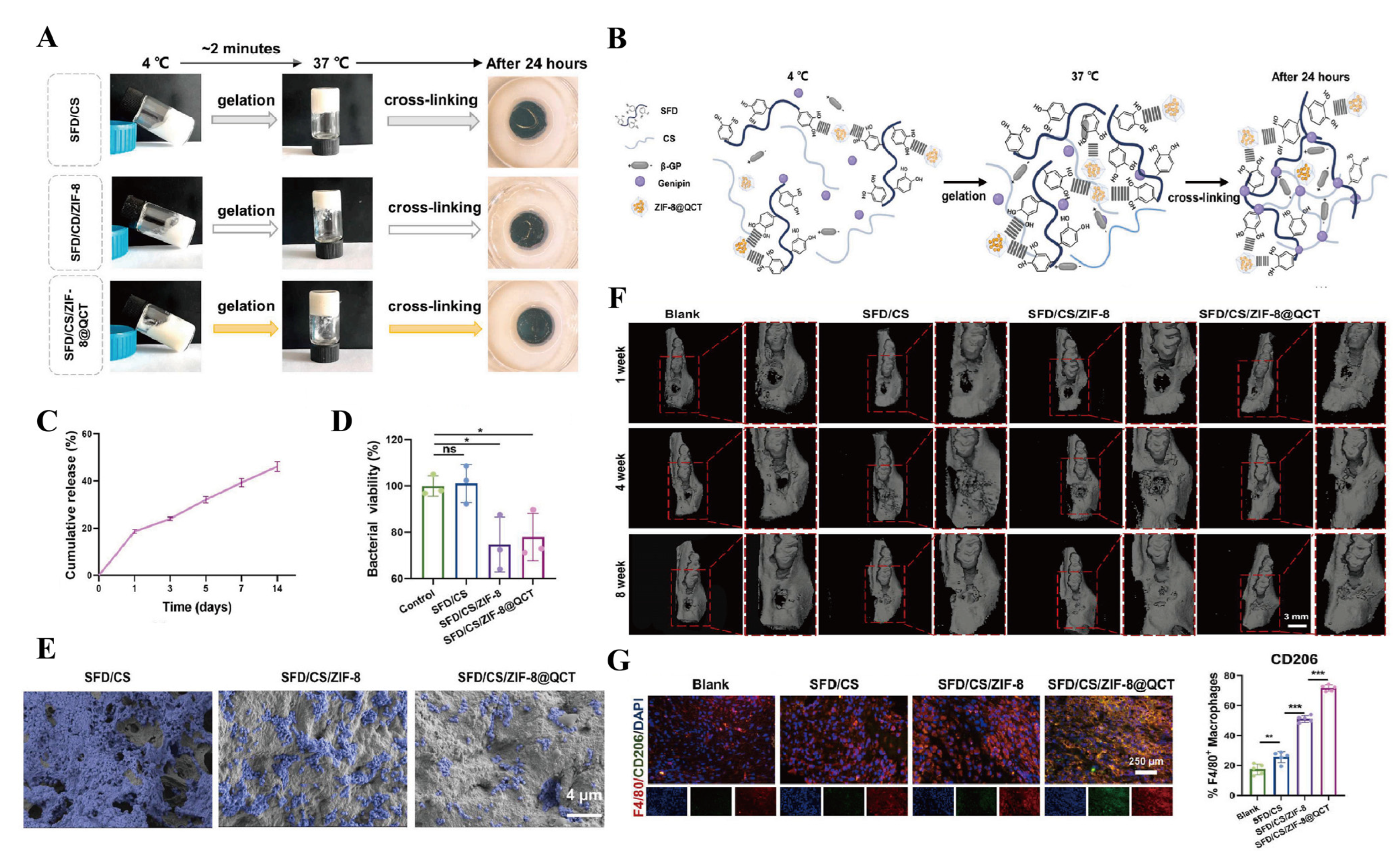
| SFD/CS/ZIF-8@QCT | Anti-inflammation; antibacterial effect; osteogenesis; stop bleeding; cell recruitment | 37 °C | Periodontitis bone defects | [104] |
| Hydrogel + EPO + FK506 | Anti-inflammation | 37 °C | Periodontitis bone defects | [110] |
| PCc/CPO/AsA/sEVs hydrogel | Dual antibacterial effect; osteogenesis | 37 °C | Periodontitis bone defects | [94] |
| bFGF/Dex lipo-gel | Angiogenesis; osteogenesis | 31/32 °C | Cranial defects | [101] |
| MCP10 + GF | Angiogenesis; innervation | 37 °C | Cranial defects | [111] |
| DCB/GP | Mechanical stability; osteogenesis | 37 °C | Radius defects | [74] |
| CoQ10/collagen hydrogel | Anti-inflammation | 25 °C | Extraction sockets of type II diabetic patients | [107] |
| PF127-BIO | Anti-inflammation | 37 °C | Periodontitis bone defects | [103] |
| CGF/Gel | Osteogenesis | 37 °C | Radius defects | [112] |
| FA-Qu-MEs@Gel | ROS scavenging ability; anti-inflammation | 37 °C | Periodontitis bone defects | [113] |
| MSC-EC-F127DA | Osteogenesis; angiogenesis | 25 °C/405 nm light source for 30 s | Extraction sockets | [92] |
| Hydrogel + Lentiv-GFP-PDGFBB | Osteoinductivity | 37 °C | Cranial defects | [114] |
| DEX@CHAp/Res@CHAp/CoI I/PLEL | Anti-inflammation | 37 °C | Femoral defects | [115] |
| T2DM/PPP-MM-S | ROS scavenging ability; osteogenesis | 35 °C | Diabetic periodontal bone defects | [116] |
| OVX-Inf-DVG | Anti-inflammation; treatment of osteoporosis | 37 °C | Femur metaphyseal fracture | [117] |
| GB@SIS | Anti-inflammation | 37 °C | Diabetic bone defect | [108] |
| 2Mg@PEG-PLGA gel | ROS scavenging ability; anti-inflammation | 37 °C | Osteoporotic bone defects | [118] |
| Material Name | Highlighting the Advantages | Gelation Conditions | Application | Refs. |
|---|---|---|---|---|
| 177Lu/AMP@CG | Cartilage protection; radionuclide retention | 37 °C | OA | [122] |
| MPA@MS@CS/SF | Mechanical stability; biocompatibility | 37 °C | Articular cartilage defects | [129] |
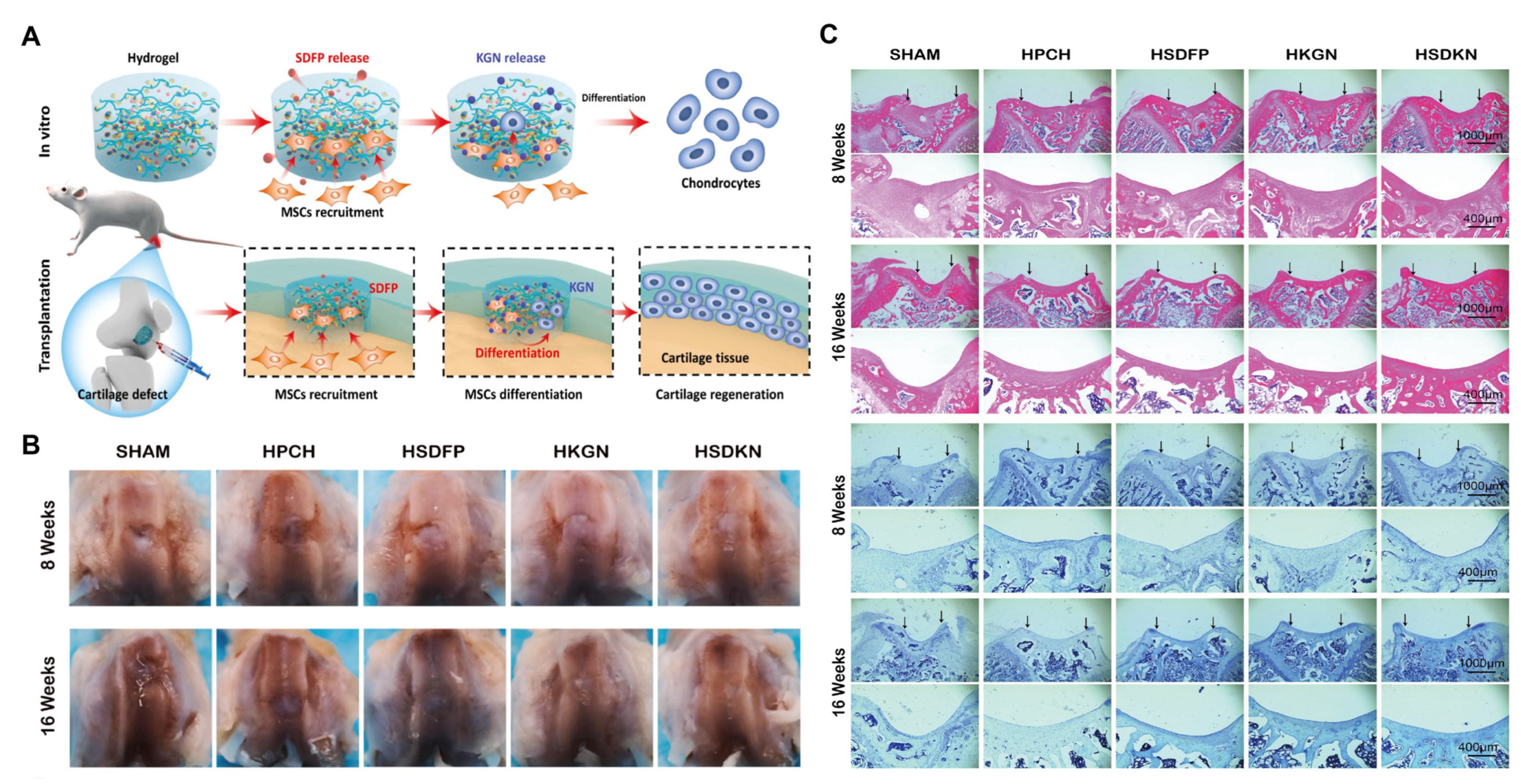
| HSDKN | Stem cell recruitment; chondrogenic differentiation | 37 °C | Articular cartilage defects | [124] |
| D-PDP@MC-Gel | Fibrocartilage hyalinization; fibrosis inhibition | 37 °C | Articular cartilage defects | [130] |
| PF127/GlcNAc | Cartilage protection; autophagy promotion | 37 °C | Articular cartilage defects | [123] |
| Exo@Gel | Anti-inflammation | 37 °C | OA | [131] |
| Exo-Gel | Local retention of exosomes | 37 °C | OA | [132] |
| p(VCL-co-HEMA) | Osteogenesis; cell sheet engineering | 37 °C | Bone and cartilage defects | [133] |
| αApoE-loaded hydrogel | Endochondral ossification | 37 °C | Bone fracture | [134] |
| PC + (HA-CP) | Anti-inflammation | 37 °C | RA | [128] |
| HA/PLL-ACM/TH | Chondrogenic differentiation | 37 °C | Osteochondral defects | [135] |
| Hyd CS/PF/BGP | Anti-inflammation | 37 °C | OA | [136] |
| CS1-PF25-TPP | High drug retention rate | 37 °C | OA | [137] |
| DMM + Gel@EXmodAtf5 | Activate chondrocyte autophagy; maintain mitochondrial function | 37 °C | OA | [138] |
| Material Name | Highlighting the Advantages | Gelation Conditions | Application | Refs. |
|---|---|---|---|---|
| CS/GP/HPC–GO–PPR | Antibacterial effect | 37 °C | Infected skin wounds | [158] |
| BP@Gel | Antibacterial effect; angiogenesis; collagen deposition | 37 °C | Infected skin wounds | [159] |
| CS + ASCs-Exos | Sustained exosome release; synergistic immunomodulation | 37 °C | Burned skin wounds | [160] |
| DHM-OTH | Oxygen-producing | 37 °C | Diabetic skin wounds | [161] |
| LL37@ZPF-2 Gel | Antibacterial effect; high encapsulation | 28 °C | Infected skin wounds | [162] |
| PF-OMSF@JK/DPSCs | Regulating and inducing stem cell differentiation | 37 °C | Spinal cord injury | [156] |
| PFLD hydrogel | Antibacterial effect; antioxidant properties | 33 °C | Infected skin wounds | [163] |
| Gel + TNF-R1 NV | Anti-inflammation | 37 °C | Burned skin wounds | [164] |
| 12%Hypo(rhNGF) | Nerve regeneration | 37.3 °C | Neurogenic keratitis | [165] |
| CIC Gel | Anti-inflammation | 37 °C | Muscle defect in sepsis | [166] |
| PH-127/PH | Skin cell proliferation; antibacterial effect | 37 °C | Infected skin wounds | [167] |
| PF-127 + HUCMSCs + SAP | Antibacterial action; angiogenesis; cell proliferation | 37 °C | Infected skin wounds | [168] |
| HAP/PDA/PF-127 | Nerve neovascularization | 37 °C | Sciatic nerve injury | [169] |
| Ni4Cu2/F127 | Enzyme-like activity | 37 °C | Skin wounds | [170] |
| DPSCs-PF127 | Angiogenesis | 37 °C | Fallopian tube mucosa defects | [171] |
| Lfcin-Nio/HG | Antibacterial effect; | 33–37 °C | Infected skin wounds | [172] |
| H-hUCESC-CM | anti-inflammation | 37 °C | Inflammatory bowel disease | [173] |
| CME-AgNPs-F127/F68 | Antibacterial effect | 37 °C | Infected skin wounds | [174] |
| PTX-NCS-gel | Antineoplastic | 37 °C | Breast cancer | [175] |
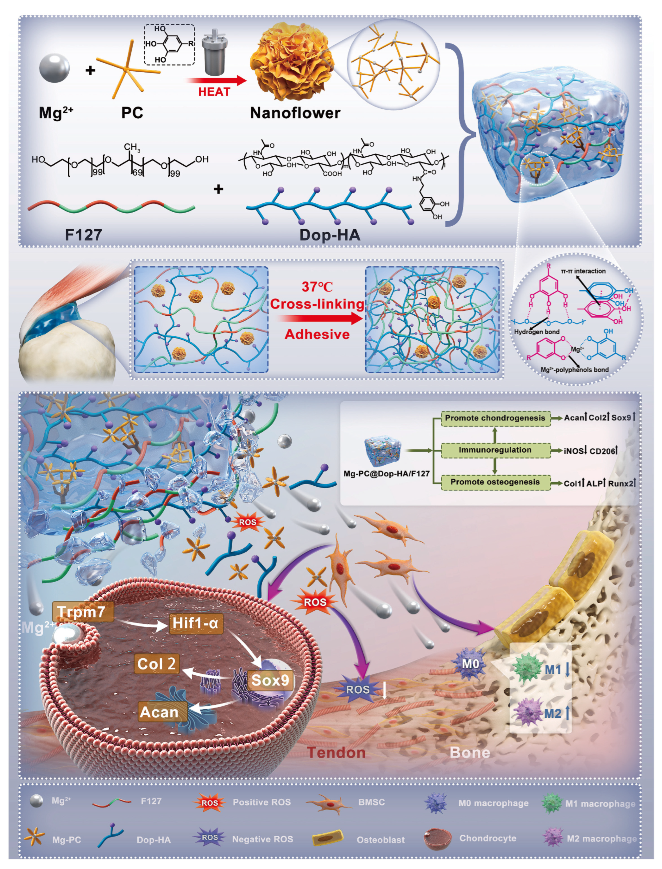
| Mg-PC@Dop-HA/F127 | Anti-inflammation; ROS scavenging ability | 37 °C | Tendon–bone interface | [151] |
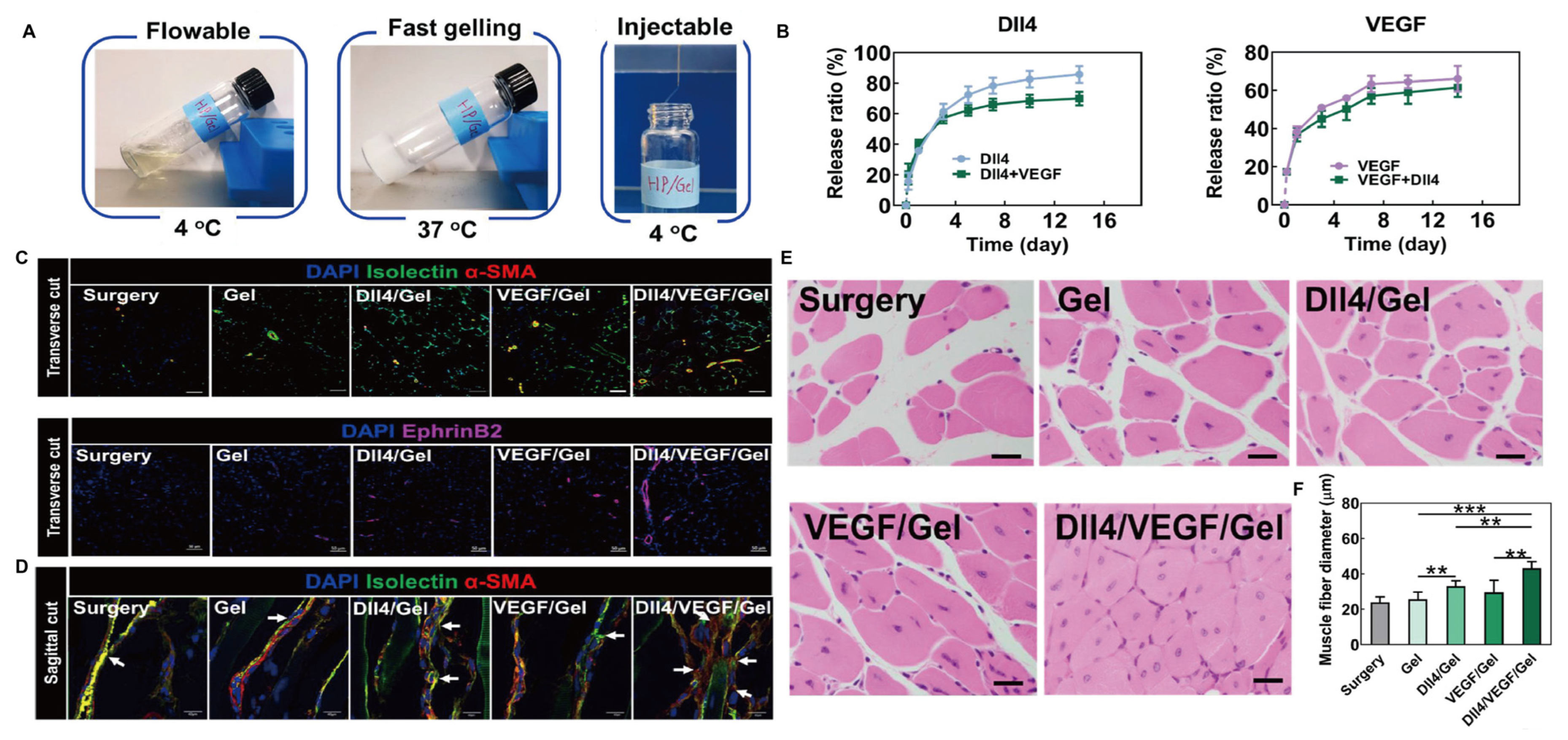
| DII4/VEGF/Gel | Cell proliferation; angiogenesis | 37 °C | Limb ischemia | [154] |
| PNI/RA-Amps3/E | Angiogenesis; fibroblast proliferation | 32 °C | Skin wounds | [176] |
| p(NIPAM-co-HEMIN)/ALG-EDA/AgNPs | Antibacterial effect; antioxidant dual-function | 37 °C | Diabetic skin wounds | [177] |
| TQ@PEG-PAF-Cur | Bacterium-targeted ROS generation | 30 °C | Infected skin wounds | [178] |
| CRO-HBC | Anti-inflammation; ROS scavenging ability | 37 °C | Burned skin wounds | [141] |
| HBCA3 | Antibacterial effect | 48 °C | Infected skin wounds | [179] |
| Implant + Hydrogel + Cisplatin | Antineoplastic | 37 °C | Osteosarcoma | [180] |
| Hydrogel nanocomposite | Removal of deep tumor cells | 37 °C | Postoperative treatment of glioblastoma | [148] |
| MCC@ZIF-8@Cur | Anti-inflammation; ROS scavenging ability | 28 °C | Diabetic skin wounds | [142] |
| UCMSC-bFGF-ECM-HP | Regulation of mitochondrial function | 37 °C | SCI | [181] |
| QTF@PNAGA hydrogel | Anti-tumor properties | 47 °C | Skin cancer | [182] |
| Gel + NEP1-40 | Protect neurons; promote axon growth | 37 °C | Brachial plexus nerve root avulsion injury | [183] |
| LPS/PPP | Anti-tumor properties | 37 °C | Colorectal cancer | [184] |
| UGN-102 | Anti-tumor properties | 37 °C | LG-NMIBC | [149] |
| G12G13MExos@Hydrogel | Anti-inflammation; neural stem cell differentiation | 34 °C | SCI | [157] |
| 5FU-Alg-Np-HG | High drug retention rate | 34 °C | Skin cancer | [185] |
| ASGP/SA/PNIPAM | Cell proliferation | 37 °C | Diabetic skin wounds | [186] |
| ILGA@Gel | Hemostasis; anti-inflammation; antibacterial effect; avoid reinfection | 37 °C | Skin wounds | [143] |
| PPP + CNTF | Anti-inflammation; antioxidant; neuroprotection | 37.5 °C | Optic nerve | [187] |
| Tissue Type | Key Advantages | Major Challenges |
|---|---|---|
| Bone | Adaptability of irregular bone defect shapes and treatment; slow release of GFs and anti-inflammatory drugs; no need for a second operation | Insufficient mechanical strength for large-segment bone defect repair; the degradation and regeneration rates are Difficult to match; insufficient long-term stability |
| Cartilage | Minimally invasive injection; sustained intra-articular drug release; enhance targeting ability; no need for a second operation | Insufficient long-term stability; low integration efficiency with native tissue |
| Skin | Simple operation; multiple response; combined antimicrobial/anti-inflammatory action | Unstable tissue adhesion; vulnerability to infection |
| Tumor | Minimally invasive; localized high-dose delivery; enhance targeting ability; photothermal combination; magnetic-thermal combination | Insufficient long-term stability; carrier biocompatibility issues; insufficient clinical validation |
| Tendon–bone interface | Minimally invasive; enhance targeting ability; no need for a second operation | Dynamic stress-induced interface failure; insufficient long-term stability |
| Muscle | Minimally invasive; no need for a second operation | Material displacement during contraction; limited efficacy in large-volume defect repair; the degradation and regeneration rates are difficult to match; insufficient long-term stability |
| Nerve | Mechanical compatibility; no need for a second operation | Blood–nerve barrier restrictions; insufficient long-term stability |
Disclaimer/Publisher’s Note: The statements, opinions and data contained in all publications are solely those of the individual author(s) and contributor(s) and not of MDPI and/or the editor(s). MDPI and/or the editor(s) disclaim responsibility for any injury to people or property resulting from any ideas, methods, instructions or products referred to in the content. |
© 2025 by the authors. Licensee MDPI, Basel, Switzerland. This article is an open access article distributed under the terms and conditions of the Creative Commons Attribution (CC BY) license (https://creativecommons.org/licenses/by/4.0/).
Share and Cite
Lv, T.; Chen, Y.; Li, N.; Liao, X.; Heng, Y.; Guo, Y.; Hu, K. A Comprehensive Review of Thermosensitive Hydrogels: Mechanism, Optimization Strategies, and Applications. Gels 2025, 11, 544. https://doi.org/10.3390/gels11070544
Lv T, Chen Y, Li N, Liao X, Heng Y, Guo Y, Hu K. A Comprehensive Review of Thermosensitive Hydrogels: Mechanism, Optimization Strategies, and Applications. Gels. 2025; 11(7):544. https://doi.org/10.3390/gels11070544
Chicago/Turabian StyleLv, Tianyang, Yuzhu Chen, Ning Li, Xiaoyu Liao, Yumin Heng, Yayuan Guo, and Kaijin Hu. 2025. "A Comprehensive Review of Thermosensitive Hydrogels: Mechanism, Optimization Strategies, and Applications" Gels 11, no. 7: 544. https://doi.org/10.3390/gels11070544
APA StyleLv, T., Chen, Y., Li, N., Liao, X., Heng, Y., Guo, Y., & Hu, K. (2025). A Comprehensive Review of Thermosensitive Hydrogels: Mechanism, Optimization Strategies, and Applications. Gels, 11(7), 544. https://doi.org/10.3390/gels11070544