Article

Taxonomy and Phylogeny of Lepiota Sect. Stenosporae (Verrucosporaceae) from Northeast China, with Six New Species and One New Record

Key Laboratory of Edible Fungal Resources and Utilization (North), Ministry of Agriculture and Rural Affairs, Jilin Agricultural University, Changchun 130118, China
*
Author to whom correspondence should be addressed.
J. Fungi 2026, 12(5), 355; https://doi.org/10.3390/jof12050355
Submission received: 30 March 2026 / Revised: 8 May 2026 / Accepted: 9 May 2026 / Published: 12 May 2026
(This article belongs to the Section Fungal Evolution, Biodiversity and Systematics)

Abstract

Lepiota sect. Stenosporae is characterized by a trichodermal or cutis-like pileus covering and spurred basidiospores. Although macroscopic similarities among its members complicate field identification, species can be delimited by combining multi-locus (ITS, nrLSU, rpb2, and tef1-α) phylogenetic analyses with distinct micro-morphological features. Using this integrative approach, we investigated specimens of sect. Stenosporae collected from Northeast China. A total of 12 species were successfully delimited, including six species new to science (Lepiota dolichospora, L. hongshiensis, L. jilinensis, L. microstenospora, L. sinocastanea, and L. sirupa) and one new record for China (L. grangei). Comprehensive morphological descriptions and line-drawing illustrations of microscopic features are provided for all recognized taxa. These findings expand the known species diversity of Lepiota in China and contribute morphological and molecular data for further systematic studies of this fungal group.

1. Introduction

The genus Lepiota (Pers.) Gray represents one of the most species-rich lineages within the family Verrucosporaceae [1]. It typically encompasses saprotrophic fungi that produce small to medium-sized, fragile basidiomata with a white spore print. These species are widely distributed across global temperate and tropical regions, commonly inhabiting forest humus layers and soil [2]. Due to their high diversity in macromorphology, coloration, and microscopic features—as well as the presence of amatoxins in certain taxa [3]—Lepiota has long been a focal point and a significant challenge in fungal taxonomy [4,5,6,7,8,9,10,11].
At the infrageneric level, the traditional sectional classification of the genus Lepiota has primarily relied on the microstructure of the pileus covering and the morphological characteristics of basidiospores [12,13,14,15]. Recent taxonomic re-assessments combining multi-locus phylogeny and morphological data [10] have resolved the long-standing polyphyly within the genus, clearly defining seven independent monophyletic clades. Based on the pileus covering type and basidiospore shape, these sections are distinguished as follows: sect. Cristatae Kühner ex Wasser features a hymeniform pileus covering and ellipsoid or spurred basidiospores; sect. Eriophorae (Bon) Reschke & Sarawi is characterized by a pileus covering composed of oblong to spherical cells and small ellipsoid basidiospores; sect. Fuscovinaceae Bon & Candusso currently accommodates only L. fuscovinacea F.H. Møller & J.E. Lange, a species unique within the genus for its fibrillose basidiomata and the absence of clamp connections; sect. Helveolae (Bon & Boiffard) Bon has a trichodermal pileus covering lacking a hymeniform basal layer, with ellipsoid to ovoid basidiospores; sect. Lepiota (Pers.) Gray possesses a trichodermal pileus covering with a hymeniform basal layer and ellipsoid, amygdaloid, or fusiform basidiospores; sect. Lilaceae Bon has a hymeniform pileus covering and ellipsoid basidiospores and sect. Stenosporae J.E. Lange ex Reschke & Sarawi is distinguished by a trichodermal or cutis-like pileus covering and characteristically spurred basidiospores.
Within this framework, Lepiota sect. Stenosporae stands out due to its distinctive spurred basidiospores and trichodermal or cutis-like pileus covering. However, the typification of this section has recently undergone a significant revision. Historically, L. pseudofelina J.E. Lange was designated as the type species [16]. Nevertheless, nomenclatural tracing indicates that this species was not included in the original concept of the section [17], and multigene phylogenetic analyses have revealed that L. pseudofelina actually belongs to Lepiota sect. Lepiota. To resolve this discrepancy and maintain taxonomic stability, a recent comprehensive phylogenomic re-assessment [10] designated L. castanea Quél. as the new type species for sect. Stenosporae. Unlike L. pseudofelina, L. castanea forms a highly stable, strongly supported monophyletic core in phylogenetic trees. Furthermore, this species perfectly aligns with the original taxonomic scope and the anatomical definition of the section due to its typical spurred basidiospores and classic trichoderm pileipellis [10]. Consequently, our study follows this updated taxonomic framework.
Although the taxonomic and nomenclatural framework of sect. Stenosporae has been recently stabilized; species within this group exhibit pronounced similarities in their macroscopic features, rendering field identification highly challenging. In China, previous taxonomic studies have recorded eight species in this section: L. castanea, L. erythrosticta (Berk. & Broome) Sacc., L. ignicolor Bres., L. luteocastanea E. Horak, L. mandarina Jun F. Liang & Zhu L. Yang, L. subcastanea Jun F. Liang & Zhu L. Yang, L. subcitrophylla Hongo, and L. tomentella J.E. Lange [18,19]. Currently, L. erythrosticta and L. luteocastanea still lack reliable molecular data support. Specifically in Northeast China, the recent literature has documented only three species (L. mandarina, L. castanea, and L. subcastanea) [19]. These contributions provide valuable baseline data for evaluating the fungal resources of the region. Building upon this foundation, the application of modern multi-locus phylogenetic approaches is essential to more comprehensively reveal the actual species composition and diversity in this area.
To address this gap, the present study details the results of an intensive four-year field investigation conducted in Northeast China. By employing an integrative taxonomic approach that combines thorough morphological observations with multi-locus phylogenetic analyses, 12 species of sect. Stenosporae were successfully delimited from this region. Among them, six are described as species new to science, one is reported as a new record for China, and the remaining five are identified as known species (including L. tomentella J.E. Lange and L. pilodes Vellinga & Huijser, newly discovered in this region). Comprehensive morphological descriptions and line-drawing illustrations of microscopic features are provided for all recognized taxa.

2. Materials and Methods

2.1. Morphological Studies

Specimens were collected from Jilin Province and Heilongjiang Province, Northeast China, from July 2023 to August 2025. Macro-morphological characters and ecological data were recorded in situ, supplemented by high-resolution photographs of the basidiomata in their natural habitats. Color changes upon bruising or injury were documented; all color descriptions were based on a standard color chart [20]. Morphological descriptions follow established taxonomic standards [12,21]. The dried specimens are deposited in the Fungarium of Jilin Agricultural University (HMJAU/FJAU).
Global distribution data for specific taxa were retrieved and verified using the Global Biodiversity Information Facility (GBIF; https://www.gbif.org, accessed on 8 May 2026) [22].

2.2. DNA Extraction, PCR Amplification, Sequencing

Total genomic DNA was extracted from fresh or dried specimens using the NuClean PlantGen DNA Kit (CWBIO, Beijing, China). Four gene fragments were amplified via PCR: the internal transcribed spacer (ITS), the nuclear ribosomal large subunit (nrLSU), the second largest subunit of RNA polymerase II (rpb2), and the translation elongation factor 1-alpha (tef1-α). The primer pairs used were ITS1F/ITS4 [23,24], LR0R/LR5 [25], 6F/RPB2-7.1R [26], and EF1-983F/EF1-1567R [27], respectively. PCR protocols and thermal cycling conditions were based on previous studies [28]. Amplicons were sequenced by Sangon Biotech (Shanghai, China), and all resulting sequences were deposited in GenBank.

2.3. Phylogenetic Studies

To evaluate the phylogenetic relationships and accurately delimit species within Lepiota sect. Stenosporae, a combined multi-locus dataset (ITS, nrLSU, rpb2, and tef1-α) was assembled. The dataset comprised a total of 173 ITS, 60 nrLSU, 36 rpb2, and 24 tef1-α sequences (accession numbers are provided in Table 1). Among these, 150 sequences were newly generated in this study (88 ITS, 29 nrLSU, 13 rpb2, and 20 tef1-α), while the remaining sequences were retrieved from the GenBank database. Based on established taxonomic frameworks for Lepiota [10], representative species from other sections of Lepiota and the closely related genus Echinoderma were included to construct the phylogenetic tree of Lepiota sect. Stenosporae. Sequences of Cystolepiota s.l. were selected as the outgroup to root the phylogenetic trees.
Table 1. Information on DNA sequences used in the phylogenetic analyses. Sequences newly generated in this study are shown in bold. “—” means data not available. Capital letters after voucher represent Holotype “H”.
TaxonVoucherOriginITSLSUrpb2tef1References
Cystolepiota hongshiensisHMJAU68205ChinaOR947187OR960533PP465917[28]
C. luteosquamulosaHMJAU67711ChinaOR233619OR240263PP465910[28]
Echinoderma “eriophorum”RITF2533ChinaMK685378MK685376MK705800Direct Submission
E. asperumHAW: JKS142USAMK412598[29]
E. asperumHMJAU37543ChinaPZ184987This study
E. flavidoasperumKUN-HKAS 87905 ChinaMN810147MN810098MN820969MN820903[30]
E. hystrixC-F-13684 HDenmarkPQ152708[11]
E. perplexumKaiR1713GermanyPP594623PP594751PP841165[10]
Lepiota  aff. castaneaFJAU78168ChinaPZ185037PZ185090PZ213593This study
L.  aff. castaneaFJAU78195ChinaPZ185027PZ185087This study
L. aff. castaneaFJAU78167ChinaPZ185040PZ185092PZ213594This study
L. aff. castaneaFJAU78281ChinaPZ185019This study
L. aff. subcastaneaFJAU78179ChinaPZ185000PZ185077PZ213592This study
L. aff. subcastaneaFJAU78189ChinaPZ185070PZ185103PZ213591This study
L. aff. subcastaneaFJAU78286ChinaPZ184994This study
L. aff. subcastaneaFJAU78178ChinaPZ185007This study
L. aff. subcastaneaFJAU78193ChinaPZ184992This study
L. aff. subcastaneaFJAU78285ChinaPZ185016This study
L. aff. subcastaneaFJAU78177ChinaPZ185020This study
L. aff. subcastaneaFJAU78180ChinaPZ185002PZ185079This study
L. alopochroaMFLU 09-0178ThailandHQ647294[7]
L. andegavensis8-X-1994, P.D.H. Roux 2121FranceAY176461[4]
L. andegavensisT1-2LebanonMZ088077Direct Submission
L. atrobrunneodiscaHSA 115 HChinaOP724226OP724232[31]
L. aurantiicolorSeSa176 HBeninPP594563PP594681PP841188[11]
L. baiyunensisB22052705 HChinaOQ547186OQ547188[32]
L. baiyunensisFJAU78273ChinaPZ185049This study
L. baiyunensisFJAU79130ChinaPZ185072This study
L. boudieriFJAU78282ChinaPZ185062This study
L. boudieriFJAU78221ChinaPZ185017This study
L. boudieriHKAS 5803ChinaEU416280[33]
L. brunneoaurantiaUEH-F0006 HPakistanOR464184[34]
L. brunneoincarnataFJAU78269ChinaPZ185051This study
L. brunneoincarnataFJAU78256ChinaPZ185014This study
L. brunneoincarnataHMAS 63488ChinaEU416302[33]
L. brunneoolivaceaSeSa214 HBeninPP594574PP594693PP841201[11]
L. brunneopileataLAH:37842PakistanOQ970543[35]
L. castaneaN.J. Dam 97020NetherlandsAY176463[4]
L. castaneaFJAU78164ChinaPZ185052PZ185095PZ213573PZ213585This study
L. castaneaFJAU78163ChinaPZ185038PZ185091This study
L. castaneaFJAU78165ChinaPZ185004This study
L. castaneaHKAS 48817ChinaEU416282[33]
L. castaneaS7GermanyOL527673[3]
L. castaneaFJAU78176ChinaPZ185025This study
L. castaneaFJAU78175ChinaPZ185024This study
L. castaneaFJAU78166ChinaPZ184997This study
L. castaneidiscaE.C. Vellinga 2516 (UC)USAAF391065[36]
L. cf. erythrostrictaCL_MART_06.091MartiniquePP594625[11]
L. citrophyllaHNL502981LaosKX711968[9]
L. citrophyllaFJAU78220ChinaPZ185028This study
L. cristataHKAS49258ChinaEU081937[37]
L. cristataFJAU79132ChinaPZ185056This study
L. cristataFJAU79133ChinaPZ185059This study
L. dolichosporaFJAU78183 HChinaPZ185053PZ185096PZ213574PZ213587This study
L. dolichosporaFJAU78184ChinaPZ185060PZ185098This study
L. dolichosporaFJAU78185ChinaPZ185012PZ185082This study
L. dolichosporaFJAU78192ChinaPZ185046This study
L. elseaeAH:40487 HSpainNR_158471[38]
L. faiaebravaeBCN-IC11111501PortugalPP622390[39]
L. flavonigrescensSeSa305 HBeninPP594596PP594717PP841226[11]
L. flavostipitataSeSa182BeninPP594568PP594686PP841193[11]
L. fuscovinacea E.C. Vellinga 2255NetherlandsAY176372AY176373[5]
L. grangeiKaiR1768GermanyPP594624PP594638PP841166[11]
L. grangeiFJAU78231ChinaPZ185018This study
L. grangeiFJAU78230ChinaPZ185071PZ185104This study
L. griseovirensSeSa91GermanyPP594548PP594665PP841272[11]
L. helveolaS.D. Russell HRL2181CanadaMH979466Direct Submission
L. hongshiensisFJAU78254 HChinaPZ185036PZ185089This study
L. hongshiensisFJAU78255ChinaPZ185023PZ185086This study
L. hongshiensisFJAU78295ChinaPZ185009This study
L. ignicolor17X1999 H. A. HuijserNetherlandsAY176472[4]
L. jilinensisFJAU78248 HChinaPZ185021PZ185085PZ213570PZ213580This study
L. jilinensisFJAU78249ChinaPZ185035PZ185088PZ213571PZ213579This study
L. lahorensisT18/LAH:10002012 HPakistanKT182475[40]
L. lilaceaE.C. Vellinga 1873UKGQ203820[41]
L. lilaceostriataSeSa280 HBeninPP594588PP594709PP841217[11]
L. longisterigmataSeSa179 HBeninPP594565PP594683PP841190[11]
L. maerimensisMFLU 12-2036ThailandMW251839MW251847[42]
L. magnisporaHMJAU33688ChinaPZ184986This study
L. magnisporaZ.L. Yang 2521ChinaAF391006[36]
L. mandarinaHKAS 50028 HChinaKM214811KM214816[18]
L. metulisporaHMGID25584UnknownMK651632Direct Submission
L. metulisporaFJAU79138ChinaPZ184988This study
L. metulisporaFJAU79137ChinaPZ184989This study
L. microstenosporaFJAU78246 HChinaPZ185011PZ185081PZ213567PZ213581This study
L. microstenosporaFJAU78247ChinaPZ185043PZ185094PZ213572PZ213582This study
L. neophanaRITF2402UnknownMK651599Direct Submission
L. neophanaFJAU79131ChinaPZ185055This study
L. neophanaE.C. Vellinga 3955 (UC)USAGQ203811[41]
L. omninoflavaKUN-HKAS 106734 HTChinaMN810157MN810092MN820951MN820923[30]
L. omninoflavaHMJAU68258ChinaOR936203[28]
L. pallidiochraceaFJAU79135ChinaPZ185050This study
L. pallidiochraceaFJAU79136ChinaPZ185067This study
L. pallidiochraceaHKAS:45579ChinaNR_158462[7]
L. pilodesSeSa382GermanyPP594615PP841248[11]
L. pilodesFJAU78232ChinaPZ185001PZ185078PZ213588This study
L. pilodesFJAU78283ChinaPZ185048This study
L. pilodesE.C. Vellinga 3234 (UC)USAEF080865[43,44]
L. poliochloodesMFLU 081272ThailandHQ647296[7]
L. pseudovenenosaSeSa302 HBeninPP594593PP594714PP841223[11]
L. rhodophyllaUC(USA-CA):1860004 HUSANR_119624[45]
L. sinocastaneaFJAU78172ChinaPZ185065PZ185101PZ213577PZ213583This study
L. sinocastaneaFJAU78160ChinaPZ185039This study
L. sinocastaneaFJAU78171 HChinaPZ185063PZ185100PZ213584This study
L. sinocastaneaFJAU78162ChinaPZ185044This study
L. sinocastaneaFJAU78170ChinaPZ185041PZ185093PZ213595This study
L. sinocastaneaFJAU78161ChinaPZ184998PZ185076PZ213564This study
L. sirupaFJAU78262ChinaPZ185057This study
L. sirupaFJAU78261 HChinaPZ185058PZ185097PZ213575PZ213586This study
L. sosuensisJBSD:CA3Dominican RepublicNR_184875[46]
L. spiculataJBSD:127426Dominican RepublicMK696156MK696155MK696576MK696577[47]
L. squamulodiffractaCA21 HTDominican RepublicKR022006[46]
L. squamulosaHMJAU68251ChinaOR936197[28]
L. squamulosaFJAU79134ChinaPZ185047This study
L. subalbaCWU(MYC)8416UkraineOK041522[48]
L. subalbaSeSa11AustriaOL527683[48]
L. subcastaneaHMJAU 3889ChinaKM214814[18]
L. subcastaneaHKAS 45633 HChinaKM214812KM214817[18]
L. subcastaneaHKAS49183ChinaMK651652MK685373Direct Submission
L. tomentellaFJAU78152ChinaPZ185010PZ185080PZ213566PZ213589This study
L. tomentellaFJAU78159ChinaPZ184999This study
L. tomentellaFJAU78157ChinaPZ185022This study
L. tomentellaFJAU78158ChinaPZ185045This study
L. tomentellaFJAU78151ChinaPZ185061PZ185099PZ213576PZ213590This study
L. tomentellaRITF570UnknownMK651653Direct Submission
L. tomentellaFJAU78154ChinaPZ185008This study
L. tomentellaFJAU78155ChinaPZ185005This study
L. tomentellaFJAU78153ChinaPZ185015This study
L. tomentellaSeSa58GermanyPP594536PP594652PP841259[11]
L. tomentellaH.A.Huijser (L)NetherlandsEF080868[43]
L. tyrianthinaSeSa213BeninPP594573PP594692PP841200[11]
L. vellinganaMCR09PakistanHE974764[49]
L. xanthophyllaE.C. Vellinga 2240 (L)NetherlandsAY176405AY176406[5]
Lepiota sp.CUH AM839IndiaOR594353Direct Submission
Lepiota sp.PS201885ThailandOP020473Direct Submission
Lepiota sp.110114MFBPC084ChinaMW488365Direct Submission
Lepiota sp.MCVE 480UnknownFJ998390Direct Submission
Lepiota sp.SeSa110BeninPP594556PP594674PP841180[11]
Lepiota sp.SeSa255BeninPP594582PP594703PP841211[11]
Lepiota sp.SeSa346BeninPP594608PP594731PP841240[11]
Lepiota sp.ZRL20180888ChinaPV607760PV607733PV614778PV614794[50]
Lepiota sp.HKAS 82453ChinaMN810130MN810085[30]
Lepiota sp.BG0014PakistanOM809699Direct Submission
Lepiota sp.BG0015PakistanOM811292Direct Submission
Lepiota sp.MFLU 09-0183ThailandHQ647297Direct Submission
Lepiota sp.E.C. Vellinga 2603USAAY176481[4]
Lepiota sp.E.C. Vellinga 2601 (UC)USAAY176479[4]
Lepiota sp.S.D. Russell iNaturalist #63266348USAOM522742Direct Submission
Lepiota sp.S.D. Russell iNaturalist # 8607856USAMN906140Direct Submission
Lepiota sp.MycoMap #7207USAOM522727Direct Submission
Lepiota sp.FJAU78209ChinaPZ185013PZ185084PZ213569PZ213596This study
Lepiota sp.FJAU78210ChinaPZ185068PZ185102PZ213578This study
Lepiota sp.FJAU78191ChinaPZ185026This study
Lepiota sp.FJAU78264ChinaPZ185029This study
Lepiota sp.FJAU78190ChinaPZ184996This study
Lepiota sp.FJAU78181ChinaPZ185006This study
Lepiota sp.FJAU78188ChinaPZ184993This study
Lepiota sp.FJAU78298ChinaPZ185034This study
Lepiota sp. FJAU78268ChinaPZ185033This study
Lepiota sp.FJAU78266ChinaPZ185030This study
Lepiota sp.FJAU25097ChinaPZ184990This study
Lepiota sp.FJAU78270ChinaPZ185069This study
Lepiota sp.FJAU78252ChinaPZ184995This study
Lepiota sp.FJAU78253ChinaPZ185042This study
Lepiota sp.FJAU78267ChinaPZ185032This study
Lepiota sp.FJAU78257ChinaPZ185003This study
Lepiota sp.FJAU78156ChinaPZ185054This study
Lepiota sp.FJAU78272ChinaPZ185066This study
Lepiota sp.FJAU78259ChinaPZ185031This study
Lepiota sp.HMJAU28816ChinaPZ184991This study
Lepiota sp.FJAU78229ChinaPZ184985PZ185083PZ213568PZ213565This study
Melanophyllum eyreiFJAU79139ChinaPZ185064This study
M. haematospermumK(M):176342UKMZ159454Direct Submission
Sequences for each locus were aligned independently using MAFFT v.7.110 [51], followed by manual inspection and further optimization in MEGA v.7.0.26 [52]. Unreliable alignment regions and gaps were removed using the “-automated1” command in trimAl [53]. The aligned sequences were then concatenated using PhyloSuite v.2 [54], generating a combined alignment of 2780 base pairs (bp) in length. The sequence partitions were defined as follows: ITS (1–710 bp), nrLSU (711–1579 bp), rpb2 (1580–2232 bp), and tef1-α (2233–2780 bp). Gaps were treated as missing data.
Phylogenetic reconstructions were performed using Maximum Likelihood (ML) and Bayesian Inference (BI) methods. To address the evolutionary heterogeneity among different gene regions, partitioned analyses were applied to the combined dataset. For the ML analysis, ModelFinder v.3.0.1 [55] was used to select the best-fit substitution models under the Akaike Information Criterion (AIC). The ML tree was inferred using IQ-TREE v.3.0.1 [56] with an edge-linked partition model. The selected best-fit models were TVM+F+I+R5 for ITS, GTR+F+I+R2 for nrLSU, TIM2+R3 for rpb2, and TIM3+I+R3 for tef1-α. Branch support was assessed using 1000 ultrafast bootstraps [57] and the Shimodaira–Hasegawa approximate likelihood-ratio test (SH-aLRT) [58].
For the BI analysis, ModelFinder v.3.0.1 [55] was employed to determine the optimal partition models based on the Bayesian Information Criterion (BIC), resulting in GTR+F+I+G4 for ITS and nrLSU, HKY+I+G4 for rpb2, and GTR+I+G4 for tef1-α. The BI analysis was conducted using MrBayes v.3.2.7a [59], running two parallel chains for 4,981,000 generations. The analysis was terminated when the average standard deviation of split frequencies (ASDSF) fell below 0.006. The initial 22% of the sampled data were discarded as burn-in. The resulting phylogenetic trees were visualized and annotated using FigTree v.1.4.3 [60] and tvBOT v.2.6.1 [61].

3. Results

3.1. Phylogenetic Analyses

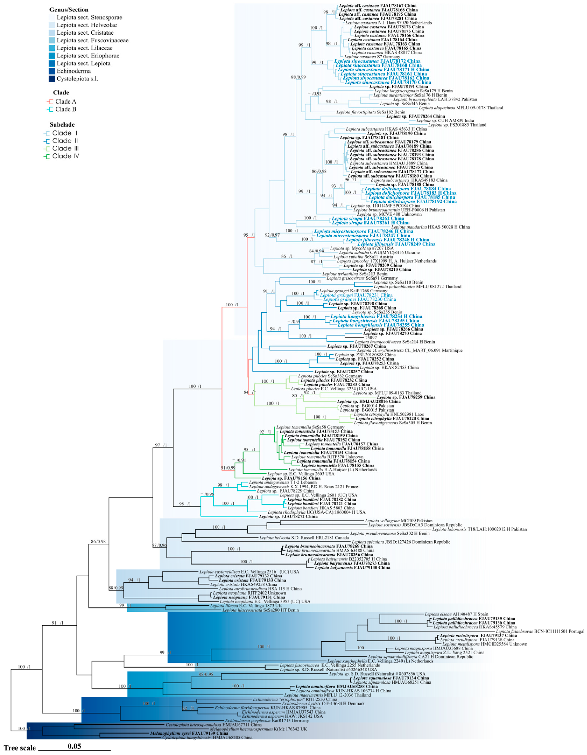
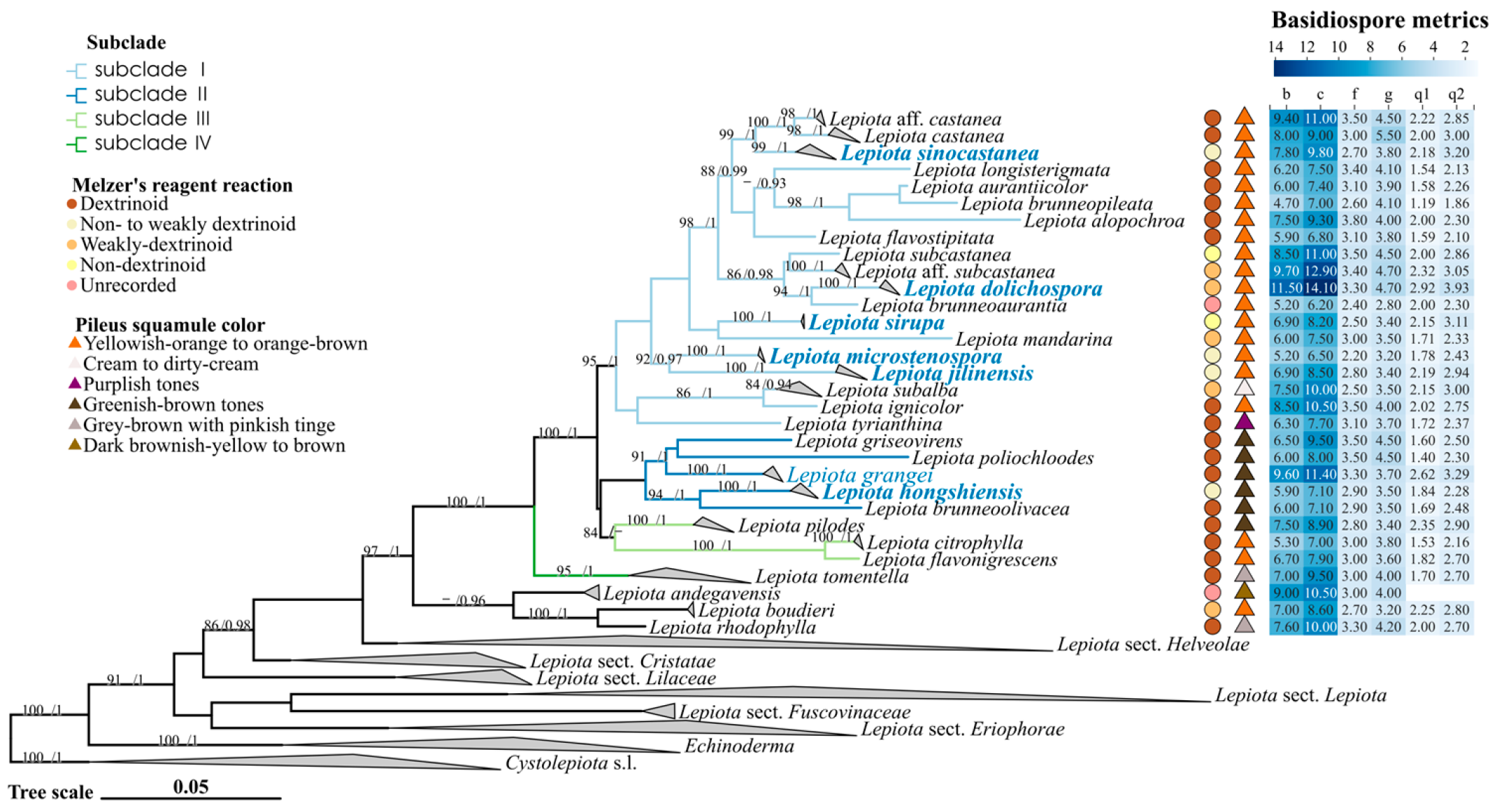
The phylogenetic relationships of Lepiota sect. Stenosporae were reconstructed using a concatenated dataset of ITS, nrLSU, rpb2, and tef1-α. The topologies generated by Maximum Likelihood (ML) and Bayesian Inference (BI) were congruent; thus, only the BI tree is presented (Figure 1 and Figure 2), with branch support values indicated at the nodes (UFBoot > 80%/PP > 0.90). In the phylogenetic reconstruction, newly described species are highlighted in bold blue, new records are indicated in blue, and newly generated sequences are marked in bold black. Using Cystolepiota s.l. as the outgroup, the Lepiota was resolved as a robust monophyletic group (91/1), within which sect. Stenosporae (97/1) was also recovered as a monophyletic lineage. Internally, the section diverged into two major well-supported clades, clade A (100/1) and clade B (98/1), which correlate with distinct types of pileus covering. Clade A is characterized by a trichodermal pileus covering, whereas clade B possesses a cutis-like pileus covering.
Within clade A (Figure 1), four distinct subclades (subclades I–IV) were identified. Subclade I (95/1) predominantly comprises species with orange-brown tones in their pileus scales, accommodating five new species described herein: Lepiota sinocastanea, L. dolichospora, L. sirupa, L. microstenospora, and L. jilinensis. Morphologically, the lamellae and stipe base of L. sinocastanea turn orange-red upon bruising, a characteristic rarely observed in other members of this subclade. Lepiota dolichospora is readily distinguished from its closely related species, L. subcastanea and L. brunneoaurantia, by its significantly larger and elongated basidiospores (Qav = 3.31). Lepiota sirupa, L. microstenospora, and L. jilinensis each occupy distinct phylogenetic lineages. Notably, L. sirupa possesses non-dextrinoid basidiospores, an uncommon trait within this subclade, and is currently known only from its type locality. Lepiota microstenospora and L. jilinensis are resolved as sister taxa; however, the former can be differentiated from the latter by its smaller basidiospores and narrower cheilocystidia. Subclades II and III are recovered as sister clades, although both their respective internal nodes and their most recent common ancestral node lack significant statistical support. Subclade II comprises species with greenish-brown pileus scales, including the new species L. hongshiensis and the new record for China, L. grangei. Lepiota hongshiensis, currently known only from its type locality, is phylogenetically most closely related to L. brunneoolivacea Sarawi. However, the lamellae of the latter shift to brownish-red upon maturation, a characteristic absent in L. hongshiensis. Although previously recorded from Europe, North America, South America, and various parts of Asia (including Western Asia, East Asia, and Southeast Asia), the occurrence of L. grangei in China is confirmed here for the first time based on molecular evidence. Subclade III (84/–) includes three species: L. citrophylla (Berk. & Broome) Sacc., L. flavonigrescens Sarawi & Reschke, and L. pilodes. Subclade IV (91/0.99) predominantly consists of L. tomentella along with two undetermined sequences labeled as Lepiota sp.
Conversely, clade B (98/1) (Figure 1) currently comprises only three known species, namely L. andegavensis, L. boudieri, and L. rhodophylla, along with three undetermined sequences labeled as Lepiota sp.

3.2. Taxonomy

Lepiota dolichospora T. Bau & X.Y. Zhou, sp. nov.
MycoBank No: MB862979
Holotype: CHINA. Jilin Province, Huadian City, Hongshi National Forest Park, 27 August 2023, X.Y. Zhou and T. Bau, FJAU78183.
Etymology: “dolichospora” is derived from the Greek ‘dolichos’ (long) and ‘spora’ (spore), referring to the remarkably elongated and slender basidiospores, which characterize this species within the section Stenosporae.
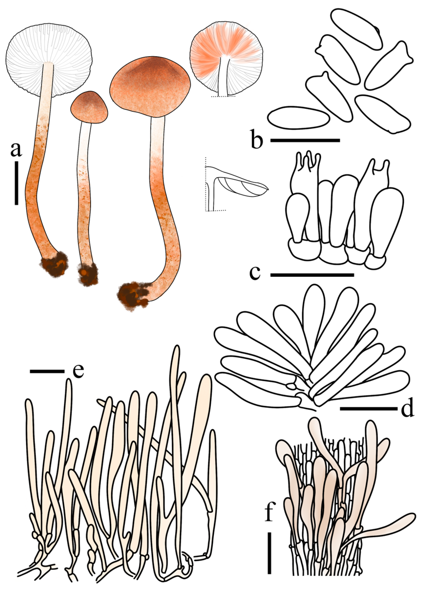
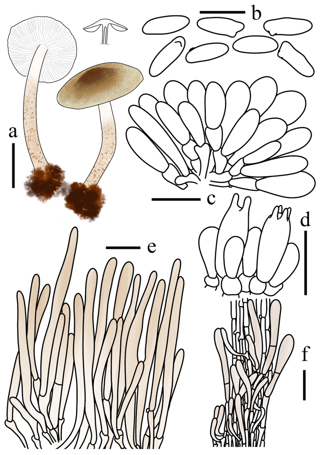
Diagnosis: Lepiota dolichospora is primarily distinguished by its small-sized basidiomata with a light orange to deep orange pileus covered in minute squamules; basidiospores that are remarkably elongated and spurred, measuring 11.5–14.1 × 3.3–4.7 μm with an exceptionally high average Q-value (Qav = 3.31) and showing a weakly dextrinoid reaction; cheilocystidia that are predominantly narrowly utriform to utriform; and a pileus covering structured as a trichoderm with slightly thick-walled terminal cells. It differs from L. subcastanea by its much longer and narrower spores (Qav = 2.48 in L. subcastanea) and from L. brunneoaurantia by its significantly larger spore dimensions.
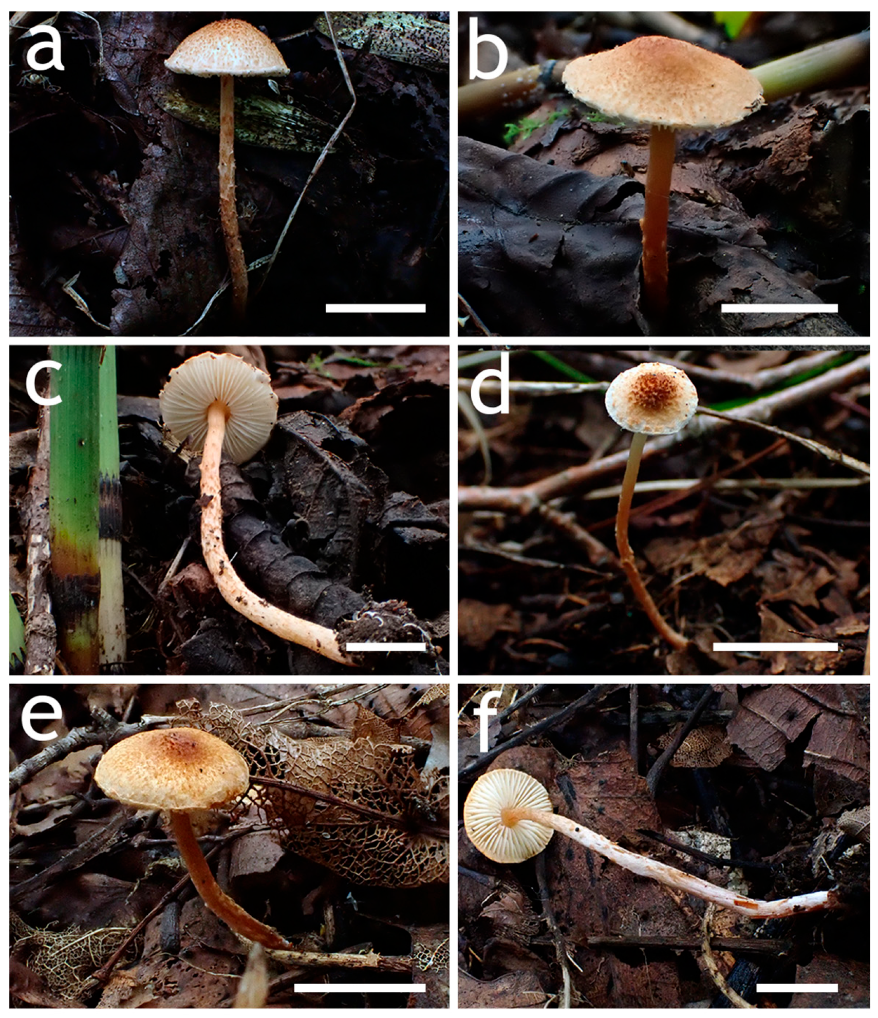
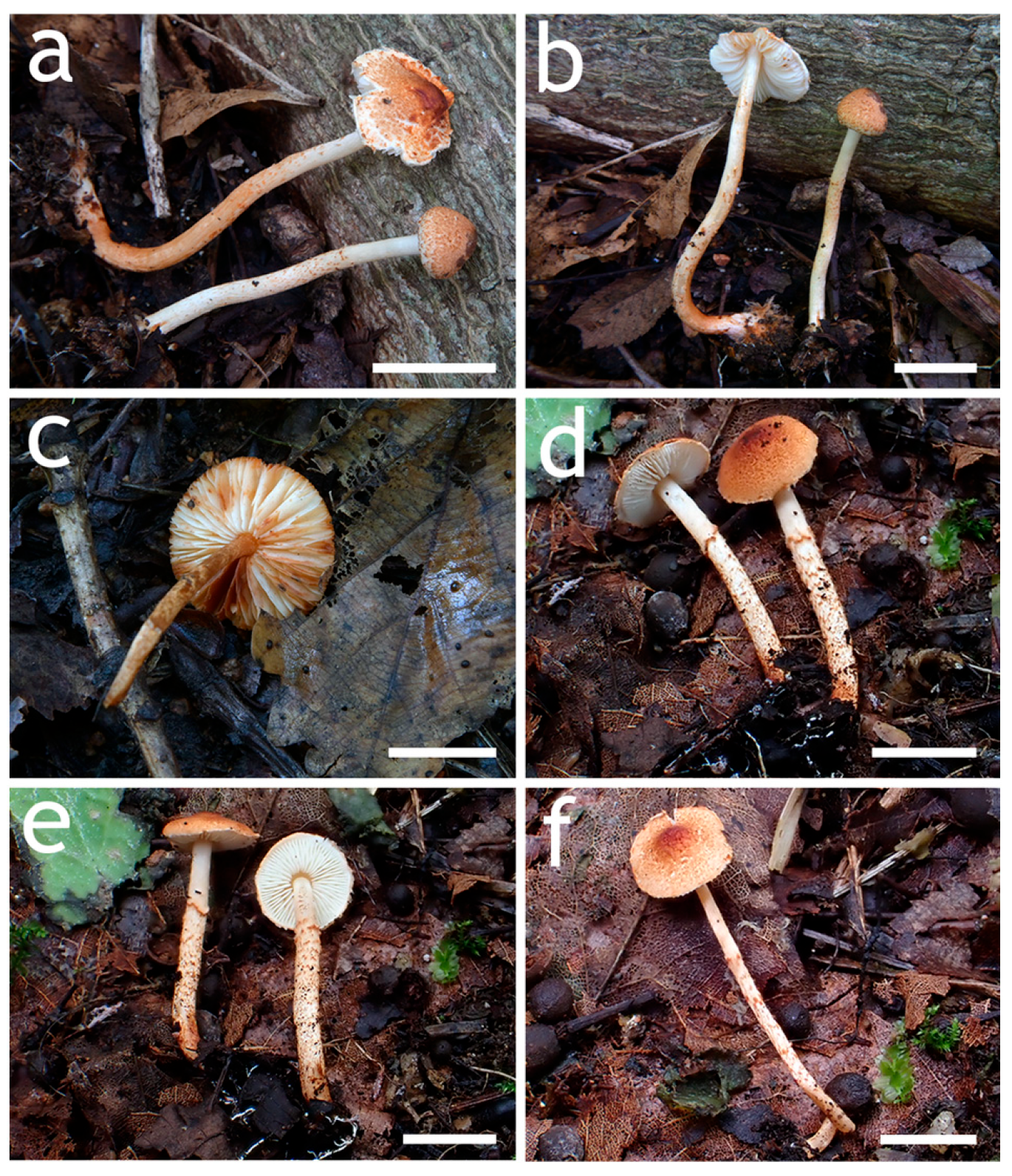
Basidiomata small. Pileus 0.8–2.3 cm in diam., campanulate when young, expanding to plano-convex at maturity; surface white, densely covered in light orange (6A3–A5) to deep orange minute squamules; central squamules brownish orange (7C6–C8); margin with veil remnants concolorous with the squamules. Context thin, white, unchanging when bruised. Lamellae free,, crowded, interspersed with lamellulae of unequal length, ventricose, white to cream; edge entire, concolorous. Stipe 3.8–8.1 × 0.2–0.4 cm, subcylindrical, slightly thickening towards the base, white to orange white (6A2), with a distinct annular zone; surface nearly glabrous above the annular zone, lower portion densely ornamented with light orange (6A3–A5) to deep orange (6A6–A8) squamules arranged in interrupted annular bands. Odour and taste not recorded.
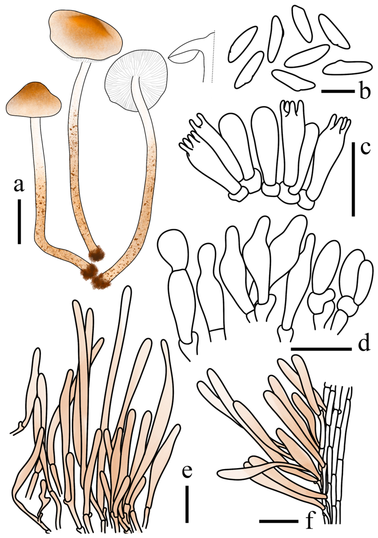
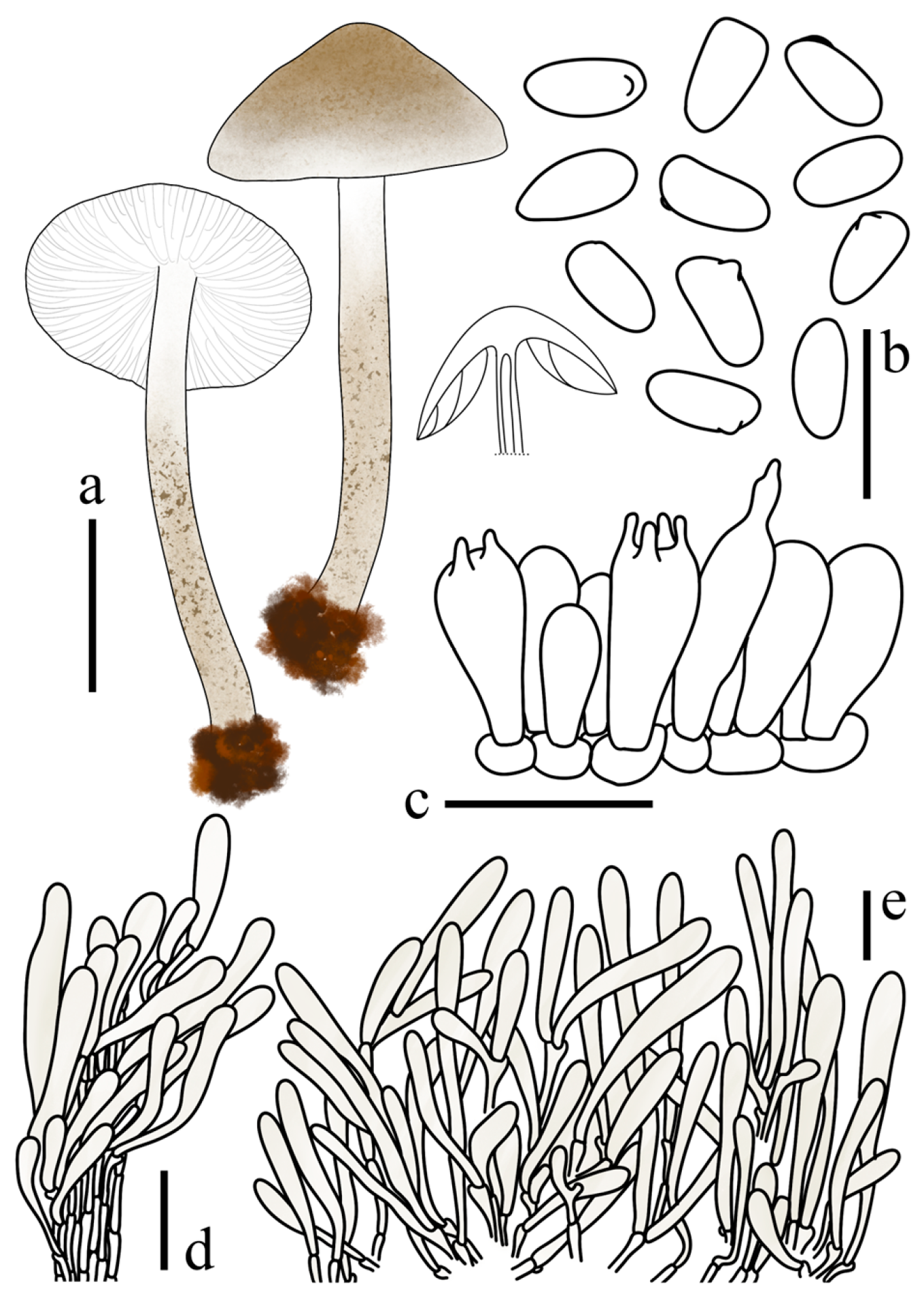
Basidiospores (40/2/2) 11.5–14.1 × 3.3–4.7 μm, avl × avw = 12.9 × 3.9 μm, Q = 2.92–3.93, Qav = 3.31, spurred, remarkably elongated, sub-triangular in side view, long-ellipsoid in frontal view; wall hyaline, thin, weakly dextrinoid. Basidia 17–27 × 6–11 μm, clavate, 4- (2-) spored, thin-walled. Lamella edge sterile. Cheilocystidia 21–38 × 5–11 μm, predominantly narrowly utriform to utriform, occasionally fusiform, hyaline, thin-walled. Pleurocystidia absent. Pileus covering a trichoderm; terminal cells 65–218 × 7–17 μm, erect, cylindrical to clavate, with rounded apices and slightly thick walls, containing light orange (6A3–A5) to greyish orange (6B3–B5) intracellular pigments. Stipe covering a cutis; terminal cells 32–180 × 9–16 μm, appressed, cylindrical to clavate, containing light orange (6A3–A5) to greyish orange (6B3–B5) intracellular pigments. Clamp connections present.
Habit and Habitat. Solitary on soil within the humus layer of broad-leaved forests dominated by Betula spp., Fraxinus mandshurica Rupr., and Juglans mandshurica Maxim.
Known distribution: Jilin Province, China.
Additional specimens examined: CHINA. Jilin Province, Jiaohe City, Shansongling, 24 July 2022, W.N. Hou, X. Wang, FJAU78185, FJAU78192; Huadian City, Hongshi National Forest Park, 16 August 2024, X.Y. Zhou FJAU78184.
Notes: Lepiota dolichospora is nested within the L. subcastanea complex of Lepiota sect. Stenosporae. This complex currently encompasses several taxa, including L. subcastanea Jun F. Liang & Zhu L. Yang [18], L. aff. subcastanea, L. brunneoaurantia Azeem & Jabeen [34], and L. subcastanea var. bispora Jun F. Liang & Zhu L. Yang [6].
Morphologically, L. dolichospora is best recognized by its exceptionally elongated, spurred basidiospores. It possesses the slenderest spores within the complex (Qav = 3.31), which readily distinguishes it from closely related taxa that exhibit relatively broader spore profiles, such as L. subcastanea (Qav = 2.48) [19], L. aff. subcastanea (Qav = 2.83), and L. subcastanea var. bispora (Qav = 2.35) [6].
Phylogenetically, L. dolichospora forms a well-supported, independent monophyletic lineage (100/1). Although it is recovered as a sister group to L. brunneoaurantia, the two are morphologically distinct; the latter possesses significantly smaller basidiospores (5.2–6.2 × 2.4–2.8 µm) compared to those of the new species (11.5–14.1 × 3.3–4.7 μm).
Within this complex, L. aff. subcastanea exhibits a distinct morphological transition. Despite being phylogenetically distinct from L. dolichospora, its spore length (averaging 11.0 × 3.9 μm) is intermediate between those of L. subcastanea and L. dolichospora. Furthermore, the most remarkable taxonomic trait of this lineage is the instability in sterigmata number, featuring a nearly equal proportion of 2-spored and 4-spored basidia—a condition exceptionally rare within sect. Stenosporae, where 4-spored basidia typically predominate.
This variation in basidia bears resemblance to L. subcastanea var. bispora [6] described from Tibet, which is characterized primarily by 2-spored basidia. However, the Tibetan variety possesses significantly broader basidiospores (up to 5.5 μm in width). Given that the variety remains poorly documented and lacks molecular data, it is currently difficult to determine whether Lepiota aff. subcastanea is conspecific with L. subcastanea var. bispora.
Lepiota hongshiensis T. Bau & X.Y. Zhou, sp. nov.
MycoBank No: 862980
Holotype: CHINA. Jilin Province, Huadian City, Hongshi National Forest Park, 16 August 2024, Z. Q. Chen, FJAU78254.
Etymology: “hongshiensis” refers to the type locality, Hongshi National Forest Park in Huadian City, Jilin Province, China, where all known specimens of this species were collected.
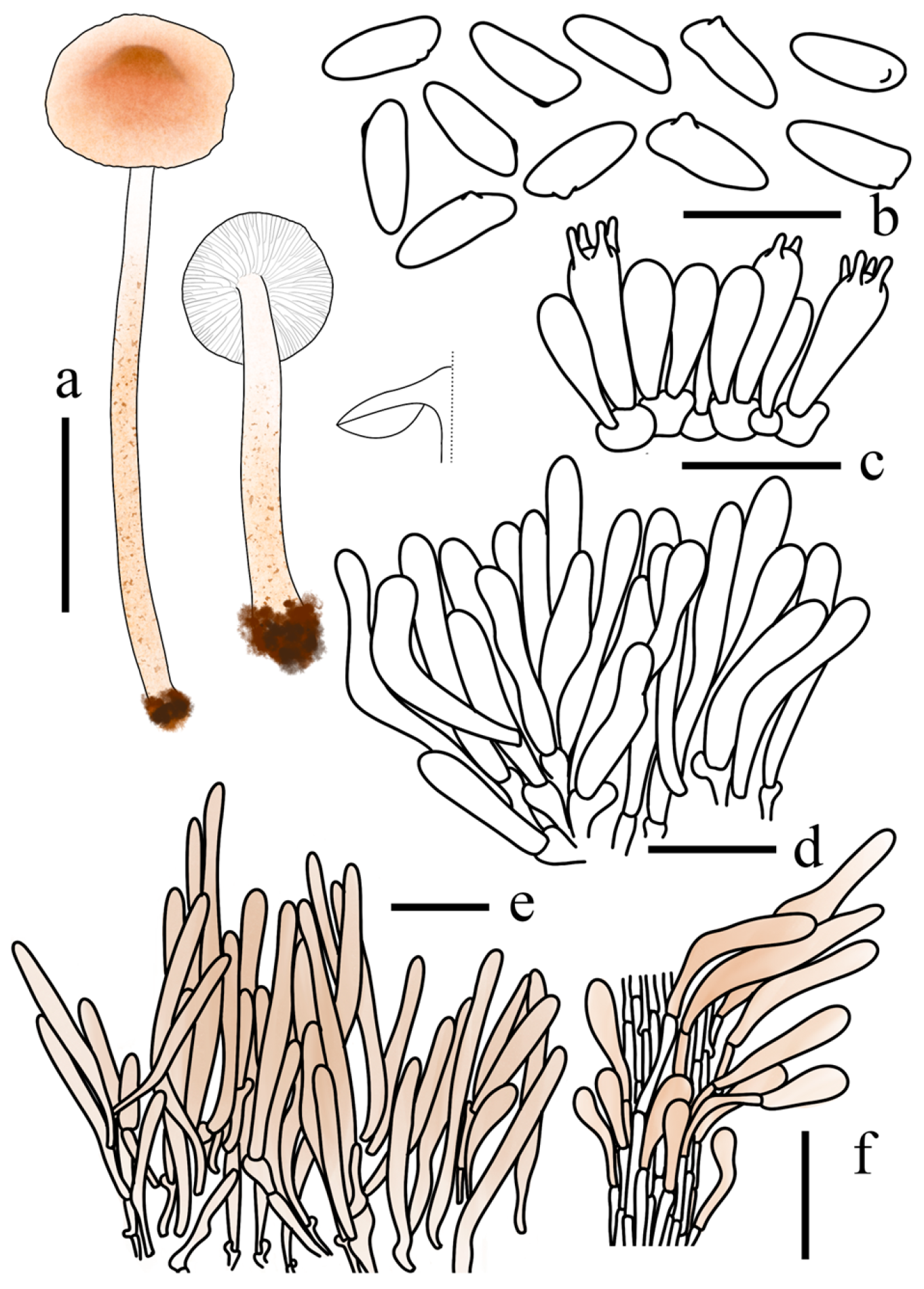
Diagnosis: Lepiota hongshiensis is characterized by its small-sized basidiomata with a pileus densely covered in greyish yellow to olive brown minute squamules; a stipe surface ornamented with squamules arranged in interrupted annular bands; remarkably non-weakly dextrinoid and spurred basidiospores measuring 5.9–7.1 × 2.9–3.5 μm; notably absent or not distinctly differentiated cheilocystidia; and a trichodermal pileus covering.
Basidiomata small. Pileus 0.9–1.8 cm in diam., campanulate when young, then expanding to plano-convex; surface white, densely clothed in minute greyish yellow (4C4–C8) to olive brown (4D6–D8) squamules. Context thin, white, unchanging when bruised. Lamellae free, crowded, interspersed with lamellulae of unequal length, ventricose, white to cream; edge entire, concolorous. Stipe 2.9–3.4 × 0.2–0.3 cm, subcylindrical, slightly thickening towards the base, white to cream; surface nearly glabrous above the annular zone, lower portion densely ornamented with greyish yellow (4C4–C8) to olive brown (4D6–D8) squamules arranged in interrupted annular bands. Odour and taste not recorded.
Basidiospores (40/2/2) 5.9–7.1 × 2.9–3.5 μm, avl × avw = 6.6 × 3.2 μm, Q = 1.84–2.28, Qav = 2.01, spurred, sub-triangular in profile, base occasionally nearly straight, ellipsoid in frontal view; wall hyaline, smooth, non-to weakly dextrinoid. Basidia 15–27 × 5–10 μm, clavate, 4- (2-) spored, thin-walled. Lamella edge fertile. Cheilocystidia not distinctly differentiated. Pleurocystidia absent. Pileus covering a trichoderm; terminal cells 61–239 × 9–19 μm, erect, cylindrical to clavate, with rounded apices and slightly thick walls, containing greyish yellow (4B2–B3) to light blond (4C2–C4) intracellular pigments. Stipe covering a cutis; terminal cells 63–122 × 9–21 μm, appressed, cylindrical to clavate, containing light blond (4C2–C4) intracellular pigments. Clamp connections present.
Habit and Habitat. Solitary to scattered on soil within the humus layer of broad-leaved forests dominated by Fraxinus mandshurica, Quercus mongolica Fisch. ex Ledeb., Juglans mandshurica, Betula spp., and Ulmus spp.
Known distribution: Jilin Province, China.
Additional specimens examined: CHINA. Jilin Province, Huadian City, Hongshi National Forest Park, 23 August 2024, R.H. Lin and T. Bau, FJAU78255; 15 August 2025, Y.F. Han, FJAU78295.
Notes: Based on our microscopic examination of the two specimens of Lepiota hongshiensis, no differentiated terminal cells or protruding hymenial structures were observed along the lamellar edges. Consequently, this species is characterized by the absence of cheilocystidia.
Phylogenetically, L. hongshiensis is nested within a clade primarily composed of species with greenish-brown pileus tones, including L. griseovirens Maire, L. poliochloodes Vellinga & Huijser, L. grangei (Eyre) Kühner, L. brunneoolivacea Sarawi, and L. pilodes Vellinga & Huijser.
Morphologically, however, L. hongshiensis clearly differs from these close relatives by its persistently white to cream lamellae, unchanging context, and relatively small basidiospores (5.9–7.1 × 2.9–3.5 μm). In contrast, the initially white to cream lamellae of L. griseovirens develop orange-brown spots with age, and the species possesses significantly larger and broader spores [(6.0–)6.5–9.5(–11.0) × (3.0–)3.5–4.5 μm] [12]. Similarly, L. poliochloodes is differentiated by its context and lamellae, which turn pinkish to pale orange-brown [12]. L. grangei is characterized by dense, dark-brown punctate squamules on the lower stipe and markedly larger spores (9.6–11.4 × 3.3–3.7 μm). Furthermore, the lamellae of L. brunneoolivacea shift to brownish-red upon maturation [11], while L. pilodes develops orange-brown spots on the lamellae and forms larger spores [(7.0–)8.0–10.0(–11.5) × 3.0–4.0(–4.5) μm].
These morphological distinctions, particularly the stable color of the hymenophore and the specific spore dimensions, support the recognition of L. hongshiensis as an independent species within this lineage.
Lepiota jilinensis T. Bau & X.Y. Zhou, sp. nov.
MycoBank No: 862981
Holotype: CHINA. Jilin Province, Jiaohe City, Qianjin Forest Farm, 25 August 2023, M. Liu and T. Bau, FJAU78248.
Etymology: “jilinensis” refers to Jilin Province, Northeast China, where all the specimens of this species were collected.
Diagnosis: Lepiota jilinensis is characterized by its pileus densely clothed in light orange to brownish orange squamules; a trichodermal pileus covering; spurred basidiospores that appear rectangular to sub-triangular in profile; and clavate to broadly clavate cheilocystidia.
Basidiomata small. Pileus 1–1.9 cm in diam., lenticular; surface white, adorned with light orange (6A3–A5) to brownish orange (7C6–C8) squamules; squamules densely aggregated and darker toward the disc. Context thin, white, unchanging when bruised. Lamellae free, crowded, alternating with several tiers of lamellulae, ventricose, white to cream; edge entire, concolorous. Stipe 4.1–5.0 × 0.1–0.2 cm, subcylindrical, light orange (6A3–A5) to brownish orange (7C6–C8); surface nearly glabrous above the annular zone, lower portion clothed in brownish orange (7C6–C8) squamules. Odour and taste not recorded.
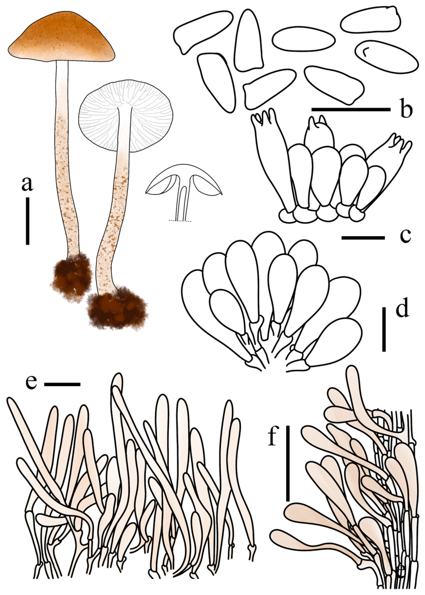
Basidiospores (40/2/2) (6.6–) 6.9–8.5 × 2.8–3.4 μm, avl × avw = 7.6 × 3.1 μm, Q = 2.19–2.94, Qav = 2.64, spurred, rectangular to sub-triangular in profile, cylindrical to sub-fusiform in frontal view; supra-hilar area not depressed or occasionally slightly depressed; base more or less with an extended spur; ventral side sometimes weakly ventricose; dorsal side occasionally slightly depressed at the base; wall slightly thickened, hyaline, smooth, non- to weakly dextrinoid. Basidia 14–25 × 4–8 μm, clavate, 4- (2-) spored, thin-walled. Lamella edge sterile. Cheilocystidia 19–35 × 9–18 μm, clavate to broadly clavate, occasionally sphaeropedunculate, hyaline, thin-walled. Pleurocystidia absent. Pileus covering a trichoderm; terminal cells 70–306 × 9–19 μm, erect, cylindrical to clavate, with rounded apices and slightly thick walls, containing light orange (6A3–A5) to greyish orange (6B3–B5) intracellular pigments; with short clavate cells, containing greyish orange (6B3–B5) intracellular pigments. Stipe covering a cutis; terminal cells 50–167 × 7–15 μm, appressed, cylindrical to clavate, containing light orange (6A3–A5) to greyish orange (6B3–B5) intracellular pigments. Clamp connections present.
Habit and Habitat. Solitary to scattered on soil within the humus layer of broad-leaved forests dominated by Fraxinus mandshurica, Quercus mongolica, and Juglans mandshurica.
Known distribution: Jilin Province, China.
Additional specimens examined: CHINA. Jilin Province, Huadian City, Hongshi National Forest Park, 28 August 2023, Z.Q. Chen and T. Bau, FJAU78249.
Notes: Lepiota jilinensis is a characteristic orange-brown member of sect. Stenosporae that shares morphological similarity with several other lineages within the section. However, it can be reliably distinguished by a combination of basidiospore dimensions, Q values, and chemical reactions.
Within the orange-brown complex, L. jilinensis is primarily defined by its medium-sized, non- to weakly dextrinoid basidiospores. This feature readily separates it from the large-spored taxa, such as L. dolichospora and L. subcastanea, as well as from the relatively small-spored species like L. microstenospora.
Furthermore, although L. jilinensis, L. sinocastanea, and L. sirupa all possess medium-sized basidiospores, they are clearly distinct based on other characters. L. sinocastanea is distinguished by its lamellae discoloring upon bruising and L. sirupa by its cylindrical to narrowly clavate cheilocystidia. In contrast, the lamellae of L. jilinensis remain unchanged when injured, and it possesses clavate to broadly clavate cheilocystidia.
Lepiota microstenospora T. Bau & X.Y. Zhou, sp. nov.
MycoBank No: 862982
Holotype: CHINA. Jilin Province, Jiaohe City, Qianjin Forest Farm, 23 July 2022, W.N. Hou, FJAU78246.
Etymology: “microstenospora” refers to the remarkably small basidiospores (5.7 × 2.8 μm), which represent the smallest spore dimensions recorded within the section Stenosporae to date.
Diagnosis: Lepiota microstenospora is primarily characterized by its pileus surface densely clothed in light orange to brownish orange, erect, fasciculate, and floccose squamules; a trichodermal pileus covering; remarkably small and spurred basidiospores (5.2–6.5 × 2.2–3.2 μm) that appear rectangular to sub-triangular in profile; and narrowly clavate to clavate cheilocystidia.
Basidiomata small. Pileus 0.7–1.2 cm in diam., plano-convex, with a distinct blunt umbo at the center; surface white, densely clothed in light orange (6A2–A5) to brownish orange (7C6–C8), erect, fasciculate, and floccose squamules. Context extremely thin, white, unchanging when bruised. Lamellae free, crowded, with lamellulae, ventricose, white to cream; edge entire, concolorous. Stipe 3.0–3.6 × 0.1–0.2 cm, subcylindrical, slightly thickening towards the base, white to light orange (6A2–A5) above the annular zone, light orange (6A2–A5) or brownish orange (7C6–C8) below; surface ornamented with concolorous floccose squamules. Stipe context reddish brown (9E5–E8). Odour and taste not recorded.
Basidiospores (40/2/2) 5.2–6.5 × 2.2–3.2 μm, avl × avw = 5.7 × 2.8 μm, Q = 1.78–2.43, Qav = 2.05, spurred, sub-triangular to rectangular in profile, oblong in frontal view; wall hyaline, smooth, non- to weakly dextrinoid. Basidia 18–22 × 4–8 μm, clavate, 4- (2-) spored, thin-walled. Lamella edge sterile. Cheilocystidia 23–38 × 5–9 μm, narrowly clavate to clavate, hyaline, thin-walled. Pleurocystidia absent. Pileus covering a trichoderm; terminal cells 89–247 × 6–15 μm, erect, cylindrical to clavate, with rounded apices and slightly thick walls, containing light orange (6A3–A5) to greyish orange (6B3–B5) intracellular pigments. Stipe covering a cutis; terminal cells 52–160 × 9–20 μm, appressed, cylindrical to clavate, containing light orange (6A3–A5) to greyish orange (6B3–B5) intracellular pigments. Clamp connections present.
Habit and Habitat. Solitary to scattered on soil within the humus layer of broad-leaved forests dominated by Quercus mongolica, Juglans mandshurica, Betula spp., and Ulmus spp.
Known distribution: Jilin Province, China.
Additional specimens examined: CHINA. Jilin Province, Jiaohe City, Qianjin Forest Farm, 25 August 2023, X. Wang and T. Bau, FJAU78247.
Notes: Although Lepiota microstenospora shares the orange-brown pileus coloration typical of other members in the Lepiota sect. Stenosporae complex, it is well separated by its exceptionally small basidiospores. Furthermore, the pileus center of this species features distinct, sub-erect, tufted, and dark tomentose squamules.
Within this complex, other small-spored species such as L. brunneoaurantia, L. flavostipitata Sarawi, and L. brunneopileata A. Rehman, Afshan, Usman & Khalid differ morphologically and chemically. Lepiota brunneoaurantia has markedly shorter and narrower cheilocystidia [(8.6–)8.9–13.1(–13.8) × (3.8–)4.2–5.9(–6.2) μm] [34]. In contrast to the non- to weakly dextrinoid basidiospores of L. microstenospora, those of L. flavostipitata and L. brunneopileata are distinctly dextrinoid [11,35]. Additionally, L. brunneopileata lacks the sub-erect, tufted squamules observed in the new species [35].
Lepiota sinocastanea T. Bau & X.Y. Zhou, sp. nov.
MycoBank No: 862983
Holotype: CHINA. Jilin Province, Ji’an City, Wunufeng National Forest Park, 25 July 2025, X. Y. Zhou, FJAU78171.
Etymology: The epithet “sinocastanea” refers to the Chinese origin (Sino-) of the species and its morphological resemblance (-castanea) to the original description of Lepiota castanea Quélet.
Diagnosis: Lepiota sinocastanea is characterized by its small-sized basidiomata with light orange to brownish orange scales; lamellae white to cream, bruising light orange (6A2–A5) to greyish orange (6B3–B6) when damaged; spurred basidiospores measuring 7.8–9.8 × 2.7–3.8 μm, non- to weakly dextrinoid in Melzer’s reagent; and a distinct phylogenetic position at the base of the L. castanea complex. It differs from the European L. castanea s.l. by its relatively small spores and the non- to weaker dextrinoid reaction.
Basidiomata small. Pileus 0.6–1.7 cm in diam., obtuse conical when young, becoming plano-convex to applanate at maturity; surface white, with a dense pileus covering composed of light orange (6A3–A6) to brownish orange (7C6–C8) scales. Context thin, white. Lamellae free, crowded, interspersed with lamellulae, ventricose, white to cream, bruising light orange (6A2–A5) to greyish orange (6B3–B6) when damaged; edge entire, concolorous. Stipe 2.1–5.0 × 0.2–0.3 cm, subcylindrical, slightly thickening towards the base, white; surface nearly smooth above the annulus, lower portion densely covered with light orange (6A3–A6) to brownish orange (7C6–C8) fine granular scales arranged in discontinuous, zonate bands; base bruising greyish orange (6B3–B6) upon injury. Odour and taste not recorded.
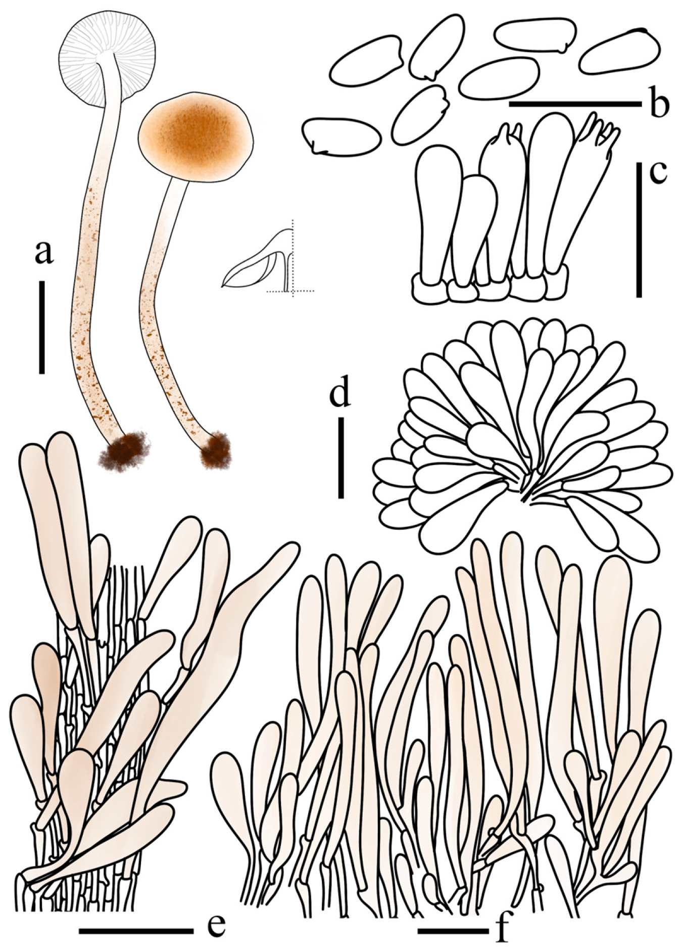
Basidiospores (40/2/2) 7.8–9.8 × 2.7–3.8 μm, avl × avw = 8.6 × 3.3 μm, Q = 2.18–3.20, Qav = 2.59, sub-triangular in profile with a distinct spurred base, long-ellipsoid to sub-fusiform in frontal view; apex often slightly acute; wall hyaline, smooth, slightly thick, non- to weakly dextrinoid. Basidia 16–25 × 6–9 μm, 4-(2-)spored, clavate, thin-walled. Lamella edge sterile. Cheilocystidia 24–44 × 4–10 μm, cylindrical, hyaline, thin-walled. Pleurocystidia absent. Pileus covering a trichoderm; terminal cells 59–340 × 8–20 μm, erect, cylindrical to clavate, with rounded apices and slightly thick walls, containing light orange (6A3–A5) to greyish orange (6B3–B5) intracellular pigments. Stipe covering a cutis; terminal cells 55–150 × 8–16 μm, appressed, cylindrical to clavate, containing light orange (6A3–A5) to greyish orange (6B3–B5) intracellular pigments. Clamp connections present.
Habit and Habitat. Solitary to scattered on soil within the humus layer of broad-leaved forests dominated by Fraxinus mandshurica, Quercus mongolica, Betula spp., and Ulmus spp.
Known distribution: Jilin and Heilongjiang Provinces, China.
Additional specimens examined: CHINA. Jilin Province, Jiaohe City, Qianjin Forest Farm, 24 July 2022, X. Wang, FJAU78162, Ji’an City, Wunufeng National Forest Park, 8 August 2023, Q.R. Liu, FJAU78160; Changchun City, Jingyuetan National Forest Park, 28 July 2025, X.Y. Zhou, FJAU78172; 8 August 2025, W. Sun, FJAU78170; Huadian City, Hongshi National Forest Park, 30 July 2025, Y.F. Han, FJAU78173; Heilongjiang Province, Tahe County, Guqi Valley National Wetland Park, 25 July 2024, H. Cheng, FJAU78161.
Notes: Phylogenetically, Lepiota sinocastanea is positioned at the base of the L. castanea core clade and the Lepiota aff. castanea lineage, forming an independent sister group.
Although L. sinocastanea shares macroscopic features with other members of this complex, it can be distinguished by its relatively small basidiospores (7.8–9.8 × 2.7–3.8 μm, avl × avw = 8.6 × 3.3 μm, Q = 2.18–3.20). In contrast, both L. castanea [8.7–10.4(–11.1) × 3.0–3.7(–4.7) μm] and L. aff. castanea [(8.9–)9.4–11(–11.4) × 3.5–4.5 μm] observed in this study possess relatively large spores. Furthermore, the basidiospores of L. sinocastanea are non- to weakly dextrinoid, unlike the distinctly dextrinoid spores of L. castanea and L. aff. castanea. Macroscopically, L. sinocastanea is unique within this complex in possessing lamellae that discolor upon bruising. These morphological, chemical, and phylogenetic divergences strongly support its recognition as an independent species.
Additionally, our study revealed another phylogenetically distinct lineage within the L. castanea complex, designated here as L. aff. castanea. Since its basidiospore dimensions and other core morphological characteristics overlap substantially with those of L. castanea, and no stable diagnostic features have been identified, we provisionally adopt open nomenclature for this allied lineage pending further collections.
Lepiota sirupa T. Bau & X.Y. Zhou, sp. nov.
MycoBank No: MB862977
Holotype: CHINA. Jilin Province, Huadian City, Hongshi National Forest Park, 16 August 2024, X.Y. Zhou, FJAU78261.
Etymology: “sirupa” is derived from the Latin noun sirupus (syrup), referring to the brownish orange color of the pileus squamules, which resembles syrup.
Diagnosis: Lepiota sirupa is primarily characterized by its pileus densely clothed in floccose squamules with a color transition from light orange (6A2–A5) to brownish orange (7C6–C8); a stipe ornamented with squamules arranged in interrupted annular bands below the annular zone; non-dextrinoid, slender basidiospores (Qav = 2.67) appearing spurred or rectangular in profile; cylindrical to narrowly clavate cheilocystidia frequently possessing transverse septa; and a pileus covering structured as a trichoderm.
Basidiomata small. Pileus 0.9–1.1 cm in diam., convex to plano-convex, with an inconspicuous blunt umbo at the center; surface white, densely clothed in light orange (6A2–A5) to brownish orange (7C6–C8) floccose squamules. Context thin, white, unchanging when bruised. Lamellae free, crowded, interspersed with lamellulae, ventricose, white to cream; edge entire, concolorous. Stipe 2.7–2.9 × 0.1–0.3 cm, subcylindrical, slightly thickening towards the base, white to light orange (6A2–A5); surface nearly glabrous above the annular zone, lower portion densely ornamented with floccose to floccular squamules arranged in interrupted annular bands. Odour and taste not recorded.
Basidiospores (40/2/2) 6.9–8.2 × 2.5–3.4 μm, avl × avw = 7.5 × 2.8 μm, Q = 2.15–3.11, Qav = 2.67, spurred, rectangular to sub-triangular in profile, slightly ventricose on the adaxial side, long-ellipsoid in frontal view; wall hyaline, thin, non-dextrinoid. Basidia 16–28 × 5–9 μm, clavate, 4- (2-) spored, hyaline, thin-walled. Lamella edge sterile. Cheilocystidia 26–53 × 5–9 μm, cylindrical to narrowly clavate, occasionally with transverse septa, hyaline, thin-walled. Pleurocystidia absent. Pileus covering a trichoderm; terminal cells 76–219 × 6–13 μm, erect, cylindrical to clavate, with rounded apices and slightly thick walls, containing light orange (6A3–A5) to greyish orange (6B3–B5) intracellular pigments. Stipe covering a cutis; terminal cells 56–189 × 5–12 μm, appressed, cylindrical to clavate, containing light orange (6A3–A5) to greyish orange (6B3–B5) intracellular pigments. Clamp connections present.
Habit and Habitat. Solitary to scattered on soil within the humus layer of broad-leaved forests dominated by Fraxinus mandshurica, Quercus mongolica, Juglans mandshurica, Betula spp., and Ulmus spp.
Known distribution: Jilin Province, China.
Additional specimens examined: CHINA. Jilin Province, Huadian City, Hongshi National Forest Park, 16 August 2024, X.Y. Zhou, FJAU78262.
Notes: Lepiota sirupa is an orange-brown member of sect. Stenosporae. It shares macroscopic features and medium-sized basidiospores with L. jilinensis but differs in having cylindrical to narrowly clavate cheilocystidia (clavate to broadly clavate in L. jilinensis). Furthermore, the non-dextrinoid basidiospores separate L. sirupa from dextrinoid-spored relatives in this section, such as the L. castanea complex. Macroscopically, it is further characterized by unchanging white to cream lamellae and a stipe that does not discolor upon bruising.
New record for China
Lepiota grangei (Eyre) Kühner, Bull. mens. Soc. linn. Soc. Bot. Lyon 3: 79 (1934)
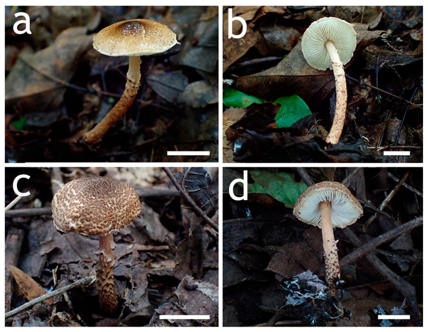
Basidiomata small. Pileus 1.8–2.3 cm in diam., white to cream, covered with light olive brown (4D4–D8) squamules; scales at the center dense, tomentose, olive brown (4F7–F8). Context white. Lamellae free, crowded, interspersed with lamellulae of unequal length, ventricose, white to cream; edge entire, concolorous. Stipe 3.5–5.2 × 0.3–0.4 cm, subcylindrical, cream; surface nearly glabrous above the annular zone, lower portion densely covered with olive brown (4F7–F8) granular squamules below, arranged in intermittent bands. Odour and taste not recorded.
Basidiospores (40/2/2) 9.6–11.4 × 3.3–3.7 μm, avl × avw = 10.3 × 3.5 μm, Q = 2.62–3.29, Qav = 2.89, subtriangular in profile with a distinct suprahilar depression and a prominent basal spur, subfusiform in frontal view; ventral side slightly ventricose; dorsal side ventricose but often depressed in the lower-middle part; apex gradually tapering; wall hyaline, slightly thick, dextrinoid. Basidia 20–30 × 6–11 μm, clavate, 4- (rarely 2-) spored, hyaline, thin-walled. Lamella edge sterile. Cheilocystidia 26–38 × 6–8 μm, clavate, often with a simple septum, hyaline, thin-walled. Pleurocystidia absent. Pileus covering a trichoderm; terminal cells 116–330 × 10–17 μm, erect, cylindrical to clavate, with up to 3 clampless septa, with rounded apices, slightly thick walls, containing greyish yellow (4B2–B3) to light blond (4C2–C3) parietal and intracellular pigments. Stipe covering a cutis; terminal cells 52–193 × 7–15 μm, appressed, cylindrical to clavate, containing light blond (4C2–C3) intracellular pigments. Clamp connections present.
Known distribution: Widespread in Europe (e.g., Germany and the Netherlands), also recorded from North America (USA) and South America [12,22]. In Asia, it has been documented in Thailand, South Korea [62], and Northern China (Jilin Province, this study).
Specimens measured: CHINA. Jilin Province, Huadian City, Hongshi National Forest Park, 16 August 2024, T.Y. Zhang, FAJU78230; Jilin City, Songhua Lake Scenic Area, 20 September 2024, J.L. Wei, FAJU78231.
Notes: Lepiota grangei is a well-documented species in Europe, typically characterized by pileus squamules with more pronounced green, bluish-green, or grayish-blue tones [12,63]. In contrast, our specimens from Northern China exhibit a light olive brown to olive brown color range. This variation likely reflects intraspecific plasticity, as the pileus color of this species has previously been described as varying from blue-green to brownish-green [63].
Phylogenetically, members of subclade II share these distinctive greenish-brown pileus tones. Geographically, L. griseovirens is known from Africa and Europe [11,63], L. poliochloodes from Europe and Thailand [7,63], and L. brunneoolivacea from Benin [11]. In China, only three species—L. grangei, L. hongshiensis, and L. pilodes—possess greenish-brown pileus squamules. However, L. hongshiensis is easily distinguished by its exceptionally small basidiospores (5.9–7.1 × 2.9–3.5 μm) and the absence of cheilocystidia. Although L. pilodes also produces relatively small spores (7.5–8.9 × 2.8–3.4 μm), it lacks septa in the terminal cells of the pileus covering. L. grangei is recognized by its relatively large basidiospores (9.6–11.4 × 3.3–3.7 μm), clavate cheilocystidia, and the presence of conspicuous septa in the terminal cells of the pileus covering.
Key to the 17 species of Lepiota sect. Stenosporae characterized by yellowish-orange to orange-brown pileus squamules
1. Pileus covering a cutisL. boudieri
1’. Pileus covering a trichoderm2
2. Lamellae pale yellow to greyish yellow3
2’. Lamellae white to cream4
3. Basidiomata turning dark brown to black upon dryingL. flavonigrescens
3’. Basidiomata unchanging upon dryingL. citrophylla
4. Basidiospores inamyloid, weakly dextrinoid, or reaction unknown5
4’. Basidiospores distinctly dextrinoid12
5. Basidiospores with average length > 9.0 μm6
5’. Basidiospores with average length < 9.0 μm7
6. Basidiospores narrowly cylindrical, 11.5–14.1 × 3.3–4.7 μm (avl × avw = 12.9 × 3.9 μm), Q = 2.92–3.93, Qav = 3.31L. dolichospora
6’. Basidiospores broader, 8.5–11.0 × 3.5–4.5 μm (avl × avw = 9.7 × 4.0 μm), Q = 2.00–2.86, Qav = 2.48L. subcastanea
7. Basidiospores with average length < 7.0 μm8
7’. Basidiospores with average length > 7.0 μm10
8. Distributed in South Asia (Pakistan)L. brunneoaurantia
8’. Distributed in East Asia (China)9
9. Basidiospores relatively small (avl × avw = 5.7 × 2.8 μm); cheilocystidia narrowly clavate to clavateL. microstenospora
9’. Basidiospores relatively large (avl × avw = 6.5 × 3.2 μm); cheilocystidia narrowly clavate, rarely narrowly utriform or fusiformL. mandarina
10. Lamellae changing colour upon bruisingL. sinocastanea
10’. Lamellae unchanging upon bruising11
11. Cheilocystidia 19–35 × 9–18 μm, clavate to broadly clavate L. jilinensis
11’. Cheilocystidia 26–53 × 5–9 μm, cylindrical to narrowly clavateL. sirupa
12. Basidiospores 7.5–14.0 μm in length13
12’. Basidiospores 4.7–7.5 μm in length14
13. Cheilocystidia narrowly clavate to fusiform or utriformL. castanea
13’. Cheilocystidia cylindrical to narrowly clavate L. alopochroa
14. Distributed in Asia (Pakistan)L. brunneopileata
14’. Distributed in Africa (Benin)  15
15. Basidia with distinctly long sterigmata, 5–10(–20) μmL. longisterigmata
15’. Basidia without distinctly long sterigmata16
16. Stipe apex white to pale yellowL. flavostipitata
16’. Stipe apex white to creamL. aurantiicolor
Note: Lepiota ignicolor Bres. is excluded from this key due to unresolved taxonomic uncertainties. Morphologically, its basidiospore size range and cheilocystidia shape significantly overlap with those of L. castanea. Regarding molecular data, the sequence labeled as L. ignicolor in our phylogenetic tree (GenBank accession: AY176472) [4] lacks a corresponding morphological description for its voucher specimen. Conversely, available morphological descriptions of this species [19] lack supporting molecular data. Given this discrepancy, the true taxonomic concept of L. ignicolor remains ambiguous. The morphological data utilized in this key are derived from the present study and the following cited literature [7,11,18,19,35].

4. Discussion

4.1. Phylogenetic Framework and Sectional Delimitation

Our phylogenetic analysis, based on a combined four-gene dataset (ITS, nrLSU, rpb2, and tef1-α), demonstrates that Lepiota sect. Stenosporae constitutes a well-supported monophyletic group within Lepiota (97/1). The section is clearly bifurcated into two major evolutionary lineages based on pileus covering micromorphology: Clade A (trichoderm) and Clade B (cutis-like) [43,64]. This topology is highly consistent with previous taxonomic frameworks [7,9,10,11], further validating the phylogenetic significance of the pileus covering in sectional delimitation.
Within Clade A, Subclade I exhibits high genetic divergence despite pronounced macroscopic similarities. Except for a few color-specialized taxa like the white-toned L. subalba [48] and purple-toned L. tyrianthina [11], members of this subclade possess similar yellowish-orange to orange-brown pileus scales. These macroscopic similarities among genetically distinct species present a challenge for field identification. Consequently, the taxonomic key provided herein is intended to assist in species-level differentiation.

4.2. Evolutionary Complexity and Phenotypic Diversity

In contrast to the well-resolved Subclade I, the remaining lineages in Clade A exhibit more complex evolutionary patterns. Subclade II is generally characterized by greenish-brown tones. Meanwhile, Subclade III, comprising three known species, further exemplifies the discordance between macromorphology and phylogeny. Although L. citrophylla [48] and L. flavonigrescens [11] share yellowish-orange to orange-brown pilei with members of Subclade I, they are readily separated by their distinct yellow-toned lamellae—a key diagnostic feature absent in the latter. In contrast, L. pilodes exhibits greenish-brown tones typically associated with Subclade II; however, our topology reveals that it does not cluster with the Subclade II lineage, instead phylogenetically aligning with the yellow-gilled species of Subclade III. This suggests that comparable color phenotypes (such as greenish-brown or orange-brown) have likely evolved independently across different lineages within the section.
Furthermore, Subclade IV (L. tomentella) is distinguished by grey-brown tones with a pinkish tinge, yet it exhibits significant intraspecific genetic variation, indicating potential cryptic diversity. In contrast, Clade B displays a much broader color spectrum, encompassing dark brownish-yellow, orange-brown, and pinkish-grey tones. This distribution of color phenotypes across the section underscores the taxonomic complexity within sect. Stenosporae. Further studies incorporating genomic-scale data are required to precisely resolve the evolutionary trajectories of these complex lineages.

4.3. Taxonomic Considerations of the Lepiota castanea Complex

Historically, the Lepiota castanea complex has received extensive attention due to the high variability in basidiospore dimensions and other microscopic features. The original description recorded relatively small basidiospores (8–9 µm) [65], whereas it was subsequently proposed, based on morphological boundaries, that the large-spored lineage (13.5–14.5 µm) be delimited as a distinct species, L. rufidula Bres. [66]. Conversely, based on extensive morphological studies of European material, a continuous distribution of spore sizes (7.0–14.0 µm) was observed, leading to the adoption of a broad species concept [12].
Our molecular phylogenetic data provide a clear genetic basis for understanding this variation. The analysis reveals that the complex is composed of at least three genetically independent evolutionary lineages, indicating that the morphological variations observed in previous studies correspond to distinct evolutionary branches.
The spores of the new species L. sinocastanea, average 8.6 × 3.3 µm, aligning closely with the original records of L. castanea [65]. Its basal evolutionary position within the complex, its non- to weakly dextrinoid reaction in Melzer’s reagent, and the distinct discoloration of the lamellae upon bruising strongly support its recognition as an independent East Asian lineage. Furthermore, another Chinese lineage (referred to herein as L. aff. castanea) possesses relatively large spores (averaging 10.0 × 3.9 µm), which is consistent with the morphological concept of the large-spored taxa. Given the biogeographic isolation between East Asia and Europe, and the current lack of sequences from the type locality of L. rufidula, we conservatively adopt open nomenclature for this lineage, pending further validation through broader global sampling.

4.4. Conclusions

Based on field surveys in Northeast China, this study describes six new species and one new regional record within Lepiota sect. Stenosporae. These findings expand the known species diversity of this section in East Asia and demonstrate that macroscopically similar lineages actually represent distinct evolutionary branches. Because macroscopic traits often exhibit convergent evolution, resolving complex groups in this section requires integrating multi-locus phylogeny with stable micro-morphological features (e.g., basidiospore dimensions and cheilocystidia shapes). While this study clarifies the taxonomy of several East Asian taxa, further global sampling remains necessary to fully resolve the evolutionary boundaries within sect. Stenosporae.

Author Contributions

Conceptualization, X.-Y.Z. and T.B.; methodology, X.-Y.Z.; software, X.-Y.Z.; validation, X.-Y.Z. and T.B.; formal analysis, X.-Y.Z.; investigation, X.-Y.Z. and T.B.; resources, X.-Y.Z. and T.B.; data curation, X.-Y.Z. and T.B.; writing—original draft preparation, X.-Y.Z.; writing—review and editing, X.-Y.Z. and T.B.; visualization, X.-Y.Z. and T.B.; supervision, T.B.; project administration, T.B.; funding acquisition, T.B. All authors have read and agreed to the published version of the manuscript.

Funding

This work was supported by the Ministry of Education Innovation Team (No. IRT1134, IRT-15R25).

Institutional Review Board Statement

Not applicable.

Informed Consent Statement

Not applicable.

Data Availability Statement

All the sequences have been deposited in GenBank (https://www.ncbi.nlm.nih.gov, accessed on 24 March 2026) and MycoBank (https://www.mycobank.org, accessed on 24 March 2026). For peer-review purposes, the concatenated phylogenetic alignment and detailed specimen metadata are temporarily available in the Zenodo repository (DOI https://doi.org/10.5281/zenodo.19253651, accessed on 8 May 2026).

Acknowledgments

We thank our supervisor and laboratory colleagues for their support and assistance with fieldwork and laboratory research.

Conflicts of Interest

The authors declare no conflicts of interest.

References

  1. Kalichman, J.; Kirk, P.M.; Matheny, P.B. A compendium of generic names of agarics and Agaricales. Taxon 2020, 69, 425–447. [Google Scholar] [CrossRef] [Scilit]
  2. Vellinga, E.C. Ecology and distribution of Lepiotaceous fungi (Agaricaceae)—A review. Nova Hedwig. 2004, 78, 273–300. [Google Scholar] [CrossRef] [Scilit]
  3. Sarawi, S.; Shi, Y.-N.; Lotz-Winter, H.; Reschke, K.; Bode, H.B.; Piepenbring, M. Occurrence and chemotaxonomical analysis of amatoxins in Lepiota spp. (Agaricales). Phytochemistry 2022, 195, 113069. [Google Scholar] [CrossRef] [Scilit] [PubMed]
  4. Vellinga, E.C. Phylogeny of Lepiota (Agaricaceae)—Evidence from nrITS and nrLSU sequences. Mycol. Prog. 2003, 2, 305–322. [Google Scholar] [CrossRef] [Scilit]
  5. Vellinga, E.C. Genera in the family Agaricaceae: Evidence from nrITS and nrLSU sequences. Mycol. Res. 2004, 108, 354–377. [Google Scholar] [CrossRef] [Scilit] [PubMed]
  6. Liang, J.F. Study on Taxonomy of Lepiota from China and Molecular Phylogeny of the Genus with Notes on Population Genetics of L. cristata. Ph.D. Thesis, Kunming Institute of Botany, Chinese Academy of Sciences, Kunming, China, 2007. [Google Scholar]
  7. Sysouphanthong, P.; Hyde, K.; Chukeatirote, E.; Bahkali, A.; Vellinga, E. Lepiota (Agaricales) in northern Thailand—1. L. section Stenosporae. Mycotaxon 2011, 117, 53–85. [Google Scholar] [CrossRef] [Scilit]
  8. Vellinga, E.C.; Sysouphanthong, P.; Hyde, K.D. The family Agaricaceae: Phylogenies and two new white-spored genera. Mycologia 2011, 103, 494–509. [Google Scholar] [CrossRef] [Scilit]
  9. Sysouphanthong, P.; Thongklang, N.; Suwannapoom, C.; Nuangmek, W.; Hyde, K.D. Lepiota section Stenosporae (Agaricaceae) two new records to Lao People’s Democratic Republic. Chiang Mai J. Sci. 2020, 47, 49–56. [Google Scholar]
  10. Sarawi, S.; Piepenbring, M.; Reschke, K. Phylogenetic and taxonomic re-assessment of the genera Echinoderma and Lepiota. Fungal Syst. Evol. 2025, 15, 235–263. [Google Scholar] [CrossRef] [Scilit]
  11. Sarawi, S.; Reschke, K.; Jagora, A.; Yorou, N.; Le Pogam, P.; Piepenbring, M. Hidden gems of Benin: Unravelling the diversity of Lepiota spp. through phylogenetic, morphological, and chemotaxonomic exploration. Persoonia 2025, 55, 313–360. [Google Scholar] [CrossRef] [Scilit] [PubMed]
  12. Vellinga, E.C. Lepiota (Pers.: Fr.) S. F. Gray. In Flora Agaricina Neerlandica: Critical Monographs on Families of Agarics and Boleti Occurring in The Netherlands; Noordeloos, M.E., Kuyper, T.W., Vellinga, E.C., Eds.; A.A. Balkema Publishers: Lisse, The Netherlands, 2001; Volume 5, pp. 64–169. [Google Scholar]
  13. Knudsen, H. A revision of Lepiota sect. Echinatae and Amyloideae (Agaricaceae) in Europe. Bot. Tidsskr. 1980, 75, 121–155. [Google Scholar]
  14. Singer, R. The Agaricales in Modern Taxonomy; Koeltz Scientific Books: Koenigstein, Germany, 1986; pp. 1–981. [Google Scholar]
  15. Bon, M. Les Lepiotes; CRDP de Picardie: Amiens, France, 1993; pp. 1–142. [Google Scholar]
  16. Singer, R. The Agaricales in modern taxonomy. Lilloa 1951, 22, 1–832. [Google Scholar]
  17. Lange, J. Studies in the Agarics of Denmark. Part II. Amanita Lepiota Coprinus Dan. Bot. Ark. 1915, 2, 1–60. [Google Scholar]
  18. Liang, J.F. Taxonomy and phylogeny in Lepiota sect. Stenosporae from China. Mycologia 2016, 108, 56–69. [Google Scholar] [CrossRef] [Scilit]
  19. Yang, Z.L.; Ge, Z.W.; Liang, J.F. Fungi Lepiotoidei, Agaricaceae; Science Press: Beijing, China, 2019; Volume 52, pp. 1–228. [Google Scholar]
  20. Kornerup, A.; Wanscher, J.H. Methuen Handbook of Colour; Methuen and Co., Ltd.: London, UK, 1967; pp. 1–252. [Google Scholar]
  21. Vellinga, E.C. Critical Monographs on Families of Agarics and Boleti Occurring in The Netherlands; A.A. Balkema Publishers: Rotterdam, The Netherlands, 1988; Volume 1, pp. 54–66. [Google Scholar]
  22. GBIF.org. GBIF Occurrence Search: Lepiota grangei. Available online: https://www.gbif.org/occurrence/search?taxon_key=7847622 (accessed on 8 May 2026).
  23. White, T.J.; Bruns, T.; Lee, S.; Taylor, J. Amplification and direct sequencing of fungal ribosomal RNA genes for phylogenetics. In PCR Protocols: A Guide to Methods and Applications; Innis, M.A., Gelfand, D.H., Sninsky, J., White, T.J., Eds.; Academic Press: New York, NY, USA, 1990; pp. 315–322. [Google Scholar]
  24. Gardes, M.; Bruns, T.D. ITS primers with enhanced specificity for basidiomycetes—Application to the identification of mycorrhizae and rusts. Mol. Ecol. 1993, 2, 113–118. [Google Scholar] [CrossRef] [Scilit]
  25. Vilgalys, R.; Hester, M. Rapid genetic identification and mapping of enzymatically amplified ribosomal DNA from several Cryptococcus species. J. Bacteriol. 1990, 172, 4238–4246. [Google Scholar] [CrossRef] [Scilit] [PubMed]
  26. Liu, Y.J.; Whelen, S.; Hall, B.D. Phylogenetic relationships among ascomycetes: Evidence from an RNA polymerse II subunit. Mol. Biol. Evol. 1999, 16, 1799–1808. [Google Scholar] [CrossRef] [Scilit]
  27. Rehner, S.A.; Buckley, E. A Beauveria phylogeny inferred from nuclear ITS and EF1-α sequences: Evidence for cryptic diversification and links to Cordyceps teleomorphs. Mycologia 2005, 97, 84–98. [Google Scholar] [CrossRef] [Scilit]
  28. Zhou, X.Y.; Bau, T. Four new species of Cystolepiota (Agaricaceae, Agaricales) from northeastern China. Front. Microbiol. 2024, 15, 1358612. [Google Scholar] [CrossRef] [Scilit]
  29. Stallman, J.K.; Hemmes, D.E.; Hynson, N.A.; Shintaku, M.H. Lepiota punaensis sp. nov. from Hawai ‘i Island, and a discussion of L. elaiophylla. Mycotaxon 2020, 135, 471–489. [Google Scholar] [CrossRef] [Scilit]
  30. Hou, Y.J.; Ge, Z.W. New species of Echinoderma and Lepiota (Agaricaceae) from China. Phytotaxa 2020, 447, 221–236. [Google Scholar] [CrossRef] [Scilit]
  31. Mao, N.; Xia, L.; Xu, Y.Y. Lepiota atrobrunneodisca (Agaricaceae, Agaricales), a new species with a hymeniform pileus covering from North China. Phytotaxa 2023, 595, 186–198. [Google Scholar] [CrossRef] [Scilit]
  32. Liang, Y.-S.; Xu, Y.-W.; Ouyang, M.; Huang, X.-X.; Lin, Z.-J.; Qiu, L.-H. Lepiota baiyunensis sp. nov. (Agaricales, Agaricaceae) from Baiyun Mountain, China. Phytotaxa 2023, 606, 1–15. [Google Scholar] [CrossRef] [Scilit]
  33. Liang, J.F.; Yang, Z.L.; Xu, J.P.; Ge, Z.W. Two new unusual Leucoagaricus species (Agaricaceae) from tropical China with blue-green staining reactions. Mycologia 2010, 102, 1141–1152. [Google Scholar] [CrossRef] [Scilit]
  34. Azeem, M.; Kiran, M.; Jabeen, S. Molecular phylogeny and morphological characterization revealed Lepiota brunneoaurantia, a new species in L. sect. Stenosporae from Margalla Hills, Pakistan. Phytotaxa 2024, 636, 61–73. [Google Scholar] [CrossRef] [Scilit]
  35. Rehman, A.; Usman, M.; Afshan, N.U.S.; Khalid, A.N. Taxonomy and phylogeny reveal two novel species of genus Lepiota (Agaricaceae, Agaricales) from Punjab, Pakistan. Plant Syst. Evol. 2024, 310, 8. [Google Scholar] [CrossRef] [Scilit]
  36. Vellinga, E.C. Studies in Lepiota IV. Lepiota cristata and L. castaneidisca. Mycotaxon 2001, 80, 297–306. [Google Scholar] [CrossRef] [Scilit]
  37. Liang, J.; Xu, J.; Yang, Z. Divergence, dispersal and recombination in Lepiota cristata from China. Fungal Divers. 2009, 38, e124. [Google Scholar]
  38. Caballero, A.; Vizzini, A.; Munoz, G.; Contu, M.; Ercole, E. Lepiota elseae (Agaricales, Agaricaceae), a new species of section Lepiota from Spain. Phytotaxa 2015, 201, 188–196. [Google Scholar] [CrossRef] [Scilit]
  39. Paz, A.; Lavoise, C. Balsamia sotxoi y Lepiota faiae-bravae, dos nuevas especies de hongos secotioides. Bol. Micol. FAMCAL 2024, 19, 9–25. [Google Scholar]
  40. Qasim, T.; Khalid, A.; Vellinga, E. A new species of Lepiota, Lepiota lahorensis, from Lahore, Pakistan. Turk. J. Bot. 2016, 40, 419–426. [Google Scholar] [CrossRef] [Scilit]
  41. Vellinga, E.C. Lepiota in California: Species with a hymeniform pileus covering. Mycologia 2010, 102, 664–674. [Google Scholar] [CrossRef] [Scilit]
  42. Tibpromma, S.; Hyde, K.D.; Jeewon, R.; Maharachchikumbura, S.S.; Liu, J.K.; Bhat, D.J.; Jones, E.G.; McKenzie, E.H.; Camporesi, E.; Bulgakov, T.S. Fungal diversity notes 491–602: Taxonomic and phylogenetic contributions to fungal taxa. Fungal Divers. 2017, 83, 1–261. [Google Scholar] [CrossRef] [Scilit]
  43. Vellinga, E.C. Lepiotaceous fungi in California, USA 2-Lepiota rhodophylla sp. nov. Mycotaxon 2006, 98, 205–212. [Google Scholar] [CrossRef] [Scilit]
  44. Vellinga, E.C.; Davis, R.M. Lepiotaceous fungi from California, USA 1-Leucoagaricus amanitoides sp. nov. Mycotaxon 2006, 98, 197–204. [Google Scholar] [CrossRef] [Scilit]
  45. Schoch, C.L.; Robbertse, B.; Robert, V.; Vu, D.; Cardinali, G.; Irinyi, L.; Meyer, W.; Nilsson, R.H.; Hughes, K.; Miller, A.N. Finding needles in haystacks: Linking scientific names, reference specimens and molecular data for Fungi. Database 2014, 2014, bau061. [Google Scholar] [CrossRef] [Scilit]
  46. Justo, A.; Angelini, C.; Bizzi, A. Two new species and a new record of Lepiota (Basidiomycota, Agaricales) from the Dominican Republic. Mycol. Prog. 2015, 14, 56. [Google Scholar] [CrossRef] [Scilit]
  47. Angelini, C.; Vizzini, A.; Justo, A.; Bizzi, A.; Davoli, P.; Kaya, E. First report of a neotropical agaric (Lepiota spiculata, Agaricales, Basidiomycota) containing lethal α-amanitin at toxicologically relevant levels. Front. Microbiol. 2020, 11, 1833. [Google Scholar] [CrossRef] [Scilit] [PubMed]
  48. Prylutskyi, O.; Zinenko, O.; Havrysh, P. First records of three Lepiota species (Agaricales, Basidiomycota) from Ukraine, with notes on a poorly known species, Lepiota subalba. Ukr. Bot. J. 2021, 78, 373–380. [Google Scholar] [CrossRef] [Scilit]
  49. Nawaz, R.; Khalid, A.; Hanif, M.; Razaq, A. Lepiota vellingana sp. nov. (Basidiomycota, Agaricales) a new species from Lahore, Pakistan. Mycol. Prog. 2013, 12, 727–732. [Google Scholar] [CrossRef] [Scilit]
  50. Li, J.X.; Zhao, R.L.; Phurbu, D.; Xing, R.; Liu, D.M.; He, M.Q.; Ling, Z.L.; Han, X.X.; Feng, H.Y.; Zhu, X.Y. Disentangling taxonomic chaos in Agaricaceae sl: An integrative phylogenomic framework with divergence dating reconstructs classification. Fungal Divers. 2025, 135, 745–881. [Google Scholar] [CrossRef] [Scilit]
  51. Katoh, K.; Rozewicki, J.; Yamada, K.D. MAFFT online service: Multiple sequence alignment, interactive sequence choice and visualization. Brief. Bioinform. 2019, 20, 1160–1166. [Google Scholar] [CrossRef] [Scilit]
  52. Kumar, S.; Stecher, G.; Tamura, K. MEGA7: Molecular evolutionary genetics analysis version 7.0 for bigger datasets. Mol. Biol. Evol. 2016, 33, 1870–1874. [Google Scholar] [CrossRef] [Scilit]
  53. Capella-Gutiérrez, S.; Silla-Martínez, J.M.; Gabaldón, T. trimAl: A tool for automated alignment trimming in large-scale phylogenetic analyses. Bioinformatics 2009, 25, 1972–1973. [Google Scholar] [CrossRef] [Scilit]
  54. Zhao, D.; Ye, T.; Gao, F.; Jakovlić, I.; La, Q.; Tong, Y.; Liu, X.; Song, R.; Liu, F.; Lian, Z.-m.; et al. PhyloSuite v2: The development of an all-in-one, efficient and visualization-oriented suite for molecular dating analysis and other advanced features. iMeta 2025, 4, e70095. [Google Scholar] [CrossRef] [Scilit]
  55. Kalyaanamoorthy, S.; Minh, B.Q.; Wong, T.K.F.; von Haeseler, A.; Jermiin, L.S. ModelFinder: Fast model selection for accurate phylogenetic estimates. Nat. Methods 2017, 14, 587–589. [Google Scholar] [CrossRef] [Scilit]
  56. Nguyen, L.-T.; Schmidt, H.A.; Von Haeseler, A.; Minh, B.Q. IQ-TREE: A fast and effective stochastic algorithm for estimating maximum-likelihood phylogenies. Mol. Biol. Evol. 2015, 32, 268–274. [Google Scholar] [CrossRef] [Scilit] [PubMed]
  57. Minh, B.Q.; Nguyen, M.A.T.; Von Haeseler, A. Ultrafast approximation for phylogenetic bootstrap. Mol. Biol. Evol. 2013, 30, 1188–1195. [Google Scholar] [CrossRef] [Scilit] [PubMed]
  58. Guindon, S.; Dufayard, J.F.; Lefort, V.; Anisimova, M.; Hordijk, W.; Gascuel, O. New algorithms and methods to estimate maximum-likelihood phylogenies: Assessing the performance of PhyML 3.0. Syst. Biol. 2010, 59, 307–321. [Google Scholar] [CrossRef] [Scilit] [PubMed]
  59. Ronquist, F.; Teslenko, M.; Van Der Mark, P.; Ayres, D.L.; Darling, A.; Höhna, S.; Larget, B.; Liu, L.; Suchard, M.A.; Huelsenbeck, J.P. MrBayes 3.2: Efficient Bayesian phylogenetic inference and model choice across a large model space. Syst. Biol. 2012, 61, 539–542. [Google Scholar] [CrossRef] [Scilit]
  60. Rambaut, A. FigTree, a Graphical Viewer of Phylogenetic Trees, version 1.4.3; Institute of Evolutionary Biology, University of Edinburgh: Edinburgh, UK, 2016. [Google Scholar]
  61. Xie, J.M.; Chen, Y.R.; Cai, G.J.; Cai, R.R.; Hu, Z.; Wang, H. Tree Visualization By One Table (tvBOT): A web application for visualizing, modifying and annotating phylogenetic trees. Nucleic Acids Res. 2023, 51, W587–W592. [Google Scholar] [CrossRef] [Scilit]
  62. Sysouphanthong, P.; Hyde, K.D.; Chukeatirote, E.; Vellinga, E.C. A review of genus Lepiota and its distribution in east Asia. Curr. Res. Environ. Appl. Mycol. 2011, 1, 161–176. [Google Scholar] [CrossRef]
  63. Vellinga, E.C.; Huijser, H.A. Notulae ad Floram agaricinam neerlandicam—XXI. Lepiota section Stenosporae. Persoonia 1993, 15, 223–240. [Google Scholar]
  64. Mornand, J. Une nouvelle lépiote, Lepiota andegavensis sp. nov. Doc. Mycol. 1982, 12, 41–43. [Google Scholar]
  65. Quélet, L. Quelques espèces critiques ou nouvelles de la Flore Mycologique de France (1). C. R. Assoc. Fr. Av. Sci. 1881, 9, 661–675. [Google Scholar]
  66. Migliozzi, V.; Zecchin, G. Studio della Sezione Stenosporae (Lange) Kühner del genere Lepiota s.s. 4ª parte. Descrizione di Lepiota cortinarius Lange, Lepiota rufidula Bresadola (= L. castanea sensu auctores, = L. ignipes Locquin ex Bon) e Lepiota ignicolor Bresadola. Micol. Ital. 2000, 29, 22–29. [Google Scholar]
Figure 1. Bayesian phylogenetic tree of Lepiota sect. Stenosporae inferred from a combined dataset of ITS, nrLSU, rpb2, and tef1-α sequences. New species are indicated in bold blue, while new record for China are shown in blue. Sequences newly generated in this study are highlighted in bold black. Bayesian posterior probabilities (PP > 0.90) and Maximum Likelihood bootstrap support values (UFBoot > 80%) are indicated at the nodes (UFBoot/PP).
Jof 12 00355 g001
Figure 2. Bayesian phylogenetic tree of Lepiota sect. Stenosporae inferred from a combined dataset of ITS, nrLSU, rpb2, and tef1-α sequences. To emphasize the topological relationships within sect. Stenosporae, unidentified lineages (labeled as “Lepiota sp.”), and clades unrelated to this section have been collapsed. New species are indicated in bold blue, and new record for China are shown in blue. In the basidiospore metrics heatmap, b–c × f–g represents the range of basidiospore dimensions, while q1–q2 denotes the range of the Q value (length/width ratio). Bayesian posterior probabilities (PP > 0.90) and Maximum Likelihood bootstrap support values (UFBoot/80%) are indicated at the nodes (UFBoot/PP).
Jof 12 00355 g002
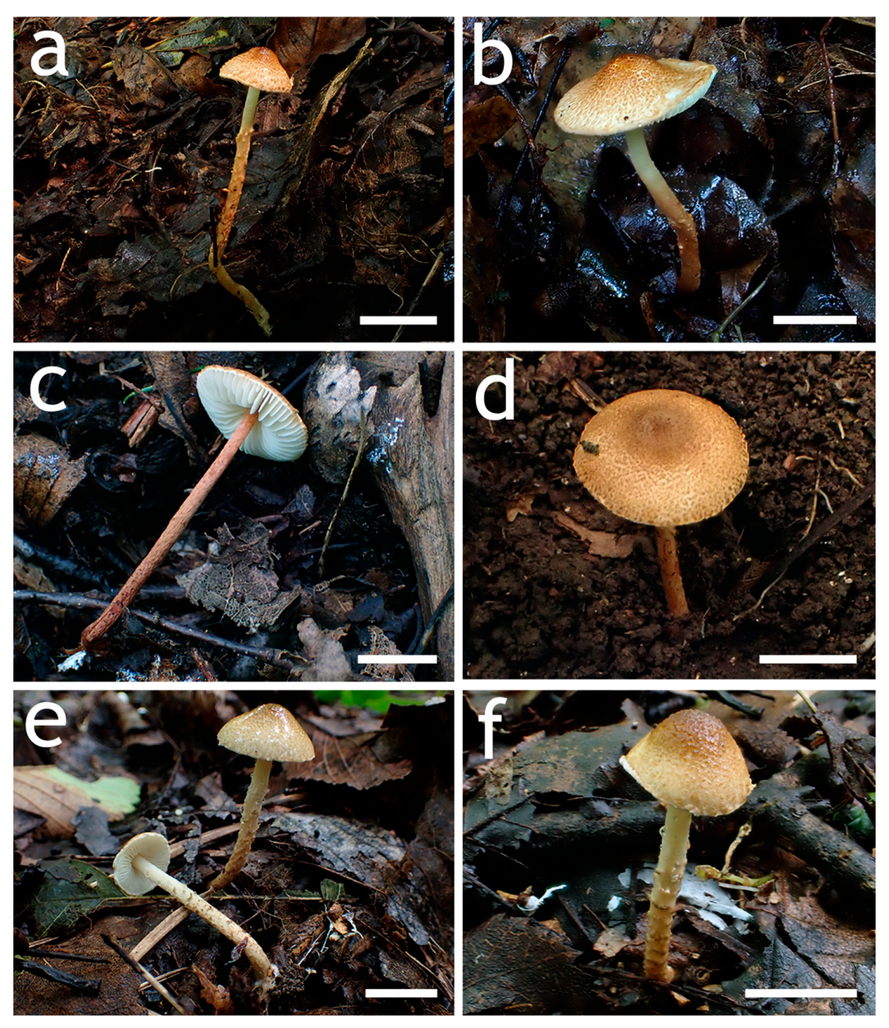
Figure 3. Basidiomata of Lepiota sect. Stenosporae species. (ac) L. dolichospora ((a) FJAU78185; (b) FJAU78184; (c) FJAU78183); (d,e) L. hongshiensis ((d) FJAU78295: (e,f) FJAU78254). Scale bars = 1 cm.
Jof 12 00355 g003
Figure 4. Lepiota dolichospora (FJAU78183, holotype). (a) Basidiomata; (b) Basidiospores; (c) Basidia; (d) Cheilocystidia; (e) Pileus covering; (f) stipe covering. Scale bars: (a) =1cm; (b) =10 µm; (c,d) =20 µm; (e,f) =50 µm.
Jof 12 00355 g004
Figure 5. Lepiota hongshiensis (FJAU78254, holotype). (a) Basidiomata; (b) Basidiospores; (c) Basidia; (d) stipe covering; (e) Pileus covering. Scale bars: (a) =1 cm; (b) =10 µm; (c) =20 µm; (d,e) =50 µm.
Jof 12 00355 g005
Figure 6. Basidiomata of Lepiota sect. Stenosporae species. (ac) L. jilinensis ((a) FJAU78248; (b,c) FJAU78249); (df) L. microstenospora ((d) FJAU78246; (e,f) FJAU78247); (af) Scale bars = 1 cm.
Jof 12 00355 g006
Figure 7. Lepiota jilinensis (FJAU78248, holotype). (a) Basidiomata; (b) Basidiospores; (c) Basidia; (d) Cheilocystidia; (e) Pileus covering; (f) stipe covering. Scale bars: (a) =1 cm; (b) =10 µm; (c,d) =20 µm; (e,f) =50 µm.
Jof 12 00355 g007
Figure 8. Lepiota microstenospora (FJAU78246, holotype). (a) Basidiomata; (b) Basidiospores; (c) Basidia; (d) Cheilocystidia; (e) stipe covering; (f) Pileus covering. Scale bars: (a) =1 cm; (b) =10 µm; (c,d) =20 µm; (e,f) =50 µm.
Jof 12 00355 g008
Figure 9. Basidiomata of Lepiota sect. Stenosporae species. (ac) L. sinocastanea. ((a,b) FJAU78171; (c) FJAU78172); (df) L. sirupa ((d,e) FJAU78261; (f) FJAU78262). Scale bars = 1 cm.
Jof 12 00355 g009
Figure 10. Lepiota sinocastanea (FJAU78171, holotype). (a) Basidiomata; (b) Basidiospores; (c) Basidia; (d) Cheilocystidia; (e) Pileus covering; (f) stipe covering. Scale bars: (a) =1 cm; (b) =10 µm; (c,d) =20 µm; (e,f) =50 µm.
Jof 12 00355 g010
Figure 11. Lepiota sirupa (FJAU78261, holotype). (a) Basidiomata; (b) Basidiospores; (c) Basidia; (d) Cheilocystidia; (e) Pileus covering; (f) stipe covering. Scale bars: (a) =1 cm; (b) =10 µm; (c,d) =20 µm; (e,f) =50 µm.
Jof 12 00355 g011
Figure 12. Basidiomata of Lepiota grangei. (a,b) FJAU78230; (c,d) FJAU78231. Scale bars = 1 cm.
Jof 12 00355 g012
Figure 13. Lepiota grangei (FJAU78230). (a) Basidiomata; (b) Basidiospores; (c) Cheilocystidia; (d) Basidia; (e) Pileus covering; (f) stipe covering. Scale bars: (a) =1 cm; (b) =10 µm; (c,d) =20 µm; (e,f) =50 µm.
Jof 12 00355 g013
Disclaimer/Publisher’s Note: The statements, opinions and data contained in all publications are solely those of the individual author(s) and contributor(s) and not of MDPI and/or the editor(s). MDPI and/or the editor(s) disclaim responsibility for any injury to people or property resulting from any ideas, methods, instructions or products referred to in the content.