Article

Spectrometric Characterization of Clinical and Environmental Isolates of Aspergillus Series Versicolores

1
Mycology and Parasitology Department, Caen University Hospital, UNICAEN, Normandie University, 14000 Caen, France
2
ToxEMAC-ABTE, UNICAEN & UNIROUEN, Normandie University, 14000 Caen, France
*
Authors to whom correspondence should be addressed.
J. Fungi 2023, 9(9), 868; https://doi.org/10.3390/jof9090868
Submission received: 29 June 2023 / Revised: 18 August 2023 / Accepted: 21 August 2023 / Published: 23 August 2023
(This article belongs to the Special Issue Recent Advances in Taxonomy, Phylogeny and Evolution of Fungi)

Abstract

:
Aspergillus series Versicolores are molds distributed among 17 species, commonly found in our environment, and responsible for infections. Since 2022, a new taxonomy has grouped them into 4 major lineages: A. versicolor, A. subversicolor, A. sydowii, and A. creber. Matrix-Assisted Laser Desorption/Ionization Time-Of-Flight Mass Spectrometry (MALDI-TOF MS) could be a faster and more cost-effective alternative to molecular techniques for identifying them by developing a local database. To evaluate this technique, 30 isolates from Aspergillus series Versicolores were used. A total of 59 main spectra profiles (MSPs) were created in the local database. This protocol enabled accurate identification of 100% of the extracted isolates, of which 97% (29/30) were correctly identified with a log score ≥ 2.00. Some MSPs recorded as Aspergillus versicolor in the supplier’s database could lead to false identifications as they did not match with the correct lineages. Although the local database is still limited in the number and diversity of species of Aspergillus series Versicolores, it is sufficiently effective for correct lineage identification according to the latest taxonomic revision, and better than the MALDI-TOF MS supplier’s database. This technology could improve the speed and accuracy of routine fungal identification for these species.

1. Introduction

Aspergillus series Versicolores is a taxonomic group of Aspergillus section Nidulantes. These fungi are commonly found in soil [1], food [2], caves, and indoor environments with bioaerosols and moldy surfaces [3]. They have a significant impact on the quality of agricultural products, as well as on indoor air quality, and can cause health problems in humans and animals through exposure to their spores and mycotoxins such as sterigmatocystin and aflatoxins [4].
Although Aspergillus series Versicolores is not a common cause of human infections, these fungi can cause allergic reactions such as allergic bronchopulmonary aspergillosis [5,6] and lead to a variety of infections, including invasive pulmonary aspergillosis [7], fungal sinusitis [8], onychomycosis [9], and endophthalmitis [10]. These infections are generally associated with immunocompromised individuals, such as those with human immunodeficiency virus/acquired immunodeficiency syndrome (HIV/AIDS) [10] or organ transplant recipients [11]. It is therefore important to identify the exact species involved in human pathologies in order to adapt treatment [12].
Aspergillus series Versicolores has undergone several taxonomic revisions over the years. The earliest taxonomic studies of Aspergillus versicolor date back to the early 20th century based on morphological characteristics [13]. In 2008, Peterson introduced molecular approaches to the study of fungal systematics, leading to a more integrated approach to taxonomy that combined molecular and morphological data [14]. In more recent years, further taxonomic revisions have been carried out using a combination of morphological, molecular, and chemical data and have revealed 17 species of Aspergillus series Versicolores [15,16]. In many studies, misidentifications have been observed between A. versicolor species and other species of Aspergillus series Versicolores [12]. As molecular and genomic tools continue to improve, a new revision of taxonomy was proposed in 2022 and was based on a combination of phylogenetic, morphological, and molecular features, including spore morphology, growth temperature, DNA sequence analysis, and osmotic tolerance. The Versicolores series was reduced to four species, namely the A. subversicolor lineage and the A. sydowii lineage, each containing a single species, while the A. versicolor lineage contained nine species (A. amoenus, A. austroafricanus, A. fructus, A. griseoaurantiacus, A. hongkongensis, A. pepii, A. protuberus, A. tabacinus, and A. versicolor), and the A. creber lineage including six species (A. creber, A. cvjetkovicii, A. jensenii, A. puulaauensis, A. tennesseensis, and A. venenatus) [17].
Although molecular and morphological identification methods have been useful for the identification of Aspergillus series Versicolores, they have some limitations, particularly with regard to their similarity, intraspecific variability [18], and basic local alignment search tool (BLAST) similarity searches using different genes, which result in different identifications [17]. Matrix-Assisted Laser Desorption/Ionization Time-Of-Flight Mass Spectrometry (MALDI-TOF MS) has become the reference technique for the rapid identification and classification of microorganisms, including fungi [19,20,21]. Several MALDI-TOF MS systems are available worldwide, such as the Bruker Biotyper, but their database may not be able to distinguish closely related species within the Aspergillus series Versicolores. First of all, the lack of completeness of these databases and their late updates in relation to taxonomic revisions may simply explain the misidentifications. Thus, several studies have reported difficulties in identifying rare Aspergillus species using the Bruker database [22,23,24]. Moreover, for Aspergillus species of the Versicolores series, their high level of intraspecific variability can result in overlapping mass spectra, making it difficult to distinguish between species.
To overcome the weaknesses of the databases provided by MALDI-TOF MS suppliers, it is possible to generate local databases. Several teams have demonstrated their usefulness for correct species identification, in particular for Achromobacter [25], Clostridia [26], and lice [27].
The objective of this study was to evaluate the interest of spectrometric identification of Aspergillus species of the Versicolores series by creating a local database at the University Hospital of Caen.

2. Materials and Methods

2.1. Clinical and Environmental Isolates

The 30 isolates used to develop the local mass spectrometry local database are described in Table 1.
They were obtained from clinical samples received at the Mycology Laboratory of the Caen University Hospital (France) and from environmental samples collected by the ToxEMAC team, which has the largest French collection of Aspergillus isolates of the Versicolores series. We evaluated 8 species of the series, some of which were represented by a single isolate, such as A. amoenus, A. fructus, A. protuberus, A. puulaauensis, and A. tabacinus, while others were represented by multiple isolates (A. creber, n = 10; A. jensenii, n = 4; A. sydowii, n = 11). All isolates were preserved in the ToxEMAC mycological bank (stored at −80 °C).

2.2. Molecular Identification

All the steps leading to the molecular identification of the isolates used in this study have already been described previously [4]: collection of environmental and clinical samples, fungal culture on malt extract agar (MEA), DNA extraction, and purification using the NucleoSpin Plant II kit and NucleoSpin gDNA Clean-up kit (Macherey-Nagel, Duren, Germany), respectively, and PCR using the beta-tubulin gene (BenA) with Bt2a/Bt2b primers and DNA sequencing.

2.3. Generation of the Local Database

2.3.1. Culture

The isolates were grown on Sabouraud-chloramphenicol-gentamicin agar (SAB) (ThermoFisher Scientific, Waltham, MA, USA), malt extract agar medium supplemented with 0.02% (m/v) chloramphenicol (MEA) (Cooper, Melun, France), and CZAPEK agar (CZA) (Cooper, Melun, France), and were all incubated at 30 °C for 2–14 days. The complete extraction was performed on a sterile tube containing 2 mL of Sabouraud-chloramphenicol liquid medium (Liquid SAB) (Biomérieux, Lyon, France) and was incubated at 24 h. The detection of a fungal colony was identified each time by phenotypic and MS methods. For each culture on SAB and MEA media, a first extraction was performed the day following the start of fungal growth (young colonies) and possibly repeated two days in a row (mature colonies), which was considered a first attempt. In case of failure, a new extraction was carried out in the same way after subculture on a new medium for young and mature colonies (second attempt). In case of another failure, the previous step was repeated (third attempt) only for cultures on SAB medium. Extraction tests were carried out first on young colonies, and when this worked, new attempts were made on older colonies. The objective was therefore to generate two main spectra profiles (MSPs) (from young and mature colonies) in a single culture medium attempt to assess whether there was a spectral difference between young and mature colonies considered.

2.3.2. Protein Extraction

Filamentous fungi grown on the medium were collected by scraping the surface of the colonies with a sterile loop (VWR International SAS, Radnor, PA, USA). Lentil-sized fungal material (hyphae and spores) was deposited in a sterile Eppendorf tube containing 300 μL of high-pressure liquid chromatography (HPLC) water (VWR International SAS, Radnor, PA, USA) and 900 μL of absolute ethanol (VWR International SAS, Radnor, PA, USA) for protein extraction.
Filamentous fungi grown in liquid SAB medium were centrifuged at 13,000× g for 10 min, then the pellet was washed 3 times with 1 mL of HPLC water. The pellet was resuspended in a hydro-alcoholic solution (300 μL of HPLC water and 900 μL of absolute ethanol).
The fungal material in the hydro-alcoholic suspension was immediately extracted. Extraction began with a 10 min centrifugation at 13,000× g. The dried pellet was resuspended in 25 μL of 70% formic acid (Sigma-Aldrich, Paris, France). Then, 25 μL of 100% acetonitrile (VWR International SAS, Radnor, PA, USA) was added and incubated for 10 min. The sample was centrifuged at 13,000× g for 2 min just before being deposited on a target. A 1 μL drop of supernatant was deposited onto a polished steel target plate (MTP 96 target plate polished steel, Bruker Daltonics GmbH, Bremen, Germany) and left to air dry. Each deposit was then covered with 1 μL of matrix solution (In vitro diagnostics (IVD) Matrix α-cyano-4-hydroxycinnamic acid (HCCA)-portioned Bruker Daltonics GmbH, Bremen, Germany). Samples were deposited in 8 replicates for each extract.
We also tested 11 isolates (representing 1 isolate for each species, with 2 isolates for A. sydowii, 2 isolates for A. creber, and 2 isolates for A. jensenii, with 1 clinical and 1 environmental isolate for A. creber and A. jensenii) by direct transfer (or scraping) of fungal material onto a MALDI plate and addition of formic acid to bypass the extraction protocol explained just before.

2.3.3. Spectral Acquisition

Data acquisition was carried out according to Bruker recommendations (MALDI Biotyper: creation of MSP Protocol V1.4, Janvier 2021). Spectra acquisition was performed with Microflex LT/SH MALDI-TOF MS (Bruker Daltonics GmbH, Bremen, Germany) using linear positive mode with mass range m/z 2000 to 20,000, and an acceleration voltage of 20 kV by the AutoXecute function of Flexcontrol v3.4 software (Bruker Daltonics GmbH, Bremen, Germany) with the “MBT_Autox” method (accumulation = 240 shots). The calibration of the MALDI-TOF MS was automatically performed using a bacterial test standard (BTS) deposit (Bruker Daltonics GmbH, Bremen, Germany) for mass calibration according to the manufacturer’s instructions. Spectra were acquired 3 times on each deposit, resulting in 24 spectra by the extract.

2.3.4. Quality Control

Data were imported and analyzed using flexAnalysis software v3.4 (Bruker Daltonics GmbH, Bremen, Germany). All spectra were represented with baseline subtraction, smoothing, and automatic scaling on the y-axis. First, the mass accuracy of the BTS was checked (mass deviation < 300 ppm for each peak). Then, the 24 spectra were checked to meet 2 objectives between 6000 and 7000 m/z to eliminate outliers’ peaks or flatline spectra, and to ensure that the mass deviation between the smallest mass spectrum and the largest mass spectrum was a maximum of 500 ppm. The supplier recommends a minimum of 20 out of 24 spectra that meet the quality requirements described above.

2.3.5. Creation of MSP

Each MSP was created from a minimum of 20 spectra of the same extract using the “MSP creation” function of MaldiBioTyper Compass Explorer® v4.1 software with standard parameters (maximum mass error for each raw spectrum, 2000 Da; desired mass error for the MSP, 200 Da; minimum desired frequency for a peak, 25%; maximum number of peaks for MSP creation, 70). The MSP created was then compared with the Bruker database to avoid any risk of contamination or errors in molecular or phenotypic identification. When a MSP was created, we attempted to obtain a second MSP with two additional days of incubation to obtain MSPs with both young and mature colonies for each of these isolates. The MSPs created were used to generate a new local database named “Fungi CAEN”.

2.4. Evaluation of MSP Library Performance

The 30 isolates were tested using the extraction protocol for filamentous fungi from the manufacturer’s instructions. The spectra generated were compared with the two combined databases (Bruker-Daltonics MALDI Biotyper Filamentous Fungi (V4, August 2021) containing 856 MSPs and “Fungi CAEN” containing 59 MSPs). We used the Bruker user manual to interpret the logarithmic score results: high-confidence identification (≥2.00), low-confidence identification (1.70–1.99), and no possible organism identification (<1.70).
We also used three clinical isolates, firstly identified as A. versicolor using the “Filamentous Fungi” Bruker database, which were then identified using PCR sequencing as described above and tested against our local database in order to evaluate its performance because these isolates were not included in our local database.

3. Results

3.1. Interest of Culture Media

Regarding fungal growth on the SAB medium and its cost-effectiveness for spectrometric identification, there were 11 failures in the first and second attempts to create MSPs, probably due to prolonged incubation time in the incubator (Table 2). We observed that the optimum incubation time on the SAB medium for extracting these fungi was two days for A. amoenus, A. fructus, and A. sydowii, three days for A. protuberus and A. creber, between four and seven days for A. jensenii, five days for A. puulaauensis, and seven days for A. tabacinus. It should be noted that MSPs were easier to obtain for fast-growing species, such as A. sydowii, whereas slower-growing species yielded poor results in terms of peptide extraction.
On the MEA medium, there were nine failures in the first attempt to create MSPs and eight failures in the second (Table 2). There was no third attempt for this medium due to a lack of time to reconduct them manually. Incubation times were longer on average, with three days for A. amoenus, A. fructus, and A. sydowii, between eight and nine days for A. jensenii, between nine and twelve days for A. creber, and up to fourteen days for A. puulaauensis. There were not enough spectra for A. protuberus to be included in the database on this culture medium (Table 2).
We observed only failures on the CZA medium. Finally, we tested the liquid SAB medium for the isolates most difficult to extract from SAB, MEA, and CZA media (no MSP obtained or only one from SAB medium): three MSPs were generated from the eight extracted isolates tested (two A. creber isolates and one A. puulaauensis isolate) (Table 2). The easiest species to extract was A. sydowii, as it could be collected more easily than the others (high conidiogenesis). Furthermore, A. sydowii, A. amoenus, and A. fructus grew faster than the others and had a less dry appearance than those that remained longer in the incubator, resulting in a better yield of spectra. The success rate on the first trial was higher with MEA (21/30, 70%) than with the SAB medium (19/30, 63%), but the incubation period was longer. Extraction works better on young colonies regardless of the type of medium. The onset of growth may vary in duration depending on the culture media (longer on MEA than on SAB). However, if we ensured prompt handling from the early stages of growth on the SAB medium, we achieved an excellent success rate on the third attempt (11/11, 100%).

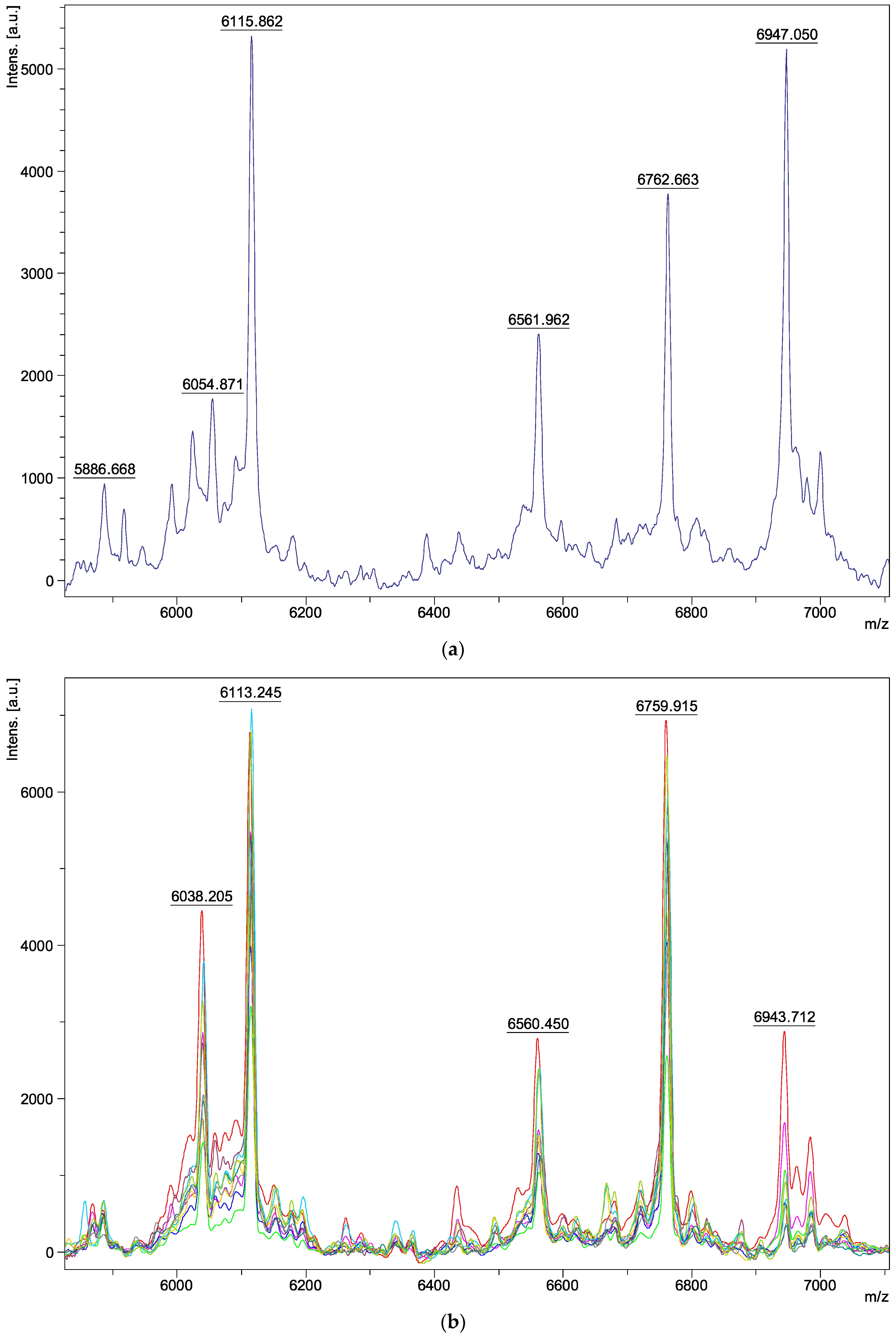
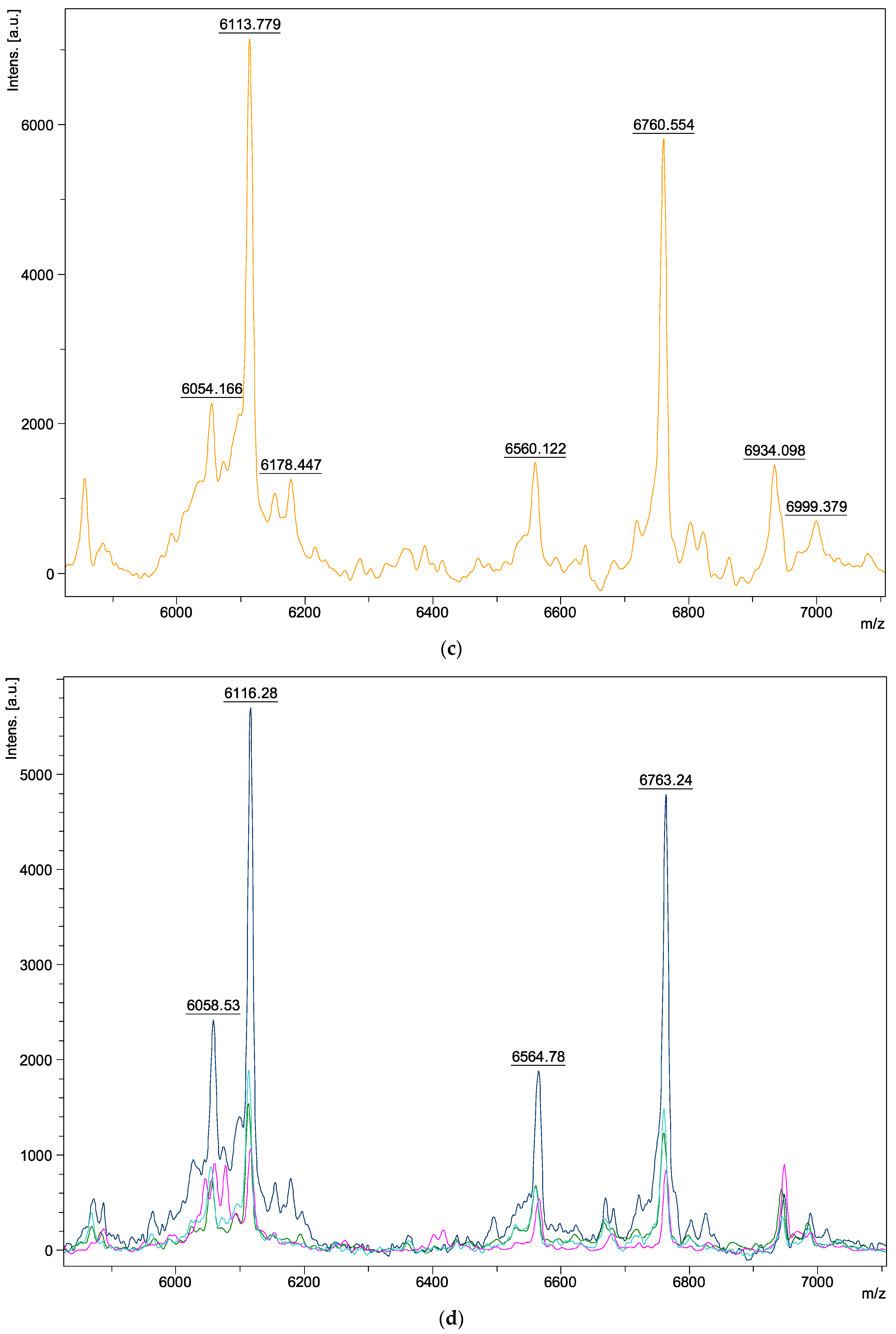
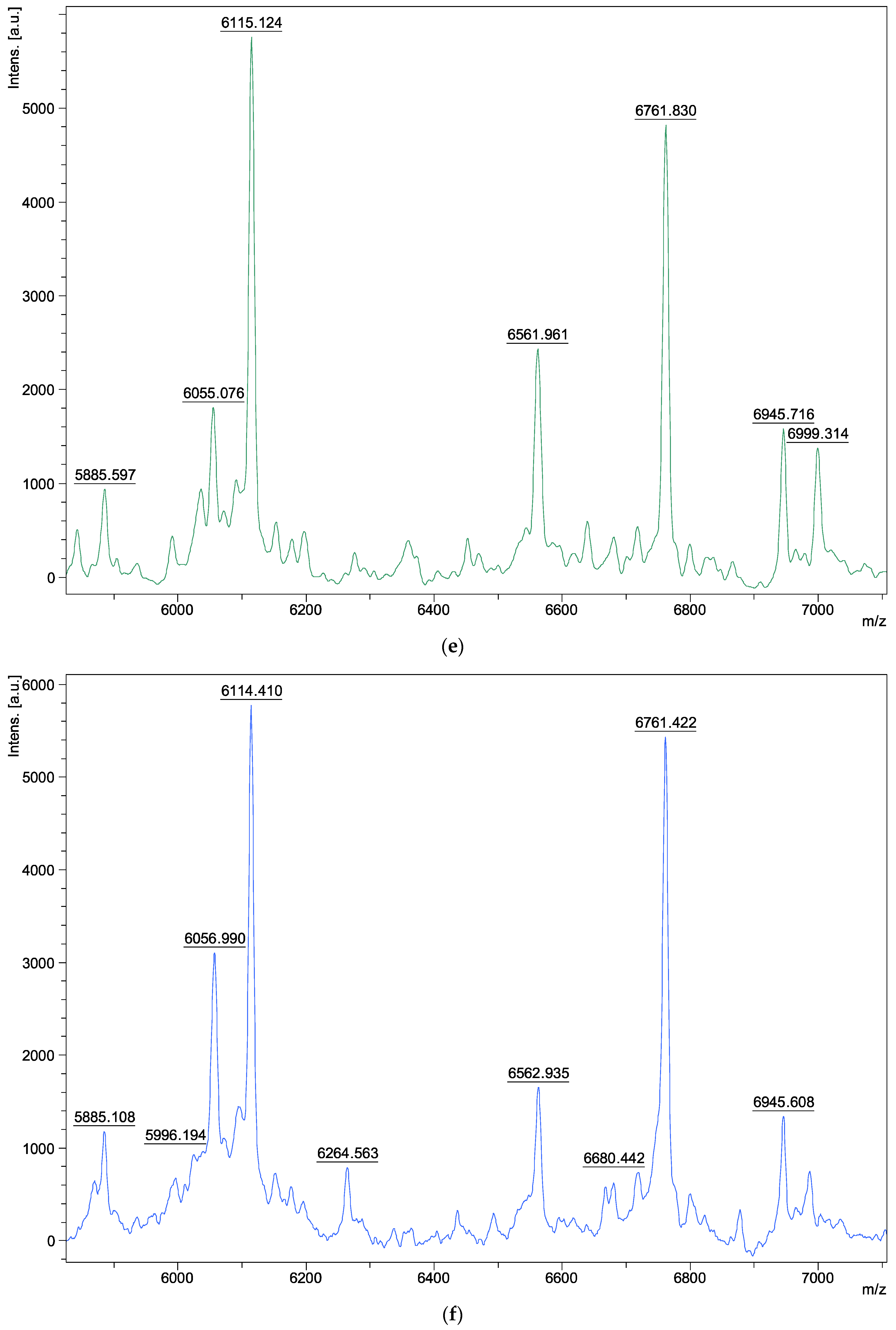
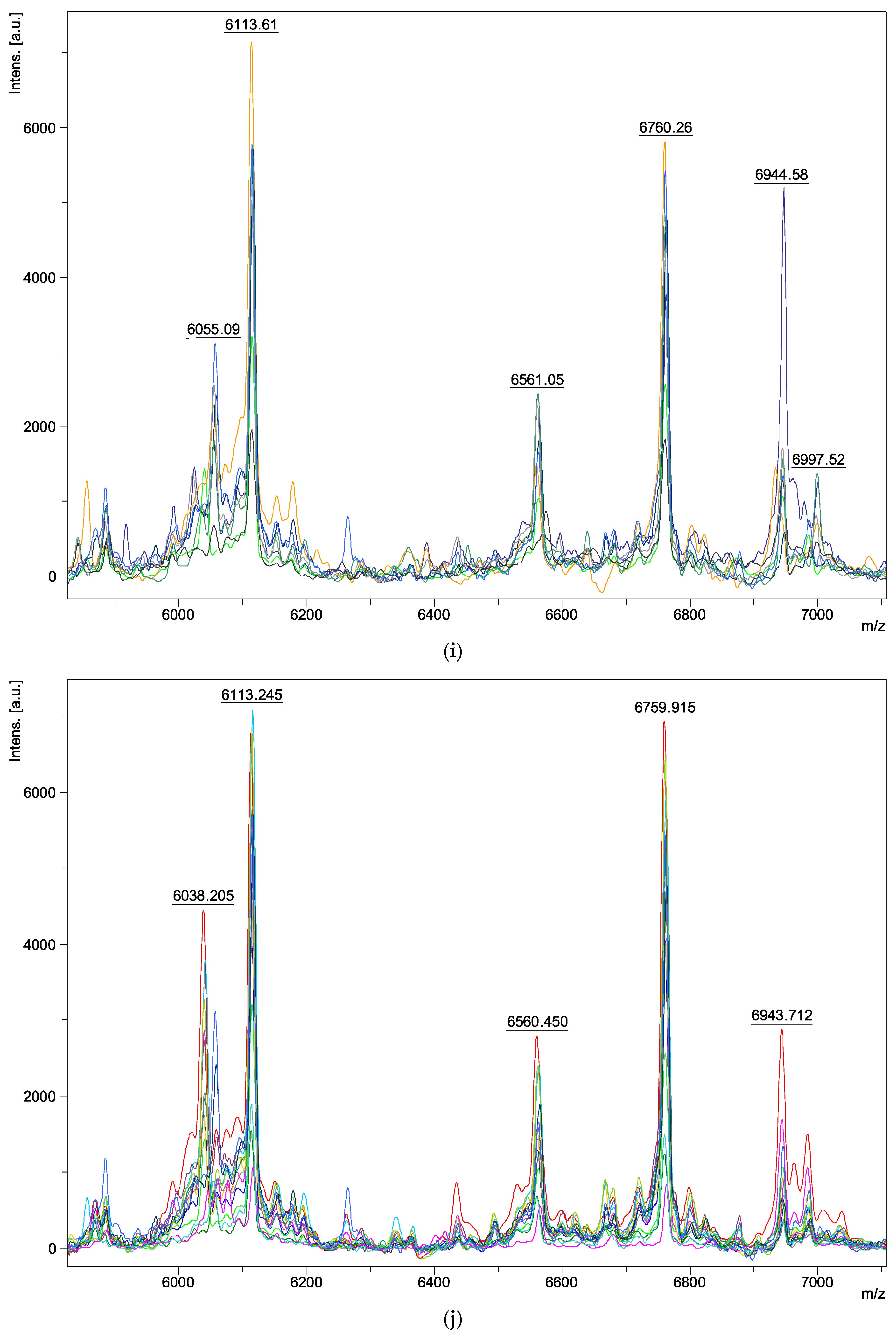
3.2. Characteristics of the Local Database

The thirty isolates, from eight different Aspergillus species of the Versicolores series, were used to create the local spectrometric database, generating fifty-nine MSPs: three for A. amoenus, nineteen for A. creber, two for A. fructus, six for A. jensenii, one for A. protuberus, three for A. puulaauensis, twenty-three for A. sydowii, and two for A. tabacinus. We were able to create MSPs with both young and mature colonies for only four isolates (two A. sydowii, one A. creber, and one A. amoenus) (Table 2).
Of the 1416 spectra obtained from these 59 MSPs, 130 were eliminated during a visual analysis of the identification spectra due to insufficient quality or obvious dissimilarity with the other replicates. We were able to obtain a MSP from mature colonies for only four isolates (i.e., 4/30, 13% of isolates): two A. sydowii isolates on an SAB medium (A. sydowii lineage), and A. amoenus and A. creber on an MEA medium (A. creber lineage). Thus, there were two MSPs in the database for those species on SAB and MEA medium (Table 2). For all other species, no conclusive results were obtained from mature colonies (difficult to collect due to low metabolism). In total, we obtained 32 MSPs from the SAB medium, 24 MSPs from the MEA medium, 0 MSPs from the CZA medium, and 3 MSPs from the SAB liquid (Table 2).
Regarding the characteristics of the spectra, we obtained peaks ranging from 3 to 15 kDa in molecular weight. The analysis was carried out between 6000 and 7000 m/z, as the most characteristic and intense ions of the fungal macromolecule profile were detected in this range. Spectra within the same species appeared identical (Figure 1a–h). Two peaks, at 6115 ± 2 and 6762 ± 2 m/z, were found in all isolates belonging to the Aspergillus series Versicolores (Figure 1i). However, variations were observed in some peaks between the different species: three peaks differed at 6039–6058, 6562–6575, and 6934–6949 m/z depending on the species analyzed (Figure 1i). There were no visible spectral differences between clinical and environmental isolates of the same species (Figure 1a–h) and between young and mature colonies (Figure 2a–d). Furthermore, no spectral differences were detected within the same isolate, depending on the medium used for its extraction (SAB, MEA, liquid SAB) (example of A. puulaauensis isolate in Figure 3).

3.3. Evaluation of the Local Database Performance and Comparison with the Bruker Database

To verify our database, each MSP created was post-analyzed against the Bruker and the local databases. Regarding the latter, species identification was correct for 100% of the clinical and environmental isolates extracted. A total of 97% of isolates (29/30) were correctly identified with a log score ≥2.00 (Table 3). Only one isolate (isolate 28) was identified with a score of 1.98 as A. versicolor with the Bruker database and as A. sydowii with a score of 1.97 with our database. It should be noted that other species were also identified with a log score ≥2.00 (Table 3). Species with only one isolate, such as A. amoenus, A. fructus, A. protuberus, or A. tabacinus from the A. versicolor lineage of the 2022 taxonomic revision, matched with several species of this lineage due to the absence of MSPs in the database. However, isolates of A. fructus, A. protuberus, and A. tabacinus matched also with other lineages such as A. sydowii, A. puulaauensis, and A. creber, but with a log score <2.00. For the A. creber lineage, A. creber isolates mainly matched with A. creber and sometimes with A. puulaauensis, which belong to the same lineage; A. jensenii and A. puulaauensis isolates matched with all three species of the A. creber lineage. For the A. sydowii lineage, all isolates matched with A. sydowii except for one (isolate 27), which matched once with A. amoenus (with a log score ≥2.00), which does not belong to the same lineage. If we consider all the identification profiles of the 30 isolates (30 × 10 = 300), 273 were associated with the local database and 27 with the Bruker database. According to the 2022 taxonomic revision, the “Fungi CAEN” local database showed 97.1% correct identification at the lineage level (265/273) and 78.8% (215/273) at the species level, with a log score ≥1.70. With a log score ≥2.00, better results were obtained, with a correct match of 99.6% (224/225) at the lineage level and 82.7% (186/225) at the species level (Table 3).
Concerning the performance of the Bruker database in relation to the thirty isolates tested, A. versicolor 120227_14 ETL was identified for A. amoenus, A. fructus, A. protuberus, and A. tabacinus; A. versicolor 392 UGB and A. versicolor D_16_256_8_1 LLH for A. amoenus and A.fructus; A. versicolor 1343 MPA for A. amoenus, A. fructus, and A. sydowii (six times); A. versicolor F51 LLH for A. creber (six times) and A. puulaauensis; A. versicolor DSM 63292 DSM only for A. creber; and A. sydowii 2008_141783 MUZ and A. versicolor 2009_137364 MUZ only for A. sydowii (Table 3). Thus, the “Filamentous Fungi” Bruker database showed a correct identification of 44.4% (12/27) at the lineage level (corresponding only to A. versicolor and A. sydowii lineages) and 0.7% (2/27) at the species level (corresponding only to A. versicolor and A. sydowii species), with a log score ≥1.70. With a log score ≥2.00, the correct identification rate decreased to 33.3% (6/18) at the lineage level and 0% (0/18) at the species level (Table 3).
The Aspergillus versicolor strains in the Bruker database did not always correspond to the A. versicolor species, according to the new taxonomic revisions. It is difficult to know to which species exactly these reference strains can be identified; it is likely that A. versicolor F51 LLH and A. versicolor DSM 63292 DSM are actually A. creber, and that A. versicolor 2009_137364 MUZ is A. sydowii. On the other hand, A. sydowii 2008_141783 MUZ appears to be well identified. For the other strains, there are not enough spectra to make a comparison (Table 3).
To speed up turnaround times in laboratory routine, we wanted to find out whether the simplified extraction method provided by Bruker (i.e., direct transfer of fungal material onto the MALDI plate and addition of formic acid) resulted in the correct identification of Aspergillus isolates of the Versicolores series. The results of this technique for eleven isolates were highly inconsistent and only worked with one isolate (isolate 30), with a log score ≥2.00: A. versicolor 120227_14 ETL (2.31), A. versicolor D_16_256_8_1 LLH (2.15), and A. versicolor 392 UGB (2.04) from the Bruker database and A. tabacinus (2.30) from the local database. For one isolate (isolate 21), all ten profiles were below the organism identification threshold (<1.70) but matched with A. sydowii from the local database (seven profiles) and A. versicolor 2009_137364 MUZ, A. versicolor 1343 MPA, and A. sydowii 2008_141783 MUZ from the Bruker database (three profiles). No spectrum was detected for the remaining nine isolates.
In addition, three clinical isolates previously identified as A. versicolor in the Bruker database and then identified by PCR sequencing as A. sydowii, A. tabacinus, and A. creber, were tested in our local database. A. sydowii matched 100% to A. sydowii from the “Fungi CAEN” database with a score ≥2.00 (four profiles ≥ 2.00 and six profiles between 1.70 and 2.00). A. tabacinus matched with A. tabacinus (1.97), A. fructus (1.92 and 1.76), A. protuberus (1.90), and A. amoenus (1.84) from our “Fungi CAEN” database; and A. versicolor 120227_14 ETL (2.26), A. versicolor 392 UGB (1.98), A. versicolor D_16_256_8_1 LLH (1.93), A. versicolor DSM 63292 DSM (1.77), and A. sydowii F68 RLH (1.88) from the Bruker database. A. creber matched to A. creber (seven profiles ≥ 2.00 and one profile between 1.70 and 2.00) from “Fungi CAEN” and A. versicolor F51 LLH (2.18) and A. versicolor DSM 63292 DSM (1.99) from the Bruker database. Finally, the “Fungi CAEN” local database showed 100% correct identification at the lineage level (23/23) and 82.6% (19/23) at the species level, with a log score ≥ 1.70, whereas the Bruker database showed a correct identification of 57.1% at the lineage level (4/7) and 0% (0/7) at the species level, with a log score ≥1.70.

4. Discussion

Many Aspergillus species of the Versicolores series are closely related and share similar morphological and molecular characteristics [17,18]. This can make accurate identification challenging using traditional methods. Molecular techniques, on the other hand, are expensive, time-consuming, and poorly suited to laboratory routines. MALDI-TOF MS has proven to be a valuable tool for differentiating species on the basis of their unique protein profiles. The high discriminatory power of MALDI-TOF MS, combined with its low cost and speed, is particularly important for the identification of closely related species, as even small differences in protein profiles can distinguish them [28].
Several studies have shown that MALDI-TOF MS can be successfully used to identify fungi in this series. Recently, Reeve et al. used MALDI-TOF MS technology to characterize 40 environmental isolates of Aspergillus series Versicolores [29]. They found consistent identifications with the Bruker database, identifying A. versicolor 2009_137364 MUZ and A. versicolor F51 LLH, which did not belong to the same spectral-linkage groups. In our database, A. versicolor 2009_137364 MUZ and A. versicolor F51 LLH matched with A. sydowii and A. creber, respectively. Masih et al. evaluated the performance of MALDI-TOF MS for the identification of different and rare Aspergillus, including A. versicolor and A. sydowii [30]. The Bruker database identified only eight (38%) of the twenty-three rare Aspergillus isolates compared with 95% for their local mycology database. Alanio et al. demonstrated the usefulness of MALDI-TOF MS for the identification of different Aspergillus species: 138 out of 140 (98.6%) were correctly identified. Two atypical isolates could not be identified, but no isolate was misidentified (specificity: 100%) [31]. It should be noted that the sensitivity of MALDI-TOF MS varies depending on the Aspergillus species. According to the study by Shao et al., MALDI-TOF MS performed well in identifying species of the Aspergillus Fumigati and Terrei sections, but performed poorly in distinguishing certain closely related species of the Nigri, Flavi, and Nidulantes sections, which include Aspergillus of the Versicolores series [24].
Another study by Vidal-Aguiar et al. compared the use of the MALDI-TOF MS with other molecular characterization methods, such as DNA sequencing, to identify different Aspergillus species [22]. The MALDI-TOF MS was found to have similar accuracy to the DNA sequencing method with no discordant identifications, but was much faster and less expensive. It is important to note that while this study focused on Aspergillus sections Fumigati, Flavi, Nigri, and Terrei, it also included Aspergillus series Versicolores, with the species A. creber, A. sydowii, and A. tabacinus each represented by one isolate. Species identification was obtained for one hundred and ninety-one isolates (95.5%), and only nine (4.5%) could not be identified at the species level, grown on solid or liquid media [22].
We have created the very first local MALDI-TOF MS database for the different species of Aspergillus series Versicolores from French clinical and environmental isolates. Following the new taxonomic classification system for Aspergillus series Versicolores, published by Sklenář et al. in 2022, our results correspond perfectly to this new taxonomy with a correct identification rate of 97.1% at the lineage level compared to the “Filamentous Fungi” Bruker database, which achieved a correct identification of only 44.4%. Despite the lack of isolates for some species, we managed to identify the main lineages of this new taxonomy (A. sydowii, A. creber, and A. versicolor) [17]. As far as supplier databases are concerned, it would be wise to rename the reference spectra “Aspergillus series Versicolores” instead of “Aspergillus versicolor”, until the taxonomic revision has been integrated.
Nevertheless, our local database still has some limitations. First, some species were rarely found in clinical or environmental samples, and were therefore represented by a single isolate: A. amoenus, A. fructus, A. protuberus, A. puulaauensis, and A. tabacinus. All these isolates belong to the A. versicolor lineage in the new 2022 taxonomy, except A. puulaauensis, which belongs to the A. creber lineage. The isolates tested in this study come exclusively from France, particularly Normandy, and some species were absent from our database, notably the A. subversicolor lineage in the new 2022 taxonomy. Given the numerous samples in different biotopes over time in this geographical area (indoor air of different homes, hospital air, agricultural environment, and human clinical samples), we can still conclude that our collection of Aspergillus series Versicolores is representative of the local fungal ecology [18]. Five species were represented by a single isolate and nine species were not present in our database, which may explain the lack of intraspecific variability in the database and why the MALDI-TOF MS technique may not be able to differentiate them from other species [17,32]. Therefore, it will be necessary to include additional isolates belonging to these species or new species when they become available in order to expand the database and improve its reliability.
Only four MSPs were created from mature colonies (two A. sydowii, one A. creber, and one A. amoenus). As their spectra show no differences, it is unnecessary to generate spectra at variable growth times for these species. The method recommended by Bruker for the identification of fungi is the full-extraction protocol. Some studies have also optimized this protocol to improve sensitivity [33,34], while some research teams have suggested the utilization of alternate matrix types, including sinapinic acid or 3,4-dihydroxycinnamic acid [20]. Even if it is difficult to change the matrix in a medical diagnostic laboratory subject to accreditation, these proposals are promising. We tested direct transfer, but it was not as effective as the full-extraction protocol. However, it may be interesting in the future to implement an identification protocol using a rapid method, similar to that used for yeasts [34].
Fungal growth conditions have a significant impact on the variability and quality of spectra, which are affected by culture media and incubation time. The media we used have not been validated by Bruker, which could explain the numerous failures on MEA and CZA media, where colonies were drier, whereas the SAB medium seemed to perform better [35].
To conclude, the use of MALDI-TOF MS for the identification of Aspergillus series Versicolores has certain limitations. The first is the intraspecific variability of these fungi, which can lead to overlapping spectra between closely related species, as shown in the study by Becker et al. [32]. Our database is therefore effective for complete identification of the lineage according to the latest taxonomic revision, but not of the species. Another limitation is the lack of reference spectra or misidentified reference spectra in commercial databases for some species within Aspergillus series Versicolores. This can result in misidentification or no identification, which may require additional confirmatory tests, such as DNA sequencing [22,23,24]. Finally, the accuracy of MALDI-TOF MS can be affected by the quality of sample preparation, age of culture, and growing conditions [35].

5. Conclusions

The taxonomic history of Aspergillus series Versicolores reflects the evolution of the means used to better characterize it. The use of multiple lines of evidence, including morphological, molecular, and chemical data, has led to a more nuanced understanding of the diversity within this group of fungi, resulting in the four lineages proposed in the last taxonomic revision of 2022 [17]. MALDI-TOF MS represents a reliable and efficient method for the identification of Aspergillus series Versicolores. This technology can improve the speed and accuracy of routine fungal identification in microbiology laboratories. The implementation and availability of this database, through its possible inclusion in the update of the supplier’s database, will help improve microbiological diagnosis and assess the exact role of these species in an environmental and clinical context.

Author Contributions

Conceptualization, O.J., A.G., A.L., A.C. and J.B.; methodology, O.J., A.L. and J.B.; validation, O.J., A.C., and J.B.; investigation, O.J. and J.B.; writing—original draft preparation, O.J.; writing—review and editing, O.J., A.G., A.L., A.C., D.G. and J.B.; supervision, J.B. All authors have read and agreed to the published version of the manuscript.

Funding

This research received no external funding.

Institutional Review Board Statement

Not applicable.

Informed Consent Statement

Not applicable.

Data Availability Statement

The data of the local database are available from the corresponding author, upon reasonable request.

Conflicts of Interest

The authors declare no conflict of interest.

References

  1. Domsch, K.H.; Gams, W.; Anderson, T.H. Compendium of Soil Fungi; Academic Press: London, UK; New York, NY, USA, 1980; ISBN 978-0-12-220401-2. [Google Scholar]
  2. Pitt, J.I.; Hocking, A.D. Fungi and Food Spoilage; Springer: New York, NY, USA, 2009; ISBN 978-0-387-92207-2. [Google Scholar]
  3. Delanoë, A.; Heutte, N.; Gente, S.; Séguin, V.; Garon, D. Relationships between Exposure to Bioaerosols, Moldy Surface and Symptoms in French Mold-Damaged Homes. Atmosphere 2020, 11, 223. [Google Scholar] [CrossRef]
  4. Géry, A.; Lepetit, C.; Heutte, N.; Séguin, V.; Bonhomme, J.; Garon, D. Cellular Cytotoxicity and Oxidative Potential of Recurrent Molds of the Genus Aspergillus Series Versicolores. Microorganisms 2022, 10, 228. [Google Scholar] [CrossRef] [PubMed]
  5. De, H.; Azad, S.M.; Giri, P.P.; Pal, P.; Ghosh, A.; Maitra, A. Two Cases of Non-Cystic Fibrosis (CF) Bronchiectasis with Allergic Bronchopulmonary Aspergillosis. Respir. Med. Case Rep. 2017, 20, 68–71. [Google Scholar] [CrossRef]
  6. Benndorf, D.; Müller, A.; Bock, K.; Manuwald, O.; Herbarth, O.; Von Bergen, M. Identification of Spore Allergens from the Indoor Mould Aspergillus Versicolor. Allergy 2008, 63, 454–460. [Google Scholar] [CrossRef] [PubMed]
  7. Charles, M.P.; Noyal, M.J.; Easow, J.M.; Ravishankar, M. Invasive Pulmonary Aspergillosis Caused by Aspergillus Versicolor in a Patient on Mechanical Ventilation. Australas. Med. J. 2011, 4, 632–634. [Google Scholar] [CrossRef]
  8. Gomes, C.C.; Pinto, L.C.C.; Victor, F.L.; da Silva, E.A.B.; Ribeiro, A.d.A.; Sarquis, M.I.d.M.; Camões, I.C.G. Aspergillus in Endodontic Infection near the Maxillary Sinus. Braz. J. Otorhinolaryngol. 2015, 81, 527–532. [Google Scholar] [CrossRef] [PubMed]
  9. Moreno, G.; Arenas, R. Other Fungi Causing Onychomycosis. Clin. Dermatol. 2010, 28, 160–163. [Google Scholar] [CrossRef]
  10. Perri, P.; Campa, C.; Incorvaia, C.; Parmeggiani, F.; Lamberti, G.; Costagliola, C.; Sebastiani, A. Endogenous Aspergillus Versicolor Endophthalmitis in an Immuno-Competent HIV-Positive Patient. Mycopathologia 2005, 160, 259–261. [Google Scholar] [CrossRef]
  11. Baddley, J.W.; Marr, K.A.; Andes, D.R.; Walsh, T.J.; Kauffman, C.A.; Kontoyiannis, D.P.; Ito, J.I.; Balajee, S.A.; Pappas, P.G.; Moser, S.A. Patterns of Susceptibility of Aspergillus Isolates Recovered from Patients Enrolled in the Transplant-Associated Infection Surveillance Network. J. Clin. Microbiol. 2009, 47, 3271–3275. [Google Scholar] [CrossRef]
  12. Géry, A.; Séguin, V.; Eldin de Pécoulas, P.; Bonhomme, J.; Garon, D. Aspergilli Series Versicolores: Importance of Species Identification in the Clinical Setting. Crit. Rev. Microbiol. 2023, 49, 485–498. [Google Scholar] [CrossRef]
  13. Smith, G. A Manual of the Aspergilli. Nature 1946, 157, 462. [Google Scholar] [CrossRef]
  14. Peterson, S.W. Phylogenetic Analysis of Aspergillus Species Using DNA Sequences from Four Loci. Mycologia 2008, 100, 205–226. [Google Scholar] [CrossRef] [PubMed]
  15. Houbraken, J.; Kocsubé, S.; Visagie, C.M.; Yilmaz, N.; Wang, X.-C.; Meijer, M.; Kraak, B.; Hubka, V.; Bensch, K.; Samson, R.A.; et al. Classification of Aspergillus, Penicillium, Talaromyces and Related Genera (Eurotiales): An Overview of Families, Genera, Subgenera, Sections, Series and Species. Stud. Mycol. 2020, 95, 5–169. [Google Scholar] [CrossRef] [PubMed]
  16. Jurjevic, Z.; Peterson, S.W.; Horn, B.W. Aspergillus Section Versicolores: Nine New Species and Multilocus DNA Sequence Based Phylogeny. IMA Fungus Glob. Mycol. J. 2012, 3, 59–79. [Google Scholar] [CrossRef] [PubMed]
  17. Sklenář, F.; Glässnerová, K.; Jurjević, Ž.; Houbraken, J.; Samson, R.A.; Visagie, C.M.; Yilmaz, N.; Gené, J.; Cano, J.; Chen, A.J.; et al. Taxonomy of Aspergillus Series Versicolores: Species Reduction and Lessons Learned about Intraspecific Variability. Stud. Mycol. 2022, 102, 53–93. [Google Scholar] [CrossRef] [PubMed]
  18. Géry, A.; Rioult, J.-P.; Heutte, N.; Séguin, V.; Bonhomme, J.; Garon, D. First Characterization and Description of Aspergillus Series Versicolores in French Bioaerosols. J. Fungi 2021, 7, 676. [Google Scholar] [CrossRef] [PubMed]
  19. Patel, R. A Moldy Application of MALDI: MALDI-ToF Mass Spectrometry for Fungal Identification. J. Fungi 2019, 5, 4. [Google Scholar] [CrossRef] [PubMed]
  20. Li, T.-Y.; Liu, B.-H.; Chie, C. Characterization of Aspergillus Spores by Matrix-Assisted Laser Desorption/Ionization Time-of-Flight Mass Spectrometry. Rapid Commun. Mass Spectrom. 2000, 14, 2393–2400. [Google Scholar] [CrossRef]
  21. Barker, K.R.; Kus, J.V.; Normand, A.-C.; Gharabaghi, F.; McTaggart, L.; Rotstein, C.; Richardson, S.E.; Campigotto, A.; Tadros, M. A Practical Workflow for the Identification of Aspergillus, Fusarium, Mucorales by MALDI-TOF MS: Database, Medium, and Incubation Optimization. J. Clin. Microbiol. 2022, 60, e01032-22. [Google Scholar] [CrossRef]
  22. Vidal-Acuña, M.R.; Ruiz-Pérez de Pipaón, M.; Torres-Sánchez, M.J.; Aznar, J. Identification of Clinical Isolates of Aspergillus, Including Cryptic Species, by Matrix Assisted Laser Desorption Ionization Time-of-Flight Mass Spectrometry (MALDI-TOF MS). Med. Mycol. 2018, 56, 838–846. [Google Scholar] [CrossRef]
  23. Imbert, S.; Normand, A.C.; Gabriel, F.; Cassaing, S.; Bonnal, C.; Costa, D.; Lachaud, L.; Hasseine, L.; Kristensen, L.; Schuttler, C.; et al. Multi-Centric Evaluation of the Online MSI Platform for the Identification of Cryptic and Rare Species of Aspergillus by MALDI-TOF. Med. Mycol. 2019, 57, 962–968. [Google Scholar] [CrossRef] [PubMed]
  24. Shao, J.; Wang, Q.; Wei, L.; Wan, Z.; Li, R.; Yu, J. Limitations of Matrix-Assisted Laser Desorption/Ionization Time-of-Flight Mass Spectrometry for the Identification of Aspergillus Species. Med. Mycol. 2022, 60, myab084. [Google Scholar] [CrossRef]
  25. Garrigos, T.; Neuwirth, C.; Chapuis, A.; Bador, J.; Amoureux, L.; Andre, E.; Barbier, E.; Caillon, J.; Cardot-Martin, E.; Cattoir, V.; et al. Development of a Database for the Rapid and Accurate Routine Identification of Achromobacter Species by Matrix-Assisted Laser Desorption/Ionization–Time-of-Flight Mass Spectrometry (MALDI-TOF MS). Clin. Microbiol. Infect. 2021, 27, e1–e126. [Google Scholar] [CrossRef]
  26. Asare, P.T.; Lee, C.-H.; Hürlimann, V.; Teo, Y.; Cuénod, A.; Akduman, N.; Gekeler, C.; Afrizal, A.; Corthesy, M.; Kohout, C.; et al. A MALDI-TOF MS Library for Rapid Identification of Human Commensal Gut Bacteria from the Class Clostridia. Front. Microbiol. 2023, 14, 1104707. [Google Scholar] [CrossRef]
  27. Ouarti, B.; Laroche, M.; Righi, S.; Meguini, M.N.; Benakhla, A.; Raoult, D.; Parola, P. Development of MALDI-TOF Mass Spectrometry for the Identification of Lice Isolated from Farm Animals. Parasite 2020, 27, 28. [Google Scholar] [CrossRef] [PubMed]
  28. Reeve, M.A.; Buddie, A.G.; Pollard, K.M.; Varia, S.; Seier, M.K.; Offord, L.C.; Cock, M.J.W. A Highly-Simplified and Inexpensive MALDI-TOF Mass Spectrometry Sample-Preparation Method with Broad Applicability to Microorganisms, Plants, and Insects. J. Biol. Methods 2018, 5, e103. [Google Scholar] [CrossRef] [PubMed]
  29. Reeve, M.A.; Caine, T.S.; Buddie, A.G. Spectral Grouping of Nominally Aspergillus Versicolor Microbial-Collection Deposits by MALDI-TOF MS. Microorganisms 2019, 7, 235. [Google Scholar] [CrossRef]
  30. Masih, A.; Singh, P.K.; Kathuria, S.; Agarwal, K.; Meis, J.F.; Chowdhary, A. Identification by Molecular Methods and Matrix-Assisted Laser Desorption Ionization–Time of Flight Mass Spectrometry and Antifungal Susceptibility Profiles of Clinically Significant Rare Aspergillus Species in a Referral Chest Hospital in Delhi, India. J. Clin. Microbiol. 2016, 54, 2354–2364. [Google Scholar] [CrossRef]
  31. Alanio, A.; Beretti, J.-L.; Dauphin, B.; Mellado, E.; Quesne, G.; Lacroix, C.; Amara, A.; Berche, P.; Nassif, X.; Bougnoux, M.-E. Matrix-Assisted Laser Desorption Ionization Time-of-Flight Mass Spectrometry for Fast and Accurate Identification of Clinically Relevant Aspergillus Species. Clin. Microbiol. Infect. 2011, 17, 750–755. [Google Scholar] [CrossRef]
  32. Becker, P.T.; de Bel, A.; Martiny, D.; Ranque, S.; Piarroux, R.; Cassagne, C.; Detandt, M.; Hendrickx, M. Identification of Filamentous Fungi Isolates by MALDI-TOF Mass Spectrometry: Clinical Evaluation of an Extended Reference Spectra Library. Med. Mycol. 2014, 52, 826–834. [Google Scholar] [CrossRef]
  33. Cassagne, C.; Ranque, S.; Normand, A.C.; Fourquet, P.; Thiebault, S.; Planard, C.; Hendrickx, M.; Piarroux, R. Mould Routine Identification in the Clinical Laboratory by Matrix-Assisted Laser Desorption Ionization Time-Of-Flight Mass Spectrometry. PLoS ONE 2011, 6, e28425. [Google Scholar] [CrossRef] [PubMed]
  34. Normand, A.C.; Gabriel, F.; Riat, A.; Cassagne, C.; Bourgeois, N.; Huguenin, A.; Chauvin, P.; Geyter, D.; Bexkens, M.; Rubio Garcia, E.; et al. Optimization of MALDI-ToF Mass Spectrometry for Yeast Identification: A Multicenter Study. Med. Mycol. 2019, 58, 639–649. [Google Scholar] [CrossRef] [PubMed]
  35. Valentine, N.; Wunschel, S.; Wunschel, D.; Petersen, C.; Wahl, K. Effect of Culture Conditions on Microorganism Identification by Matrix-Assisted Laser Desorption Ionization Mass Spectrometry. Appl. Environ. Microbiol. 2005, 71, 58–64. [Google Scholar] [CrossRef] [PubMed]
Figure 1. Spectrum of different species of Aspergillus series Versicolores on SAB between 6000 and 7000 m/z: (a) A. amoenus N = 1; (b) A. creber N = 10; (c) A. fructus N = 1; (d) A. jensenii N = 4; (e) A. protuberus N = 1; (f) A. puulaauensis N = 1; (g) A sydowii N = 11; (h) A tabacinus N = 1; (i) 1 isolate of each species; (j) A. creber lineage N = 15; (k) A. sydowii lineage N = 11; (l) A. versicolor lineage N = 4. N, number of isolates for each species (ah) and for each lineage (jl).
Jof 09 00868 g001aJof 09 00868 g001bJof 09 00868 g001cJof 09 00868 g001dJof 09 00868 g001eJof 09 00868 g001f
Figure 2. Comparison of spectrum between young and mature colonies of different species of Aspergillus series Versicolores between 6000 and 7000 m/z: (a) A. amoenus 1 (E) on MEA medium (green: young colonies; brown: mature colonies); (b) A. creber 10 (E) on MEA medium (light blue: young colonies; dark blue: mature colonies); (c) A. sydowii 19 (C) on SAB medium (grey: young colonies; blue: mature colonies); (d) A. sydowii 23 (C) on SAB medium (blue: young colonies; orange: mature colonies).
Jof 09 00868 g002aJof 09 00868 g002b
Figure 3. Spectrum of Aspergillus puulaauensis on different mediums between 6000 and 7000 m/z (green = SAB liquid; blue = SAB; red = MEA).
Jof 09 00868 g003
Table 1. List and origin of Aspergillus series Versicolores isolates.
SpeciesIsolate NumberOrigins
Aspergillus amoenus1 (E)Mold-damaged home
Aspergillus creber2 (E)Cancer treatment center
3 (E)Cancer treatment center
4 (E)Serpula lacrymans-damaged home
5 (C)Skin scales of right foot
6 (C)Skin scales of right foot
7 (C)Sputum
8 (E)Mold-damaged home
9 (E)Mold-damaged home
10 (E)Mold-damaged home
11 (E)Mold-damaged home
Aspergillus fructus12 (C)Nail of big toe
Aspergillus jensenii13 (C)Scalp
14 (C)Bronchoalveolar lavage fluid
15 (C)Bronchoalveolar lavage fluid
16 (E)Mold-damaged home
Aspergillus protuberus17 (E)Cancer treatment center
Aspergillus puulaauensis18 (C)Armpit skin
Aspergillus sydowii19 (C)Bronchoalveolar lavage fluid
20 (C)Bronchoalveolar lavage fluid
21 (C)External auditory canal
22 (C)Bronchoalveolar lavage fluid
23 (C)Bronchoalveolar lavage fluid
24 (C)Bronchoalveolar lavage fluid
25 (C)Bronchoalveolar lavage fluid
26 (C)Nail
27 (C)Bronchoalveolar lavage fluid
28 (C)Bronchoalveolar lavage fluid
29 (C)Bronchoalveolar lavage fluid
Aspergillus tabacinus30 (C)Sputum (cystic fibrosis)
E, environmental isolate; C, clinical isolate.
Table 2. Results of 59 MSPs of Aspergillus series Versicolores on different culture media.
SpeciesIsolate NumberCulture MediumNumber of MSPs Obtained
SABMEACZALIQUID SAB
First
Attempt
Second
Attempt
Third AttemptFirst
Attempt
Second
Attempt
First AttemptFirst
Attempt
Aspergillus amoenus1 I1 MSP//2 MSPs/0 MSP/3
Aspergillus creber2 (E)0 MSP0 MSP1 MSP0 MSP0 MSP0 MSP1 MSP2
3 (E)0 MSP0 MSP1 MSP1 MSP///2
4 (E)0 MSP0 MSP1 MSP0 MSP0 MSP0 MSP1 MSP2
5 (C)0 MSP0 MSP1 MSP1 MSP///2
6 (C)1 MSP//0 MSP0 MSP0 MSP0 MSP1
7 (C)0 MSP0 MSP1 MSP0 MSP1 MSP0 MSP/2
8 (E)1 MSP//0 MSP0 MSP0 MSP0 MSP1
9 (E)0 MSP0 MSP1 MSP1 MSP///2
10 (E)1 MSP//2 MSPs///3
11 (E)0 MSP0 MSP1 MSP1 MSP///2
Aspergillus fructus12 (C)1 MSP//1 MSP/0 MSP/2
Aspergillus jensenii13 (C)1 MSP//1 MSP///2
14 (C)0 MSP0 MSP1 MSP0 MSP0 MSP0 MSP0 MSP1
15 (C)1 MSP//1 MSP///2
16 (E)0 MSP0 MSP1 MSP0 MSP0 MSP0 MSP0 MSP1
Aspergillus protuberus17 (E)0 MSP0 MSP1 MSP0 MSP0 MSP0 MSP0 MSP1
Aspergillus puulaauensis18 (C)0 MSP0 MSP1 MSP1 MSP/0 MSP1 MSP3
Aspergillus sydowii19 (C)2 MSPs//1 MSP///3
20 (C)1 MSP//1 MSP///2
21 (C)1 MSP//1 MSP///2
22 (C)1 MSP//1 MSP///2
23 (C)2 MSPs//0 MSP0 MSP0 MSP/2
24 (C)1 MSP//1 MSP///2
25 (C)1 MSP//1 MSP///2
26 (C)1 MSP//1 MSP///2
27 (C)1 MSP//1 MSP///2
28 (C)1 MSP//1 MSP///2
29 (C)1 MSP//1 MSP///2
Aspergillus tabacinus30 (C)1 MSP//1 MSP/0 MSP/2
E, environmental isolate; C, clinical isolate; SAB, Sabouraud-chloramphenicol-gentamicin agar; MEA, malt extract agar; CZA, CZAPEK agar. 0 MSP = Failed attempt, 1 MSP = obtained from a young colony, 2 MSPs = obtained from young and mature colonies. /, not performed because MSP was already obtained during the previous attempt.
Table 3. Comparison of the Bruker database (Filamentous Fungi) and the local database (“Fungi CAEN”).
SpeciesIsolates NumberBruker DatabaseLocal DatabaseTotal Number of Profiles (/10)
Log Score ≥ 2.00Number of Profiles1.70 ≤ Log Score < 2.00Number of ProfilesLog Score ≥ 2.00Number of Profiles1,70 ≤ Log Score < 2.00Number of Profiles
Aspergillus amoenus1 (E)Aspergillus versicolor 120227_14 ETL1 0Aspergillus amoenus2 010
Aspergillus versicolor 392 UGB1 Aspergillus tabacinus1
Aspergillus versicolor D_16_256_8_1 LLH1 Aspergillus fructus2
Aspergillus versicolor 1343 MPA1 Aspergillus protuberus1
Aspergillus creber2 (E)Aspergillus versicolor F51 LLH1 0Aspergillus creber7 010
Aspergillus versicolor DSM 63292 DSM1 Aspergillus puulaauensis1
3 (E) 0 0Aspergillus creber9 010
Aspergillus puulaauensis1
4 (E)Aspergillus versicolor F51 LLH1 0Aspergillus creber8 010
Aspergillus puulaauensis1
5 (C) 0 0Aspergillus creber9 010
Aspergillus puulaauensis1
6 (C)Aspergillus versicolor F51 LLH1 0Aspergillus creber8 010
Aspergillus puulaauensis1
7 (C) 0 0Aspergillus creber10 010
8 (E)Aspergillus versicolor F51 LLH1 0Aspergillus creber9 010
9 (E)Aspergillus versicolor F51 LLH1 0Aspergillus creber9 010
10 (E) 0 0Aspergillus creber10 010
11 (E)Aspergillus versicolor F51 LLH1 0Aspergillus creber8 010
Aspergillus puulaauensis1
Aspergillus fructus12 (C)Aspergillus versicolor 120227_14 ETL1Aspergillus versicolor 1343 MPA1Aspergillus fructus2Aspergillus protuberus110
Aspergillus versicolor 392 UGB1Aspergillus amoenus1Aspergillus puulaauensis1
Aspergillus versicolor D_16_256_8_1 LLH1Aspergillus tabacinus1
Aspergillus jensenii13 (C) 0 0Aspergillus jensenii4 010
Aspergillus creber3
Aspergillus puulaauensis3
14 (C) 0 0Aspergillus jensenii3Aspergillus jensenii110
Aspergillus creber2Aspergillus creber3
Aspergillus puulaauensis1
15 (C) 0 0Aspergillus jensenii5 010
Aspergillus creber4
Aspergillus puulaauensis1
16 (E) 0 0Aspergillus jensenii3Aspergillus jensenii210
Aspergillus creber2Aspergillus creber2
Aspergillus puulaauensis1
Aspergillus protuberus17 (E) 0Aspergillus versicolor 120227_14 ETL1Aspergillus protuberus1Aspergillus fructus210
Aspergillus tabacinus1Aspergillus sydowii3
Aspergillus amoenus1Aspergillus puulaauensis1
Aspergillus puulaauensis18 (C)Aspergillus versicolor F51 LLH1 0Aspergillus puulaauensis3 010
Aspergillus jensenii2
Aspergillus creber4
Aspergillus sydowii19 (C)Aspergillus versicolor 1343 MPA1 0Aspergillus sydowii8Aspergillus sydowii110
20 (C) 0 0Aspergillus sydowii9Aspergillus sydowii110
21 (C) 0 0Aspergillus sydowii9Aspergillus sydowii110
22 (C)Aspergillus versicolor 1343 MPA1 0Aspergillus sydowii6Aspergillus sydowii310
23 (C) 0Aspergillus sydowii 2008_141783 MUZ1Aspergillus sydowii2Aspergillus sydowii710
24 (C) 0Aspergillus versicolor 1343 MPA1Aspergillus sydowii5Aspergillus sydowii410
25 (C) 0Aspergillus sydowii 2008_141783 MUZ1Aspergillus sydowii8Aspergillus sydowii110
26 (C) 0 0Aspergillus sydowii10 010
27 (C)Aspergillus versicolor 1343 MPA1 0Aspergillus sydowii8 010
Aspergillus amoenus1
28 (C) 0Aspergillus versicolor 1343 MPA1 0Aspergillus sydowii810
Aspergillus versicolor 2009_137364 MUZ1
29 (C)Aspergillus versicolor 1343 MPA1 0Aspergillus sydowii9 010
Aspergillus tabacinus30 (C)Aspergillus versicolor 120227_14 ETL1 0Aspergillus tabacinus2Aspergillus fructus110
Aspergillus amoenus1Aspergillus jensenii2
Aspergillus protuberus1Aspergillus amoenus1
Aspergillus fructus1
Total number of profiles 18 9 225 48300
E, environmental isolate; C, clinical isolate.
Disclaimer/Publisher’s Note: The statements, opinions and data contained in all publications are solely those of the individual author(s) and contributor(s) and not of MDPI and/or the editor(s). MDPI and/or the editor(s) disclaim responsibility for any injury to people or property resulting from any ideas, methods, instructions or products referred to in the content.

