Article

High Diversity of Fusarium Species in Onychomycosis: Clinical Presentations, Molecular Identification, and Antifungal Susceptibility

1
Department of Dermatology, Chang Gung Memorial Hospital, Linkou Branch, Taoyuan 333423, Taiwan
2
College of Medicine, Chang Gung University, Taoyuan 333323, Taiwan
3
Department of Dermatology and Aesthetic Medicine Center, Jen-Ai Hospital, Taichung 412224, Taiwan
4
Department of Plant Pathology, National Chung Hsing University, Taichung 402202, Taiwan
5
Research Laboratory of Medical Mycology, Chang Gung Memorial Hospital, Linkou Branch, Taoyuan 33323, Taiwan
*
Author to whom correspondence should be addressed.
J. Fungi 2023, 9(5), 534; https://doi.org/10.3390/jof9050534
Submission received: 14 March 2023 / Revised: 10 April 2023 / Accepted: 27 April 2023 / Published: 30 April 2023
(This article belongs to the Special Issue New Perspectives for Superficial Fungal Infections)

Abstract

:
Fusarium are uncommon but important pathogenic organisms; they cause non-dermatophyte mould (NDM) onychomycosis. Patients typically respond poorly to treatment owing to Fusarium’s native resistance to multiple antifungal drugs. However, epidemiological data for Fusarium onychomycosis are lacking in Taiwan. We retrospectively reviewed the data of 84 patients with positive Fusarium nail sample cultures at Chang Gung Memorial Hospital, Linkou Branch between 2014 and 2020. We aimed to investigate the clinical presentations, microscopic and pathological characteristics, antifungal susceptibility, and species diversity of Fusarium in patients with Fusarium onychomycosis. We enrolled 29 patients using the six-parameter criteria for NDM onychomycosis to determine the clinical significance of Fusarium in these patients. All isolates were subjected to species identification by sequences and molecular phylogeny. A total of 47 Fusarium strains belonging to 13 species in four different Fusarium species complexes (with Fusarium keratoplasticum predominating) were isolated from 29 patients. Six types of histopathology findings were specific to Fusarium onychomycosis, which may be useful for differentiating dermatophytes from NDMs. The results of drug susceptibility testing showed high variation among species complexes, and efinaconazole, lanoconazole, and luliconazole showed excellent in vitro activity for the most part. This study’s primary limitation was its single-centre retrospective design. Our study showed a high diversity of Fusarium species in diseased nails. Fusarium onychomycosis has clinical and pathological features distinct from those of dermatophyte onychomycosis. Thus, careful diagnosis and proper pathogen identification are essential in the management of NDM onychomycosis caused by Fusarium sp.

1. Introduction

Fusarium is a widely distributed hyaline mould genus with at least 300 phylogenetically different species in 23 species complexes [1]. These species have notable human pathogenicity; however, despite their diversity, only a few, such as F. solani species complex (FSSC), F. oxysporum species complex (FOSC), and F. fujikuroi species complex (FFSC), cause disease in humans [2]. In immunocompetent patients, locally invasive onychomycosis and keratitis are the most frequent manifestations; however, in immunocompromised patients, severe disseminated disease may cause mortality [3]. Owing to late diagnosis, intrinsic resistance to azole antifungals, and the emergence of multidrug resistant strains due to agricultural antifungal overuse, treatment of fusariosis is a major challenge.
Fusarium onychomycosis accounts for 0.97–6% of cases of onychomycosis [4], and Fusarium is a causative agent in 9–44% of cases of non-dermatophyte mould (NDM) onychomycosis [5]. Fusarium is a common environmental and agricultural fungus which could be a contaminant of clinical nail samples. Therefore, repeated culture may be required to determine the true pathogenicity. Trauma, soil contact, and walking barefoot are the primary causes of Fusarium onychomycosis, which preferentially affects the big toe with clinical phenotypes of superficial, subungual, or acute paronychia. Clinically important Fusarium species are typically resistant to all antifungals including azoles, echinocandins, and polyenes [6]. This, along with the diversity of pathogenic Fusarium species, highlights the importance of fungal culture and molecular identification [7].
Climate, environmental, and socioeconomic factors may also have affected the epidemiological profiles of causal onychomycosis agents throughout time [8]. However, there remains a lack of studies regarding the clinicopathological and epidemiological characteristics of Fusarium onychomycosis in Taiwan. Thus, we aimed to investigate the clinical presentations, microscopic and pathological characteristics, and Fusarium species diversity in patients with Fusarium onychomycosis.

2. Materials and Methods

We retrospectively reviewed data obtained from 84 patients with positive nail cultures of Fusarium at the Chang Gung Memorial Hospital, Linkou branch, between 2014 and 2020. Demographic data, history of soil contact, associated predisposing factors (including diabetes mellitus, malignancy, and an immunocompromised status), treatment (topical or systemic antifungals, surgery, and laser therapy), and prognosis were collected from medical records and by telephone interview. Photographs of the affected nails were taken with patient consent, and this study was reviewed and approved by the Institutional review board (IRB) of the Chang Gung Medical Foundation (approval number 202101575B0). Patient consent was waived by the IRB.
Diagnosis of Fusarium onychomycosis was made based on the six-parameter criteria for NDM onychomycosis proposed by Gupta et al., as follows: (1) identification of NDMs by microscopy using potassium hydroxide (KOH) preparation, (2) culture isolation of NDMs, (3) repeated isolation of the same NDM in culture, (4) failure to isolate a dermatophyte in culture, (5) culture of the same NDM from 5 out of 20 inoculations of nail fragments, and (6) NDM identification using molecular techniques or histological findings [9]. In this study, we made a diagnosis of Fusarium onychomycosis when parameter (4) and at least two to three other parameters were fulfilled.

2.1. Sample Collection, Culture, and Microscopic/Histopathological Examination

Diseased portions of subungual nail debris or plates were collected using a nail clipper or scalpel. Debris was pre-treated with 20% KOH and examined by microscopy to detect fungal elements. Nail plates were sent for histopathological examination and stained with haematoxylin and eosin and Periodic acid–Schiff stains. For fungal culturing, nail debris was inoculated on both inhibitory mould (CMP®, Creative Life Sciences, Taipei, Taiwan) and Mycosel agar (BD Difco™, BD, Franklin Lakes, NJ, USA) plates and incubated at 25 °C. The fungus grown was purified by subculture on Sabouraud’s dextrose agar plates (BD Difco™) for morphological identification and molecular study.

2.2. Molecular Identification

All Fusarium isolates were subjected to sequence-based molecular identification. The fungal genomic DNA was obtained using the Smart LabAssist (TANBead, TANBead, Taoyuon City, Taiwan) automatic DNA extraction machine. The internal transcribed spacers (ITS) of ribosomal DNA were amplified with primers—ITS1 (TCCGTAGGTGAACCTGCGG) and ITS4 (TCCTCCGCTTATTGATATGC); the partial transcription elongation factor-1α (TEF-1α) gene was amplified with primers EF1 (ATGGGTAAGGARGACAAGAC) and EF2 (GGARGTACCAGTSATCATG). Polymerase chain amplification (PCR) products were confirmed by electrophoresis, purified, and sequenced using an ABI Prism 3730 xl DNA analyser (Applied Biosystems, Foster City, California, USA). Sequences generated in this study were deposited at the DNA Data Bank of Japan (DDBJ) [10]. Preliminary identification performed by comparing the sequences of each Fusarium isolate with sequences deposited in the Fusarium MLST (Multilocus Sequence Typing) database at the Mycobank website (https://fusarium.mycobank.org/ (accessed on 15 February 2022)) and the Fusarium Database (http://isolate.fusariumdb.org (accessed on 15 February 2022)). Identification was confirmed by phylogenetic analysis.
Based on preliminary identification results from the Fusarium MLST database, sequences of Fusarium species similar to the strains used in this study were downloaded from GenBank (https://www.ncbi.nlm.nih.gov/genbank/ (accessed on 15 June 2022)). Atractium crassum was selected as the outgroup for subsequent analysis, and sequences were first aligned by multiple alignment using fast Fourier transform (online version; https://mafft.cbrc.jp/alignment/server/ (accessed on 15 June 2022)). During manual inspection, any poorly aligned regions were removed using Gblocks [11]. Finally, the TEF-1α and ITS regions were concatenated for subsequent analyses. A maximum-likelihood tree was generated using the workflow in IQ-TREE 2.1.3 [12], and DNA models were automatically selected by the built-in ModelFinder algorithm [13]. The support value of the nodes was calculated from 1000 repeated slow standard nonparametric bootstrap. A Bayesian inference tree was obtained by analysing the same dataset with MrBayes v3.2.6 [14]. The analysis started with two MCMC chains of 1,000,000 generations, and one tree was kept every 1000 generations. The last three quarters of the 1000 trees obtained were used to compute the final consensus tree, and trees were visualized using MEGA 7 [15]. All analyses were performed using a Linux Mint 20.3 (64-bit) operating system, and to ensure reproducibility, random seeds were explicitly set to 56 wherever necessary.

2.3. Antifungal Susceptibility

Forty-seven specimens were included from 29 patients with positive fungal culture. Drug susceptibility tests were performed in accordance with the third edition of M38: Reference Method for Broth Dilution Antifungal Susceptibility Testing of Filamentous Fungi, published by the Clinical and Laboratory Standards Institute [16]. Candida parapsilosis ATCC 22019, C. krusei ATCC 6258, and Trichophyton mentagrophytes ATCC MYA-4439 were used as controls. Antimicrobial agents, and the range of concentration tested included amphotericin B (AMB; 16–0.031 μg/mL), terbinafine (TRB; 32–0.063 μg/mL), fluconazole (FLC; 64–0.125 μg/mL), itraconazole (ITC; 32–0.063 μg/mL), efinaconazole (EFC; 4–0.008 μg/mL), lanoconazole (LNC; 4–0.008 μg/mL), luliconazole (LLC; 4–0.008 μg/mL), voriconazole (VRC; 16–0.031 μg/mL), and natamycin (NAT; 32–0.063 μg/mL). Minimal inhibitory concentration (MIC) for all antifungals to the fungal isolate were determined by 100% mycelium growth inhibition following 48 h of incubation at 35 °C.

3. Results

Fusarium was isolated from the nail samples of 84 patients, 55 of whom did not fulfil the criteria for diagnosis of NDM onychomycosis and were excluded. Finally, 29 patients were enrolled for further analysis.

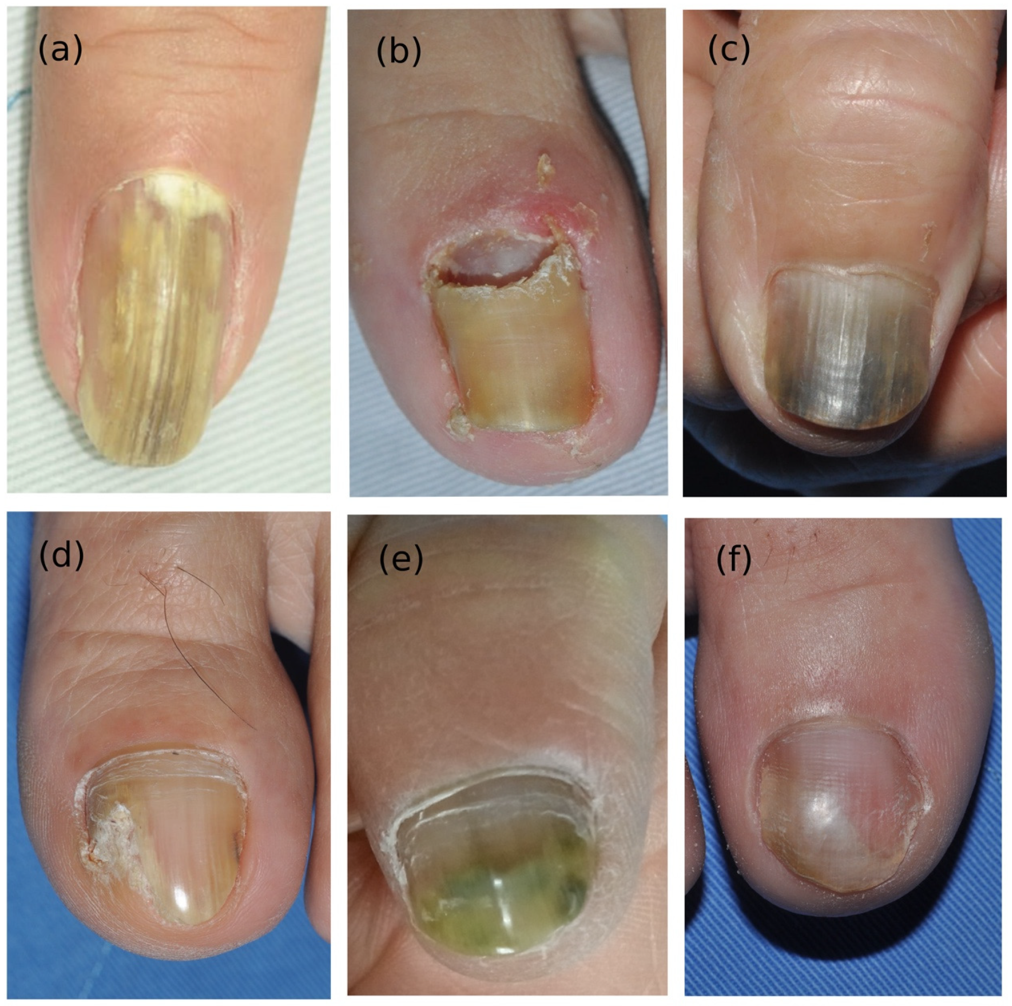
3.1. Demographic Data and Clinical Manifestations

After rigorous clinical, histological, and mycological confirmation, 29 patients were diagnosed with Fusarium onychomycosis (Table 1). There were 13 men (44.8%) and 16 women (55.2%), with the mean age of 55 (3–87) years. Average disease duration was 20 (1–108) months. Six (20.7%) patients had a personal history of gardening or soil contact, and seven (24.1%) had diabetes (n = 2), immunocompromise (n = 2), and underlying cancer (n = 3) as predisposing factors.
The most commonly involved nails were toenails (n = 16), especially the first toe (n = 15); however, there was one case in bilateral thumbs and six in fingernails. Six patients had both fingernail and toenail onychomycosis; however, none of the six had predisposing factors of diabetes, immunocompromise, or cancer. Clinical manifestations of Fusarium onychomycosis differed from those of classic dermatophyte onychomycosis; they included yellow to greenish discoloration, onycholysis, paronychia (mild or severe), and proximal subungual onychomycosis (PSO) (Figure 1).

3.2. Direct Microscopic and Histological Findings

Direct microscopic examination (DME) was performed on three patients, with frequent branching irregularly septated hyphae (Figure 2e), chlamydospore-like swelling (Figure 2f), terminal swelling and beading, and adventitious sporulation observed (Figure 2g). Among 29 patients, 21 had histopathological evidence of nail plate invasion. The histological characteristics of Fusarium onychomycosis could be categorized into six patterns, which provide clues for differentiating NDM onychomycosis from dermatophyte onychomycosis (Figure 3, Table 2): (1) presence of frequently branching irregularly septated hyphae, (2) arbitrarily widening hyphae, (3) dermatophytoma-like fungal mass, (4) thin hyphae embedded in nail specimen, (5) moniliform hyphae, and (6) hyphae with terminal swelling. The most encountered pathological finding was frequently branching irregularly shaped hyphae (Figure 3a), followed by moniliform hyphae (Figure 3e), and hyphae with terminal swelling (Figure 3f).

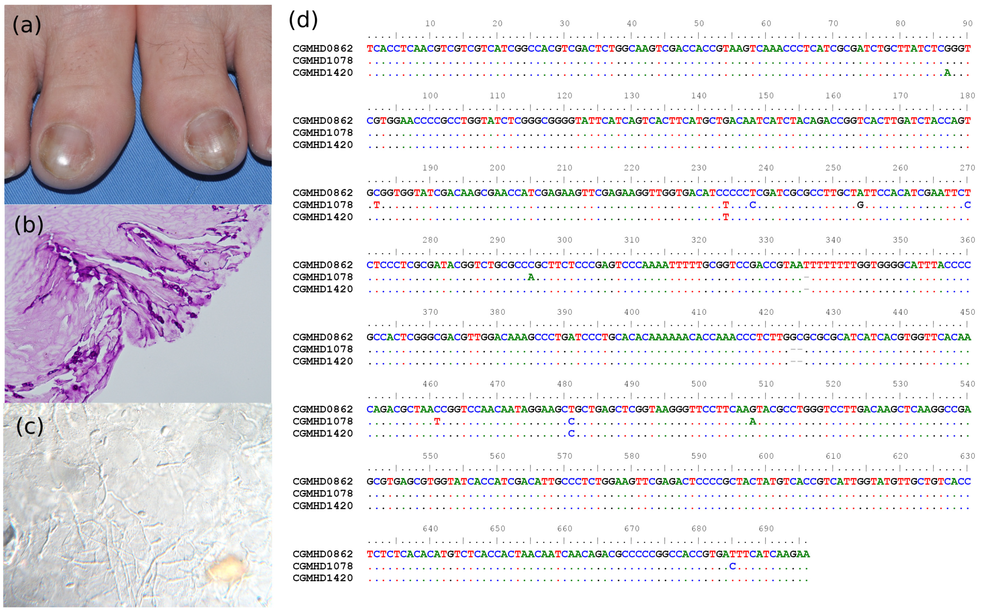
3.3. Molecular Identification

A total of 47 Fusarium strains were isolated, and the TEF-1α and ITS regions of all strains were successfully amplified and sequenced. The lengths of sequences obtained were 506–530 bp (DDBJ accession no. LC697741-LC697787) and 665–704 bp (DDBJ accession no. LC687503-LC687549). Based on phylogenetic analyses (Figure 4), the causative pathogens of confirmed cases of Fusarium onychomycosis were the FSSC (n = 23, including F. keratoplasticum, F. falciforme, F. solani, Fusarium lichenicola, F. suttonianum, Nectria bolbophylli, Fusarium sp.), Fusarium incarnatum-equiseti species complex (FIESC, n = 2, F. arcuatisporum, F. pernambucanum), FFSC (n = 3, F. annulatum, F. denticulatum), and FOSC (n = 2, F. curvatum and Fusarium sp.). One patient was infected by two Fusarium species at the same time (Figure 2).

3.4. Antifungal Susceptibility Testing

Antifungal susceptibility results are presented in Table 3. Obvious susceptibility differences between and within species complex were noted. In general, all isolates had very high MICs to FLC and ITC and low MICs to EFC, LNC, and LLC. FSSC had higher TRB MICs than non-FSSC, although FIESC had only one strain in the group, and the rough data are not so precise. The range of MICs are similar in VRC and NAT, and lower in AMB. Three F. keratoplasticum isolates were resistant to all antifungals, and one of them had a lower MIC to LNC.

3.5. Treatment Response and Prognosis

Among 13 patients who received topical antifungal agents alone (Table 4), more than half (53.8%) had poor response, while two had good response. Nine patients received combination therapy (TRB and topical antifungal agents); however, more than half (55%) of them still had a poor response. Two patients received combination therapy (ITC and topical antifungal agents); one had a good response while the other had a poor response. Only three patients received TRB/ITC treatment with continuous topical antifungal agents, contributing to the good response observed in them.

3.6. Presentation of Two Special Cases

Case 1. A 60-year-old woman presented with yellow to greenish discoloration on her right 2nd finger, left thumb, and 2nd and 4th fingers for 2–3 months (Figure 2a,b). She had systemic lupus erythematosus (SLE) and was under treatment for oral azathioprine. She denied gardening history or contact with soil. Direct microscopic examination of the nail specimen demonstrated irregularly segmented hyphae with frequent branching, adventitious sporulation, chlamydospore-like swelling, and thin hyphae, which was different from that of dermatophytes (Figure 2e–g). Histopathology of the nail revealed septated hyphae and chlamydospores (Figure 2h). Fungal cultures from diseased nails all grew Fusarium, and molecular identification showed that her right thumb was infected by F. keratoplascum (CGMHD 0974), and her left thumb and 2nd and 4th fingers were infected by F. solani (CGMHD 0975, CGMHD 0976, CGMHD 0977). Repeated culture 3 months later also revealed the same results. The patient responded poorly to oral itraconazole (200 mg/day) for 3 months and was later treated with oral terbinafine (250 mg/day) combined with nail debridement and topical sulconazole solutions. A growth of new nails was noted three months later (Figure 2c,d).
Case 2. A 55-year-old female patient had yellow to greyish discoloration on the bilateral big toes for several years (Figure 5a). She was a case of HBV chronic hepatitis and goiter of the thyroid. She denied gardening habit or contact to soil or other underlying disease, such as diabetes, malignancy, or under immunosuppressive treatment. Histopathology of the diseased nail demonstrated septated hyphae with a beaded appearance which invaded the nail plate (Figure 5b). Direct microscopic examination revealed septated hyphae and adventitious sporulation (Figure 5c). Six Fusarium isolates were cultured from the diseased nails during the two years of follow-ups. Molecular identification proved that all of them were F. keratoplasticum, but of three different genotypes based on the TEF-1α sequences (Figure 5d) The patient initially received oral griseofulvin 500 mg/day and topical antifungals for 21 days, but in vain. The patient received intermittent nail debridement and treatment with topical sulconazole solution in the following 3–4 years. New healthy nails finally grew with negative culture results. No recurrence was noted.

4. Discussion

Diagnosis of Fusarium onychomycosis is challenging because NDMs are common contaminants of nails. Published diagnostic criteria vary, and there is no consensus [9]. Approximately 42.8% false negative cases of NDM onychomycosis may be misdiagnosed when only negative dermatophyte microscopic examination and repeated culture are performed [17]. Gupta et al. proposed using three of their six clinical guideline criteria (KOH identification, isolation in culture, repeated isolation, inoculum counting [18], dermatophyte exclusion, and histological proof) to rule out dermatophyte contamination, and this remains the most widely used diagnostic method [8]. Although this method cannot perfectly prevent misdiagnosis of false negative and contaminants, it is straightforward and useful in aiding clinicians in clinical practice. For example, regarding inoculum counting, Gupta et al. had pointed out the low predictive value of inoculum counting as 23.2% of the time [19]. Similar histology finding may also be found in dermatophyte histology, but special appearance of dermatophytoma, irregulated septated hyphae, and terminal swelling are seldom seen in dermatophyte. The systemic review and comparison of histology difference in NDM and dermatophyte is important but still lacking, except in our clinical observations. When only using DME positive and negative dermatophyte culture for NDM diagnosis, there are only 53.6% sensitivity and 70.3% specificity [20]. Classical criteria include positive DME and repeated culture with 92.7% accuracy without the possibility of contaminants, but this method is difficult to be used in clinical practice [20]. Therefore, we follow the criteria of Gupta et al. and furthermore, 21 out of 29 patients in our research had histopathological evidence of fungal invasion with signs of NDM histology features, which can decrease rates of contaminants. New diagnostic methods, including molecular methods and techniques involving PCR, are seeing increasing application and importance. The commercialization of PCR kits may improve fungal diagnosis in the future [8].
In the clinical presentation of Fusarium onychomycosis, only 27.5% of patients had predisposing factors such as diabetes, immunosuppression, and cancers. The majority of Fusarium subtypes vary across the research, with proximal subungual onychomycosis (PSO), total dystrophic onychomycosis (TDO), and paronychia previously regarded as the most common clinical phenotypes [7,21]. However, distal lateral subungual onychomycosis (DLSO) and onycholysis are reportedly the more predominant subtypes [5,22,23,24,25]. FOSC is predominant in DLSO cases, and FSSC is more commonly involved in PSO, TDO, and SWO phenotypes according to Uemura et al. [25], but research from the north of Iran demonstrated diversity species among different subtypes of onychomycosis, with F. proliferatum, F. keratoplasticum, and F. falciforme predominated in DLSO, and variable appearance in PSO, TDO and endonyx onychomycosis [26]. The clinical differences between Fusarium and dermatophytes onychomycoses are not clear; however, some clues are available. Fusarium onychomycosis is most implicated in (1) periungual inflammation of the nail matrix and purulent discharge [4,9,27], (2) resistance to empirical antifungal treatment [25], (3) trauma history or nail dystrophy and absence of tinea pedis [28], and (4) involvement of the big toes (fingernails are only occasionally involved as combination symptoms) [23,24,29]. In the present study, there were six DLSO, two PSO (one overlapping paronychia), one WSO, and one DLSO phenotype with onychodystrophy from 10 cases.
The histological presentation of Fusarium onychomycosis is only mentioned in the case of reports and is rarely systemically reviewed [30]. Lavorato et al. compared the performance of mycology and histology for dermatophyte and NDM onychomycoses and revealed that direct microscopy was more sensitive for NDM and that nail clippings for histopathology were better for dermatophyte onychomycosis [31]. However, this research only collected DLSO pattern onychomycosis, and only 28.5% cases were Fusarium onychomycosis. Although we cannot differentiate dermatophyte onychomycosis from NDM onychomycosis simply by histology, there may be additional clues. Among the six recognized patterns, the most frequently seen patterns in our study were frequently branching irregularly septated hyphae, moniliform hyphae, and hyphae with terminal swelling. Direct microscopic examination with the findings of chlamydospores with beaded appearance hyphae, terminal enlarging, and adventitious sporulation are also helpful for differentiation.
The pathogenesis of Fusarium’s invasion of human nails has been previously elucidated [32]. In vitro, Fusarium species can destroy the stratum corneum through keratolysis without additional nutrients [32]. Further, marked protease activity has been detected in FSSC [33]. Flavia et al. demonstrated that Fusarium oxysporum invades nail plates, resulting in the ex-vitro formation of biofilm composed of hyphae, conidia, and extra matrix. The nail unit is a site of immune privilege with low expression of major histocompatibility antigens, dysfunction of antigen presenting cells, and inhibition of natural killer cell activity [34]. However, studies comparing NDM and dermatophyte onychomycoses in terms of levels of myotoxins, keratinase, and proteases along with components of fungal biofilms remain scarce.
Prior to this study, most of our patients received combination (topical and systemic antifungals—TRB and ITC) or destructive (laser and surgery) therapy. Among them, 53.8% showed poor response to all treatment. In review articles, few treatment methods are listed, with 26.7% clinical and 13.9% mycological cure rates reported [8,25] Gupta et al. proposed a treatment algorithm for NDM onychomycosis using a combination of topical (EFC, Tavaborole, and LLC) and systemic (ITC and TRB) therapies [8], with ITC used as daily or pulse therapy (400 mg/pulse once a week for 3 weeks); both showed mild-to-moderate evidence of Fusarium clearance [35,36]. TRB is commonly used for onychomycosis, but the drug resistance rate is relatively high and requires combination with topical antifungals or keratolytics [37]. Verrier et al. reported that oral TRB and ITC are not effective in the treatment of Fusarium onychomycosis [38]. If treatment fails, an antifungal susceptibility test is indicated, and alternatives should be considered. There is one report of treating recalcitrant Fusarium falciforme with posaconazole pulse therapy (800 mg/pulse one week for each month, with total four months) in the literature; this reportedly achieved clinical and mycological improvement [39]. Combination therapy using topical EFC, oral ITC, and oral fosravuconazole are also approved for the treatment of onychomycosis in Japan [24]. Further, topical treatment with AMB for a year has shown a reduction in recalcitrant cases [40]. Other ablative treatment procedures such as two sessions of Qs Nd-YAG laser therapy (532 nm and 1064 nm) one month apart for patients with FSSC onychomycosis showed good response [41], while another study used 1340 nm laser monotherapy, resulting in persistent onychomycosis (91%) under mycological tests for one year [42]. Methylene blue-mediated photodynamic therapy is another choice, which may be superior to 5% amorolfine nail lacquer for NDM onychomycosis [43].
Currently, there are no clinical breakpoints for antifungal drugs against different Fusarium species. In this study, TRB, FLC, and ITC all showed poor improvement in treatment. However, AMB, EFC, LNC, LLC, VRC, and NAT showed better results and should be considered for clinical applications according to MIC results (Table 3). In the literature, The MIC levels from the north of Iran are compatible with our findings, as LLC and LNC was in the range of 1–0.001 μg/mL [26]. Based on Uemura et al., MIC in Fusarium onychomycosis, ITC, FLC, and 5-Fluorocytosine showed high resistance tendency; TRB showed variable resistance tendency [25]; and VRC and AMB showed low resistance tendency. The recently developed antifungal EFC has shown good treatment response in intractable cases [24,44], and olorofim has also shown promise as a candidate [45] after showing in vitro activity against FSSC and FOSC.
Identification of Fusarium to the species level is challenging for clinical laboratories as the morphological characteristics required for identification are few and require experience with the genus. Furthermore, the recent Fusarium taxonomy is based on molecular phylogeny, making identification by morphology alone unreliable. Among existing reviews and case studies on Fusarium onychomycosis, five identified the pathogen to the genus level and 11 to the Fusarium specie level; only five studies performed molecular identification to the species complex level [4,5,7,22,23,24,26,29,30,32,46,47,48,49,50,51,52,53,54]. Molecular identification is typically performed using phylogenetic analysis of sequences of ITS and TEF-1α, with RNA polymerase II’s second largest subunit (RPB2) genes sometimes used for better differentiation between species. Molecular identification can provide clues to the source and process of the infection, and the importance of repeated and accurate fungal culture reminds patients to pay attention to pathogen control in public health to identify and avoid the source of Fusarium colonization and invasion. Differences between clinical manifestation and antifungal susceptibility testing highlighted the importance of accurate molecular classification.

5. Conclusions

Although Fusarium onychomycosis accounts for 1–6% of cases of onychomycosis, its frequent resistance to treatment highlights its importance [5]. Cutaneous fusarium infections can serve as the origin of disseminated and invasive infection poor response to empirical antifungals. Therefore, accurate diagnosis through histological and molecular identification is required. Positive culture results for Fusarium species from nail samples are not a proof of infection; further histological proof or positive repeated culture results for the same species of Fusarium are required. Molecular identification (ITS + TEF-1α) and phylogenetic analysis can be applied for pathogen species confirmation. If a positive culture of Fusarium is simply due to colonization, then destruction of the colony and hygiene to prevent colonization is enough. However, if there is a true infection, then combination therapy (using topical and oral antifungal agents) and even surgical debridement are required. Dermatologists will do well to partner with mycologists specialized in Fusarium identification and application. In this article, we highlighted the clinicopathological features of Fusarium onychomycosis and provided six histopathological hints for differentiating Fusarium onychomycosis from dermatophyte onychomycosis. However, much work is needed to provide a standard effective treatment protocol for Fusarium onychomycosis.
This study has some limitations. The first is its design as a single-centre retrospective study. Additionally, owing to a lack of follow up with clinical photos, we could not determine and classify the percentage of accurate onychomycosis subtypes in this study. Last but not least, although current diagnostic criteria for Fusarium onychomycosis is not perfect, it is crucial for helping clinicians in diagnosis and treatment application.

Author Contributions

L.-Y.L. contributed to the project administration, original draft writing, data collection, and design; P.-L.S. carried out the project administration, conceptualization, writing review and editing, and supervision; J.-H.O. performed the molecular identification; Y.-C.F. performed sequencing and antifungal susceptibility testing; R.C.-Y.H. and Y.-H.C. contributed to the data sharing. All authors have read and agreed to the published version of the manuscript.

Funding

This research received no external funding.

Institutional Review Board Statement

This study was reviewed and approved by the IRB of the Chang Gung Medical Foundation (approval number 202101575B0).

Informed Consent Statement

Patient consent was waived by the IRB.

Data Availability Statement

The data presented in the manuscript are available on request from the corresponding authors.

Acknowledgments

We would like to thank Gini-Wu, Chin-Yi Yang, Yao-Yu Chang, Hong-Shang Hong, Ming-Hui Chi, and Chih-Hsun Yang for providing the patients.

Conflicts of Interest

The authors declare no conflict of interest.

References

  1. Geiser, D.M.; Al-Hatmi, A.; Aoki, T.; Arie, T.; Balmas, V.; Barnes, I.; Bergstrom, G.C.; Bhattacharyya, M.K.K.; Blomquist, C.L.; Bowden, R.; et al. Phylogenomic analysis of a 55.1 kb 19-gene dataset resolves a monophyletic Fusarium that includes the Fusarium solani Species Complex. Phytopathology 2020, 111, 1064–1079. [Google Scholar] [CrossRef]
  2. Nucci, M.; Anaissie, E. Fusarium infections in immunocompromised patients. Clin. Microbiol. Rev. 2007, 20, 695–704. [Google Scholar] [CrossRef]
  3. Nucci, F.; Nouer, S.A.; Capone, D.; Anaissie, E.; Nucci, M. Fusariosis. Semin. Respir. Crit. Care Med. 2015, 36, 706–714. [Google Scholar] [CrossRef]
  4. Guilhermetti, E.; Takahachi, G.; Shinobu, C.S.; Svidzinski, T.I. Fusarium spp. as agents of onychomycosis in immunocompetent hosts. Int. J. Dermatol. 2007, 46, 822–826. [Google Scholar] [CrossRef]
  5. Diongue, K.; Ndiaye, M.; Seck, M.C.; Diallo, M.A.; Badiane, A.S.; Ndiaye, D. Onychomycosis Caused by Fusarium spp. in Dakar, Senegal: Epidemiological, Clinical, and Mycological Study. Dermatol. Res. Pract. 2017, 2017, 1268130. [Google Scholar] [CrossRef]
  6. Al-Hatmi, A.M.; Meis, J.F.; de Hoog, G.S. Fusarium: Molecular Diversity and Intrinsic Drug Resistance. PLoS Pathog. 2016, 12, e1005464. [Google Scholar] [CrossRef]
  7. Gupta, C.; Jongman, M.; Das, S.; Snehaa, K.; Bhattacharya, S.N.; Seyedmousavi, S.; van Diepeningen, A.D. Genotyping and In Vitro Antifungal Susceptibility Testing of Fusarium Isolates from Onychomycosis in India. Mycopathologia 2016, 181, 497–504. [Google Scholar] [CrossRef]
  8. Gupta, A.K.; Summerbell, R.C.; Venkataraman, M.; Quinlan, E.M. Nondermatophyte mould onychomycosis. J. Eur. Acad. Dermatol. Venereol. 2021, 35, 1628–1641. [Google Scholar] [CrossRef]
  9. Gupta, A.K.; Drummond-Main, C.; Cooper, E.A.; Brintnell, W.; Piraccini, B.M.; Tosti, A. Systematic review of nondermatophyte mold onychomycosis: Diagnosis, clinical types, epidemiology, and treatment. J. Am. Acad. Dermatol. 2012, 66, 494–502. [Google Scholar] [CrossRef]
  10. Mashima, J.; Kodama, Y.; Kosuge, T.; Fujisawa, T.; Katayama, T.; Nagasaki, H.; Okuda, Y.; Kaminuma, E.; Ogasawara, O.; Okubo, K.; et al. DNA data bank of Japan (DDBJ) progress report. Nucleic Acids Res. 2016, 44, D51–D57. [Google Scholar] [CrossRef]
  11. Talavera, G.; Castresana, J. Improvement of phylogenies after removing divergent and ambiguously aligned blocks from protein sequence alignments. Syst. Biol. 2007, 56, 564–577. [Google Scholar] [CrossRef] [PubMed]
  12. Minh, B.Q.; Schmidt, H.A.; Chernomor, O.; Schrempf, D.; Woodhams, M.D.; von Haeseler, A.; Lanfear, R. IQ-TREE 2: New Models and Efficient Methods for Phylogenetic Inference in the Genomic Era. Mol. Biol. Evol. 2020, 37, 1530–1534. [Google Scholar] [CrossRef] [PubMed]
  13. Kalyaanamoorthy, S.; Minh, B.Q.; Wong, T.K.F.; von Haeseler, A.; Jermiin, L.S. ModelFinder: Fast model selection for accurate phylogenetic estimates. Nat. Methods 2017, 14, 587–589. [Google Scholar] [CrossRef]
  14. Ronquist, F.; Teslenko, M.; van der Mark, P.; Ayres, D.L.; Darling, A.; Hohna, S.; Larget, B.; Liu, L.; Suchard, M.A.; Huelsenbeck, J.P. MrBayes 3.2: Efficient Bayesian phylogenetic inference and model choice across a large model space. Syst. Biol. 2012, 61, 539–542. [Google Scholar] [CrossRef] [PubMed]
  15. Kumar, S.; Stecher, G.; Tamura, K. MEGA7: Molecular Evolutionary Genetics Analysis Version 7.0 for Bigger Datasets. Mol. Biol. Evol. 2016, 33, 1870–1874. [Google Scholar] [CrossRef]
  16. Clinical and Laboratory Standards Institute (CLSI). M38: Reference Method for Broth Dilution Antifungal Susceptibility Testing of Filamentous Fungi, 3rd ed.; Approved Standard; Clinical and Laboratory Standards Institute (CLSI): Wayne, PA, USA, 2008. [Google Scholar]
  17. Bombace, F.; Iovene, M.R.; Galdiero, M.; Martora, F.; Nicoletti, G.F.; D’Andrea, M.; Della Pepa, M.E.; Vitiello, M. Non-dermatophytic onychomycosis diagnostic criteria: An unresolved question. Mycoses 2016, 59, 558–565. [Google Scholar] [CrossRef]
  18. Walshe, M.M.; English, M.P. Fungi in nails. Br. J. Dermatol. 1966, 78, 198–207. [Google Scholar] [CrossRef]
  19. Gupta, A.K.; Cooper, E.A.; MacDonald, P.; Summerbell, R.C. Utility of inoculum counting (Walshe and English criteria) in clinical diagnosis of onychomycosis caused by nondermatophytic filamentous fungi. J. Clin. Microbiol. 2001, 39, 2115–2121. [Google Scholar] [CrossRef]
  20. Summerbell, R.C.; Cooper, E.; Bunn, U.; Jamieson, F.; Gupta, A.K. Onychomycosis: A critical study of techniques and criteria for confirming the etiologic significance of nondermatophytes. Med. Mycol. 2005, 43, 39–59. [Google Scholar] [CrossRef]
  21. Dignani, M.C.; Anaissie, E. Human fusariosis. Clin. Microbiol. Infect. 2004, 10, 67–75. [Google Scholar] [CrossRef]
  22. Ranawaka, R.R.; Nagahawatte, A.; Gunasekara, T.A. Fusarium onychomycosis: Prevalence, clinical presentations, response to itraconazole and terbinafine pulse therapy, and 1-year follow-up in nine cases. Int. J. Dermatol. 2015, 54, 1275–1282. [Google Scholar] [CrossRef] [PubMed]
  23. Calado, N.B.; Sousa, F., Jr.; Gomes, N.O.; Cardoso, F.R.; Zaror, L.C.; Milan, E.P. Fusarium nail and skin infection: A report of eight cases from Natal, Brazil. Mycopathologia 2006, 161, 27–31. [Google Scholar] [CrossRef] [PubMed]
  24. Noguchi, H.; Matsumoto, T.; Kimura, U.; Hiruma, M.; Kano, R.; Yaguchi, T.; Ihn, H. Non-dermatophyte Mould Onychomycosis in Japan. Med. Mycol. J. 2020, 61, 23–31. [Google Scholar] [CrossRef] [PubMed]
  25. Uemura, E.V.G.; Barbosa, M.D.S.; Simionatto, S.; Al-Harrasi, A.; Al-Hatmi, A.M.S.; Rossato, L. Onychomycosis Caused by Fusarium Species. J. Fungi 2022, 8, 360. [Google Scholar] [CrossRef]
  26. Haghani, I.; Shams-Ghahfarokhi, M.; Dalimi Asl, A.; Shokohi, T.; Hedayati, M.T. Prevalence, genetic diversity and antifungal susceptibility profiles of F. fujikuroi, F. solani and Fusarium incarnatum-equiseti species complexes from onychomycosis in north of Iran. Mycoses 2022, 65, 1030–1039. [Google Scholar] [CrossRef]
  27. Baran, R.; Tosti, A.; Piraccini, B.M. Uncommon clinical patterns of Fusarium nail infection: Report of three cases. Br. J. Dermatol. 1997, 136, 424–427. [Google Scholar] [CrossRef]
  28. Gupta, A.K.; Ryder, J.E.; Baran, R.; Summerbell, R.C. Non-dermatophyte onychomycosis. Dermatol. Clin. 2003, 21, 257–268. [Google Scholar] [CrossRef]
  29. Ranawaka, R.R.; de Silva, N.; Ragunathan, R.W. Onychomycosis caused by Fusarium sp in Sri Lanka: Prevalence, clinical features and response to itraconazole pulse therapy in six cases. J. Dermatol. Treat. 2008, 19, 308–312. [Google Scholar] [CrossRef]
  30. Rammlmair, A.; Muhlethaler, K.; Haneke, E. Fusarium onychomycoses in Switzerland-A mycological and histopathological study. Mycoses 2019, 62, 928–931. [Google Scholar] [CrossRef]
  31. Lavorato, F.G.; Guimarães, D.A.; Premazzi, M.G.; Piñeiro-Maceira, J.M.; Bernardes-Engemann, A.R.; Orofino-Costa, R. Performance of mycology and histopathology tests for the diagnosis of toenail onychomycosis due to filamentous fungi: Dermatophyte and non-dermatophyte moulds. Mycoses 2017, 60, 587–593. [Google Scholar] [CrossRef]
  32. Galletti, J.; Negri, M.; Grassi, F.L.; Kioshima-Cotica, E.S.; Svidzinski, T.I. Fusarium spp. is able to grow and invade healthy human nails as a single source of nutrients. Eur. J. Clin. Microbiol. Infect. Dis. 2015, 34, 1767–1772. [Google Scholar] [CrossRef] [PubMed]
  33. Gugnani, H.C. Nondermatophytes filamentous keratinophilic fungi and their role in human infection. Rev. Iberoam. Micol. 2000, 17, 109–114. [Google Scholar]
  34. Ito, T.; Meyer, K.C.; Ito, N.; Paus, R. Immune privilege and the skin. Curr. Dir. Autoimmun. 2008, 10, 27–52. [Google Scholar] [CrossRef] [PubMed]
  35. De Doncker, P.R.; Scher, R.K.; Baran, R.L.; Decroix, J.; Degreef, H.J.; Roseeuw, D.I.; Havu, V.; Rosen, T.; Gupta, A.K.; Piérard, G.E. Itraconazole therapy is effective for pedal onychomycosis caused by some nondermatophyte molds and in mixed infection with dermatophytes and molds: A multicenter study with 36 patients. J. Am. Acad. Dermatol. 1997, 36, 173–177. [Google Scholar] [CrossRef] [PubMed]
  36. Gupta, A.K.; Gregurek-Novak, T.; Konnikov, N.; Lynde, C.W.; Hofstader, S.; Summerbell, R.C. Itraconazole and terbinafine treatment of some nondermatophyte molds causing onychomycosis of the toes and a review of the literature. J. Cutan. Med. Surg. 2001, 5, 206–210. [Google Scholar] [CrossRef] [PubMed]
  37. Al-Hatmi, A.M.S.; Bonifaz, A.; Ranque, S.; Sybren de Hoog, G.; Verweij, P.E.; Meis, J.F. Current antifungal treatment of fusariosis. Int. J. Antimicrob. Agents 2018, 51, 326–332. [Google Scholar] [CrossRef]
  38. Verrier, J.; Bontems, O.; Baudraz-Rosselet, F.; Monod, M. Oral terbinafine and itraconazole treatments against dermatophytes appear not to favor the establishment of Fusarium spp. in nail. Dermatology 2014, 228, 225–232. [Google Scholar] [CrossRef] [PubMed]
  39. Al-Hatmi, A.M.; Bonifaz, A.; Calderón, L.; Curfs-Breuker, I.; Meis, J.F.; van Diepeningen, A.D.; de Hoog, G.S. Proximal subungual onychomycosis caused by Fusarium falciforme successfully cured with posaconazole. Br. J. Dermatol. 2015, 173, 253–255. [Google Scholar] [CrossRef] [PubMed]
  40. Lurati, M.; Baudraz-Rosselet, F.; Vernez, M.; Spring, P.; Bontems, O.; Fratti, M.; Monod, M. Efficacious treatment of non-dermatophyte mould onychomycosis with topical amphotericin B. Dermatology 2011, 223, 289–292. [Google Scholar] [CrossRef]
  41. Khurana, A.; Chowdhary, A.; Sardana, K.; Gautam, R.K.; Sharma, P.K. Complete cure of Fusarium solani sp. complex onychomycosis with Qs NdYAG treatment. Dermatol. Ther. 2018, 31, e12580. [Google Scholar] [CrossRef]
  42. Espírito-Santo, G.A.D.; Leite, D.P., Jr.; Hoffmann-Santos, H.D.; Dias, L.B.; Hahn, R.C. 1340nm Laser Therapy For Onychomycosis: Negative Results of Prospective Treatment of 72 Toenails and a Literature Review. J. Clin. Aesthetic Dermatol. 2017, 10, 56–61. [Google Scholar]
  43. Bowornsathitchai, N.; Thammahong, A.; Shoosanglertwijit, J.; Kitsongsermthon, J.; Wititsuwannakul, J.; Asawanonda, P.; Boontaveeyuwat, E. Methylene blue-mediated photodynamic therapy may be superior to 5% amorolfine nail lacquer for non-dermatophyte onychomycosis. Photodermatol. Photoimmunol. Photomed. 2021, 37, 183–191. [Google Scholar] [CrossRef] [PubMed]
  44. Hirose, M.; Noguchi, H.; Matsumoto, T.; Kimura, U.; Hiruma, M.; Kano, R.; Yaguchi, T.; Fujimoto, N.; Satoh, T.; Ihn, H. Ungual hyalohyphomycosis caused by Fusarium cugenangense. Clin. Case Rep. 2020, 8, 3533–3538. [Google Scholar] [CrossRef] [PubMed]
  45. Badali, H.; Cañete-Gibas, C.; Patterson, H.; Sanders, C.; Mermella, B.; Garcia, V.; Mele, J.; Fan, H.; Wiederhold, N.P. In vitro activity of olorofim against clinical isolates of the Fusarium oxysporum and Fusarium solani species complexes. Mycoses 2021, 64, 748–752. [Google Scholar] [CrossRef] [PubMed]
  46. Rosa, P.D.; Heidrich, D.; Correa, C.; Scroferneker, M.L.; Vettorato, G.; Fuentefria, A.M.; Goldani, L.Z. Genetic diversity and antifungal susceptibility of Fusarium isolates in onychomycosis. Mycoses 2017, 60, 616–622. [Google Scholar] [CrossRef]
  47. Castro Lopez, N.; Casas, C.; Sopo, L.; Rojas, A.; Del Portillo, P.; Cepero de Garcia, M.C.; Restrepo, S. Fusarium species detected in onychomycosis in Colombia. Mycoses 2009, 52, 350–356. [Google Scholar] [CrossRef]
  48. Motamedi, M.; Ghasemi, Z.; Shidfar, M.R.; Hosseinpour, L.; Khodadadi, H.; Zomorodian, K.; Mirhendi, H. Growing Incidence of Non-Dermatophyte Onychomycosis in Tehran, Iran. Jundishapur J. Microbiol. 2016, 9, e40543. [Google Scholar] [CrossRef]
  49. Bansal, Y.; Singla, N.; Kaistha, N.; Sood, S.; Chander, J. Molecular identification of Fusarium species complex isolated from clinical samples and its antifungal susceptibility patterns. Curr. Med. Mycol. 2019, 5, 43–49. [Google Scholar] [CrossRef]
  50. Godoy, P.; Nunes, E.; Silva, V.; Tomimori-Yamashita, J.; Zaror, L.; Fischman, O. Onychomycosis caused by Fusarium solani and Fusarium oxysporum in Sao Paulo, Brazil. Mycopathologia 2004, 157, 287–290. [Google Scholar] [CrossRef]
  51. Tosti, A.; Piraccini, B.M.; Lorenzi, S. Onychomycosis caused by nondermatophytic molds: Clinical features and response to treatment of 59 cases. J. Am. Acad. Dermatol. 2000, 42, 217–224. [Google Scholar] [CrossRef]
  52. Phaitoonwattanakij, S.; Leeyaphan, C.; Lertrujiwanit, K.; Bunyaratavej, S. Predisposing factors, clinical features and treatment outcomes of Fusarium onychomycosis and comparison of its characteristics with Neoscytalidium onychomycosis. J. Mycol. Med. 2021, 31, 101165. [Google Scholar] [CrossRef] [PubMed]
  53. Ranawaka, R.R.; Nagahawatte, A.; Gunasekara, T.A.; Weerakoon, H.S.; de Silva, S.H. Randomized, double-blind, comparative study on efficacy and safety of itraconazole pulse therapy and terbinafine pulse therapy on nondermatophyte mold onychomycosis: A study with 90 patients. J. Dermatol. Treat. 2016, 27, 364–372. [Google Scholar] [CrossRef] [PubMed]
  54. Van Diepeningen, A.D.; Feng, P.; Ahmed, S.; Sudhadham, M.; Bunyaratavej, S.; de Hoog, G.S. Spectrum of Fusarium infections in tropical dermatology evidenced by multilocus sequencing typing diagnostics. Mycoses 2015, 58, 48–57. [Google Scholar] [CrossRef] [PubMed]
Figure 1. Clinical manifestations of Fusarium onychomycosis, including yellow and greenish discoloration (a,b,e), onycholysis (af), severe paronychia (b), and proximal subungual onychomycosis (a,b). The diversity of clinical presentation highlights the importance of fungus culture and species identification.
Jof 09 00534 g001
Figure 2. A 60-year-old female who had Fusarium onychomycosis on fingernails. Fusarium keratoplasticum was isolated from right 2nd fingernail and F. solani was isolated from left 1st, 2nd, and 4th fingernails. (a,b) Yellowish green discoloration on the right second finger, left thumb, and left second and fourth fingers for 2–3 months. (c,d) Three months after combination therapy with systemic terbinafine and topical sulconazole solution treatment; green discoloration of nails regressed with new nail regrowth. (eg) Direct microscopic examination showing frequently branching irregularly septated hyphae, adventitious sporulation, and chlamydospore like swelling. (h) Sections showing twisted broad septated hyphae (Hematolysin and eosin stain, original magnification 200×).
Jof 09 00534 g002
Figure 3. Histopathological characteristics of Fusarium onychomycosis classified into six subgroups. (a) Frequently branching irregularly septated hyphae, (b) Arbitrarily widening hyphae, (c) Dermatophytoma-like fungal mass, (d) Thin hyphae embedded in the nail specimen, (e) Moniliform hyphae, and (f) Hyphae with terminal swelling (Hematolysin and eosin stain, original magnification 200×).
Jof 09 00534 g003
Figure 4. The maximum likelihood phylogenetic tree inferred from the concatenated TEF-1α and ITS regions. Bootstrap support value (ML) and Bayesian posterior probability (BP) higher than 50 and 0.6 are given at each node as ML/BP. Nodes with a support of 100/1.0 are shown in bold. The strains isolated in this study are shown in bold. Type strains are indicated with a superscripted T.
Jof 09 00534 g004
Figure 5. A 55-year-old female had Fusarium onychomycosis on her bilateral big toes. (a) Yellowish grey discoloration and onycholysis on the big toes. (b) Histopathology study showing septated and moniliform hyphae invasion into the nail specimen. (Hematoxysin and eosin stain, original magnification 200×) (c) Direct microscopic examination revealed septated hyphae and adventitious sporulation. (d) Transcription elongation factor-1α (TEF-1α) gene sequences of the isolates showed that the F. keratoplasticum in her toenails belonged to three different strain types: CGMHD0862 (left big toe), CGMHD1078 (right big toe), and CGMHD1420 (big toe, site not specified).
Jof 09 00534 g005
Table 1. Clinical and mycological characteristics in 29 patients with Fusarium onychomycosis.
Sex/AgeImmune StatusLocationDuration
(Month)
SpeciesTreatmentPrognosisContact to Soil
FFSCF/77ICRight big toenail2Fusarium denticulatumTAFGRYes
F/61ICRight middle finger1Fusarium annulatumSurgeryGRYes
F/84Lung cancerFingernails6Fusarium annulatumITC + TAFGRNo
FIESCF/65Paraneoplastic pemphigusFingernails108Fusarium pernambucanum (FIESC 17)ITC + TAFPoRNo
M/46Tuberous sclerosis, left RCCToenails3Fusarium arcuatisporum (FIESC 7)TAFPaRNo
FOSCF/54ICRight big toenail53Fusarium curvatumTAFLosNo
M/36ICBilateral big toenails4Fusarium sp.ITC + TAFGRNo
FSSCF/60SLEFingernails7Fusarium keratoplasticum (FSSC 2)
Fusarium solani (FSSC 5)
ITC/TRB + TAFGRNo
M/21ICLeft big toenail4Fusarium solani (FSSC 5)TAFPaRNo
M/43ICBilateral big toenail1Fusarium falciforme (FSSC 3 + 4)TAFPoRNo
F/58DMRight big toenail32Fusarium falciforme (FSSC 3 + 4)TRB + TAFPoRNo
F/45ICLeft big toenail4Fusarium keratoplasticum (FSSC 2)TRB +TAFGRNo
F/67ICBilateral big toenail60Fusarium keratoplasticum (FSSC 2)TRB + TAFPoRNo
M/77ICBilateral thumb48Fusarium keratoplasticum (FSSC 2)TRB +TAFPoRYes
F/3ICFingernails + Toenails15Fusarium keratoplasticum (FSSC 2)TAFPoRNo
F/31ICLeft big toenail32Fusarium keratoplasticum (FSSC 2)ITC/TRB + TAFGRNo
M/34ICBilateral toenails4Fusarium keratoplasticum (FSSC 2)TRB + TAFGRNo
F/79ICFingernails + Toenails25Fusarium keratoplasticum (FSSC 2)TAFPoRNo
F/55ICBilateral big toenail41Fusarium keratoplasticum (FSSC 2)Laser + Griseolfulbin + TAFGRNo
M/60ICFingernails + Toenails41Fusarium keratoplasticum (FSSC 2)TRB + TAFPaRNo
M/45ICRight big toenail2Fusarium keratoplasticum (FSSC 2)TAFPoRNo
F/65ICRight big toenail24Fusarium keratoplasticum (FSSC 2)TRB + TAFPoRNo
M/48ICFingernails + Toenails9Fusarium keratoplasticum (FSSC 2)TAFPoRNo
F/87Terminal ileal cancerBilateral big toenail36Fusarium keratoplasticum (FSSC 2)TAFPoRNo
M/70DMLeft thumb2Fusarium suttonianum (FSSC 20)TAFGRYes
M/77ICFingernails + Toenails6Fusarium lichenicola (FSSC 16)TAFPoRYes
M/65ICRight big toenail5Nectria bolbophylliTRB + TAFGRNo
M/20ICLeft 2nd fingernail1Fusarium sp.TAFLosNo
F/61ICFingernails + Toenails5Fusarium sp.TRB + TAFGRYes
Abbreviations: F: female; M: male; IC: immunocompetent; RCC: renal cell carcinoma; SLE: systemic lupus erythematosus; DM: diabetes mellitus; GR: Good response; PaR: Partial response; PoR: poor response; Los: Loss of follow up; TAF: Topical antifungals; ITC: Itraconazole; TRB: Terbinafine.
Table 2. Six characteristic histopathology findings in Fusarium onychomycosis.
(a) Frequently Branching Irregularly Septated Hyphae(b) Arbitrarily Widening Hyphae(c) Dermatophytoma Like Fungal Mass(d) Thin Hyphae(e) Moniliform Hyphae(f) Hyphae with Terminal
Swelling
FSSC (N = 23)
F. keratoplasticum413 74
F. falciforme1 1
F. solani SC1
F. suttonianum1 1
F. lichenicola1
Nectria bolbophylli1
Fusarium species1 1 1
FFSC (N = 3)
F. denticulatum1 1
F. annulatum2
FOSC (N = 2)
F. curvatum1
FIESC (N = 2)
F. pernambucanum 1
F. arcuatisporum 1
Total14241105
Table 3. The Fusarium species of clinical isolates and their minimum inhibitory concentration of 9 drugs (μg/mL) and GenBank accession numbers.
SpeciesRLMM No. Accession Number
AMBTRBFLCITCEFCLNCLLCVRCNATITSEF1a
FFSCF. annulatumCGMHD024814>64>320.250.0630.03144LC687503LC697741
F. annulatumCGMHD291324>64>320.50.1250.06348LC687504LC697742
F. denticulatumCGMHD110112>6440.1250.031<0.00814LC687507LC697745
FIESCF. arcuatisporumCGMHD0667NDNDNDNDNDNDNDNDNDLC687505LC697743
F. pernambucanumCGMHD05501>32>64320.50.0630.06324LC687537LC697775
FOSCF. curvatumCGMHD043618>64>320.50.1250.06344LC687506LC697744
Fusarium sp.CGMHD359424>64>320.50.1250.06388LC687546LC697784
Fusarium sp.CGMHD369942>64>320.50.1250.06388LC687547LC697785
FSSCF. falciformeCGMHD04141>32>64>3210.50.2588LC687508LC697746
F. falciformeCGMHD28762>32>64>3210.1250.03148LC687509LC697747
F. keratoplasticumCGMHD02342>32>64>3220.50.12584LC687510LC697748
F. keratoplasticumCGMHD03211>32>64>3240.250.12584LC687511LC697749
F. keratoplasticumCGMHD05492>32>64>3220.50.12584LC687512LC697750
F. keratoplasticumCGMHD0562>16>32>64>32>4>4>4>16>32LC687513LC697751
F. keratoplasticumCGMHD06582>32>64>3220.250.06384LC687514LC697752
F. keratoplasticumCGMHD06662>32>64>3220.250.12584LC687515LC697753
F. keratoplasticumCGMHD06832>32>64>3220.250.12584LC687516LC697754
F. keratoplasticumCGMHD06932>32>64>3220.250.06384LC687517LC697755
F. keratoplasticumCGMHD07021>32>64>3210.250.06384LC687518LC697756
F. keratoplasticumCGMHD07404>32>64>3220.250.06384LC687519LC697757
F. keratoplasticumCGMHD08214>32>64>3220.250.06384LC687520LC697758
F. keratoplasticumCGMHD08622>32>64>3220.50.12584LC687521LC697759
F. keratoplasticumCGMHD0974>16>32>64>32>40.25>4>16>32LC687522LC697760
F. keratoplasticumCGMHD10782>32>64>3220.50.12584LC687523LC697761
F. keratoplasticumCGMHD12202>32>64>3220.250.06384LC687524LC697762
F. keratoplasticumCGMHD12352>32>64>320.50.1250.03124LC687525LC697763
F. keratoplasticumCGMHD12682>32>64>32210.125164LC687526LC697764
F. keratoplasticumCGMHD14201>32>64>3220.250.06384LC687527LC697765
F. keratoplasticumCGMHD18752>32>64>320.50.1250.03144LC687528LC697766
F. keratoplasticumCGMHD19834>32>64>3220.250.063164LC687529LC697767
F. keratoplasticumCGMHD22234>32>64>3220.250.06384LC687530LC697768
F. keratoplasticumCGMHD26174>32>64>3220.250.06384LC687531LC697769
F. keratoplasticumCGMHD32972>32>64>320.50.1250.06324LC687532LC697770
F. keratoplasticumCGMHD33352>32>64>3210.250.06344LC687533LC697771
F. keratoplasticumCGMHD39784>32>64>3220.250.06384LC687534LC697772
F. keratoplasticumCGMHD45682>32>64>3210.250.06384LC687535LC697773
F. lichenicolaCGMHD22131>32>64>320.50.1250.063416LC687536LC697774
F. solaniCGMHD05300.5>32>64>3210.250.06388LC687538LC697776
F. solaniCGMHD09751>32>64>3220.50.06384LC687539LC697777
F. solaniCGMHD09761>32>64>3220.50.06384LC687540LC697778
F. solaniCGMHD09771>32>64>3220.50.06384LC687541LC697779
F. solaniCGMHD10801>32>64>3220.50.06384LC687542LC697780
F. solaniCGMHD13290.5>32>64>3210.50.12584LC687543LC697781
Fusarium sp.CGMHD0943>16>32>64>32>40.5>4>16>32LC687544LC697782
Fusarium sp.CGMHD0944432>64>320.50.1250.03148LC687545LC697783
F. suttonianumCGMHD19110.5>32>64>3210.50.2548LC687548LC697786
Nectria bolbophylliCGMHD02254>32>64>3220.50.125164LC687549LC697787
Abbreviations: RLMM: Research Laboratory of Medical Mycology, Chang Gung Memorial Hospital, Linkou Branch, Taoyuan, Taiwan; AMB: amphotericin B, TRB: terbinafine, FLC: fluconazole, ITC: itraconazole, EFC: efinaconazole, LNC: lanoconazole, LLC: luliconzole, VRC: voriconazole, NAT: natamycin; ND: not done due to loss of sporulation.
Table 4. Treatment response and prognosis of Fusarium onychomycosis.
Treatment MethodsGRPaRPoRLos
Itraconazole + topical antifungals (N = 2)1010
Terbinafine + topical antifungals (N = 9)3150
Itraconazole/Terbinafine + topical antifungals (N = 3)3000
Topical antifungals only (N = 13)2272
Laser + Griseofulvin + topical antifungals (N = 1)1000
Surgery (N = 1)1000
Total number (N = 29)113132
Abbreviations: GR: Good response; PaR: Partial response; PoR: poor response; Los: Loss of follow up.
Disclaimer/Publisher’s Note: The statements, opinions and data contained in all publications are solely those of the individual author(s) and contributor(s) and not of MDPI and/or the editor(s). MDPI and/or the editor(s) disclaim responsibility for any injury to people or property resulting from any ideas, methods, instructions or products referred to in the content.

