Article

Bioclimatic Origin Shapes Phylogenetic Structure of Tirmania (Pezizaceae): New Species and New Record from North Africa

by
Fatima El-Houaria Zitouni-Haouar
1,*,
Martin I. Bidartondo
2,3,
Gabriel Moreno
4,
Juan Ramón Carlavilla
4,
José Luis Manjón
4,
Samir Neggaz
1 and
Saida Hanane Zitouni-Nourine
5
1
Laboratory of Biology of Microorganisms and Biotechnology, Department of Biotechnology, Faculty of Natural and Life Sciences, Oran 1 Ahmed Ben Bella University, Oran 31000, Algeria
2
Department of Life Sciences, Imperial College London, Silwood Park, Ascot, London SL5 7PY, UK
3
Ecosystem Stewardship, Royal Botanic Gardens, Kew, Richmond TW9 3DS, UK
4
Departamento de Ciencias de la Vida, Facultad de Biología, Universidad de Alcalá, 28805 Alcalá de Henares, Madrid, Spain
5
Pharmaceutical Development Research Laboratory, Department of Pharmacy, Faculty of Medicine, Oran 1 Ahmed Ben Bella University, Oran 31000, Algeria
*
Author to whom correspondence should be addressed.
J. Fungi 2023, 9(5), 532; https://doi.org/10.3390/jof9050532
Submission received: 17 March 2023 / Revised: 19 April 2023 / Accepted: 23 April 2023 / Published: 29 April 2023
(This article belongs to the Special Issue Phylogeny and Taxonomy of Ascomycete Fungi)

Abstract

:
The phylogenetic relationships among Tirmania were investigated using the internal transcribed spacer (ITS) and large subunit (LSU) regions of the nuclear-encoded ribosomal DNA (rDNA) and compared with morphological and bioclimatic data. The combined analyses of forty-one Tirmania samples from Algeria and Spain supported four lineages corresponding to four morphological species. Besides the two previously described taxa, Tirmania pinoyi and Tirmania nivea, here we describe and illustrate a new species, Tirmania sahariensis sp. nov., which differs from all other Tirmania by its distinct phylogenetic position and its specific combination of morphological features. We also present a first record of Tirmania honrubiae from North Africa (Algeria). Our findings suggest that restrictions imposed by the bioclimatic niche have played a key role in driving the speciation process of Tirmania along the Mediterranean and Middle East.

1. Introduction

Tirmania Chatin is an edible hypogeous desert truffle mostly endemic to arid and desertic areas of the Mediterranean region and the Middle East. Species of this genus have long been appreciated as a popular delicacy of the Arabian diet and were frequently used in the ethnomedicine of the North African Bedouins to treat several ailments. Moreover, in some areas of the Arabian Gulf region, Tirmania was considered as an extremely prestigious food item. Indeed, Gulf royal families claimed the truffle crop during seasons of abundance and had truffle lands patrolled until most of the crop was harvested [1]. Similarly to other mycorrhizal desert truffles, Tirmania species form a symbiotic mycorrhizal relationship with the roots of exclusive host plants. Members of the Cistaceae family and most notably annual and perennial species of the Helianthemum genus have been identified as the most common host plants preferred by the two desert truffles sister species Tirmania and Terfezia [1,2,3,4,5,6]). These host plants species were the main key behind the domestication achievement of desert truffles as a niche crop. In fact, successful desert truffle plantations led to the cultivation of two Terfezia species, Terfezia claveryi and Terfezia boudieri, mycorrhizing two species of Helianthemum, Helianthemum almeriense and Helianthemum sessiliflorum, respectively. Good desert truffle yields were obtained with these Helianthemum mycorrhized seedlings prepared in a nursery and planted afterwards in appropriate cultivation plots with effective irrigation regimes [7].
The genus Tirmania was raised by Chatin [8,9] to accommodate desert truffle specimens received from Algeria (North Africa) displaying at that time a specific combination of morphological criteria characterized mostly by a white peridium and smooth, ellipsoid spores. The etymology of the generic name Tirmania came from M. Tirman, the general governor of Algeria who sent the type specimens to Chatin, while the type species of the genus earned the epithet of Tirmania africana in reference to its origin [8]. Two Tirmania species Tirmania cambonii and Tirmania ovalispora were proposed subsequently by Chatin and Patouillard, respectively [8,10]. However, the work of Trappe [11] synonymized the Tirmania species described previously and proposed the new combination Tirmania nivea to represent the Tirmania taxon with broadly ellipsoid, smooth spores. He further offered a new effective taxonomic identification tool allowing the distinction of Tirmania from its closest taxa based on the amyloid reaction (green to blue color) of its asci in response to Melzer’s solution. This specific amyloid reaction allowed the transfer of Tirmania from the Terfeziaceae [11] to the Pezizaceae family [12]. Molecular phylogenetic studies that have been undertaken later on the Pezizaceae clearly confirmed and demonstrated that Tirmania belongs to this family [3,13,14,15,16,17,18]. On the other hand, Maire [19] described a new Tirmania species with globose minutely roughened spores which he affiliated to the Terfezia group under the name Terfezia pinoyi despite noting great similarities between the described specimen and the type species of Tirmania. This new species was transferred many years later to Tirmania by Malençon [20]. Alsheikh and Trappe [1] published a global taxonomic monograph of the genus Tirmania. They studied morphological and anatomical features of many fresh ascomata and several valuable herbarium specimens of Tirmania and concluded that all the different Tirmania taxa described previously were erected based on modest morphological differences and should be represented under two species namely Tirmania nivea and Tirmania pinoyi.
Tirmania was traditionally restricted to North Africa and Western Asia [1,11]) until the first record of this genus was reported from the Tabernas Desert (Almería, Southern Spain) of the Iberian Peninsula [21]. Three years later, Moreno et al. [22] re-examined the material studied by Moreno-Arroyo et al. [21] at the the Real Jardín Botánico of Madrid. The collection was originally described and recorded as T. pinoyi; however, Moreno et al. [22] emended the taxonomic misidentification at the species level leading to the abolishment of T. pinoyi from the Spanish hypogeous mycoflora and, consequently, amended the European Catalogue of hypogeous fungi by the introduction of a first new T. nivea record.
Recent molecular taxonomic revisions on the genus Terfezia have proved that this taxon is the most species-rich among the desert truffles. Taxonomic revisions on this genus are still ongoing and discoveries of new species considerably increased the richness of Terfezia over the Mediterranean and the Middle Eastern region [5,23,24,25,26,27,28,29,30,31]. However, the sister genus Tirmania has always been regarded as a genetically conserved, less diverse taxon. Nonetheless, a third Tirmania species Tirmania honrubiae sp. nov. was recently described from the Middle East (United Arab Emirates) [32]. The main goal of the present study was to investigate the diversity of the genus Tirmania in North Africa with a more extensive sampling from Algeria. Novel Tirmania species and a new record are herein described from North Africa by means of morphological and molecular phylogenetic investigations of rDNA sequences. Target genes were also sequenced for the herbarium specimen of the first T. nivea record in Spain. In addition, we provide an updated taxonomic key to species of Tirmania revised so far in light of new molecular and morphological data. Ecological factors especially climatic zones of the Tirmania DNA sequences representing all the major clades of the genus along its geographic distribution area in the Mediterranean and Middle Eastern countries were determined according to the Köppen–Geiger global climate classification to provide new insights into the factors driving speciation and evolutionary phenomena in Tirmania.

2. Materials and Methods

2.1. Field-Collected Fungal Specimens

Specimens of Tirmania, commonly called white desert truffles, were collected from the Western steppe and desert regions of Algeria which are characterized by a cold/hot arid steppe climate and cold/hot arid desert climate, respectively (Table 1; Figure 1), according to the Köppen–Geiger global climate classification [33]. The locations of desert truffles ascocarps were detected by cracked humps on the soil surface formed by the swelling of the fruitbodies beneath the ground. The typical ascomata cracks were often found near to the common natural host plants of desert truffles, Helianthemum spp., or to some other xerophilous plants. Ascocarps were extracted from the cracks using wooden or metal digging sticks. Tirmania specimens were primarily selected among the harvested desert truffles based on their whitish peridium. The presumed Tirmania ascomata were then sun-dried on a sieve and stored in sealed paper bags, labelled with collection details and macromorphological characteristics, pending further morphological and genetic identification. The Spanish field-collected Tirmania specimen MA fungi 37352, harvested from the Tabernas Desert (Almería, Southern Spain) and housed in the herbarium of the Real Jardín Botánico of Madrid, was also studied and used as a reference material from the Iberian Peninsula. Several dried samples representing the three major genetic clades of Tirmania encountered in Algeria, including the new species and the new record, were deposited in the Herbarium AH of the University of Alcalá de Henares, Spain, under voucher specimen numbers listed in Table 1.

2.2. Morphological Characterization

Macromorphological features including color, structure, shape, and size of ascomata (peridium and gleba) were described from fresh specimens. Micromorphology of the inner and outer structure of the peridium was examined and recorded from rehydrated hand-sectioned dried ascocarp tissues. Spore and asci shape, color, and number of ascospores per ascus were evaluated in distilled water, 5% KOH, and cotton blue-lactophenol. Small samples of gently squashed gleba were observed in Melzer’s reagent [34] to assess the amyloid reaction of asci and spore walls. Spore and asci dimensions were determined from gleba squash preparations in distilled water mounts on at least 50 mature spores and asci with the aid of an Olympus CX22 light microscope equipped with an ocular micrometer. For scanning electron microscopy (SEM) observations, samples were firstly prepared according to the critical point drying technique prior to mounting, following Moreno et al. [35]. Ascospore ornamentation was examined and photographed using the scanning electron microscopy (SEM) Zeiss DSM-950 instrument at the University of Alcalá (Spain).
Figure 1. Maximum likelihood (ML) phylogram of Tirmania species inferred from the concatenated DNA sequence data of ITS and 28S rDNA gene regions with Eremiomyces innocentii and Eremiomyces magnisporus as outgroups. RAxML bootstrap support (BS) values equal to or above 70% and Bayesian posterior probability (PP) scores equal to or greater than 0.95 are shown at the nodes. The sequences obtained in the present study are highlighted in bold. Bar = 2 changes/100 characters.
Jof 09 00532 g001
Table 1. Geographic origin, Ecological data, and GenBank accession IDs of the newly sequenced Tirmania collections and reference sequences used in the molecular phylogenetic study. BSK: Cold arid steppe climate; BSH: Hot arid steppe climate; BWK: Cold arid desert climate; BWH: Hot arid desert climate. N/F = no details found. N/A: no data available.
SpeciesVoucher SpecimenCountry of OriginLocalityHost PlantBioclimatic
Zone (Köppen–Geiger Climate Classification)
GenBank Accession
IDs
ITS28S LSU
Tirmania pinoyi lineage
Tirmania pinoyiAH 49194AlgeriaNaama, Mecheria, El BiodhHelianthemum sp.BSKOM845235OM845236
Tirmania pinoyiTM2AlgeriaNaama, Mecheria, El BiodhHelianthemum sp.BSKOM845238OM854800
Tirmania pinoyiTM3AlgeriaNaama, Mecheria, El BiodhHelianthemum sp.BSKOM845239OM845241
Tirmania pinoyiAH 49192AlgeriaNaama, Mecheria, BougarneHelianthemum sp.BSKOM845245N/A
Tirmania pinoyiTM5AlgeriaNaama, Mecheria, BougarneHelianthemum sp.BSKOM845247OM978172
Tirmania pinoyiTM6AlgeriaNaama, Mecheria, BougarneHelianthemum sp.BSKOM978174OM978175
Tirmania pinoyiTM14AlgeriaTiaret, Ksar Chellala, Zmalet El-Amir Abdelkader, BouchouatHelianthemum hirtum, Helianthemum salicifoliumBSKOM985023OM978180
Tirmania pinoyiTM15AlgeriaTiaret, Ksar Chellala, Zmalet El-Amir Abdelkader, BouchouatHelianthemum hirtum, Helianthemum salicifoliumBSKOM978182OM978183
Tirmania pinoyiTM16AlgeriaTiaret, Ksar Chellala, Zmalet El-Amir Abdelkader, BouchouatHelianthemum hirtum, Helianthemum salicifoliumBSKOM978184N/A
Tirmania pinoyiTM17AlgeriaTiaret, Ksar Chellala, Zmalet El-Amir Abdelkader, BouchouatHelianthemum hirtum, Helianthemum salicifoliumBSKOM978185N/A
Tirmania pinoyiTM18AlgeriaTiaret, Ksar Chellala, Zmalet El-Amir Abdelkader, BouchouatHelianthemum hirtum
Helianthemum salicifolium
BSKOM985024OM978187
Tirmania pinoyiAH 49197AlgeriaTiaret, Ksar Chellala, Zmalet El-Amir Abdelkader, BouchouatHelianthemum hirtum
Helianthemum salicifolium
BSKOM854801OM845246
Tirmania pinoyiTM20AlgeriaTiaret, Hamadia, Rechaiga, BenhamedHelianthemum hirtumBSKOM978186N/A
Tirmania pinoyiTM21AlgeriaTiaret, Hamadia, Rechaiga, BenhamedHelianthemum hirtumBSKOM985024N/A
Tirmania pinoyiTM22AlgeriaTiaret, Hamadia, Rechaiga, BenhamedHelianthemum hirtumBSKOM978189OM978191
Tirmania pinoyiTM24AlgeriaTiaret, Hamadia, Rechaiga, BenhamedHelianthemum hirtumBSKOM978190OM978192
Tirmania pinoyiTM25AlgeriaTiaret, Hamadia, Rechaiga, BenhamedHelianthemum hirtumBSKOM985026OM978193
Tirmania pinoyiTM26AlgeriaTiaret, Hamadia, Rechaiga, BenhamedHelianthemum hirtumBSKOM985027OM985030
Tirmania pinoyiTM30AlgeriaTiaret, Hamadia, Rechaiga, BenhamedHelianthemum hirtumBSKOM978207N/A
Tirmania pinoyiBd1IranHormozgan, BishederazHelianthemum salicifoliumBSKGQ888697 *N/A
Tirmania pinoyij601SyriaDamascoN/FBSKMG920185 *N/A
Tirmania pinoyiSTBD1IranHormozgan, BishederazHelianthemum salicifoliumBSKHM352549 *N/A
Tirmania honrubiae lineage
Tirmania honrubiaeTM34AlgeriaBéchar, Béni Ounif, Oued NamousHelianthemum lippiiBWHOM978206OM978218
Tirmania honrubiaeTM35AlgeriaBéchar, Béni Ounif, Oued NamousHelianthemum lippiiBWHOM978219OP871363
Tirmania honrubiaeAH 49191AlgeriaBéchar, Béni Ounif, Oued NamousHelianthemum lippiiBWHOM985039OM985040
Tirmania honrubiaeTM37AlgeriaBéchar, Abadla, HammaguirHelianthemum lippiiBWHOM985028N/A
Tirmania honrubiaeAH 49316AlgeriaBéchar, Abadla, HammaguirHelianthemum lippiiBWHOM985029N/A
Tirmania honrubiaeTM39AlgeriaBéchar, Abadla, HammaguirHelianthemum lippiiBWHOM985031OM978232
Tirmania honrubiaeAH 49195AlgeriaBéchar, Abadla, HammaguirHelianthemum lippiiBWHOM985036OM985035
Tirmania honrubiaeAH 49317AlgeriaBéchar, Abadla, HammaguirHelianthemum lippiiBWHOM985032N/A
Tirmania honrubiaeTM42AlgeriaBéchar, Abadla, Oglat BraberHelianthemum lippiiBWHOM985034OM978234
Tirmania honrubiaeAH 49319AlgeriaBéchar, Abadla, Oglat BraberHelianthemum lippiiBWHOM985037OM978235
Tirmania honrubiaeAH 49193AlgeriaBéchar, Abadla, Oglat BraberHelianthemum lippiiBWHOM985038N/A
Tirmania honrubiaeTM45AlgeriaBéchar, Abadla, Oglat BraberHelianthemum lippiiBWHOM985033N/A
Tirmania honrubiaeAH 49318AlgeriaBéchar, Abadla, Oglat BraberHelianthemum lippiiBWHOM978236N/A
Tirmania honrubiaeTM47AlgeriaBéni Abbès, BoulaadamHelianthemum lippiiBWHOP871121OP871282
Tirmania honrubiaeTM48AlgeriaBéni Abbès, BoulaadamHelianthemum lippiiBWHOM978238N/A
Tirmania honrubiaeTM49AlgeriaLaghouat, Hassi R’MelHelianthemum sp.BWHOP871283OP871360
Tirmania honrubiaeTM50AlgeriaLaghouat, Hassi R’MelHelianthemum sp.BWHOP871359OP871362
Tirmania honrubiaej286United Arab EmiratesAbu Dhabi, GhantootHelianthemum lippiiBWHMG949283 *N/A
Tirmania honrubiaej294United Arab EmiratesAbu Dhabi, GhantootHelianthemum lippiiBWHMG949284 *N/A
Tirmania honrubiaej297United Arab EmiratesAbu Dhabi, GhantootHelianthemum lippiiBWHMG949282 *N/A
Tirmania honrubiaej299United Arab EmiratesAbu Dhabi, GhantootHelianthemum lippiiBWHMG949287 *N/A
Tirmania honrubiaej304United Arab EmiratesAbu Dhabi, GhantootHelianthemum lippiiBWHMG949285 *N/A
Tirmania honrubiaej344United Arab EmiratesAbu Dhabi, Seih SadiraHelianthemum lippiiBWHMG949288 *N/A
Tirmania honrubiaej359United Arab EmiratesAbu Dhabi, GhantootHelianthemum lippiiBWHMG949289 *N/A
Tirmania honrubiaej361United Arab EmiratesAbu Dhabi, GhantootHelianthemum lippiiBWHMG949286 *N/A
Tirmania nivea lineage
Tirmania niveaMA Fungi 37352SpainAlmeria, Tabernas DesertProbably Helianthemum almerienseBSKOM978320OM978321
Tirmania niveatab04SpainAlmeria, Tabernas DesertN/FBSKAF276666 *N/A
Tirmania nivea17084IsraelZe’elimN/FBSHJF908770 *N/A
Tirmania sahariensis sp. nov. lineage
Tirmania sahariensisTM9AlgeriaNaama, Ain SefraHelianthemum lippiiBWKOM985805OM985806
Tirmania sahariensisTM10AlgeriaNaama, Ain SefraHelianthemum lippiiBWKOM985804OM985791
Tirmania sahariensisAH 49198AlgeriaNaama, Ain SefraHelianthemum lippiiBWKOM985893OM985892
Tirmania sahariensisAH 49196AlgeriaNaama, Ain SefraHelianthemum lippiiBWKOM985807N/A
Tirmania sahariensisTM13AlgeriaNaama, Ain SefraHelianthemum lippiiBWKOM985808N/A
Tirmania sahariensisTM31AlgeriaBéni Abbès, Tabelbala, Oued DaouraHelianthemum lippiiBWHOM985790N/A
Tirmania sahariensisTM32AlgeriaBéni Abbès, Tabelbala, Oued DaouraHelianthemum lippiiBWHOM985792N/A
Tirmania sahariensisTM33AlgeriaBéchar, Abadla, Hammaguir, Erg SerhenHelianthemum lippiiBWHOM985793N/A
Tirmania niveaFaqZ1QatarN/FN/FBWHKJ947347 *N/A
Tirmania niveaFaqZ2QatarN/FN/FBWHKJ947357 *N/A
Tirmania niveaniv05KuwaitN/FHelianthemum salicifoliumBWHAF276668 *N/A
Tirmania niveaSi2IranKerman, Baghaat, SirjanHelianthemum salicifoliumBWKFJ197820 *N/A
* Sequences retrieved from GenBank. The others are new sequences obtained in the present study.

2.3. Genomic DNA Extraction, PCR Amplification, and Sequencing of Ribosomal DNA

The majority of the Algerian Tirmania samples (93%) were genetically analyzed at the Jodrell laboratory of the Royal Botanic Gardens (Kew, London, UK). Genomic DNA was extracted from dried glebal tissues following the CTAB method of Gardes and Bruns [36] with DNA binding and purification according to Bidartondo et al. [37]. After rigorous disinfection of the peridium, 20 mg of clean glebal tissues were removed from the inner part of ascomata to be suspended in 300 µL of cetyltrimethyl ammonium bromide (CTAB) lysis buffer (100-mM Tris-HC1 (pH 8.0), 1.4 M NaCl, 20 mM EDTA, 2% CTAB, 1% PVP-40). The buffer-suspended samples were frozen at −20 °C for 30 min and then thawed in a 45 °C heat block. The glebal tissues were crushed afterwards with a micropestle and incubated at 65 °C for 30 min; then, 300 µL of chloroform was added to each sample and mixed twice by pulse vortexing for 10 sec. Following centrifugation at 13,200 rpm for 15 min, the upper phase (~200 µL) was removed from each tube and transferred to a new centrifuge tube. The DNA was then purified using a GeneClean III kit (Qbiogene, Carlsbad, CA, USA) as described by the manufacturer. Polymerase chain reaction (PCR) amplifications were performed on two target ribosomal genes (ITS and LSU) generally considered as fungal DNA barcoding markers. The internal transcribed spacer (ITS) and 28S large subunit (LSU) regions of nuclear-encoded ribosomal DNA (rDNA) were amplified using the fungal-specific primers ITS1-F/ITS4 [36,38]) and the primers pair LR0R-LR5 [39,40], respectively. Aliquots of 2.5 μL of DNA templates from the Tirmania samples were combined with 7.5 μL of 2× PicoMaxx® high-fidelity PCR Master mix (Stratagene, CedarCreek, TX, USA). The PCR cycling was carried out on an Eppendorf Mastercycler Pro® thermocycler equipped with a vapo protect lid (Eppendorf, Hamburg, Germany). Amplifications were performed with an initial denaturation step at 95 °C for 2 min, followed by 32 cycles of denaturation at 95 °C for 40 s, annealing at 53 °C for 30 s and extension at 72 °C for 3 min 50 s, with a final extension step of 72 °C for 5 min. The PCR amplicons were confirmed by checking with ethidium-bromide-stained agarose gels (1.5%) electrophoresed in 0.5× TBE buffer at 80 V for 25 min and visualized using a UV-light imaging unit UVP GelStudio (Analytik Jena; Upland, CA, USA). Positive PCR products were then purified with exonuclease I and shrimp alkaline phosphatase ExoSAP-IT (USB/GE Healthcare, Buckinghamshire, UK) following the manufacturer’s instructions. The DNA sequencing was performed on an ABI 3730 Genetic Analyzer (Applied Biosystems, Foster City, CA, USA) using a BigDye v.3.1 Cycle Sequencing Kit (Applied Biosystems, Foster City, CA, USA) following purification by ethanol and EDTA precipitation. The DNA extraction, PCR amplifications and ITS-LSU rDNA sequencing of the 7% remaining Algerian Tirmania samples investigated in the present work were performed at the University of Alcalá de Henares (Spain) as described by Zitouni-Haouar et al. [5,41]. The molecular characterization of the Spanish Tirmania sample MA Fungi 37352 was also conducted at the University of Alcalá de Henares. The DNA extraction was performed on a dry specimen, employing a modified protocol based on Murray and Thompson [42]. PCR amplifications targeting the ITS and LSU rDNA sequences were conducted according to Mullis and Faloona [43]. Amplification reactions included 35 cycles with an annealing temperature of 54 °C. The PCR products were checked using 1% agarose gel electrophoresis. Positive amplicons were purified and sequenced with one or both PCR primers at the ALVALAB (Oviedo, Spain). All the ITS and LSUgenerated sequences have been submitted to GenBank under the accession numbers reported in Table 1.

2.4. Molecular Phylogeny Construction

Preliminary taxonomic identification of the ITS and 28S rDNA sequences generated in this study was achieved by conducting a similarity search using the BLAST algorithm [44] of GenBank (http://www.ncbi.nlm.nih.gov/blast (accessed on 5 January 2022)). Each electrophoretogram was checked visually and its corresponding sequence was edited manually using Chromas software version 2.1.10 in order to remove low-quality peaks. DNA sequences obtained in the present work were then aligned to those most similar in a single combined ITS and 28S rDNA alignment using the MUSCLE algorithm [45] implemented in MEGA 11.0. software [46]. The closest reference sequences were selected to represent the three currently accepted taxa in Tirmania from Díez et al. [3], Jamali and Banihashemi [47], Osmundson et al. [48], and Morte et al. [32]. Sequences from Eremiomyces innocentii and Eremiomyces magnisporus were chosen for outgroup comparison. Maximum likelihood phylogenies and Bayesian analyses were performed on the final alignment to infer phylogenetic relationships among the Tirmania group. Analyses of the best-scoring maximum likelihood tree were conducted in RAxML-HPC2 on XSEDE [49] using 1.000 bootstrap replications. Evolutionary models were determined using jModelTest [50] with GTR + G for ITS and GTR + G + I for LSU being selected as the best models. Bayesian phylogenetic inference was carried out using MrBayes 3.2.6 [51]. Four simultaneous independent chains were run from random trees for 10.000.000 generations. Trees were sampled every 1000th generation, and the first 25% of the sampled trees from each run were discarded as burn-in. Only significant branch support is displayed at the nodes with a maximum likelihood bootstrap support (MLB) ≥ 70% and Bayesian posterior probability (PP) ≥ 0.95.

3. Results

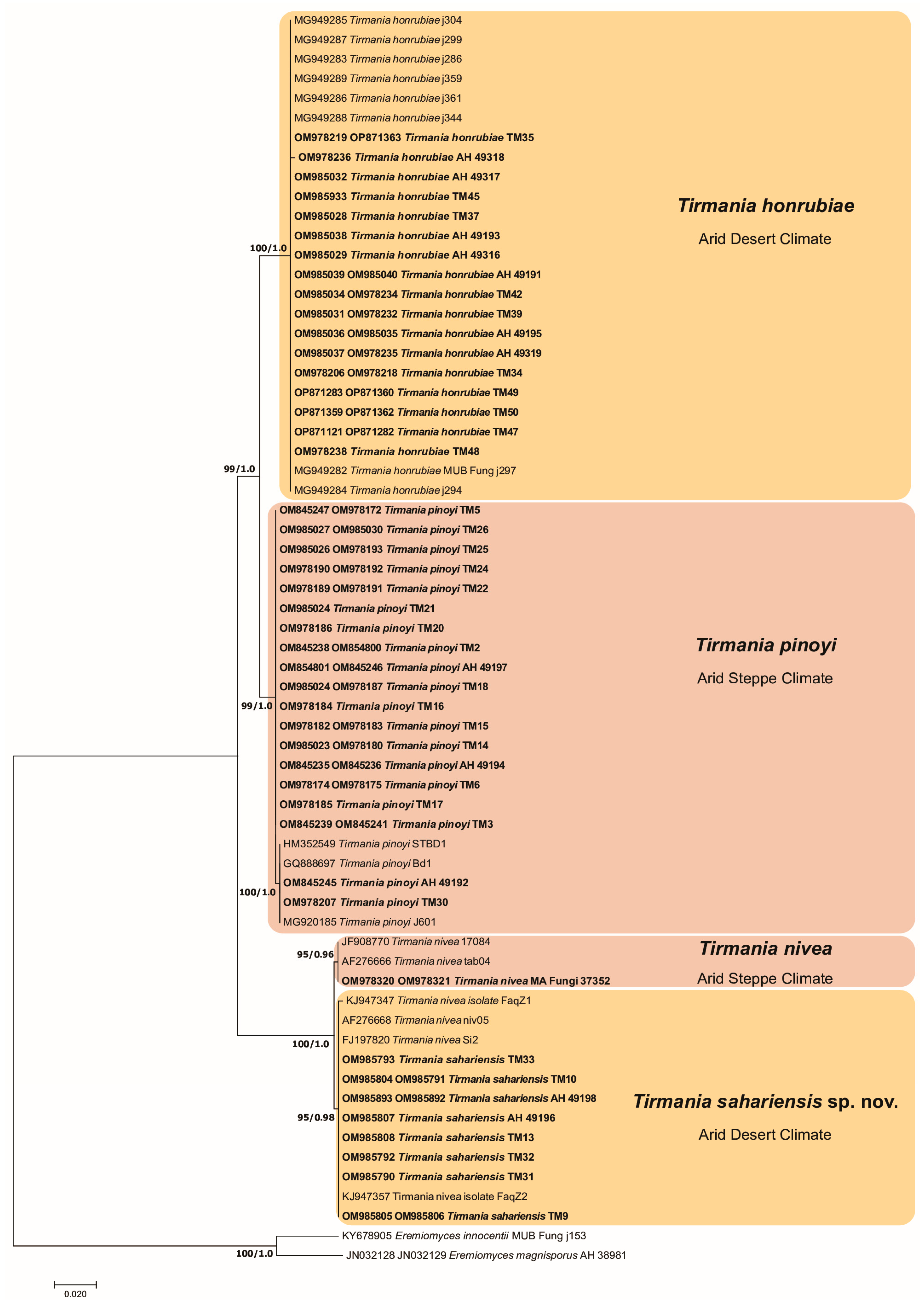
3.1. Phylogenetic Analysis

The combined phylogenetic tree accommodated 89 sequences listed in Table 1. The ITS and LSU amplicons size of the 45 genotyped Tirmania ascomata were 602 and 634 bp in the final alignment, with 217 and 63 variable positions, respectively. The maximum likelihood and Bayesian inference trees yielded similar topologies for both ITS and 28S rDNA gene regions, and the concatenated data set identified the same supported clades. Phylogenetic analysis of ITS and LSU sequence data from the Tirmania specimens investigated in this study in relationship with the Tirmania reference sequences retrieved from GenBank revealed two major lineages highly supported by bootstrap and posterior probabilities values (BS: 99%, PP: 1.00; BS: 100%, PP: 1.00). The two major lineages separated into four strongly supported and well-defined clades corresponding to four morphologically distinct Tirmania species. Moreover, each genetic clade identified in Tirmania ITS + LSU phylogenies correlated well with a specific bioclimatic pattern. Two of the four clades represented two Tirmania taxa previously reported from the Mediterranean region and the Middle East, namely T. pinoyi and T. nivea. Several rDNA sequences from Algerian samples studied in the present work formed a robustly supported clade (BS: 100%, PP: 1.00) with T. honrubiae sequences retrieved from GenBank supporting their affiliation to this taxon and representing, therefore, a first record of this species from North Africa. The remaining fourth clade was formed by Tirmania samples showing specific morphological characters not encountered even in their genetically closest sister species T. nivea, thus, evidencing their distinct taxonomic status. We, therefore, propose a novel taxonomic combination to accommodate samples of the fourth Tirmania clade. The designation of Tirmania sahariensis as a new species is supported by ITS/LSU rDNA analyses and morphological features (Figure 1).

3.2. Taxonomy

Tirmania sahariensis Zitouni-Haouar, Bidartondo, G. Moreno, Carlavilla & Manjón sp. nov.
MycoBank MB847872
GenBank: OM985893, OM985892 (Holotype).
Type. ALGERIA, Naama, Ain Sefra, under Helianthemum lippii (L.) Dum. Cours., hypogeous, mostly solitary, 12 April 2012, F.E.-H. Zitouni-Haouar (holotype: AH49198, paratype: AH49196).
Diagnosis.Tirmania sahariensis differs from its sisters Tirmania species (T. nivea, T. pinoyi and T. honrubiae) by having a combination of smooth globose and ellipsoid spores compared to its closest phylogenetically related species T. nivea which is characterized by strictly ellipsoid smooth spores [22]. This species is also recognized by its unusual maximum number of spores per ascus which can reach 10 spores. The distinct phylogenetic position of T. sahariensis separates it furthermore from the rest of Tirmania species. T. honrubiae is characterized by globose spores ornamented by low-rounded warts. T. pinoyi has spherical minutely reticulate spores.
Etymology.“sahariensis” epithet in reference to its Saharan habitat.
Description. Ascomata hypogeous to partially or occasionally completely emergent at maturity; subglobose to ellipsoid, sometimes lobed or turbinate with small basal attachment, smooth or covered with shallow crevices; 2.2–7.4 × 3.3–9.4 cm in size, 7–125 g fresh weight; off white or white yellowish when young, becoming yellowish brown to orange brown with age or after desiccation (Figure 2a–d). Gleba solid, fleshy, with subglobose to elongate pale yellow to light pink pockets of fertile tissue, separated by whitish sterile veins (Figure 2e). Odor and taste very pleasant.
Peridium ± 200 µm thick, consisting of two layers. The outer layer is composed of appressed, interwoven hyphae, 5–11 μm in diameter at septa, with inflated pale yellow or hyaline scattered outermost cells, walls 0.5–1 μm thick. The inner peridium differentiated as hyaline, interwoven septate hyphae (textura intricata), 13–30 μm broad with innermost cells subglobose, inflated to 20–42 μm diam, thin-walled and hyaline. Asci amyloid; mostly ellipsoid to pyriform or subglobose; 50–96 × 39–55 μm, with short stipitate (4–) 7–15 × 8–27 (–37) μm; walls 0.5–1.5 μm thick, harboring 6–(8–10) spores either exclusively ellipsoid or mixed with 1 to 3 globose spores (Figure 2f–i). Ascospores ellipsoid to subglobose, (10–) 11–14 × 12.5–18.5 (–19) μm, or globose, (10.5–) 12 × 15 μm diam.; hyaline with numerous small lipid guttules in immature spores merging into a single large guttule at maturity with a de Bary bubble (in Melzer’s reagent); the walls are 1–1.5 μm thick and two-layered: outer layer smooth, inner layer smooth to occasionally very minutely roughened with irregularly dispersed small ridges (up to 0.4 μm high) (Figure 2f–m).
Distribution and Ecology. ALGERIA, Naama, Ain Sefra; Béchar, Abadla, Hammaguir, Erg Serhen; Béni Abbès, Tabelbala, Oued Daoura. Hypogeous, solitary or gregarious (two ascomata), in sandy alkaline soil associated with Helianthemum lippii. This species is locally called «terfess labiadh» which means white truffle. Ascocarps are collected from the beginning of December to the end of May.
Additional collections examined. ALGERIA, Naama, Ain Sefra, April 2012, leg. F.E.-H. Zitouni-Haouar, paratype AH49196 (ITS sequence GenBank OM985807), TM9 (ITS sequence GenBank OM985805, LSU sequence GenBank OM985806), TM10 (ITS sequence GenBank OM985804, LSU sequence GenBank OM985791), TM13 (ITS sequence GenBank OM985808); ALGERIA, Béni Abbès, Tabelbala, Oued Daoura, April 2012, leg. F.E.-H. Zitouni-Haouar, TM31 (ITS sequence GenBank OM985790), TM32 (ITS sequence GenBank OM985792); ALGERIA, Béchar, Abadla, Hammaguir, Erg Serhen, March 2015, leg. F.E.-H. Zitouni-Haouar, TM33 (ITS sequence GenBank OM985793).
Tirmania nivea (Desf.: Fr.) Trappe.
Descriptions. See Moreno et al. [22]
Collections examined. SPAIN, Almería, Tabernas Desert, in sandy soils, 29 May 1995, leg. P. Rodriguez, MA-Fungi 37,352 (ITS sequence GenBank OM978320, LSU sequence GenBank OM978321).
Tirmania honrubiae Morte, Bordallo & Ant. Rodr.
Description. Ascomata hypogeous, 1–5 cm below soil surface, to partially emergent; subglobose to turbinate with small basal attachment; 2.8–9 × 3–10 cm, 10–197 g fresh weight (Figure 3a–c). Gleba solid, fleshy, with yellowish to creamy sterile veins bordering white to pale pink pockets of fertile tissue (Figure 3c). Odor pleasant and strong even after four days; taste agreeable.
Peridium 0.46–1.47 mm thick, covered with superficial to deep irregular depressions, orangish yellow to pale brown–orange becoming dark brown with age (Figure 3a–c). Asci (4–) 6–8 spored; very weakly amyloid (the faint blue color does not exceed asci walls in Melzer’s reagent) to non-amyloid (even from fresh mature samples); subglobose or ellipsoid to pyriform; 63–94 × 48–67 μm, with a short stem 12–24 × 10–15 μm; walls usually not more than 1 μm thick (Figure 3d,e). Ascospores globose; 15–20 μm diam.; hyaline with a single large guttule and a de Bary bubble at maturity; the wall ± 1 μm thick consists of two layers: outer layer smooth, inner layer roughened with ridges and low rounded warts (0.3–0.8 μm high) which seem to protrude out beyond the outer wall layer at maturity. Young spores appear ornamented with a well-developed reticulum (reticular walls 0.3–0.5 μm thick) which is completely replaced by warts in very mature spores (Figure 3d–i).
Distribution and Ecology. ALGERIA, Béchar, Béni Ounif, Oued Namous; Béchar, Abadla, Hammaguir; Oglat Braber; Béni Abbès, Boulaadam; Laghouat, Hassi R’Mel. Hypogeous, solitary, in sandy calcareous, alkaline soil associated with Helianthemum lippii. Ascocarps are collected from the beginning of November to the end of April. Tirmania honrubiae is locally called «terfess lahmar» which means red truffle.
Collections examined. ALGERIA, Béchar, Béni Ounif, Oued Namous, March 2012, leg. F.E.-H. Zitouni-Haouar, TM34 (ITS sequence GenBank OM978206, LSU sequence GenBank OM978218), TM35 (ITS sequence GenBank OM978219, LSU sequence GenBank OP871363), AH49191 (ITS sequence GenBank OM985039, LSU sequence GenBank OM985040); ALGERIA, Béchar, Abadla, Hammaguir, April 2012, leg. F.E.-H. Zitouni-Haouar, TM37 (ITS sequence GenBank OM985028), AH49316 (ITS sequence GenBank OM985029), TM39 (ITS sequence GenBank OM985031, LSU sequence GenBank OM978232), AH49195 (ITS sequence GenBank OM985036, LSU sequence GenBank OM985035), March 2013, AH49317 (ITS sequence GenBank OM985032); ALGERIA, Béchar, Abadla, Oglat Braber, April 2015, leg. F.E.-H. Zitouni-Haouar, TM42 (ITS sequence GenBank OM985034, LSU sequence GenBank OM978234), AH49319 (ITS sequence GenBank OM985037, LSU sequence GenBank OM978235), AH49193 (ITS sequence GenBank OM985038), March 2011, TM45 (ITS sequence GenBank OM985933), AH49318 (ITS OM978236); ALGERIA, Béni Abbès, Boulaadam, April 2012, leg. F.E.-H. Zitouni-Haouar, TM47 (ITS sequence GenBank OP871121, LSU sequence GenBank OP871282), TM48 (ITS sequence GenBank OM978238); ALGERIA, Laghouat, Hassi R’Mel, March 2016, leg. F.E.-H. Zitouni-Haouar, TM49 (ITS sequence GenBank OP871283, LSU sequence GenBank OP871360), TM50 (ITS sequence GenBank OP871359, LSU sequence GenBank OP871362).
Tirmania pinoyi (Maire) Malençon
Description. Ascomata hypogeous; subglobose to ovoid, or irregularly shaped with flattened basal attachment; 4–7 × 5.5–16.5 cm, 13–480 g fresh weight (Figure 4a–d). Gleba solid, fleshy, with white to light yellow or pale pink islets of fertile tissue surrounded by creamy sterile veins (Figure 4d). Odor and taste pleasant.
Peridium 0.3–1.4 mm thick, superficially to deeply cracked, light brown or yellowish brown becoming slightly darker with age (Figure 4b–d). Asci amyloid; 6–8 spored at maturity; mostly ellipsoid to ovoid, occasionally pyriform; 50–85 × 30–62 μm, excluding stalk ≤30 µm long; the walls 0.5–1.5 µm thick (Figure 4e–g). Ascospores globose; 15–20 μm broad.; hyaline with one large guttule and a de Bary bubble which may be lacking at times; the wall up to 1.6 μm thick formed of two layers: outer layer, 0.5–1 μm thick, smooth; inner layer, 0.3–0.6 μm thick, roughened with a not well-defined reticulum (reticular walls showing an irregular polygonal shape) which protrude into the outer wall layer at maturity (Figure 4e–i).
Distribution and Ecology. ALGERIA, Tiaret, Bouchouat; Benhamed; Mecheria, El Biodh; Bougarne. Hypogeous, mostly solitary, in sandy loam calcareous, alkaline soil associated with Helianthemum hirtum, Helianthemum salicifolium, and Helianthemum sp. This species is locally known as «Belhourech or Chehba» which means truffle with light color. Ascomata are collected from the beginning of February to the middle of June.
Collections examined. ALGERIA, Naama, Mecheria, El Biodh, April 2012, leg. F.E.-H. Zitouni-Haouar, AH49194 (ITS sequence GenBank OM845235, LSU sequence GenBank OM845236), TM2 (ITS sequence GenBank OM845238, LSU sequence GenBank OM854800), TM3 (ITS sequence GenBank OM845239, LSU sequence GenBank OM845241); ALGERIA, Naama, Mecheria, Bougarne, March 2013, leg. F.E.-H. Zitouni-Haouar, AH49192 (ITS sequence GenBank OM845245), TM5 (ITS sequence GenBank OM845247, LSU sequence GenBank OM978172), TM6 (ITS sequence GenBank OM978174, LSU sequence GenBank OM978175); ALGERIA, Tiaret, Ksar Chellala, Zmalet El-Amir Abdelkader, Bouchouat, May 2012, leg. F.E.-H. Zitouni-Haouar, TM14 (ITS sequence GenBank OM985023, LSU sequence GenBank OM978180), TM15 (ITS sequence GenBank OM978182, LSU sequence GenBank OM978183), TM16 (ITS sequence GenBank OM978184), TM17 (ITS sequence GenBank OM978185), TM18 (ITS sequence GenBank OM985024, LSU sequence GenBank OM978187), AH49197 (ITS sequence GenBank OM854801, LSU sequence GenBank OM845246); ALGERIA, Tiaret, Hamadia, Rechaiga, Benhamed, February 2009, leg. F.E.-H. Zitouni-Haouar, TM20 (ITS sequence GenBank OM978186), TM21 (ITS sequence GenBank OM985024), TM22 (ITS sequence GenBank OM978189, LSU sequence GenBank OM978191), TM24 (ITS sequence GenBank OM978190, LSU sequence GenBank OM978192), TM25 (ITS sequence GenBank OM985026, LSU sequence GenBank OM978193), TM26 (ITS sequence GenBank OM985027, LSU sequence GenBank OM985030), TM30 (ITS sequence GenBank OM978207).
Taxonomic key to Tirmania species
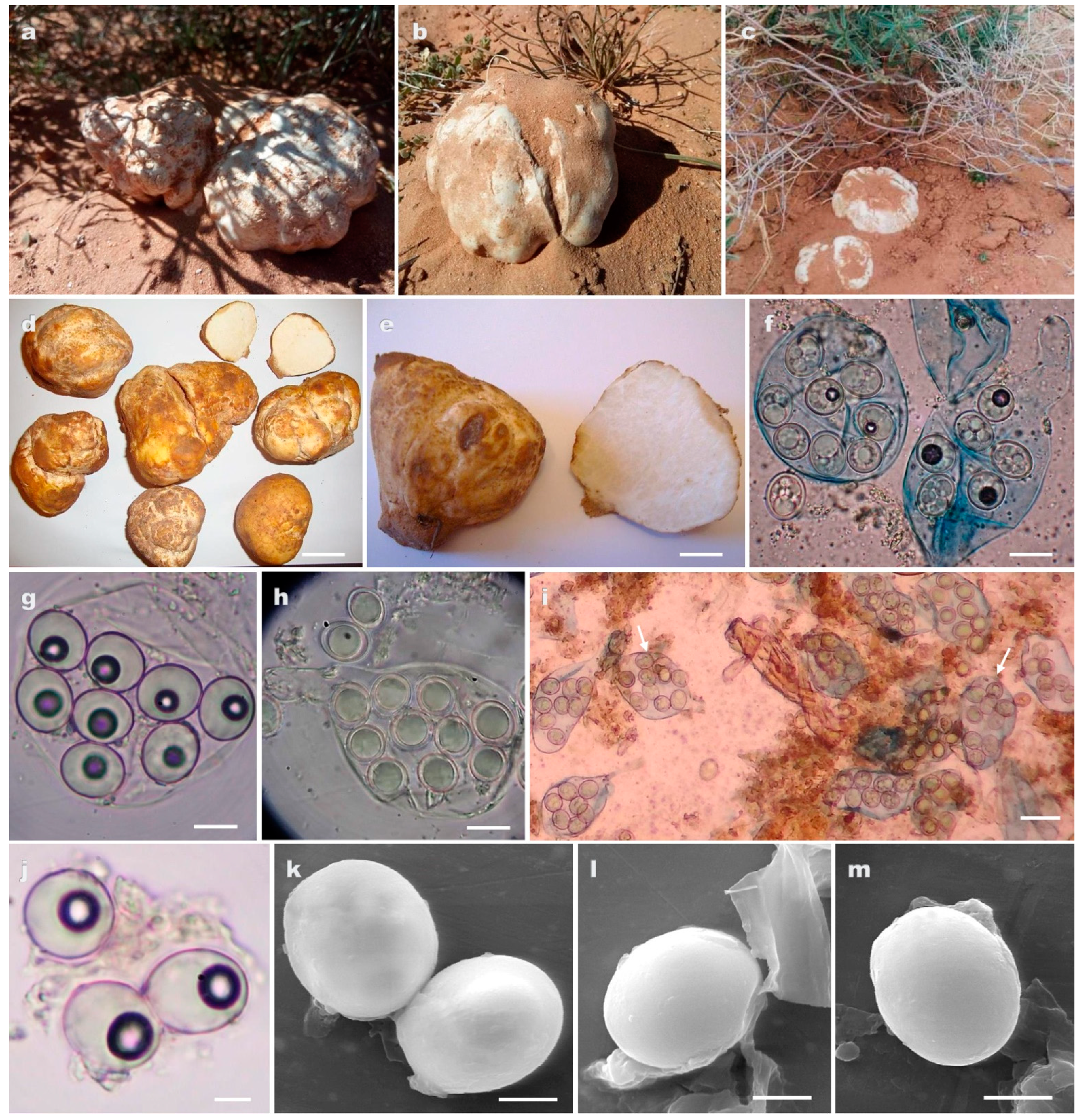
  • Peridium orangish yellow to pale brown–orange, spores globose roughened with ridges and low rounded warts……..………………………………… T. honrubiae (clade 1)
  • Peridium light brown or yellowish brown, spores globose roughened with a not well-defined reticulum………………………………………………………T. pinoyi (clade 2)
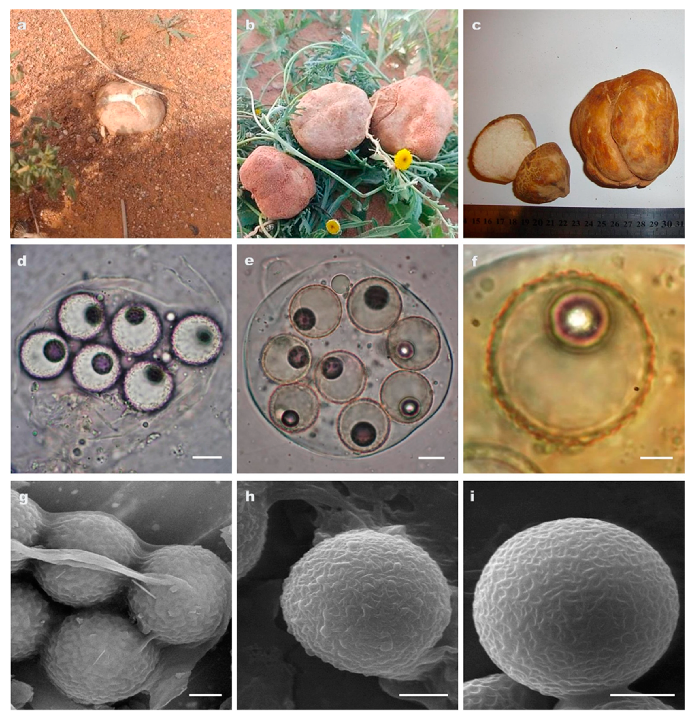
  • Peridium off white or yellow to cinnamon, spores smooth exclusively ellipsoid, asci (4–6)–8 spored …………………………………………………………………T. nivea (clade 3)
  • Peridium off white or white yellowish, spores smooth ellipsoid or globose, asci 6–(8–10) spored..……………………………………………………………T. sahariensis (clade 4)

4. Discussion

Phylogenetic and morphological data presented in this study demonstrated that the Tirmania genus is even more diverse than previously suspected. The phylogenetic structure of this taxon correlated substantially with morphological features and bioclimatic origin. The main morphological character separating the two major clusters identified in Tirmania phylogenies was spore shape. Indeed, the first major group was formed of T. pinoyi and T. honrubiae with exclusively globose spores, whereas the second one grouped T. nivea and T. sahariensis showing a mixture of spore shapes with mostly ellipsoid form. However, the species delineation intracluster relates to ascospore morphological characters in addition to the bioclimatic origin. Thus, the major cluster of Tirmania species with globose spores includes two genetic lineages on the basis of spore ornamentation (reticulate in T. pinoyi and warty in T. honrubiae) and according to the bioclimatic niche of the samples, which were collected from a cold arid steppe (BSk) for T. pinoyi and a hot arid desert (BWh) for T. honrubiae (Figure 1, Table 1) according to the Köppen–Geiger global climate classification [33] (Figure 5). The same situation was observed with the second major cluster of Tirmania species with mostly ellipsoid spores which bifurcated into two lineages mainly on the basis of spore form (exclusively ellipsoid in T. nivea versus ellipsoid and globose in T. sahariensis) and bioclimatic origin. Here again, species delimitation followed the two bioclimatic patterns observed in the first major cluster. Hence, while samples of T. nivea were harvested from cold/hot arid steppe (BSk/BSh), those of T. sahariensis originated from cold/hot arid desert (BWk/BWh) (Table 1, Figure 1 and Figure 5). This finding suggests that ecological factors and, notably, bioclimatic origin seem to drive the phylogenetic structure of the genus leading to allopatric speciation. Allopatry is generally caused by a geographic barrier that consists of suboptimal environmental conditions for the species in question (e.g., deserts, mountains, or oceans) [52]. The two sets of allopatric siblings species (T.pinoyi/T.honrubiae, T.nivea/T.sahariensis) observed in each Tirmania major cluster have dissimilar climatic niche envelopes (Arid steppe/Desert climates) and different geographic areas which separate them phylogenetically. Thus, the bioclimatic area range could have a possible role in driving allopatric speciation of the recently discovered Tirmania taxon T. honrubiae Morte, Bordallo & Ant. Rodr. and the new Tirmania species T. sahariensis sp. nov. described here, as a response to the natural selective pressures imposed by the hostile conditions of their desert (BWk/BWh) habitats (Figure 1). Natural selection is a central factor affecting speciation and plays an important role in producing phenotypic and genetic diversity within a population. It is the driving force that acts on the existing features of two populations with reference to the ecological differences of their habitats and leads to the evolution of adaptive features [53] (Chethana et al., 2021). Díez et al. [3] were the first to study the intrageneric relationships underlying the phylogeny of the two major desert truffle genera, Terfezia and Tirmania. They hypothesized that host specialization and edaphic tolerances, especially soil pH (fungus and/or host tolerances), might have played a key role in the speciation of Terfezia species. However, these authors could not propose the same hypotheses for Tirmania taxa given that they worked on only five specimens. They, therefore, recommended larger surveys to confirm whether T. nivea and T. pinoyi also have different edaphic tolerances and/or host adaptations. On the other hand, the lack of data on the exact taxonomic identity and phylogenies of the Helianthemum species associated with the Tirmania samples investigated in the present work calls into question whether the genetic pattern observed in this desert truffle group led to cospeciation with the host plant. In cospeciation, strong congruence (topological similarity) is usually expected between the host and symbiont phylogenies [54]. Further cophylogenetic studies of desert truffle samples with their respective host plants candidates are, therefore, needed to infer the mechanisms of speciation in desert truffles. The phylogenetic diversity examined in this study was strongly supported by morphological evidence and most notably by ascospore and ascus features. Results from the present analyses highlight the importance of spore characters as the most reliable micromorphological features to delimit desert truffle taxa. This was the case also for Terfezia and Picoa where the species delineation concept was strongly dependent on spore morphological characters, although this diagnostic feature was at times inconspicuous or featureless for delimiting some taxa within these groups. Indeed, morphological similarities recorded in ascospore ornamentation or the absence of this in immature samples were likely the main difficulties that prevailed in the species delimitation of Terfezia and Picoa, respectively, and which led also to serious taxonomic misidentifications [5,41]. The newly recorded Tirmania species from North Africa, T. honrubiae, is a further desert truffle species which seems to have been misidentified in the past as T. pinoyi due to the great micromorphological similarities between the two taxa. The spore ornamentation which is regarded as the most effective diagnostic tool allowing the discrimination between these two species is hardly detectable with a light microscope and is only perceivable in mature spores under SEM. Alsheikh and Trappe [1] reported that a specimen of T. pinoyi collected from an Algerian region (Ain Sefra) characterized by a cold arid desert climate (BWK) was initially deposited in the Patouillard Herbarium (FH) as Terfezia boudieri. The morphological and molecular analyses in accordance with bioclimatic data reported here suggest that this specimen may be affiliated to T. honrubiae given that it presents globose spores with a BWK bioclimatic origin. The erroneous taxonomic identification of this sample supports our suggestion as the low warts observed on the surface of T. honrubiae mature spore strongly resemble those of T. boudieri. Although the amyloid reaction of the asci wall is considered as a useful taxonomic identification tool to differentiate Tirmania from its closest desert truffles species [11], mature asci even from fresh T. honrubiae samples analyzed in the present work were very weakly amyloid to mostly non-amyloid in response to Melzer’s solution. Previous studies reported that the amyloid reaction of the ascus wall, which is considered as the cardinal feature of Pezizaceae, seems to be a symplesiomorphic character which has been lost in several lineages of hypogeous taxa (e.g., in those including Marcelleina, Cazia, Terfezia, and some species of Pachyphloeus) [13,14,17] and which seems to be disappearing in T. honrubiae. The European Catalogue of hypogeous fungi has also been subject to taxonomic confusion regarding the first report of the genus Tirmania from Europe. Indeed, Moreno-Arroyo et al. [21] examined a Tirmania material from the Tabernas Desert of Almería (Southern Spain, BSK) and recorded it as T. pinoyi. The misdiagnosis was revealed later when Moreno et al. [22] studied the same material and reported that it displayed ellipsoid spores (never spherical) typical of T. nivea. The molecular and phylogenetic analysis presented here and performed on the same Tirmania material from the Tabernas Desert (MA Fungi 37352) (Figure 1, Table 1) analyzed in the two previous studies supported the emendation made by Moreno et al. [22] regarding the abolition of T. pinoyi from the Spanish hypogeous fungal flora and the record of T. nivea as the unique representative species of the genus Tirmania in the European mycoflora. The T. nivea combination was originally proposed by Trappe [11] to represent the Tirmania taxon with broadly ellipsoid, smooth spores. The phylogenetic inferences based on the rDNA sequence dataset and the mixture of globose and ellipsoid spores as well as the eight- to ten-spored asci observed in specimens of the T. nivea sister lineage (Figure 1) provide strong evidence for the erection of the new Tirmania species, T. sahariensis sp. nov.

5. Conclusions

This work addressed the intrageneric relationships among Tirmania species using morphological and phylogenetic analysis and considering bioclimatic information. Our analyses provide the first insights into the influence of climatic range area on the evolution of Tirmania species. The genetic structure of this desert truffle group seems to be related to climatic niche divergence leading to allopatric speciation. We confirmed the existence of four distinct genetic lineages within this group corresponding to four morphological species with a new species and a new record. Further phylogenomic analysis from whole genomic sequencing of Tirmania strains are needed in order to refine phylogenies and to fully understand the observed genetic pattern. Phylogenies presented in this study have led to a taxonomic update of Tirmania and provided essential phylogenetic background for the conservation and management of these highly valued edible fungi.

Author Contributions

Conceptualization, F.E.-H.Z.-H., M.I.B., G.M. and J.L.M.; methodology, F.E.-H.Z.-H. and M.I.B.; investigation, F.E.-H.Z.-H., M.I.B., G.M. and J.R.C.; data analysis, F.E.-H.Z.-H., J.R.C., S.N. and S.H.Z.-N.; resources, M.I.B., F.E.-H.Z.-H., J.L.M. and G.M.; writing—original draft preparation, F.E.-H.Z.-H.; writing—review and editing, all authors. All authors have read and agreed to the published version of the manuscript.

Funding

This research received no external funding.

Institutional Review Board Statement

Not applicable.

Informed Consent Statement

Not applicable.

Data Availability Statement

All sequences generated in this study were submitted to GenBank.

Acknowledgments

We sincerely thank Laura MARTINEZ-SUZ from the Royal Botanic Gardens (Jodrell Laboratory, Kew, UK) for kindly providing some sequencing reagents. We wish to express our gratitude to A. PRIEGO and J.A. PÉREZ from the University of Alcalá De Henares (Spain) for the valuable assistance in the SEM observations and J. REJOS, curator of the AH Herbarium (University of Alcalá). We would like to acknowledge Varda KAGAN-ZUR and Samad JAMALI for providing invaluable data on collection locality of some reference Tirmania samples used in the present work. This research was made possible through the financial support of the Royal Botanic Gardens (Kew, UK), the Spanish Ministry of Education and Culture, and the Algerian General Directorate of Scientific Research and Technological Development (DGRSDT), and the Algerian Ministry of Higher Education and Scientific research (MESRS).

Conflicts of Interest

The authors declare no conflict of interest.

References

  1. Alsheikh, A.M.; Trappe, J.M. Desert truffles: The genus Tirmania. Trans. Br. Mycol. Soc. 1983, 81, 83–90. [Google Scholar] [CrossRef]
  2. Alsheikh, A.M. Taxonomy and Mycorrhizal Ecology of the Desert Truffles in the Genus Terfezia. Ph.D. Thesis, Oregon State University, Corvallis, OR, USA, 1994; 239p. [Google Scholar]
  3. Díez, J.; Manjón, J.L.; Martin, F. Molecular phylogeny of the mycorrhizal desert truffles (Terfezia and Tirmania), host specificity and edaphic tolerance. Mycologia 2002, 94, 247–259. [Google Scholar] [CrossRef] [PubMed]
  4. Zitouni-Haouar, F.E.-H.; Fortas, Z.; Chevalier, G. Morphological characterization of mycorrhizae formed between three Terfezia species (desert truffles) and several Cistaceae and Aleppo pine. Mycorrhiza 2014, 24, 397–403. [Google Scholar] [CrossRef]
  5. Zitouni-Haouar, F.E.-H.; Carlavilla, J.R.; Moreno, G.; Manjon, J.L.; Fortas, Z. Genetic diversity of the genus Terfezia (Pezizaceae, Pezizales): New species and new record from North Africa. Phytotaxa 2018, 334, 183–194. [Google Scholar] [CrossRef]
  6. Morte, A.; Gutiérrez, A.; Navarro-Ródenas, A. Advances in desert truffle mycorrhization and cultivation. In Mushrooms, Humans and Nature in a Changing World; Springer: Cham, Switzerland, 2020; pp. 205–219. [Google Scholar]
  7. Morte, A.; Kagan-Zur, V.; Navarro-Ródenas, A.; Sitrit, Y. Cultivation of desert truffles—A crop suitable for arid and semi-arid zones. Agronomy 2021, 11, 1462. [Google Scholar] [CrossRef]
  8. Chatin, A. Contribution à l′histoire naturelle de la truffe. Bull. Société Bot. Fr. 1891, 38, 54–64. [Google Scholar] [CrossRef]
  9. Chatin, A. La Truffe; J.B. Baillière et Fils: Paris, France, 1892. [Google Scholar]
  10. Patouillard, N. Énumération des champignons observés en Tunisie. In Exploration Scientifique de la Tunisie; Imprimerie Nationale: Paris, France, 1892. [Google Scholar]
  11. Trappe, J.M. A synopsis of the Carbomycetaceae and Terfeziaceae (Tuberales). Trans. Br. Mycol. Soc. 1971, 57, 85–92. [Google Scholar] [CrossRef]
  12. Trappe, J.M. The orders, families and genera of hypogeous ascomycotina (truffles and their relatives). Mycotaxon 1979, 9, 297–340. [Google Scholar]
  13. Hansen, K.; Læssøe, T.; Pfister, D.H. Phylogenetics of the Pezizaceae, with an emphasis on Peziza. Mycologia 2001, 93, 958–990. [Google Scholar] [CrossRef]
  14. Hansen, K.; Lobuglio, K.F.; Pfister, D.H. Evolutionary relationships of the cup-fungus genus Peziza and Pezizaceae inferred from multiple nuclear genes: RPB2, β-tubulin, and LSU rDNA. Mol. Phylogenet. Evol. 2005, 36, 1–23. [Google Scholar] [CrossRef]
  15. Ferdman, Y.; Aviram, S.; Roth-Bejerano, N.; Trappe, J.M.; Kagan-Zur, V. Phylogenetic studies of Terfezia pfeilii and Choiromyces echinulatus (Pezizales) support new genera for southern African truffles: Kalaharituber and Eremiomyces. Mycol. Res. 2005, 109, 237–245. [Google Scholar] [CrossRef] [PubMed]
  16. Tedersoo, L.; Hansen, K.; Perry, B.A.; Kjøller, R. Molecular and morphological diversity of pezizalean ectomycorrhiza. New Phytol. 2006, 170, 581–596. [Google Scholar] [CrossRef] [PubMed]
  17. Læssøe, T.; Hansen, K. Truffle trouble: What happened to the Tuberales? Mycol. Res. 2007, 111, 1075–1099. [Google Scholar] [CrossRef]
  18. Kovács, G.M.; Trappe, J.M. Nomenclatural History and Genealogies of Desert Truffles. In Soil Biology; Kagan-Zur, V., Roth-Bejerano, N., Sitrit, Y., Morte, A., Eds.; Springer: Berlin/Heidelberg, Germany, 2014; Volume 38, pp. 21–37. [Google Scholar] [CrossRef]
  19. Maire, R. Notes mycologiques. Ann. Mycol. 1906, 4, 329–335. [Google Scholar]
  20. Malençon, G. Champignons hypogés du Nord de, l’Afrique–I. Ascomycetes. Persoonia 1973, 7, 261–279. [Google Scholar]
  21. Moreno-Arroyo, B.; Gomez, J.; Calonge, F.D. Aportaciones a la micoflora hipogea Ibérica. Boletín Soc. Micol. Madr. 1997, 22, 91–95. [Google Scholar]
  22. Moreno, G.; Díez, J.; Manjon, J.L. Picoa lefebvrei and Tirmania nivea, two rare hypogeous fungi from Spain. Mycol. Res. 2000, 104, 378–381. [Google Scholar] [CrossRef]
  23. Kovács, G.M.; Balazs, T.K.; Calonge, F.D.; Martín, M.P. The diversity of Terfezia desert truffles: New species and a highly variable species complex with intrasporocarpic nrDNA ITS heterogeneity. Mycologia 2011, 103, 841–853. [Google Scholar] [CrossRef]
  24. Bordallo, J.J.; Rodríguez, A.; Honrubia, M.; Morte, A. Terfezia canariensis sp. nov. una nueva especie de trufa encontrada en las Islas Canarias. Cantarela 2012, 56, 1–8. [Google Scholar]
  25. Bordallo, J.J.; Rodríguez, A.; Muñoz-Mohedano, J.M.; Suz, L.M.; Honrubia, M.; Morte, A. Five new Terfezia species from the Iberian Peninsula. Mycotaxon 2013, 124, 189–208. [Google Scholar] [CrossRef]
  26. Bordallo, J.J.; Rodríguez, A.; Kaounas, V.; Camello, F.; Honrubia, M.; Morte, A. Two new Terfezia species from Southern Europe. Phytotaxa 2015, 230, 239–249. [Google Scholar] [CrossRef]
  27. Bordallo, J.J.; Rodriguez, A.; Santos-Silva, C.; Louro, R.; Muñoz-Mohedano, J.M.; Morte, A. Terfezia lusitanica, a new mycorrhizal species associated to Tuberaria guttata (Cistaceae). Phytotaxa 2018, 357, 141–147. [Google Scholar] [CrossRef]
  28. Bordallo, J.J.; Rodriguez, A.; Morte, A. Terfezia morenoi. Fungal Planet description sheets. Persoonia 2018, 40, 324–325. [Google Scholar] [CrossRef]
  29. Moreno, G.; Manjón, J.L.; Alvarado, P. A new Terfezia from Spain. Boletín Soc. Micol. Madr. 2019, 43, 55–60. [Google Scholar]
  30. Rodríguez, A.; Navarro-Ródenas, A.; Morte, A.; Cabero, J.; Luque, D. Terfezia dunensis. Fungal Planet description sheets. Persoonia 2019, 43, 410–411. [Google Scholar] [CrossRef]
  31. Louro, R.; Nobre, T.; Santos-Silva, C. Terfezia solaris-libera sp. nov., a new mycorrhizal species within the spiny-spored lineages. J. Mycol. Mycol. Sci. 2020, 3, 1–13. [Google Scholar] [CrossRef]
  32. Morte, A.; Bordallo, J.J.; Rodriguez, A. Tirmania honrubiae. Fungal Planet description sheets. Persoonia 2018, 40, 328–329. [Google Scholar] [CrossRef]
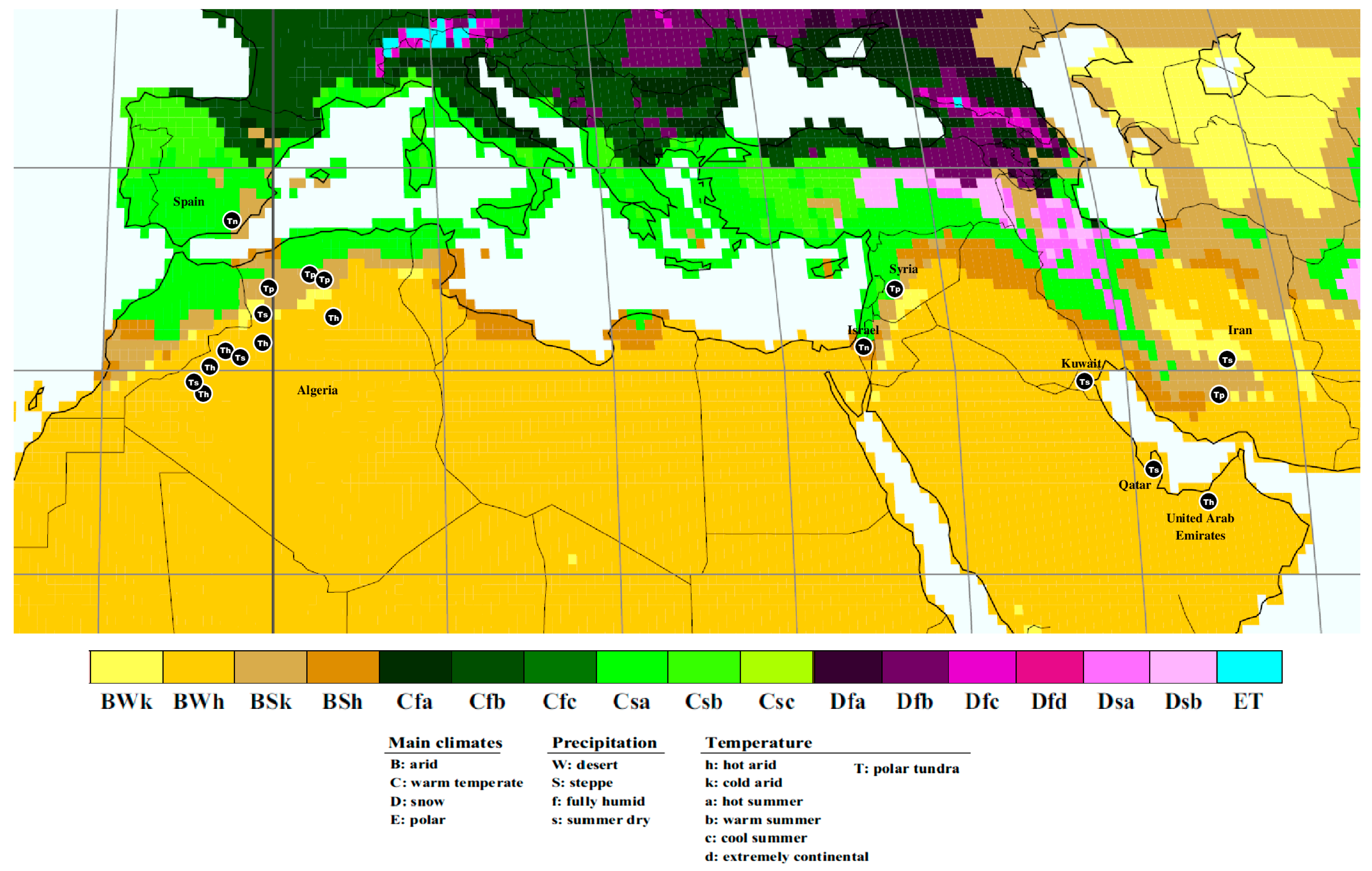
  33. Kottek, M.; Grieser, J.; Beck, C.; Rudolf, B.; Rubel, F. World map of the Köppen-Geiger climate classification updated. Meteorol. Z. 2006, 15, 259–263. [Google Scholar] [CrossRef]
  34. Langeron, K. Précis de Mycologie; Ed. Masson: Paris, France, 1952; pp. 367–397. [Google Scholar]
  35. Moreno, G.; Altés, A.; Ochoa, C.; Wright, J.E. Contribution to the study of the family Tulostomataceae in Baja California, Mexico. Mycologia 1995, 87, 96–120. [Google Scholar] [CrossRef]
  36. Gardes, M.; Bruns, T.D. ITS primers with enhanced specificity for Basidiomycetes-application to the identification of mycorrhizae and rusts. Mol. Ecol. 1993, 2, 113–118. [Google Scholar] [CrossRef]
  37. Bidartondo, M.I.; Burghardt, B.; Gebauer, G.; Bruns, T.D.; Read, D.J. Changing partners in the dark: Isotopic and molecular evidence of ectomycorrhizal liaisons between forest orchids and trees. Proc. R. Soc. Lond. B 2004, 271, 1799–1806. [Google Scholar] [CrossRef]
  38. White, T.J.; Bruns, T.D.; Lee, S.; Taylor, J.W. Amplification and direct sequencing of fungal ribosomal RNA genes for phylogenetics. In PCR Protocols: A Guide to Methods and Applications; Innis, M.A., Gelfand, D.H., Sninsky, J., White, T.J., Eds.; Academic Press: London, UK, 1990; 482p. [Google Scholar]
  39. Vilgalys, R.; Hester, M. Rapid genetic identification and mapping of enzymatically amplified ribosomal DNA from several Cryptococcus species. J. Bacteriol. 1990, 172, 4238–4246. [Google Scholar] [CrossRef] [PubMed]
  40. Cubeta, M.A.; Echandi, E.; Abernethy, T.; Vilgalys, R. Characterization of anastomosis groups of binucleate Rhizoctonia species using restriction analysis of an amplified ribosomal RNA gene. Phytopathology 1991, 81, 1395–1400. [Google Scholar] [CrossRef]
  41. Zitouni-Haouar, F.E.-H.; Alvarado, P.; Sbissi, I.; Boudabous, A.; Fortas, Z.; Moreno, G.; Manjón, J.L.; Gtari, M. Contrasted genetic diversity, relevance of climate and host plants, and comments on the taxonomic problems of the genus Picoa (Pyronemataceae, Pezizales). PLoS ONE 2015, 10, e0138513. [Google Scholar] [CrossRef] [PubMed]
  42. Murray, M.G.; Thompson, W.F. Rapid isolation of high molecular weight plant DNA. Nucleic Acids Res. 1980, 8, 4321–4325. [Google Scholar] [CrossRef] [PubMed]
  43. Mullis, K.B.; Faloona, F.A. Specific synthesis of DNA in vitro via a polymerase-catalyzed chain reaction. Methods Enzymol. 1987, 155, 335–350. [Google Scholar] [CrossRef]
  44. Altschul, S.F.; Madden, T.L.; Schaffer, A.A.; Zhang, J.H.; Zhang, Z.; Miller, W.; Lipman, D.J. Gapped BLAST and PSI-BLAST: A new generation of protein database search programs. Nucleic Acids Res. 1997, 25, 3389–3402. [Google Scholar] [CrossRef]
  45. Edgar, R.C. MUSCLE: Multiple sequence alignment with high accuracy and high throughput. Nucleic Acids Res. 2004, 32, 1792–1797. [Google Scholar] [CrossRef]
  46. Tamura, K.; Stecher, G.; Kumar, S. MEGA11: Molecular Evolutionary Genetics Analysis Version 11. Mol. Biol. Evol. 2021, 38, 3022–3027. [Google Scholar] [CrossRef]
  47. Jamali, S.; Banihashemi, Z. Hosts and Distribution of Desert Truffles in Iran, based on morphological and molecular criteria. J. Agric. Sci. Technol. 2012, 14, 1379–1396. [Google Scholar]
  48. Osmundson, T.W.; Robert, V.A.; Schoch, C.L.; Baker, L.J.; Smith, A.; Robich, G.; Mizzan, L.; Garbelotto, M.M. Filling gaps in biodiversity knowledge for macrofungi: Contributions and assessment of an herbarium collection DNA barcode sequencing project. PLoS ONE 2013, 8, e62419. [Google Scholar] [CrossRef] [PubMed]
  49. Miller, M.A.; Pfeiffer, W.; Schwartz, T. Creating the CIPRES Science Gateway for inference of large phylogenetic trees. In Proceedings of the 2010 Gateway Computing Environments Workshop (GCE), New Orleans, LA, USA, 14 November 2010; Volume 14, pp. 1–8. [Google Scholar]
  50. Posada, D. jModelTest: Phylogenetic model averaging. Mol. Biol. Evol. 2008, 25, 1253–1256. [Google Scholar] [CrossRef] [PubMed]
  51. Ronquist, F.; Teslenko, M.; Van der Mark, P.; Ayres, D.L.; Darling, A.; Hohna, S.; Larget, B.; Liu, L.; Suchard, M.A.; Huelsenbeck, J.P. MrBayes 3.2: Efficient Bayesian phylogenetic inference and model choice across a large model space. Syst. Biol. 2012, 61, 539–542. [Google Scholar] [CrossRef] [PubMed]
  52. Wiens, J.J.; Graham, C.H. Niche conservatism: Integrating evolution, ecology, and conservation biology. Annu. Rev. Ecol. Evol. Syst. 2005, 36, 519–539. [Google Scholar] [CrossRef]
  53. Chethana, K.T.; Manawasinghe, I.S.; Hurdeal, V.G.; Bhunjun, C.S.; Appadoo, M.A.; Gentekaki, E.; Raspé, O.; Promputtha, I.; Hyde, K.D. What are fungal species and how to delineate them? Fungal Divers. 2021, 109, 1–25. [Google Scholar] [CrossRef]
  54. Giraud, T.; Refrégier, G.; Le Gac, M.; de Vienne, D.M.; Hood, M.E. Speciation in fungi. Fungal Genet. Biol. 2008, 45, 791–802. [Google Scholar] [CrossRef] [PubMed]
Figure 2. (am) Tirmania sahariensis sp. nov.: (ac) ascomata in situ, under their host plant Helianthemum lippii; (c,d) ascomata showing peridial surface; (e) gleba in cross section; (f) light microscopy of asci harboring a mixture of ellipsoid and globoses spores in Melzer’s reagent; (g) ascus with exclusively ellipsoid spores; (h,i) asci containing ten spores (arrows); (j) ellipsoid and globoses ascospores; (km) scanning electron microscopy of ascospores. Scale bars: 3.3 cm (d), 2.1 cm (e), 15 μm (f,h), 10 μm (g), 30 μm (i), and 5 μm (jm).
Jof 09 00532 g002
Figure 3. (ai) Tirmania honrubiae: (a,b) ascomata in situ; (c) ascoma covered with deep depressions and gleba in cross section; (d) light microscopy of 6-spored ascus; (e) octosporus ascus in Melzer’s reagent showing the very weakly amyloid reaction where the faint blue color does not exceed the ascus wall; (f) ornamentation detail of ascospore in Melzer’s reagent under light microscope; (g,h) scanning electron microscopy of mature ascospores roughened with ridges and low-rounded warts; (i) immature young spore ornamented with a well-developed reticulum. Scale bars: 9 μm (d,e), 8 μm (f), and 5 μm (gi).
Jof 09 00532 g003
Figure 4. (ai) Tirmania pinoyi: (a) cracks on soil caused by ascoma expansion; (b) ascoma after removing ground surface near to the host plant Helianthemum hirtum (arrows); (c) ascoma in situ; (d) ascomata showing peridial surface and gleba in cross section; (e,f) light microscopy of asci containing ascospores; (g) amyloid reaction of octosporus ascus in Melzer’s reagent; (h) scanning electron microscopy of mature ascospores roughened with a not well-defined reticulum; (i) detail of ascospore surface under SEM. Scale bars: 1.8 cm (d), 12 μm (e,f), 8 μm (g), 5 μm (h), and 2 μm (i).
Jof 09 00532 g004
Figure 5. Global bioclimatic distribution of the four Tirmania species according to the world map of Köppen–Geiger climate classification updated (Map from Kottek et al. [33]). Tp: T. pinoyi, Th: T. honrubiae, Tn: T. nivea, Ts: T. sahariensis.
Jof 09 00532 g005
Disclaimer/Publisher’s Note: The statements, opinions and data contained in all publications are solely those of the individual author(s) and contributor(s) and not of MDPI and/or the editor(s). MDPI and/or the editor(s) disclaim responsibility for any injury to people or property resulting from any ideas, methods, instructions or products referred to in the content.

