Article

Biodiversity and Bioprospecting of Fungal Endophytes from the Antarctic Plant Colobanthus quitensis

by
Laura Bertini
1,†,
Michele Perazzolli
2,3,†,
Silvia Proietti
1,
Gloria Capaldi
1,
Daniel V. Savatin
4,
Valentina Bigini
4,
Claudia Maria Oliveira Longa
3,
Marina Basaglia
5,
Lorenzo Favaro
5,
Sergio Casella
5,
Benedetta Fongaro
6,
Patrizia Polverino de Laureto
6,* and
Carla Caruso
1,*
1
Department of Ecological and Biological Sciences, University of Tuscia, Largo dell’Università, 01100 Viterbo, Italy
2
Centre Agriculture Food Environment (C3A), University of Trento, Via E. Mach 1, 38098 San Michele all’Adige, Italy
3
Research and Innovation Centre, Fondazione Edmund Mach, Via E. Mach 1, 38098 San Michele all’Adige, Italy
4
Department of Agriculture and Forest Sciences, University of Tuscia, Via S. Camillo de Lellis, 01100 Viterbo, Italy
5
Department of Agronomy Food Natural Resources Animals and Environment, University of Padova, Viale dell’Università 16, Legnaro, 35020 Padova, Italy
6
Department of Pharmaceutical and Pharmacological Sciences, University of Padova, Via F. Marzolo 5, 35131 Padova, Italy
*
Authors to whom correspondence should be addressed.
These authors contributed equally to this work.
J. Fungi 2022, 8(9), 979; https://doi.org/10.3390/jof8090979
Submission received: 4 August 2022 / Revised: 13 September 2022 / Accepted: 14 September 2022 / Published: 19 September 2022
(This article belongs to the Special Issue Biological Activity of Fungi: Interaction with the Environment)

Abstract

:
Microorganisms from extreme environments are considered as a new and valuable reservoir of bioactive molecules of biotechnological interest and are also utilized as tools for enhancing tolerance to (a)biotic stresses in crops. In this study, the fungal endophytic community associated with the leaves of the Antarctic angiosperm Colobanthus quitensis was investigated as a new source of bioactive molecules. We isolated 132 fungal strains and taxonomically annotated 26 representative isolates, which mainly belonged to the Basidiomycota division. Selected isolates of Trametes sp., Lenzites sp., Sistotrema sp., and Peniophora sp. displayed broad extracellular enzymatic profiles; fungal extracts from some of them showed dose-dependent antitumor activity and inhibited the formation of amyloid fibrils of α-synuclein and its pathological mutant E46K. Selected fungal isolates were also able to promote secondary root development and fresh weight increase in Arabidopsis and tomato and antagonize the growth of pathogenic fungi harmful to crops. This study emphasizes the ecological and biotechnological relevance of fungi from the Antarctic ecosystem and provides clues to the bioprospecting of Antarctic Basidiomycetes fungi for industrial, agricultural, and medical applications.

1. Introduction

Antarctica is one of the last pristine environments on Earth and represents a valuable area for bioprospecting [1]. Its microbial diversity is largely unexplored and potentially encloses microorganisms endowed with activities of high relevance for white, green, and red biotechnologies [2]. To enable growth under the extreme environmental conditions of Antarctica, microbes evolved several adaptations strategies which are often accompanied by modifications to both gene regulation and metabolic pathways, increasing the possibility of finding unique functional metabolites of biotechnological importance. These adaptations include cellular storage of cryoprotectants such as sugars and polyols to maintain turgor pressure [3], high levels of unsaturated membrane phospholipids to stabilize membranes [4], and fungal melanin for protection against freezing and UV radiation [5], and the production of antifreeze proteins as well as cold-active enzymes [6]. Thus, microbial extremophiles turn out to be a chemical reservoir of extremolytes and extremozymes that have the potential to be outstanding resources for the development of a bio-based economy [2,7]. Extremozymes are enzymes that have evolved molecular mechanisms of adaptation to extreme physicochemical conditions and might have relevant applications in industrial biotransformation processes [2]. They naturally exhibit features that are usually gained by means of laborious synthetic biology approaches or can be used as a scaffold to design tailor-made biocatalysts for application in eco-friendly industrial processes [8]. Most extremozymes characterized from Antarctic filamentous fungi and yeasts are classified as hydrolases, but some oxidoreductases, such as laccase and superoxide dismutase, have also been reported [9] and references therein. A thorough study by Loperena et al. [10] reported the screening of extracellular enzymatic activities from 120 bacteria, 31 yeasts, and 10 filamentous fungi isolated from different environmental samples of Maritime Antarctica, highlighting that the most frequent and highest activities were from hydrolases. In particular, amylase, caseinase, lipase, and gelatinase activities were mainly detected in bacterial isolates, whereas yeasts and filamentous fungi more frequently showed lipase and cellulase activities, and little or no detectable pectinase, xylanase, or amylase activities [10].
Besides extremozymes, microorganisms living under stressful environmental conditions are also a valuable source of extremolytes and secondary metabolites, which can constitute up to 25% of dry cell weight [2,7]. The possible commercial applications of extremolytes include anticancer drugs, antioxidants, cell-cycle-blocking agents, and sunscreens, among others [11]. Tian and colleagues [12] reviewed 219 novel natural products from the Arctic and the Antarctic microorganisms, lichen, moss, and marine faunas endowed with antimicrobial, antiviral, and cytotoxic activities towards numerous cell lines as well as free radical scavenging properties ([12] and references therein). In particular, new asterric acid derivatives, peptaibols, epipolythiodioxopiperazines, polyketides, terpenes and sesquiterpenes endowed with multiple biological activities were isolated from Antarctic soil and deep-sea derived fungi [12]. Moreover, extracts from filamentous fungi coming from different Antarctic environments were found to be active against Xanthomonas phytopathogens [13]. The biological activity of secondary metabolites from endophytic fungi associated with the only two endemic vascular plants of Maritime Antarctica, i.e., Deschampsia antarctica Desv. and Colobanthus quitensis (Kunth) Bartl, is less characterized and, to date, only a few fungal extracts from endophytes of these plants were found to possess Leishmanicidal and antitumoral activities [14]. In addition to the importance of extremophiles in bioprospecting for bioactive molecules, they also exert a pivotal role in modulating plant and animal performance in stressful conditions. Such organisms are inhabited by microorganisms forming metaorganisms, whose functionality is strictly dependent by the dynamic crosstalk between the community components [15]. Interestingly, this kind of functional symbiosis has also been reported for the two Antarctic angiosperms [16,17]. C. quitensis leaves metatranscriptome disclosed transcripts belonging to fungi, bacteria, viruses, and mycoviruses revealing the holobiontic nature of the plant [18,19,20]. The microbial communities associated with both Antarctic vascular plants have been widely reported to enhance their tolerance to stress, improving the uptake of nutrients and water, and the resistance to UV-B radiation, drought, saline stress, and simulated global warming [20,21,22,23,24,25,26]. Interestingly, Antarctic plant symbionts were also found to improve the ecophysiological performance of agricultural species under abiotic stress conditions [27,28]. Thus, great interest is growing around the possibility to exploit extremophilic microbes as a biotechnological tool for a sustainable agriculture, even though only a few reports are available to date regarding the use of extremophiles as potential biocontrol agents for crop disease management and as potential enhancers of crop productivity [29]. With the advent of the genomic era, genome and metagenome mining technologies have been progressively applied to discover genes that are likely to produce novel bioactive compounds [30]. However, culture-dependent isolation techniques, followed by bioactivity-guided fractionation and identification of purified compounds, are still necessary for applied studies [9] and indispensable for the molecular and functional characterization of microorganisms.
In this study, we identified fungal endophytes from the leaves of the Antarctic angiosperm C. quitensis. Moreover, selected fungal isolates have been used as a source to identify metabolites and enzymes for bioprospecting purposes. In addition, plant–fungus interaction experiments were carried out on model plants with the aim of evaluating their possible use as biocontrol agents and/or plant growth promoters in eco-friendly solutions for sustainable agriculture.

2. Materials and Methods

2.1. Samples Collection

Sampling was carried out in the proximity of the Henryk Arctowski Antarctic Research station, King George Island, Maritime Antarctica (62°14′ S, 58°48′ W). Colobanthus quitensis plantlets were collected from three sites (S1, 62°9′44.58″ S, 58°27′58.68″ W; S2, 62°9′49.62″ S, 58°28′7.02″ W; S3, 62°9′52.90″ S, 58°28′21.31″ W) differing in the distance from the coastline, soil composition, altitude, temperature, and wind exposure. Besides collecting plant samples grown in open areas (OA samples), plantlets were also collected inside small greenhouses open on the top (Open Top Chambers, OTCs), placed only in S2 and S3, which determine an increase of about 4 °C during midday, mimicking the effect of global warming (OTC samples). Following sampling, plants were placed in 2 mL cryo-vials filled with 0.2 mL of sterile water and stored at 4 °C until isolation of fungal endophytes that was performed at the arrival of the samples in Italy.

2.2. Isolation of Culturable Fungal Endophytes

C. quitensis plants were surface-disinfected as described in [31] with 70% ethanol for 1 min, 2% sodium hypochlorite for 1.5 min, and 70% ethanol for 1 min. Plants were washed three times with sterilized distilled water (2 min each) and samples (0.5 g) were ground in stainless jars using a mixer-mill disruptor (MM 400, Retsch, Haan, Germany) at 25 Hz for 2 min in the presence of 1 mL 0.85% NaCl. Culturable fungi were isolated as previously described [32] by plating serial dilutions of each suspension (100 µL aliquots) on Potato Dextrose Agar (PDA) (Oxoid Ltd., Basingstoke, Hampshire, UK) supplemented with 0.25% lactic acid to inhibit bacterial growth [33,34]. As control of plant surface sterilization, aliquots (100 µL) of the last washing solution were plated to confirm the absence of microbial growth. Plates were incubated at 15 ± 1 °C and 25 ± 1 °C and fungal colony-forming units per gram of plant fresh weight (CFU g−1) were assessed daily for up to 60 days [35]. Three biological replicates were analyzed for each of five conditions (S1.OA, S2.OA, S2.OTC, S3.OA, S3.OTC) and two technical replicates were performed for each sample. The absence of bacterial growth was confirmed on all plates according to colony morphology and microscopic analyses.

2.3. Identification and Selection of Fungal Endophytes

Colony morphology and microscopic characteristics were examined starting from the eighth day after inoculation on PDA plates incubated at 25 °C. Fungal isolates were initially divided into different ‘morphotypes’ based on morphological features such as colony texture, degree of sporulation, obverse and reverse colony colors, production of soluble pigments and exudates. Sporulating fungi were identified based on the morphological characteristics of the conidiophores and conidia after fungal material was mounted in lactophenol, cotton blue or acid fuchsin for microscopical observation under the Eclipse 80i microscope (Nikon Inc., Melville, NY, USA). The isolates were identified through their cultural and morphological profiles compared with available literature [36,37]. To reduce redundancy, one representative isolate for each morphotype was selected and taxonomically annotated by amplification of the fungal internal transcribed spacer (ITS) region with the DreamTaq DNA Polymerase (Thermo Fisher Scientific, Waltham, MA, USA) using specific primer pairs (ITS1f: 5′-TCCGTTGGTGAACCAGCGG-3′ and ITS4r: 5′-TCCTCCGCTTATTGATATGC-3′). PCR products were purified by the NucleoSpin PCR Clean-up purification kit (Macherey-Nagel, Dueren, Germany) and sequenced with an ABIPRISM 3730xl DNA analyzer (Applied Biosystems, Thermo Fisher Scientific). ITS sequences were deposited in the NCBI database (http://www.ncbi.nlm.nih.gov; accessed on 1 June 2022) (Table S1). ITS sequences were aligned (BLASTn) against the nucleotide ITS database of fungi type and reference material of the NCBI (http://www.ncbi.nlm.nih.gov; accessed on 1 March 2022) and genus rank annotation was adopted when equal BLAST top score similarity values (ranging from 96 to 100% of sequence identity) were obtained for species belonging to the same genus, according to [38]. The phylogenetic tree was constructed on ITS sequences with the two best BLASTn hits using the neighbor-joining method and a bootstrap analysis with 1000 replicates with MEGA version 7.0 [39].

2.4. Fungal Growth and Samples Preparation

Fungal isolates were grown at 25 °C on PDA plates or in 100 mL flasks containing 30 mL Potato Dextrose Broth (PDB, Oxoid Ltd., Basingstoke, Hampshire, UK) inoculated with 5 mm agar plugs cut from the margin of fungal colonies developed on PDA plates. The flasks were incubated at 25 °C under shaking (150 rpm) in the dark. After 3–4 weeks, the cultures were aseptically centrifuged at 5000 rpm for 15 min to obtain cell-free supernatants; pellets were washed twice with sterile NaCl 0.9% and both supernatants and pellets were lyophilized. The lyophilized supernatants were resuspended in one tenth of sterile deionized water and used for antibacterial activity evaluations and enzymes in vitro assays. Pellets were used for metabolites extraction as described below.

2.5. Antibacterial Activity Evaluation

The antimicrobial properties of concentrated supernatants from the newly isolated endophytic fungi were assessed by the agar well diffusion method against a range of pathogens and model microorganisms. Bacterial isolates used for screening, included Grampositive and Gram-negative microorganisms (Listeria innocua 652, Listeria monocytogenes ATCC 19117, Bacillus cereus ATCC 11778, Staphylococcus aureus, Pseudomonas aeruginosa PAO 1 GFP, Escherichia coli APEC 13422_1_CER78, Escherichia coli APEC 18042/2, Salmonella enteritidis, Salmonella gallinarum, Enterobacter sp. CCH15). Bacterial isolates were grown in Nutrient Broth at 37 °C under shaking (200 rpm). The 24 h cultures were diluted to 106 CFU mL−1 with sterile NaCl 0.9%. One-hundred microliters of the bacterial suspensions were spread on nutrient agar plates used for the well diffusion assays; plates were maintained at room temperature while digging 5 mm diameter wells in the agar using a sterile glass pipette. Wells were then filled with 50 μL of concentrated supernatants buffered at pH 7 with NaOH. The plates were maintained for about 1 h at room temperature to allow diffusion and then incubated at 37 °C for 24 h before measuring inhibition zone diameters. Triplicate plates were prepared for each fungal sample and bacterial isolate.

2.6. Extracellular Enzyme Activities

The amylolytic, lipolytic, cellulolytic, pectinolytic, and proteolytic activities of fungi were evaluated on solid media, added with specific substrates, placing 5 mm fungal plugs on the plates, as described in [40]. After incubation for 7–15 days at 25 °C, the appearance of haloes, indicating enzyme activity around the fungal colonies, was observed. Proteolytic activity was assessed using both skimmed milk and gelatin as substrates. Isolates exhibiting the highest extracellular amylolytic and cellulolytic production based on their hydrolysis haloes were selected and enzymatic activities on starch and carboxymethylcellulose (CMC) were measured in the concentrated supernatants at different pH, i.e., 3, 4, 5, 6, 7, as described in [40,41]. Enzymatic activities were expressed as nanokatals mL−1 (nKat mL−1), defined as the enzyme activity needed to produce 1 nmol of glucose per second per mL of culture.

2.7. Fungal Metabolite Extraction

Metabolite extraction was carried out from fungal isolates of Trametes sp. S2.OA.C_F6, Lenzites sp. S3.OA.B_F6 and Sistotrema sp. S1.OA.C_F2 according to [42,43]. The lyophilized fungal culture (200 mg) was treated with 60% cold methanol containing 1% formic acid (5 mL). After 30 s, the sample was frozen in liquid nitrogen for 5 min and then thawed on ice. Methanol extracts were centrifuged twice at 13,000 rpm for 15 min at 4 °C and the pellet was re-extracted. The water extracts were obtained by dissolving 200 mg of lyophilized fungal culture in 10 mL of hot water (80 °C) for 2 h and the solution was filtered on Whatman filter paper. Both extracts were lyophilized and stored at −80 °C. The samples were resuspended in the appropriate buffer depending on the experiments just before the analysis.

2.8. Determination of Total Phenolic and Total Flavonoid Content

Total phenolic content (TPC) of extracts was obtained according to Folin–Ciocalteu’s method [44,45]. All analyses were carried out with a Perkin Elmer Lambda-25 spectrophotometer (Shelton, CT, United State). The absorbance was measured at 765 nm. TPC was expressed as mg of Gallic acid equivalents (GAE) g−1 dry weight [46,47]. The total flavonoid content (TFC) was obtained by using the aluminum chloride colorimetric method [48,49]. For each sample, 30 μL of fungal extract (40 mg mL−1, dry weight) was mixed with 120 μL of absolute MeOH, 6 μL of 0.75 M AlCl3, 6 μL of 1 M CH3COONa, and 170 μL H2O. After incubation at room temperature for 40 min, the mixture absorbance was measured at 415 nm. TFC was expressed as mg of quercetin equivalents (QE) g−1 dry weight.

2.9. Cell Viability Assay

Primary cultures of human lung epithelial HBEC3-KT (ATCC® No. CRL-4051™) and neuroblastoma SH-SY5Y (ATCC® No. CRL-2266™) cells were cultured at 37 °C under 5% CO2 for 2 days in 75 cm2 flasks (NUNC, Roskilde, Denmark). The flasks were pre-treated with gelatin and a Dulbecco’s Modified Eagle Medium/Nutrient Mixture F-12 (DMEM-F12) supplemented with 10% fetal bovine serum, 1% penicillin-streptomycin, 1% glutamine, and 0.8% human epidermal growth factor. Confluent cells were detached using a trypsin-EDTA solution and then sedimented by centrifugation at 100× g for 5 min. After removing the supernatant, the cell pellets were resuspended in fresh medium, counted in a Bürker Counting Chambers, and seeded at 5000 cells per well in 96-well plates (NUNC, Roskilde, Denmark). The Viability assay with PrestoBlue reagent was performed according to the manufacturer’s protocol (Invitrogen, Thermo Fisher Scientific, Waltham, MA, USA). After exposure of the cells to 100 μL of fungal extracts at increasing concentrations (0.5, 1, and 5 mg mL−1) for 24 and 48 h, the viability was detected by recording the absorbance at 570 nm by using a VICTOR® Nivo™ system. The cellular viability was expressed as the percentage of viability as related to untreated cells.

2.10. Expression and Purification of Recombinant Human α-Synuclein

Human α-Synuclein (Syn) and its mutant E46K were expressed in E. coli BL21 (DE3) cell line transfected with the pT7–7/α-syn plasmid [50]. Overexpression of the protein was achieved by growing the cells in LB medium at 37 °C to an absorbance at 600 nm of 0.6, followed by induction with 0.5 mM isopropyl-thiogalactopyranoside. The purification of the recombinant protein was carried out following a previously described procedure [50].

2.11. Aggregation of α-Synuclein In Vitro

Recombinant lyophilized protein was dissolved in 20 mM sodium phosphate buffer, pH 7.4, and then filtered with a 0.22 µm PVDF membrane (Millipore, Bedford, MA, USA). Protein concentration was estimated by UV spectroscopy using the molar absorption at 280 nm for Syn and E46K samples (εM = 5960 M−1 cm−1). The final concentration was 70 µM (1 mg mL−1). The fibril formation assay was carried out in an Eppendorf Thermomixer Compact (Eppendorf, Hamburg, Germany) incubating the samples for up to 7 days (168 h) at 37 °C under shaking at 750 rpm in the absence and in the presence of Trametes sp. S2.OA.C_F6 methanol extract (1:10, Syn (or E46K)/extract, w/w). Aliquots were collected at 0, 72, and 168 h and analyzed by the Thioflavin T (ThT) binding assay [51] and far UV circular dichroism (CD) measurements. A Cary Eclipse fluorescence spectrophotometer (Agilent Technologies, Santa Clara, CA, USA) was used to measure the dye emission in the 455–600 nm range, after excitation at 440 nm, of a freshly prepared 25 µM ThT solution in 25 mM sodium phosphate buffer, pH 6.0, filtered with a 0.22 µm PES membrane, at a final Syn (or E46K) concentration of 60 µg mL−1. Far-UV CD spectra were recorded on a Jasco (Tokyo, Japan) J-710 spectropolarimeter, using a 1.0 mm path length quartz cell and a protein concentration of 7 μM. The mean residue ellipticity [θ] (degree cm2 dmol−1) was calculated from the formula [θ] = (θobs/10) (MRW/lc), where θobs is the observed ellipticity in degrees; MRW is the mean residue molecular weight of the protein; l is the optical path length in cm, and c is the protein concentration in g ml−1. The spectra were recorded in PBS. Transmission Electron Microscopy (TEM) was performed by negative staining method, in which 5 µL of the sample were diluted to 0.25 mg mL−1 of protein; the sample was dried on the plastic grid and subsequently stained with 10 µL of 1% (w/v) uranyl acetate solution. A Tecnai G2 12 Twin TEM microscope (FEI Company, Hillsboro, OR, USA) was used for sample imaging.

2.12. Plant Growth Conditions and Plant-Fungi Co-Cultivation Experiments

Tomato seeds (Solanum lycopersicum L. cv. AKRAI F1) were surface sterilized with 95% (v/v) ethanol for 5 min followed by incubation in 1% (v/v) sodium hypochlorite for 7 min. Arabidopsis thaliana ecotype Columbia 0 (Col-0) were surface sterilized for 10 min, under gentle shaking with 0.08% (v/v) sodium hypochlorite containing 0.01% SDS. Following sterilization, seeds were washed 3 times with sterile water and then placed on Petri dishes (90 mm diameter) containing half-strength MS nutrient medium (2.245 g L−1 MS powder) [52]. The medium was supplemented with sucrose 0.5% (w/v) (Sigma-Aldrich, Burlington, MA, USA) and solidified with agar 0.8% (w/v) (Duchefa Biochemie, Haarlem, The Netherlands) at pH 5.8. Tomato seed plates were incubated vertically in a growth chamber at 23 °C with a 16 h light/8 h dark photoperiod, whereas Arabidopsis seeds were first vernalized overnight at 4 °C, under dark conditions, and then moved to a growth chamber at 24 °C with a 14 h light/10 h dark photoperiod. When all seeds were germinated, seedlings of the same dimension were transferred on Petri dishes of 150 mm diameter. Two fungal plugs of 5 mm diameter, cut from the growing edge of Trametes sp. S2.OA.C_F6 and Lenzites sp. S3.OA.B_F6 cultures on PDA plates (Duchefa Biochemie, Haarlem, The Netherlands), were placed at a distance of 5 cm from the roots, and co-cultivation experiments were protracted for 3 days for tomato and 7 days for Arabidopsis. In half of the plates, a furrow was made using a sterile scalpel to physically separate the seedlings from contact with the fungus. At the end of the exposure period, fresh weight and root length were measured for each seedling. In the control experiments, plantlets were grown in the absence of the fungus. Plate digital photographs were taken at a resolution of 600 dpi (dots per inch) with an Epson Expression V800 Photo scanning system. The primary root length of all plantlets was measured from the starting point of the root differentiation zone and quantified by using the freely available ImageJ software (http://rsbweb.nih.gov/ij/, accessed on 10 February 2022).

2.13. Co-Cultures of Antarctic Fungi and Fungal Phytopathogens

The interaction between Trametes sp. S2.OA.C_F6, Lenzites sp. S3.OA.B_F6, Peniophora sp. S3.OTC.C_F1 and Sistotrema sp. S1.OA.C_F2 with the phytopathogens Fusarium graminearum and Botrytis cinerea was tested in vitro. All fungal isolates were grown on PDA medium in 90 mm Petri dishes, and their growth rate was recorded measuring the mycelium radial growth. For co-cultivation experiments, a 5 mm diameter fungal plug, cut from the growing edge of each Antarctic fungal isolate, was placed on PDA medium (90 mm Petri dishes) on the opposite side from the pathogen. The plates were sealed with parafilm and incubated at 24 °C for 7 days in the light. The same co-cultivation experiment was carried out on PDA medium supplemented with bromophenol blue 0.005% (v/v), as a pH indicator (yellow, pH < 3.5; blue, pH > 4.6). The formation of a yellow halo within the bromophenol blue medium indicates the acidification of the medium induced by the mycelium.

2.14. Statistical Analysis

All data were reported as mean values ± standard deviation (SD) of biological triplicates. Statistical analysis was carried out using one-way ANOVA with a p-value < 0.05 regarded as statistically significant. In the analysis the p-values are indicated as * <0.05, ** <0.01, *** <0.001. The differences between control and experimental samples were determined by t-test (Microsoft Excel program for Windows, v.2005 and Origin 9) or Tukey post hoc test (GraphPad Prism 7.0 Software Inc., San Diego, CA, USA), as indicated in the figure legends.

3. Results and Discussion

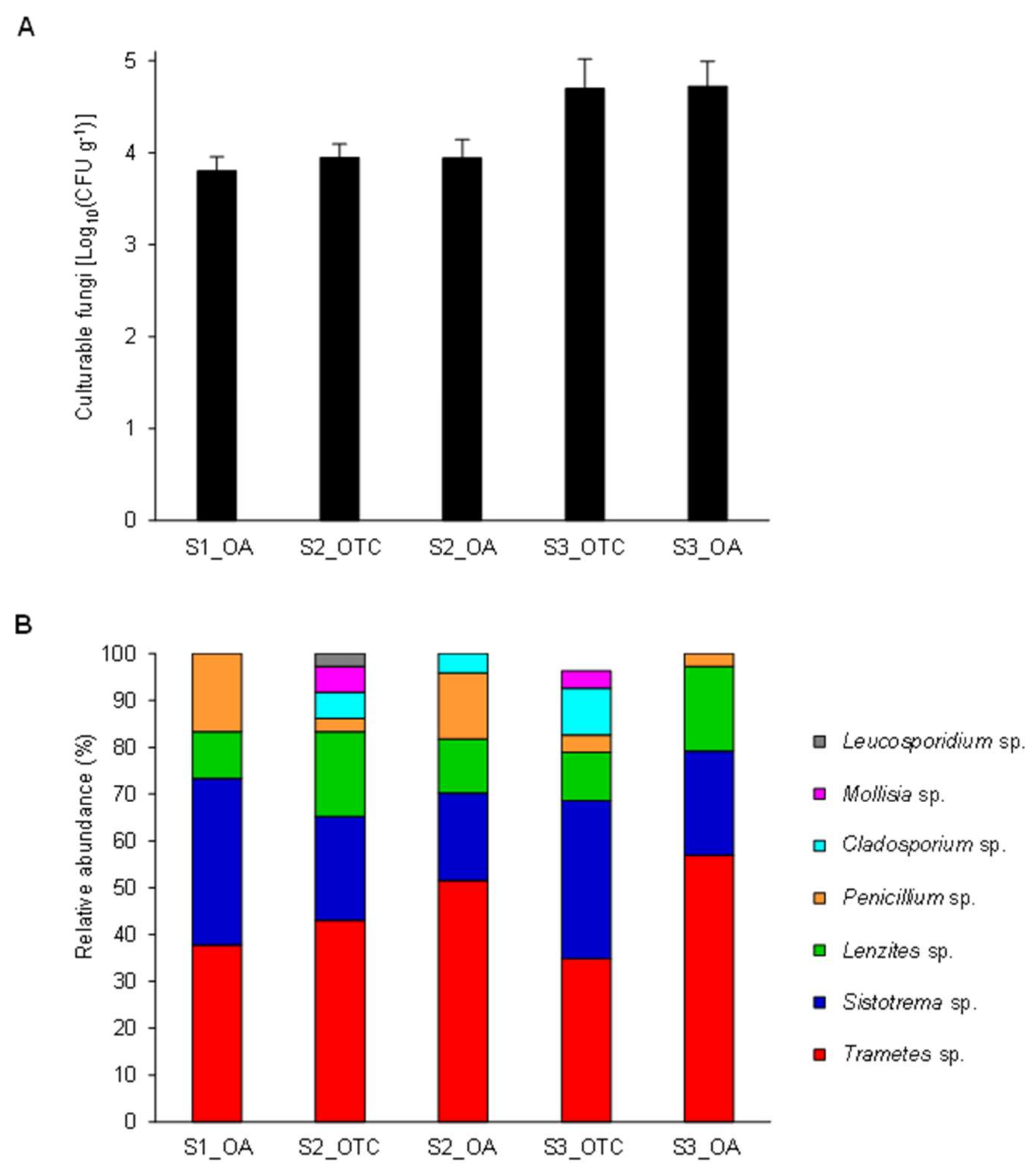
3.1. Isolation and Identification of Endophytic Fungi from the Antarctic Plant Colobanthus quitensis

A total of 132 endophytic fungi were isolated only from the plates incubated at 25 ± 1 °C with no CFU differences among the collection sites (Figure 1A), and no fungal CFUs were found during incubation at 15 ± 1 °C. The absence of fungal growth at 15 ± 1 °C suggests that growth conditions of Antarctic isolates require further optimizations to allow their growth on artificial media in vitro. For example, additional growth media, possibly containing C. quitensis plant extracts or supplementary nutritional factors, are required to improve the growth of Colobanthus quitensis endophytes in vitro and to better assess their performance at low temperatures. On the other hand, it should be also considered that Antarctic endophytic fungi may be protected by the plants from the direct exposure to the harsh environmental conditions, growing at temperatures slightly higher than the outside. Thus, the optimal growth temperature could be different than expected. Taxonomic annotation of fungal isolates by morphological analysis revealed the dominance of Trametes sp., Sistotrema sp., Lenzites sp., and Penicillium sp. (Figure 1B). To reduce redundancy for the functional characterization, 26 representative fungal isolates were selected according to the colony characteristics. The sequence of their ITS confirmed the dominance of Trametes sp. (seven isolates), Sistotrema sp. (seven isolates), Lenzites sp. (two isolates), Penicillium sp. (two isolates), Peniophora sp. (two isolates) and highlighted the presence of one isolate of Cladosporium sp., Fusarium sp., Leucosporidium sp., Mollisia sp., Phlebia sp., and Ypsilina sp. (Table S1 and Figure S1). Although the possibility of having isolated siblings from the replicas cannot be totally ruled out, it seems unlikely due their differential enzymatic activities found among the selected endophytic fungal isolates (see Section 3.2 below).
Ascomycota is the most abundant phylum of culturable fungi isolated from C. quitensis samples in Antarctica [14,35,53]. In particular, Penicillium sp. has been identified as one of the dominant fungal genera of C. quitensis rhizosphere, roots, and leaves, indicating that cosmopolitan cold-adapted taxa can establish ecological relationships with their plant host [54]. Moreover, taxa phylogenetically near to fungi that can cause diseases in plants and animals, such as Fusarium, Cladosporium, Botrytis, and Alternaria, have been previously isolated from Antarctic plants [35,55], corroborating their possible functional roles on C. quitensis. Filamentous Basidiomycetes have rarely been isolated from plants inhabiting the Antarctic Peninsula and even their typical role in wood decay has been acquired by Ascomycetes in Antarctica [56]. More commonly, Basidiomycetes have been isolated from the Antarctic soil, such as Sistotrema brinkmannii that was isolated from the Antarctic Dry Valleys [56], whence other Basidiomycetes are being recorded by molecular approaches [57]. This indicates that further studies are required to better characterize the distribution and frequency of these fungal taxa in Antarctica. Basidiomycota members were often detected in root-associated endophytic fungal communities from Arctic regions [58]. Among the Basidiomycetes isolated in this study, Phlebia sp., Trametes sp. and Peniophora sp. are known to cause a white-rot type of wood decay, and they have been reported as endophytes from a variety of hosts across wide geographic locations [59], suggesting that further functional and genomic characterizations (e.g., sequencing of more than one genetic marker) are required to improve the taxonomic annotation at the level of species and to verify the existence of endemic taxa adapted for the association with herbaceous Antarctic plants. Pegler et al. [60] reported the presence of Trametes versicolor on introduced wood brought to South Georgia, indicating that the possible introduction of non-endemic taxa should be considered as well. However, other decomposer Basidiomycetes can presumably exist naturally in bryophyte and grass ecosystems [61].

3.2. Antimicrobial and Enzymatic Activity of Selected Newly Isolated Endophytic Fungi

Among the representative fungal isolates, eight were selected according to the taxonomic annotation and collection site to be screened for the production of industrially relevant extracellular enzymes. Specifically, we selected four isolates of Trametes sp., two of Lenzites sp., one of Sistotrema sp., and one of Peniophora sp. (Table 1). The majority of the tested fungal isolates exhibited hydrolytic activities on starch, lipid, cellulose, pectin, and proteins (Table 1), and they showed no antibacterial activity against strains of Listeria innocua, Listeria monocytogenes, Bacillus cereus, Staphylococcus aureus, Pseudomonas aeruginosa, Escherichia coli, Salmonella enteritidis, Salmonella gallinarum, and Enterobacter sp.
Peniophora sp. S3.OTC.C_F1 produced hydrolysis halos only on cellulose. On the other hand, the isolates of Trametes sp. displayed broad extracellular enzymatic profiles. Lipolytic and pectinolytic enzymes were common, although the limited size of the resulting hydrolysis halos indicates low activity. Limited proteolytic action on skimmed milk was also detected in all Trametes sp. and Lenzites sp. isolates, while with gelatin as the substrate, only Trametes sp. S1.OA.A_F2 produced a clear hydrolysis halo. The different ability to utilize the gelatin or the casein contained in skimmed milk could be due to the substrate specificity of the enzymes produced by the isolates as reported in [62,63].
Cellulases and starch-degrading enzymes produced the largest hydrolysis halos, recalling the importance of these enzymes in fungal nutrition and colonization [64,65]. Trametes sp. S1.OA.A_F2, Trametes sp. S2.OTC.C_F5, Trametes sp. S2.OA.A_F5 and Lenzites sp. S3.OA.B_F6 showed the most marked amylolytic and cellulolytic halos (Table 1) and were selected to further characterize their enzymes by in vitro assays. Concentrated supernatants were then used in liquid assays in the presence of soluble starch or carboxymethylcellulose to assess the optimal pH value for their amylases or cellulases, respectively. Lenzites sp. S3.OA.B_F6 produced the highest amylolytic activity that reached the value of 4.676 nkat mL−1 (Table S2). A pH of 5 was found to be optimal, while at pH 4 and 7, the enzymatic activity decreased to 50 and 20%, respectively. Trametes sp. S1.OA.A_F2, Trametes sp. S2.OTC.C_F5 and Trametes sp. S2.OA.A_F5 produced cellulases and amylases in low quantities, and their activities ranged from 0.201 (pH 4) to 0.494 nkat mL−1 (pH 4) for cellulases and from 0.155 (pH 7) to 0.513 (pH 7) nkat mL−1 for amylases (Table S2).
Various types of extracellular enzymes were produced by the Antarctic fungal isolates. Endophytic fungi are ubiquitous in plant tissue and usually possess the specific hydrolytic systems required for depolymerize vegetal biomass, thus enabling the colonization process. Endophytic and phytopathogenic fungi seem to have similar invasion mechanisms, and many known pathogenic species are being isolated as endophytic in a range of plants [66]. The extracellular complex consists of the hydrolytic enzymes responsible for polysaccharide degradation and the oxidative ligninolytic system, which degrades lignin and opens phenyl rings [67,68]. For these reasons, assessing the bioactive potentials of the endophytes can pave the way for eco-friendly applications as a source of enzymes. Endophytic fungi remain an unexplored group of filamentous fungi, and their potential as enzyme producers has not been deeply explored yet. Enzymes from endophytic fungi isolated in extremophilic environments could be economically relevant and very interesting for the production of industrial biocompounds [69,70]. As an example, taking into consideration their aptitude to hydrolyze the complex structure of lignocellulose and the production of lipases, amylases, and proteases, extremophile endophytic fungi may become new sources of industrially relevant enzymes, suitable for the degradation of lignocellulosic biomass in the production of fuel ethanol and other value-added products. To evaluate the real potential of these endophytic fungi as sources of industrial biocatalysts, their physicochemical properties should be further characterized. Moreover, extensive studies of cultivation techniques, submerged or solid-state, are needed at both a laboratory and industrial scale to raise enzyme yields and make the production economically attractive.

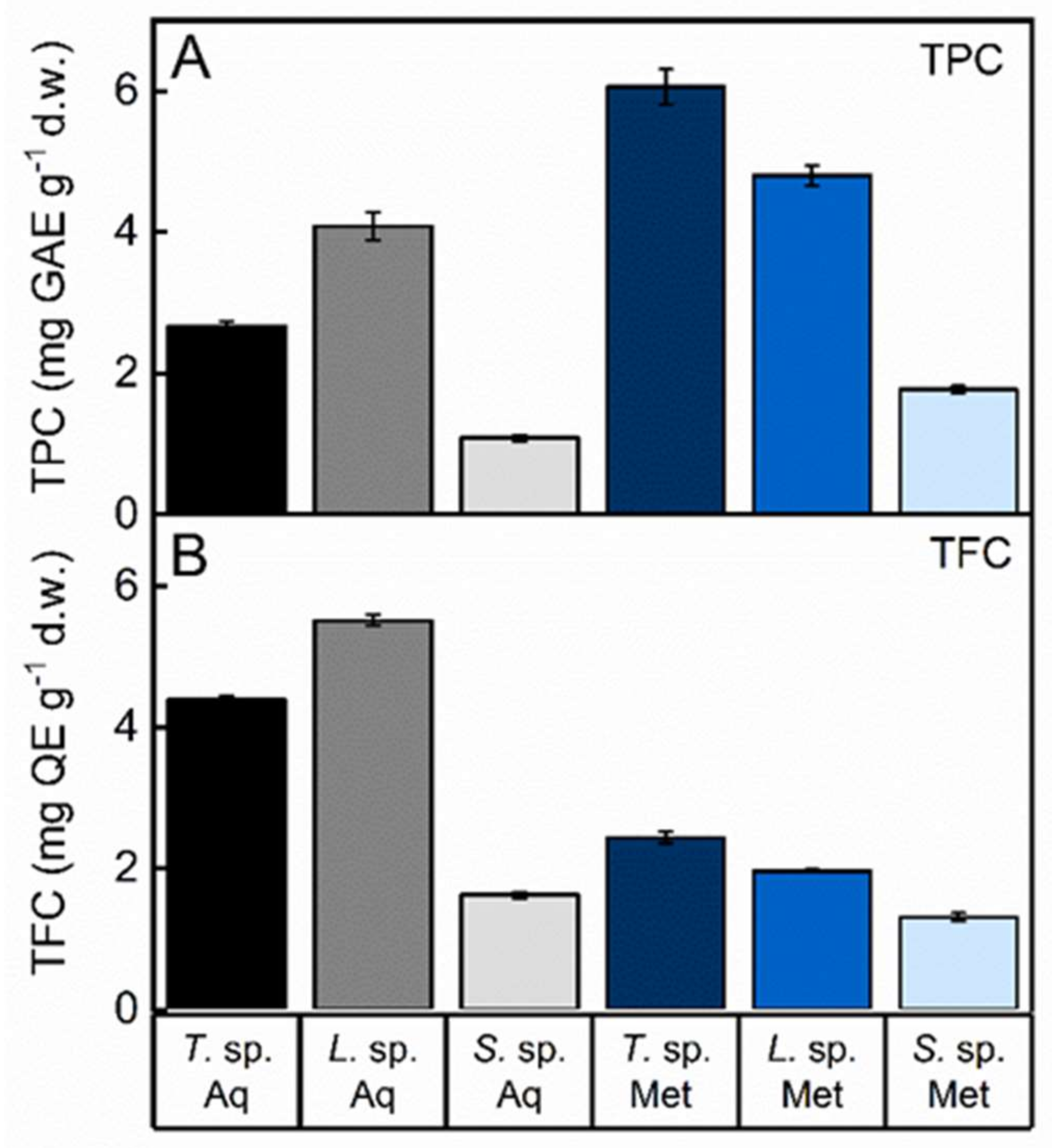
3.3. Total Phenolic and Flavonoid Content of Fungal Extracts

Fungi represent a recognized source of natural products, crucial to health and with wide applications. A multitude of bioactive compounds, such as polysaccharides, terpenes, polyphenols, lectins, peptides, and proteins, have been isolated and exploited for their biological properties [71,72]. The high and increasing number of fungal genome sequences demonstrated that their biosynthetic potential is still underestimated since many pathways involved in the biosynthesis of secondary metabolites are silent under standard cultivation conditions [73]. Hence, due to the limitations in fungal cultivability and the lack of effective methods of exhaustive isolation of metabolites, the exploitation of fungi as a source of potential bioactive products is still scarce. Therefore, it is very important to characterize fungi living in peculiar life conditions such as in Antarctica, since environmental stressors as well as their host could induce the activation of normally cryptic pathways under different growth conditions [74]. Aqueous and methanol extracts were obtained from three Antarctic fungal isolates, i.e., Trametes sp. S2.OA.C_F6, Lenzites sp. S3.OA.B_F6 and Sistotrema sp. S1.OA.C_F2, and the total phenolic (TPC) and flavonoid (TFC) contents were measured due to their importance as signal molecules known to respond to changing environments. The two solvents used have been shown to give the highest yield in the extraction procedure. In particular, alcohols were proved the best solvents for phenol extraction [75]. The TPC was expressed in terms of gallic acid equivalent per gram of dry weight (GAE g−1) (standard curve equation: y = 1.1872x − 0.0235, R2 = 0.9956). The values obtained for the concentration of total phenols ranged from 1.0857 ± 0.011 mg GAE g−1 for the aqueous extract of Sistotrema sp. S1.OA.C_F2 to 6.0580 ± 0.062 mg GAE g−1 for the methanol extract of Trametes sp. S2.OA.C_F6 (Figure 2A and Table S3).
Flavonoids, ubiquitous in plants and present in fungi, are a group of secondary metabolites derived from the phenylpropanoid pathway. In plants, they have important physiological roles in signaling and in response to changing environments. These compounds are widely studied for their diverse and important pharmacological activities such as antiviral, antimicrobial, antiallergic, antibacterial, anti-inflammatory, antifungal, cardioprotective, antiangiogenic, antithrombotic, antiproliferative, antitumor, antimalarial, apoptosis-inducing and antioxidant activities [76]. Flavonoids also play an important role in the prevention of degenerative diseases associated with oxidative stress [77,78]. Several data suggested the role of flavonoids in the establishment of plant roots endosymbiosis with certain fungi [79]. The total flavonoid content of the Antarctic fungal isolates was expressed as quercetin equivalent per gram of dry weight (QE g−1) (standard curve equation: y = 0.8688x − 0.0112, R2 = 0.9946), as reported in Figure 2B and Table S3. The methanol extract of Sistotrema sp. exhibited the lowest concentration of flavonoids (1.309904 ± 0.014 mg QE g−1), while the highest was found in the aqueous extract of Lenzites sp. (5.51562 ± 0.022 mg QE g−1). Indeed, the aqueous extraction produced the highest yield of both TPC and TFC in Lenzites sp. compared to the other fungi. Conversely, methanol was found to be more effective in extracting both phenols and flavonoids from Trametes sp. compared to the other fungi. Furthermore, our results showed that Sistotrema sp. produced lower amounts of both TPC and TFC regardless of the solvent used.

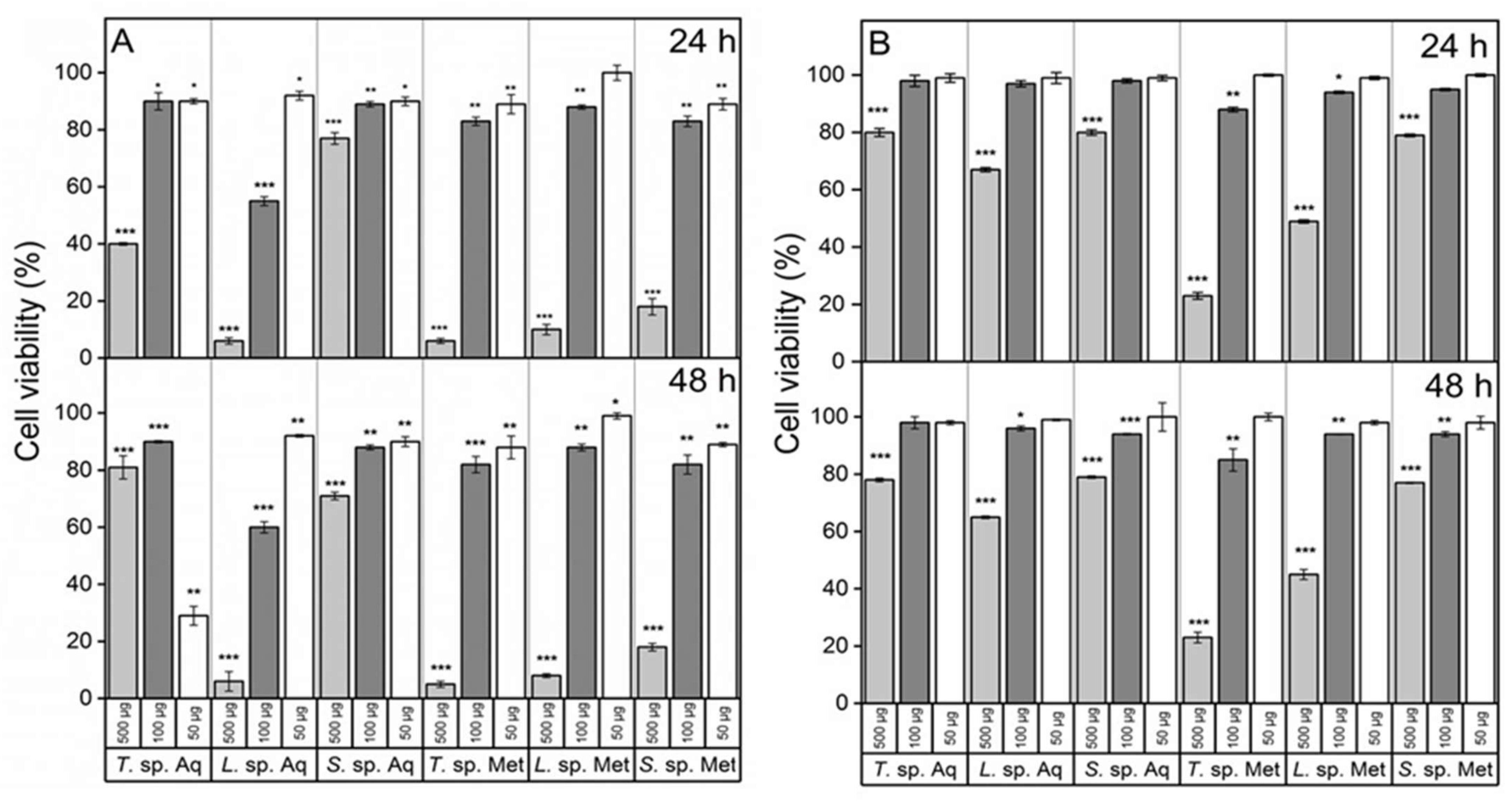
3.4. Cancer Cells Appear to Be Sensitive to Fungal Extracts

The relatively high phenolic and flavonoid contents of the fungal extracts make these Antarctic species an interesting source of bioactive compounds for medicinal applications. The presence of phenols and flavonoids in plants or microorganisms was often correlated to antioxidant activity. In human organisms, the imbalance between reactive oxygen species (ROS) and the antioxidant system may cause oxidative stress, leading to cellular damage and subsequently to various diseases such as neurodegenerative disorders, aging, and cancer [80]. To verify whether our fungal extracts could impact human cell growth, we investigated their effect on the viability of two cell lines, one from human neuroblastoma (SH-SY5Y, tumor cells) and another from human lung epithelial cells (HBEC, non-tumor cells). As shown in Figure 3A,B, the extracts inhibited the growth of both cell lines in a dose-dependent manner, and the effect increased slightly over time. Moreover, the organic extracts showed greater cytotoxicity than the aqueous ones in all samples, which could be due to a higher phenol content found in all extracts [81]. Furthermore, all extracts appeared to exert a greater effect on cancer cells. Trametes sp. S2.OA.C_F6 methanol extract showed the strongest growth inhibitory activity for both tumor and non-tumor cell lines (IC50 = 1.69 ± 0.12 mg mL−1, IC50 = 2.42 ± 0.26 mg mL−1, respectively). On the other hand, as for the aqueous extracts, the most cytotoxic was that from Lenzites sp. S3.OA.B_F6 for both cell lines (IC50 = 1.89 ± 0.18 mg mL−1, IC50 = 15.76 ± 0.95 mg mL−1, respectively). Interestingly, other authors demonstrated that, by using the MTT assay, the ethanol extract of Lenzites betulina strongly reduced the viability of several types of cancer cells, and this effect appeared to be induced through ROS generation, reduction of the matrix metalloproteinases, P53 up-regulation, and Bcl2, pro-caspase-3, pro-caspase-9 and proline-rich acidic protein down-regulation [82]. The cytotoxic effect of our extracts appeared to be more intense than that of extracts derived from non-Antarctic species, although tested on different cell lines [82]. Interestingly, a positive correlation between TPC and the ability to inhibit cell growth was observed for all extracts regardless of the types of used cells.

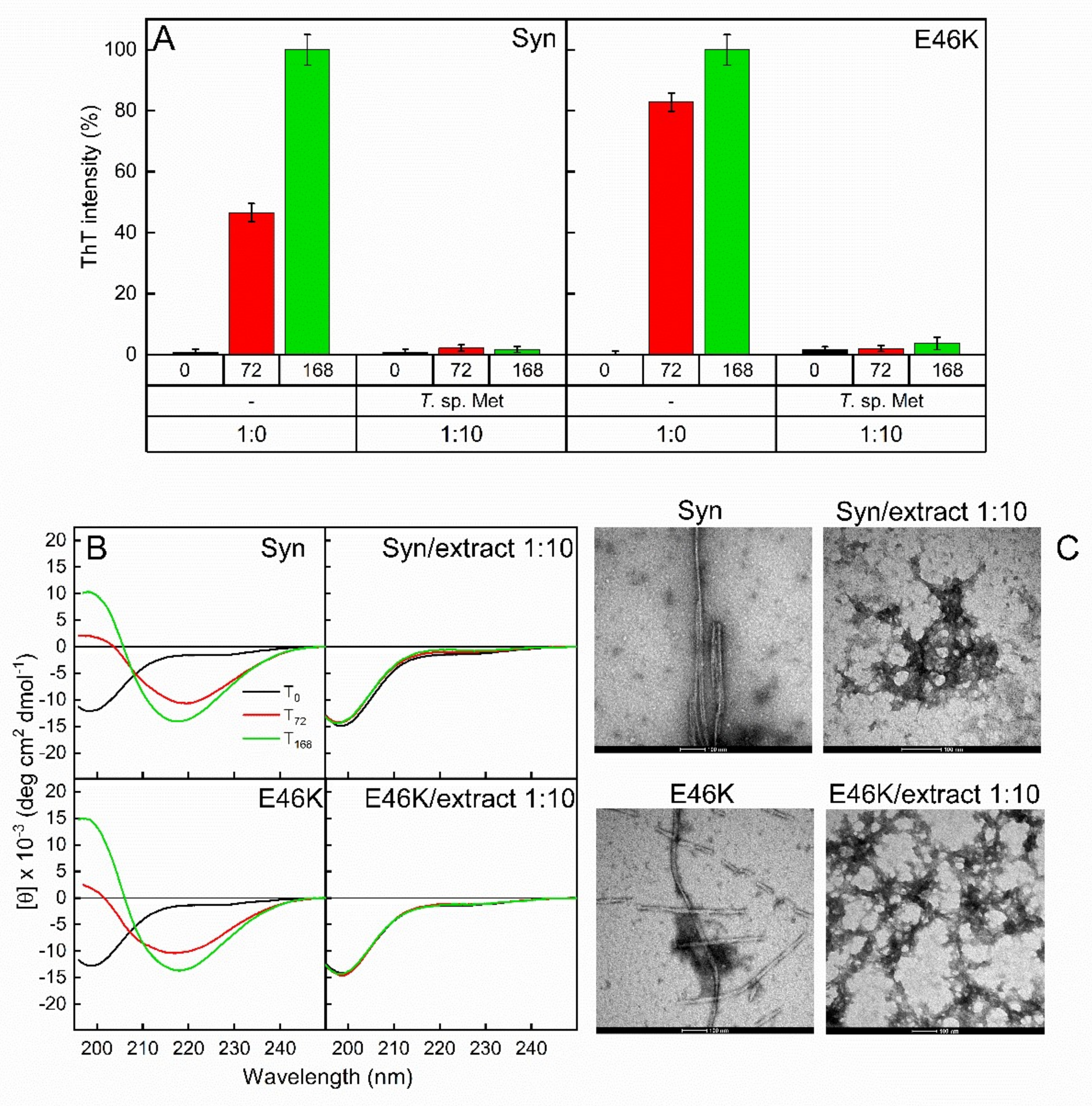
3.5. Fungal Extracts Affect the Formation of Amyloid Fibrils

Parkinson’s disease is a neurodegenerative disorder associated with the amyloid aggregation of the presynaptic protein α-synuclein (Syn) and its familiar mutants [83]. Although several molecular events underlying Syn aggregation are still unclear, oxidative stress is known to contribute to this process. Some metabolites extracted from different fungi showed neuroprotective effects in vivo [84]. Polyphenols, pure or present in crude extracts, could protect against neurodegenerative disorders interfering with the oxidative stress mechanism in the brain and/or by reducing the aggregation propensity of Syn [85,86,87]. In this study, we analyzed the effects of the methanol extract of Trametes sp. S2.OA.C_F6 on the fibrillation process of Syn and its familiar mutant E46K. This extract was selected for its high content of polyphenols. Both proteins were incubated at 37 °C under shaking to promote protein fibrillation, in the presence or absence of the fungal extract. As assessed by Thioflavin T (ThT) assay (Figure 4A), amyloid fibrils were present in both Syn and its mutant already after 72 h of incubation, and their amount increased over time. At this stage, the protein secondary structure underwent conformational change (Figure 4B). Indeed, according to their unfolded nature [88], the proteins exhibited typical CD spectra of random coil conformation (black lines) at time 0, while adopting β-sheet secondary structure after incubation. As shown in Figure 4B, the β-sheet conformation is highlighted as a minimum at 218 nm in CD spectra recorded at 72 h and is more pronounced after 168 h of incubation. TEM pictures revealed the presence of structures compatible with mature unbranched fibrils with a diameter of 6–10 nm and length of 1 μm in the samples incubated for 168 h (Figure 4C), as previously observed [87]. The intensity of the ThT fluorescence emission at 485 nm of Syn and E46K samples was strongly reduced when the proteins were incubated with the methanol extract of Trametes sp. (1:10, Syn (or E46K)/extract, w/w), indicating the absence of fibrillar material in the samples (Figure 4A). This result was confirmed by far-UV CD analysis (Figure 4B). In fact, analyzing CD spectra after 72 and 168 h of incubation in the presence of the fungal extract, a predominantly random coil conformation was detected in both samples, indicating the absence of the transition to β-sheet secondary structure and, therefore, the absence of amyloid fibrils (Figure 4B). Moreover, TEM analysis indicated the presence of amorphous aggregates with a morphology completely different from amyloid fibrils. In conclusion, Trametes sp. methanol extract strongly hampers the amyloidogenic process of Syn and its mutant, as previously observed by using small molecules of natural origin [87,89].

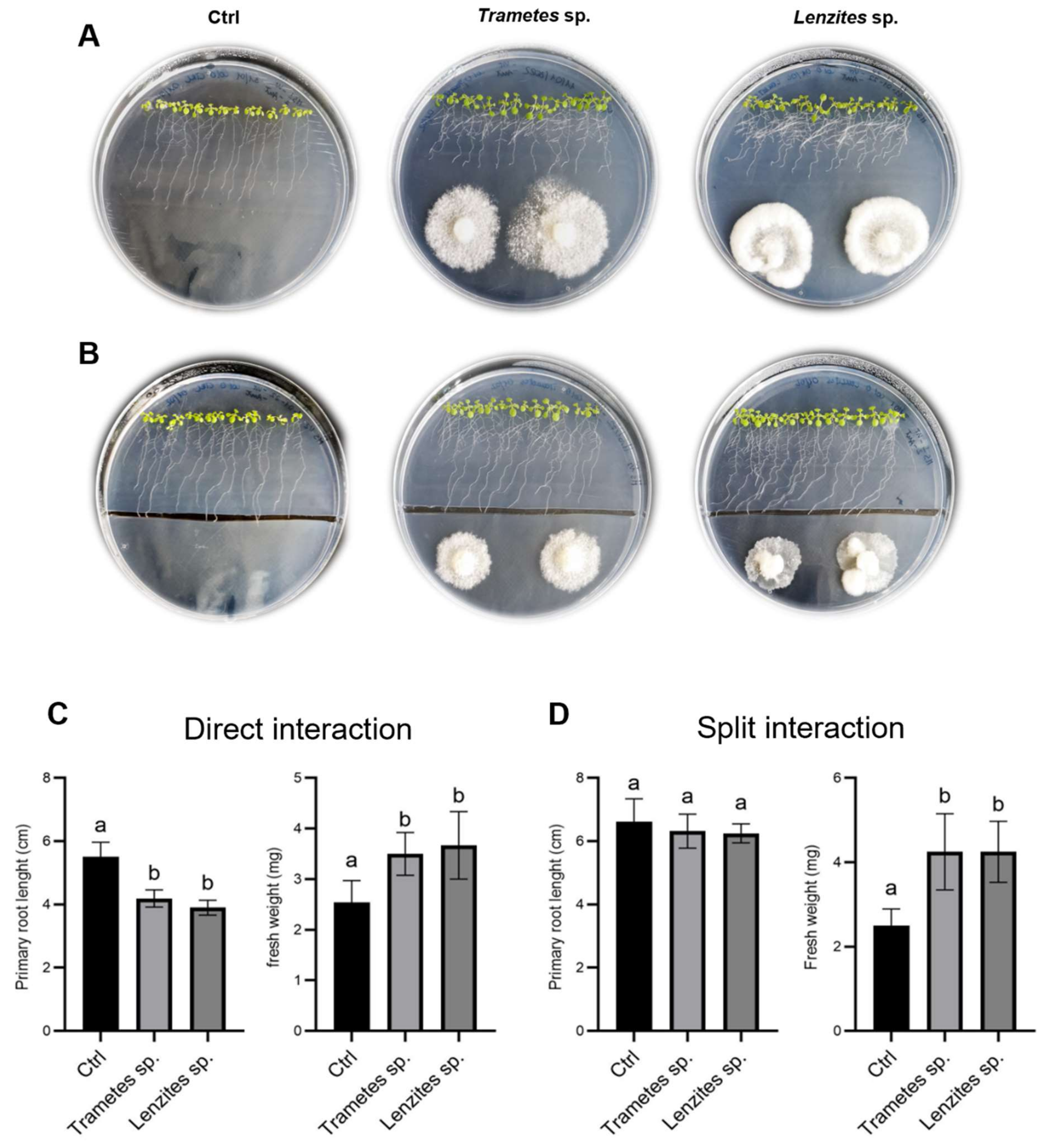
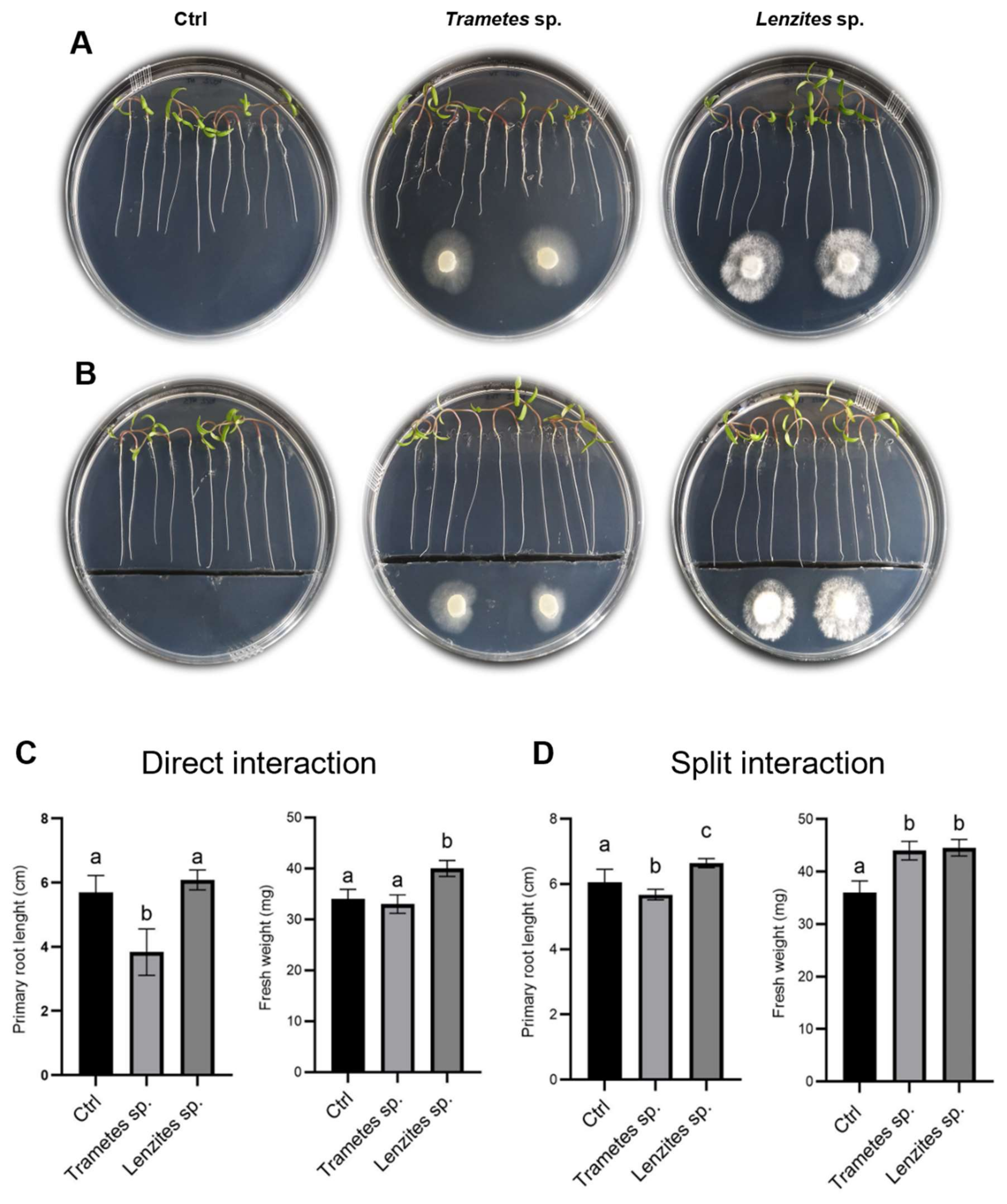
3.6. Antarctic Fungi Affect the Root Architecture in Arabidopsis and Tomato

Several studies have reported the beneficial effects of plant-associated microbial communities on plant growth and fitness, even in extreme environments [20,21,22,23,24,25,26]. The knowledge of the influence of Antarctic endophytic fungi on plants of agricultural interest is still in its infancy, but it deserves to be deepened for the peculiarity of extremophilic microorganisms. To test the effect of the Antarctic Basidiomycetes Trametes sp. S2.OA.C_F6 and Lenzites sp. S3.OA.B_F6 on plant growth, co-cultivation experiments were carried out with Arabidopsis thaliana Col-0 and tomato (Solanum lycopersicum L.-cv. AKRAI F1) seedlings. To discriminate between the effects triggered by volatile or soluble molecules, both split and contact interaction experiments were carried out, respectively. As for Arabidopsis, at 7 days post-inoculum (dpi), the direct contact interaction caused a significant shortening of the primary root length (Figure 5A,C) and a simultaneous increase of the growth of secondary roots, leading to an overall enhancement in root density as compared to the control. Moreover, both fungi promoted the development of the aerial part, inducing a statistically significant increase in plant fresh weight (Figure 5C). On the other hand, the inhibitory effect on primary root length was lost in the split interaction (Figure 5B,D), while the growth promotion of secondary roots and aerial part was maintained, as evidenced by the statistically significant increase in plant fresh weight (Figure 5D).
This result suggested that diffusible soluble molecules secreted by the fungi could inhibit primary root length, while fungal volatile organic compounds (VOCs) could induce secondary roots formation and aerial part development. In tomato, the presence of the furrow in the plates did not substantially influence the effect of the fungus on the considered plant parameters and the two fungal isolates showed an opposite behavior on primary root length, which was significantly inhibited by Trametes sp. S2.OA.C_F6 and enhanced by Lenzites sp. S3.OA.B_F6. Moreover, both fungi had a positive effect on plant fresh weight (Figure 6, all panels).
These results suggested that volatile signals would be the key factors during either Lenzites sp. S3.OA.B_F6– or Trametes sp. S2.OA.C_F6–tomato interactions. Interestingly, similar effects were reported for Trichoderma–Arabidopsis interaction, where plant growth enhancement was mainly attributed to fungus-emitted VOCs, especially terpenes, whereas root morphology alteration was principally ascribed to fungus-induced regulation of auxin distribution in apical and lateral roots [90,91,92]. It has also been reported that acidification of the culture medium by Trichoderma is able to induce auxin redistribution within Arabidopsis columella root cap cells, causing root tip bending and growth inhibition [93]. To test whether Antarctic isolates were able to induce acidification of the medium, we grew both fungi on PDA plates supplemented with bromophenol blue as a pH indicator. Interestingly, we found that they were able to strongly acidify the medium (Figure S1), and it could be hypothesized that Arabidopsis plants adapt the root apparatus to the acidification via lateral root production. In general, our results suggest that both Antarctic fungi behave similarly to Trichoderma when interacting with plants, establishing beneficial relationships with them. This discovery opens up new perspectives on the possible exploitation of these fungi for sustainable agriculture. To this end, the experiment will be scaled-up growing plants first in pots and then in the field.

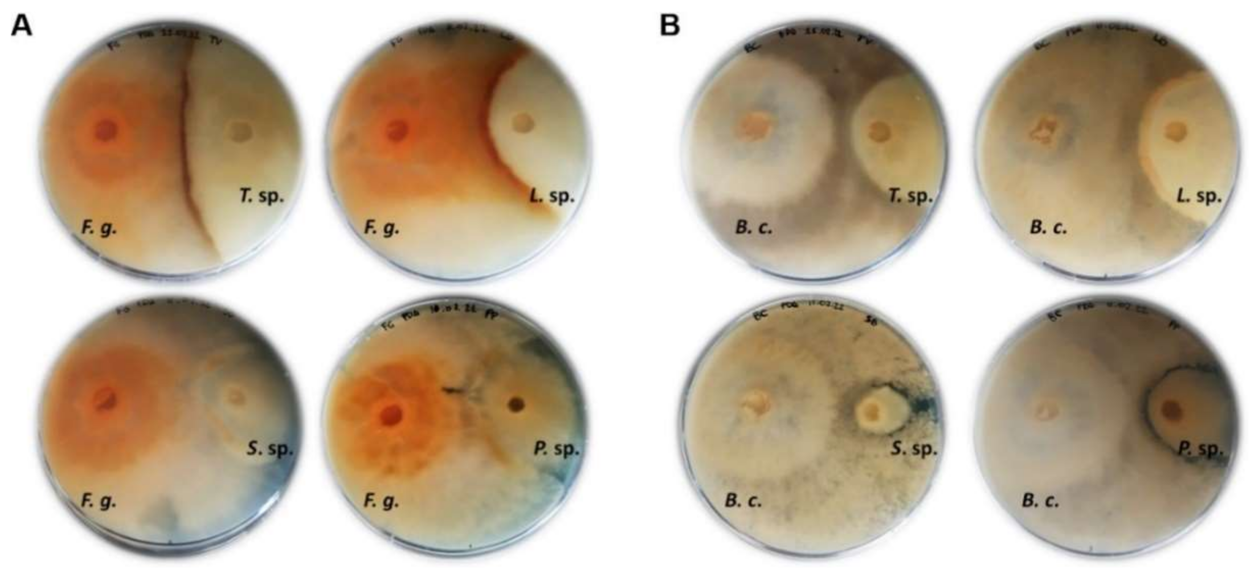
3.7. Antarctic Fungi Antagonize the Growth of Crop Plant Pathogenic Fungi

Co-culturing of fungi has been reported as a successful tool to induce the activation of cryptic pathways, which trigger the production of new fungal secondary metabolites with potential biotechnological applications [94]. Specifically, the antagonistic reaction can stimulate the biosynthesis of bioactive natural molecules in the interaction zone to counteract the spreading of the antagonist [95]. In this work, the ability of the Antarctic fungal isolates of Trametes sp. S2.OA.C_F6, Lenzites sp. S3.OA.B_F6, Sistotrema sp. S1.OA.C_F2 and Peniophora sp. S3.OTC.C_F1 to antagonize or inhibit the growth of phytopathogenic fungi, i.e., Fusarium graminearum and Botrytis cinerea, was investigated by growing fungi in co-cultures. The observed antagonistic reactions included overgrowth, contact inhibition, and distance inhibition. Specifically, in both co-cultures with F. graminearum (Figure 7A) and B. cinerea (Figure 7B), either Trametes sp. S2.OA.C_F6 or Lenzites sp. S3.OA.B_F6 showed a strongly pigmented mycelium in the interaction zone, accompanied by an antagonistic effect on the phytopathogen growth. This phenotype might be explained by the production of volatile or diffusible antifungal metabolites, which include both signaling and reacting compounds, by one or both isolates as a result of the interaction [96]. On the other hand, Sistotrema sp. S1.OA.C_F2 seemed to be almost completely overwhelmed by the phytopathogens, whereas Peniophora sp. S3.OTC.C_F1 was the only isolate displaying distance inhibition against B. cinerea. For a better interpretation of the results, it is worth mentioning that the growth rates of Trametes sp. S2.OA.C_F6 and Lenzites sp. S3.OA.B_F6 are similar to that of F. graminearum and B. cinerea (7–10 days to cover the whole 90 mm Petri dish), whereas Peniophora sp. S3.OTC.C_F1 and Sistotrema sp. S1.OA.C_F2 grow slower than the phytopathogenic fungi (around 15 and 20 days to cover the whole 90 mm Petri dish, respectively), and thus, their antagonistic reaction could be slightly impaired.
Previous works reported that extracellular alkalinization contributes to fungal pathogenesis against both plants and microbes and acidification could reverse pathogenicity [97,98]. Thus, to understand the behavior of co-cultured fungi with respect to their ability to influence the pH of the growth medium, we also carried out the co-culturing experiments on PDA plates supplemented with bromophenol blue (Figure S2). The ability of Trametes sp. S2.OA.C_F6 and Lenzites sp. S3.OA.B_F6 to acidify the medium (Figure S1) could be hypothesized as one of the mechanisms adopted by the two fungal isolates to counteract the phytopathogen spreading. As for the other fungal isolates, none of them seemed able to acidify the medium (Figure S2). Examining the antagonistic potential of Antarctic fungi against important phytopathogens is the initial step in using these isolates for applications in plant biocontrol programs used in agriculture.
In conclusion, the search for novel bio-products in poorly explored environments is emerging as the key to providing solutions for many relevant issues, and Antarctic environments are increasingly recognized as precious locations for bioprospecting. This study emphasizes the ecological and biotechnological relevance of fungi from the Antarctic ecosystem. The fungal isolates used in this work have proved to be valuable sources of bioactive molecules ranging from industrial to pharmaceutical and agricultural applications, even though more studies are necessary to allow their biotechnological exploitation.

Supplementary Materials

The following supporting information can be downloaded at: https://www.mdpi.com/article/10.3390/jof8090979/s1, Figure S1: Phylogenetic tree of representative endophytic fungi of Colobanthus quitensis; Figure S2: Representative images showing Trametes sp. and Lenzites sp. isolates grown for 7 days on PDA solid medium in the presence or absence of bromophenol blue, as a pH indicator; Figure S3: Co-cultivation of Fusarium graminearum and Botrytis cinerea and Antarctic fungal isolates on PDA solid medium at 10 dpi in the presence of bromophenol blue as a pH indicator. Table S1: taxonomic annotation of representative endophytic fungi of Colobanthus quitensis; Table S2: highest cellulolytic and amylolytic activities of selected Antarctic fungal isolates detected in vitro on carboxymethylcellulose or soluble starch; Table S3: total phenolic content and total flavonoid content of aqueous or methanol extract of Trametes sp. S2.OA.C_F6, Lenzites sp. S3.OA.B_F6 and Sistotrema sp. S1.OA.C_F2.

Author Contributions

Conceptualization, C.C., L.B., M.P., P.P.d.L., M.B. and D.V.S.; Methodology and Validation, L.B., C.C. and S.P.; Formal Analysis, G.C., L.B., S.P., C.M.O.L., B.F., L.F. and V.B.; Investigation, C.C., P.P.d.L. and L.B.; Resources, C.C., M.P., P.P.d.L., M.B., D.V.S. and S.C.; Writing—Original Draft Preparation, L.B., C.C., M.P., P.P.d.L. and M.B.; Writing—Review and Editing, S.P., L.F., B.F., D.V.S. and S.C.; Visualization, L.B., C.C. and S.P.; Supervision, C.C., L.B., M.P. and P.P.d.L.; Project Administration, C.C., L.B., M.P. and P.P.d.L.; Funding Acquisition, L.B., M.P. and P.P.d.L. All authors have read and agreed to the published version of the manuscript.

Funding

This research was funded by the Ministero dell’Università e della Ricerca (MUR) in the framework of the National Program for Antarctic Research (PNRA), grant number PNRA18_00147 (Impact of climate change on plant–microbe interaction: a focus on tripartite plant–fungus–virus relationship and on their bioactive compounds) and by the University of Padova DOR2251254/22, DOR2107797/21 and DOR2084579/20.

Institutional Review Board Statement

Not applicable.

Informed Consent Statement

Not applicable.

Data Availability Statement

Fungal ITS sequences presented in this study are available in NCBI (http://www.ncbi.nlm.nih.gov; accessed on 1 June 2022); accession numbers for each fungal isolate are indicated in Table S1.

Acknowledgments

The authors thank Alessandra Piccirillo (Dipartimento di Biomedicina Comparata e Alimentazione of University of Padova) for providing isolates for antibacterial evaluation experiments and Bianca Vicelli (University of Trento) for taxonomic annotation based on ITS sequencing.

Conflicts of Interest

The authors declare no conflict of interest.

References

  1. Danilovich, M.E.; Sánchez, L.A.; Acosta, F.F.; Delgado, O.D. Antarctic bioprospecting: In pursuit of microorganisms producing new antimicrobials and enzymes. Polar Biol. 2018, 41, 1417–1433. [Google Scholar] [CrossRef]
  2. Raddadi, N.; Cherif, A.; Daffonchio, D.; Neifar, M.; Fava, F. Biotechnological applications of extremophiles, extremozymes and extremolytes. Appl. Microbiol. Biotechnol. 2015, 99, 7907–7913. [Google Scholar] [CrossRef] [PubMed]
  3. Timling, I.; Taylor, D.L. Peeking through a frosty window: Molecular insights into the ecology of Arctic soil fungi. Fungal Ecol. 2012, 5, 419–429. [Google Scholar] [CrossRef]
  4. Weinstein, R.N.; Montiel, P.O.; Johnstone, K. Influence of growth temperature on lipid and soluble carbohydrate synthesis by fungi isolated from fellfield soil in the maritime Antarctic. Mycologia 2000, 92, 222–229. [Google Scholar] [CrossRef]
  5. Coleine, C.; Stajich, J.E.; de los Ríos, A.; Selbmann, L. Beyond the extremes: Rocks as ultimate refuge for fungi in drylands. Mycologia 2021, 113, 108–133. [Google Scholar] [CrossRef] [PubMed]
  6. Robinson, C.H. Cold adaptation in Arctic and Antarctic fungi. New Phytol. 2001, 151, 341–353. [Google Scholar] [CrossRef]
  7. Zucconi, L.; Canini, F.; Temporiti, M.E.; Tosi, S. Extracellular enzymes and bioactive compounds from Antarctic terrestrial fungi for bioprospecting. Int. J. Environ. Res. Public Health 2020, 17, 6459. [Google Scholar] [CrossRef]
  8. Krüger, A.; Schäfers, C.; Schröder, C.; Antranikian, G. Towards a sustainable biobased industry—Highlighting the impact of extremophiles. New Biotechnol. 2018, 40, 144–153. [Google Scholar] [CrossRef]
  9. Duarte, A.W.F.; dos Santos, J.A.; Vianna, M.V.; Vieira, J.M.F.; Mallagutti, V.H.; Inforsato, F.J.; Wentzel, L.C.P.; Lario, L.D.; Rodrigues, A.; Pagnocca, F.C.; et al. Cold-adapted enzymes produced by fungi from terrestrial and marine Antarctic environments. Crit. Rev. Biotechnol. 2017, 38, 600–619. [Google Scholar] [CrossRef]
  10. Loperena, L.; Soria, V.; Varela, H.; Lupo, S.; Bergalli, A.; Guigou, M.; Pellegrino, A.; Bernardo, A.; Calviño, A.; Rivas, F.; et al. Extracellular enzymes produced by microorganisms isolated from maritime Antarctica. World J. Microbiol. Biotechnol. 2012, 28, 2249–2256. [Google Scholar] [CrossRef]
  11. Gabani, P.; Singh, O.V. Radiation-resistant extremophiles and their potential in biotechnology and therapeutics. Appl. Microbiol. Biotechnol. 2013, 97, 993–1004. [Google Scholar] [CrossRef] [PubMed]
  12. Tian, Y.; Li, Y.-L.; Zhao, F.-C. Secondary Metabolites from Polar Organisms. Mar. Drugs 2017, 15, 28. [Google Scholar] [CrossRef] [PubMed]
  13. Purić, J.; Vieira, G.; Cavalca, L.B.; Sette, L.D.; Ferreira, H.; Vieira, M.L.C.; Sass, D.C. Activity of Antarctic fungi extracts against phytopathogenic bacteria. Lett. Appl. Microbiol. 2018, 66, 530–536. [Google Scholar] [CrossRef] [PubMed]
  14. Santiago, I.F.; Alves, T.M.A.; Rabello, A.; Sales Junior, P.A.; Romanha, A.J.; Zani, C.L.; Rosa, C.A.; Rosa, L.H. Leishmanicidal and antitumoral activities of endophytic fungi associated with the Antarctic angiosperms Deschampsia antarctica Desv. and Colobanthus quitensis (Kunth) Bartl. Extremophiles 2012, 16, 95–103. [Google Scholar] [CrossRef] [PubMed]
  15. Bang, C.; Dagan, T.; Deines, P.; Dubilier, N.; Duschl, W.J.; Fraune, S.; Hentschele, U.; Hirtf, H.; Hültera, N.; Lachnitb, T.; et al. Metaorganisms in extreme environments: Do microbes play a role in organismal adaptation? Zoology 2018, 127, 1–19. [Google Scholar] [CrossRef]
  16. Longton, R.E. Vegetation Ecology and Classification in the Antarctic Zone. Can. J. Bot. 1979, 57, 2264–2278. [Google Scholar] [CrossRef]
  17. Smith, R.I.L. The enigma of Colobanthus quitensis and Deschampsia antarctica in Antarctica. In Antarctic Biology in a Global Context; Huiskes, A.H.L., Gieskes, W.W.C., Rozema, J., Schorno, R.M.L., van der Vies, S.M., Wolff, W.J., Eds.; Backhuys Publishers: Leiden, The Netherlands, 2003; pp. 234–239. [Google Scholar]
  18. Arthofer, W.; Bertini, L.; Caruso, C.; Cicconardi, F.; Delph, L.F.; Fields, P.D.; Ikeda, M.; Minegishi, Y.; Proietti, S.; Rotthamer, H.; et al. Genomic Resources Notes accepted 1 February 2015–31 March 2015. Mol. Ecol. Resour. 2015, 15, 1014–1015. [Google Scholar]
  19. Nibert, M.L.; Manny, A.R.; Debat, H.J.; Firth, A.E.; Bertini, L.; Caruso, C. A barnavirus sequence mined from a transcriptome of the Antarctic pearlwort Colobanthus quitensis. Arch. Virol. 2018, 163, 1921–1926. [Google Scholar] [CrossRef]
  20. Ballesteros, G.I.; Torres-Díaz, C.; Bravo, L.A.; Balboa, K.; Caruso, C.; Bertini, L.; Proietti, S.; Molina-Montenegro, M.A. In silico analysis of metatranscriptomic data from the Antarctic vascular plant Colobanthus quitensis: Responses to a global warming scenario through changes in fungal gene expression levels. Fungal Ecol. 2020, 43, 100873. [Google Scholar] [CrossRef]
  21. Torres-Díaz, C.; Gallardo-Cerda, J.; Lavin, P.; Oses, R.; Carrasco-Urra, F.; Atala, C.; Acuña-Rodríguez, I.S.; Convey, P.; Molina-Montenegro, M.A. Biological interactions and simulated climate change modulates the ecophysiological performance of Colobanthus quitensis in the Antarctic ecosystem. PLoS ONE 2016, 11, 10. [Google Scholar] [CrossRef]
  22. Gallardo-Cerda, J.; Levihuan, J.; Lavín, P.; Oses, R.; Atala, C.; Torres-Díaz, C.; Cuba-Díaz, M.; Barrera, A.; Molina-Montenegro, M.A. Antarctic rhizobacteria improve salt tolerance and physiological performance of the Antarctic vascular plants. Polar Biol. 2018, 41, 1973–1982. [Google Scholar] [CrossRef]
  23. Ramos, P.; Rivas, N.; Pollmann, S.; Casati, P.; Molina-Montenegro, M.A. Hormonal and physiological changes driven by fungal endophytes increase Antarctic plant performance under UV-B radiation. Fungal Ecol. 2018, 34, 76–82. [Google Scholar] [CrossRef]
  24. Barrera, A.; Hereme, R.; Ruiz-Lara, S.; Larrondo, L.F.; Gundel, P.E.; Pollmann, S.; Molina-Montenegro, M.A.; Ramos, P. Fungal endophytes enhance the photoprotective mechanisms and photochemical efficiency in the Antarctic Colobanthus quitensis (Kunth) Bartl. exposed to UV-B radiation. Front. Ecol. Evol. 2020, 8, 122. [Google Scholar] [CrossRef]
  25. Hereme, R.; Morales-Navarro, S.; Ballesteros, G.; Barrera, A.; Ramos, P.; Gundel, P.E.; Molina-Montenegro, M.A. Fungal endophytes exert positive effects on Colobanthus quitensis under water stress but neutral under a projected climate change scenario in Antarctica. Front. Microbiol. 2020, 11, 264. [Google Scholar] [CrossRef] [PubMed]
  26. Oses-Pedraza, R.; Torres-Díaz, C.; Lavín, P.; Retamales-Molina, P.; Atala, C.; Gallardo-Cerda, J.; Acuña-Rodríguez, I.S.; Molina-Montenegro, M.A. Root endophytic Penicillium promotes growth of Antarctic vascular plants by enhancing nitrogen mineralization. Extremophiles 2020, 24, 721–732. [Google Scholar] [CrossRef]
  27. Molina-Montenegro, M.A.; Oses, R.; Torres-Diaz, C.; Atala, C.; Zurita-Silva, A.; Ruiz-Lara, S. Root-endophytes improve the ecophysiological performance and production of an agricultural species under drought condition. AoB Plants 2016, 8, plw062. [Google Scholar] [CrossRef]
  28. Acuña-Rodríguez, I.S.; Hansen, H.; Gallardo-Cerda, J.; Atala, C.; Molina-Montenegro, M.A. Antarctic extremophiles: Biotechnological alternative to crop productivity in saline soils. Front. Bioeng. Biotechnol. 2019, 7, 22. [Google Scholar] [CrossRef]
  29. Yadav, A.N. Beneficial plant-microbe interactions for agricultural sustainability. J. Appl. Biol. Biotechnol. 2021, 9, 1–4. [Google Scholar] [CrossRef]
  30. Chávez, R.; Fierro, F.; García-Rico, R.O.; Vaca, I. Filamentous fungi from extreme environments as a promising source of novel bioactive secondary metabolites. Front. Microbiol. 2015, 6, 903. [Google Scholar] [CrossRef]
  31. Galambos, N.; Compant, S.; Moretto, M.; Sicher, C.; Puopolo, G.; Wäckers, F.; Sessitsch, A.; Pertot, I.; Perazzolli, M. Humic Acid Enhances the Growth of Tomato Promoted by Endophytic Bacterial Strains through the Activation of Hormone-, Growth-, and Transcription-Related Processes. Front. Plant Sci. 2020, 11, 82267. [Google Scholar] [CrossRef]
  32. Arrigoni, E.; Albanese, D.; Longa, C.M.; Angeli, D.; Donati, C.; Ioriatti, C.; Pertot, I.; Perazzolli, M. Tissue age, orchard location and disease management influence the composition of fungal and bacterial communities present on the bark of apple trees. Environ. Microbiol. 2020, 22, 2080–2093. [Google Scholar] [CrossRef] [PubMed]
  33. Elazegui, F.A.; Castilla, N.P.; Dalisay, T.U.; Mew, T.W. Causal agent of red stripe disease of rice. Plant Dis. 2004, 88, 1310–1317. [Google Scholar] [CrossRef] [PubMed]
  34. Orpin, J.B.; Mzungu, I.; Usman-Sani, H. Fungal Infestation of Garri Sold around Dutsinma Metropolis. J. Proteom. Bioinform. 2020, 13, 1–4. [Google Scholar]
  35. Rosa, L.H.; Almeida Vieira Mde, L.; Santiago, I.F.; Rosa, C.A. Endophytic fungi community associated with the dicotyledonous plant Colobanthus quitensis (Kunth) Bartl. (Caryophyllaceae) in Antarctica. FEMS Microb. Ecol. 2010, 73, 178–189. [Google Scholar] [CrossRef] [PubMed]
  36. Ellis, M.B. Dematiaceous Hyphomycetes; Commonwealth Mycological Institute: Egham, UK, 1971; p. 608. [Google Scholar]
  37. Watanabe, T. Pictorial Atlas of Soil and Seed Fungi: Morphologies of Cultured Fungi and Key to Species, 3rd ed.; CRC Press: Boca Raton, FL, USA, 2010; p. 426. [Google Scholar]
  38. Hofstetter, V.; Buyck, B.; Eyssartier, G.; Schnee, S.; Gindro, K. The unbearable lightness of sequenced-based identification. Fungal Divers. 2019, 96, 243–284. [Google Scholar] [CrossRef]
  39. Kumar, S.; Stecher, G.; Tamura, K. MEGA7: Molecular Evolutionary Genetics Analysis Version 7.0 for Bigger Datasets. Mol. Biol. Evol. 2016, 33, 1870–1874. [Google Scholar] [CrossRef]
  40. Favaro, L.; Corich, V.; Giacomini, A.; Basaglia, M.; Casella, S. Grape marcs as unexplored source of new yeasts for future biotechnological applications. World J. Microbiol. Biotechnol. 2013, 29, 1551–1562. [Google Scholar]
  41. Favaro, L.; Jooste, T.; Basaglia, M.; Rose, S.H.; Saayman, M.; Görgens, J.F.; Casella, S.; van Zyl, W.H. Codon-optimized glucoamylase sGAI of Aspergillus awamori improves starch utilization in an industrial yeast. Appl. Microbiol. Biotechnol. 2012, 95, 957–968. [Google Scholar] [CrossRef]
  42. Lim, C.K.; Ahmad, R.; Marzuki, N.F.; Goh, Y.K.; Azizan, K.A.; Goh, Y.K.; Goh, K.J. Optimization of metabolite extraction protocols for untargeted metabolite profiling of mycoparasitic Scytalidium parasiticum using LC-TOF-MS. Sains Malays. 2018, 47, 3061–3068. [Google Scholar] [CrossRef]
  43. Rašeta, M.; Popović, M.; Knežević, P.; Šibul, F.; Kaišarević, S.; Karaman, M. Bioactive phenolic compounds of two medicinal mushroom species Trametes versicolor and Stereum subtomentosum as antioxidant and antiproliferative agents. Chem. Biodivers. 2020, 17, 12. [Google Scholar] [CrossRef]
  44. Singleton, V.L. Analysis of total phenols and other oxidation substrates and antioxidants by means of Folin-Ciocalteu reagent. Methods Enzymol. 1999, 299, 152–178. [Google Scholar]
  45. Way, M.L. A comparison of laboratory analysis methods for total phenolic content of cider. Beverages 2020, 6, 55. [Google Scholar] [CrossRef]
  46. Shirazi, O.U. Determination of total phenolic, flavonoid content and free radical scavenging activities of common herbs and spices. J. Pharmacogn. Phytochem. 2014, 3, 104–108. [Google Scholar]
  47. Siddiqui, N. Spectrophotometric determination of the total phenolic content, spectral and fluorescence study of the herbal Unani drug Gul-e-Zoofa (Nepeta bracteata Benth). J. Taibah Univ. Med. Sci. 2017, 12, 360–363. [Google Scholar] [CrossRef] [PubMed]
  48. Chang, C.C.; Yang, M.H.; Wen, H.J.; Chern, J.C. Estimation of total flavonoid content in propolis by two complementary colorimetric methods. J. Food Drug Anal. 2002, 10, 178–182. [Google Scholar]
  49. Ghosh, M.; Sinha, B.N.; Seijas, J.A.; Vázquez-Tato, M.P.; Feás, X. Flavonoids and phenolic compounds from Litsea polyantha Juss. bark. In Medicinal and Natural Products Chemistry, Proceedings of the 18th International Electronic Conference On Synthetic Organic Chemistry (ECSOC-18), Lugo, Spain, 1–30 November 2014; Seijas, J.A., Vázquez-Tato, M.P., Lin, S.K., Eds.; MDPI: Basel, Switzerland, 2015. [Google Scholar]
  50. De Franceschi, G.; Frare, E.; Bubacco, L.; Mammi, S.; Fontana, A.; Polverino de Laureto, P. Molecular insights into the interaction between alpha-synuclein and docosahexaenoic acid. J. Mol. Biol. 2009, 394, 94–107. [Google Scholar] [CrossRef] [PubMed]
  51. LeVine, H., 3rd. Thioflavine T interaction with synthetic Alzheimer’s disease beta-amyloid peptides: Detection of amyloid aggregation in solution. Protein Sci. 1993, 2, 404–410. [Google Scholar] [CrossRef] [PubMed]
  52. Murashige, T.; Skoog, F. A revised medium for rapid growth and bioassays with tobacco tissue cultures. Physiol. Plant. 1962, 15, 473–497. [Google Scholar] [CrossRef]
  53. Santiago, I.F.; Rosa, C.A.; Rosa, L.H. Endophytic symbiont yeasts associated with the Antarctic angiosperms Deschampsia antarctica and Colobanthus quitensis. Polar Biol. 2017, 40, 177–183. [Google Scholar] [CrossRef]
  54. Coelho, L.d.C.; de Carvalho, C.R.; Rosa, C.A.; Rosa, L.H. Diversity, distribution, and xerophilic tolerance of cultivable fungi associated with the Antarctic angiosperms. Polar Biol. 2021, 44, 379–388. [Google Scholar] [CrossRef]
  55. Rosa, L.H.; Zani, C.L.; Cantrell, C.L.; Duke, S.O.; Dijck, P.V.; Desideri, A.; Rosa, C.A. Fungi in Antarctica: Diversity, Ecology, Effects of Climate Change, and Bioprospection for Bioactive Compounds. In Fungi of Antarctica, 1st ed.; Rosa, L., Ed.; Springer: Cham, Switzerland, 2019; pp. 1–17. [Google Scholar]
  56. Arenz, B.E.; Blanchette, R.A.; Farrell, R.L. Fungal diversity in Antarctic soils. In Antarctic Terrestrial Microbiology, 1st ed.; Cowan, D.A., Ed.; Springer: Berlin, Germany, 2014; pp. 35–53. [Google Scholar]
  57. Canini, F.; Geml, J.; D’Acqui, L.P.; Buzzini, P.; Turchetti, B.; Onofri, S.; Ventura, S.; Zucconi, L. Fungal diversity and functionality are driven by soil texture in Taylor Valley, Antarctica. Fungal Ecol. 2021, 50, 101041. [Google Scholar] [CrossRef]
  58. Botnen, S.; Vik, U.; Carlsen, T.; Eidesen, P.B.; Davey, M.L.; Kauserud, H. Low host specificity of root-associated fungi at an Arctic site. Mol. Ecol. 2014, 23, 975–985. [Google Scholar] [CrossRef] [PubMed]
  59. Martin, R.; Gazis, R.; Skaltsas, D.; Chaverri, P.; Hibbett, D. Unexpected diversity of basidiomycetous endophytes in sapwood and leaves of Hevea. Mycologia 2015, 107, 284–297. [Google Scholar] [CrossRef] [PubMed]
  60. Pegler, D.N.; Spooner, B.M.; Lewis Smith, R.I. Higher Fungi of Antarctica, the Subantarctic Zone and Falkland Islands. Kew Bull. 1980, 35, 499–562. [Google Scholar] [CrossRef]
  61. Held, B.W.; Blanchette, R.A. Deception Island, Antarctica, harbors a diverse assemblage of wood decay fungi. Fungal Biol. 2017, 121, 145–157. [Google Scholar] [CrossRef]
  62. De Giori, G.S.; de Valdez, G.F.; de Ruiz Holgado, A.P.; Oliver, G. Effect of pH and temperature on the proteolytic activity of lactic acid bacteria. J. Dairy Sci. 1985, 68, 2160–2164. [Google Scholar] [CrossRef]
  63. Pansare, A.C.; Venugopal, V.; Lewis, N.F. A note on nutritional influence on extracellular protease synthesis in Aeromonas hydrophila. J. Appl. Bacteriol. 1985, 58, 101–104. [Google Scholar] [CrossRef]
  64. Corrêa, R.C.G.; Rhoden, S.A.; Mota, T.R.; Azevedo, J.L.; Pamphile, J.A.; de Souza, C.G.M.; Polizeli, M.L.T.M.; Bracht, A.; Peralta, R.M. Endophytic fungi: Expanding the arsenal of industrial enzyme producers. J. Ind. Microbiol. Biotechnol. 2014, 41, 1467–1478. [Google Scholar] [CrossRef]
  65. Shankar Naik, B.; Abrar, S.; Krishnappa, M. Industrially Important Enzymes from Fungal Endophytes. In Recent Advancement in White Biotechnology through Fungi; Yadav, A., Mishra, S., Singh, S., Gupta, A., Eds.; Springer: Cham, Switzerland, 2019; Volume 1, pp. 263–280. [Google Scholar]
  66. Kogel, K.-H.; Franken, P.; Hückelhoven, R. Endophyte or parasite—What decides? Curr. Opin. Plant Pathol. 2006, 9, 358–363. [Google Scholar] [CrossRef]
  67. Robl, D.; da Silva Delabona, P.; Mergel, C.M.; Rojas, J.D.; dos Santos Costa, P.; Pimentel, I.C.; Vicente, V.A.; da Cruz Pradella, J.G.; Padilla, G. The capability of endophytic fungi for production of hemicellulases and related enzymes. BMC Biotechnol. 2013, 13, 1–12. [Google Scholar] [CrossRef]
  68. Queiroz, C.B.D.; Santana, M.F. Prediction of the secretomes of endophytic and non-endophytic fungi reveals similarities in host plant infection and colonization strategies. Mycologia 2020, 112, 491–503. [Google Scholar] [CrossRef] [PubMed]
  69. Huang, Z.; Cai, X.; Shao, C.; She, Z.; Xia, X.; Chen, Y.; Yang, J.; Zhou, S.; Lin, Y. Chemistry and weak antimicrobial activities of phomopsins produced by mangrove endophytic fungus Phomopsis sp. ZSU-H76. Phytochemistry 2008, 69, 1604–1608. [Google Scholar] [CrossRef] [PubMed]
  70. Zabalgogeazcoa, I.; Oleaga, A.; Pérez-Sánchez, R. Pathogenicity of endophytic entomopathogenic fungi to Ornithodoros erraticus and Ornithodoros moubata (acari: Argasidae). Vet. Parasitol. 2008, 158, 336–343. [Google Scholar] [CrossRef]
  71. Demain, A.L.; Velasco, J.; Adrio, J.L. Industrial mycology: Past, present, and future. In Handbook of Industrial Mycology, 1st ed.; An, Z., Ed.; CRC Press: Boca Raton, FL, USA, 2004; pp. 1–25. [Google Scholar]
  72. Bérdy, J. Bioactive Microbial Metabolites. A Personal View. J. Antibiot. 2005, 58, 1–26. [Google Scholar] [CrossRef] [PubMed]
  73. Brakhage, A.A. Regulation of fungal secondary metabolism. Nat. Rev. 2012, 11, 21–32. [Google Scholar] [CrossRef] [PubMed]
  74. Flores-Vallejo, R.D.C.; Folch-Mallol, J.L.; Sharma, A.; Cardoso-Taketa, A.; Alvarez-Berber, L.; Villarreal, M.L. ITS2 ribotyping, in vitro anti-inflammatory screening, and metabolic profiling of fungal endophytes from the Mexican species Crescentia alata Kunth. S. Afr. J. Bot. 2020, 134, 213–224. [Google Scholar] [CrossRef]
  75. Oyetayo, O.V.; Nieto-Camacho, A.; Ramırez-Apana, T.M.; Baldomero, R.E.; Jimenez, M. Total Phenol, Antioxidant and Cytotoxic Properties of Wild Macrofungi Collected from Akure Southwest Nigeria. Jordan J. Biol. Sci. 2012, 6, 105–110. [Google Scholar] [CrossRef]
  76. Dixon, R.A.; Pasinetti, G.M. Flavonoids and Isoflavonoids: From Plant Biology to Agriculture and Neuroscience. Plant Physiol. 2010, 154, 453–457. [Google Scholar] [CrossRef]
  77. Hodgson, J.M.; Croft, K.D.; Puddey, I.B.; Mori, T.A.; Beilin, L.J. Soybean isoflavonoids and their metabolic products inhibit in vitro lipoprotein oxidation in serum. J. Nutr. Biochem. 1996, 7, 664–669. [Google Scholar] [CrossRef]
  78. Andersen, M.; Markham, K.R. Flavonoids, Chemistry, Biochemistry and Applications; CRC Press/Taylor & Francis: Boca Raton, FL, USA, 2006; pp. 1–1239. [Google Scholar]
  79. Abdel-Lateif, K.; Bogusz, D.; Hocher, V. The role of flavonoids in the establishment of plant roots endosymbioses with arbuscular mycorrhiza fungi, rhizobia and Frankia bacteria. Plant Signal. Behav. 2012, 7, 636–641. [Google Scholar] [CrossRef]
  80. Lushchak, V.I. Free radicals, reactive oxygen species, oxidative stress and its classification. Chem. Biol. Interact. 2014, 224, 164–175. [Google Scholar] [CrossRef] [PubMed]
  81. Selassie, C.D.; Kapur, S.; Verma, R.P.; Rosario, M. Cellular Apoptosis and Cytotoxicity of Phenolic Compounds: A Quantitative Structure-Activity Relationship Study. J. Med. Chem. 2005, 48, 7234–7242. [Google Scholar] [CrossRef] [PubMed]
  82. Tapojyoti, S.; Swapan, K.G. Anti-cancer property of Lenzites betulina (L) Fr. on cervical cancer cell lines and its anti-tumor effect on HeLa-implanted mice. bioRxiv 2019. [Google Scholar] [CrossRef]
  83. Poewe, W.; Seppi, K.; Tanner, C.M.; Halliday, G.M.; Brundin, P.; Volkmann, J.; Schrag, A.E.; Lang, A.E. Parkinson disease. Nat. Rev. Dis. Primers 2017, 3, 17013. [Google Scholar] [CrossRef] [PubMed]
  84. Yurchenko, E.A.; Menchinskaya, E.S.; Pislyagin, E.A.; Trinh, P.T.H.; Ivanets, E.V.; Smetanina, O.F.; Yurchenko, A.N. Neuroprotective Activity of Some Marine Fungal Metabolites in the 6-Hydroxydopamin- and Paraquat-Induced Parkinson’s Disease Models. Mar. Drugs 2018, 16, 457. [Google Scholar] [CrossRef]
  85. Macedo, D.; Tavares, L.; McDougall, G.J.; Vicente Miranda, H.; Stewart, D.; Ferreira, R.B.; Tenreiro, S.; Outeiro, T.F.; Santos, C.N. (Poly)phenols protect from α-synuclein toxicity by reducing oxidative stress and promoting autophagy. Hum. Mol. Genet. 2015, 24, 1717–1732. [Google Scholar] [CrossRef] [Green Version]
  86. Polverino de Laureto, P.; Acquasaliente, L.; Palazzi, L. Polyphenols as potential therapeutic drugs in neurodegeneration. In Neuroprotection—New Approaches and Prospects, 1st ed.; Otero-Losada, M., Capani, F., Lloret, S.P., Eds.; IntechOpen: London, UK, 2019; Volume 3, pp. 37–67. [Google Scholar]
  87. Palazzi, L.; Leri, M.; Cesaro, S.; Stefani, M.; Bucciantini, M.; Polverino de Laureto, P. Insight into the molecular mechanism underlying the inhibition of α-synuclein aggregation by hydroxytyrosol. Biochem. Pharmacol. 2020, 173, 113722. [Google Scholar] [CrossRef]
  88. Weinreb, P.H.; Zhen, W.; Poon, A.W.; Conway, K.A.; Lansbury, P.T., Jr. NACP, a protein implicated in Alzheimer’s disease and learning, is natively unfolded. Biochemistry 1996, 35, 13709–13715. [Google Scholar] [CrossRef]
  89. Fongaro, B.; Cappelletto, E.; Sosic, A.; Spolaore, B.; Polverino de Laureto, P. 3,4-Dihydroxyphenylethanol and 3,4-dihydroxyphenylacetic acid affect the aggregation process of E46K variant of α-synuclein at different extent: Insights into the interplay between protein dynamics and catechol effect. Protein Sci. 2022, 31, e4356. [Google Scholar] [CrossRef]
  90. Contreras-Cornejo, H.A.; Macías-Rodríguez, L.; Cortés-Penagos, C.; López-Bucio, J. Trichoderma virens, a plant beneficial fungus, enhances biomass production and promotes lateral root growth through an auxin-dependent mechanism in Arabidopsis. Plant Physiol. 2009, 149, 1579–1592. [Google Scholar] [CrossRef]
  91. Nieto-Jacobo, M.F.; Steyaert, J.M.; Salazar-Badillo, F.B.; Nguyen, D.V.; Rostás, M.; Braithwaite, M.; De Souza, J.T.; Jimenez-Bremont, J.F.; Ohkura, M.; Stewart, A.; et al. Environmental growth conditions of Trichoderma spp. affects indole acetic acid derivatives, volatile organic compounds, and plant growth promotion. Front. Plant Sci. 2017, 8, 102. [Google Scholar] [CrossRef] [PubMed]
  92. González-Pérez, E.; Ortega-Amaro, M.A.; Salazar-Badillo, F.B.; Bautista, E.; Douterlungne, D.; Jiménez-Bremont, J.F. The Arabidopsis-Trichoderma interaction reveals that the fungal growth medium is an important factor in plant growth induction. Sci. Rep. 2018, 8, 16427. [Google Scholar] [CrossRef] [PubMed]
  93. Pelagio-Flores, R.; Esparza-Reynoso, S.; Garnica-Vergara, A.; López-Bucio, J.; Herrera-Estrella, A. Trichoderma-induced acidification Is an early trigger for changes in Arabidopsis root growth and determines fungal phytostimulation. Front. Plant Sci. 2017, 8, 822. [Google Scholar] [CrossRef] [PubMed]
  94. Netzker, T.; Fischer, J.; Weber, J.; Mattern, D.J.; König, C.C.; Valiante, V.; Schroeckh, V.; Brakhage, A.A. Microbial communication leading to the activation of silent fungal secondary metabolite gene clusters. Front. Microbiol. 2015, 6, 299. [Google Scholar] [CrossRef]
  95. Bertrand, S.; Bohni, N.; Schnee, S.; Schumpp, O.; Gindro, K.; Wolfender, J.-L. Metabolite induction via microorganism co-culture: A potential way to enhance chemical diversity for drug discovery. Biotechnol. Adv. 2014, 32, 1180–1204. [Google Scholar] [CrossRef] [PubMed]
  96. Schulz, B.; Haas, S.; Junker, C.; Andrée, N.; Schobert, M. Fungal endophytes are involved in multiple balanced antagonisms. Curr. Sci. 2015, 109, 39–45. [Google Scholar]
  97. Masachis, S.; Segorbe, D.; Turrà, D.; Leon-Ruiz, M.; Fürst, U.; El Ghalid, M.; Leonard, G.; López-Berges, M.S.; Richards, T.A.; Felix, G.; et al. A fungal pathogen secretes plant alkalinizing peptides to increase infection. Nat. Microbiol. 2016, 1, 16043. [Google Scholar] [CrossRef]
  98. Palmieri, D.; Vitale, S.; Lima, G.; Di Pietro, A.; Turrà, D. A bacterial endophyte exploits chemotropism of a fungal pathogen for plant colonization. Nat. Commun. 2020, 11, 5264. [Google Scholar] [CrossRef]
Figure 1. Endophytic fungi from Colobanthus quitensis leaves. (A) Colony-forming units (CFU) of culturable endophytic fungi per fresh plant weight unit (g) were evaluated for C. quitensis plantlets harvested in open areas (OA samples) as well as inside open-top chambers (OTC samples). Culturable fungi were grown at 25 ± 1 °C for 21 days on potato dextrose agar (PDA) supplemented with 0.25% lactic acid. No additional colonies could be observed for a longer incubation time. Mean Log10 (CFU g−1) and standard deviation values from three replicates (each as a pool of plants) are presented for each sample. (B) Relative abundances of taxa of culturable endophytic fungi assessed by morphological analyses. No significant differences were found among the collection sites (OA and OTC), according to Tukey’s test (p > 0.05).
Jof 08 00979 g001
Figure 2. Total phenolic content (TPC) (A) and total flavonoid content (TFC) (B) of the aqueous (Aq) and methanol (Met) fungal extracts of Trametes sp. S2.OA.C_F6 (T. sp.), Lenzites sp. S3.OA.B_F6 (L. sp.) and Sistotrema sp. S1.OA.C_F2 (S. sp.). TPC was expressed in terms of gallic acid equivalent (GAE) g−1 dry weight; TFC was expressed as quercetin equivalent (QE) g−1 dry weight. Data were reported as mean values ± standard deviation (SD) of triplicates.
Jof 08 00979 g002
Figure 3. Cell viability test. SH-SY5Y (A) and HBEC (B) cells exposed for 24 and 48 h to increasing concentrations (50, 100, 500 µg) of aqueous (Aq) or methanol (Met) fungal extracts of Trametes sp. S2.OA.C_F6 (T. sp.), Lenzites sp. S3.OA.B_F6 (L. sp.) and Sistotrema sp. S1.OA.C_F2 (S. sp.). Results were expressed as a percentage of the corresponding untreated cultures. Error bars indicate the standard error of three independent experiments carried out in triplicate. Asterisks indicate statistically significant differences: *, p < 0.05; **, p < 0.01; ***, p < 0.001.
Jof 08 00979 g003
Figure 4. Amyloid formation inhibition probed by ThT fluorescence assay (A), circular dichroism (CD) (B) and TEM analysis (C). α-Synuclein (Syn) and its familial mutant (E46K) were left to aggregate in the absence (1:0) and in the presence (1:10) of methanol extract of Trametes sp. S2.OA.C_F6 (T. sp. Met). Aliquots taken from the mixtures were collected after 0 (black), 72 (red) and 168 h (green) of incubation. The TEM scale bar in every picture corresponds to 100 nm. Error bars indicate the standard error of three independent experiments carried out in triplicate.
Jof 08 00979 g004
Figure 5. Effect of Trametes sp. S2.OA.C_F6 and Lenzites sp. S3.OA.B_F6 isolates on Arabidopsis growth. (A,B) Representative photos of 12-day-old Arabidopsis (Col-0) plantlets grown on MS½ medium at 7 dpi. (A) Direct interaction experiments and (B) split interaction experiments. (C,D) Physiological parameter measurements. Primary root length (cm) and fresh weight (mg) measured in direct interaction experiments and (C) split interaction experiments (D). Bar plots represent mean ± SD (n = 15). Different letters indicate significant differences (one-way ANOVA, p < 0.05; Tukey post hoc test, p < 0.05).
Jof 08 00979 g005
Figure 6. Effect of Trametes sp. S2.OA.C_F6 and Lenzites sp. S3.OA.B_F6 isolates on tomato growth. (A,B) Representative photos of 8-day-old tomato plantlets grown on MS½ medium at 3 dpi. (A) Direct interaction experiments and (B) split interaction experiments. (C,D) Physiological parameter measurements. Primary root length (cm) and fresh weight (mg) measured in direct interaction experiments (C) and split interaction experiments (D). Bar plots represent mean ± SD (n = 15). Different letters indicate significant differences (one-way ANOVA, p < 0.05; Tukey post hoc test, p < 0.05).
Jof 08 00979 g006
Figure 7. Co-cultivation of Fusarium graminearum (A) and Botrytis cinerea (B) (on the left of the plates) and Antarctic fungal isolates on PDA solid medium at 10 dpi. F. g.: Fusarium graminearum; B. c.: Botrytis cinerea; T. sp.: Trametes sp.; L. sp.: Lenzites sp.; S. sp.: Sistotrema sp.; P. sp.: Peniophora sp.
Jof 08 00979 g007
Table 1. Screening for extracellular enzymatic activities of newly isolated Antarctic fungal isolates. Qualitative extracellular enzyme activity was determined by the appearance of haloes (diameter in mm) around the colony as follows: −, no halo; +, 5–20 mm halo; +++, ≥30 mm halo.
Fungal IsolateActivity
AmylolyticLipolyticCellulolyticPectinolyticProteolytic (Skimmed Milk)Proteolytic
(Gelatin)
Trametes sp. S1.OA.A_F2++++++++
Trametes sp. S2.OTC.C_F5+++++++
Trametes sp. S2.OA.A_F5+++++++
Trametes sp. S2.OA.C_F6++++
Lenzites sp. S2.OTC.C_F8+++
Lenzites sp. S3.OA.B_F6++++++
Sistotrema sp. S1.OA.C_F2++
Peniophora sp. S3.OTC.C_F1+
Publisher’s Note: MDPI stays neutral with regard to jurisdictional claims in published maps and institutional affiliations.

