Article

Diversity of Ascomycota in Jilin: Introducing Novel Woody Litter Taxa in Cucurbitariaceae

1
Internationally Cooperative Research Center of China for New Germplasm Breeding of Edible Mushroom, Jilin Agricultural University, Changchun 130118, China
2
College of Plant Protection, Jilin Agricultural University, Changchun 130118, China
3
Center of Excellence in Fungal Research, Mae Fah Luang University, Chiang Rai 57100, Thailand
4
School of Science, Mae Fah Luang University, Chiang Rai 57100, Thailand
*
Authors to whom correspondence should be addressed.
J. Fungi 2022, 8(9), 905; https://doi.org/10.3390/jof8090905
Submission received: 15 July 2022 / Revised: 12 August 2022 / Accepted: 23 August 2022 / Published: 26 August 2022
(This article belongs to the Special Issue Ascomycota: Diversity, Taxonomy and Phylogeny)

Abstract

:
Cucurbitariaceae has a high biodiversity worldwide on various hosts and is distributed in tropical and temperate regions. Woody litters collected in Changchun, Jilin Province, China, revealed a distinct collection of fungi in the family Cucurbitariaceae based on morphological and molecular data. Phylogenetic analyses of the concatenated matrix of the internal transcribed spacer (ITS) region, the large subunit (LSU) of ribosomal DNA, the RNA polymerase II subunit (rpb2), the translation elongation factor 1-alpha (tef1-α) and β-tubulin (β-tub) genes indicated that the isolates represent Allocucurbitaria and Parafenestella species based on maximum likelihood (ML), maximum parsimony (MP) and Bayesian analysis (BPP). We report four novel species: Allocucurbitaria mori, Parafenestella changchunensis, P. ulmi and P. ulmicola. The importance of five DNA markers for species-level identification in Cucurbitariaceae was determined by Assemble Species by Automatic Partitioning (ASAP) analyses. The protein-coding gene β-tub is determined to be the best marker for species level identification in Cucurbitariaceae.

1. Introduction

Fungi are known to have a high diversity; however, the number of named and classified fungi is still lower than the estimated number of species [1,2,3,4]. This could be because several regions are yet to be explored. China is the third largest country in the world by area, with several different climatic conditions [5,6,7,8]. Jilin is a province located in northeast (NE) China where the temperature is hot and dry in summers and has a harsh winter with temperatures down to −20 °C [9]. The vegetation in the eastern mountains includes tree genera such as the Betula, Fraxinus, Juglans, Larix, Pinus, Quercus, Salix, Sorbus and Ulmus [10]. These trees are common in the northern hemisphere and in temperate climates [11].
The family Cucurbitariaceae was established by Winter [12], and it is characterized by clustered ascomata and scattered, black, and shiny ostioles, surrounded with olivaceous-to-brown hyphae and having yellow-to-dark olivaceous, brown and muriform ascospores [13,14,15]. Asexual morphs are known to occur as pycnidia with hyaline conidia [14]. Cucurbitariaceae has received much attention in recent years, and it includes 13 genera: Allocucurbitaria Valenz.-Lopez, Stchigel, Guarro & Cano, Astragalicola Jaklitsch & Voglmayr, Cucitella Jaklitsch & Voglmayr, Cucurbitaria Gray (=Pleurostromella Petr.), Fenestella Tul. & Tul C., Neocucurbitaria Wanas., E.B.G. Jones & K.D. Hyde, Paracucurbitaria Valenz.-Lopez Stchigel, Guarro & Cano, Parafenestella Jaklitsch & Voglmayr, Protofenestella Jaklitsch & Voglmayr, Rhytidiella Zalasky, Seltsamia Jaklitsch & Voglmayr, Syncarpella Theiss. & Syd. and Synfenestella Jaklitsch & Voglmayr [13]. Jaklitsch et al. [15] provided a comprehensive study of fenestelloid clades of Cucurbitariaceae using fresh collections. Various type specimens were verified, and all the genera of Cucurbitariaceae formed a well-supported clade in a multi-locus phylogeny [15]. However, the phylogenetic placement of Rhytidiella and Syncarpella remain to be confirmed as they lack molecular data [15]. Fenestella, Neocucurbitaria and Parafenestella have a wide distribution mainly in temperate regions and can be found on various hosts [14,16,17,18,19]. For example, Parafenestella salicum was found on the twigs of Salix alba and Fenestella parafenestrata on the branches of Quercus robur in Austria, while Neocucurbitaria subcaespitosa was isolated from the twigs of Sorbus aria in Switzerland [14,15].
This study mainly focuses on ascomycetous fungi from the northern part of China. The novel taxa are introduced based on morphology and molecular data. In this study, Allocucurbitaria was used to demonstrate important characteristics for distinguishing the asexual morph at the generic level. This study also determines the best barcode out of five DNA markers for species delineation in Cucurbitariaceae by applying assemble species by automatic partitioning (ASAP) analyses.

2. Materials and Methods

2.1. Collection and Isolation

Dried branches of Morus alba, Populus species and Ulmus pumila were collected from Jilin Agricultural University in Changchun, Jilin Province, China (longitude: 125.410385; latitude: 43.810433). Specimens were kept in sealed paper bags indicating the location, time and host details. The specimens were processed following Senanayake et al. [20] for isolation. Single-spore isolation was performed using potato dextrose agar (PDA) and incubated at 25 °C in the dark [16]. Germinated ascospores were transferred aseptically to PDA and grown at 25 °C for 2 weeks. Pure cultures were deposited at the Engineering Research Center of the Chinese Ministry of Education for Edible and Medicinal Fungi at the Jilin Agricultural University (CCMJ), Changchun, China, and type specimens were deposited in the Herbarium of Mycology, Jilin Agricultural University (HMJAU). The new taxa were registered with Mycobank [17,18].

2.2. Morphological Observation

The specimens were examined using a Zeiss Stemi 2000C stereomicroscope equipped with a Leica DFC450C (Leica, Heidelberg, Germany) digital camera. A thin section of partial ascoma was prepared and placed on glass slides with a drop of sterile water. The structure and size of microcharacters were observed and photographed using a digital Axiocam 506 color camera equipped with Zeiss Image A2 (Zeiss, Oberkochen, Germany). Fructification of asexual morph in the sterile culture was observed after four weeks of incubation in the dark.

2.3. DNA Extraction, PCR Amplification and Sequencing

Genomic DNA was extracted using NuClean PlantGen DNA Kit (CWBIO, Taizhou, China) according to the manufacturer’s protocol. The internal transcribed spacer region of ribosomal DNA (ITS) [21], the large subunit (LSU) of ribosomal DNA [22], the RNA polymerase II second-largest subunit (rpb2) [23], the translation elongation factor 1-alpha (tef1-α) and beta-tubulin (β-tub) were amplified as described in Table 1. The amplification reactions were performed using 20 μL PCR mixtures containing 9 μL of ddH2O, 10 μL of 2× EsTaq MasterMix (Dye), 0.4 μL of DNA template and 2 μL of 2 μmol/μL of each forward and reverse primer. All PCR products were visualized with electrophoresis using a 1% agarose gel. The PCR products were sequenced by Sangon Biotech (Shanghai) Co., Ltd., China.

2.4. Phylogenetic Analysis

The sequence data were assembled using Geneious Prime 2021 (Biomatters Ltd., Auckland, New Zealand). The closest matches for the new strains were obtained using BLASTn searches (http://www.blast.ncbi.nlm.nih.gov/, accessed on 17 December 2021), and reference sequence data were downloaded from recent publications [14,15]. The sequences were aligned with MAFFT version 7 (https://mafft.cbrc.jp/alignment/server/, accessed on 8 July 2022) [27], and ambiguous nucleotides were manually adjusted following visual examination in AliView version 1.26 [28]. Leading or trailing gaps exceeding the primer binding site were trimmed from the alignments, and the alignment gaps were treated as missing data. The concatenation of the multilocus data was created using Sequence Matrix version 1.8 [29].
Phylogenetic analyses were conducted using maximum likelihood, maximum parsimony and Bayesian inference methods. Maximum likelihood analysis was performed using RAxML-HPC2 on XSEDE on the CIPRES web portal (http://www.phylo.org/portal2/, accessed on 8 July 2022) [30,31,32]. The GTR+I+G model of nucleotide evolution was used for the datasets, and RAxML rapid bootstrapping of 1000 pseudo-replicates was performed [33]. The best-fit evolutionary models for individual and combined datasets were estimated under the Akaike information criterion (AIC) using jModeltest 2.1.10 on the CIPRES web portal for posterior probability [34]. The GTR+I+G model was the best model for the datasets. Maximum parsimony analysis of the combined matrices was performed using a parsimony ratchet approach. Descriptive tree statistics for parsimony (Consistency Index [CI], Homoplasy Index [HI] Tree Length [TL], Retention Index [RI] and Relative Consistency Index [RC]) were calculated for the trees generated under the different optimality criteria. The resulting best trees were then analyzed using PAUP and subjected to a heuristic search with TBR branch swapping (MulTrees option in effect, steepest descent option not in effect) [35]. Bayesian inference analyses were conducted using MrBayes v. 3.2.6 on the CIPRES web portal. Simultaneous Markov chains were run for seven million generations, and trees were sampled every 100th generation [36]. The phylogenetic trees were visualized in FigTree 1.4.3 [37] and edited in Adobe Illustrator CS v. 6 (Adobe, San Jose, CA, USA).

2.5. Analysis of Matrix Partitions by Assemble Species by Automatic Partitioning

Puillandre et al. [38] introduced the assemble species by automatic partitioning (ASAP) method to build species partitions. The ASAP method circumscribes species partitions using an implementation of a hierarchal clustering algorithm based on pairwise genetic distances (Kimura 2-Parameter). The pairwise genetic distances are used to build a list of partitions ranked by a score that is computed using the probabilities of groups to define panmictic species. The ASAP delimitations were run on the online version (https://bioinfo.mnhn.fr/abi/public/asap/ (accessed on 13 January 2022)) using single-locus datasets that included 107 strains of Cucurbitariaceae. The partition with the lowest ASAP score is known to represent the best partitions [38,39], and thus partitions with the lowest ASAP score were considered for each dataset [39,40].

3. Results

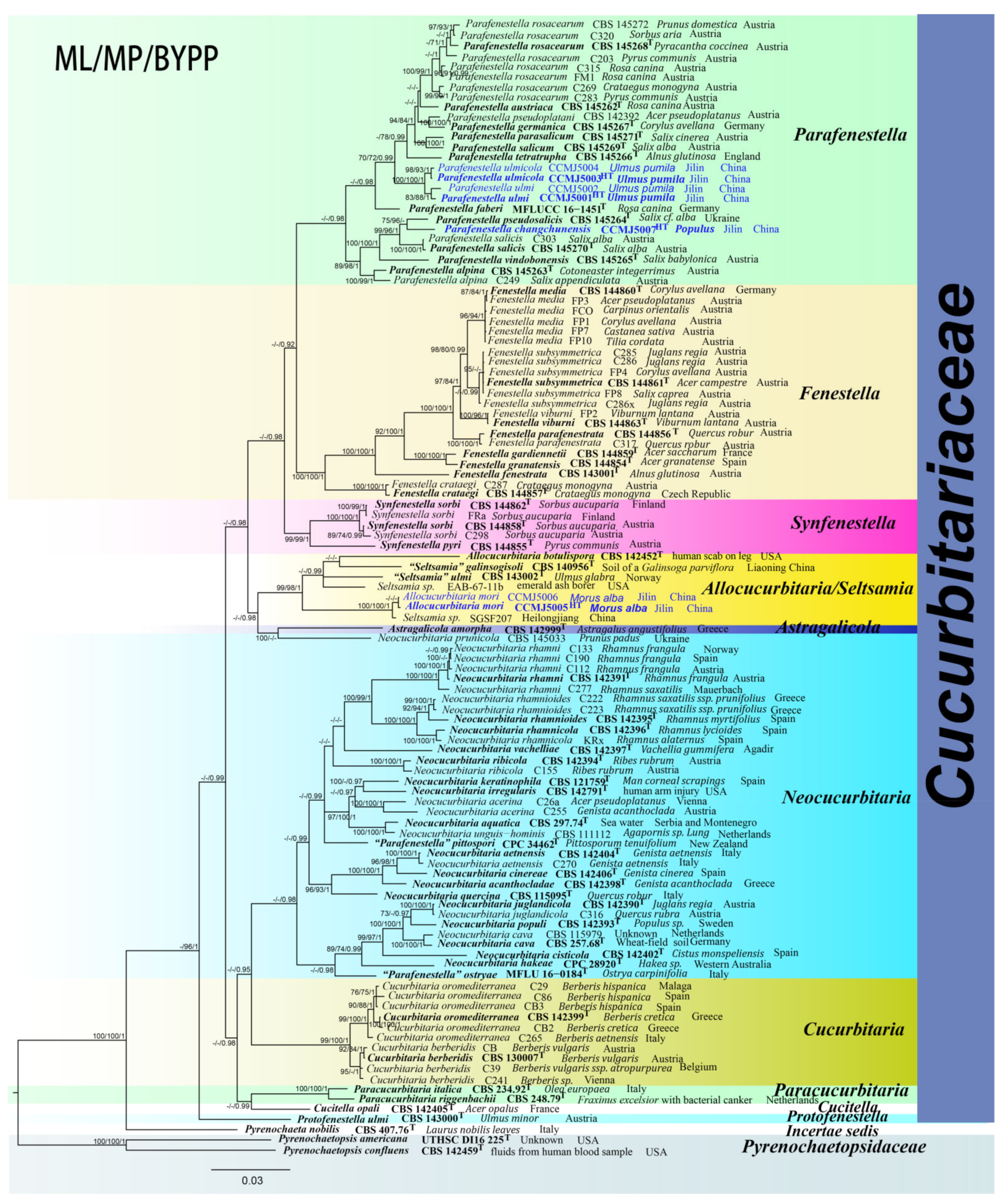
3.1. Phylogenetic Analyses

The final concatenated dataset comprised 110 ingroup taxa and two outgroup taxa, with 4607 characters including gaps (651 bases for ITS, 911 bases for LSU, 1063 bases for rpb2, 1281 bases for tef1, and 701 bases for β-tub). The RAxML analysis yielded a best-scoring tree with a final ML optimization likelihood value of −39123.587750. The matrix consisted of 1740 distinct alignment patterns, with 25.90% undetermined characters or gaps. Estimated base frequencies were as follows: A = 0.234707, C = 0.269983, G = 0.265086, T = 0.230223; substitution rates AC = 1.287870, AG = 4.563896, AT = 1.434736, CG = 1.144629, CT = 6.919700, GT = 1.000000; proportion of invariable sites I = 0.606319; gamma distribution shape parameter α = 0.967784. The maximum parsimony dataset consisted of 1230 parsimony-informative characters and 246 variable characters. The parsimony analysis yielded 256 most parsimonious trees out of 1000 (TL = 6467, CI = 0.368, RI = 0.806, RC = 0.296, HI = 0.632). In the BPP analysis, 2437 trees were sampled after the 20% burn-in with a stop value of 0.009904. The maximum parsimony dataset consisted of 3132 parsimony-informative characters and 241 variable characters. The parsimony analysis yielded 512 most parsimonious trees out of 1000 (TL = 6468, CI = 0.368, RI = 0.806, RC = 0.297, HI = 0.632). In the BPP analysis, 1461 trees were sampled after the 20% burn-in with a stop value of 0.009955. The phylogenetic trees generated from the ML, MP and BPP had similar topologies (Figures S6 and S7).
In the ML analysis of the ITS region, Parafenestella ulmi (CCMJ 5001 and CCMJ 5002) and P. ulmicola (CCMJ 5003 and CCMJ 5004) clustered together with high support (ML = 95%), while P. changchunensis (CCMJ 5007) formed a clade with P. vindobonensis (CBS 145256) with relatively low support (ML = 63%) in Parafenestella (Figure S1). Parafenestella ostryae (MFLU 16-0184) and P. pittospori (CPC 34462) resided in the Neocucurbitaria clade (Figure 1) similar to the combined dataset. Allocucurbitaria mori (CCMJ 5005 and CCMJ 5006) formed a clade with A. botulispora (CBS 142452), Seltsamia galinsogisoli (CBS 140956), S. ulmi (CBS 143002) and two unidentified Seltsamia species (EAB-67-11b and SGSF207) (ML = 100%). The LSU locus could not accurately distinguish taxa at the genus and species level in Cucurbitariaceae (Figure S2). In the ML analysis of β-tub gene, P. ulmi (CCMJ 5001 and CCMJ 5002) and P. ulmicola (CCMJ 5003 and CCMJ 5004) formed a clade with high support (ML = 94%), while P. changchunensis (CCMJ 5007) clustered with P. pseudosalicis (CBS 145264) with moderate support (ML = 71%). Allocucurbitaria mori (CCMJ 5005 and CCMJ 5006) and A. botulispora (CBS 142452) formed a clade with moderate support (ML = 54%, Figure S5). In the tef1-α analysis, Parafenestella ulmi (CCMJ 5001 and CCMJ 5002) and P. ulmicola (CCMJ 5003 and CCMJ 5004) formed a clade with relatively high support (ML = 89%) (Figure S4). Parafenestella changchunensis (HMJAU 60182) formed a clade with P. salicis (CBS 145270 and C303), P. pseudosalicis (CBS 145264), P. vindobonensis (CBS 145265) and P. alpina (CBS 145263 and C249) with relatively high support (ML = 79%). Allocucurbitaria mori (CCMJ 5005 and CCMJ 5006) clustered with Synfenestella pyri (CBS 144855) with low support (ML = 41%).
In the multi-locus phylogenetic analysis, Parafenestella ulmi (CCMJ 5001 and CCMJ 5002) and P. ulmicola (CCMJ 5003 and CCMJ 5004) formed a clade with high support (ML = 100%; MP = 100%; BPP = 1.00). Parafenestella changchunensis (CCMJ 5007) clustered with P. pseudosalicis (CBS 145264) and P. salicis (CBS 145270 and C303) with high support (ML = 99%; MP = 96%; BPP = 1.00). Parafenestella changchunensis (CCMJ 5007) is closely related to P. pseudosalicis (ML = 75%; MP = 96%). The fresh collections from Morus alba revealed a new species Allocucurbitaria mori (CCMJ 5005 and CCMJ 5006). The two isolates (CCMJ 5005 and CCMJ 5006) formed a close relationship to an unidentified Seltsamia species (SGSF207) with strong statistical support (ML = 100%; MP = 100%; BPP = 1.00).
Figure 1. The Bayesian 50% majority-rule consensus phylogram based on a concatenated ITS, LSU, rpb2, tef1 and β-tub dataset of Cucurbitariaceae. The tree is rooted with Pyrenochaetopsis americana (UTHSC DI16225) and P. confluens (CBS 142459). Bootstrap support values for maximum likelihood and maximum parsimony analysis greater than 70% (ML = left; MP = middle) and Bayesian posterior probabilities ≥ 0.90 (BPP, right) are shown at the nodes. The new species are indicated in blue. The type-derived strains are indicated in bold and marked with T.
Jof 08 00905 g001

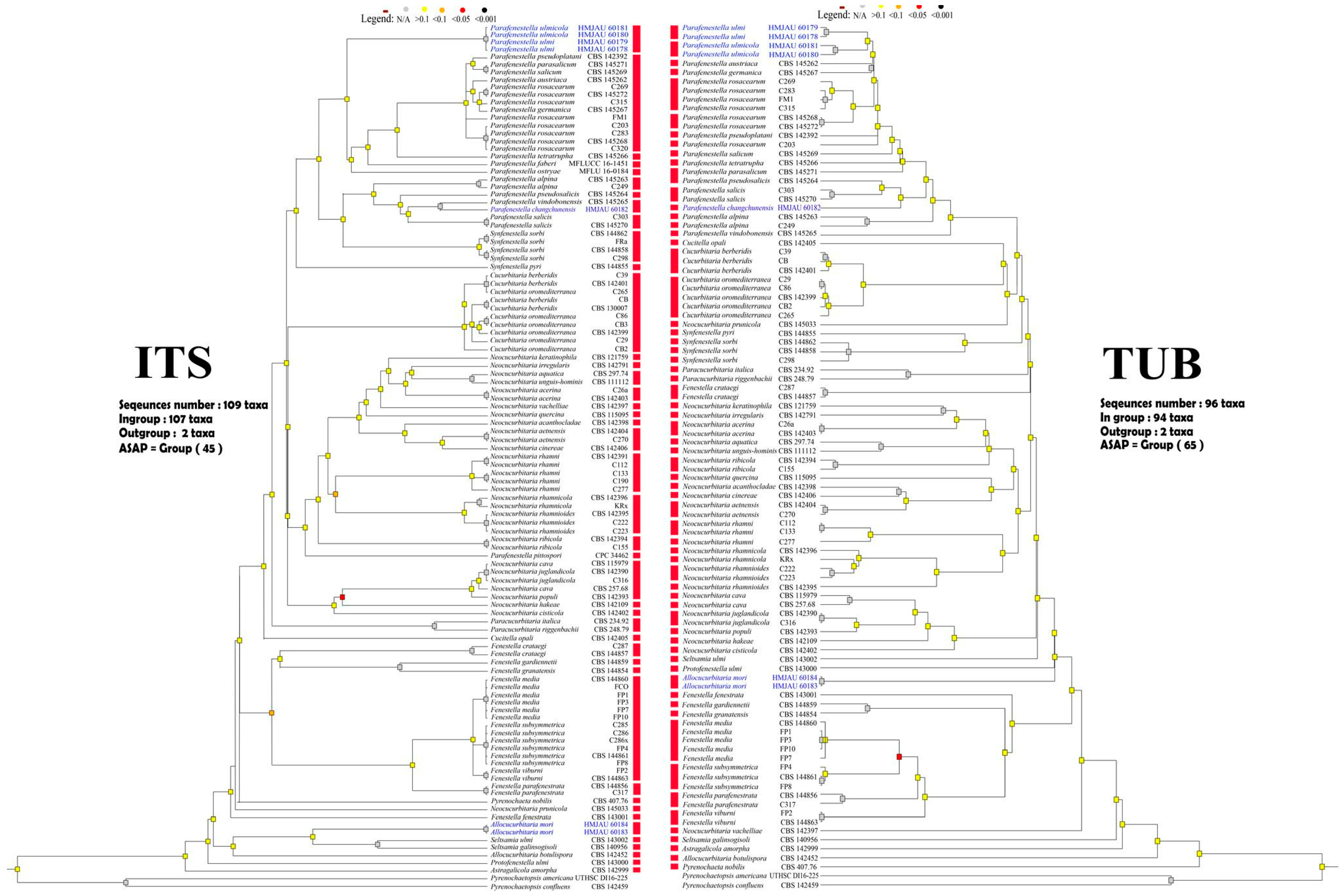
3.2. ASAP: Assemble Species by Automatic Partitioning
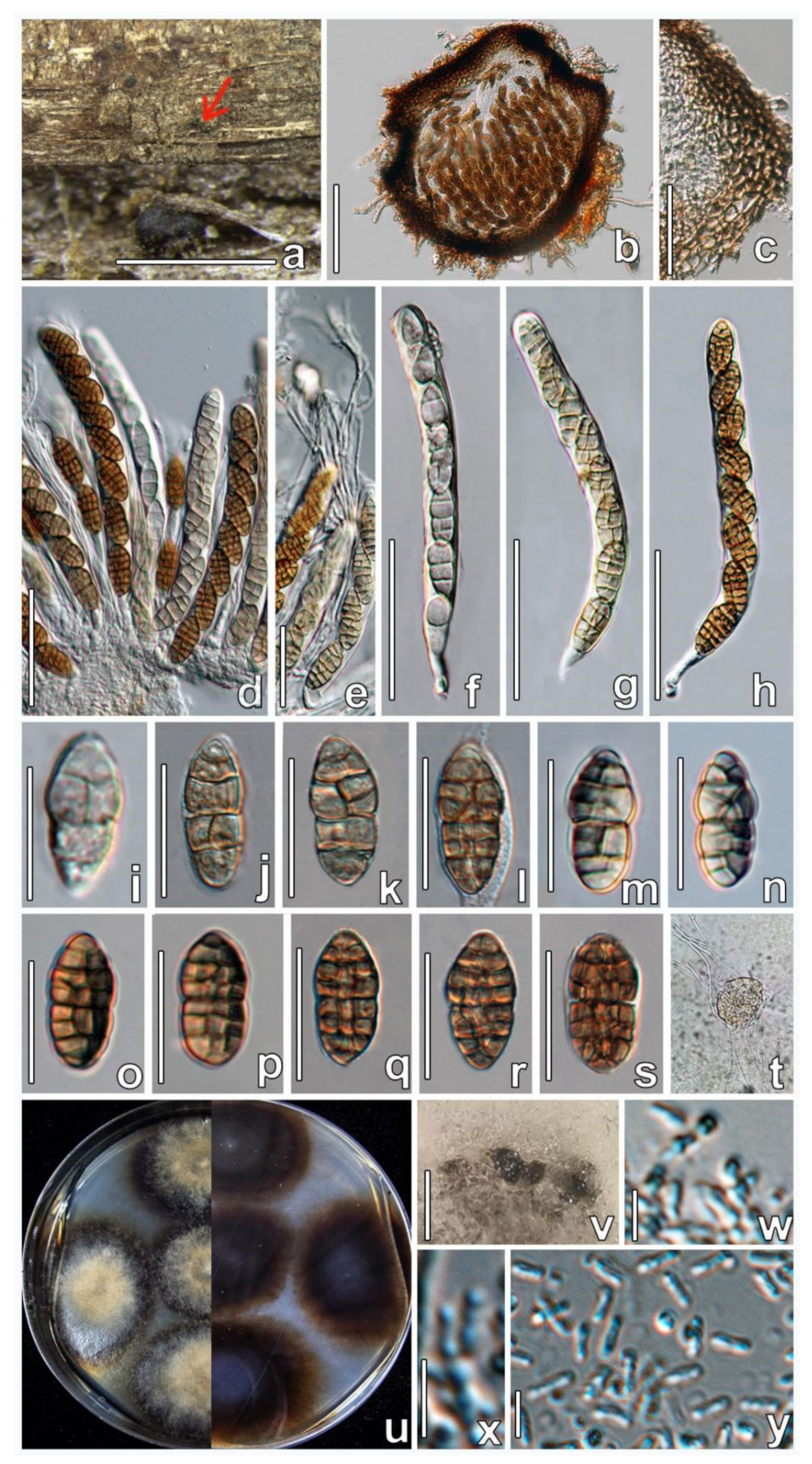
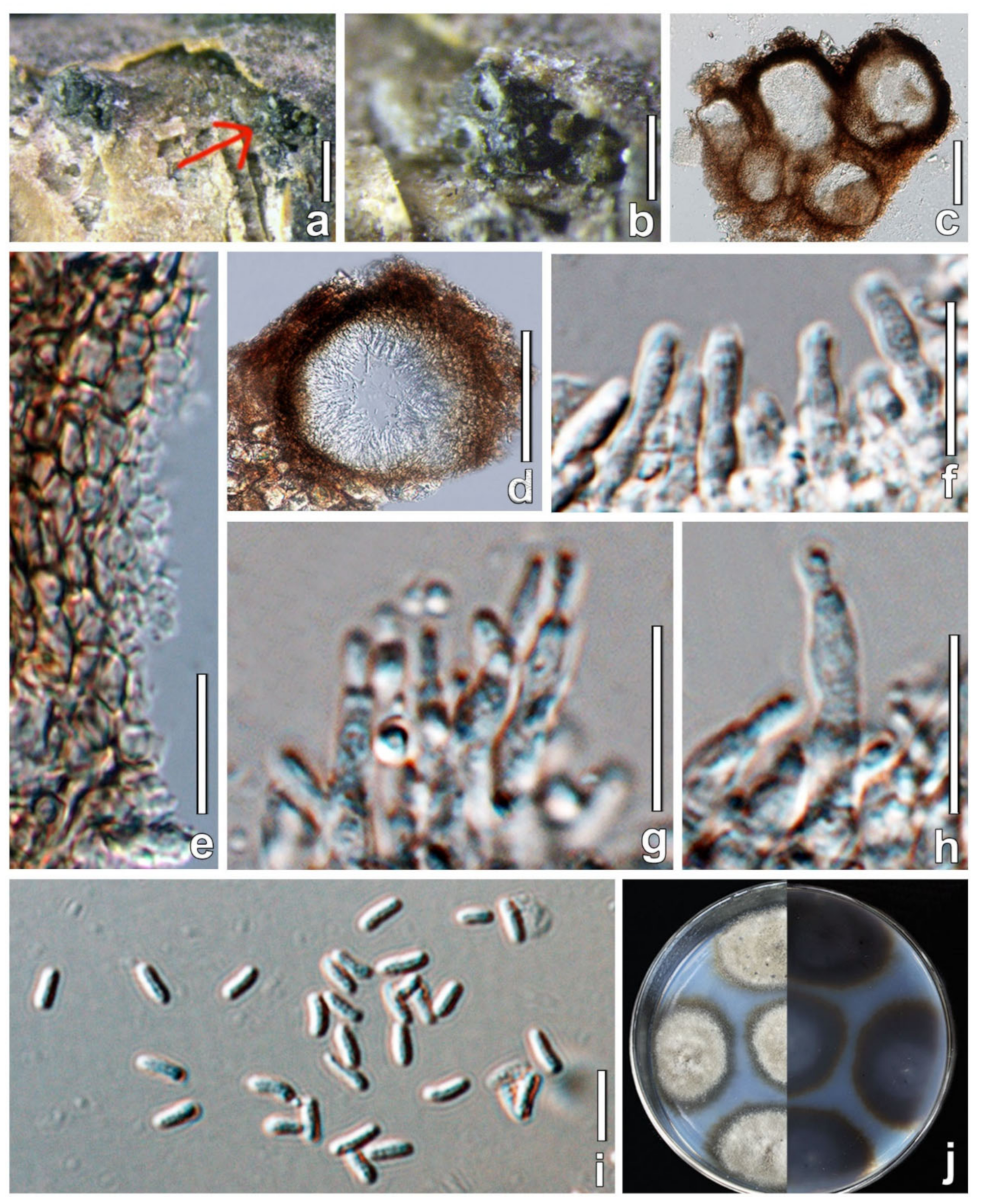
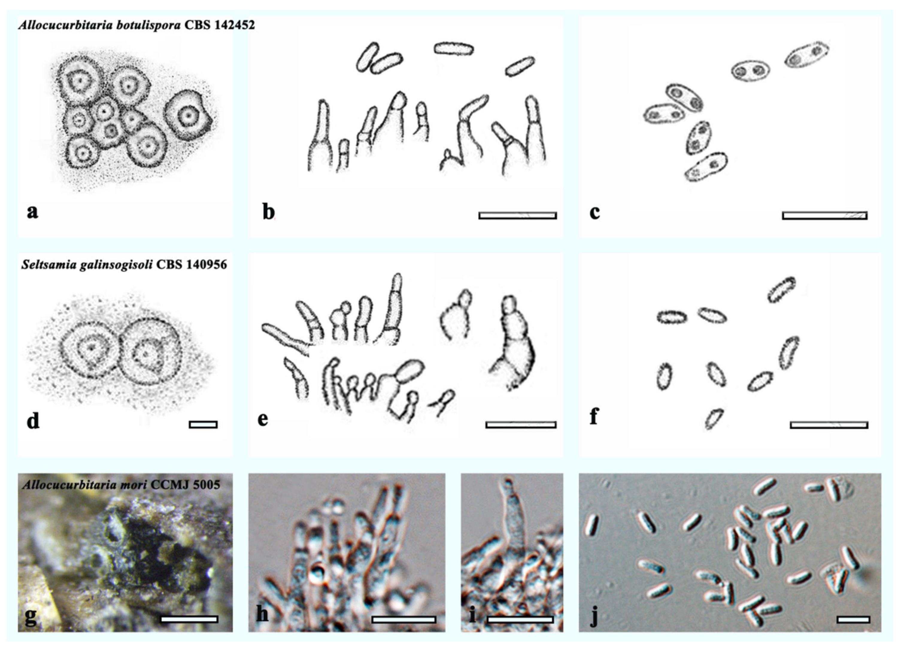
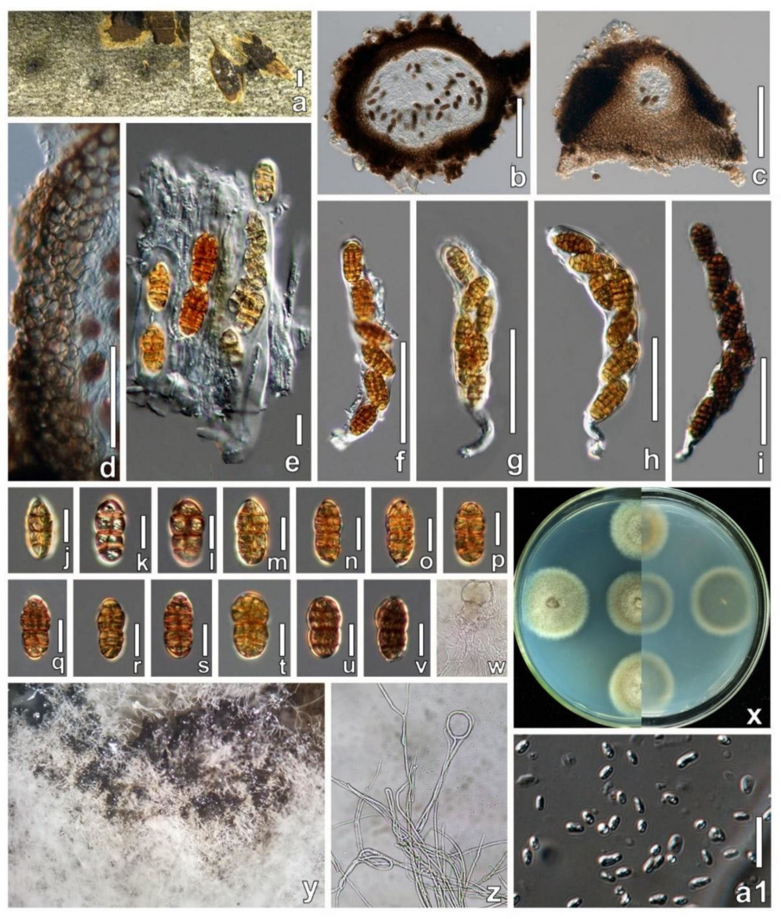
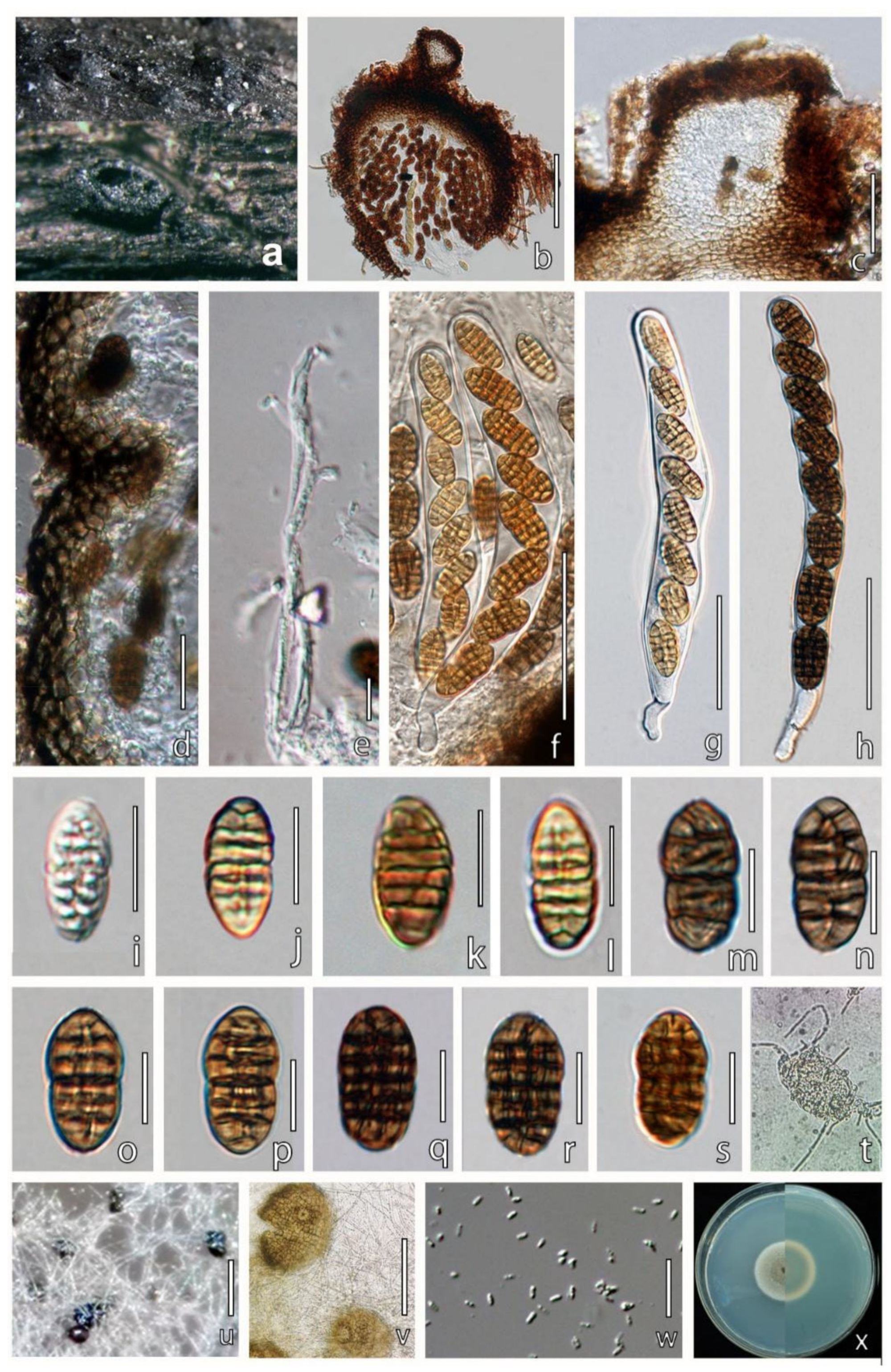
Five single-locus datasets were used that comprised 110 sequences of ITS, 109 sequences of LSU, 101 sequences of rpb2, 96 sequences of β-tub and 88 sequences of tef1. The ASAP analysis of the ITS region assigned all members of Cucurbitariaceae into 45 groups (Figure 2); β-tub gene into 65 groups (Figure 2); LSU into 43 groups (Figure S8); rpb2 gene into 65 groups (Figure S9); tef1 gene into 45 groups (Figure S10).
The ASAP analysis recovered P. ulmi (CCMJ 5001 and CCMJ 5002), P. ulmicola (CCMJ 5003 and CCMJ 5004) and twelve other strains including P. pseudoplatani (CBS 142392), P. austriaca (CBS 145262), P. rosacearum (C203, FM1, C269, C283, CBS 145268, C315, CBS145272, C320), P. germanica (CBS 145267) and P. tetratrupha (CBS 145266) as one group in the LSU data. Parafenestella changchunensis (CCMJ 5007) and P. pseudosalicis (CBS 145264) were recovered as one group in the LSU data. The ASAP analysis of the ITS region recovered P. ulmi and P. ulmicola as one group (Figure 2). The ASAP result of the β-tub gene was similar to the combined dataset (Figure 2). Parafenestella ulmi and P. ulmicola were not delineated by the tef1 and rpb2 genes (Figures S9 and S10). Parafenestella changchunensis, P. pseudosalicis (CBS 145264) and P. salicis (CBS 145270 and C303) were recovered as one group in the tef1 data. Allocucurbitaria mori (CCMJ 5005 and CCMJ 5006) grouped with Synfenestella pyri (CBS 144855) in the ASAP analysis of the tef1 gene, but both were recovered as individual groups in the ITS, LSU, rpb2, and β-tub datasets.
In the ASAP analysis, the β-tub gene was the best marker for identifying Parafenestella and Allocucurbitaria taxa. Parafenestella ulmi and P. ulmicola were recovered as a group in ASAP analysis of the ITS and other markers but were recovered as separate groups in the β-tub dataset (similar to the combined dataset). Parafenestella changchunensis and P. vindobonensis (CBS 145265) were recovered as a group in the ITS region but were recovered as distinct species in the β-tub dataset. Allocucurbitaria mori was recovered as an individual group in all single-marker analyses (except tef1 gene). Based on the current results, the β-tub gene is the best marker for the identification of Cucurbitariaceae taxa at the species level.

3.3. Taxonomy

Allocucurbitaria mori W.X. Su, Phukhams. & Y. Li, sp. nov. (Figure 3).
MycoBank Number: MB844413.
Etymology: Named after the host genus Morus.
Holotype: HMJAU 60183.
Description: Saprobic on dead twigs of Morus alba.
Sexual morph: Undetermined.
Asexual morph: Stromata poorly developed, multiloculate, with 5–8 locules forming groups in stromata, immersed. Conidiomata 108–180 × 103–201 μm ( x ¯ = 142 × 143 μm, n = 6), pycnidia, solitary or aggregated, sometimes confluent, semi-immerged, visible as black protrusions, globose to ellipsoid, coriaceous, black, without distinguishable ostioles. Pycnidial wall 5–9 μm wide, thick-walled, composed of 7–10 layers of thin-walled cells of textura angularis, dark brown on the outside to gradually lighter on the inside, inner layer subhyaline, lining layer bearing conidiogenous cells. Conidiophores reduced to conidiogenous cells. Conidiogenous cells 6–14 × 1–5 μm ( x ¯ = 10 × 2 μm, n = 30), enteroblastic, solitary, long cylindrical, arising from the inner layer of conidioma, smooth-walled, hyaline. Conidia 3–5 × 1–2 μm ( x ¯ = 4 × 1.5 μm, n = 50), oblong, hyaline, aseptate, with a minute guttule, smooth.
Cultural characters: Colonies on MEA reaching 32–38 mm diam after 4 weeks at 25 °C. Cultures from above, gray at the center, dense in the middle, sparse at the edge, circular, papillate, black lumps produced on the surface of cultures, white at the edge.
Material examined: CHINA, Jilin Province, Changchun, Jilin Agricultural University, from Morus alba (Moraceae) twigs, 20 May 2021, Wenxin Su and C. Phukhamsakda, S057 (HMJAU 60183, holotype); ex-type living culture, CCMJ5005; isotype = HMJAU 60184; ex-isotype living culture, CCMJ5006.
GenBank accession numbers: CCMJ5005: LSU = OL897171, ITS = OL996120, tef1 = OL944601, rpb2 = OL944505, and β-tub = OL898725. CCMJ5006: LSU = OL897172, ITS = OL996121, tef1-α = OL944602, rpb2 = OL944506 and β-tub = OL898720.
Notes: Allocucurbitaria mori (CCMJ5005 and CCMJ5006) formed a separate clade in Allocucurbitaria/Seltsamia with high support (ML = 98%; MP = 97%; BPP = 1.00). Morphologically, A. mori (HMJAU 60183) is similar to A. botulispora (CBS 142452) and S. galinsogisoli (CBS 140956) in having cylindrical, enteroblastic, solitary conidiogenous cells and aseptate conidia [41,42] (Figure 4). However, S. galinsogisoli (CBS 140956) has longer conidia, while A. botulispora (CBS 142452) has distinct guttulate at the conidia ends [41,42].
A BLASTn search of the ITS region of A. mori strain CCMJ 5005 showed a high query cover and similarity (99.80%) to an unidentified Seltsamia sp. (SGSF207) from soil. However, there are no other loci available in public databases for comparison. Hence, we introduce Allocucurbitaria mori as a novel species, and this is the first report of Allocucurbitaria on Morus tree [41,42,43].
Figure 3. Allocucurbitaria mori (HMJAU 60183, holotype) The red arrow indicates the conidiomata in face view. (a,b) Appearance of conidiomata on host substrate. (c,d) Vertical section of partial conidiomata. (e) Section of partial conidioma wall. (fh) Conidiogenous cells and conidia. (i) Conidia. (j) Culture characteristics on PDA. Scale bars: (a) = 500 µm; (b) = 200 µm; (c,f) = 100 µm; (d) = 50; (e,g,h) = 10 µm; (i) = 5 µm.
Jof 08 00905 g003
Figure 4. Morphology of related taxa in the Allocucurbitaria clade. (ac) Characters of Allocucurbitaria botulispora were redrawn from Valenzuela-Lopez et al. [41]: (a) Pycnidia. (b,c) Conidiogenous cells and conidia. (df) Characters of Seltsamia galinsogisoli redrawn from Zhang et al. [42]: (d) Pycnidia. (e,f) Conidiogenous cells and conidia. (gj) Characters of Allocucurbitaria mori (CCMJ 5005): (g) Appearance of conidiomata of Allocucurbitaria mori on host substrate. (hj) Conidiogenous cells and conidia. Scale bars: (b,c,e,f,h,i) = 10 µm; (d) = 20 µm; (g) = 200 µm; (j) = 5 µm.
Jof 08 00905 g004
Parafenestella changchunensis W. X. Su, Phukhams. & Y. Li, sp. nov. (Figure 5).
MycoBank Number: MB844412.
Etymology: referring to Changchun City where the sample was collected.
Holotype: HMJAU 60182.
Description: Saprobic on dead stems of Populus L.
Sexual morph: Ascomata 174–416 × 226–486 μm ( x ¯ = 280 × 353 μm, n = 5), single or gregarious, scattered, globose to depressed globose, submerged, visible as black dots and protruding host surface, solitary or aggregated. Ostioles 61 × 100 μm, center, protruding filled with periphyses. Peridium 12–27 μm wide, thick-walled, composed of 6–10 wall layers, outer part comprising dark brown cells of textura angularis, inner layer thin-walled, dark brown from the outside radiating light brown cells to hyaline towards the inside. Hamathecium of dense, 1.6–2.0 μm ( x ¯ = 1.7 μm, n = 10) wide, filamentous, septate, cellular pseudoparaphyses surrounding asci. Asci 95–138 × 16–21 μm ( x ¯ = 121 × 18 μm, n = 10), 6–8 ascospores, bitunicate, fissitunicate, broad cylindrical, some curved, short-pedicellate, apically rounded with an ocular chamber. Ascospores 18–25 × 8–13 μm ( x ¯ = 21 × 10 μm, n = 30), uniseriate, partially overlapping, fusiform to oval, slightly asymmetrical, the middle of ascospores is slightly contracted, with 4–6 transverse septa, 2–3 vertical septa, the upper part is slightly larger than the lower part, light yellow to dark brown.
Asexual morph: Pycnidia produced in PDA after 2 weeks of incubation in the dark, mycelium white. Conidiomata confluent or scattered, superficial, covered with dense vegetative hyphae, with turbid whitish drops, globose, black. Conidia 5–8 × 2.5–4.5 μm ( x ¯ = 6.5 × 3.7 μm, n = 30), oblong to allantoid, hyaline, aseptate, with 1–2 guttules.
Culture characteristics: Colonies on PDA, reaching 26–31 mm diam after 2 weeks at 25 °C. Culture from above, mycelium dense and producing hyphal coil structures; from the center to the outer edge, the color changes from grey to greyish-green to white, with obvious concentric wheel patterns, a clear radiation pattern at the back, round.
Material examined: CHINA, Jilin Province, Changchun, Jilin Agricultural University, from dead stems of Populus L. (Salicaceae), 18 April 2021, Wenxin Su, S12-16 (HMJAU 60182, holotype); ex-type living culture, CCMJ5007.
GenBank accession numbers: CCMJ5007: LSU = OL897170, SSU = OL891808, ITS = OL996119, tef1 = OL944600, and β-tub = OL898719.
Notes: In our phylogenetic analysis, P. changchunensis (CCMJ5007) is closely related to P. pseudosalicis (CBS 145264) with moderate support (ML = 75%; MP = 96 %; Figure 1). Parafenestella changchunensis is morphologically similar to P. pseudosalicis in having immersed, concave apex ascomata, with the upper part of young ascospores often wider, ends concolorous and smooth walled [14]. The immature spores of P. changchunensis have four horizontal septa and form 2–3 vertical septa during the maturation process. However, the immature spores of P. pseudosalicis have 2 transverse septa turning into 2–4 longitudinal septa during the maturation process [15]. Parafenestella changchunensis mycelium nodules gradually form fruiting bodies on the medium, while there are no reports of the asexual morph of P. pseudosalicis [15].
A BLASTn search of the ITS region of P. changchunensis (CCMJ 5007) showed a high similarity and query cover (98.81%) to P. vindobonensis (CBS 145265). The β-tub sequence of P. changchunensis (CCMJ 5007) showed a high query cover and similarity (96.82%) to P. pseudosalicis (C301). There were 0.96% (6/627 bases), 0.34% (3/885), 1.78% (13/730) and 7.99% (43/538 bases) base differences in the ITS, LSU, tef1-α and β-tub genes between P. changchunensis (CCMJ 5007) and P. vindobonensis (CBS 145265), excluding gaps. There were 1.75% (11/627 bases), 0.11% (1/885), 1.10% (8/730) and 3.16% (17/538 bases) base differences in the ITS, LSU, tef1-α and β-tub genes of P. changchunensis (CCMJ 5007) and P. pseudosalicis strain C301, excluding gaps. Therefore, we introduce P. changchunensis as a novel species, and this is the first report of Parafenestella on the Populus tree [14,15].
Figure 5. Parafenestella changchunensis (HMJAU 60182, holotype). (a) Ascomata on host surface. (b) Vertical section through partial ascoma. (c) Ostioles. (d) Partial peridium. (e) Pseudoparaphyses. (fi) Asci. (jv) Development stages of ascospores. (w) Germinating ascospore (x) Culture characteristics on PDA. (y) Pycnidia. (z) Hyphal coil structures formed by mycelia. (a1) Conidia. Scale bars: (a) = 500 µm; (b,c) = 100 µm; (d,e) = 20 µm; (fi) = 50 µm; (jv,a1) = 10 µm.
Jof 08 00905 g005
Parafenestella ulmi W.X. Su, Phukhams., & Y. Li, sp. nov. (Figure 6).
MycoBank Number: MB844410.
Etymology: Named after the host genus Ulmus.
Holotype: HMJAU 60178.
Description: Saprobic on dead stems of Ulmus pumila.
Sexual morph: Ascomata 170–225 × 194–260 μm ( x ¯ = 201 × 229 μm, n = 5), immersed, visible as black spots or having a convex surface, solitary, scattered, globose to ellipsoid, flat at the base, coriaceous, black. Peridium 19–39 μm wide, composed of 6–10 layers, outer part comprising dark brown cells of textura angularis, inner layer comprising thin-walled, light brown cells of textura angularis. Hamathecium of dense, 1.5–4.5 μm wide ( x ¯ = 2.2 μm, n = 20), filamentous, septate, pseudoparaphyses surrounding asci. Asci 115–181 × 11–15 μm ( x ¯ = 132 × 13 μm, n = 20), 8 ascospores, bitunicate, cylindrical, mostly curved, short-pedicellate, apically rounded with an ocular chamber, clearly visible when immature. Ascospores 18–24 × 8–12 μm ( x ¯ = 22 × 10 μm, n = 30), uniseriate to partially overlapping, broadly ellipsoid, slightly pointed at both ends, 5–8 transversely septate, 1–2 vertically septate, mature spores constricted at the middle septum, slightly curved, initially hyaline, becoming yellowish to brown at maturity, the cell above median septum slightly wider, smooth-walled.
Asexual morph: Pycnidia produced in PDA after 2 weeks of incubation in the dark, mycelium greenish, 1–3 μm ( x ¯ = 2.2 μm, n = 20), uniloculate, confluent or scattered, superficial, covered with dense vegetative hyphae, globose, dark brown to black. Conidiogenous cells 18–24 × 8–12 μm ( x ¯ = 22 × 10 μm, n = 30), enteroblastic, phialidic, determinate, discrete, solitary, short cylindrical or conical, straight, with broad base, hyaline. Conidia 3–5 × 1–2 μm ( x ¯ = 4.3 × 1.5 μm, n = 30), long ellipsoid to cylindrical, aseptate, with two small guttulate at the polar ends, hyaline, smooth-walled.
Culture characteristics: Colonies on PDA, reaching 45–48 mm diam after two weeks at 25 °C. Culture from above the center to the outer edge, the color radiating from black to dark green to yellow and white edges, with obvious concentric wheel patterns, dense intermediate hyphae and sparse white mycelium at the outer circle; reverse greenish-black, round.
Material examined: CHINA, Jilin Province, Changchun, Jilin Agricultural University, from Ulmus pumila (Ulmaceae) stem litter, 15 March 2021, Wenxin Su and C. Phukhamsakda, S12 (HMJAU 60178, holotype); ex-type living culture, CCMJ 5001, isotype = HMJAU 60179; ex-isotype living culture, CCMJ 5002.
GenBank accession numbers: CCMJ5001: LSU = OL897166, SSU = OL891806, ITS = OL996115, tef1-α = OL944596, rpb2 = OL944501, and β-tub = OL898723. CCMJ5002: LSU = OL897167, ITS = OL996116, tef1-α = OL944597, rpb2 = OL944502, and β-tub = OL898717.
Notes: In our phylogenetic analysis, P. ulmi (CCMJ 5001 and CCMJ 5002) and P. ulmicola (CCMJ 5003 and CCMJ 5004) formed a clade in Parafenestella with high statistical support (ML = 100%; MP = 100%; BPP = 1.00; Figure 1). Both P. ulmi and P. ulmicola were found on dead branches of Ulmus pumila in Jilin Province, China, which lies in the temperate zone. Parafenestella taxa are mainly recorded in Austria, followed by England, Germany and Ukraine, which are all temperate countries [15]. Morphologically, the ascomata of P. ulmi and P. ulmicola are semi-immersed, visible as black spots or convex surfaces. The asci of P. ulmi are longer than P. ulmicola but similar in width (132 × 13 vs. 119 × 13 μm). The immature ascospores of P. ulmi present 2–3 transverse septa without longitudinal septate, but the spores have 4–8 transverse septa with 1–3 longitudinal septate at mature stages. The ascospores of P. ulmicola showed indentation when immature that disappeared during maturation. The ascospores of P. ulmicola showed 5–8 transverse septa and 1–2 vertically septate after maturity with less constriction at the septum. The ascospores of P. ulmi are yellowish to brown, while P. ulmicola have dark brown ascospores at maturity. In PDA, the colonies of P. ulmicola have wavy and aggregated colony edges. The colonies of P. ulmi are blue-black (reverse view) with black-green edges, while P. ulmicola is gray-brown with white edges.
A BLASTn search of the ITS region of P. ulmi strain CCMJ 5001 showed a high query cover and similarity (96.45%) to P. tetratrupha (CBS 145266) while the β-tub sequence of P. ulmi strain CCMJ 5001 showed a high similarity and query cover (97.07%) to P. germanica strain C307. Therefore, we introduce P. ulmi as a novel species.
Figure 6. Parafenestella ulmi (HMJAU 60178, holotype). (a) Ascomata on host surface. (b) Vertical section through ascoma. (c) Partial peridium in vertical section. (d,e) Asci arrangement along with pseudoparaphyses. (fh) Development stages of asci. (is) Development stages of ascospores. (t) Germinating ascospore. (u) Four-week-old culture characteristics on PDA. (v) Pycnidia formed in sterile culture after two weeks of incubation on PDA. (w,x) Conidiogenous cells and conidia. (y) Conidia. Scale bars: (a) = 500 µm; (b) = 100 µm; (ch) = 50 µm; (is) = 20 µm; (v) = 200 µm; (wy) = 5 µm.
Jof 08 00905 g006
Parafenestella ulmicola W.X. Su, Phukhams., & Y. Li, sp. nov. (Figure 7).
MycoBank Number: MB844411.
Etymology: Named after the host genus Ulmus.
Holotype: HMJAU 60180.
Description: Saprobic on twigs debris of Ulmus pumila L.
Sexual morph: Ascomata 242–434 × 310–462 μm ( x ¯ = 306 × 359 μm, n = 5) μm wide, semi-immersed, visible as a convex hemisphere, globose to subglobose, solitary or mostly aggregated, scattered, coarse-walled, coriaceous, black, with a papilla. Ostiole 21 × 24 μm, centrally located. Peridium 21–68 μm wide, composed of 11–20 wall layers, with dark brown cells of textura angularis. Asci 105–153 × 11–14 μm ( x ¯ = 119 × 13 μm, n = 20), 8 ascospores, bitunicate, fissitunicate, broadly cylindrical, apically rounded, some curved, short-pedicellate, ocular chamber is not visible at maturity. Ascospores 17–22 × 8–12 μm ( x ¯ = 19 × 9 μm, n = 30), uniseriate, rarely overlapping, broadly oval, blunt at both ends, narrow towards the ends, with 4–8 transversely septate, 1–3 vertically septate, constricted at the middle septum, initially hyaline, becoming yellowish to brown at maturity, smooth-walled.
Asexual morph: Pycnidia produced in cultures on PDA after four weeks of incubation in the dark, mycelium greenish, 41–158 μm diam, covered with white mycelium, ellipsoid, semi-immersed, scattered or aggregated, black, ostiole central. Peridium with brown cells of textura angularis. Conidia 1.4–2.5 × 0.6–0.9 μm ( x ¯ = 1.9 × 0.7 μm, n = 30), cylindrical to allantoid, hyaline, smooth, aseptate, with a minute guttulate.
Culture characteristics: Colonies on PDA reaching 35–41 mm diam after 2 weeks at 25 °C. Culture from above the center to the outer edge, the color changes from grey to taupe to white, with obvious concentric wheel patterns; a few weeks later, the outer circle hyphae grow into round dark green hyphae with a thin surface.
Material examined: CHINA, Jilin Province, Changchun, Jilin Agricultural University, from Ulmus pumila (Ulmaceae) twigs debris, 15 March 2021, Wenxin Su and C. Phukhamsakda, S16 (HMJAU 60180, holotype); ex-type living culture, CCMJ 5003, isotype = HMJAU 60181; ex-isotype living culture, CCMJ 5004.
GenBank accession numbers: CCMJ5003: LSU = OL897168, SSU = OL891807, ITS = OL946117, tef1-α = OL944598, rpb2 = OL944503 and β-tub = OL898724. CCMJ5004: LSU = OL897169, ITS = OL996118, tef1-α = OL944599, rpb2 = OL944504 and β-tub = OL898719.
Notes: Sixteen Parafenestella species are listed in Species Fungorum [44], of which six species were reported on Rosaceae, four on Salicaceae and three on Betulaceae, while one species was reported on Pittosporaceae, Salicaceae and Sapindaceae [14,15,45,46]. Parafenestella ulmicola (CCMJ 5003 and CCMJ 5004) is closely related to P. ulmi (CCMJ 5001 and CCMJ 5002) within Parafenestella (ML = 100%; MP = 100%; BPP = 1.00, Figure 1). There were 2.31% (12/518) base differences in the β-tub, 0.14% (1/733) base differences in the tef1-α and 0.27% (2/736) base differences in the rpb2 gene between P. ulmicola (CCMJ 5003 and CCMJ 5004) and P. ulmi (CCMJ 5001 and CCMJ 5002), excluding gaps. There were no base differences in the ITS and LSU sequences.
Figure 7. Parafenestella ulmicola (HMJAU 60180, holotype). (a) Ascomata on host surface. (b) Vertical section through ascoma. (c) Ostiole. (d) Partial peridium wall. (e) Pseudoparaphyses. (fh) Asci. (is) Developmental stages of ascospores. (t) Germinating ascospore. (u) Pycnidia produced in four weeks old cultures on PDA. (v) Conidiomata. (w) Conidia. (x) Four weeks old culture on PDA. Scale bars: (b) = 100 µm; (c) = 50 µm; (d,e) = 20 µm; (fh) = 50 µm; (is) = 10 µm; (u) = 200 µm; (v) = 100 µm; (w) = 5 µm.
Jof 08 00905 g007
Parafenestella ulmi and P. ulmicola are phylogenetically close to P. tetratrupha but differ from P. tetratrupha by having a less longitudinal septa being visible at the surface [20]. Parafenestella tetratrupha ascospores are ellipsoid, yellow-brown to reddish-brown to dark brown, with 1–3 main septa, 8–17 distinct transverse and 2–4 longitudinal septa; they are darker and longer than P. ulmi and P. ulmicola (26.5–33.5 × 13–16.5 vs. 18–24 × 8–12 vs. 17–22 × 8–12 µm) and have more transverse septa than P. ulmi and P. ulmicola (Table 2). In the multi-locus phylogenetic analysis, although P. rosacearum was divided into six groups (Figure 1), it was still identified as one species because the tef1-α sequences of C203, C283 and C309 are almost the same. The rpb2 sequences of strains C203, C315, FM1 and FP11 are identical, while C269 and C283 differ from C203, C283 and C309 by 20 nucleotides [15]. In the phylogenetic analysis, P. germanica and P. pseudoplatani clustered in the same clade as P. parasalicum and P. salicum. These strains were identified as different species due to morphological distinctiveness [15]. The ascospores of P. germanica were larger than P. pseudoplatani (29–39.5 × 13–16.5 vs. 25–29 × 12–14 µm). The ascospores of P. parasalicum were larger than P. salicum (36–44 × 15.8–19.3 vs. 27–33 × 12.5–16 µm) (Table 3). There were 0.40% (2/494) base differences in the ITS, 2.28% (16/701) base differences in β-tub, 0.51% (4/789) base differences in tef1-α and 1.41% (15/1063) base differences in rpb2 between P. germanica and P. pseudoplatani. There were 3.42% (24/701) base differences in β-tub, 1.90% (15/789) base differences in tef1-α and 1.32% (14/1063) base differences in rpb2 between P. parasalicum and P. salicum. Thus, the species boundaries of P.ulmi and P. ulmicola were justified based on their distinct morphological traits and nucleotides differences. Therefore, we introduce P. ulmicola as a novel species, and this is first report of Parafenestella on Ulmus trees.

4. Discussion

The family Cucurbitariaceae was introduced by Winter [12] and typified by Cucurbitaria berberidis (Pers.) Gray [46]. Members of this family occur worldwide and are commonly recorded in Austria, Germany, England and Ukraine as saprobic or necrotrophic on various substrates including plant debris, soil and wood [14,15,47]. Although ribosomal markers and the ITS region are important for phylogenetic analyses, other loci are often needed for better resolution at the species level [48,49,50,51]. The ITS region can have low support values on key evolutionary nodes and cannot be used to accurately classify species in most genera [52,53]. Housekeeping genes and protein-coding genes such as act, β-tub, cal, gapdh, rpb2 and tef1 are thus usually recommended for a stable and reliable topology in phylogenetic analyses [54,55,56].
In this study, ASAP [38] was used to determine the most informative loci for Parafenestella. The β-tub gene provided the best species level identification of Parafenestella, followed by rpb2, tef1-α, ITS and LSU based on ASAP analyses (Figure 2, Figures S8–S11). ASAP analyses based on the β-tub gene provided the best resolution of P. ulmi and P. ulmicola, in addition to P. changchunensis, P. pseudosalicis and P. salicis (Figure 2). The ITS region is an important marker; however, it could not delineate between P. pseudoplatani (CBS 142392), P. parasalicum (CBS 145271), P. salicum (CBS 145269), P. austriaca (CBS 145262), P. germanicola (CBS 145267) and P. rosacearum (C203, C269, C283, C315, C320, CBS 145272, CBS 145268, FM1) as they were recovered as a group in ASAP analysis. In the ASAP analysis of the β-tub gene, this clade was divided into seven groups: (1) P. austriaca (CBS 145262), (2) P. germanicola (CBS 145267), (3) P. rosacearum (C269, C283, C315, FM1), (4) P. rosacearum (CBS 145272, CBS 145268) and P. rosacearum (C203), (5) P. pseudoplatani (CBS 142392), (6) P. parasalicum (CBS 145271) and (7) P. salicum (CBS 145269) (Figure 2). The β-tub gene exists in all eukaryotes and is involved in the formation of the spindle during cell division [57]. β-tubulin plays an important role in defining the characteristics of species [58]. The ASAP analysis of the β-tub gene likely reflects the interspecific relationship within Parafenestella. Thus, we encourage the inclusion of β-tub in the phylogenetic studies of Parafenestella species. This result is also supported by the phylogeny of single genes, two loci datasets (ITS + β-tub, Figure S14); ITS + rpb2, Figure S12); ITS + tef1-α, Figure S13) and multi-loci dataset (Figures S7 and S11).
Valenzuela-Lopez et al. [58] established Allocucurbitaria in Cucurbitariaceae based on morphological and phylogenetic analysis. Allocucurbitaria botulispora (CBS 142452) was classified as Pyrenochaeta species [43]. Valenzuela-Lopez et al. [41] examined the morphology of Pyrenochaeta and suggested that A. botulispora was more similar to phoma-like taxa. As it clustered in Cucurbitariaceae, the authors classified the species under the genus Allocucurbitaria within Cucurbitariaceae [41]. Seltsamia was introduced with the unique characteristics of pleomassaria-like fungus [14]. There is no confirmed report of the holomorph character of the type species (S. ulmi), and thus the generic status is constrained. Three species of Allocucurbitaria are listed in Species Fungorum [44], with one species reported on Ulmus glabra in Norway, one species from soil in China and one species reported from diseased human scab in the USA [41,59]. Notably, the Allocucurbitaria strains can be saprophyte and can harbor soil and/or opportunistic fungal disease in humans [41,42,43]. We provide the first report of Allocucurbitaria on dead twigs of Populus morus.
Parafenestella is the fourth most speciose genera in Cucurbitariaceae (Cucurbitaria 94 species; Fenestella 28 species; Neocucurbitaria 21 species; Parafenestella 14 species; Syncarpella 7 species; Rhytidiella 4 species; Allocucurbitaria 2 species; Astragalicola 2 species; Paracucurbitaria 2 species; Synfenestella 2 species; Cucitella 1 species; Protofenestella 1 species; Seltsamia 1 species) [44]. Parafenestella species are commonly distributed over temperate areas including northeast China but are rarely found in the tropical regions [11,13]. All three novel species in this study were collected during early spring in Changchun, Jilin Province, China. Jilin Province (40°52′~46°18′ N) belongs to a temperate continental climate, and the study of similar vegetation from similar climates is likely to result in many Parafenestella taxa [60]. We speculate that extensive investigations in the temperate regions would result in numerous Parafenestella members. Climate conditions also affect the infection degree of Cucurbitariaceae fungi to hosts, as temperatures below 0 °C may stop fungal development [15]. The age of the host including branch size and thickness may also affect the development of Cucurbitariaceae [15].
Parafenestella is characterized by immersed to erumpent and aggregated or clusters of ascomata [15]. The number of ascomata in Parafenestella (as a cluster) is often less than 10, which is higher than in Fenestella and Synfenestella [14,15]. Parafenestella does not form distinct pseudostromata, while Fenestella forms a pustular pseudostroma appearing as bumps, and Synfenestella forms conspicuous pseudostromatic pustules on pseudostromata [15]. The ascospores of Parafenestella are irregularly arranged and partially overlapping, while the ascospores of Fenestella and Synfenestella are borne in a uniseriate arrangement [14,15]. The sexual morph of Cucurbitariaceae is usually found on the wood and bark of trees and shrubs (Corylus avellana, Prunus domestica, Rosa canina, Sorbus aucuparia) [15]. The asexual morph of Parafenestella has not been reported from the natural host and is successfully produced only in culture [14,15]. However, pycnidia in artificial culture often lack conidiophores, which could be due to environmental conditions [61].

Supplementary Materials

The following are available online at https://www.mdpi.com/article/10.3390/jof8090905/s1, Figure S1: The best-scoring RAxML tree based on a concatenated ITS dataset. Figure S2: The best-scoring RAxML tree based on a concatenated LSU dataset. Figure S3: The best-scoring RAxML tree based on a concatenated rpb2 dataset. Figure S4: The best-scoring RAxML tree based on a concatenated tef1 dataset. Figure S5: The best-scoring RAxML tree based on a concatenated tub2 dataset. Figure S6. The best-scoring RAxML tree based on a concatenated ITS, LSU, rpb2, tef1 and tub2 dataset. Figure S7. Phylogram generated from maximum parsimony analysis based on combined ITS, LSU, rpb2, tef1 and tub2 dataset. Figure S8: Phylogram generated from ASAP analysis using LSU sequence data. Figure S9: Phylogram generated from ASAP analysis using rpb2 sequence data. Figure S10: Phylogram generated from ASAP analysis using tef1 sequence data. Figure S11: Phylogram generated from ASAP analysis using ITS, LSU, rpb2, tef1 and tub2 dataset. Figure S12: The best-scoring RAxML tree based on a concatenated ITS + rpb2 dataset. Figure S13: The best-scoring RAxML tree based on a concatenated ITS + tef1 dataset. Figure S14: The best-scoring RAxML tree based on a concatenated ITS + tub2 dataset.

Author Contributions

Conceptualization, Y.L. and C.P.; Writing—original draft and formal analysis, W.S.; Data curation, W.S., R.X., C.P. and C.S.B.; Investigation, W.S. and C.P.; Methodology, W.S., R.X., C.P., C.S.B., S.T. and Y.D.; Supervision, Y.L. and C.P.; Writing—review & editing, W.S., C.S.B. and C.P.; funding acquisition, Y.L. and C.P. All of the authors have read and approved the final draft. All authors have read and agreed to the published version of the manuscript.

Funding

This research was funded by the National Natural Science Foundation of China (NSFC) for granting a Youth Science Fund Project (number 32100007) and the Program of Creation and Utilization of Germplasm of Mushroom Crop of “111” Project (No. D17014).

Institutional Review Board Statement

Not applicable.

Informed Consent Statement

Not applicable.

Data Availability Statement

All sequences generated in this study were submitted to GenBank.

Acknowledgments

Chayanard Phukhamsakda (Postdoctoral number 271007) would like to thank Jilin Agricultural University. Chitrabhanu S. Bhunjun would like to thank the National Research Council of Thailand (NRCT) grant “Total fungal diversity in a given forest area with implications towards species numbers, chemical diversity and biotechnology” (grant no. N42A650547). The authors would like to thank Yong Ping Fu from Jilin Agricultural University, Changchun, China.

Conflicts of Interest

The authors declare no conflict of interest.

References

  1. Hawksworth, D.L. The magnitude of fungal diversity: The 1.5 million species estimate revisited. Mycological 2001, 105, 1422–1432. [Google Scholar] [CrossRef]
  2. Bhunjun, C.S.; Niskanen, T.; Suwannarach, N.; Wannathes, N.; Chen, Y.J.; McKenzie, E.H.; Maharachchikumbura, S.S.; Buyck, B.; Zhao, C.L.; Fan, Y.G.; et al. The numbers of fungi: Are the most speciose genera truly diverse? Fungal Divers. 2022, 27, 387–462. [Google Scholar] [CrossRef]
  3. Phukhamsakda, C.; Nilsson, R.H.; Bhunjun, C.S.; de Farias, A.R.; Sun, Y.R.; Wijesinghe, S.N.; Raza, M.; Bao, D.F.; Lu, L.; Tibpromma, S.; et al. The numbers of fungi: Contributions from traditional taxonomic studies and challenges of metabarcoding. Fungal Divers. 2022, 28, 327–386. [Google Scholar] [CrossRef]
  4. Wu, B.; Hussain, M.; Zhang, W.; Stadler, M.; Liu, X.; Xiang, M. Current insights into fungal species diversity and perspective on naming the environmental DNA sequences of fungi. Mycology 2019, 10, 127–140. [Google Scholar] [CrossRef]
  5. Liu, J.; Diamond, J. China’s environment in a globalizing world. Nature 2005, 435, 1179–1186. [Google Scholar] [CrossRef] [PubMed]
  6. Luo, Z.L.; Hyde, K.D.; Liu, J.K.; Bhat, D.J.; Bao, D.F.; Li, W.L.; Su, H.Y. Lignicolous freshwater fungi from China II: Novel Distoseptispora (Distoseptisporaceae) species from northwestern Yunnan Province and a suggested unified method for studying lignicolous freshwater fungi. Mycosphere 2018, 9, 444–461. [Google Scholar] [CrossRef]
  7. Zheng, H.; Wan, Y.K.; Li, J.; Rafael, F.C.R.; Yu, Z.F. Phialolunulospora vermispora (Chaetosphaeriaceae, Sordariomycetes), a novel asexual genus and species from freshwater in southern China. MycoKeys 2020, 76, 17. [Google Scholar] [CrossRef]
  8. Zhang, Z.K.; Wang, X.C.; Zhuang, W.Y.; Cheng, X.H.; Zhao, P. New species of Talaromyces (Fungi) isolated from soil in Southwestern China. Biology 2021, 10, 745. [Google Scholar] [CrossRef]
  9. Zheng, P. China’s Geography; China Intercontinental Press: Beijing, China, 2006. [Google Scholar]
  10. Zhang, X.; Wang, W.C.; Fang, X.Q.; Ye, Y. Vegetation of Northeast China during the late seventeenth to early twentieth century as revealed by historical documents. Reg. Environ. Change 2011, 11, 869–882. [Google Scholar] [CrossRef]
  11. Yuan, D.Y.; Zhu, L.J.; Cherubini, P.; Li, Z.S.; Zhang, Y.D.; Wang, X.C. Species-specific indication of 13 tree species growth on climate warming in temperate forest community of northeast China. Ecol. Indic. 2021, 133, 108389. [Google Scholar] [CrossRef]
  12. Winter, H.G. Pilze—Ascomyceten. In GL Rabenhorst’s Kryptogamen-Flora von Deutschland, Oesterreich und der Schweiz; Verlag von Eduard Kummer: Leipzig, Germany, 1885; Volume 1, pp. 65–528. [Google Scholar]
  13. Wijayawardenem, N.N.; Hyde, K.D.; Al-Ani, L.K.T.; Tedersoo, L.; Haelewaters, D.; Rajeshkumar, K.C.; Zhao, R.L.; Aptroot, A.; Leontyev, D.V.; Saxena, R.K.; et al. Outline of fungi and funguslike taxa. Mycosphere 2020, 11, 1060–1456. [Google Scholar] [CrossRef]
  14. Jaklitsch, W.M.; Checa, J.; Blanco, M.N.; Olariaga, I.; Tello, S.; Voglmayr, H. A preliminary account of the Cucurbitariaceae. Stud. Mycol. 2018, 90, 71–118. [Google Scholar] [CrossRef] [PubMed]
  15. Jaklitsch, W.M.; Voglmayr, H. Fenestelloid clades of the Cucurbitariaceae. Persoonia 2020, 44, 1–40. [Google Scholar] [CrossRef] [PubMed]
  16. Monkai, J.; Tibpromma, S.; Manowong, A.; Mapook, A.; Norphanphoun, C.; Hyde, K.D.; Promputtha, I. Discovery of three novel Cytospora species in Thailand and their antagonistic potential. Diversity 2021, 13, 488. [Google Scholar] [CrossRef]
  17. Index Fungorum. 2022. Available online: http://www.indexfungorum.org/names/names.asp (accessed on 11 April 2022).
  18. Jayasiri, S.C.; Hyde, K.D.; Ariyawansa, H.; Bhat, D.J.; Buyck, B.; Cai, L.; Dai, Y.C.; Abd-Elsalam, K.A.; Ertz, D.; Hidayat, I.; et al. The faces of fungi database: Fungal names linked with morphology, phylogeny and human impacts. Fungal Divers. 2015, 74, 3–18. [Google Scholar] [CrossRef]
  19. De Hoog, G.S.; Gerrits van den Ende, A.H.G. Molecular diagnostics of clinical strains of filamentous basidiomycetes. Mycoses 1998, 41, 183–189. [Google Scholar] [CrossRef]
  20. Senanayake, I.C.; Rathnayaka, A.R.; Marasinghe, D.S.; Calabon, M.S.; Gentekaki, E.; Lee, H.B.; Hurdeal, V.G.; Pem, D.; Dissanayake, L.S.; Wijesinghe, S.N.; et al. Morphological approaches in studying fungi: Collection, examination, isolation, sporulation and preservation. Mycosphere 2020, 11, 2678–2754. [Google Scholar] [CrossRef]
  21. White, T.J.; Bruns, T.D.; Lee, S.B.; Taylor, J.W. Amplification and direct sequencing of fungal ribosomal RNA genes for phylogenetics. PCR Protoc. Guid. Methods Appl. 1990, 18, 315–322. [Google Scholar]
  22. Vilgalys, R.; Hester, M. Rapid genetic identification and mapping of enzymatically amplified ribosomal DNA from several Cryptococcus species. J. Bacteriol. 1990, 172, 4238–4246. [Google Scholar] [CrossRef]
  23. Voglmayr, H.; Akulov, O.Y.; Jaklitsch, W.M. Reassessment of Allantonectria, phylogenetic position of Thyronectroidea, and Thyronectria caraganae sp. nov. Mycol. Prog. 2016, 15, 921. [Google Scholar] [CrossRef] [PubMed]
  24. Carbone, I.; Kohn, L.M. A method for designing primer sets for speciation studies in filamentous ascomycetes. Mycologia 1999, 91, 553–556. [Google Scholar] [CrossRef]
  25. Rehner, S.A.; Buckley, E. A Beauveria phylogeny inferred from nuclear ITS and EF1-α sequences: Evidence for cryptic diversification and links to Cordyceps teleomorphs. Mycologia 2005, 97, 84–98. [Google Scholar] [CrossRef]
  26. O’Donnell, K.; Cigelnik, E.; Weber, N.S.; James, M.T. Phylogenetic relationships among ascomycetous truffles and the true and false morels inferred from 18S and 28S ribosomal DNA sequence analysis. J. Mol. Evol. 1997, 89, 48–65. [Google Scholar] [CrossRef]
  27. Katoh, K.; Rozewicki, J.; Yamada, K.D. MAFFT online service: Multiple sequence alignment, interactive sequence choice and visualization. Brief. Bioinform. 2019, 20, 1160–1166. [Google Scholar] [CrossRef]
  28. Larsson, A. AliView: A fast and lightweight alignment viewer and editor for large datasets. Bioinformatics 2014, 30, 3276–3278. [Google Scholar] [CrossRef]
  29. Vaidya, G.; Lohman, D.J.; Meier, R. SequenceMatrix: Concatenation software for the fast assembly of multi-gene datasets with character set and codon information. Cladistics 2011, 27, 171–180. [Google Scholar] [CrossRef]
  30. Hall, M.A. Correlation-Based feature selection for machine learning. PhD Thesis, The University of Waikato, Hamilton, New Zealand, 1999. [Google Scholar]
  31. Stamatakis, A.; Hoover, P.; Rougemont, J. A rapid bootstrap algorithm for the RAxML web servers. Syst. Biol. 2008, 57, 758–771. [Google Scholar] [CrossRef]
  32. Stamatakis, A. RAxML version 8: A tool for phylogenetic analysis and post-analysis of large phylogenies. Bioinformatics 2014, 30, 1312–1313. [Google Scholar] [CrossRef]
  33. Stamatakis, A. RAxML-VI-HPC: Maximum likelihood-based phylogenetic analyses with thousands of taxa and mixed models. Bioinformatics 2006, 22, 2688–2690. [Google Scholar] [CrossRef]
  34. Darriba, D.; Taboada, G.L.; Doallo, R.; Posada, D. jModelTest 2: More models, new heuristics and parallel computing. Nat. Methods 2012, 9, 772. [Google Scholar] [CrossRef] [PubMed]
  35. Voglmayr, H.; Jaklitsch, W.M. Corynespora, Exosporium and Helminthosporium revisited—New species and generic reclassification. Stud. Mycol. 2017, 87, 43–76. [Google Scholar] [CrossRef] [PubMed]
  36. Ronquist, F.; Huelsenbeck, J.P. MrBayes 3: Bayesian phylogenetic inference under mixed models. Bioinformatics 2003, 19, 1572–1574. [Google Scholar] [CrossRef]
  37. Rambaut, A. FigTree v1.4, Tree Figure Drawing Tool. 2014. Available online: http.ac.uk/software/figtree (accessed on 8 July 2022).
  38. Puillandre, N.; Brouillet, S.; Achaz, G. ASAP: Assemble species by automatic partitioning. Mol Ecol Resour. 2021, 21, 609–620. [Google Scholar] [CrossRef] [PubMed]
  39. Sung, G.H.; Hywel-Jones, N.L.; Sung, J.M.; Luangsa-ard, J.J.; Shrestha, B.; Spatafora, J.W. Phylogenetic classification of Cordyceps and the clavicipitaceous fungi. Stud. Mycol. 2007, 57, 5–59. [Google Scholar] [CrossRef]
  40. Bhunjun, C.S.; Phukhamsakda, C.; Tibpromma, S.; McKenzie, E.H.C.; Bhat, D.J.; Li, Y.; Hyde, K.D. Microfungi associated with dead leaves of Dasymaschalon obtusipetalum, Garcinia propinqua and Mammea harmandii, with the description of 20 new and rare species. Mycosphere 2022, in press. [Google Scholar]
  41. Valenzuela-Lopez, N.; Cano-Lira, J.F.; Guarro, J.; Sutton, D.A.; Wiederhold, N.; Crous, P.W.; Stchigel, A.M. Coelomycetous Dothideomycetes with emphasis on the families Cucurbitariaceae and Didymellaceae. Stud. Mycol. 2018, 90, 1–69. [Google Scholar] [CrossRef]
  42. Zhang, T.Y.; Wu, Y.Y.; Zhang, M.Y.; Cheng, J.; Dube, B.; Yu, H.J.; Zhang, Y.X. New antimicrobial compounds produced by Seltsamia galinsogisoli sp. nov., isolated from Galinsoga parviflora as potential inhibitors of FtsZ. Sci. Rep. 2019, 9, 8319. [Google Scholar] [CrossRef]
  43. Valenzuela-Lopez, N.; Sutton, D.A.; Cano-Lira, J.F.; Paredes, K.; Wiederhold, N.; Guarro, J.; Stchigel, A.M. Coelomycetous fungi in the clinical setting: Morphological convergence and cryptic diversity. Mol. Phylogenet. Evol. 2017, 55, 552–567. [Google Scholar] [CrossRef]
  44. Species Fungorum. 2022. Available online: http://www.speciesfungorum.org/Names/Names.asp (accessed on 20 May 2022).
  45. Wanasinghe, D.N.; Phookamsak, R.; Jeewon, R.; Wen, J.L.; Hyde, K.D.; Jones, E.B.G.; Camporesi, E.; Promputtha, I. A family level rDNA based phylogeny of Cucurbitariaceae and Fenestellaceae with descriptions of new Fenestella species and Neocucurbitaria gen. nov. Mycosphere 2017, 8, 397–414. [Google Scholar] [CrossRef]
  46. Crous, P.W.; Wingfield, M.J.; Lombard, L.; Roets, F.; Swart, W.J.; Alvarado, P.; Carnegie, A.J.; Moreno, G.; Luangsa-ard, J.; Thangavel, R. Fungal Planet description sheets: 951–1041. Persoonia 2019, 43, 223. [Google Scholar] [CrossRef] [PubMed]
  47. Doilom, M.; Liu, J.K.; Jaklitsch, W.M.; Ariyawansa, H.; Wijayawardene, N.N.; Chukeatirote, E.; Zhang, M.; McKenzie, E.H.C.; Geml, J.; Voglmayr, H.; et al. An outline of the family Cucurbitariaceae. Sydowia 2013, 65, 167–192. [Google Scholar]
  48. Xu, R.; Su, W.X.; Tian, S.Q.; Bhunjun, C.S.; Tibpromma, S.; Hyde, K.D.; Li, Y.; Phukhamsakda, C. Synopsis of leptosphaeriaceae and introduction of three new taxa and one new record from China. J. Fungi 2022, 8, 416. [Google Scholar] [CrossRef]
  49. Bhunjun, C.S.; Dong, Y.; Jayawardena, R.S.; Jeewon, R.; Phukhamsakda, C.; Bundhun, D.; Hyde, K.D.; Sheng, J. A polyphasic approach to delineate species in Bipolaris. Fungal Divers. 2020, 102, 225–256. [Google Scholar] [CrossRef]
  50. Chethana, K.W.; Manawasinghe, I.S.; Hurdeal, V.G.; Bhunjun, C.S.; Appadoo, M.A.; Gentekaki, E.; Raspé, O.; Promputtha, I.; Hyde, K.D. What are fungal species and how to delineate them? Fungal Divers. 2021, 109, 1–25. [Google Scholar] [CrossRef]
  51. Pem, D.; Jeewon, R.; Chethana, K.W.T.; Hongsanan, S.; Doilom, M.; Suwannarach, N.; Hyde, K.D. Species concepts of Dothideomycetes: Classification, phylogenetic inconsistencies and taxonomic standardization. Fungal Divers. 2021, 109, 283–319. [Google Scholar] [CrossRef]
  52. Sharma, V.; Salwal, R. Molecular markers and their use in taxonomic characterization of Trichoderma spp. In Molecular Markers in Mycology; Singh, B.P., Gupta, V.K., Eds.; Springer: Cham, Switzerland, 2017; pp. 37–52. [Google Scholar]
  53. Bhunjun, C.S.; Phukhamsakda, C.; Jayawardena, R.S.; Jeewon, R.; Promputtha, I.; Hyde, K.D. Investigating species boundaries in Colletotrichum. Fungal Divers. 2021, 107, 107–127. [Google Scholar] [CrossRef]
  54. Hou, Z.Q.; Zhu, X.S.; Chen, Y.; Wu, Y.; Wang, Y.; Li, C.Y. Advances in research and application of DNA barcoding in plant pathogenic fungi. J. Agric. Sci. Technol. 2021, 49, 1247–1252. [Google Scholar]
  55. Niraikulam, A.; Hyungdon, Y.; Natarajan, S. Protein Coding Genes for Better Resolution of Phylogenetic Analysis. J. Biotechnol. 2010, 5, 74. [Google Scholar]
  56. Phukhamsakda, C.; McKenzie, E.H.C.; Phillips, A.J.L.; Jones, E.B.G.; Jayarama Bhat, D.J.; Stadler, M.; Bhunjun, C.S.; Wanasinghe, D.N.; Thongbai, B.; Camporesi, E.; et al. Microfungi associated with Clematis (Ranunculaceae) with an integrated approach to delimiting species boundaries. Fungal Divers. 2020, 102, 1–203. [Google Scholar] [CrossRef]
  57. Chen, H.R. Structural and Functional Relationship between JWA and α-Tubulin. Master’s Thesis, Nanjing Medical University, Nanjing, China, 2004. [Google Scholar]
  58. Samson, R.A.; Seifert, K.A.; Kuijpers, A.F.A.; Houbraken, J.A.M.P.; Frisvadet, J.C. Phylogenetic analysis of Penicillium subgenus Penicillium using partial β-tubulin sequences. Stud Mycol. 2004, 49, 175–200. [Google Scholar]
  59. Magaña-Dueñas, V.; Stchigel, A.M.; Cano-Lira, J.F. New Coelomycetous fungi from freshwater in Spain. J. Fungi 2021, 7, 368. [Google Scholar] [CrossRef] [PubMed]
  60. Chen, Y.L.; Xu, T.L.; Veresoglou, S.D.; Hu, H.W.; Hao, Z.P.; Hu, Y.J.; Liu, L.; Deng, Y.; Rillig, M.C.; Chen, B.D. Plant diversity represents the prevalent determinant of soil fungal community structure across temperate grasslands in northern China. Soil Biol. Biochem. 2017, 110, 12–21. [Google Scholar] [CrossRef]
  61. De Gruyter, J.; Woudenberg, J.H.C.; Aveskamp, M.M.; Verkley, G.J.M.; Groenewald, J.Z.; Crous, P.W. Systematic reappraisal of species in Phoma section Paraphoma, Pyrenochaeta and Pleurophoma. Mycologia 2010, 102, 1066–1081. [Google Scholar] [CrossRef] [PubMed]
Figure 2. Dendrogram from ASAP analysis based on two datasets (ITS and β-tub markers). The results of species delimitation are indicated by red bars. Sequences generated in this study are in blue.
Jof 08 00905 g002
Table 1. The PCR primers and amplifying conditions used in this study.
Amplification Loci
(Primer Pair Forward/Reverse)
PCR ConditionsReferences
ITS (ITS5/ITS4)An initial denaturation step of 5 min at 94 °C, followed by 35 cycles of 30 s at 94 °C, 30 s at 56 °C and 90 s at 72 °C, and a final extension step of 10 min at 72 °C, and 10 °C for holding temperatureWhite et al. [21]
rpb2 (fRPB2-5F/fRPB2-7cR)Vilgalys et al. [23]
tef1 (2218F/983R)Carbone and Kohn [24]
Rehner and Buckley [25]
LSU (LROR/LR5)An initial denaturation step of 5 min at 94 °C, followed by 35 cycles of 30 s at 94 °C, 45 s at 53 °C and 90 s at 72 °C, and a final extension step of 10 min at 72 °C, and 10 °C for holding temperatureVilgalys and Hester [22]
Β-tub (T1/Bt2b)O’Donnell and Cigelnik [26]
Table 2. The dataset used for phylogenetic analysis. The type-derived sequences are in bold.
TaxonStrainHost/SubstrateTypification
Status
GenBank Accession Numbers
ITSLSUrpb2tef1-αβ-tub
Allocucurbitaria botulisporaCBS 142452human scab on legHolotypeLT592932LN907416LT593070LT593001
Allocucurbitaria moriHMJAU 60183Morus albaHolotypeOL996120OL897171OL944505OL944601OL898725
Allocucurbitaria moriHMJAU 60184Morus albaIsotypeOL996121OL897172OL944506OL944602OL898720
Astragalicola amorphaCBS 142999Astragalus angustifoliusHolotypeMF795753MF795753MF795795MF795842MF795883
Cucitella opaliCBS 142405Acer opalusHolotypeMF795754MF795754MF795796MF795843MF795884
Cucurbitaria berberidisC39Berberis vulgaris subsp. atropurpureaMF795755MF795755MF795797MF795844MF795885
Cucurbitaria berberidisCBBerberis vulgarisMF795757MF795757MF795799MF795846MF795887
Cucurbitaria berberidisCBS 130007 = CB1Berberis vulgarisEpitypeMF795758MF795758MF795800
Cucurbitaria berberidisCBS 142401 = C241Berberis sp.MF795756MF795756MF795798MF795845MF795886
Cucurbitaria oromediterraneaC265Berberis aetnensisMF795762MF795762MF795804MF795850MF795891
Cucurbitaria oromediterraneaC29Berberis hispanicaMF795759MF795759MF795801MF795847MF795888
Cucurbitaria oromediterraneaC86Berberis hispanicaMF795760MF795760MF795802MF795848MF795889
Cucurbitaria oromediterraneaCB2Berberis creticaMF795763MF795763MF795805MF795851MF795892
Cucurbitaria oromediterraneaCB3Berberis hispanicaMF795764MF795764MF795806MF795852
Cucurbitaria oromediterraneaCBS 142399 = C229Berberis creticaHolotypeMF795761MF795761MF795803MF795849MF795890
Fenestella crataegiC287Crataegus monogynaMK356281MK356281MK357554MK357598
Fenestella crataegiCBS 144857 = C314Crataegus monogynaEpitypeMK356282MK356282MK357512MK357555MK357599
Fenestella fenestrataCBS 143001 = FP9Alnus glutinosaEpitypeMF795765MF795765MF795807MF795853MF795893
Fenestella gardiennetiiCBS 144859 = FMAcer saccharumHolotypeMK356283MK356283MK357513MK357556MK357600
Fenestella granatensisCBS 144854 = C279Acer granatenseHolotypeMK356284MK356284MK357514MK357557MK357601
Fenestella mediaCBS 144860 = FPCorylus avellanaEpitypeMK356285MK356285MK357515MK357558MK357602
Fenestella mediaFCOCarpinus orientalisMK356286MK356286MK357516MK357559
Fenestella mediaFP1Corylus avellanaMK356287MK356287MK357517MK357560MK357603
Fenestella mediaFP3Acer pseudoplatanusMK356288MK356288MK357518MK357561MK357604
Fenestella mediaFP7Castanea sativaMK356289MK356289MK357519MK357562MK357605
Fenestella mediaFP10Tilia cordataMK356290MK356290MK357520MK357563MK357606
Fenestella parafenestrataCBS 144856 = C306Quercus roburHolotypeMK356291MK356291MK357521MK357564MK357607
Fenestella parafenestrataC317Salix sp.MK356292MK356292MK357522MK357565MK357608
Fenestella subsymmetricaCBS 144861 = FP6Acer campestreHolotypeMK356297MK356297MK357525MK357569MK357610
Fenestella subsymmetricaC285Juglans regiaMK356293MK356293MK357523MK357566
Fenestella subsymmetricaC286Juglans regiaMK356294MK356294MK357567
Fenestella subsymmetricaC286xJuglans regiaMK356295MK356295
Fenestella subsymmetricaFP4Corylus avellanaMK356296MK356296MK357524MK357568MK357609
Fenestella subsymmetricaFP8Salix capreaMK356298MK356298MK357526MK357570MK357611
Fenestella viburniCBS 144863 = FVLViburnum lantanaHolotypeMK356300MK356300MK357528MK357572MK357613
Fenestella viburniFP2Viburnum lantanaMK356299MK356299MK357527MK357571MK357612
Neocucurbitaria acanthocladaeCBS 142398 = C225Genista acanthocladaHolotypeMF795766MF795766MF795808MF795854MF795894
Neocucurbitaria acerinaC26aAcer pseudoplatanusMF795767MF795767MF795809MF795855MF795895
Neocucurbitaria acerinaCBS 142403 = C255Acer pseudoplatanusMF795768MF795768MF795810MF795856MF795896
Neocucurbitaria aetnensisCBS 142404 = C261Genista aetnensisHolotypeMF795769MF795769MF795811MF795857MF795897
Neocucurbitaria aetnensisC270Genista aetnensisMF795770MF795770MF795812MF795858MF795898
Neocucurbitaria aquaticaCBS 297.74Sea waterHolotypeLT623221EU754177LT623278LT623238
Neocucurbitaria cavaCBS 115979AY853248EU754198LT623273LT623234
Neocucurbitaria cavaCBS 257.68Wheat-field soilEpitypeJF740260EU754199LT717681KT389844
Neocucurbitaria cinereaeCBS 142406 = KU9Genista cinereaHolotypeMF795771MF795771MF795813MF795859MF795899
Neocucurbitaria cisticolaCBS 142402 = C244Cistus monspeliensisHolotypeMF795772MF795772MF795814MF795860MF795900
Neocucurbitaria hakeaeCBS 142109 = CPC 28920Hakeasp.HolotypeKY173436KY173526KY173593KY173613
Neocucurbitaria irregularisCBS 142791Subcutaneous tissue from injured human armHolotypeLT592916LN907372LT593054LT592985
Neocucurbitaria juglandicolaC316Quercus rubraMK356301MK356301MK357529MK357573MK357614
Neocucurbitaria juglandicolaCBS 142390 = BW6Juglans regiaHolotypeMF795773MF795773MF795815MF795861MF795901
Neocucurbitaria keratinophilaCBS 121759From human corneal scrapings (keratitis)HolotypeEU885415LT623215LT623275LT623236
Neocucurbitaria populiCBS 142393 = C28Populussp.HolotypeMF795774MF795774MF795816MF795862MF795902
Neocucurbitaria prunicolaCBS 145033Prunus padusMK442594MK442534MK442668MK442737
Neocucurbitaria quercinaCBS 115095Quercus roburNeotypeLT623220GQ387619LT623277LT623237
Neocucurbitaria rhamniCBS 142391 = C1Rhamnus frangulaEpitypeMF795775MF795775MF795817MF795863
Neocucurbitaria rhamniC112Rhamnus frangulaMF795776MF795776MF795818MF795864MF795903
Neocucurbitaria rhamniC133Rhamnus frangulaMF795777MF795777MF795819MF795865MF795904
Neocucurbitaria rhamniC190Rhamnus frangulaMF795778MF795778MF795820MF795866
Neocucurbitaria rhamniC277Rhamnus saxatilisMF795779MF795779MF795821MF795867MF795905
Neocucurbitaria rhamnicolaCBS 142396 = C185Rhamnus lycioidesHolotypeMF795780MF795780MF795822MF795868MF795906
Neocucurbitaria rhamnicolaKRxRhamnus alaternusMF795781MF795781MF795823MF795869MF795907
Neocucurbitaria rhamnioidesC222Rhamnus saxatilis subsp. prunifoliusMF795783MF795783MF795825MF795871MF795909
Neocucurbitaria rhamnioidesC223Rhamnus saxatilis subsp. prunifoliusMF795784MF795784MF795826MF795872MF795910
Neocucurbitaria rhamnioidesCBS 142395 = C118Rhamnus myrtifoliusHolotypeMF795782MF795782MF795824MF795870MF795908
Neocucurbitaria ribicolaCBS 142394 = C55Ribes rubrumHolotypeMF795785MF795785MF795827MF795873MF795911
Neocucurbitaria ribicolaC155Ribes rubrumMF795786MF795786MF795828MF795874MF795912
Neocucurbitaria unguis-hominisCBS 111112Agapornis sp. LT623222GQ387623LT623279LT623239
Neocucurbitaria vachelliaeCBS 142397 = C192Vachellia gummiferaHolotypeMF795787MF795787MF795829MF795875MF795913
Paracucurbitaria italicaCBS 234.92Olea europaeaHolotypeLT623219EU754176LT623274LT623235
Paracucurbitaria riggenbachiiCBS 248.79Fraxinus excelsiorwith bacterial cankerHolotypeLT903672GQ387608LT903673LT900365
Parafenestella alpinaCBS 145263 = C198Cotoneaster integerrimusHolotypeMK356302MK356302MK357530MK357574MK357615
Parafenestella alpinaC249Salix appendiculataMK356303MK356303MK357531MK357575MK357616
Parafenestella austriacaCBS 145262 = C152Rosa caninaHolotypeMK356304MK356304MK357532MK357576MK357617
Parafenestella changchunensisHMJAU 60182PopulusL.HolotypeOL996119OL897170OL944600OL898719
Parafenestella faberiMFLUCC 16-1451Rosa caninaHolotypeKY563071KY563074
Parafenestella germanicaCBS 145267 = C307Corylus avellanaHolotypeMK356305MK356305MK357533MK357577MK357618
Parafenestella ostryaeMFLU 16-0184Ostrya carpinifoliaKY563072KY563075
Parafenestella pittosporiCPC 34462Pittosporum tenuifoliumHolotypeMN562098MN567606
Parafenestella pseudoplataniCBS 142392 = C26Acer pseudoplatanusHolotypeMF795788MF795788MF795830MF795876MF795914
Parafenestella pseudosalicisCBS 145264 = C301Salixcf. albaHolotypeMK356307MK356307MK357535MK357579MK357620
Parafenestella rosacearumCBS 145268 = C309Pyracantha coccineaHolotypeMK356311MK356311MK357539MK357583MK357624
Parafenestella rosacearumC203Pyrus communisMK356308MK356308MK357536MK357580MK357621
Parafenestella rosacearumC269Crataegus monogynaMK356309MK356309MK357537MK357581MK357622
Parafenestella rosacearumC283Pyrus communisMK356310MK356310MK357538MK357582MK357623
Parafenestella rosacearumC315Rosa caninaMK356312MK356312MK357540MK357584MK357625
Parafenestella rosacearumC320Sorbus ariaMK356315MK356315MK357543MK357587
Parafenestella rosacearumCBS 145272 = FP11Prunus domesticaMK356314MK356314MK357542MK357586MK357627
Parafenestella rosacearumFM1Rosa caninaMK356313MK356313MK357541MK357585MK357626
Parafenestella salicisCBS 145270 = C313Salix albaNeotypeMK356317MK356317MK357545MK357589MK357629
Parafenestella salicisC303Salix albaMK356316MK356316MK357544MK357588MK357628
Parafenestella salicumCBS 145269 = C311Salix albaHolotypeMK356318MK356318MK357546MK357590MK357630
Parafenestella tetratruphaCBS 145266 = C304Alnus glutinosaEpitypeMK356319MK356319MK357547MK357591MK357631
Parafenestella ulmiHMJAU 60178Ulmus pumilaL.HolotypeOL996115OL897166OL944501OL944596OL898723
Parafenestella ulmiHMJAU 60179Ulmus pumilaL.IsotypeOL996116OL897167OL944502OL944597OL898717
Parafenestella ulmicolaHMJAU 60180Ulmus pumilaL.HolotypeOL996117OL897168OL944503OL944598OL898724
Parafenestella ulmicolaHMJAU 60181Ulmus pumilaL.IsotypeOL996118OL897169OL944504OL944599OL898718
Parafenestella vindobonensisCBS 145265 = C302Salix babylonicaHolotypeMK356320MK356320MK357548MK357592MK357632
Protofenestella ulmiCBS 143000 = FP5Ulmus minorHolotypeMF795791MF795791MF795833MF795879MF795915
Pyrenochaeta nobilisCBS 407.76 = AFTOL-ID 1856Laurus nobilis leavesNeotypeMF795792MF795792MF795834MF795880MF795916
Pyrenochaetopsis americanaUTHSC DI16-225HolotypeLT592912LN907368LT593050LT592981
Pyrenochaetopsis confluensCBS 142459Deep tissue/ fluids from human blood sampleHolotypeLT592950LN907446LT593089LT593019
Seltsamia galinsogisoliCBS 140956 = CGMCC 3.17981 =SYPF 7336Soil of a Galinsoga parvifloraEpitypeKU759584KU759581
Seltsamia sp.EAB-67-11bEmerald ash borerMT777389
Seltsamia sp.SGSF207MK192899
Seltsamia ulmiCBS 143002 = L150Ulmus glabraHolotypeMF795794MF795794MF795836MF795882MF795918
Synfenestella pyriCBS 144855 = C297Pyrus communisHolotypeMK356321MK356321MK357549MK357593MK357633
Synfenestella sorbiC298Sorbus aucupariaMK356325MK356325MK357553MK357597MK357636
Synfenestella sorbiCBS 144858 = C196Sorbus aucupariaHolotypeMK356324MK356324MK357552MK357596MK357635
Synfenestella sorbiCBS 144862 = FRSorbus aucupariaEpitypeMK356322MK356322MK357550MK357594MK357634
Synfenestella sorbiFRaSorbus aucupariaMK356323MK356323MK357551MK357595
Table 3. Synopsis of sexual morph characteristics of eleven Parafenestella species with the newly introduced species in bold.
TaxonSexual Morph
AscomataAsciAscospores
P. alpina240–375 μm diam, globose, subglobose or pyriform, usually tightly aggregated in bark on a perithecial host fungus in small numbers, with brown to black, subicular hyphae.170–208 × 18.5–21.5 μm, cylindrical to oblong, a short stipe and simple or knob-like base, containing 6–8 ascospores in uniseriate arrangement.24–30.5 × 12–14 μm, typically ellipsoid to fusoid often inequilateral, pale or yellowish-brown, eventually dark brown, with 7–15 transverse and 2–4 longitudinal septa.
P. austriaca283–431 μm diam, subglobose to pyriform, scattered or aggregated, basally and laterally surrounded by subhyaline to dark brown subicular hyphae.159–205 × 16–19.5 μm, cylindrical, with a short stipe and simple or knob-like base, containing 4–8 ascospores in uniseriate arrangement.27–32.5 × 13–15 µm, broadly ellipsoid, symmetric, dark brown or dark reddish-brown, with 9–14 distantly spaced transverse and 3–5 longitudinal septa.
P. changchunensis280 × 353 μm, globose to depressed globose, solitary or aggregated forming visible black bumps submerged under bark.95–138 × 16–21 μm, broad cylindrical, short-pedicellate, curved, some curved, 6–8 spores ocular chamber is not visible at maturity, uniseriate arrangement.18–25 × 8–13 μm, fusiform to oval, light yellow to dark brown, developing 2 main septa, 4–6 transverse septa, 2–3 longitudinal septa.
P. faberi300–500 μm diam, tightly or loosely aggregated in small numbers, with ostiolar, partly erumpent through bark fissures, maxing with Cytospora species.135–180 × 18.5–23.5 μm, cylindrical to oblong or narrowly clavate, a short stipe and simple or knob-like base, 4–8 ascospores in uniseriate to partly biseriate arrangement.28.5–36 × 12.5–16 µm, variable in shape, pale or yellowish-brown to dark brown, with 1–4 main septa, 7–14 transverse and 1–5 longitudinal septa.
P. germanica230–450 μm diam, black, solitary or in small groups on inner bark or on the ostiolar level of old Diaporthe decedens.140–173 × 17.5–22 μm, cylindrical to oblong, with a short stipe and simple or knob-like base, containing 2–8 ascospores (obliquely or overlapping), uniseriate arrangement.29–39.5 × 13–16.5 μm, ellipsoid to broadly fusoid, turning yellow to yellow-brown to dark brown, with 1–3 main septa, 8–15 transverse and 3–6 longitudinal septa.
P. parasalicum270–400 μm diam, immersed in bark, globose, subglobose or pyriform, forming groups, maxing with Cytospora species.185–219 × 22–27 μm, cylindrical to oblong, with a short stipe and simple or knob-like base, containing 4–8 ascospores (overlapping, obliquely), uniseriate to partly biseriate arrangement.36–44 × 15.8–19.3 μm, fusoid or ellipsoid, yellow-brown to dark brown, with 2 main septa, 11–16 distinct transverse septa and 3–5 longitudinal septa.
P. pseudosalicis300–400 diam, subglobose to subpyriform, immersed in bark or on ascomata of an effete perithecial fungus, often with concave apex, covered with subicular hyphae.186–215 × 17.5–19 μm, cylindrical to oblong, with a short stipe and simple or knob-like base, containing 4–8 ascospores in uniseriate arrangement.25–29 × 12–14 μm, ellipsoid, yellow-brown to dark brown, with 1–3 main septa, 7–11 transverse and 2–4 longitudinal septa, with minute guttules.
P. rosacearum285–432 μm diam, globose, subglobose to subpyriform, immersed on often blackened inner bark, scattered or in small groups, erumpent through bark fissures.181–240 × 19–22 μm, cylindrical to oblong, with a short-contorted stipe and simple or knob-like base, containing 2–8 ascospores in uniseriate, rarely partly biseriate arrangement.28–35 × 13.5–16.5 μm, ellipsoid, symmetric to inequilateral, yellow-brown to dark brown, with 1–3 main septa, 7–15 transverse and 2–5 longitudinal septa.
P. salicis275–442 μm diam, globose, subglobose to pyriform or subconical, immersed below the epidermis on inner bark, partly erumpent through bark fissures.141–188 × 16–19 μm, cylindrical to oblong, with a short stipe and simple or knob-like base, containing 1–8 ascospores in (obliquely) uniseriate to partly biseriate arrangement.23–29 × 11–13.5 μm, ellipsoid to fusoid, symmetric, golden yellow-brown (when fresh) to dark brown, with 1–3 main septa, 5–11 transverse and 1–3 longitudinal septa.
P. salicum270–420 diam, globose, subglobose or pyriform, immersed in bark, the inner bark layers connected to the host, scattered or aggregate, cover with subicular hyphae.181–228 × 19.5–24 μm, cylindrical, with a short stipe and simple or knob-like base, containing 6–8 ascospores in (overlapping) uniseriate arrangement.27–33 × 12.5–16 μm, broadly ellipsoid to broadly fusoid, first 2-celled and hyaline, turning golden yellow to dark brown or dark reddish-brown, with 9 –14 transverse and 3–4 longitudinal septa.
P. tetratrupha300–500 μm diam, globose, subglobose or pyriform, immersed, tightly or loosely aggregated in whitish to dark brown subiculum, erumpent through fissures.154–229 × 18.5–22.2 μm, cylindrical to oblong, with a short stipe and simple or knob-like base, containing 2–8 ascospores in uniseriate arrangement.26.5–33.5 × 13–16.5 μm, ellipsoid, yellow-brown to reddish-brown to dark brown, with 1–3 main septa, 8–17 distinct transverse and 2–4 longitudinal septa.
P. ulmi170–225 × 194–260 μm, globose to ellipsoid, immersed under the host epidermis, visible as black spots or having a convex surface.115–181 × 11–15 μm, cylindrical, mostly curved, short-pedicellate, containing 8 ascospores, uniseriate to partially overlapping.18–24 × 8–12 μm, broadly ellipsoid, yellowish to brown, with 5–8 transversely septate, 1–2 longitudinal septa.
P. ulmicola242–434 × 310–462 μm, globose to subglobose, on the surface, semi-immersed, visible as a convex hemisphere, with a papilla.105–153 × 11–14 μm, broad cylindrical, some curved, short-pedicellate, containing 8 ascospores, short-pedicellate, uniseriate, rarely overlapping.17–22 × 8–12 μm, broadly oval, yellowish to brown, with 4–8 transversely septate and 1–3 vertical septate.
P. vindobonensis308–425 μm diam, globose, subglobose or pyriform, immersed in bark, partially erumpent, tightly aggregated in small groups on inner bark mixing with pseudostromata of a Cytospora sp.179–214 × 13.5–15.5 μm, cylindrical, with a short stipe and simple or knob-like base, containing 4–8 ascospores in uniseriate arrangement.24.5–30.5 × 9.5–11 μm, oblong, fusoid or narrowly ellipsoid, turning yellowish to medium brown, 1–6 main septa, when mature with 7–11 thick transverse and 1–3 septa, containing minute droplets.
Publisher’s Note: MDPI stays neutral with regard to jurisdictional claims in published maps and institutional affiliations.

