Review

Trichoderma: An Eco-Friendly Source of Nanomaterials for Sustainable Agroecosystems

1
Biology Department, Science and Humanities College, Shaqra University, Alquwayiyah 11726, Saudi Arabia
2
Plant Pathology Research Institute, Agricultural Research Center (ARC), 9-Gamaa St., Giza 12619, Egypt
3
Plant Pathology Department, Faculty of Agriculture, Cairo University, Giza 12613, Egypt
4
Biotechnology English Program, Faculty of Agriculture, Cairo University, Giza 12613, Egypt
5
Department of Microbiology, Vivekanandha Arts and Science College for Women, Sankari 637303, Tamil Nadu, India
6
Electron Microscopy and Nanoscience Laboratory, Punjab Agricultural University, Ludhiana 141004, Punjab, India
7
Department of Microbiology, Nicolaus Copernicus University, Lwowska 1, 87100 Torun, Poland
*
Author to whom correspondence should be addressed.
J. Fungi 2022, 8(4), 367; https://doi.org/10.3390/jof8040367
Submission received: 1 March 2022 / Revised: 21 March 2022 / Accepted: 31 March 2022 / Published: 2 April 2022
(This article belongs to the Special Issue Fungal Nanotechnology 2.0)

Abstract

:
Traditional nanoparticle (NP) synthesis methods are expensive and generate hazardous products. It is essential to limit the risk of toxicity in the environment from the chemicals as high temperature and pressure is employed in chemical and physical procedures. One of the green strategies used for sustainable manufacturing is microbial nanoparticle synthesis, which connects microbiology with nanotechnology. Employing biocontrol agents Trichoderma and Hypocrea (Teleomorphs), an ecofriendly and rapid technique of nanoparticle biosynthesis has been reported in several studies which may potentially overcome the constraints of the chemical and physical methods of nanoparticle biosynthesis. The emphasis of this review is on the mycosynthesis of several metal nanoparticles from Trichoderma species for use in agri-food applications. The fungal-cell or cell-extract-derived NPs (mycogenic NPs) can be applied as nanofertilizers, nanofungicides, plant growth stimulators, nano-coatings, and so on. Further, Trichoderma-mediated NPs have also been utilized in environmental remediation approaches such as pollutant removal and the detection of pollutants, including heavy metals contaminants. The plausible benefits and pitfalls associated with the development of useful products and approaches to trichogenic NPs are also discussed.

Graphical Abstract

1. Introduction

The use of myco-nanoparticles in agriculture is still in its early phases of research, especially in terms of their interactions with agriculturally beneficial microorganisms. A few in vitro experiments have been published, while in vivo investigations are currently taking place in greenhouse conditions [1,2,3]. Microorganisms, often known as nanofactories or nanoparticle producers, have potential because their cellular machinery may be altered to make the synthesis of NPs easier [4,5]. The biogenic synthesis based on fungi has several benefits in terms of efficiency and the generation of diverse metabolites under optimal circumstances. Furthermore, because fungi are natural producers of a wide range of antimicrobial compounds, using them as a capping agent of nanoparticles might result in a synergistic antimicrobial impact with metal NPs against pathogenic microorganisms [6].
Among the microbial agents, significant diversity of intracellular and extracellular proteins and enzymes that function as reducing agents makes fungal bioagents more suited than bacteria [7]. Trichoderma strains have a long history of effectiveness as biocontrol agents against a variety of pathogenic microorganisms. Furthermore, recent research has demonstrated that these fungi improve plant resilience, growth, and development, resulting in increased yield output [8,9]. One of the strategies for decreasing the harmful effects of heavy metals on plants is to use Trichoderma harzianum. Several Trichoderma species have been identified for the synthesis of a variety of important secondary metabolites such as plant growth regulators, antibiotics, and enzymes, which are mostly utilized to defend plants against pathogens [9]. Additionally, the metabolites produced and secreted by Trichoderma species in the culture filtrates are known to possess antimicrobial, anticancer, and antioxidant properties [10].
Recently, the use of agrochemicals incorporating nanostructured materials has emerged as a viable agricultural option [11]. Several Trichoderma species have been employed in nanotechnology, primarily for the synthesis of metal nanoparticles (Figure 1). Their resistance to many nano compounds has recently been discovered, but little is known about their contribution to the production of metallic NPs via tolerance to these chemicals and how these aspects affect Trichoderma relationships. Enzymes such as reductases, which can operate as bioreductive agents in the biofabrication of NPs, can be considered the key biocomponents for the Trichoderma-mediated mycosynthesis of NPs [12]. Trichoderma is an easy-to-manage fungus with several physiological and technological advantages [13]. The biosynthesis of metal nanoparticles using the advantageous Trichoderma hyphal extracts is a straightforward, environmentally friendly, and cost-effective method. Secondary metabolites released by T. harzianum operate as capping and reducing agents, which contribute to the biological activity and green synthesis of AgNPs using Trichoderma [14]. It is a quick, economically feasible, environmentally safe, nontoxic metal nanoparticle synthesis technique well-suited for large-scale production [15]. As a result, utilization of this green bio-based, environmentally friendly, and economically feasible approach can be a plausible alternative solution for the successful manufacturing of sustainable nanomaterials. In this review, the information on various uses of Trichoderma genus to develop techniques for the mycosynthesis of metal NPs and their uses in agroecosystems is presented. Further, the probable role of novel Trichoderma-NP bioconjugates as a viable alternative for sustainable agriculture will be identified.

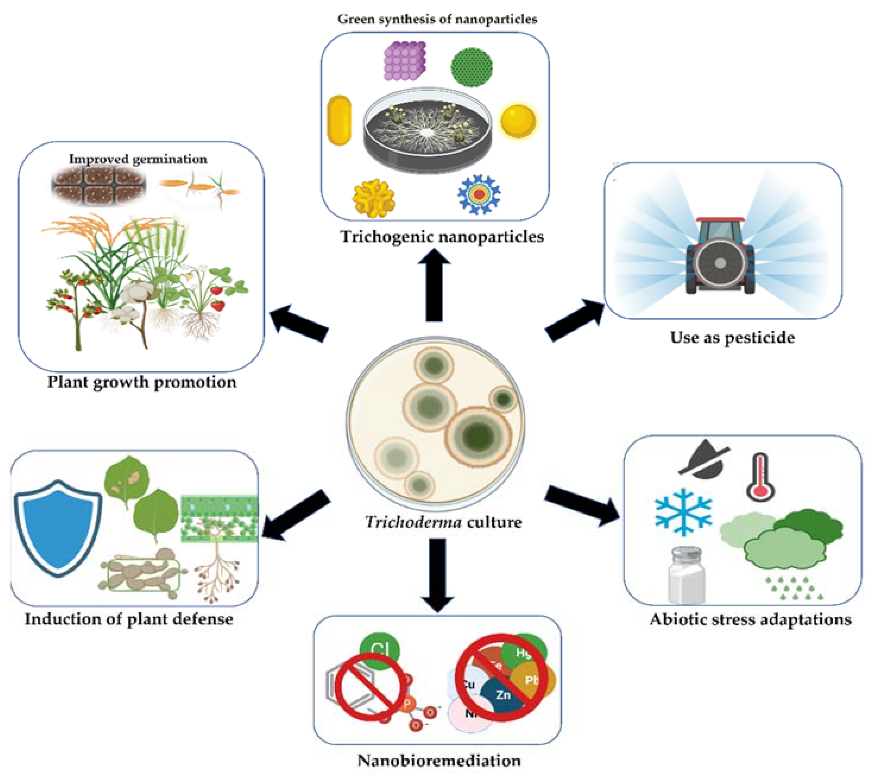
2. Beneficial Effects of Trichoderma in Agroecosystems

The Trichoderma genus includes fungal species that are economically significant because of their plant growth and performance-promoting actions, such as enhanced nutrient availability, the mycoparasitism of plant pathogens, and the priming of plant defense. The biodiversity of the genus Trichoderma provides fresh insight into their uses, particularly in agriculture and industry, as well as their potential use as biofungicides and biofertilizers in the field. Trichoderma spp. boost plant development, initiate plant defense, and aid plant growth in response to a variety of biotic and abiotic challenges, such as soil dryness, excessive salt content, and the presence of poisonous metal ions [16]. Trichoderma spp. are well-known plant growth-promoting fungi (PGPFs) with the potential to compete with pathogenic microorganisms while also promoting plant health [17]. Trichoderma species are extremely significant as biocontrol agents against a variety of plant pathogenic fungi and, hence, serve as viable alternatives to synthetic fungicides. Trichoderma species have been used as biological control agents for the management of plant pathogenic microbes, including fungi and bacteria [18]. Trichoderma and their secondary metabolites released into the rhizosphere may have an impact on plant development and nutrition, as well as the induction of systemic resistance and the biocontrol of pathogenic bacteria [18]. Plants and Trichoderma communicate bidirectionally via numerous signal molecules, resulting in a beneficial symbiotic relationship [16]. It can colonize plant roots by sensing nutrients secreted from the roots in the rhizosphere; interact with plants by producing various MAMP (microbe-associated molecular pattern) and damage-associated molecular pattern (DAMP) molecules; stimulate an immune response, suppressing many plant pathogens through the use of systemic acquired resistance (SAR) and induced systemic resistance (ISR) mechanisms; and act as a systemic resistance inducer in the plant [16,17]. Colonization of roots and leaves by Trichoderma can prime plant defense, allowing for powerful plant responses to following pathogen threats [19]. However, in addition to biopesticide action, certain Trichoderma strains have been shown to exhibit biostimulant activity, plant growth promotion, enhanced yield and nutritional quality, and the ability to mitigate the negative effects of abiotic stressors [20,21,22]. Therefore, Trichoderma has evolved from a mycoparasitic biocontrol agent (BCA) to one with numerous features such as pathogen antagonism, pathogen competition for resources, induction of systemic resistance in the host, overall plant growth promotion, and reduction of abiotic stressors. Furthermore, while being previously described primarily as soil and root colonizers, it is now clear that numerous Trichoderma species are endophytic [23]. As a result, it is not surprising that Trichoderma is found to be an effective beneficial biological agent, with active ingredients in over 200 agricultural products such as biopesticides, biofertilizers, bio-growth enhancers, and bio-stimulants that are marketed across the globe [15,24,25].
Beneficial fungi, including the Trichoderma species, are integral components of the decomposer microflora population, which plays a vital role in bioconcentration, decontamination, and even degradation/removal of the xenobiotics added to the ecosystem due to intentional and/or unintentional incorporation of a variety of contaminants. This removal or decontamination is referred to as ‘bioremediation’ and involves the use of microbial enzymes to convert toxic metal compounds into nonhazardous chemicals [26]. In a research report, T. viride exhibited the ability to break down and use nitrogenous (trinitrotoluene, TNT) explosives at doses of 50 and 100 ppm as the N-source to meet the nitrogen requirements for normal development [27]. Besides nitrogenous explosives, the potential hydrocarbon-degrading abilities of Trichoderma species can be useful to bioremediate diesel oil spills in an aquatic ecosystem, ensuring the protection of the environment [28]. Another report reiterated the use of Trichoderma species, particularly T. harzianum strain T22, for ensuring the biodegradation of diesel fuel, allowing it to be used as a carbon source [29]. Further, the physiological and metabolic versatility of various Trichoderma fungal species can be utilized for the remediation of heavy metals from different eco-niches. T. lixii CR700 demonstrated excellent Cu removal capability across a wide pH range. In the removal of Cu, T. lixii CR700 employs simultaneous surface sorption and accumulation processes [30].
Different Trichoderma species are, thus, effective natural decomposition agents to speed up the degradation of organic materials. The benefit will be two-fold as the nutrients released from the xenobiotic will be available in the plant rhizosphere and can be utilized and taken up by the plant [9]. The presence of Trichoderma with alfalfa seedlings boosted the soil’s N, P, and K content and alfalfa biomass [31]. The conjugate use of the Trichoderma and chemical fertilizer improved the plant’s nutritional quality, productivity, and vegetative and reproductive development. This can cut agricultural expenses while also reducing pollutants [32]. Overall, Trichoderma acts as a nutrient mobilizer, improving the quality and yield components of the crops.
Many studies have indicated that Trichoderma spp. are particularly efficient at degrading pesticides and can be useful in an integrated pest control strategy as well as for reducing pesticide residual effects. Trichoderma spp. have been reported to degrade benzimidazole fungicide (Carbendazim) [33], chlorpyrifos [34], penthiopyrad [35], and 2,2-dichlorovinyl dimethyl phosphate (DDVP) [36]. Wu et al. [37] proposed the tolerance mechanism of T. asperellum TJ01 to dichlorvos. Trichoderma spp. produce a variety of volatile organic compounds (VOCs) possessing various chemical properties. These metabolites are critical in agricultural, food, and pharmaceutical applications [38]. T. reesei has not been proven to be hazardous to humans [39]. The biosynthesis of silver nanoparticles by the fungus T. reesei is preferable in terms of safety, economics, and large-scale production capability [39]. As a result, the use of Trichoderma should be encouraged since it offers sustainable agriculture by minimizing the use of hazardous chemicals in agriculture (Figure 2).

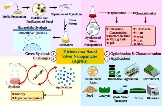
3. Biosynthesis of Nanoparticles by Trichoderma Genus

Fungal nanotechnology is one of the most popular options because of the vast range of benefits it has over bacteria, actinomycetes, plants, and other organisms in terms of physicochemical qualities [40]. When it comes to the biological generation of NPs, fungi outperform the majority of microorganisms in terms of efficiency. This is due to the ability of the fungi to produce a large variety of bioactive metabolites and metal accumulation properties and have improved processes, all of which are beneficial [41]. Because of their capacity to tolerate metals and accumulate metals in their tissues, fungi have emerged as an important branch for the biosynthesis of nanoparticles [42]. Green synthesis of nanoparticles is nontoxic as it involves the utilization of safe reagents, which makes it more cost-effective and eco-friendlier compared to the traditional methods [14]. Fungi offer several other benefits, such as simplicity of management and cultivation, no requirement of complicated components, production of a large amount of biomass and metabolites, high cell wall-binding capability, and ability to absorb large amounts of metal [43]. The next section describes the production of myconanoparticles from several Trichoderma species.

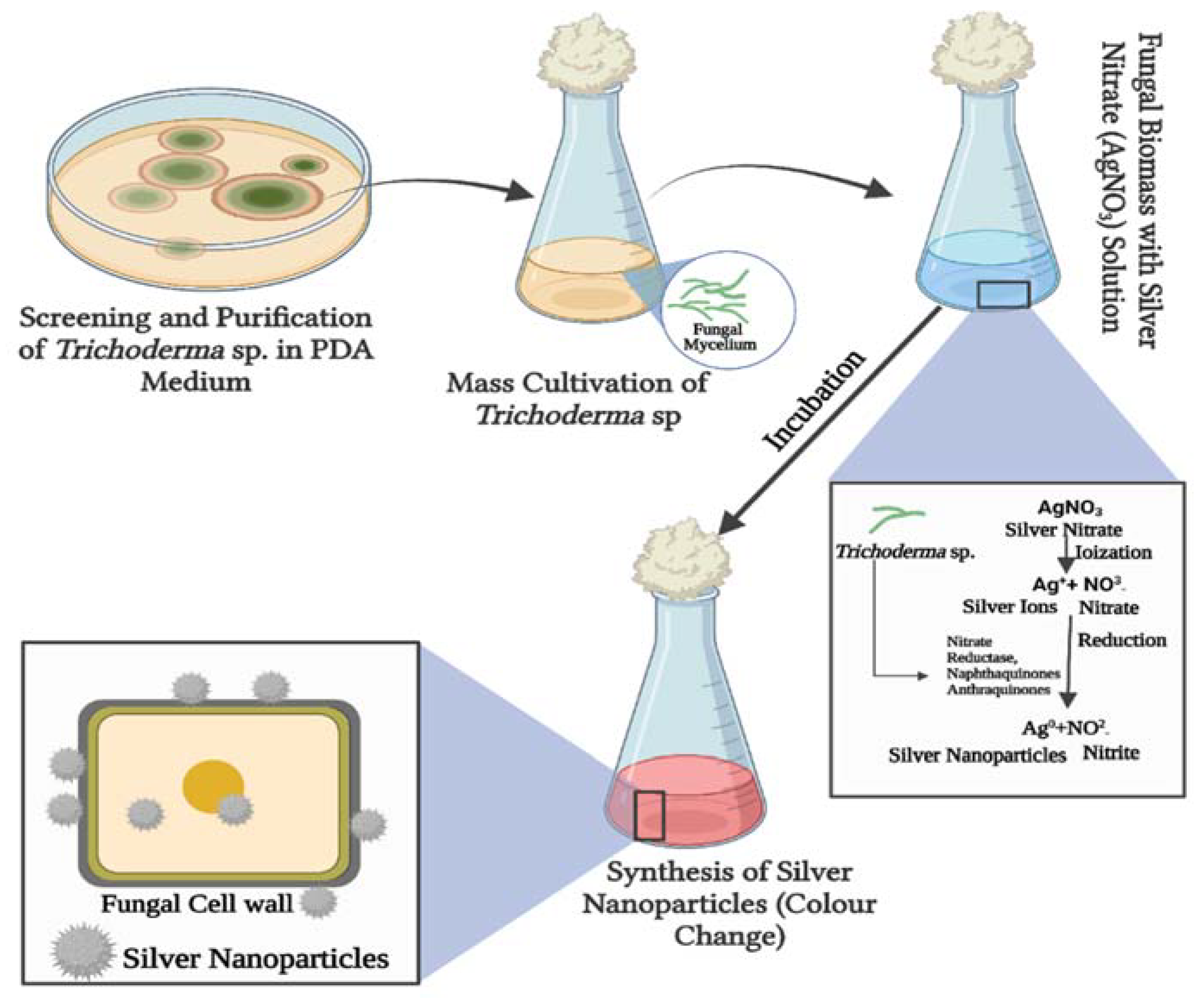
3.1. Silver Nanoparticles

The first report on the biosynthesis of silver nanoparticles with regulated properties from a nonpathogenic and economically viable biocontrol agent involved the incubation of the cell-free filtrate of T. asperellum for 5 days at 25 °C containing AgNO3 (1 mM) [44]. The use of UV–vis spectroscopy revealed the kinetics of the reaction with a strong surface plasmon resonance band observed at 410 nm, indicating the synthesis of silver nanoparticles. Based on the results of TEM and XRD studies, the size of the silver nanoparticles ranged from 13 to 18 nm [44]. T. reesei has also been used for the extracellular biosynthesis of AgNPs for the first time [39], allowing for the synthesis of AgNPs on an industrial scale. Fluorescence emission spectroscopy was used to produce detailed information on the progress of the decrease of silver nitrate (formation of silver nanoparticles) on the nanosecond timescale. The quantitative analysis of the reaction products was carried out using Fourier transform infrared spectroscopy (FTIR). The TEM images revealed the morphologically varied forms and crystalline nature of the AgNPs, with diameters ranging from 5 to 50 nm. Likewise, another study showcased T. virens to be the most efficient producer of AgNPs when compared to a total of 75 isolates from five distinct Trichoderma species. Every 24 h, the highest plasmon band was recorded at 420 nm, reaching maximum intensity at 120 h. The morphology of NPs was validated by high-resolution transmission electron microscopy (HRTEM), which revealed that nanoparticles were single or aggregated, spherical and homogeneous in shape, and ranged in size from 8 to 60 nm [45].
T. harzianum cell filtrate was used to synthesize AgNPs in a simple green and eco-friendly manner, without the need for any toxic reducing agents, capping agents, or dispersion agents. Temperature and AgNO3 concentration both had a considerable impact on AgNP production, as evidenced by UV–vis spectra. AgNPs were shown to be stable for three months, as confirmed through the DLS study. The synthesized AgNPs possessed face-centered cubic symmetry with a size range of 10–51 nm [46]. T. harzianum extracellular filtrate is an entomopathogenic fungus used for AgNP synthesis with distinct characteristics. The reduction rate of silver ions was assessed using a UV-visible spectrophotometer, which revealed a maximum absorption at 433.5 nm. Fourier transform infrared spectroscopy identified specific fungal metabolite functional groups responsible for the synthesis of AgNPs. The linkage between amino acid residues was shown by the amide group through the vibration of the N–H bend. X-ray diffraction analysis confirmed that the mycosynthesized AgNPs were oval in shape, crystalline in nature, and monodispersed in colloidal form, as well as exhibiting high purity. The surface properties of the generated AgNPs, as depicted by scanning electron microscopy, involved no direct contact even inside aggregates, indicating that AgNPs have been stabilized by a capping agent, and the size was around 10–20 nm [47]. Elgorban et al. [48] studied the synthesis of biogenic silver nanoparticles at room temperature in darkness using the fungus T. viride. Centrifugation was used to increase the concentration of AgNPs. The bioreduction of silver nanoparticles (AgNPs) was seen spectrophotometrically, and the AgNPs under investigation were characterized using UV–vis, TEM, and SEM. T. viride AgNPs were reported to be stable and polydispersed globular particles, with diameters ranging from 1 to 50 nm. They can be used as a reducing agent in the biogenic production of silver nanoparticles. T. harzianum-derived AgNPs possessed a spherical shape, acceptable polydispersity, and a size distribution between 20 and 30 nm, with no aggregates [4]. Biological silver nanoparticles were generated extracellularly utilizing the fungus T. longibrachiatum, with the fungal cell filtrate employed as a reducing and stabilizing agent in the nanoparticle manufacturing process. Incubation at 28 °C for 72 h with fungal biomass without agitation resulted in AgNP biosynthesis. The production of spherical nanoparticles with sizes ranging from 5 to 25 nm was observed by TEM [49]. Saravanakumar and Wang [50] reported, for the first time, the synthesis of anisotropic structured AgNPs using T. atroviride and investigated their biomedical properties. The observation of plasmon resonance at 390–400 nm in the UV–vis spectrum verified the formation of AgNPs. FTIR, transmission electron microscopy, and EDX examination revealed a significant percentage signal of anisotropic structural AgNPs, with sizes ranging from 15 to 25 nm. The Trichoderma filtrate contained biochemicals capable of bio-reducing silver nitrate (AgNO3), which are spherical NPs and nontoxic at low concentrations [51].
Extracellular biosynthesis of AgNPs from AgNO3 solution using T. reesei PF biomass cell-free water extract (CFE) has been described [52]. The optimal generation of AgNPs was achieved when T. reesei fungi were cultured in conditions containing 0.1 percent corn steep liquor, 10% biomass extracted to produce CFE, and 10 mM AgNO3. HRTEM micrographs verified the presence of Ag metallic nanoparticles in the crystal phase. The TEM pictures revealed the development of AgNPs clusters with sizes ranging from 1–4 to 15–25 nm. The NPs were stabilized by the capping action of the biomolecules, as indicated by FTIR spectroscopy. The zeta-potential of the prepared AgNPs was negative [52]. AgNPs were effectively synthesized using T. harzianum filtrates cultured in the presence and absence of enzymatic stimulation of the S. sclerotiorum cell wall, resulting in nanoparticles with distinct physicochemical properties. The DLS approach was utilized to determine hydrodynamic diameters, yielding mean hydrodynamic diameters of 57.02 ± 1.75 nm for AgNP-TS and 81.84 ± 0.67 nm for AgNP-T, respectively. The NTA approach yielded nanoparticle sizes and concentrations that differed in AgNP-TS and AgNP-T particle sizes of 88.0 ± 7.3 and 182.5 ± 6.9 nm, respectively [43]. T. longibrachiatum DSMZ 16,517 mycelial cell-free filtrate (MCFF) bioreduced silver ions (Ag+) to their metallic nanoparticle state (Ag0), as shown by AgNPs. The DLS examination revealed average AgNP size and zeta potential values of 17.75 nm and 26.8 mV, suggesting the stability of the synthesized AgNPs. The crystallinity of the mycosynthesized AgNPs, with an average size of 61 nm, was confirmed by the XRD pattern. The FESEM and HRTEM images revealed non-agglomerated spherical, triangular, and cuboid AgNPs, with sizes ranging from 5 to 11 nm. The FTIR analysis of the mycosynthesized AgNPs confirmed MCFF’s activity as a reducing and capping agent [53]. T. atroviride-hosted Chiliadenus montanus was shown to be the best candidate for the synthesis of mycogenic AgNPs among all investigated species. HRTEM was used to describe these AgNPs, which revealed a dispersion of spherical AgNPs ranging in size from 10 to 15 nm. By using the agar well diffusion technique, mycosynthesized AgNPs were compared to chemically synthesized AgNPs for their antibacterial, anticandidal, and antifungal activities over six pathogenic bacteria and four pathogenic fungi in vitro [54]. Chitin-induced exo-metabolites extracted from the varied and stress-tolerant T. fusant Fu21 were used to synthesize green AgNPs with sizes ranging from 59.66 to 4.18 nm (SEM), the spherical shape of nanoparticles with proven purity, stability (51.2 mV zeta potential), and nanoparticle polydispersity [55].
Amazon fungus Trichoderma sp. strain (TCH 01) was isolated from Bertholletia excelsa (Brazil-nut) seeds, and the soil was identified to biosynthesize the AgNP, as demonstrated by the RPSL band and UV–vis spectroscopy analysis. According to the TEM picture, the particles were polydispersed and spherical in form. AgNPs varied in size from 14 to 25 nm. All AgNPs exhibited incipient instability for the zeta potential. When grown at pH 5 for 9 days, smaller nanoparticles and higher polydispersity indexes were obtained [56]. Among 15 isolates of the T. species tested for the manufacture of AgNPs, a cell-free aqueous filtrate of T. virens HZA14 generating gliotoxin produced the best yield for the production of AgNPs. Electron microscopy tests revealed that AgNPs were 5–50 nm in size and had spherical and oval forms with smooth surfaces [57]. UV–visible spectrophotometry, FTIR, EDS, DLS, XRD, and SEM were used to analyze the AgNPs produced from T. harzianum culture filtrate. The surface plasmon resonance of synthesized particles created a peak with a center wavelength of 438 nm. According to the DLS research, the average size of AgNPs is 21.49 nm. SEM was used to determine the average size of AgNPs, which was 72 nm. The cubic crystal structure determined by XRD analysis validated the particles’ identification as silver nanoparticles [15]. As an alternative to traditional chemical and physical techniques, a green method for the synthesis of AgNPs has been provided. T. reesei fungus biomass was exploited as a green and renewable source of reductase enzymes and metabolites capable of converting Ag+ ions into AgNPs. T. reesei is an appropriate reagent for the production of monodisperse AgNPs that are stabilized by the capping effect of biomolecules. Trichogenic synthesis of AgNPs mediated by the Trichoderma genus is shown in Figure 3.

3.2. Zinc Oxide Nanoparticles

T. harzianum (PGT4), T. reesei (PGT5), and T. reesei (PGT13) monocultures and co-cultures (PGT4 + PGT5 + PGT13) generated secondary metabolites useful in the mycosynthesis of zinc oxide nanoparticles (ZnO NPs). Trichoderma—D-glucanzinc oxide nanoparticles (T—D-glu-ZnO NPs) were synthesized under optimal conditions using the fungal mycelial water extract (FWME) obtained from T. harzianum. The effective conjugation of D-glucan from barley with T-ZnONPs was confirmed by PACE and FTIR. The optimized T—D-glu-ZnO NPs had a spherical form with a mean size of 30.34 nm. T—D-glu-ZnONPs greatly suppressed the development of Staphylococcus aureus within roundworms while also enhancing roundworm growth [58]. Co-cultivation can induce the production of new secondary metabolites more effectively than monocultures. Another study involving the biosynthesis of ZnONPs showed the formation of NPs with crystalline structures free of impurities, according to PXRD analysis. The size of the crystalline particles ranged between 12 and 35 nm. These biosynthesized ZnONPs showed antibacterial efficacy against the rice cause of Bacterial Leaf Blight, Xanthomonas oryzae pv. oryzae [59]. T. harzianum is a possible fungal antagonist that is employed in the extracellular manufacture of ZnONPs. TEM imaging revealed that ZnONPs’ crystalline structure comprises hexagonal, spherical, and rod-shaped particles in a mixture of exceptionally small particles. The size range of the generated ZnONPs was 8–23 nm [3].

3.3. Copper Nanoparticles

T. asperellum cell-free extract was used to create copper oxide nanoparticles (TA-CuONPs). TA-CuO NPs were found to be crystalline with spherical particles. The CuONPs ranged in size from 10 to 190 nm, with an average diameter of 110 nm [60]. Consolo et al. [61] revealed, for the first time, the numerous extracellular biosynthesis of NPs from T. harzianum, as well as the synthesis of CuO and ZnONPs from this fungus. Biogenically synthesized Ag, CuO, and ZnO nanoparticles were made by employing a cell filtrate of a strain of T. harzianum as a reducer and stabilizer agent. When the ZnO NPs were evaluated against different target microorganisms, the potential of Ag and CuO for phytopathogen control was emphasized. Biocontrol agents Pseudomonas fluorescens, T. atroviride, and Streptomyces griseus were used in the production of copper and silica nanoparticles. T. harzianum was effectively used in the synthesis of copper nanoparticles. CuNPs were tested for antibacterial properties against two bacteria, Staphylococcus aureus and Escherichia coli, using a simple green and eco-friendly method [62]. UV–vis spectrometry, TEM, and EDAX analysis were used to analyze silica and copper nanoparticles. Nanoparticles were discovered to be aggregated and irregularly spherical. The size of silica nanoparticles varied from 12 to 22 nm, whereas the size of copper nanoparticles ranged from 5 to 25 nm [63].

3.4. Selenium Nanoparticles

The activity of biosynthesized selenium nanoparticles (SeNPs) using T. asperellum culture filtrate against Sclerospora graminicola, the cause of mildew disease in pearl millet, was higher. SeNPs were found to be hexagonal, near-spherical, and irregular in form, with sizes ranging from 49.5 to 312.5 nm. The size of SeNPs was inversely related to their biological activity [64]. Selenium nanoparticles (TSNPs) biosynthesized from T. harzianum JF309 were compared to conventional SNPs. The liquid metabolites of eight Trichoderma strains were collected, and modified biogenic synthesis was achieved. SNPs were found to be spherical or pseudo-spherical in shape, but TSNPs were more irregular. TSNPs were discovered to be somewhat larger than standard SNPs. The antifungal impact of TSNPs was far superior to that of standard SNPs [65]. The Trichoderma sp. WL-Go culture broth was used to create simple and cost-effective selenium nanoparticles. SeNPs had a partial size range of 20–220 nm, with an average diameter of 147.1 nm [66].

3.5. Other Nanoparticles

Gold nanoparticles were synthesized through a very fast and environment-friendly method within 10 min; this was the first time for such rapid biosynthesis of growth-promoting and plant pathogen control AuNPs. T. viride cell-free extract was treated with HAuCl4 at 30 °C for 10 min. TEM analysis confirmed that the NPs were widely dispersed and scattered in nature, with the bulk of them being spherical in form. The crystalline nature was verified by the SAED pattern. The size of the AuNPs ranged from 20 to 30 nm [67]. Chitosan nanoparticles were biogenically synthesized from T. viride and described using UV–vis spectroscopy, with FTIR verifying the functional groups of chitosan nanoparticles as OH, N–H, C–H, C=O, C–O, C–N, and P=O and electron microscopy demonstrating the roughly spherical form. DLS analysis determined the average size of chitosan nanoparticles to be 89.03 nm [68]. The T. harzianum (MF780864) isolate demonstrated the extracellular production of SiO2NPs from rice husks. UV, FTIR, DLS, and TEM were used to describe SiO2 NPs, which had a size of around 89 nm and shapes of oval, rod, and cubical particles [69].
The possibility of synthesizing FeNPs from Trichoderma species using simple methods has also been investigated. The presence of alkene, carboxyl, and phenol groups showed that the NPs were capped by the organism following the redox event. The generation of FeNPs from fungi such as Trichoderma species, which has the potential to generate more FeNPs than other bioresources, was proven [70]. T. harzianum is a biocontrol agent that is employed in the green manufacture of biogenic iron oxide nanoparticles for stabilization. Dynamic light scattering, nanoparticle tracking analysis, scanning electron microscopy, X-ray diffraction, and Fourier transform infrared spectroscopy techniques were used to assess the physicochemical properties of nanoparticles, and the results showed that the average size diameter of hematite (Fe2O3) nanoparticles are 207.2 nm [71]. Table 1 presents many metal NPs produced by different Trichoderma species, including AgNPs, ZnONPs, CuNPs and CuONPs, SeNPs, and other NPs, each with unique features and antimicrobial potential.

4. Production of Metal NPs by Hypocrea
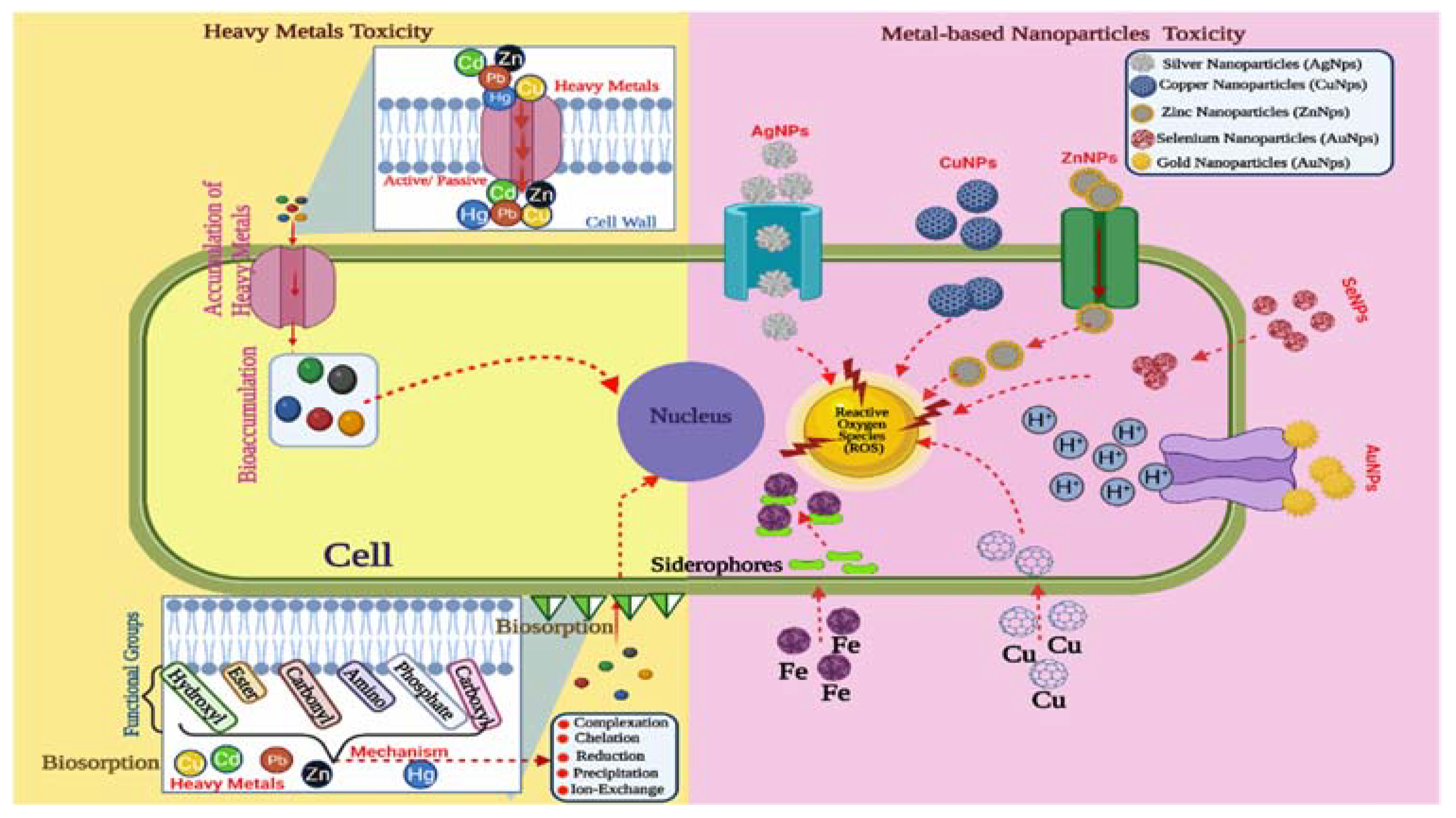
In pure culture, cultures obtained from Hypocrea lixii ascospores developed the morphological species T. harzianum. T. harzianum is known as a cosmopolitan, ubiquitous species associated with a wide variety of substrates [72]. The ability of the fungus Hypocrea lixii to ingest and reduce copper ions to copper NPs was investigated. This approach verified the existence of proteins as stabilizing and capping agents around the copper nanoparticles. These results showed that the dead biomass of H. lixii is an economically and technically feasible solution for wastewater bioremediation and a possible candidate for industrial-scale synthesis of copper NPs [73]. Rapid synthesis of gold nanoparticles is possible in less than a minute using cell-free extracts of T. viride and recombinant H. lixii at various reaction temperatures. Another study was the first report of T. sp. involved in the rapid synthesis of gold nanoparticles that may function as an antimicrobial agent as well as an effective biocatalyst [67]. The fungus H. lixii’s dead biomass was discovered to be an effective instrument for the extracellular and intracellular synthesis of nickel oxide NPs as well as the absorption of hazardous metal ions from an aqueous solution. The fungus’s dead biomass played a significant role in the formation of metallic NPs, which are easily oxidized to nickel oxide in the media. The dead biomass also functioned as a stabilizer during the creation of nickel oxide NPs and so might be employed for nickel ion uptake during bioremediation procedures [74]. In addition, a new species, H. virens, has been discovered to be a teleomorph of T. virens, a species commonly used in biological control applications. H. virens, for example, is a possible biocontrol agent agonist of the Ceratocystis paradoxa pathogen that causes pineapple disease on sugarcane. The fungus H. virens was tested for its ability to synthesize AgNPs. A H. virens fungal biomass of extracellular and intracellular extract to aqueous silver nitrate solution for 72 h revealed that the appearance of a dark-brown color in the fungal biomass after reaction with Ag+ ions was a clear indicator of metal ion reduction and the formation of AgNPs by the fungal biomass [45].

5. Toxicity

In a recent study, the blood kinetics and tissue distribution of 20, 80 and 110 nm AgNPs were studied in rats [75]. The silver nanoparticles disappeared quickly from the blood and were distributed to all animal organs, independent of the NP size [75]. As a result, it can be identified that the AgNPs may have different toxicity and, hence, be associated with a distinct health risk. After repeated intravenous injections of silver nanoparticles, accumulation was observed [76]. The toxicity of AgNPs was determined by the dose and particle size [50]. In terms of toxicity, comparisons of cell lines 3T3 (mouse embryo fibroblasts), HeLa (human cervical adenocarcinoma), HaCaT (human keratinocytes), V79 (Chinese hamster pulmonary fibroblasts), and A549 (human epithelial adenocarcinoma) with controls revealed that the biogenic nanoparticles exhibited cytotoxic and genotoxic effects that varied depending on the cell line used and the exposure concentration. An interesting observation was that in the majority of these assays, the effects increased in direct proportion to the doses employed and were most strong at concentrations higher than those used in S. sclerotiorum inhibition evaluations. Concerning the impacts of nanoparticles in soil, there was evidence of some partial effects on bacteria, with probable microbiota recovery over time. Because the AgNP-T nanoparticles had no detrimental impacts on soybean germination and development, it is possible to conclude that this approach constitutes the first step towards the potential control of white mold in soybean crops using nanotechnology [4]. T. atroviride-induced AgNPs triggered cell changes, inhibition, or damage, as well as cytoplasmic compression in a concentration-dependent way. AgNPs significantly reduced cell viability with an inhibitory dose of 16.5 g/mL against human breast cancer cells (MDA-MB-231). Cell viability increased significantly with drug dosage concentration (NPs) against IMR 90, U251, and A549 lung cells, and human breast cancer cells MCF-7 and MDA-MB-231 [50]. The cytotoxicity and genotoxicity of AgNP-TS and AgNP-T nanoparticles were shown to be low in V79, 3T3, and HaCat cell lines. Guilger-Casagrande and colleagues [43] used, for the first time, photothermolysis to confirm the anticancer activity of biogenic CuO NPs. The TA-CuO NPs caused the photothermolysis of A549 cancer cells through ROS production, nucleus damage, mitochondrial membrane potential (m), and regulatory protein expression [60]. When SNPs and TSNPs were tested for cytotoxic action using human viability and mortality, neither SNPs nor TSNPs demonstrated hypertoxicity or lethality for the three cells [65]. At a concentration of 200 g ml−1, it possessed multi-mode antimicrobial action and has been determined to be nontoxic to humans. The lowest inhibitory concentration of green nanoformulations for the greater degrading activity of the pathogen fungal mycelium was determined to be 20 g Ag/mL green nanoformulations [55]. T—D-glu-ZnO NPs were not hazardous to NIH3T3 cells but had a dose-dependent inhibitory impact on human pulmonary carcinoma A549 cells, according to a cytotoxicity investigation. T-ZnO NPs and T—D-glu-ZnO NPs caused cancer cell death by necrosis and apoptosis, respectively [58]. The zebrafish model was used to assess the toxicity; there was no toxicity of nanocopper and nanosilica when exposed to 0.5, 3, and 30 g concentrations in respect of embryo viability, hatching rate, body mass index, and heartbeat counts [63]. The cytotoxicity of biogenic iron oxide nanoparticles was evaluated using several cell lines and Allium cepa assays to measure the genotoxicity. They did not impact cell viability when compared to controls and did not cause changes in the mitotic index at the quantities used. The presence of iron oxide nanoparticles did not affect seed germination [71].

6. Understanding the Mechanism of Synthesis of Trichogenic NPs

Fungal biosynthetic techniques can be grouped into intracellular and extracellular synthesis based on where nanoparticles are produced. Extracellular synthesis of nanoparticles, for example, is still being developed in terms of understanding the mechanisms of synthesis, simple downstream processing, and quick scale-up processing.

6.1. Bioactive Metabolites

The use of T. longibrachiatum for AgNP biosynthesis revealed the existence of proteins and their binding to AgNPs via carbonyl groups of amino acid residues and peptides, which might have contributed to their stability and prevention of agglomeration. Proteins on the surface of AgNPs function as capping agents [49]. There are 35 metabolites that have been discovered as significant variations between selenium nanoparticles biosynthesized from T. harzianum (TSNP) and standard SNPs, which included organic acids, sugars, amino acids, and carbohydrate metabolism intermediates. Among these, 27 are strong antifungal agents with higher levels in TSNPs than in SNPs. Many acids, sugars, and their derivatives are used in the coating of TSNPs, including heptonic acid, ferulate, fumaric acid, threonic acid, glucose, and mannitol. All of these organic compounds in aqueous formulations capped the selenium NPs and acted as stabilizers while also increasing the antagonistic capability of biosynthesized selenium NPs against pathogens [65]. While using T. virens HZA14 for AgNP biosynthesis revealed the interaction patterns of protein, carbohydrate, and heterocyclic compound molecules with AgNPs, the maximum yield was associated with gliotoxin [57]. Aromatic amino acids such as tyrosine and tryptophan were found in the FTIR spectra of Trichoderma spp. medium. The electrostatic attraction of negatively charged carboxylate groups by free amine groups, cysteine residues, or some proteins secreted by the fungus during the formation of AgNPs can bind to them. The release of extracellular protein molecules from fungus is responsible for the synthesis and stability of AgNPs [56]. The interaction of different functional groups of exometabolites with Ag was validated using FTIR for the production of green AgNPs and may be responsible for stabilization. T. fusant Fu21 inoculated with SM-containing chitin was used for GC–MS profiling to identify functional groups and compounds, which were discovered to be alkanes, dicarboxylic acid, aromatic ketones, amino acid, hetero-acyclic compounds, ketose sugar, sugar alcohol, aliphatic amines, polyol compounds, steroidal pheromones, and carbocyclic sugars [55]. Secondary metabolites released by T. harzianum operated as capping and reducing agents, providing consistency and contributing to biological activity determined by LC-MS/MS. The most common compounds included 1-benzoyl-3-[(S)-((2S,4R,8R)-8-ethylquinuclidin-2-yl] thiourea (6-methoxyquinolin-4-yl) methyl, puerarin, genistein, isotalatizidine, and ginsenoside [15]. The existence of numerous functional groups of biomolecules and capping protein, enclosing biosynthesized SiO2 NPs corresponding to carbonyl residues, alcohol, nitrile, acid chloride, alkene bands, and peptide bonds of the proteins involved, was verified by FTIR analysis [69]. The presence of functional groups of alkene, alkane, and alcohol in the FTIR test suggested that these may have participated in the SeNP production processes [66]. The presence of functional groups was verified by FTIR analysis of biosynthesized silica and copper nanoparticles using biocontrol agents, resulting in effective synthesis [63]. According to the FTIR analysis results, phenolic, proteins, amino acids, aldehydes, ketone, and other functional groups were involved in the reduction, capping, and stability of zinc oxide NPs [3].

6.2. Enzymes

While many microbial species can produce metal NPs, the mechanism of nanoparticle biosynthesis has not been identified as yet [77,78]. For its survival, T. harzianum produces enzymes and metabolites that are involved in the breakdown of silver nitrate into Ag+ ions and NO3, owing to the possible action of hydrolytic/nitrate reductase enzymes. Through the catalytic activity of extracellular fungal secondary metabolites, the toxic Ag+ ions are further converted to nontoxic (Ag0 = biosilver) metallic nanoparticles in this procedure. The bioreduction of silver is based on the presence of relevant functional groups in the extracellular filtrate of T. harzianum, which is more effective than other fungi and is nontoxic to humans [47,79]. FTIR and surface-enhanced resonance were used. Raman spectroscopy revealed a viable mechanism for the synthesis of silver nanoparticles in T. asperellum [44]. The procedure consisted of two critical steps: bioreduction of AgNO3 to produce AgNPs, followed by stabilization and/or encapsulation with a suitable capping agent. The cell’s defense mechanism for silver detoxification has been proposed as a biological process for AgNP production [80]. Extracellular enzyme secretion provides the advantage of obtaining large quantities in a relatively pure state, free of other cellular proteins associated with the organism, and can be easily processed by filtering the cells and isolating the enzyme for nanoparticle synthesis from the cell-free filtrate. T. reesei is thought to be the most effective extracellular enzyme producer compared to other filamentous fungi [39], and it has a long history of producing industrial enzymes [81]. The NADH co-enzyme, as well as NADH-dependent enzymes such as nitrate reductase, are present in Trichoderma genus strains and are essential in the formation of nanoparticles and capping that gives greater stability [39,82]. The determination of hydrolytic enzyme-specific activity revealed that AgNP-TS had a greater specific activity of NAGase and chitinase than AgNP-T. Concerning the filtrates, T. harzianum exposure to the cell wall of S. sclerotiorum enhanced the specific activity of the enzyme NAGase [43]. T. harzianum produces enzymes and metabolites involved in breaking strong ionic bonds between silver and nitrate ions for its own survival, perhaps by the action of hydrolytic/nitrate reductase enzymes. Extracellular fungal secondary metabolite enzymatic activity transforms dangerous Ag+ ions into nontoxic biosilver NPs [14].

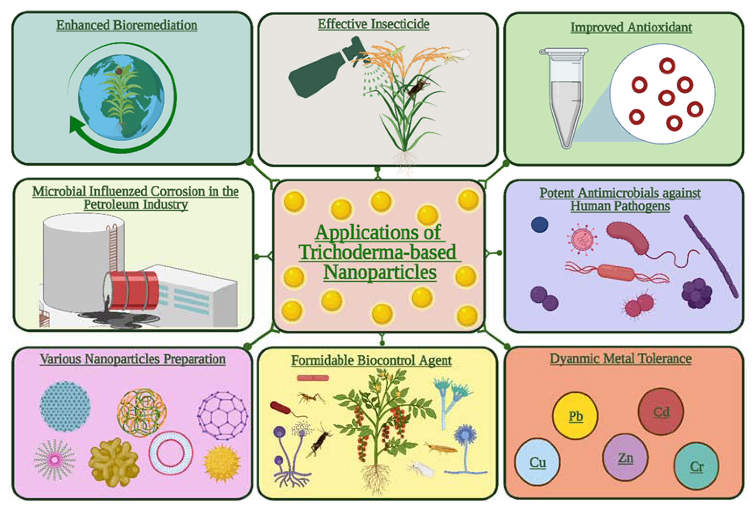
7. Applications of Trichoderma-Mediated NPs in the Agri-Food Sector

Crop production invariably involves rampant use of chemical pesticides, insecticides, and herbicides that have been exhibiting bioaccumulation of toxic chemical residues potentially hazardous to plant ecosystems [40]. This problem has shifted the research foci to the identification of more effective alternatives such as nano-based agrochemicals. The intrusion of nano-interventions in the agri-food sector is being extended to extract the benefits showcased by nanotechnology applications in electronics, pharmaceutical, biomedical, paint, and cosmetics industries. However, the chemically synthesized nanoparticles have a high cost and substantial eco-toxicity (https://www.reportlinker.com/p06193716/Nanotechnology-Services-Global-Market-Report.html?utm_source=GNW, accessed on 20 February 2022). Their rampant use in open field conditions is anticipated to enhance the negative ecological and health hazards, besides the huge input costs. Therefore, green nanoparticles have been speculated to address these issues related to synthetic NPs. Various fungal species may create mycogenic nanoparticles that might stimulate growth and protect crops against diseases in some prospective agricultural uses through antioxidant, antimicrobial, and plant-stimulating properties. The primary benefits of the mycogenic NP synthesis protocol(s) will be the one-pot, cost-effective, and less environmentally corrosive features. Still, the widespread usage of myco-nanoparticles may lead to a few complications. For example, on multiple applications of these NPs, the nano-toxicity aspects will render the plant beneficial microbes vulnerable to the applied nanoparticles, which might compromise their viability as well as the biocontrol and plant growth promotion benefits. Farms and agricultural consumers will soon be able to utilize myconano-functional agrochemicals, pre-harvest and post-harvest crop protection agents, sensors used in genetic equipment, and crop protection components. Additionally, the fungus-derived nanoparticles can function as useful myco-nano-sorbents and would be an attractive way to perform heavy metal biosorption from contaminated wastewater. The employment of Trichoderma genus for the synthesis of metallic nanoparticles is ecologically advantageous, time-saving, and cost-effective. Furthermore, potential agrochemicals can be designed by amalgamating NPs and Trichoderma strains to produce more sustainable products. The agri-food applications of this technology outlined below will help you understand how the Trichoderma genus is used to make nanodevices and how these trichogenic NPs are being used in various applications.

7.1. Antifungal Activity

The effect of metal chitosan nanocomposites at 100 g mL−1 in combination with Cu-tolerant Trichoderma longibrachiatum strains on cotton seedling damping-off under greenhouse conditions was also investigated. In vitro, the bimetallic blends (BBs) and Cu-chitosan nanocomposite had the best antifungal effectiveness against both R. solani anastomosis groups. These findings suggested that BBs, the Cu chitosan nanocomposite, and BBs mixed with Trichoderma may inhibit R. solani-caused cotton seedling disease in vivo. R. solani was evaluated in a greenhouse with a Trichoderma strain and shown to have a synergistic inhibitory effect with BBs [83]. The biogenic synthesis of AgNPs was carried out by use of mycelial extracts of T. harzianum supplemented in aqueous silver nitrate (1 × 10⁻³ mol L−1). In a potato dextrose agar-based poison food assay, the use of concentrations ranging from 0.15 × 10¹² and 0.31 × 10¹² NPs/mL reduced the mycelial development and the generation of new sclerotia in S. sclerotiorum [4]. Antifungal application of AgNPs resulted in a considerable decrease in the number of forming colonies for several plant pathogenic fungi, with an efficiency of up to 90% against Fusarium verticillioides, Fusarium moniliforme, Penicillium brevicompactum, Helminthosporium oryzae, and Pyricularia grisea [49]. Because mycelial development was inhibited and no new sclerotia were formed, both AgNP-TS and AgNP-T nanoparticles showed promise for controlling S. sclerotiorum. The most effective inhibition of mycelial development was accomplished with AgNP-TS, which might be attributable to the nanoparticles’ reduced hydrodynamic diameter as well as a potential biomolecule effect from the nanoparticles’ capping [43]. With 20 g Ag/mL green nanoformulations for increased pathogen fungal mycelium breakdown activity, green Ag-NPs boost antifungal action to reduce the phytopathogen Sclerotium rolfsii producing stem rot in groundnut [55]. In vitro testing of AgNPs produced with T. virens HZA14 antifungal efficacy against S. sclerotiorum revealed that hyphal development, sclerotial formation, and myceliogenic germination of sclerotia were all inhibited by 100%, 93.8%, and 100%, respectively. The direct interaction between nanoparticles and fungal cells, including AgNPs’ contact, accumulation, lamellar fragment creation, and micropore or fissure development on fungal cell walls, was demonstrated using SEM/EDS technologies [57]. When compared to T. asperellum alone and carbendazim @0.1%, chitosan nanoparticles in combination with T. asperellum were found to be superior in suppressing the mycelial development of soil-borne pathogens such as F. oxysporum, R. solani, and S. rolfsii [68]. Copper and silica nanoparticles biosynthesized with biocontrol agents suppressed P. hypolateritia and P. theae growth, suggesting that this might be a unique way of managing diseases that affect tea plantations while also improving tea quality parameters. Various nanoformulations were prepared using suitably inert and eco-friendly carrier materials employing nano copper and nanosilica [63]. In the laboratory and greenhouse, T. harizinum-mediated ZnONPs were demonstrated, for the first time, to exhibit fungicidal activity against three soil–cotton pathogenic fungi (Fusarium sp., R. solani, and Macrophomina phaseolina) [3].
Pseudomonas fluorescens and T. viride, two distinct biocontrol agents, were used to make microbial CuONPs, which were then evaluated using various analytical methods. In terms of in vitro antifungal efficacies, CuONPs synthesized from T. viride showed the highest percent growth inhibition compared to CuONPs generated from P. fluorescens. CuONPs produced from T. viride, on the other hand, demonstrated considerably stronger antifungal activity in vivo than the commonly used Bordeaux mixture [84]. Antifungal activity against three soil-borne pathogens was proven in vitro and in the greenhouse. In the three fungal infections, AgNPs greatly reduced hyphal growth. The ability of T. harzianum isolates to synthesize a wide spectrum of proteins and enzymes without the need for chemical reducers and stabilizers has been proven. Biosynthesized AgNPs have shown high potential in protecting cotton plants from the fungal invasion induced by damping-off [14]. Trichoderma species have a wide range of biotechnological uses, including acting as biofungicides to manage various plant diseases, biofertilizers to promote plant development, and synthesizing and bioremediating metal nanoparticles. Possible applications of mycogenic NPs generated by the Trichoderma genus are demonstrated in Figure 4.

7.2. Antibacterial Activity

The antibacterial activity of green nano-biosilver T. harzianum AgNPs was demonstrated against S. aureus and K. aeruginosa, as well as Gram-positive and Gram-negative bacteria, with the Gram-negative bacterium (K. pneumoniae) displaying better sensitivity. T. atroviride’s AgNPs were found to have antibacterial action against Gram-positive and Gram-negative clinical pathogens such as E. coli, P. aeruginosa, and S. aureus [46]. AgNPs had stronger antibacterial activity than AgNO3 and were similar to the positive control kanamycin. They also demonstrated DPPH scavenging action in a dose-dependent manner, with an IC50 of 45.6 g/mL. The cell-free filtrate, on the other hand, showed no inhibition against the pathogens, probably due to metabolites with the antibiotic property. As a result, the biosynthesized AgNPs can be employed as a natural antioxidant to prevent human cell damage and degenerative diseases by regulating antioxidants, pro-oxidants, and ROS levels [50]. The antibacterial activity of AgNP was evaluated against a variety of bacteria types. Gram-negative (E. coli and P. aeruginosa) bacteria had lower MBC and MIC values than Gram-positive (S. aureus and E. faecalis) bacteria, and this difference was ascribed to the bacterial cell wall structure. This suggests that Gram-negative bacteria have better antibacterial activity due to bacterial surface adsorption and oxidative stress induction; nevertheless, more research is required [56]. Gram-positive (S. aureus and B. subtilis) and Gram-negative (E. coli and R. solanacearum) pathogenic bacteria were tested, and the AgNPs synthesized using T. harzianum filtrate were reported to have significant antioxidant properties and antibacterial activity against both Gram-positive (S. aureus and B. subtilis) and Gram-negative (E. coli and R. solanacearum) bacteria, with higher activity against the Gram-negative bacteria [15]. CuNPs were effectively produced using T. harzianum, an agriculturally beneficial fungus, in a simple green and environmentally favorable way. CuNPs and T. harzianum fungus were shown to have antibacterial action against both Gram-positive and Gram-negative bacteria [62]. The antibacterial activity of the generated AgNPs against Escherichia coli was investigated [85].

7.3. Plant Growth Promotion

In greenhouse settings, Trichoderma-mediated AgNPs were evaluated against tomato wilt caused by Fusarium species. The treated plants with various doses of AgNPs showed a promoting effect on all the tested parameters in comparison with the control and Trichoderma formulation [86]. With the prolonged soaking time of silver nanoparticles solution, T. harzianum-synthesized AgNPs demonstrated an increase in the percentage of seed germination. AgNPs produced by T. harzianum had a positive effect on oilseed germination. As a result, mycogenic AgNPs have a biological assay for increasing seed viability in agriculture [87]. The biocontrol agent T. harzianum was used as a stabilizing agent in the green manufacture of biogenic iron oxide nanoparticles. The nanoparticles’ antifungal effectiveness against S. sclerotiorum (white mold) was tested in vitro. The impact of the NPs on seed germination was also examined. They were also able to promote the proliferation of Trichoderma, which inhibited the establishment of the pathogen S. sclerotiorum while having no effect on seed germination [71].

7.4. Trichoderma-Based Nanobioremediation

Fungi-based nanotechnology is quickly evolving as an effective technology to treat industrial wastewaters. T. harzianum-derived CdS-NPs were used to photocatalyze the breakdown of methylene blue in a photocatalytic cell [88]. The production of electron (e) and hole (h+) pairs, which function as powerful oxidants and reductants, caused the degradation. Adsorbed water on the CdS-NPs traps the hole (h+), resulting in the formation of a hydroxyl radical, while oxygen takes electrons from the conduction band, resulting in the formation of an anion radical. These radicals target the dye’s azo bond, causing it to degrade and produce CO2, H2O, and NH4+. Due to an effective biocatalyst that converted 4-nitrophenol to 4-aminophenol in the presence of NaBH4 and the capacity to inhibit pathogenic bacteria, biosynthesized gold nanoparticles present new hope for green bioremediation [67]. The test human pathogenic bacteria were strongly suppressed by biogenic AgNPs made from the fungus T. viride [48]. Because of their biocidal action against halotolerant planktonic sulfate-reducing bacteria, mycosynthesized AgNPs have become an appealing alternative for controlling microbially driven corrosion in the petroleum industry [53]. T. harzianum-biosynthesized SiO2 NPs were effective as a lead adsorbent from water during the bioremediation process. The content of lead in water and the muscles of Nile tilapia (Oreochromis niloticus) decreased. O. niloticus’ immune system, liver, and renal functions improved [69]. The Trichoderma genus can be employed as a possible metal biosorbent and as a powerful bioremediation agent. Some Trichoderma strains have high metal tolerance and bioaccumulation ability, making them a viable mycoremediation agent of heavy metal contamination in environmental safety. Figure 5 illustrates the pathways of tolerance to metals and nanoparticles (NPs) in Trichoderma.

7.5. Miscellaneous Advantages and Disadvantages of Trichogenic Nanoparticles

The biogenic AgNPs synthesized from the extracellular filtrate of T. harzianum have an additional benefit over physical and chemical approaches that need high pressure, energy, chemical precursors, and a high cost. This filamentous fungus has a larger capacity for binding Ag+ Ag0, making this procedure easier and less expensive for the large-scale manufacturing of NPs at the industrial level, making it cost-effective [47]. T. viride’s capacity to generate AgNPs is extremely promising for green, sustainable nanomaterial synthesis [48]. The nanoparticles demonstrated little size fluctuation as well as high physicochemical stability. As the beans were exposed to biogenic silver nanoparticles, there were no significant changes in germination and seedling development compared to the negative control [4]. T. longibrachiatum showed a possibility for the extracellular and reliable biosynthesis of silver nanoparticles. The AgNPs synthesized using this biosystem technique were generally stable up to two months after synthesis [49], and, even after six months of storage, nanocrystalline silver particles created by T. asperellum did not display significant aggregation [15,44]. The use of fungi in the biogenic synthesis of silver nanoparticles has several benefits, including the development of a capping agent from fungal biomolecules, which provides stability and can assist in biological activity. The cytotoxicity and genotoxicity of AgNP-TS and AgNP-T nanoparticles to V79, 3T3, and HaCat cell lines were both low [43]. When compared to normal pesticides, the use of nano-based formulations will be a preferable option for avoiding excess chemicals in soil [45]. T. reesei possesses simpler and less expensive culture conditions, as well as greater growth rates on both industrial and laboratory sizes, resulting in lower costs in large-scale manufacturing [39]. T. atroviride-based biosynthesized AgNPs are an economically effective and ecologically sustainable way of producing AgNPs [50]. The small-sized nanoparticles generated, their narrow size distribution, the stability attained without the use of chemicals as capping agents, and the low number of chemical reagents used are the key benefits of the described extracellular biosynthesis of AgNPs. Because of the negative zeta potential that might aid the attachment of physiologically active compounds, the generated AgNPs were appropriate carriers of such compounds [52,54]. The use of green AgNPs as an antifungal agent is seen as an environmentally friendly resource, an alternative to fungicides, and a cost-effective method [55]. The surface characteristics of the biogenic chitosan nanoparticles generated from T. viride were discovered to be positively charged and stable [68].
However, there are several drawbacks that must be resolved in order to properly employ fungus for biogenic synthesis. The engagement of the host organism for protein expression, immature synthesis techniques, and limited know-how on large-scale production technique(s) may possibly limit the benefits associated with fungus-based nanoparticle synthesis. The most critical factors that may affect the final product include knowing which fungus to employ, its growth parameters, the necessity for sterile settings, and the time it takes for the fungus to develop and finish the synthesis. Scaling-up can also create challenges, such as the need for research focusing on the mechanism associated with the development of capping layers and effective nucleation [68]. All these challenges associated with the green synthesis of various metal NPs through the use of different types of fungal biomass should be solved, and the feasibility of green synthesized nanoparticles for obtaining useful commercial applications in the agri-food industry should be identified.

8. Challenges

Mycogenic nanoparticles can be synthesized with a wide range of particle size distributions. There are issues with reproducibility as well as a lack of control over particle size, shape, and dispersion. As a result, in future efforts, the parameters of synthesis should be thoroughly investigated [89]. The development of innovative nanofungicides, nanofertilizers, and nanosorbents generated by fungi and the evaluation of these nanoformulations for the remediation of heavy metal-contaminated industrial wastewater are still in their early phases. Several problems must be overcome to scale up mycogenic nanoparticles and fulfill the demands of the real world. For example, a new nanosorbent created by diverse microorganisms must be economically advantageous and socially acceptable while also being able to meet various water quality regulations to ensure human health and environmental safety [90]. Scaling up the method is difficult since the commercial synthesis of nanoparticles still needs more study. No NP residue must be discharged into the environment as a result of the treatment method. For example, the environmental tracking of silver in the field is critical for evaluating its influence on the environment and human health. Furthermore, further study should be conducted to assess the hazardous effect of AgNPs before widespread manufacture and usage in agricultural applications [49]. The toxicity of NPs remains a key hurdle in transferring these materials from lab to industry for the time being, and this has to be addressed further.

9. Future Trends

The biogenic silver nanoparticles exhibited no deleterious effects on T. harzianum culture compared to the negative control. This shows that combining T. harzianum with nanoparticles would not affect the fungus’ development [4]. It is hoped that techniques involving the biogenic synthesis of metallic nanoparticles will be developed, taking into account not only the potential biological activity of the metal nanoparticles but also the biomolecules and organic compounds obtained from the organisms used in the synthesis, which compose the capping on the nanoparticles [43]. In order to have better control over the size and polydispersity of these nanoparticles, more research is needed to understand the specific chemical pathway leading to their production using biological means. More studies should be carried out on the creation of AgNPs-based compounds and their combination with fungicides [49]. Extracellular biosynthesized AgNPs might be used as carriers for different agrochemicals and natural bioactive molecules. There is a greater need for field trials of Trichoderma-based nanoparticles to understand nanoparticle–plant–microbe interactions and toxicity issues to develop safer and user-friendly agrochemicals.

10. Conclusions

Based on the literature review, we conclude on whether developing agrochemicals that employ Trichoderma strains to produce green NPs could be more sustainable agrochemicals. Given that the Trichoderma genus is one of the most often used fungi for the mycosynthesis of NPs, it is possible that it possesses tolerance mechanisms to these structures that might be used in combination to generate products that boost agricultural productivity while simultaneously treating plant infections in the field. The synthesis of NPs by beneficial fungi such as Trichoderma is novel, cost-effective, and eco-friendly compared to the synthesis by chemical and physical methods. However, more study is required to assess the Trichoderma and heavy metals interaction in a heterogeneous system under field circumstances. Additionally, to comprehend the potential advantages of nanoparticles produced by beneficial microbes in the agricultural sector, it is necessary to comprehend nanoparticle penetration and transport routes in plants. The shape and size of the nanoparticles have a significant impact on their reactivity, stability, and behavior. More effective agrochemical applications using composite materials are desirable because these can decrease potential economic losses and, in particular, environmental damage. For example, combining the antimicrobial impact of mycogenic nanoparticles with biofungicides for plant disease control may boost antifungal efficacy through synergistic interaction, allowing for a reduced fungicide dose and, as a result, avoiding the development of fungal pathogen resistance.

Author Contributions

K.A.A.-E., M.A.A. and H.M.A. conceptualized, reviewed, edited, and acquired funding. A.K. and M.A.A. performed the investigations. A.K. and M.R. (Mahendra Rai) also acquired the resources. M.A.A. curated the data, H.M.A., M.R. (Mythili Ravichandran) and F.K.A. wrote the first draft and edited it. M.A.A. acquired the funding. All authors have read and agreed to the published version of the manuscript.

Funding

The authors would like to thank the Deanship of Scientific Research at Shaqra University for supporting this work.

Institutional Review Board Statement

Not applicable.

Informed Consent Statement

Not applicable.

Data Availability Statement

Not applicable.

Acknowledgments

The authors would like to thank the Deanship of Scientific Research at Shaqra University for supporting this work.

Conflicts of Interest

The authors declare no conflict of interest.

References

  1. Alghuthaymi, M.A.; Rajkuberan, C.; Rajiv, P.; Kalia, A.; Bhardwaj, K.; Bhardwaj, P.; Abd-Elsalam, K.A.; Valis, M.; Kuca, K. Nanohybrid antifungals for control of plant diseases: Current status and future perspectives. J. Fungi 2021, 7, 48. [Google Scholar] [CrossRef]
  2. Hashem, A.H.; Abdelaziz, A.M.; Askar, A.A.; Fouda, H.M.; Khalil, A.M.A.; Abd-Elsalam, K.A.; Khaleil, M.M. Bacillus megaterium-Mediated Synthesis of Selenium Nanoparticles and Their Antifungal Activity against Rhizoctonia solani in Faba Bean Plants. J. Fungi 2021, 7, 195. [Google Scholar] [CrossRef] [PubMed]
  3. Zaki, S.A.; Ouf, S.A.; Albarakaty, F.M.; Habeb, M.M.; Aly, A.A.; Abd-Elsalam, K.A. Trichoderma harzianum-Mediated ZnO Nanoparticles: A Green Tool for Controlling Soil-Borne Pathogens in Cotton. J. Fungi 2021, 7, 952. [Google Scholar] [CrossRef]
  4. Guilger, M.; Pasquoto-Stigliani, T.; Bilesky-Jose, N.; Grillo, R.; Abhilash, P.C.; Fraceto, L.F.; De Lima, R. Biogenic silver nanoparticles based on Trichoderma harzianum: Synthesis, characterization, toxicity evaluation and biological activity. Sci. Rep. 2017, 7, 44421. [Google Scholar] [CrossRef] [PubMed]
  5. Mahawar, H.; Prasanna, R. Prospecting the interactions of nanoparticles with beneficial microorganisms for developing green technologies for agriculture. Environ. Nanotechnol. Monit. Manag. 2018, 10, 477–485. [Google Scholar] [CrossRef]
  6. Chauhan, A.; Anand, J.; Parkash, V.; Rai, N. Biogenic synthesis: A sustainable approach for nanoparticles synthesis mediated by fungi. Inorg. Nano-Metal Chem. 2022, 1–14. [Google Scholar] [CrossRef]
  7. Misra, M.; Chattopadhyay, S.; Sachan, A.; Sachan, S.G. Microbially synthesized nanoparticles and their applications in environmental clean-up. Environ. Technol. Rev. 2022, 11, 18–32. [Google Scholar] [CrossRef]
  8. Khan, R.A.A.; Najeeb, S.; Mao, Z.; Ling, J.; Yang, Y.; Li, Y.; Xie, B. Bioactive Secondary Metabolites from Trichoderma spp. against Phytopathogenic Bacteria and Root-Knot Nematode. Microorganisms 2020, 8, 401. [Google Scholar] [CrossRef] [PubMed] [Green Version]
  9. Zin, N.A.; Badaluddin, N.A. Biological functions of Trichoderma spp. for agriculture applications. Ann. Agric. Sci. 2020, 65, 168–178. [Google Scholar] [CrossRef]
  10. El-Rahman, A.A.E.-M.A.; El-Shafei, S.M.A.E.-A.; Ivanova, E.V.; Fattakhova, A.N.; Pankova, A.V.; El-Shafei, M.A.E.-A.; El-Morsi, E.-M.A.E.-F.; Alimova, F.K. Cytotoxicity of Trichoderma spp. Cultural Filtrate Against Human Cervical and Breast Cancer Cell Lines. Asian Pac. J. Cancer Prev. 2014, 15, 7229–7234. [Google Scholar] [CrossRef] [PubMed] [Green Version]
  11. Singh, R.P.; Handa, R.; Manchanda, G. Nanoparticles in sustainable agriculture: An emerging opportunity. J. Control. Release 2021, 329, 1234–1248. [Google Scholar] [CrossRef] [PubMed]
  12. Elegbede, J.A.; Lateef, A.; Azeez, M.A.; Asafa, T.B.; Yekeen, T.A.; Oladipo, I.C.; Aina, D.A.; Beukes, L.S.; Gueguim-Kana, E.B. Biofabrication of Gold Nanoparticles Using Xylanases Through Valorization of Corncob by Aspergillus niger and Trichoderma longibrachiatum: Antimicrobial, Antioxidant, Anticoagulant and Thrombolytic Activities. Waste Biomass Valoriz. 2020, 11, 781–791. [Google Scholar] [CrossRef]
  13. Ramírez-Valdespino, C.A.; Orrantia-Borunda, E. Trichoderma and Nanotechnology in Sustainable Agriculture: A Review. Front. Fungal Biol. 2021, 2. [Google Scholar] [CrossRef]
  14. Zaki, S.A.; Ouf, S.A.; Abd-Elsalam, K.A.; Asran, A.A.; Hassan, M.M.; Kalia, A.; Albarakaty, F.M. Trichogenic Silver-Based Nanoparticles for Suppression of Fungi Involved in Damping-Off of Cotton Seedlings. Microorganisms 2022, 10, 344. [Google Scholar] [CrossRef] [PubMed]
  15. Konappa, N.; Udayashankar, A.C.; Dhamodaran, N.; Krishnamurthy, S.; Jagannath, S.; Uzma, F.; Pradeep, C.K.; De Britto, S.; Chowdappa, S.; Jogaiah, S. Ameliorated antibacterial and antioxidant properties by Trichoderma harzianum mediated green synthesis of silver nanoparticles. Biomolecules 2021, 11, 535. [Google Scholar] [CrossRef]
  16. Madbouly, A.K. Biodiversity of Genus Trichoderma and Their Potential Applications. In Industrially Important Fungi for Sustainable Development. Fungal Biology; Abdel-Azeem, A.M., Yadav, A.N., Yadav, N., Usmani, Z., Eds.; Springer: Cham, Switzerland, 2021; pp. 429–460. [Google Scholar]
  17. Bitas, V.; Kim, H.-S.; Bennett, J.W.; Kang, S. Sniffing on Microbes: Diverse Roles of Microbial Volatile Organic Compounds in Plant Health. Mol. Plant-Microbe Interact. 2013, 26, 835–843. [Google Scholar] [CrossRef] [PubMed] [Green Version]
  18. Contreras-Cornejo, H.A.; Macías-Rodríguez, L.; Del-Val, E.; Larsen, J. Ecological functions of Trichoderma spp. and their secondary metabolites in the rhizosphere: Interactions with plants. FEMS Microbiol. Ecol. 2016, 92, fiw036. [Google Scholar] [CrossRef] [PubMed] [Green Version]
  19. Gupta, R.; Bar, M. Plant Immunity, Priming, and Systemic Resistance as Mechanisms for Trichoderma spp. Biocontrol. In Trichoderma; Sharma, A., Sharma, P., Eds.; Springer: Singapore, 2020; pp. 81–110. [Google Scholar]
  20. Hermosa, R.; Viterbo, A.; Chet, I.; Monte, E. Plant-beneficial effects of Trichoderma and of its genes. Microbiology 2012, 158, 17–25. [Google Scholar] [CrossRef] [PubMed] [Green Version]
  21. López-Bucio, J.; Pelagio-Flores, R.; Herrera-Estrella, A. Trichoderma as biostimulant: Exploiting the multilevel properties of a plant beneficial fungus. Sci. Hortic. 2015, 196, 109–123. [Google Scholar] [CrossRef]
  22. Nakkeeran, S.; Rajamanickam, S.; Vanthana, M.; Renukadevi, P.; Muthamilan, M. Harnessing the Perception of Trichoderma Signal Molecules in Rhizosphere to Improve Soil Health and Plant Health. In Trichoderma; Springer: Singapore, 2020; pp. 61–79. [Google Scholar]
  23. Chakraborty, B.N.; Chakraborty, U.; Sunar, K. Induced Immunity Developed by Trichoderma species in Plants. In Trichoderma; Springer: Singapore, 2020; pp. 125–147. [Google Scholar]
  24. Woo, S.L.; Ruocco, M.; Vinale, F.; Nigro, M.; Marra, R.; Lombardi, N.; Pascale, A.; Lanzuise, S.; Manganiello, G.; Lorito, M. Trichoderma-based Products and their Widespread Use in Agriculture. Open Mycol. J. 2014, 8, 71–126. [Google Scholar] [CrossRef] [Green Version]
  25. Meher, J.; Rajput, R.S.; Bajpai, R.; Teli, B.; Sarma, B.K. Trichoderma: A Globally Dominant Commercial Biofungicide. In Trichoderma: Agricultural Applications and Beyond; Springer: Cham, Switzerland, 2020; pp. 195–208. [Google Scholar]
  26. Ram, R.M.; Vaishnav, A.; Singh, H.B. Trichoderma spp.: Expanding Potential beyond Agriculture. In Trichoderma: Agricultural Applications and Beyond; Springer: Cham, Switzerland, 2020; pp. 351–367. [Google Scholar]
  27. Weber, R.W.S.; Ridderbusch, D.C.; Anke, H. 2,4,6-Trinitrotoluene (TNT) tolerance and biotransformation potential of microfungi isolated from TNT-contaminated soil. Mycol. Res. 2002, 106, 336–344. [Google Scholar] [CrossRef]
  28. Nazifa, T.H.; Bin Ahmad, M.A.; Hadibarata, T.; Salmiati; Aris, A. Bioremediation of diesel oil spill by filamentous fungus Trichoderma reesei H002 in aquatic environment. Int. J. Integr. Eng. 2018, 10, 14–19. [Google Scholar] [CrossRef]
  29. Elshafie, H.S.; Camele, I.; Sofo, A.; Mazzone, G.; Caivano, M.; Masi, S.; Caniani, D. Mycoremediation effect of Trichoderma harzianum strain T22 combined with ozonation in diesel-contaminated sand. Chemosphere 2020, 252, 126597. [Google Scholar] [CrossRef] [PubMed]
  30. Kumar, V.; Dwivedi, S.K. Bioremediation mechanism and potential of copper by actively growing fungus Trichoderma lixii CR700 isolated from electroplating wastewater. J. Environ. Manag. 2021, 277, 111370. [Google Scholar] [CrossRef] [PubMed]
  31. Zhang, F.; Xu, X.; Huo, Y.; Xiao, Y. Trichoderma-Inoculation and Mowing Synergistically Altered Soil Available Nutrients, Rhizosphere Chemical Compounds and Soil Microbial Community, Potentially Driving Alfalfa Growth. Front. Microbiol. 2019, 9, 3241. [Google Scholar] [CrossRef] [PubMed]
  32. Molla, A.H.; Manjurul Haque, M.; Amdadul Haque, M.; Ilias, G.N.M. Trichoderma-Enriched Biofertilizer Enhances Production and Nutritional Quality of Tomato (Lycopersicon esculentum Mill.) and Minimizes NPK Fertilizer Use. Agric. Res. 2012, 1, 265–272. [Google Scholar] [CrossRef] [Green Version]
  33. Singh, U.B.; Malviya, D.; Wasiullah; Singh, S.; Pradhan, J.K.; Singh, B.P.; Roy, M.; Imram, M.; Pathak, N.; Baisyal, B.M.; et al. Bio-protective microbial agents from rhizosphere eco-systems trigger plant defense responses provide protection against sheath blight disease in rice (Oryza sativa L.). Microbiol. Res. 2016, 192, 300–312. [Google Scholar] [CrossRef] [PubMed]
  34. Willian, G.A. Biodegradation of Chlorpyrifos by Whole Cells of Marine-Derived Fungi Aspergillus sydowii and Trichoderma sp. J. Microb. Biochem. Technol. 2015, 7, 133–139. [Google Scholar] [CrossRef] [Green Version]
  35. Podbielska, M.; Kus-Liśkiewicz, M.; Jagusztyn, B.; Piechowicz, B.; Sadło, S.; Słowik-Borowiec, M.; Twarużek, M.; Szpyrka, E. Influence of Bacillus subtilis and Trichoderma harzianum on Penthiopyrad Degradation under Laboratory and Field Studies. Molecules 2020, 25, 1421. [Google Scholar] [CrossRef] [Green Version]
  36. Sun, J.; Yuan, X.; Li, Y.; Wang, X.; Chen, J. The pathway of 2,2-dichlorovinyl dimethyl phosphate (DDVP) degradation by Trichoderma atroviride strain T23 and characterization of a paraoxonase-like enzyme. Appl. Microbiol. Biotechnol. 2019, 103, 8947–8962. [Google Scholar] [CrossRef]
  37. Wu, Q.; Ni, M.; Wang, G.; Liu, Q.; Yu, M.; Tang, J. Omics for understanding the tolerant mechanism of Trichoderma asperellum TJ01 to organophosphorus pesticide dichlorvos. BMC Genom. 2018, 19, 596. [Google Scholar] [CrossRef]
  38. Salwan, R.; Rialch, N.; Sharma, V. Bioactive Volatile Metabolites of Trichoderma: An overview. In Secondary Metabolites of Plant Growth Promoting Rhizomicroorganisms; Springer: Singapore, 2019; pp. 87–111. [Google Scholar]
  39. Vahabi, K.; Mansoori, G.A.; Karimi, S. Biosynthesis of Silver Nanoparticles by Fungus Trichoderma reesei (A Route for Large-Scale Production of AgNPs). Insciences J. 2011, 1, 65–79. [Google Scholar] [CrossRef]
  40. Abd-Elsalam, K.A. Special Issue: Fungal Nanotechnology. J. Fungi 2021, 7, 583. [Google Scholar] [CrossRef] [PubMed]
  41. Zhao, X.; Zhou, L.; Riaz Rajoka, M.S.; Yan, L.; Jiang, C.; Shao, D.; Zhu, J.; Shi, J.; Huang, Q.; Yang, H.; et al. Fungal silver nanoparticles: Synthesis, application and challenges. Crit. Rev. Biotechnol. 2018, 38, 817–835. [Google Scholar] [CrossRef]
  42. Ayad, F.; Matallah-Boutiba, A.; Rouane–Hacene, O.; Bouderbala, M.; Boutiba, Z. Tolerance of Trichoderma sp. to Heavy Metals and its Antifungal Activity in Algerian Marine Environment. J. Pure Appl. Microbiol. 2018, 12, 855–870. [Google Scholar] [CrossRef]
  43. Guilger-Casagrande, M.; Lima, R. de Synthesis of Silver Nanoparticles Mediated by Fungi: A Review. Front. Bioeng. Biotechnol. 2019, 7, 287. [Google Scholar] [CrossRef] [PubMed] [Green Version]
  44. Mukherjee, P.; Roy, M.; Mandal, B.P.; Dey, G.K.; Mukherjee, P.K.; Ghatak, J.; Tyagi, A.K.; Kale, S.P. Green synthesis of highly stabilized nanocrystalline silver particles by a non-pathogenic and agriculturally important fungus T. asperellum. Nanotechnology 2008, 19, 075103. [Google Scholar] [CrossRef] [PubMed]
  45. Prameela Devi, T.; Kulanthaivel, S.; Kamil, D.; Borah, J.L.; Prabhakaran, N.; Srinivasa, N. Biosynthesis of silver nanoparticles from Trichoderma species. Indian J. Exp. Biol. 2013, 51, 543–547. [Google Scholar]
  46. Ahluwalia, V.; Kumar, J.; Sisodia, R.; Shakil, N.A.; Walia, S. Green synthesis of silver nanoparticles by Trichoderma harzianum and their bio-efficacy evaluation against Staphylococcus aureus and Klebsiella pneumonia. Ind. Crops Prod. 2014, 55, 202–206. [Google Scholar] [CrossRef]
  47. Sundaravadivelan, C.; Padmanabhan, M.N. Effect of mycosynthesized silver nanoparticles from filtrate of Trichoderma harzianum against larvae and pupa of dengue vector Aedes aegypti L. Environ. Sci. Pollut. Res. 2014, 21, 4624–4633. [Google Scholar] [CrossRef] [PubMed]
  48. Elgorban, A.M.; Al-Rahmah, A.N.; Sayed, S.R.; Hirad, A.; Mostafa, A.A.F.; Bahkali, A.H. Antimicrobial activity and green synthesis of silver nanoparticles using Trichoderma viride. Biotechnol. Biotechnol. Equip. 2016, 30, 299–304. [Google Scholar] [CrossRef] [Green Version]
  49. Elamawi, R.M.; Al-Harbi, R.E.; Hendi, A.A. Biosynthesis and characterization of silver nanoparticles using Trichoderma longibrachiatum and their effect on phytopathogenic fungi. Egypt. J. Biol. Pest Control 2018, 28, 28. [Google Scholar] [CrossRef] [Green Version]
  50. Saravanakumar, K.; Wang, M.-H. Trichoderma based synthesis of anti-pathogenic silver nanoparticles and their characterization, antioxidant and cytotoxicity properties. Microb. Pathog. 2018, 114, 269–273. [Google Scholar] [CrossRef] [PubMed]
  51. Adebayo-Tayo, B.; Ogunleye, G.; Ogbole, O. Biomedical application of greenly synthesized silver nanoparticles using the filtrate of Trichoderma viride: Anticancer and immunomodulatory potentials. Polym. Med. 2020, 49, 57–62. [Google Scholar] [CrossRef] [PubMed]
  52. Gemishev, O.T.; Panayotova, M.I.; Mintcheva, N.N.; Djerahov, L.P.; Tyuliev, G.T.; Gicheva, G.D. A green approach for silver nanoparticles preparation by cell-free extract from Trichoderma reesei fungi and their characterization. Mater. Res. Express 2019, 6, 095040. [Google Scholar] [CrossRef]
  53. Omran, B.A.; Nassar, H.N.; Younis, S.A.; Fatthallah, N.A.; Hamdy, A.; El-Shatoury, E.H.; El-Gendy, N.S. Physiochemical properties of Trichoderma longibrachiatum DSMZ 16517-synthesized silver nanoparticles for the mitigation of halotolerant sulphate-reducing bacteria. J. Appl. Microbiol. 2019, 126, 138–154. [Google Scholar] [CrossRef] [PubMed]
  54. Abdel-Azeem, A.; Nada, A.A.; O’Donovan, A.; Kumar Thakur, V.; Elkelish, A. Mycogenic Silver Nanoparticles from Endophytic Trichoderma atroviride with Antimicrobial Activity. J. Renew. Mater. 2020, 8, 171–185. [Google Scholar] [CrossRef]
  55. Hirpara, D.G.; Gajera, H.P. Green synthesis and antifungal mechanism of silver nanoparticles derived from chitin- induced exometabolites of Trichoderma interfusant. Appl. Organomet. Chem. 2020, 34, e5407. [Google Scholar] [CrossRef]
  56. Ramos, M.M.; dos Morais, E.S.; da Sena, I.S.; Lima, A.L.; de Oliveira, F.R.; de Freitas, C.M.; Fernandes, C.P.; de Carvalho, J.C.T.; Ferreira, I.M. Silver nanoparticle from whole cells of the fungi Trichoderma spp. isolated from Brazilian Amazon. Biotechnol. Lett. 2020, 42, 833–843. [Google Scholar] [CrossRef] [PubMed]
  57. Tomah, A.A.; Alamer, I.S.A.; Li, B.; Zhang, J.-Z. Mycosynthesis of Silver Nanoparticles Using Screened Trichoderma Isolates and Their Antifungal Activity against Sclerotinia sclerotiorum. Nanomaterials 2020, 10, 1955. [Google Scholar] [CrossRef] [PubMed]
  58. Saravanakumar, K.; Jeevithan, E.; Hu, X.; Chelliah, R.; Oh, D.-H.; Wang, M.-H. Enhanced anti-lung carcinoma and anti-biofilm activity of fungal molecules mediated biogenic zinc oxide nanoparticles conjugated with β-D-glucan from barley. J. Photochem. Photobiol. B Biol. 2020, 203, 111728. [Google Scholar] [CrossRef] [PubMed]
  59. Shobha, B.; Lakshmeesha, T.R.; Ansari, M.A.; Almatroudi, A.; Alzohairy, M.A.; Basavaraju, S.; Alurappa, R.; Niranjana, S.R.; Chowdappa, S. Mycosynthesis of ZnO Nanoparticles Using Trichoderma spp. Isolated from Rhizosphere Soils and Its Synergistic Antibacterial Effect against Xanthomonas oryzae pv. oryzae. J. Fungi 2020, 6, 181. [Google Scholar] [CrossRef] [PubMed]
  60. Saravanakumar, K.; Shanmugam, S.; Varukattu, N.B.; MubarakAli, D.; Kathiresan, K.; Wang, M.-H. Biosynthesis and characterization of copper oxide nanoparticles from indigenous fungi and its effect of photothermolysis on human lung carcinoma. J. Photochem. Photobiol. B Biol. 2019, 190, 103–109. [Google Scholar] [CrossRef] [PubMed]
  61. Consolo, V.F.; Torres-Nicolini, A.; Alvarez, V.A. Mycosinthetized Ag, CuO and ZnO nanoparticles from a promising Trichoderma harzianum strain and their antifungal potential against important phytopathogens. Sci. Rep. 2020, 10, 20499. [Google Scholar] [CrossRef]
  62. Ahmadi-Nouraldinvand, F.; Afrouz, M.; Elias, S.G.; Eslamian, S. Green synthesis of copper nanoparticles extracted from guar seedling under Cu heavy-metal stress by Trichoderma harzianum and their bio-efficacy evaluation against Staphylococcus aureus and Escherichia coli. Environ. Earth Sci. 2022, 81, 54. [Google Scholar] [CrossRef]
  63. Natesan, K.; Ponmurugan, P.; Gnanamangai, B.M.; Manigandan, V.; Joy, S.P.J.; Jayakumar, C.; Amsaveni, G. Biosynthesis of silica and copper nanoparticles from Trichoderma, Streptomyces and Pseudomonas spp. evaluated against collar canker and red root-rot disease of tea plants. Arch. Phytopathol. Plant Prot. 2021, 54, 56–85. [Google Scholar] [CrossRef]
  64. Nandini, B.; Hariprasad, P.; Prakash, H.S.; Shetty, H.S.; Geetha, N. Trichogenic-selenium nanoparticles enhance disease suppressive ability of Trichoderma against downy mildew disease caused by Sclerospora graminicola in pearl millet. Sci. Rep. 2017, 7, 2612. [Google Scholar] [CrossRef]
  65. Hu, D.; Yu, S.; Yu, D.; Liu, N.; Tang, Y.; Fan, Y.; Wang, C.; Wu, A. Biogenic Trichoderma harzianum-derived selenium nanoparticles with control functionalities originating from diverse recognition metabolites against phytopathogens and mycotoxins. Food Control 2019, 106, 106748. [Google Scholar] [CrossRef]
  66. Diko, C.S.; Zhang, H.; Lian, S.; Fan, S.; Li, Z.; Qu, Y. Optimal synthesis conditions and characterization of selenium nanoparticles in Trichoderma sp. WL-Go culture broth. Mater. Chem. Phys. 2020, 246, 122583. [Google Scholar] [CrossRef]
  67. Mishra, A.; Kumari, M.; Pandey, S.; Chaudhry, V.; Gupta, K.C.; Nautiyal, C.S. Biocatalytic and antimicrobial activities of gold nanoparticles synthesized by Trichoderma sp. Bioresour. Technol. 2014, 166, 235–242. [Google Scholar] [CrossRef]
  68. Boruah, S.; Dutta, P. Fungus mediated biogenic synthesis and characterization of chitosan nanoparticles and its combine effect with Trichoderma asperellum against Fusarium oxysporum, Sclerotium rolfsii and Rhizoctonia solani. Indian Phytopathol. 2021, 74, 81–93. [Google Scholar] [CrossRef]
  69. El-Gazzar, N.; Almanaa, T.N.; Reda, R.M.; El Gaafary, M.N.; Rashwan, A.A.; Mahsoub, F. Assessment the using of silica nanoparticles (SiO2NPs) biosynthesized from rice husks by Trichoderma harzianum MF780864 as water lead adsorbent for immune status of Nile tilapia (Oreochromis niloticus). Saudi J. Biol. Sci. 2021, 28, 5119–5130. [Google Scholar] [CrossRef] [PubMed]
  70. Kareem, S.; Adeleye, T.; Ojo, R. Effects of pH, temperature and agitation on the biosynthesis of iron nanoparticles produced by Trichoderma species. IOP Conf. Ser. Mater. Sci. Eng. 2020, 805, 012036. [Google Scholar] [CrossRef]
  71. Bilesky-José, N.; Maruyama, C.; Germano-Costa, T.; Campos, E.; Carvalho, L.; Grillo, R.; Fraceto, L.F.; de Lima, R. Biogenic α-Fe2O3 Nanoparticles Enhance the Biological Activity of Trichoderma against the Plant Pathogen Sclerotinia sclerotiorum. ACS Sustain. Chem. Eng. 2021, 9, 1669–1683. [Google Scholar] [CrossRef]
  72. Chaverri, P.; Samuels, G.J. Hypocrea lixii, the teleomorph of Trichoderma harzianum. Mycol. Prog. 2002, 1, 283–286. [Google Scholar] [CrossRef]
  73. Salvadori, M.R.; Lepre, L.F.; Ando, R.A.; Oller do Nascimento, C.A.; Corrêa, B. Biosynthesis and Uptake of Copper Nanoparticles by Dead Biomass of Hypocrea lixii Isolated from the Metal Mine in the Brazilian Amazon Region. PLoS ONE 2013, 8, e80519. [Google Scholar] [CrossRef] [PubMed]
  74. Salvadori, M.R.; Ando, R.A.; Oller Nascimento, C.A.; Corrêa, B. Extra and Intracellular Synthesis of Nickel Oxide Nanoparticles Mediated by Dead Fungal Biomass. PLoS ONE 2015, 10, e0129799. [Google Scholar] [CrossRef]
  75. Kakakhel, M.A.; Sajjad, W.; Wu, F.; Bibi, N.; Shah, K.; Yali, Z.; Wang, W. Green synthesis of silver nanoparticles and their shortcomings, animal blood a potential source for silver nanoparticles: A review. J. Hazard. Mater. Adv. 2021, 1, 100005. [Google Scholar] [CrossRef]
  76. Lankveld, D.P.K.; Oomen, A.G.; Krystek, P.; Neigh, A.; de Jong, A.T.; Noorlander, C.W.; Van Eijkeren, J.C.H.; Geertsma, R.E.; De Jong, W.H. The kinetics of the tissue distribution of silver nanoparticles of different sizes. Biomaterials 2010, 31, 8350–8361. [Google Scholar] [CrossRef]
  77. Durán, N.; Marcato, P.D.; Alves, O.L.; De Souza, G.I.; Esposito, E. Mechanistic aspects of biosynthesis of silver nanoparticles by several Fusarium oxysporum strains. J. Nanobiotechnol. 2005, 3, 8. [Google Scholar] [CrossRef] [Green Version]
  78. Das, S.K.; Marsili, E. A green chemical approach for the synthesis of gold nanoparticles: Characterization and mechanistic aspect. Rev. Environ. Sci. Bio/Technol. 2010, 9, 199–204. [Google Scholar] [CrossRef] [Green Version]
  79. Benítez, T.; Rincón, A.M.; Limón, M.C.; Codón, A.C. Biocontrol mechanisms of Trichoderma strains. Int. Microbiol. 2004, 7, 249–260. [Google Scholar] [CrossRef]
  80. Thakkar, K.N.; Mhatre, S.S.; Parikh, R.Y. Biological synthesis of metallic nanoparticles. Nanomed. Nanotechnol. Biol. Med. 2010, 6, 257–262. [Google Scholar] [CrossRef]
  81. Oksanen, T.; Pere, J.; Paavilainen, L.; Buchert, J.; Viikari, L. Treatment of recycled kraft pulps with Trichoderma reesei hemicellulases and cellulases. J. Biotechnol. 2000, 78, 39–48. [Google Scholar] [CrossRef]
  82. Anil Kumar, S.; Abyaneh, M.K.; Gosavi, S.W.; Kulkarni, S.K.; Pasricha, R.; Ahmad, A.; Khan, M.I. Nitrate reductase-mediated synthesis of silver nanoparticles from AgNO3. Biotechnol. Lett. 2007, 29, 439–445. [Google Scholar] [CrossRef]
  83. Abd-Elsalam, K.A.; Vasil’kov, A.Y.; Said-Galiev, E.E.; Rubina, M.S.; Khokhlov, A.R.; Naumkin, A.V.; Shtykova, E.V.; Alghuthaymi, M.A. Bimetallic blends and chitosan nanocomposites: Novel antifungal agents against cotton seedling damping-off. Eur. J. Plant Pathol. 2018, 151, 57–72. [Google Scholar] [CrossRef]
  84. Sawake, M.M.; Moharil, M.P.; Ingle, Y.V.; Jadhav, P.V.; Ingle, A.P.; Khelurkar, V.C.; Paithankar, D.H.; Bathe, G.A.; Gade, A.K. Management of Phytophthora parasitica causing gummosis in citrus using biogenic copper oxide nanoparticles. J. Appl. Microbiol. 2022. [Google Scholar] [CrossRef] [PubMed]
  85. Gemishev, O.; Panayotova, M.; Gicheva, G.; Mintcheva, N. Green Synthesis of Stable Spherical Monodisperse Silver Nanoparticles Using a Cell-Free Extract of Trichoderma reesei. Materials 2022, 15, 481. [Google Scholar] [CrossRef]
  86. Pandey, S.; Shahid, M.; Srivastava, M.; Singh, A.; Kumar, V.; Trivedi, S.; Srivastava, Y.K. Biosynthesis of SILVER nanoparticles by Trichoderma harzianum and its effect on the germination, growth and yield of tomato plant. J. Pure Appl. Microbiol. 2015, 9, 3335–3342. [Google Scholar]
  87. Shelar, G.B.; Chavan, A.M. Myco-synthesis of silver nanoparticles from Trichoderma harzianum and its impact on germination status of oil seed. Biolife 2015, 3, 109–113. [Google Scholar] [CrossRef]
  88. Bhadwal, A.S.; Tripathi, R.M.; Gupta, R.K.; Kumar, N.; Singh, R.P.; Shrivastav, A. Biogenic synthesis and photocatalytic activity of CdS nanoparticles. RSC Adv. 2014, 4, 9484. [Google Scholar] [CrossRef]
  89. Pereira, L.; Dias, N.; Carvalho, J.; Fernandes, S.; Santos, C.; Lima, N. Synthesis, characterization and antifungal activity of chemically and fungal-produced silver nanoparticles against Trichophyton rubrum. J. Appl. Microbiol. 2014, 117, 1601–1613. [Google Scholar] [CrossRef] [PubMed] [Green Version]
  90. Shakya, M.; Rene, E.R.; Nancharaiah, Y.V.; Lens, P.N.L. Fungal-Based Nanotechnology for Heavy Metal Removal. In Nanotechnology, Food Security and Water Treatment; Springer: Berlin/Heidelberg, Germany, 2018; pp. 229–253. [Google Scholar]
Figure 1. The top ten Trichoderma species used to produce safe metal nanoparticles through mycogenic synthesis.
Jof 08 00367 g001
Figure 2. Trichoderma applications in the agricultural ecosystem. Red circle indicates stopping of the heavy metal contamination through active removal by nanomaterials. This figure was created with BioRender software.
Jof 08 00367 g002
Figure 3. Schematic illustration of biosynthesis mechanism of silver nanoparticles (AgNPs) using Trichoderma species. The present figure was created by BioRender.com.
Jof 08 00367 g003
Figure 4. Various potential applications of Trichoderma-mediated nanoparticles in agroecosystems. The present figure was created by BioRender.com.
Jof 08 00367 g004
Figure 5. The adsorption and absorption mechanisms in the cell walls caused by the presence of functional groups, proteins, or compounds that serve as chelating agents, as well as the accumulation of these in the vacuoles, are illustrated schematically in the Trichoderma tolerance mechanisms to micro- and nano-metals. Mechanisms involving antioxidant enzyme activity that reduces the damage produced by reactive oxygen species may also be present. The arrows indicate movement of the metal ions or nanoparticles affecting specific organelle in the cell. The present figure was created by BioRender.com.
Jof 08 00367 g005
Table 1. Trichoderma species employed for green synthesis metal nanoparticles.
Trichoderma Species NPsSizeShapeApplicationReferences
T. asperellumAgNPs13–18 nmcrystalline natureBiomolecular detection[44]
T. reeseiAgNPs5–50 nmvariable morphology Preparing many nanostructured materials and devices[39]
T. virensAgNPs8–60 nmround and uniform in shapeCrop protection[45]
T. harzianumAgNPs10–51 nmface centered cubic symmetry particlesAntioxidant properties and antibacterial activity[46]
T. harzianumAgNPs10–20 nmoval shaped, crystalline in natureMosquito control[47]
T. virideAgNPs1–50 nmglobular particlesAntibacterial effect against human pathogenic bacteria[48]
T. harzianumAgNPs20–30 nmspherical Control of S. sclerotiorum[4]
T. longibrachiatumAgNPs5–25 nmsphericalControl of many phytopathogenic fungi[49]
T. atrovirideAgNPs15–25 nmanisotropic structuralAntioxidant and antibacterial against clinical pathogens[50]
T. harzianumAgNP-TS57.02 ± 1.75 nmdifferent characteristicsControl of S. sclerotiorum[43]
AgNP-T81.84 ± 0.67 nm
T. reeseiAgNPs1–4 nm
15–25 nm
crystal phasecarriers of biologically active molecules[52]
T. longibrachiatum DSMZ 16517AgNPs5–11 ± 0·5 nmspherical, triangular, and cuboidControl of industrial microbes[53]
Trichoderma sp.AgNPs14–25 nmroundAntibacterial[56]
T. virens HZA14AgNPs5–50 nmspherical and oval with smooth surfacesControl of S. sclerotiorum[57]
T. atrovirideAgNPs10–15 nmsphericalControl of pathogenic bacteria and fungi[54]
T. fusant Fu21AgNPs59.66 ± 4.18 nmsphericalControl of S. sclerotiorum[55]
T. harzianumAgNPs72 nmcubic crystal structureAntioxidant properties and antibacterial activity[15]
Trichoderma spp.
co-culture
ZnONPs12–35 nmcrystal structureControl of Bacterial Leaf Blight causative in rice [59]
T. harzianum (SKCGW009)ZnONPs30.34 nmsphericalAntibacterial activity
enhanced roundworm growth
[58]
T. harzianumZnONPs8–23 nmhexagonal, spherical and rodfungicidal action against three soil–cotton pathogenic fungi[3]
T. asperellumCuONPs10–190 nmsphericaldevelopment of anticancer nanotherapeutics[60]
T. harzianumCuONPs~20 nmspherical structureAntibacterial activity[62]
T. harzianumAgNPs5–18 nmsphericalControl of plant pathogens[61]
CuONPs38–77 nmDispersed and elongated fibers in shape
ZnONPs27–40 nm in widthfan and bouquet structureControl of microorganisms
134–200 nm in length
T. atroviride and 2 other fungiCuONPs5–25 nmsphericalManagement of some tea plantation diseases[63]
SiO2NPs12–22 nm
T. asperellumSeNPs49.5–312.5 nmhexagonal, near-spherical, and irregularControl of Sclerospora graminicola, mildew disease causative in pearl[64]
T. harzianum JF309SeNPsbigger than traditional SNPirregularantifungal[65]
Trichoderma sp. WL-GoSeNPsAn average of 147.1 nmspherical and pseudo-sphericalND[66]
T. virideAuNPs20–30 nmsphericalbioremediation[67]
T. virideChitosan NPs89.03 nmnearly sphericalControl of soil borne pathogens[68]
T. harzianum MF780864SiO2NPs89 nmoval, rod, and cubicalBioremediation[69]
T. harzianumiron oxide NPs207 ± 2 nm (α-Fe2O3)Spherical shapeControl of S. sclerotiorum[71]
Publisher’s Note: MDPI stays neutral with regard to jurisdictional claims in published maps and institutional affiliations.

