Article

Endophytic Fungi Isolated from Baccharis linearis and Echinopsis chiloensis with Antifungal Activity against Botrytis cinerea

Faculty of Chemistry and Biology, University of Santiago of Chile, Bernardo O’Higgins Avenue 3363, Estación Central, Santiago 9160000, Chile
*
Authors to whom correspondence should be addressed.
J. Fungi 2022, 8(2), 197; https://doi.org/10.3390/jof8020197
Submission received: 28 December 2021 / Revised: 29 January 2022 / Accepted: 16 February 2022 / Published: 18 February 2022

Abstract

:
Botrytis cinerea is one of the most important phytopathogens in agriculture worldwide, infecting economically important crops. The main control of this fungus is by synthetic fungicides, causing the selection of resistant isolates. Compounds produced by endophytic fungi have been shown to have antifungal activity against this pathogen and can be used as an alternative to synthetic fungicides. The aim of this work was to isolate endophytic fungi from Chilean foothills in the Metropolitan Region. Ten fungi were isolated from Echinopsis chiloensis and Baccharis linearis, however, only two isolates inhibited the mycelial growth of B. cinerea by antibiosis and were identified as Epicoccum sp. and Pleosporales sp. Extracts at 200 mg L−1 from Epicoccum sp. and Pleosporales sp. showed antifungal activity against B. cinerea of 54.6 and 44.6% respectively. Active compounds in the Epicoccum sp. extracts were mainly alkaloids and phenolic compounds; meanwhile, in the Pleosporales sp. extracts, terpenes and/or saponins were responsible for the antifungal activity.

1. Introduction

Botrytis cinerea is a phytopathogenic fungus that produces the disease called gray mold within a wide range of plant species [1] and it is one of the most important plant pathogen fungi globally, causing significant economic losses during storage and transportation [1]. The main control method of this disease is by synthetic compounds [1]. However, this control method affects the environment [2] and the selection of resistant isolates of the fungus [3]. In Chile, B. cinerea resistant isolates have also been described in ‘Thompson Seedless’ table grapes. Several fungicides were tested and only around 20% of the isolates were sensitive to all these fungicides [4]. Hence, the development of new antifungals to control B. cinerea requires urgent attention.
In recent years, attention has focused on the development of new strategies based on chemical compounds obtained from microorganisms for the control of B. cinerea. The main advantages of these compounds are that they last a short time in the environment and have a varied mechanism of action that prevents the development of resistant strains of the phytopathogen [5].
In the last decade, plant endophytic fungi have increased due to their potential as producers of active secondary metabolites [6]. Most plants are colonized by endophytic fungi; it has been reported that plants are the host of one or several strains of endophytes [7]. These are microorganisms that can live inside tissue of living plants without showing any disease symptoms [8]. Endophytes can produce different secondary metabolites with biological activities such as antibacterial, fungicidal, and algicidal properties [9,10,11]. Different compounds from endophytes possessing antifungal activity against B. cinerea have been described so far, including monoterpene derivatives obtained from the plant endophytic fungus Pestalotiopsis foedan [12], hydroxyanthraquinone derivatives from Coniothyrium sp. [13], Griseofulvin, from the endophyte Xylaria sp. and dihydrocoumarin derivatives from the fungus Pezicula sp. [14,15,16].
Regarding endophytic fungi with antifungal activity isolated from Chilean plants, few works have been published. Eight gymnosperm species of southern of Chile, Araucaria araucana, Austrocedrus chilensis, Fitzroya cupressoides, Pilgerodendron uviferum, Podocarpus nubigena, Podocarpus saligna, Prumnopitys andina, and Saxegothaea conspicua were analyzed and thirty-eight endophytic fungi, some of these producing antifungal compounds, were isolated [17]. Also, it has been described that foliar endophytic fungi isolated from the Chilean tree Embothrium coccineum present low antifungal activity against B. cinerea [18] and, more recently, endophytic fungi from the endemic plant Echinopsis chiloensis with antifungal activity against B. cinerea have been described by Vidal et al. [11].
Different criteria have been considered to find endophytic fungi with antifungal activity. For instance, perennial plants of endemic origin that inhabit environments with great biodiversity increase the probability of finding diverse endophytic fungi [19]. Central Andean Precordillera of Chile has a Mediterranean climate with a very high geographic and climate heterogeneity [20,21]. These ecological conditions have allowed the development of a wide range of endemic and native plant species [21]. Research of endophytic fungi of these plants is barely developed as we mentioned above; this offers an opportunity to find endophytic fungi or their metabolites with biotechnological potential [22]
Plants such as E. chiloensis and B. Linearis inhabit this place. E. chiloensis is the most prominent cactus in central Chile and it can live in Mediterranean pluvial–seasonal and xeric–oceanic climates [23], and only one endophytic fungus has been described on this plant [11]. On the other hand, B. linearis is a dioecious, small to a medium-sized evergreen shrub that lives in drylands and is found in the Coastal and Andes Mountains in Central Chile and in Argentinian Patagonia [24]. In this plant, only mycorrhizal (Glomus sp.) and bacterial endophytes have been found but, isolation of endophytic fungi have not been described for this plant [25].
Given the great diversity of ecological systems in the area, it is expected that these plants contain various endophytic fungi with bioactive secondary metabolites that inhibit the growth of B. cinerea. Consequently, the aim of this work was to find new endophytic fungi obtained from E. chiloensis and Baccharis linearis, growing in the Central Andean Precordillera of Chile, with antifungal activities against B. cinerea.

2. Materials and Methods

2.1. Collection of Plant Materials

Samples were obtained from plant roots (E. chiloensis and B. linearis) in Camino Real, El Ingenio in central Andean Precordillera of Chile (Latitude: −33.75002; Longitude: −70.283477). Healthy tissue from plants were recollected in April 2019. Samples were kept at 4 °C in sterile Falcon tubes until use.

2.2. Isolation of Endophytes

Tissue surface sterilization was carried out as described by Silva-Hughes et al. [26] with modifications. Tissues were immersed in 70% ethanol and 0.2% Tween 20 for three minutes, then washed with sterile distilled water and treated with 2.5% sodium hypochlorite for three minutes. Lastly, tissues were washed with sterile distilled water once more. Sterilized tissues were then cut into small fragments (0.5 cm), placed in Petri dishes containing potato dextrose agar (PDA), and then supplemented with kanamycin sulfate (0.05 mg L−1) and chloramphenicol (0.034 mg L−1). Plates were incubated for fourteen days at 22 °C. Fungi obtained were transferred to new PDA plates and incubated at 22 °C. This procedure was repeated several times until obtaining a pure culture. The isolated endophytes from E. chiloensis and B. linearis were designated Ech1 to Ech6 and Bl1 to Bl4, respectively.

2.3. Antifungal Assays against B. cinerea

The strain G29 of B. cinerea was used in this study. This strain was isolated from infected grapes (Vitis vinifera) [27].

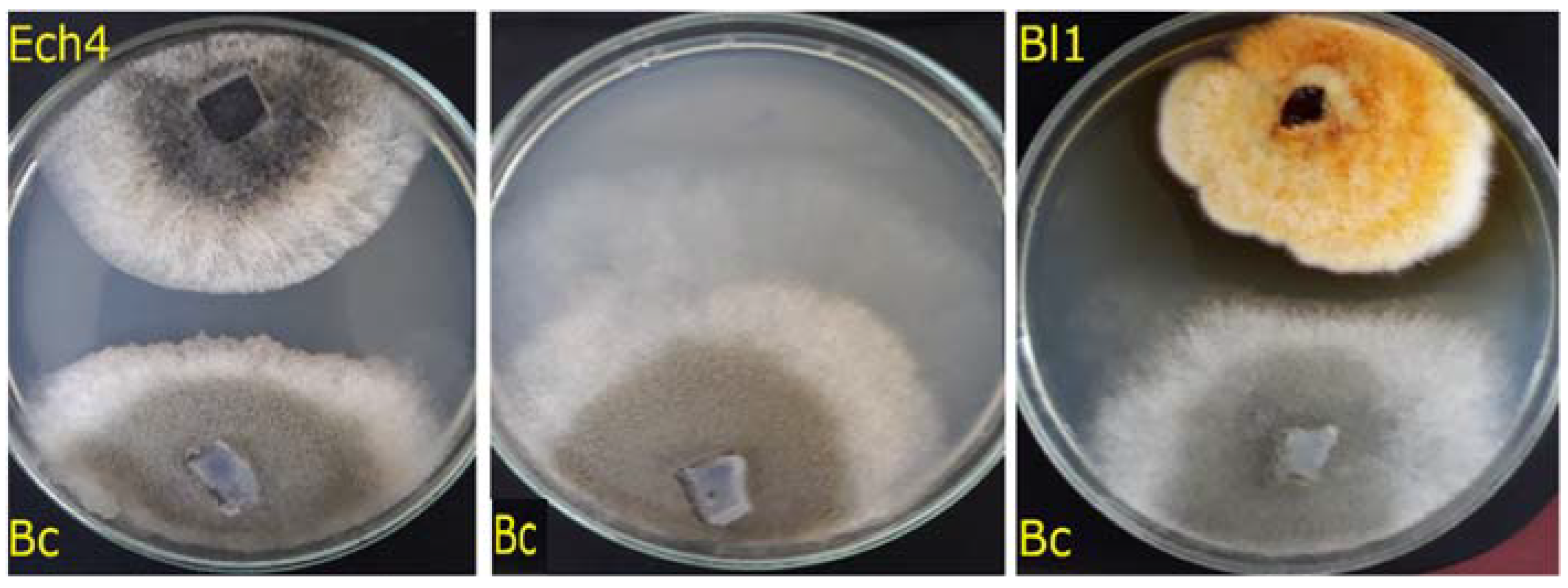
Confrontation Assays

The evaluation of the antifungal activity of the endophytes against B. cinerea was carried out using dual confrontation assays as described by Chen et al. [28], with modifications. Small discs of PDA culture medium with mycelium of the isolated endophytes and B. cinerea were inoculated in opposites sides of PDA plates (6 cm apart) supplemented with antibiotics. B. cinerea, inoculated in one side of the PDA Petri dishes, was used as a control. Inhibition percentage was calculated using the radial mycelial growth of B. cinerea, confronting the endophyte (Ri) and the radial mycelial growth of B. cinerea in the control (Rc) according to the formula (Rc − Ri)/Rc × 100. These experiments were done in triplicate.

2.4. Identification of Endophytes

For the molecular identification of the endophytic fungi, genomic DNA was purified using the CTAB method [29]. Around 200 mg of endophyte tissue obtained from axenic cultures was placed in a Falcon tube with 800 µL of CTAB buffer (3% CTAB, 1.4 M NaCl, 20 mM EDTA and 100 mM Tris-HCl pH 8.0). The tissue was vortexed using 4 mm diameter glass spheres for 3 min. Disrupted tissue was incubated at 60 °C for 30 min. Then, it was centrifuged at 8400× g for 10 min at room temperature, and the supernatant was separated from the cellular debris. The recovered supernatant was treated with 2 ng µL−1 RNAse at 37 °C for 30 min. Next, 800 µL of chloroform:isoamyl alcohol (24:1) was added, and the aqueous phase was recovered after centrifugation for 14,196× g for 10 min at room temperature. Later, 800 µL of cold isopropanol was added and incubated at 20 °C for 2 h. Subsequently, the solution was centrifuged for 14,196× g for 10 min at 4 °C. After, 500 µL absolute ethanol was added to the pellet and dried using a paper towel. Finally, DNA was resuspended in DNAse free water.
ITS regions of the rDNA and β-tubulin gene were analyzed to identify the endophytic fungi [30]. To amplify the ITS sequences, the forward primer ITS-1 (5′-TCCGTAGGTGAACCTGCGG) and reverse primer ITS-4 (5′-TCCTCCGCTTATTGATATGC) were used [31], while to amplify β-tubulin marker, the forward primer BT2aF (GGTAACCAAATCGGTGCTGCTTTC) and reverse primer BT2bR (ACCCTCAGTGTAGTGACCCTTGGC) were used [32].
PCR reaction was performed in 50 µL volumes, containing 2 µL of genomic DNA, 1 µL of forward primer (10 µM), 1 µL of reverse primer (10 µM), 21 µL of nuclease-free water, and 25 µL of GoTaq® Green Master Mix 2× (Promega, Madison WI, USA). PCR reaction mixtures for amplify ITS region were subjected to an initial denaturation at 94 °C for 3 min, and 38 cycles using the following temperatures: 94 °C for 40 s, 55 °C for 45 s, and 72 °C for 40 s; finally, an elongation was carried out at 72 °C for 5 min. The amplification program for β-tubulin PCR began with an initial denaturation at 94 °C for 5 min and 35 cycles using the following temperatures: 94 °C for 45 s, 55 °C for 45 s, and 72 °C for 2 min; finally, an elongation was carried out at 72 °C for 10 min. PCR reactions were performed in an Eppendorf® (Hamburg, Germany) MasterCycler Personal. Finally, the PCR products were sequenced by the Genomic and Bioinformatic Center of the Universidad Católica de Chile using automatic sequencing ABI PRISM 3100.
Electropherograms were manually edited using Geneious Prime® (Biomatters Ltd., Auckland, New Zealand) 2021.01 version software. A consensus sequence was created using both ITS and β-tubulin sequences, BLASTn tool was used to look for similarities with other fungi. Finally, using Geneious Prime software, consensus sequences tree construction was carried out using sequences obtained using ClustalW algorithm. Phylogenetic trees were constructed using the Tamura-Nei genetic distance model using the neighbor-joining method. Resampling for each dendrogram was 1000 using Geneious Prime software.

2.5. Extraction of Secondary Metabolites from the Most Active Endophytic Fungi

For the extraction of secondary metabolites, fungi were inoculated in PDA plates on a cellophane layer and incubated for seven days. After this, cellophane containing the fungi was discarded. Remaining culture medium was extracted using ethyl acetate. The organic phase was evaporated using a rotary evaporator at 40 °C. PDA without endophyte was used as negative control.

2.6. Antifungal Activity against B. Cinerea of the Extracts

The antifungal activity of the extracts was evaluated in vitro. Extracts dissolved in acetone were added to PDA plates at 50, 100, and 200 mg L−1. Acetone was added to PDA plates as a negative control. The final acetone concentration was identical in the control and treatment assay. After acetone evaporation in a laminar flow cabinet, the culture media were inoculated with 0.5 cm agar disks from an active growing culture of B. cinerea. Cultures were incubated in the dark at 22 °C for three days. Mycelium diameter was measured daily in two perpendicular directions. Inhibition percentages were calculated after 72 h of incubation. These experiments were done in triplicate.
A bioautography was carried out using thin-layer chromatography (TLC) to identify what fraction of the extract containing the diffusible compounds had antifungal activity [33,34]. Extract dissolved in methanol was placed in a TLC plate (silica gel 60 F254, Merck, Santiago, Chile) and separated with methanol:chloroform (9:1) as an eluent system. The separation of the compound in the TLC was visualized using UV light at 254 nm. Mobile phase methanol:chloroform (9:1) was placed in TLC plate as solvent control. Bioautography using the TLC with the separated compounds was carried out as described by Vidal et al. [11].
A preliminary characterization of the antifungal compounds in the extracts from the isolates Ech4 and Bl1 was performed using different stain solutions. Extracts were separated in TLC plates using chloroform:methanol (9:1) as an eluent system and stained with the following solutions: sulfuric acid (25% v/v) for organic compounds, Dragendorff’s reagent spray solution (Merck) for alkaloids, solution of iron (III) chloride (2% w/v iron chloride, methanol 50% v/v and 50% v/v water) for phenolics compounds, a solution of vanillin–sulfuric acid (3.5% w/v vanillin in methanol and 0.625% v/v of sulfuric acid) for terpenoids [35] and saponins [36].

2.7. Statistical Analysis

For all the non-parametrical statistical analyses, GraphPad Prism 6.01 was used. Statistical significance between treated groups and the control group by multiple t-tests and Holm-Sidak method are indicated with asterisks (p < 0.05).

3. Results

3.1. Isolation of Endophytes

Endophytic fungi were isolated from root of the endemic plant Echinopsis chiloensis and the native plant Baccharis linearis (Figure 1).
Six endophytic fungi were isolated from apparently healthy E. chiloensis and four fungi from B. linearis (Table 1).
Evaluation of the antifungal activity against B. cinerea of the isolated fungi was carried out by using the dual confrontation assay. Among these, only two isolates, Bl1 and Ech4, showed a significative inhibition of the mycelial growth of B. cinerea (p < 0.05). The calculated inhibition values were 26.4 and 44.0% for Bl1 and Ech4, respectively (Figure 2).
The formation of an inhibition halo between the mycelia of both isolates, Ech4 and Bl1, and B. cinerea (Figure 3) suggests that the antifungal effect was produced by diffusible compounds (antibiosis) secreted by Ech4 and Bl4.

3.2. Antifungal Activity against B. cinerea of the Extracts

Extracts of the secondary metabolites produced from Ech4 and Bl1 were obtained, and the antifungal activity of these extracts was evaluated against B. cinerea (Figure 4). The extract obtained from the endophytic fungus Bl1 showed a higher antifungal activity against B. cinerea than the extract obtained from the endophytic fungus Ech4 at the tested concentrations. The extracts from Bl1 and Ech4 showed a higher antifungal activity at 200 mg L−1, being 54.6 and 44.6%, respectively.
Additionally, to identify what fraction of the extracts was responsible for the antifungal activity, a bioautography assay was carried out using 2 mg of both extracts (Figure 5). Results showed that not all the compounds in the extracts had antifungal activity. Extract of the isolate Bl1 showed an inhibition halo for compounds with lower Rf values; meanwhile, the extract obtained from Ech4 also showed an inhibition halo in the zone of the compounds with higher Rf values (Figure 5).
For the preliminary characterization of the active compounds, different stain solutions were used. The bioactive fraction of the Bl1 extract was positive for Dragendorff’s reagent and iron chloride (III) solution, therefore this fraction would contain compounds of the alkaloid family, with the presence of phenolic groups, while the bioactive fraction of extract from Ech4 was positive for the vanillin/sulfuric acid solution, indicating that it could contain terpenes and saponins (Results not shown).

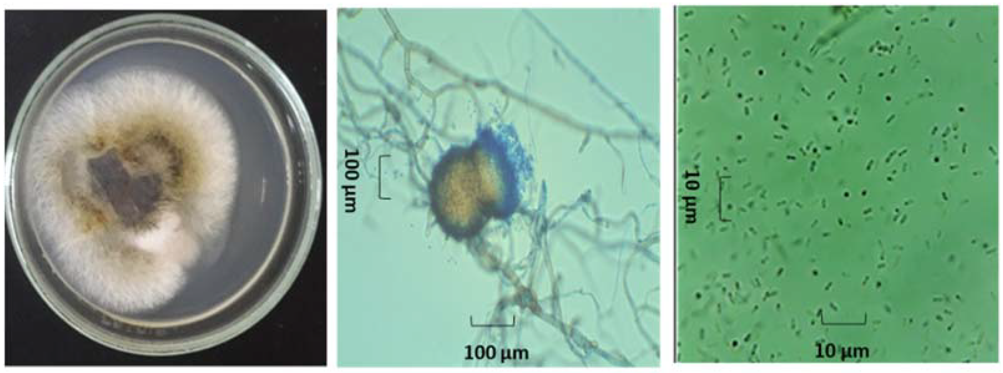
3.3. Identification of Endophytes with Antifungal Activity

For the morphological identification, axenic cultures of both endophytes (Ech4 and Bl1) were observed with or without a lactophenol cotton blue stain under a light microscope. The isolate Bl1 developed orange-pigmented, septate vegetative hyphae with branched growth. A single globular orange conidia was developed in each sporodochium. (Figure 6). On the other hand, the isolate Ech4 showed in PDA medium a branched hyaline septate hypha, yellow-pigmented conidiomata pycnidial globose, covered with some hyphal outgrowths and ovoid shaped small conidia smooth- and thin-walled, hyaline, aseptate. (Figure 7).
For the molecular identification, the ITS region and β-tubulin marker were amplified for both Ech4 and Bl1 isolates. PCR products for the ITS sequence are shown as bands arounds 500 bp and 600 bp in the agarose gel and β-tubulin sequence shown as bands arounds 300 bp. Most similar ITS and β-tubulin sequences to the isolates were searched by an alignment using the NCBI BLASTn tool. The phylogenetic tree, based on ITS and β-tubulin sequences, showed isolate Bl1 and Epicoccum spp. are clustered in the same clade (Figure 8 and Figure 9). On the other hand, the phylogenetic tree based on ITS and β-tubulin showed that the isolate Ech4 is clustered in the same clade with more than one family, such as Cucurbitariaceae, Leptosphaeriaceae, and Pleosporaceae (Figure 10 and Figure 11).

4. Discussion

In this work, ten endophytic fungi were found in roots from plants growing in Chilean Central Precordillera. Six endophytes were found from the endemic plant E. chiloensis. Previously, an endophytic fungus belonging to the genus Alternaria was isolated from the mesenchymal tissue of this plant that inhabited in the same location [11]. This difference could be explained because the diversity of fungi occurring in shoots and roots may have important variations [37]. In addition, mycelial growth within plant tissues is heterogeneous [38], and the presence of endophytes and the synthesis of their metabolites may have marked seasonal variations [39,40]. Also, various isolates from the same fungal specie, obtained from separate plant sources, can produce different secondary metabolites [41].
On the other hand, in the endemic plant B. linearis, four endophytes were found, and this is the first report of endophytic fungi isolated from this plant.
Consequently, ten endophytes were isolated in this work, a similar number as other studies. For example, fourteen endophytic fungi were isolated from five endemic plants in India using a similar isolation method [42], and around five endophytes per plant from different gymnosperm plants were found in Chile [17]. On the other hand, also a higher number of endophytes have been reported in other studies, for example, a 108 endophytes were obtained from the endemic plant Opuntia humifusa in the United States [26], and 319 fungal species were isolated from the roots of 24 plant species from Spain [43]. Nevertheless, the relatively low number of endophytes found in this study could be explained by a rigorous disinfection of the plant surface or by different growing conditions, for instance, Silva-Hughes et al. incubated plant fragments for 60 days [26], an incubation period around four times longer than in this work. Out of ten endophytic fungi, two fungi designated as Bl1 and Ech4 showed antifungal activity against B. cinerea in dual confrontation assays; this inhibition was similar to an endophytic Alternaria sp. against the same isolate of B. cinerea using the same experimental conditions [11].Other authors previously reported endophytic fungi with antifungal activity against B. cinerea in Chile, such as endophytes found in the endemic tree Embotrium coccineum [18], endophytic fungi isolated from Artemisia absinthium [44], Penicillium janczewskii and Microsphaeropsis olivacea, endophytes isolated from Chilean native gymnosperms [17], and Alternaria sp. and Aureobasidium sp. fungi isolated from Chilean endemic and native plants [11]. This suggests that the fungi found could have potential as a biocontrol agent of this phytopathogen.
In addition, it was shown that both Bl1 and Ech4 isolates inhibited the B. cinerea mycelial growth by antibiosis, similar to other endophytic fungi; for example, fungi isolated from Theobroma cacao showed antibiosis against pathogens such as Moniliophthora roreri and Phytophthora palmivora [45], and endophytic fungi isolated from Aloe vera also inhibited by antibiosis the mycelial growth of Fusarium oxysporum [46]. The inhibition halo found in both isolates indicates that the antifungal activity of the obtained endophytic fungi is due to the secretion of diffusible secondary metabolites to the culture medium.
The identification of the Bl1 and Ech4 isolates by phylogenetic analysis based on the molecular markers ITS-rDNA and β-tubulin suggests that both endophytes are grouped with species of the order Pleosporales. The order of the Pleosporales is one of the largest of Dothideomycetes [47]. In this order, families such as Didymellaceae, Didymosphaeriaceae, Pleosporaceae, Hypsostromataceae, Cucurbitariacea, and others have been identified, and, within this classification, epiphytic, endophytic, and phytopathogenic fungal species have been found [48]. In detail, the sequences of the isolate Bl1 are more closely grouped with the genus Epicoccum, while the Ech4 isolate is closely grouped with more than one family within the suborder Pleosporineae [48].
On the other hand, in the case of isolate Bl1, this grouping is confirmed with the morphological analysis of the axenic culture, where the presence of sporodochia with orange-pigmented conidiomata are characteristic to species belonging to the genus Epicoccum, such as E. italicum, E. layuense, and E. poae, and the difference between these species is the conidium size [49,50]. Epicoccum species have been extensively reviewed as ubiquitous phytopathogens that can be used as biological control agents [51]. Epicoccum spp. have been found in different plant parts [50] and may be endophytes [52] and even saprophytes or pathogens [53,54].
Axenic culture of the isolate Ech4 showed small ovoid-shaped conidia and pigmented pilose pycnidia, which is described for different species of the Pleosporales order, such as the Phoma genus [49,55,56]. However, Phoma is considered a group of paraphyletic species that are continuously renamed and reassigned to different families of the Pleosporinae suborder, in addition to some genera whose family are still considered incertae sedis [47,48,57]. Additionally, Hou et al. suggested that the use of conventional molecular markers such as ITS and β-Tubulin present limitations that can be improved with the use of the rpb2 molecular marker to arrive at more reliable phylogenies [58]. Thus, in this study it is only established that isolate Ech4 corresponds to the Pleosporales order.
Many studies have been described that fungi of the order Pleosporales and the genus Epicoccum have been isolated as endophytes and their antifungal activity against different phytopathogenic fungi has been demonstrated [59,60,61,62]. This is the first time that Epicoccum sp. have been isolated from E. chiloensis and that an endophytic fungus of the order Pleosporales have been isolated from B. linearis.
On the other hand, in this work, the results suggest that both extracts obtained from the fungal isolates inhibit the mycelial growth of B. cinerea. Compounds with antimicrobial activity have been described for Epicoccum sp. and for many other Pleosporales species, such as Coniothyrium sp. and Phoma sp. [13,63,64]. Bioautography assays results suggest that orange active fraction of the extract from the isolate Bl1 (Epicoccum sp.), belongs to a group of pigmented compounds such as polyketides, carotenoids, and flavonoids already described in the genus Epicoccum [65]. Preliminary characterization of active fractions of the extract of the Epicoccum sp. isolate presented positive reaction with Dragendorff’s stain and iron (III) chloride solution, suggesting the presence of alkaloids and phenolics compounds in the extract. Alkaloids such as epicorazine A and epicorazine B have been described for this fungus and they have shown antimicrobial activity [66]. Also, aromatic polyketides with antifungal properties have been previously found in the Epicoccum spp. [67]. Meanwhile, active fraction of the extract from the isolate Ech4 suggest the presence of antifungal compounds. Some Pleosporales species of endophytic fungi such as Phoma sp. [59] and Alternaria sp. [11] produce metabolites with antifungal activity against B. cinerea [11,68]. Since the extract from Ech4 only showed a positive reaction for the vanillin/sulfuric acid stain, this suggests the presence of terpenes and their derivatives, such as saponins [69,70]. It has been reported that similar compounds have been described for fungal species of the order Pleosporales, such as Leptosphaeria sp. producing triterpenoid saponin to improve damage in their host, Dipsacus asperoides [71], and Phoma sp. producing terpenes, such as aphidicolin, a specific inhibitor of DNA polymerase, as well as antifungal compounds such as furan and dihydrofuran derivatives [64,72].
In conclusion, two endophytic fungi, Epicoccum sp. and Pleosporales sp., with antifungal activity against B. cinerea were isolated from the roots of plants growing in the Chilean Central Precordillera. This is the first report of Epicoccum sp. being isolated from E. chiloensis and of an endophytic fungus of the order Pleosporales being isolated from B. linearis. Also, possibly alkaloids and phenolic compounds were found in the extract from Epicoccum sp., and terpenes and their derivate compounds such as saponins from the fungus Pleosporales sp., could be responsible for the antifungal activity against B. cinerea, further study of these compounds and their effects may be a new alternative for the biocontrol of phytopathogens.

Author Contributions

Conceptualization, L.M. and M.C.; methodology, C.C.; software, R.P.; writing—original draft preparation, P.C. and C.C.; writing—review and editing, P.C., C.C., R.P., M.C. and L.M.; project administration, M.C.; funding acquisition, M.C. All authors have read and agreed to the published version of the manuscript.

Funding

This research was funded by DICYT Proyecto POSTDOC_DICYT, Código 022040CT_POSTDOC.

Institutional Review Board Statement

Not applicable.

Informed Consent Statement

Not applicable.

Data Availability Statement

The data presented in this study are available on request from the corresponding author.

Acknowledgments

The authors thank to Dirección de Investigación Científica y Tecnológica (DICYT) USACH and to Universidad de Santiago de Chile grant DICYT Proyecto POSTDOC_DICYT, Código 022040CT_POSTDOC for their financial support.

Conflicts of Interest

The authors declare no conflict of interest.

References

  1. Fillinger, S.; Elad, Y. Botrytis-the Fungus, the Pathogen and Its Management in Agricultural Systems, 1st ed.; Springer: Cham, Switzerland, 2016; pp. 189–216. [Google Scholar]
  2. Baćmaga, M.; Wyszkowska, J.; Kucharski, J. The biochemical activity of soil contaminated with fungicides. J. Environ. Sci. Health Part B Pestic. Food Contam. Agric. Wastes 2019, 54, 252–262. [Google Scholar] [CrossRef]
  3. Hahn, M. The rising threat of fungicide resistance in plant pathogenic fungi: Botrytis as a case study. J. Chem. Biol. 2014, 7, 133–141. [Google Scholar] [CrossRef] [PubMed] [Green Version]
  4. Esterio, M.; Copier, C.; Román, A.; Araneda, M.J.; Rubilar, M.; Pérez, I.; Auger, J. Frequency of fungicide-resistant Botrytis cinerea populations isolated from ‘Thompson Seedless’ table grapes in the Central Valley of Chile. Sci. Investig. Agrar. 2017, 44, 295–306. [Google Scholar] [CrossRef] [Green Version]
  5. Roca-Couso, R.; Flores-Féli, J.D.; Rivas, R. Mechanisms of action of microbial biocontrol agents against Botrytis cinerea. J. Fungi 2021, 7, 1045. [Google Scholar] [CrossRef]
  6. Gholami-Shabani, M.; Shams-Ghahfarokhi, M.; Razzaghi-Abyaneh, M. Natural product synthesis by fungi: Recent trends and future prospects. In Recent Advancement in White Biotechnology Through Fungi; Springer: Cham, Switzerland, 2019; pp. 385–401. [Google Scholar]
  7. Selim, K.A.; El-Beih, A.A.; AbdEl-Rahman, T.M.; El-Diwany, A.I. Biology of endophytic fungi. Curr. Res. Environ. Appl. Mycol. 2012, 2, 31–82. [Google Scholar] [CrossRef]
  8. Jia, M.; Chen, L.; Xin, H.L.; Zheng, C.J.; Rahman, K.; Han, T.; Qin, L.P. A friendly relationship between endophytic fungi and medicinal plants: A systematic review. Front. Microbiol. 2016, 7, 1–14. [Google Scholar] [CrossRef] [Green Version]
  9. Schulz, B.; Boyle, C.; Draeger, S.; Römmert, A.K.; Krohn, K. Endophytic fungi: A source of novel biologically active secondary metabolites. Mycol. Res. 2002, 106, 996–1004. [Google Scholar] [CrossRef]
  10. Deshmukh, S.K.; Gupta, M.K.; Prakash, V.; Saxena, S. Endophytic fungi: A source of potential antifungal compounds. J. Fungi 2018, 4, 77. [Google Scholar] [CrossRef] [Green Version]
  11. Vidal, A.; Parada, R.; Mendoza, L.; Cotoras, M. Endophytic fungi isolated from plants growing in central andean precordillera of chile with antifungal activity against Botrytis cinerea. J. Fungi 2020, 6, 149. [Google Scholar] [CrossRef]
  12. Xu, D.; Zhang, B.-Y.; Yang, X.-L. Antifungal monoterpene derivatives from the plant endophytic fungus Pestalotiopsis foedan. Chem. Biodivers. 2016, 13, 1422–1425. [Google Scholar] [CrossRef]
  13. Sun, P.; Huo, J.; Kurtán, T.; Mándi, A.; Antus, S.; Tang, H.; Draeger, S.; Schulz, B.; Hussain, H.; Krohn, K.; et al. Structural and stereochemical studies of hydroxyanthraquinone derivatives from the endophytic fungus Coniothyrium sp. Chirality 2013, 25, 141–148. [Google Scholar] [CrossRef] [PubMed] [Green Version]
  14. Richardson, S.N.; Walker, A.K.; Nsiama, T.K.; McFarlane, J.; Sumarah, M.; Ibrahim, A.; Miller, J.D. Griseofulvin-producing Xylaria endophytes of Pinus strobus and Vaccinium angustifolium: Evidence for a conifer-understory species endophyte ecology. Fungal Ecol. 2014, 11, 107–113. [Google Scholar] [CrossRef]
  15. Wang, J.; Wang, G.; Zhang, Y.; Zheng, B.; Zhang, C.; Wang, L. Isolation and identification of an endophytic fungus Pezicula sp. in Forsythia viridissima and its secondary metabolites. World J. Microbiol. Biotechnol. 2014, 30, 2639–2644. [Google Scholar] [CrossRef] [PubMed]
  16. Bai, Y.-B.; Gao, Y.-Q.; Nie, X.-D.; Tuong, T.-M.; Li, D.; Gao, J.-M. Antifungal activity of griseofulvin derivatives against phytopathogenic fungi in vitro and in vivo and three-dimensional quantitative structure-activity relationship analysis. J. Agric. Food Chem. 2019, 67, 6125–6132. [Google Scholar] [CrossRef] [PubMed]
  17. Hormazábal, E.; Piontelli, E. Endophytic fungi from Chilean native gymnosperms: Antimicrobial activity against human and phytopathogenic fungi. World J. Microbiol. Biotechnol. 2009, 25, 813–819. [Google Scholar] [CrossRef]
  18. González-Teuber, M. The defensive role of foliar endophytic fungi for a South American tree. AoB Plants 2016, 8, plw050. [Google Scholar] [CrossRef] [Green Version]
  19. Strobel, G.; Daisy, B. Bioprospecting for microbial endophytes and their natural products. Microbiol. Mol. Biol. Rev. 2003, 67, 491–502. [Google Scholar] [CrossRef] [Green Version]
  20. Ovalle, C.; Del Pozo, A.; Casado, M.A.; Acosta, B.; De Miguel, J.M. Consequences of landscape heterogeneity on grassland diversity and productivity in the Espinal agroforestry system of central Chile. Landsc. Ecol. 2006, 21, 585–594. [Google Scholar] [CrossRef]
  21. Rundel, P.W.; Arroyo, M.T.; Cowling, R.M.; Keeley, J.E.; Lamont, B.B.; Vargas, P. Mediterranean biomes: Evolution of their vegetation, floras, and climate. Annu. Rev. Ecol. Evol. Syst. 2016, 47, 383–407. [Google Scholar] [CrossRef]
  22. Oses-Pedraza, R.; Hernández, V.; Campos, L.; Becerra, J.; Irribarren-Riquelme, D.; Lavín, P.; Rodríguez, J. Advances in research on biodiversity and bioprospecting of endophytic fungi in Chile. In Neotropical Endophytic Fungi, 1st ed.; Rosa, L.H., Ed.; Springer: Cham, Switzerland, 2021; Volume 1, pp. 53–91. [Google Scholar]
  23. Walter, H.E. Floral biology of Echinopsis chiloensis ssp. chiloensis (Cactaceae): Evidence for a mixed pollination syndrome. Flora Morphol. Distrib. Funct. Ecol. Plants 2010, 205, 757–763. [Google Scholar] [CrossRef]
  24. Aljaro, M.E.; Frias, D.; Montenegro, G. Life cycle of Rhachiptera limbata (Diptera, Tephritidae) and its relationship with Baccharis linearis (Compositae). Rev. Chil. Hist. Nat. 1984, 57, 123–129. [Google Scholar]
  25. Gazitúa, M.C.; Morgante, V.; Poupin, M.J.; Ledger, T.; Rodríguez-Valdecantos, G.; Herrera, C.; González-Chávez, M.D.C.; Ginocchio, R.; González, B. The microbial community from the early-plant colonizer (Baccharis linearis) is required for plant establishment on copper mine tailings. Sci. Rep. 2021, 11, 10448. [Google Scholar] [CrossRef] [PubMed]
  26. Silva-Hughes, A.F.; Wedge, D.E.; Cantrell, C.L.; Carvalho, C.R.; Pan, Z.; Moraes, R.M.; Madoxx, V.L.; Rosa, L.H. Diversity and antifungal activity of the endophytic fungi associated with the native medicinal cactus Opuntia humifusa (Cactaceae) from the United States. Microbiol. Res. 2015, 175, 67–77. [Google Scholar] [CrossRef] [PubMed]
  27. Muñoz, G.; Hinrichsen, P.; Brygoo, Y.; Giraud, T. Genetic characterisation of Botrytis cinerea populations in Chile. Mycol. Res. 2002, 106, 594–601. [Google Scholar] [CrossRef]
  28. Chen, P.-H.; Chen, R.-Y.; Chou, J.-Y. Screening and evaluation of yeast antagonists for biological control of Botrytis cinerea on strawberry fruits. Mycobiology 2018, 46, 33–46. [Google Scholar] [CrossRef] [PubMed] [Green Version]
  29. Irfan, M.; Ting, Z.T.; Yang, W.; Chunyu, Z.; Qing, M.; Lijun, Z.; Feng, L. Modification of CTAB protocol for maize genomic DNA extraction. Res. J. Biotechnol. 2013, 8, 41–45. [Google Scholar]
  30. Raja, H.A.; Miller, A.N.; Pearce, C.J.; Oberlies, N.H. Fungal identification using molecular tools: A primer for the natural products research community. J. Nat. Prod. 2017, 80, 756–770. [Google Scholar] [CrossRef]
  31. White, T.J.; Bruns, T.; Lee, S.; Taylor, J.W. Amplification and direct sequencing of fungal ribosomal RNA genes for phylogenetics. PCR Protoc. 1990, 18, 315–322. [Google Scholar] [CrossRef]
  32. Hubka, V.; Kolarik, M. β-tubulin paralogue tubC is frequently misidentified as the benA gene in Aspergillus section Nigri taxonomy: Primer specificity testing and taxonomic consequences. Persoonia 2012, 29, 1–10. [Google Scholar] [CrossRef] [Green Version]
  33. Choma, I.M.; Grzelak, E.M. Bioautography detection in thin-layer chromatography. J. Chromatogr. A 2011, 1218, 2684–2691. [Google Scholar] [CrossRef]
  34. Dissanayak, M.; Ito, S.-I.; Akakabe, Y. TLC bioautography guided detection and biological activity of antifungal compounds from medicinal plant Acorus calamus Linn. Asian J. Plant Pathol. 2015, 9, 16–26. [Google Scholar] [CrossRef] [Green Version]
  35. Visweswari, G.; Christopher, R.; Rajendra, W. Phytochemical screening of active secondary metabolites present in Withania somnifera root: Role in traditional medicine. Int. J. Pharm. Sci. Res. 2013, 4, 2770. [Google Scholar] [CrossRef]
  36. Cheok, C.Y.; Salman, H.A.K.; Sulaiman, R. Extraction and quantification of saponins: A review. Food Res. Int. 2014, 59, 16–40. [Google Scholar] [CrossRef]
  37. Bayman, P.; Lebrón, L.L.; Tremblay, R.; Lodge, D.J. Variation in endophytic fungi from roots and leaves of Lepanthes (Orchidaceae). New Phytol. 1997, 135, 143–149. [Google Scholar] [CrossRef] [PubMed] [Green Version]
  38. McGonigle, T.P.; Miller, M.H.; Evans, D.G.; Fairchild, G.L.; Swan, J.A. A new method which gives an objective measure of colonization of roots by vesicular—arbuscular mycorrhizal fungi. New Phytol. 1990, 115, 495–501. [Google Scholar] [CrossRef]
  39. Guo, L.-D.; Huang, G.-R.; Wang, Y. Seasonal and tissue age influences on endophytic fungi of Pinus tabulaeformis (Pinaceae) in the Dongling Mountains, Beijing. J. Integr. Plant Biol. 2008, 50, 997–1003. [Google Scholar] [CrossRef]
  40. Rogers, W.M.; Roberts, C.A.; Andrae, J.G.; Davis, D.K.; Rottinghaus, G.E.; Hill, N.S.; Kallenbach, R.L.; Spiers, D.E. Seasonal fluctuation of ergovaline and total ergot alkaloid concentrations in tall fescue regrowth. Crop Sci. 2011, 51, 1291–1296. [Google Scholar] [CrossRef]
  41. Susca, A.; Proctor, R.H.; Butchko, R.A.; Haidukowski, E.M.; Stea, G.; Logrieco, A.F.; Moretti, A. Variation in the fumonisin biosynthetic gene cluster in fumonisin-producing and nonproducing black aspergilli. Fungal Genet. Biol. 2014, 73, 39–52. [Google Scholar] [CrossRef] [Green Version]
  42. Anitha, D.; Vijaya, T.; Pragathi, D.; Reddy, N.V.; Mouli, K.C.; Venkateswarulu, N.; Bhargav, D.S. Isolation and characterization of endophytic fungi from endemic medicinal plants of Tirumala hills. Int. J. Life Sci. Biotechnol. Pharm. Res. 2013, 2, 367–373. [Google Scholar]
  43. Macia-Vicente, J.G.; Jansson, H.-B.; Abdullah, S.K.; Descals, E.; Salinas, J.; Lopez-Llorca, L.V. Fungal root endophytes from natural vegetation in Mediterranean environments with special reference to Fusarium spp. FEMS Microbiol. Ecol. 2008, 64, 90–105. [Google Scholar] [CrossRef] [Green Version]
  44. Noumeur, S.; Mancini, V.; Romanazzi, G. Activity of endophytic fungi from Artemisia absinthium on Botrytis cinerea. Acta Hortic. 2016, 1144, 101–104. [Google Scholar] [CrossRef]
  45. Mejia, L.; Rojas, E.I.; Maynard, Z.; Van Bael, S.; Arnold, A.E.; Hebbar, P.; Samuels, G.J.; Robbins, N.; Herre, E.A. Endophytic fungi as biocontrol agents of Theobroma cacao pathogens. Biol. Control 2008, 46, 4–14. [Google Scholar] [CrossRef]
  46. Rahul, Y.; Ajay, V.S.; Samiksha, J.; Manish, K. Antifungal and enzyme activity of endophytic fungi isolated from Ocimum sanctum and Aloe vera. Afr. J. Microbiol. Res. 2015, 9, 1783–1788. [Google Scholar] [CrossRef] [Green Version]
  47. Hongsanan, S.; Phookamsak, R.; Goonasekara, I.D.; Thambugala, K.M.; Hyde, K.D.; Bhat, J.D.; Suwannarach, N.; Cheewangkoon, R. Introducing a new pleosporalean family Sublophiostomataceae fam. nov. to accommodate Sublophiostoma gen. nov. Sci. Rep. 2021, 11, 1–10. [Google Scholar] [CrossRef]
  48. Zhang, Y.; Crous, P.W.; Schoch, C.L.; Hyde, K.D. Pleosporales. Fungal Divers. 2012, 53, 1–221. [Google Scholar] [CrossRef] [PubMed] [Green Version]
  49. Larone, D.H. Medically important fungi: A guide to identification-5th edition. Lab. Med. 2014, 45, e68–e69. [Google Scholar] [CrossRef] [Green Version]
  50. Chen, Q.; Hou, L.W.; Duan, W.J.; Crous, P.W.; Cai, L. Didymellaceae revisited. Stud. Mycol. 2017, 87, 105–159. [Google Scholar] [CrossRef]
  51. Taguiam, J.D.; Evallo, E.; Balendres, M.A. Epicoccum species: Ubiquitous plant pathogens and effective biological control agents. Eur. J. Plant Pathol. 2021, 159, 713–725. [Google Scholar] [CrossRef]
  52. Araújo, F.D.D.S.; Fávaro, L.C.D.L.; Araújo, W.L.; de Oliveira, F.L.; Aparicio, R.; Marsaioli, A.J. Epicolactone-Natural product isolated from the sugarcane endophytic fungus Epicoccum nigrum. Eur. J. Org. Chem. 2012, 27, 5225–5230. [Google Scholar] [CrossRef]
  53. Beasley, D.R.; Joyce, D.C.; Coates, L.M.; Wearing, A.H. Saprophytic microorganisms with potential for biological control of Botrytis cinerea on Geraldton waxflower flower. Aust. J. Exp. Agric. 2001, 41, 697–703. [Google Scholar] [CrossRef] [Green Version]
  54. Taguiam, J.D.; Evallo, E.; Bengoa, J.; Maghirang, R.; Balendres, M.A. Pathogenicity of Epicoccum sorghinum towards dragon fruits (Hylocereus species) and In Vitro evaluation of chemicals with antifungal activity. J. Phytopathol. 2020, 168, 303–310. [Google Scholar] [CrossRef]
  55. Aveskamp, M.M.; Verkley, G.J.M.; De Gruyter, J.; Murace, M.A.; Perelló, A.; Woudenberg, J.H.C.; Groenewald, J.Z.; Crous, P.W. DNA phylogeny reveals polyphyly of Phoma section Peyronellaea and multiple taxonomic novelties. Mycologia 2009, 101, 363–382. [Google Scholar] [CrossRef] [PubMed] [Green Version]
  56. Bennett, A.; Ponder, M.M.; Garcia-Diaz, J. Phoma infections: Classification, potential food sources, and their clinical impact. Microorganisms 2018, 6, 58. [Google Scholar] [CrossRef] [PubMed] [Green Version]
  57. De Gruyter, J.; Woudenberg, J.H.C.; Aveskamp, M.M.; Verkley, G.J.M.; Groenewald, J.Z.; Crous, P.W. Redisposition of phoma-like anamorphs in Pleosporales. Stud. Mycol. 2013, 75, 1–36. [Google Scholar] [CrossRef] [PubMed] [Green Version]
  58. Hou, L.; Groenewald, J.; Pfenning, L.; Yarden, O.; Crous, P.; Cai, L. The phoma-like dilemma. Stud. Mycol. 2020, 96, 309–396. [Google Scholar] [CrossRef]
  59. Wang, L.; Xu, B.-G.; Wang, J.-Y.; Su, Z.-Z.; Lin, F.-C.; Zhang, C.-L.; Kubicek, C.P. Bioactive metabolites from Phoma species, an endophytic fungus from the Chinese medicinal plant Arisaema erubescens. Appl. Microbiol. Biotechnol. 2012, 93, 1231–1239. [Google Scholar] [CrossRef] [PubMed]
  60. Alcock, A.; Elmer, P.; Marsden, R.; Parry, F. Inhibition of Botrytis cinerea by epirodin: A secondary metabolite from New Zealand isolates of Epicoccum nigrum. J. Phytopathol. 2015, 163, 841–852. [Google Scholar] [CrossRef]
  61. Gupta, S.; Kaul, S.; Singh, B.; Vishwakarma, R.A.; Dhar, M.K. Production of gentisyl alcohol from Phoma herbarum endophytic in Curcuma longa L. and its antagonistic activity towards leaf spot pathogen Colletotrichum gloeosporioides. Appl. Biochem. Biotechnol. 2016, 180, 1093–1109. [Google Scholar] [CrossRef]
  62. Song, X.; Li, Y.; Hu, Y.; Guo, W.; Wu, Z.; Zhang, Y.; Cao, Z. Investigation of endophytes from blueberry roots with antifungal activity and plant growth enhancement. Rhizosphere 2021, 20, 100454. [Google Scholar] [CrossRef]
  63. Kaliňák, M.; Barátová, V.; Gallová, E.; Ondrušová, Z.; Hudecová, D. Secondary metabolite production of Epicoccum sp. isolated from lignite. Acta Chim. Slovaca 2013, 6, 42–48. [Google Scholar] [CrossRef]
  64. Hussain, H.; Kock, I.; Al-Harrasi, A.; Al-Rawahi, A.; Abbas, G.; Green, I.R.; Shah, A.; Badshah, A.; Saleem, M.; Draeger, S.; et al. Antimicrobial chemical constituents from endophytic fungus Phoma sp. Asian Pac. J. Trop. Med. 2014, 7, 699–702. [Google Scholar] [CrossRef] [Green Version]
  65. Fatima, N.; Ismail, T.; Muhammad, S.A.; Jadoon, M.; Ahmed, S.; Azhar, S.; Mumtaz, A. Epicoccum sp., an emerging source of unique bioactive metabolites. Acta Pol. Pharm. Drug Res. 2016, 73, 13–21. [Google Scholar]
  66. Deffieux, G.; Filleau, M.-J.; Baute, R. New antibiotics from the fungus Epicoccum nigrum. III. Epicorazine B: Structure elucidation and absolute configuration. J. Antibiot. 1978, 31, 1106–1109. [Google Scholar] [CrossRef] [PubMed]
  67. Braga, R.M.; Padilla, G.; Araújo, W.L. The biotechnological potential of Epicoccum spp.: Diversity of secondary metabolites. Crit. Rev. Microbiol. 2018, 44, 759–778. [Google Scholar] [CrossRef] [PubMed]
  68. Shentu, X.; Zhan, X.; Ma, Z.; Yu, X.; Zhang, C.-X. Antifungal activity of metabolites of the endophytic fungus Trichoderma brevicompactum from garlic. Braz. J. Microbiol. 2014, 45, 248–254. [Google Scholar] [CrossRef] [PubMed] [Green Version]
  69. Paulsamy, S.; Karthika, K. TLC and HPTLC fingerprints of various secondary metabolites in the stem of the traditional medicinal climber, Solena amplexiacaulis. Indian J. Pharm. Sci. 2015, 77, 111–116. [Google Scholar] [CrossRef] [PubMed] [Green Version]
  70. Le, A.V.; Parks, S.E.; Nguyen, M.H.; Roach, P.D. Improving the vanillin-sulphuric acid method for quantifying total saponins. Technologies 2018, 6, 84. [Google Scholar] [CrossRef] [Green Version]
  71. Gong, A.; Zhou, T.; Xiao, C.; Jiang, W.; Zhou, Y.; Zhang, J.; Liang, Q.; Yang, C.; Zheng, W.; Zhang, C. Association between dipsacus saponin VI level and diversity of endophytic fungi in roots of Dipsacus asperoides. World J. Microbiol. Biotechnol. 2019, 35, 1–14. [Google Scholar] [CrossRef] [Green Version]
  72. Oikawa, H.; Toshima, H.; Ohashi, S.; König, W.A.; Kenmoku, H.; Sassa, T. Diversity of diterpene hydrocarbons in fungus Phoma betae. Tetrahedron Lett. 2001, 42, 2329–2332. [Google Scholar] [CrossRef]
Figure 1. Recollected plants from the Andean Precordillera of Chile. E. chiloensis (left) and B. linearis (right).
Jof 08 00197 g001
Figure 2. Antifungal effect of isolate Bl1 (black bar) and isolate Ech4 (grey bar) on B. cinerea growth. Each bar represents the average inhibition percentage of three experiments ± SD. Statistical significance when comparing treated groups and control group by multiple t-tests and Holm-Sidak method are indicated with asterisks (p < 0.05).
Jof 08 00197 g002
Figure 3. Effect of the isolates Ech4 (upper left) negative control (center) and Bl1 (upper right) on the mycelial growth of B. cinerea (bottom of both pictures).
Jof 08 00197 g003
Figure 4. Antifungal activity of the extracts obtained from the endophytic fungi Bl1 (black bars) and Ech4 (grey bars) against B. cinerea. Each bar represents the average inhibition percentage of B. cinerea cultured in three experiments per treatment ± SD. Statistical significance when comparing treated groups and control group by multiple t-tests and Holm-Sidak method are indicated with asterisks (p < 0.05).
Jof 08 00197 g004
Figure 5. Bioautography assay for the extracts. (A) Chromatogram of the extract obtained from Bl1 visualized using UV light (254 nm) (mobile phase CHCl3:MeOH 9:1) (left). Bioautography of the extract Bl1 (center). Zone of inhibition (right). (B) Chromatogram of the extract obtained from Ech4 visualized using UV light (254 nm) (left) (mobile phase CHCl3:MeOH 9:1). Bioautography of the extract Ech4 (center). Zone of inhibition (right) Red circle indicates zone of inhibition. (C) Solvent control (mobile phase CHCl3:MeOH 9:1).
Jof 08 00197 g005
Figure 6. Morphological examination of the isolate Bl1. Colony on PDA (Left), sporodochia (center), and conidium (right).
Jof 08 00197 g006
Figure 7. Morphological examination of the isolate Ech4. Colony on PDA (left), hyphae and conidiomata picnidial (center), and conidia (right).
Jof 08 00197 g007
Figure 8. Neighbor-joining tree based on fungal internal transcribed spacer (ITS) sequences of Bl1 (Accession: OK090879), 14 Epicoccum species and Pleospora welwitschiae CBS 463.84 as outgroup. Numbers labeled at each node indicate bootstrap value (percentage) from 1000 replicates. Bar, 0.04 substitutions per nucleotide position.
Jof 08 00197 g008
Figure 9. Neighbor-joining tree based on fungal tubulin sequences of Bl1 (Accession: OK319031), 11 Pleosporales species and Phoma nigrificans CBS 740.96 as outgroup. Numbers labeled at each node indicate bootstrap value (percentage) from 1000 replicates. Bar, 0.04 substitutions per nucleotide position.
Jof 08 00197 g009
Figure 10. Neighbor-joining tree based on fungal internal transcribed spacer (ITS) sequences of Ech4 (Accession: OK090880), 15 Pleosporales species. and Preussia persica CBS 117,680 as outgroup. Numbers labeled at each node indicate bootstrap value (percentage) from 1000 replicates. Bar, 0.04 substitutions per nucleotide position.
Jof 08 00197 g010
Figure 11. Neighbor-joining tree based on fungal tubulin sequences of Ech4 (Accession: OK319032), 8 Pleosporales species and Preussia procaviicola CBS 146,981 as outgroup. Numbers labeled at each node indicate bootstrap value (percentages) from 1000 replicates. Bar, 0.04 substitutions per nucleotide position.
Jof 08 00197 g011
Table 1. Endophytes isolated from the recollected plants.
Plant.Fungal Isolate
E. chiloensisEch1
Ech2
Ech3
Ech4
Ech5
Ech6
B. linearisBl1
Bl2
Bl3
Bl4
Publisher’s Note: MDPI stays neutral with regard to jurisdictional claims in published maps and institutional affiliations.

