Article

Multi-Gene Phylogenetic Analyses Revealed Five New Species and Two New Records of Distoseptisporales from China

1
School of Food and Pharmaceutical Engineering, Guizhou Institute of Technology, Guiyang 550003, China
2
Center of Excellence in Fungal Research, Mae Fah Luang University, Chiang Rai 57100, Thailand
3
Guizhou Key Laboratory of Agricultural Biotechnology, Guizhou Academy of Agricultural Sciences, Guiyang 550006, China
4
School of Science, Mae Fah Luang University, Chiang Rai 57100, Thailand
5
Engineering and Research Center for Southwest Bio-Pharmaceutical Resources of National Education Ministry of China, Guizhou University, Guiyang 550025, China
*
Author to whom correspondence should be addressed.
J. Fungi 2022, 8(11), 1202; https://doi.org/10.3390/jof8111202
Submission received: 26 September 2022 / Revised: 6 November 2022 / Accepted: 7 November 2022 / Published: 14 November 2022
(This article belongs to the Special Issue Ascomycota: Diversity, Taxonomy and Phylogeny)

Abstract

:
Eight hyphomycetes were collected as part of an investigation into the diversity of hyphomycetous fungi in China. Based on morphology and multi-loci (LSU, ITS, tef1α, and rpb2) phylogenetic analyses, five new taxa, including a new Aquapteridospora species A. hyalina and four novel Distoseptispora species, viz D. aquisubtropica, D. septata, D. tropica, and D. wuzhishanensis were introduced in Distoseptisporales (Sordariomycetes). Two new habitat records, viz Distoseptispora pachyconidia and D. xishuangbannaensis were firstly reported. Also provided in this study are detailed descriptions of eight new collections and a revised phylogenetic tree for the Distoseptisporales.

1. Introduction

The two genera, Distoseptispora (Distoseptisporaceae) and Aquapteridospora (Aquapteridosporaceae) were recently introduced to Distoseptisporales (Diaporthomycetidae) [1,2,3]. Yang et al. [4] established Aquapteridospora (Diaporthomycetidae) as a monotypic genus, with A. lignicola as the type species. In later research, Hyde et al. [1] introduced Aquapteridosporaceae to accommodate Aquapteridospora and placed this family in the order Distoseptisporales based on divergence estimates, morphological characteristics, and phylogenetic analysis. This was followed in the “Outline of Fungi and fungus-like taxa” by Wijayawardene et al. [5]. Currently, Aquapteridospora consists of five species, most of which were collected from freshwater habitats. Only, A. bambusinum was discovered on dead bamboo culms [6]. The genus is distinguished by its macronematous, mononematous, solitary, unbranched, cylindrical conidiophores; its polyblastic, pale brown conidiogenous cells; and its fusiform, euseptate, guttulate conidia [1,4]. According to phylogenetic analyses, Aquapteridospora formed a distinct sister clade to all Distoseptispora species [4,7]. Morphologically, Aquapteridospora has macronematous, mononematous and unbranched conidiophores, monoblastic conidiogenous cells, and solitary conidia, similar to Distoseptispora. However, Aquapteridospora differs from Distoseptispora in its terminal conidiogenous cells, longer conidiophores, and fusiform euseptate conidia [2,3,4,8].
The type species of Distoseptispora was designated as D. fluminicola by Su et al. [9]. In recent years, a number of Distoseptispora (Distoseptisporaceae, Distoseptisporales) species have been accepted. Currently, 55 species including 41 freshwater species and 14 terrestrial species have been accepted in Distoseptispora [2,10,11,12,13,14,15,16]. Most Distoseptispora species are located in Asia, primarily in China and Thailand, with 26 species in the former and 27 in the latter [3,17,18,19,20,21], while two species viz D. adscendens and D. leonensis [22,23] were found in Hungary and Malaysia, respectively. Most Distoseptispora species are saprobic on palms, Pandanus, Tectona, bamboo, Clematidis, and Carex including unidentified submerged wood [9,24,25,26,27,28], and only D. caricis has been reported as an endophytic species [29]. In addition, D. hyalina is the only one species known to have a sexual morph [7].
During a survey of hyphomycetes in China, eight hyphomycetous taxa were collected from Hainan and Guizhou Provinces. Based on morphological evidence and phylogenetic analyses of sequence combinations of LSU, ITS, tef1α, and rpb2, five new species in genera Aquapteridospora and Distoseptispora, namely A. hyalina, D. aquisubtropica, D. septata, D. tropica, and D. wuzhishanensis, and two new habitat records of D. pachyconidia and D. xishuangbannaensis, were identified, and their full descriptions and illustrations are provided in the present report.

2. Materials and Methods

2.1. Sample Collection, Specimen Examination, and Isolation

Fresh specimens of decaying wood were randomly collected from freshwater and terrestrial habitats in Hainan and Guizhou Province, China (Figure 1). Samples were brought back to the laboratory in plastic bags with the collection details including localities and dates. Samples were incubated at room temperature in ziplock bags or sterile moist plastic boxes for about two weeks. Colonies on decaying wood surface were examined, observed, and photographed for their appearance with stereomicroscopes (SMZ 745 and SMZ 800N, Nikon, Tokyo, Japan) from low (0.75 times) to high (5 times) magnification. Fresh colonies were picked with sterile needles at a stereomicroscope magnification of 5 times and placed on a slide with a small amount of distilled water, and then placed under a Nikon EOS 90D digital camera attached to ECLIPSE Ni compound microscope (Nikon, Tokyo, Japan) for microscopic morphological characteristics. The dimensions of conidiophores, conidiogenous cells, and conidia were measured using Tarosoft (R) Image Frame Workprogram. In the species descriptions, arithmetic means as “ x ¯ ”, and “n” stand for the number of measured elements. Photoplates were processed with Adobe PhotoShop CC 2019 (Adobe Systems, San Jose, CA, USA).
Single spore isolations were performed on water agar (WA) and germinated conidia were aseptically transferred to fresh potato dextrose agar (PDA) following the method of Senanayake et al. [30]. Cultures were grown on PDA and incubated in an incubator at 25 °C for 5 weeks and morphological characters, including color, shape, and size were recorded. The dried specimens were deposited at the Herbarium of Kunming Institute of Botany, Chinese Academy of Sciences (HKAS), Kunming, China, and the Herbarium of Guizhou Academy of Agriculture Sciences (GZAAS), Guiyang, China. Cultures were deposited in Guizhou Culture Collection, China (GZCC). Index Fungorum and Faces of Fungi numbers were acquired by the guideline in Jayasiri et al. [31] and Index Fungorum (2022) [32].

2.2. DNA Extraction, PCR Amplification, and Sequencing

Fresh fungal mycelia were scraped with sterilized toothpicks and transferred to 1.5 mL microcentrifuge tubes. Genomic DNA was extracted using the Biospin Fungus Genomic DNA Extraction Kit (BioFlux, Shanghai, China), following the manufacturer’s protocol. The primer pairs of LR0R/LR5, ITS5/ITS4, 983F/2218R, and frpb2-5f/frpb2-7cr were used to amplify the large subunit ribosomal DNA (LSU) [33], the internal transcribed spacer (ITS) [34], the translation elongation factor 1 alpha (tef1α) [35] and the RNA polymerase II second largest subunit (rpb2) gene regions [36], respectively. The amplification reactions were completed in a 50 μL reaction volume, including 2 μL DNA template, 2 μL of each forward and reverse primers, and 44 μL of 1.1 × T3 Supper PCR Mix (Qingke Biotech, Chongqing, China). Amplification reactions were carried out as follows (Table 1).
The quality of PCR amplification products was verified on 1% agarose electrophoresis gels stained with ethidium bromide. Purification and sequencing of PCR products were completed at Beijing Tsingke Biological Engineering Technology and Services Co., Ltd. (Beijing, China).

2.3. Phylogenetic Analyses

Original sequences were checked using BioEdit v 7.0.5.3 [37]. Forward and reverse sequences were assembled using SeqMan v. 7.0.0 (DNASTAR, Madison, WI, USA). The taxa used in this study were selected based on the closest matches from BLASTn search results (https://blast.ncbi.nlm.nih.gov/Blast.cgi, accessed on 15 October 2022), and from previous studies (Table 2) [7,12,28]. Sequence alignments for each locus were performed using the online multiple alignment program MAFFT version 7 (https://mafft.cbrc.jp/alignment/server/, accessed on 20 October 2022) [37,38], and auto-adjusted by trimAl tool [39]. A phylogenetic tree, which infers a phylogenetic relationship, was reconstructed based on a concatenated LSU, ITS, tef1α, and rpb2 dataset using the online CIPRES Science Gateway (https://www.phylo.org/portal2/home.action, accessed on 29 October 2022) and analyzed using Maximum Likelihood (ML), Maximum Parsimony (MP) and Bayesian Inference (BI).
The “ALTER” (http://www.sing-group.org/ALTER/, accessed on 29 October 2022) website was used to convert the aligned fasta file to the phylip format for ML analyses [40]. ML analyses were performed through the CIPRES science Gateway V. 3.3 (https://www.phylo.org/portal2/home.action, accessed on 29 October 2022) [41]. The ML analysis was carried out using the RAxML-HPC v.8 on XSEDE (8.2.12) tool using a GTRGAMMA approximation with rapid bootstrap analysis followed by 1000 bootstrap replicates [42]. Maximum Parsimony (MP) analysis was carried out by using PAUP on XSEDE (4.a168) online website [43]. The 1000 random taxa were added for a heuristic search to infer MP trees. The value of MaxTrees, which collapsed branches of zero length and saved all multiple parsimonious trees, was set to 5000. Parsimony score values of tree length (TL), consistency index (CI), retention index (RI), and homoplasy index (HI) were calculated for trees generated under different optimum criteria. Clade stability was estimated using a bootstrap analysis with 1000 replicates, and the taxa was added for the random stepwise of each with 10 replicates [44].
The aligned fasta file was converted to the nexus format file for BI analysis by using AliView v. 1.27 [40]. Bayesian Inference (BI) analyses were performed by using MrBayes on XSEDE (3.2.7a) via CIPRES [42]. The best-fit evolutionary model for the individual and combined datasets was determined using MrModeltest v. 2.3. 10 [45]. GTR + G + I substitution model was selected for LSU, ITS, tef1α, and rpb2. The posterior probabilities (PP) were determined based on Bayesian Markov chain Monte Carlo (BMCMC) sampling [46]. Four simultaneous Markov chains were run for 10,000,000 generations, and trees were sampled every 1000th generation (resulting in 10,000 trees). The first 2500 trees, which represented the burn-in phase of the analysis, were discarded. The posterior probabilities (PP) in the majority rule consensus tree were calculated by the remaining 7500 trees.
Phylogenetic trees were visualized with FigTree v. 1.4.4. Adobe PhotoShop CC 2019 (Adobe Systems, San Jose, CA, USA) and Adobe Illustrator CC 2019v. 23.1.0 (Adobe Systems, San Jose, CA, USA) were used to edit trees and figures layout. Sequences generated in this study were deposited in GenBank (Table 2).

3. Phylogenetic Results

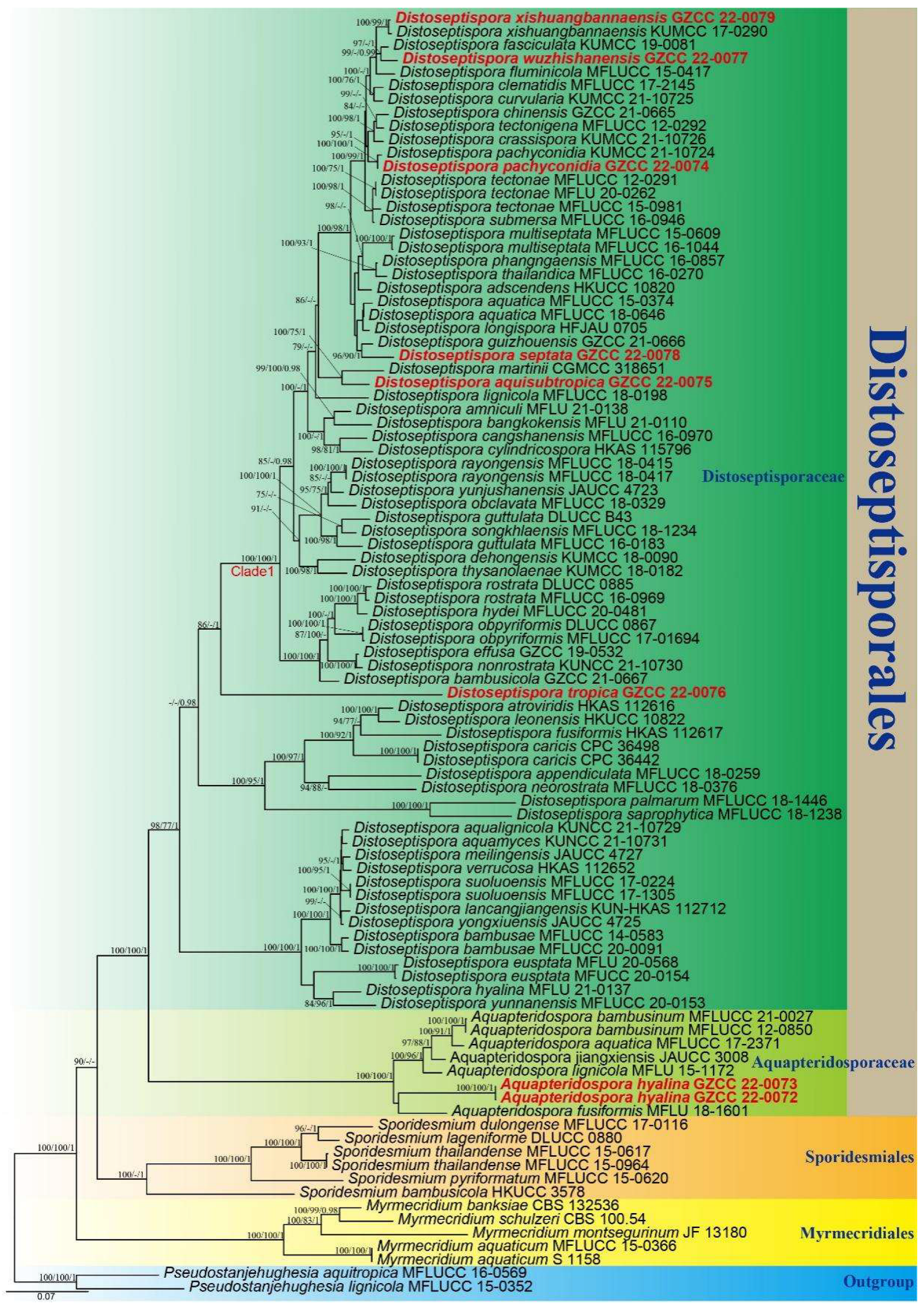
Using partial nucleotide sequences from four genes, the phylogenetic placements of our new collections were determined. The concatenated sequence matrix comprised LSU (1–855 bp), ITS (856–1448 bp), tef1α (1449–2367 bp), and rpb2 (2368–3420 bp) with a total of 3420 characters for 93 taxa and two outgroups (Pseudostanjehughesia aquitropica MFLUCC 16-0569 and Ps. lignicola MFLUCC 15-0352) [7,12,16,47,48,49,50]. Four gene analyses were conducted to compare the respective topologies and clade stabilities. There were 1733 distinct alignment patterns in the matrix, along with 31.09% undetermined characters or gaps. The ML, MP, and BI analyses of the concatenated LSU, ITS, tef1α, and rpb2 dataset yielded similar tree topologies, and Figure 2 depicts the final likelihood value of the ML analysis.
The phylogenetic tree (Figure 2) demonstrates that our eight collections represent seven species within Distoseptisporales. Two isolates (GZCC 22-0072 and GZCC 22-0073) represent the new species Aquapteridospora hyalina which forms a distinct clade from other taxa of Aquapteridospora with strong support. Distoseptispora aquisubtropica and D. martinii are distinguished by their distinct conidial characteristics. Distoseptispora septata forms a sister lineage to D. guizhouensis with well support (96% ML/90% MP/1 PP). Distoseptispora tropica (GZCC 22-0076) is a significantly distinct lineage from other taxa (Figure 2). Distoseptispora wuzhishanensis forms a sister lineage to D. fasciculata with well support (97% ML/1 PP). Our isolate GZCC 22-0079 is recognized as D. xishuangbannaensis and GZCC 22-0074 as D. pachyconidia, and their molecular data are provided, respectively.

4. Taxonomy

Aquapteridospora hyalina J. Ma and Y.Z. Lu., sp. nov., Figure 3.
Index Fungorum number: IF559932; Facesoffungi number: FoF11002.
Etymology: The epithet ‘hyalina’ referring to the colourless conidia.
Holotype: HKAS 123764.
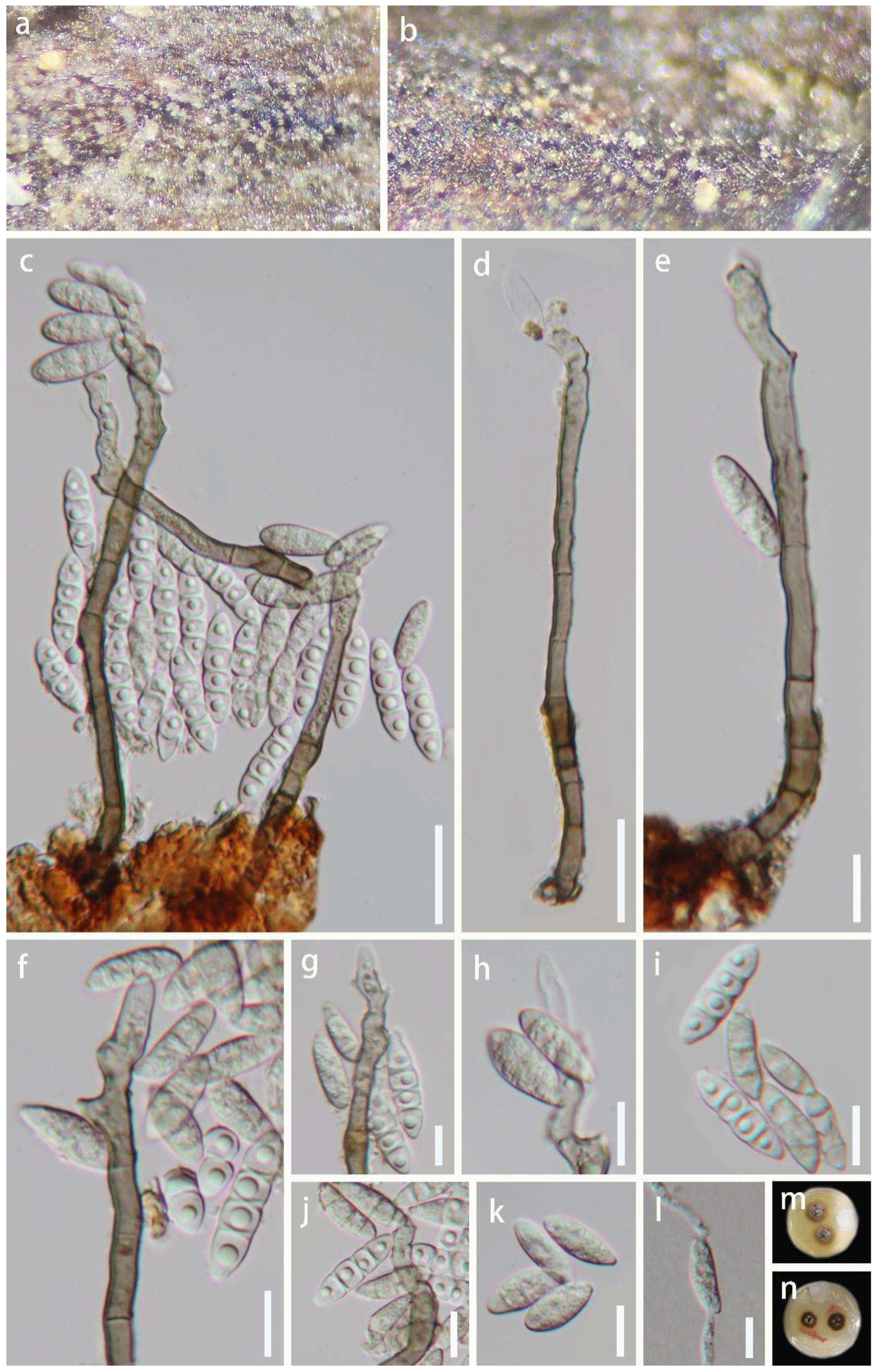
Saprobic on decaying wood in a freshwater habitat. Sexual morph: Undetermined. Asexual morph: Colonies on the natural substrate, effuse, solitary, hyaline. Mycelium mostly superficial, consisting of branched, septate, smooth, pale brown to brown hyphae. Conidiophores 68–130 × 4.5–6.5 μm ( x ¯ = 91 × 5.5 μm, n = 30), macronematous, mononematous, solitary, erect, simple, straight, or slightly flexuous, unbranched, smooth, cylindrical, 3–7-septate, thick-walled, smooth-walled, brown at the base, subhyaline to pale brown towards apex. Conidiogenous cells 25–62 × 4–6.5 μm ( x ¯ = 44 × 5 μm, n = 30), polyblastic, monoblastic, smooth-walled, terminal, sub-cylindrical or gradually tapering towards tip, sub-hyaline to pale brown, forming conidia sympodially on conspicuous denticles, bearing tiny, protuberant, circular scars. Conidia 17–28 × 4–6 μm ( x ¯ = 20 × 5.5 μm, n = 50) acropleurogenous, solitary, fusiform, 1–3-septate, truncate obtuse at septum, hyaline when young, sub-hyaline to pale brown when mature, tapering and pointed at both ends, smooth-walled, often with single guttulate in each cell when young.
Culture characteristics: Conidia were germinated on water agar and produced germ tubes within 10 h. Colonies grown on PDA, circular, flat, reaching 18 mm diam. after 25 days of incubation at 25 °C, lightly gray at the entire margin, brown in the center, reverse-side pale brown or brown at the entire margin, pale grey in the center.
Material examined: China, Hainan Province, Baoting County, Sandao Town, Yanoda Rainforest cultural tourism area, 18°46′ N, 109°65′ E, on rotting wood in a freshwater stream, 23 October 2021, Jian Ma, Y6 (HKAS 123764, holotype; GZAAS 22-0077, isotype), ex-type living culture GZCC 22-0072; idem, Y10-1 (HKAS 123763), living culture GZCC 22-0073.
Notes: The proposed new species Aquapteridospora hyalina shares similar characteristics with other species of the genus Aquapteridospora in having macronematous, mononematous, unbranched, polyblastic conidiophores, sympodial proliferations conidiogenous cells, and fusiform and acropleurogenous conidia [4]. However, A. hyalina differs from all species of Aquapteridospora in having hyaline to pale brown conidia with distinguished guttulae in each cell when young. The phylogenetic analyses confirmed that the two strains (GZCC 22-0072 and GZCC 22-0073) of A. hyalina form a distinct clade sister to A. fusiformis within Aquapteridosporaceae (Figure 2). Therefore, based on the morphological evidence with multi-gene phylogenetic results, a new species A. hyalina is introduced.
Distoseptispora aquisubtropica J. Ma and Y.Z. Lu, sp. nov., Figure 4.
Index Fungorum number: IF559686; Facesoffungi number: FoF11334.
Etymology: ‘aquisubtropica’ is derived from subtropical, which means climate type and ‘aqui’ refers to its presence in aquatic habitat.
Holotype: HKAS 124023.
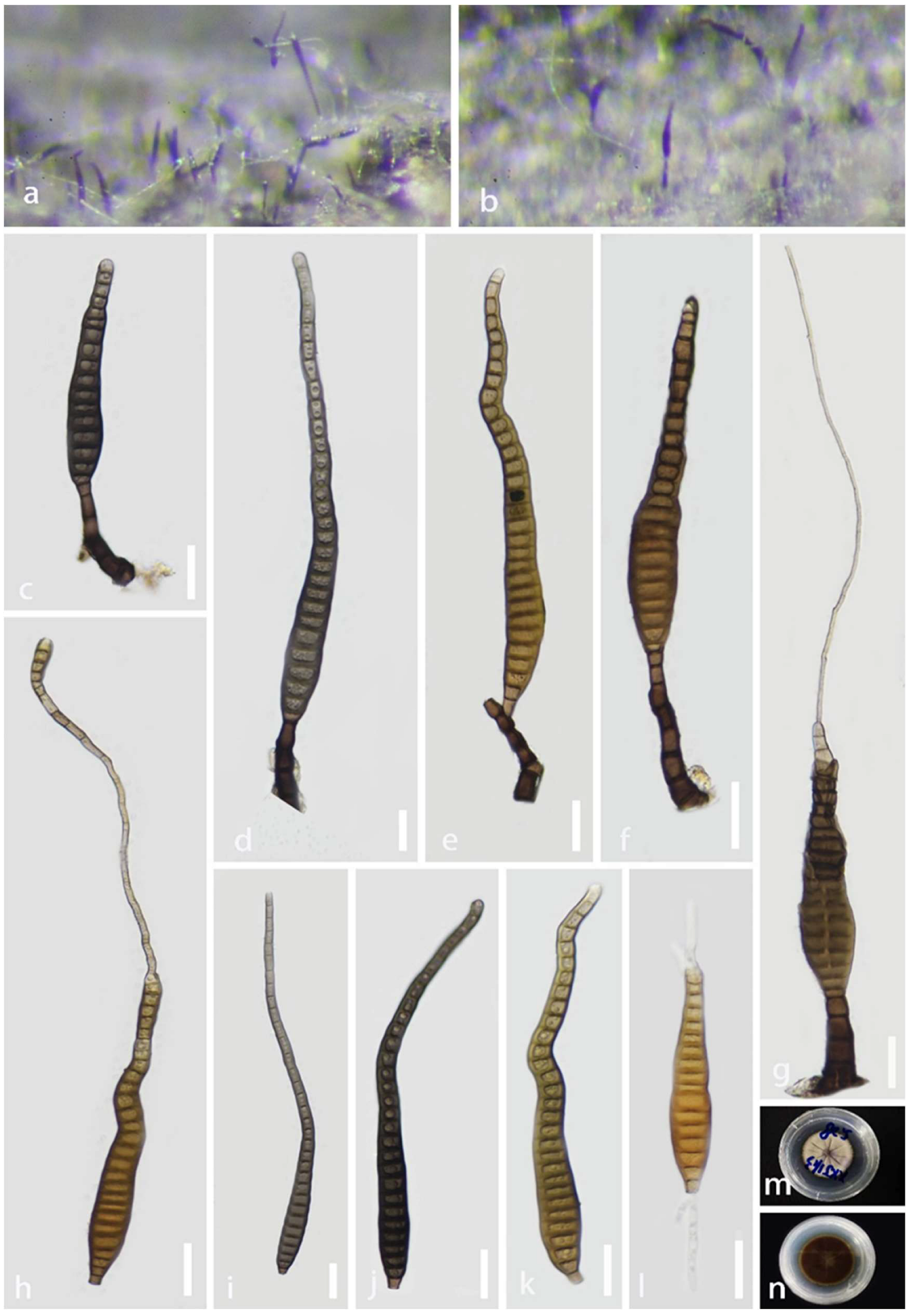
Saprobic on decaying wood submerged in a freshwater habitat. Sexual morph: Undetermined. Asexual morph: hyphomycetous. Colonies on natural substrate superficial, effuse, gregarious, smooth, septate, hairy, brown, or dark brown. Mycelium mostly immersed, composed of branched, septate, smooth, pale brown to brown hyphae. Conidiophores 16–83 × 5–11 μm ( x ¯   = 51 × 8 μm, n = 25), macronematous, mononematous, cylindrical, erect, simple, straight, or slightly flexuous, unbranched, smooth, thick-walled, solitary, brown at the base, pale brown or sub-hyaline towards the apex, 2–5-septate. Conidiogenous cells 3–11 × 3–7 μm ( x ¯ = 6 × 5.5 μm, n = 25), holoblastic, monoblastic, terminal, integrated, cylindrical, pale brown or brown, smooth. Conidia 43–278 × 11–19 μm ( x ¯   = 141 × 14.5 μm, n = 38), acrogenous, solitary, multi-distoseptate, obclavate or lanceolate, rostrate, straight or slightly curved, verrucose, guttulate, thick-walled, smooth-walled, pale brown or dark brown, olivaceous, 16–31-distoseptate, usually paler towards apex, sometimes have conspicuous hyphae attached to the conidium, rounded at apex, with a truncate base.
Culture characteristics: Conidia were germinated on water agar and produced germ tubes within 10 h. Colonies grown on PDA, circular, flat, dense, dark gray, fluffy, reaching 34 mm diam. after 25 days of incubation at 25 °C, break in the center, from below olivaceous to brown at the center, pale yellow at the entire margin.
Material examined: China, Guizhou Province, Zhenyuan County, 27°05′ N, 108°41′ E, on decaying wood submerged from a freshwater stream, 1 May 2021, Jian Ma, XXJ11-3 (HKAS 124023, holotype; GZAAS 22-0080, isotype), ex-type living culture GZCC 22-0075.
Notes: According to a BLASTn search on NCBI GenBank, the ITS and LSU sequences of the new isolate (Distoseptispora aquisubtropica) share 92.95% similarity across 85% of the query sequence coverage and 99.6% similarity across 86% of the query sequence coverage with D. martini, respectively. The phylogenetic results indicate that D. aquisubtropica forms a sister clade to D. martinii with well-supported values (100% ML/75% MP/1 PP). The morphological features of D. aquisubtropica are compatible with the Distoseptispora generic concept, while D. martini resembles Acrodictys rather than Distoseptospora [13]. Distoseptospora aquisubtropica can be distinguished from D. martini by its obclavate or lanceolate conidia, while the latter is oblate or subglobose [13]. Based on a pairwise comparison of ITS and LSU nucleotides, D. aquisubtropica differs from D. martinii in 24/453 bp (5.3%) for ITS and 5/515 bp (0.97%) for LSU, respectively. Therefore, we identified D. aquisubtropica as a novel species, according to the species delimitation guidelines proposed by Chethana et al. and Maharachchikumbura et al. [51,52].
Distoseptispora pachyconidia R. Zhu and H. Zhang. Journal of Fungi 30: 22 (2022). Figure 5.
Index Fungorum Number: IF559924; Facesoffungi number: FoF12581.
Saprobic on decaying wood in terrestrial habitats. Sexual morph: Undetermined. Asexual morph: Colonies effuse, gregarious, brown, or dark brown, hairy. Mycelium immersed and partly superficial, consisting of branched, septate, smooth, pale brown to brown hyphae. Conidiophores 14–44 × 4–7 μm ( x ¯ = 26 × 5 μm, n = 20), macronematous, mononematous, brown to dark brown, solitary, 2–4-septate, erect, straight, or flexuous, unbranched, smooth, cylindrical, singly or in groups, truncate at the apex, slightly constricted at septa. Conidiogenous cells 4–9 × 4–5.5 μm ( x ¯ = 6 × 4.5 μm, n = 20), holoblastic, monoblastic, integrated, terminal, determinate, pale brown to brown, smooth, cylindrical. Conidia 50–242 × 11–20 μm ( x ¯ = 111 × 13 μm, n = 30) acrogenous, solitary, obclavate, rostrate, smooth-walled, straight or slightly curved, up to 38-distoseptate, guttulate, olivaceous to dark brown, mostly slightly constricted at septa, tapering towards the rounded apex, truncate at the base.
Culture characteristics: Conidia were germinated on water agar and produced germ tubes within 10 h. Colonies grown on PDA, circular, mycelium flat, dense, reaching 40 mm diam. after 30 days of incubation at 25 °C, gray or brown, reverse-side dark brown.
Material examined: China, Hainan Province, Haikou City, Xiuying District, Ecological leisure trail, on decaying wood on the ground, 20°01′ N, 110°25′ E, 10 August 2021, Jian Ma, HK2 (HKAS 123754), living culture GZCC 22-0074.
Notes: Zhang et al. [16] introduced Distoseptispora pachyconidia from decaying wood in Yunnan Province, China. Analyses of multi-gene revealed that the new isolate GZCC 22-0074 clustered with D. pachyconidia. The morphological characteristics of this isolate are similar to the protologue of D. pachyconidia. However, GZCC 22-0074 differs from D. pachyconidia in having different colors of conidia (olivaceous to dark brown vs. pale-brown with a green tinge) and the number of conidial septa (up to 38 vs. 8–21-distoseptate) [16]. According to a pairwise nucleotide comparison of ITS, LSU, tef1α and rpb2, our isolate differs from the type strain of D. pachyconidia (HKAS 122179) in 1/520 bp (0.2%) for ITS, 1/852 bp (0.1%) for LSU, 1/913 bp (0.1%) for tef1α and 0/1052 bp (0%) for rpb2, respectively. Thus, the phylogenetic evidence did not show significant differences between them (Figure 2). We therefore identified the new isolate as D. pachyconidia.
Distoseptispora septata J. Ma and Y.Z. Lu, sp. nov., Figure 6.
Index Fungorum number: IF559688; Facesoffungi number: FoF11337.
Etymology: ‘septata’ referring to ‘septate’ conidia.
Holotype: HKAS 123759.
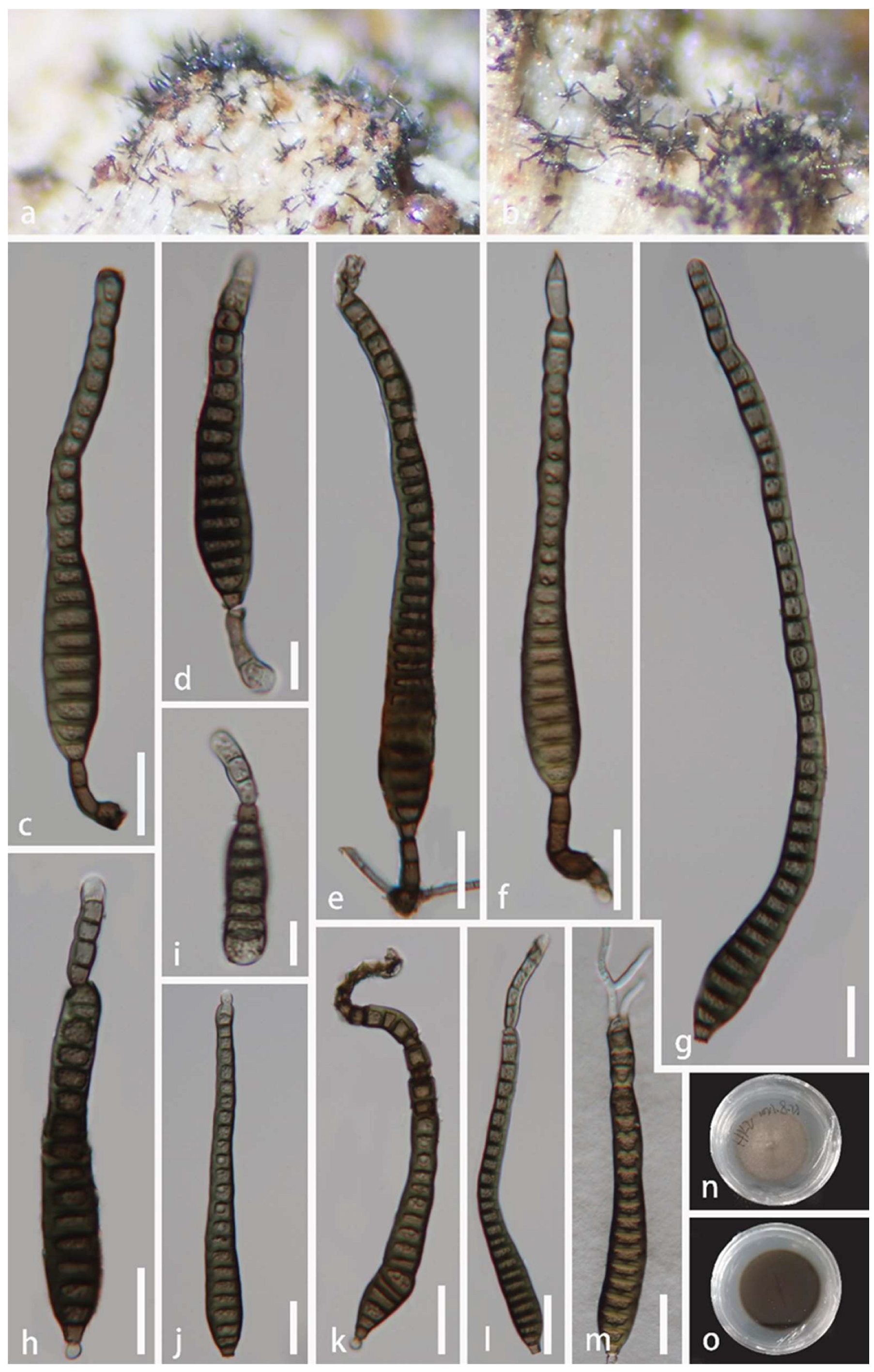
Saprobic on decaying wood submerged in a freshwater habitat. Sexual morph: Undetermined. Asexual morph: hyphomycetous. Colonies on natural substrate superficial, effuse, gregarious, smooth, septate, hairy, brown, or dark brown. Mycelium mostly immersed, composed of branched, septate, smooth, pale brown to brown hyphae. Conidiophores 23–86 × 3–7 μm ( x ¯ = 44 × 5.5 μm, n = 30), macronematous, mononematous, cylindrical, erect, simple, mostly flexuous, unbranched, smooth, thick-walled, solitary, brown, 1–6-septate. Conidiogenous cells 5–18 × 4–6 μm ( x ¯ = 9 × 5 μm, n = 30), holoblastic, monoblastic, terminal, integrated, cylindrical, pale brown or brown, smooth. Conidia 22–179 × 10–16 μm ( x ¯ = 89 × 13 μm, n = 35), acrogenous, solitary, multi-distoseptate and up to 25-septate, obclavate, rostrate, straight or slightly curved, verrucose, guttulate, thick-walled, smooth-walled, pale brown or dark brown, olivaceous-green, usually paler towards apex, rounded at apex, with a truncate base.
Culture characteristics: Conidia were germinated on water agar and produced germ tubes within 10 h. Colonies grown on PDA, circular, flat, dense, fluffy, reaching 35 mm diam. after 25 days of incubation at 25 °C, olivaceous-green, paler brown and dark brown, reverse-side dark brown.
Material examined: China, Hainan Province, Wuzhishan City, Shui Man Town, Wuzhishan National Nature Reserve, 18°92′ N, 109°63′ E, on dead wood in a freshwater stream, 26 December 2021, Xia Tang, W17 (HKAS 123759, holotype; GZAAS 22-0083, isotype), ex-type living culture GZCC 22-0078.
Notes: Distoseptispora septata shares a sister relationship to D. guizhouensis with strong support (96% ML/90% MP/1 PP). However, D. septata differs from D. guizhouensis in having smaller conidia (22–179 × 10–16 μm vs. 90–273 × 15–21 μm). Particularly, D. guizhouensis is distinguished from D. septata by distinctly flexuous conidiophores. Based on a pairwise comparison of ITS nucleotides, D. septata differs from D. guizhouensis by 13/535 bp (2.4%). Following the guidelines for defining species boundaries of Chethana et al. and Pem et al. [51,53], we therefore introduce GZCC 22-0078 as a new species.
Distoseptispora tropica J. Ma and Y.Z. Lu, sp. nov., Figure 7.
Index Fungorum number: IF559689; Facesoffungi number: FoF11335.
Etymology: ‘tropica’ derived from the climate in which the species was discovered.
Holotype: HKAS 123761.
Saprobic on dead wood, in terrestrial habitat. Sexual morph: Undetermined. Asexual morph: hyphomycetous. Colonies on dead wood, effuse, scattered or in small groups, gregarious, hairy, smooth, brown, or dark brown. Mycelium mostly immersed, composed of branched, septate, smooth, brown to dark brown. Conidiophores 60–151 × 3.5–7 μm ( x ¯ = 94 × 5 μm, n = 20), macronematous, mononematous, cylindrical, erect, straight, or slightly flexuous, unbranched, smooth, thick-walled, dark brown, solitary or caespitose, dark brown and rounded at the apex, paler brown at the upper part, 5–7 septate. Conidiogenous cells 10–21 × 3.5–6 μm ( x ¯ = 159 × 5 μm, n = 20), holoblastic, monoblastic, terminal, integrated, cylindrical, pale brown or brown, smooth. Conidia 39–75 × 7.5–10.5 μm ( x ¯ = 47 × 8 μm, n = 30), acrogenous, verrucose, solitary, multi-distoseptate, obclavate, rostrate, upper part tapering towards the apex, guttulate, thick-walled, smooth, olivaceous brown or dark brown, 5–7 distoseptate, with conspicuous hyphae attachment conidium, with a truncate base.
Culture characteristics: Conidia were germinated on water agar and produced germ tubes within 10 h. Colonies grown on PDA, circular, flat, dense, fluffy, reaching 40 mm diam. after 40 days of incubation at 25 °C, grayish brown mycelium on the surface, reverse-side dark brown.
Material examined: China, Hainan Province, Haikou City, Xiuying District, Ecological leisure trail, 20°01′ N, 110°25′ E, on decaying wood in terrestrial habitat, 10 August 2021, Jian Ma, HK9 (HKAS 123761, holotype; GZAAS 22-0081, isotype), ex-type living culture GZCC 22-0076.
Notes: Distoseptispora tropica shares an extremely similar morphology with the species of Distoseptispora, Ellisembia, and Sporidesmium [7,9,54]. However, phylogenetic analysis (Figure 2) indicates that D. tropica belongs to Distoseptispora. Distoseptispora tropica forms a distinct lineage basal to Clade 1 with strong support (100% ML/100% MP/1 PP). However, D. tropica differs from other taxa in Clade 1 in having longer conidiophores and smaller conidia.
Distoseptispora wuzhishanensis J. Ma and Y.Z. Lu, sp. nov., Figure 8.
Index Fungorum number: IF559706; Facesoffungi number: FoF11336.
Etymology: ‘wuzhishanensis’ derived from the city where the species was discovered.
Holotype: HKAS 123762.
Saprobic on decaying wood in a freshwater habitat. Sexual morph: Undetermined. Asexual morph: hyphomycetous. Colonies on substrate superficial, effuse, solitary, smooth, septate, hairy, brown, or dark brown. Mycelium mostly immersed, composed of branched, septate, smooth, pale brown to brown hyphae. Conidiophores 16–56 × 5–7 μm ( x ¯ = 35 × 6 μm, n = 25), macronematous, mononematous, cylindrical, erect, scattered or in small groups, straight or slightly flexuous, unbranched, smooth, thick-walled, brown at the base, pale brown towards the apex, 1–4 septate. Conidiogenous cells 6.5–10.5 × 4.5–6 μm ( x ¯ = 8 × 5 μm, n = 25), holoblastic, monoblastic, terminal, integrated, cylindrical, pale brown or brown, smooth. Conidia 76–143 × 11–17 μm ( x ¯ = 108 × 13.5 μm, n = 40), acrogenous, solitary, multi-distoseptate, obclavate, rostrate, straight, or slightly curved, verrucose, guttulate, thick-walled, smooth-walled, pale brown or dark brown, olivaceous-green and yellow, usually up to 22-distoseptate, strongly constricted at septa, usually paler towards apex, rounded at apex, with a truncate base.
Culture characteristics: Conidia were germinated on water agar and produced germ tubes within 10 h. Colonies grown on PDA, circular, flat, dense, fluffy, reaching 24 mm diam. after 25 days of incubation at 25 °C, olivaceous-green or brown mycelium on the surface, reverse side partly paler brown to dark brown.
Material examined: China, Hainan Province, Wuzhishan City, Shui Man Town, Wuzhishan National Nature Reserve, 18°92′ N, 109°63′ E, on dead wood in a freshwater stream, 26 December 2021, Xia Tang, W21 (HKAS 123762, holotype; GZAAS 22-0082, isotype), ex-type living culture GZCC 22-0077.
Notes: According to phylogenetic tree (Figure 2), Distoseptispora wuzhishanensis forms a close lineage to D. fasciculata with high bootstrap support (99% ML/0.99 PP). The morphological characteristics of the new taxon match well with the species concept of Distoseptispora in conidiophores and conidia. However, D. wuzhishanensis differs from D. fasciculata in having longer conidiophores (16–56 × 5–7 μm vs. 12–16 × 5–6 μm) [3].
Distoseptispora xishuangbannaensis Tibpromma and K.D. Hyde. Fungal Diversity 93: 82 (2018). Figure 9.
Index Fungorum Number: IF554554; Facesoffungi number: FoF04563.
Saprobic on decaying wood, submerged in freshwater habitats. Sexual morph: Undetermined. Asexual morph: hyphomycetous. Colonies effuse, hairy, gregarious, olivaceous, or brown. Mycelium mostly immersed, composed of branched, smooth, septate, pale brown to brown hyphae. Conidiophores 19–52 × 5–7 μm ( x ¯ = 29 × 6 μm, n = 20), macronematous, mononematous, unbranched, solitary, straight or slightly flexuous, brown at the base, pale brown towards the apex, slightly tapering distally, truncate at the apex, 1–4 septate. Conidiogenous cells 2–8.5 × 4.0–7 μm ( x ¯   = 6 × 5 μm, n = 20), blastic, holoblastic, monoblastic, terminal, integrated, cylindrical, pale brown. Conidia 88–269 × 11–15 μm ( x ¯ = 165 × 13 μm, n = 30), solitary, acrogenous, cylindric-obclavate, rostrate, straight, or slightly curved, guttulate, thick-walled, smooth-walled, green-brown or brown, up to 42-distoseptate, usually paler olivaceous towards apex, rounded at apex, with a truncate base.
Culture characteristics: Conidia were germinated on water agar and produced germ tubes within 10 h. Colonies grown on PDA, circular, fluffy, reaching 36 mm diam. after 28 days of incubation at 25 °C, lightly grey in the center, olivaceous at the entire margin, reverse side pale brown and brown mycelium.
Material examined: China, Hainan Province, Lingshui Lizu Autonomous County, Diaoluoshan National Nature Reserve, 18°43′ N, 109°43′ E, on rotting wood in a freshwater habitat, 24 August 2021, Jian Ma, DL40 (HKAS 123760), living culture GZCC 22-0079.
Notes: Tibpromma et al. [20] introduced Distoseptispora xishuangbannaensis from dead leaf sheaths of Pandanus utilis in China. Analyses of multiple genes revealed that our new isolate (GZCC 22-0079) is closely related to D. xishuangbannaensis. Distoseptispora xishuangbannaensis possesses macronematous, straight or slightly flexuous conidiophores, holoblastic, monoblastic, brown conidiogenous cells, and acrogenous, cylindrical-obclavate conidia. The morphological characteristics of our isolate share similar characters with the holotype of D. xishuangbannaensis (HKAS 101809), but our isolate has longer and wider conidiophores (19–52 × 5–7 μm vs. 12–17 × 2–5 μm) and smaller conidia (88–269 × 11–15 μm vs. 160–305 × 8–15 μm) [20]. According to a pairwise nucleotide comparison of ITS, tef1α and rpb2, our new isolate differs from D. xishuangbannaensis in 5/498 bp (1%) for ITS, 3/898 bp (0.3%) for tef1α and 2/1052 bp (0.2%) for rpb2, respectively, and the phylogenetic result did not show significant differences between the new isolate and D. xishuangbannaensis (Figure 2). We therefore identified the new isolate as D. xishuangbannaensis.

5. Discussion

In recent years, studies on the number of saprotrophic fungi have received extensive attention [55,56]. For example, freshwater fungi occur in streams and other aquatic bodies [2,57], Calabon et al. [51] listed 3870 species, and numerous taxa are still being discovered [1]. Calabon et al. [51] listed 22 Distoseptispora species from freshwater and this study brings the total to 40 species. Analyses of morphological characteristics and molecular data indicate that eight collections, including five new species, namely A. hyalina, D. aquisubtropica, D. septata, D. tropica, and D. wuzhishanensis are introduced. Five Aquapteridospora epithets (A. lignicola, A. fusiformis, A. bambusinum, A. jiangxiensis, and A. aquatica) are listed in Index Fungorum [32,58]. Morphologically, A. hyalina corresponds well with the generic concept of Aquapteridospora [4,6,58]. However, the new isolate differs from other new species of genus Aquapteridospora by having conidia that have a truncate obtuse at their septum. Multi-gene analyses indicated that A. hyalina, a species phylogenetically distinct in the genus Aquapteridospora, was most closely related to A. fusiformis with weak support.
Morphologically, the asexual morph of Distoseptispora resembles Sporidesmium taxa [9]. Yang et al. [7] reported the existence of the sexual morph of Distoseptispora. Combining the morphological characteristics and the phylogenetic evidence of all species in genus Distoseptispora, we found that some Distoseptispora species form sister clades in phylogeny but have different morphologies. For example, D. martini and D. aquisubtropica have a close phylogenetic relationship. However, D. martini has distinctive oblate or subglobose conidia, while D. aquisubtropica has obclavate or lanceolate conidia. In addition, there are some species with similar morphologies but are genetically unrelated. For example, D. tropica and D. verrocosa have an identical morphology of conidiophores and conidia, but the two species have a distant phylogenetic relationship. Considering this phenomenon, additional molecular data and morphological characteristics are required for verification and expansion.
Previous studies of Distoseptispora have primarily been conducted in China (Guizhou, Yunnan, Sichuan, and Jiangxi Provinces) and in Thailand, which are subtropical and tropical regions. In this paper, eight collections were discovered in freshwater and terrestrial habitats in the Provinces of Hainan and Guizhou, China. Thus, it is unclear whether it has a close relationship with the climate. It may be a result of the limited geographical regions sampled.

Author Contributions

The specimens in this study were collected by J.M., X.-J.X. and Y.-Z.L. Morphological data were collected by J.M. and X.-J.X. Molecular data and phylogenetic analyses were performed by J.M. and X.-J.X. Writing the original draft, and review and editing were completed by J.M., J.-Y.Z., X.-J.X., Y.-P.X., X.T., S.B., J.-C.K. and Y.-Z.L. All authors have read and agreed to the published version of the manuscript.

Funding

This work was funded by the Science and Technology Foundation of Guizhou Province ([2020]1Y058), the National Natural Science Foundation of China (NSFC 31900020), China Postdoctoral Science Foundation (2020M683657XB), Youth Science and Technology Talent Development Project from Guizhou Provincial Department of Education (QJHKYZ [2021]263) and Guizhou Province high-level talent innovation and entrepreneurship merit funding project (No. 202104).

Institutional Review Board Statement

Not applicable.

Informed Consent Statement

Not applicable.

Data Availability Statement

All sequences generated in this study were submitted to GenBank (https://www.ncbi.nlm.nih.gov/genbank/, accessed on 16 May 2022).

Acknowledgments

We would like to thank Shaun Pennycook (Manaaki Whenua Landcare Research, New Zealand) for advising on fungal nomenclature. Jian Ma would like to thanks Na Wu, Xing-Juan Xiao, Meng-Lan Chen, Xue-Mei Chen, Chuan-Gen Lin, Yao Feng, Ya-ya Chen, Hong-zhi Du their valuable suggestions and help. We thank Abhaya Balasuriya under the Reinventing Visiting Professor Program 2022, Mae Fah Luang University for his valuable correction and suggestion.

Conflicts of Interest

The authors declare no conflict of interest.

References

  1. Hyde, K.D.; Bao, D.F.; Hongsanan, S.; Chethana, K.; Yang, J.; Suwannarach, N. Evolution of freshwater Diaporthomycetidae (Sordariomycetes) provides evidence for five new orders and six new families. Fungal Divers. 2021, 107, 71–105. [Google Scholar] [CrossRef]
  2. Luo, Z.L.; Hyde, K.D.; Liu, J.K.; Maharachchikumbura, S.S.N.; Jeewon, R.; Bao, D.F.; Bhat, D.J.; Lin, C.G.; Li, W.L.; Yang, J.; et al. Freshwater Sordariomycetes. Fungal Divers. 2019, 99, 451–660. [Google Scholar] [CrossRef] [Green Version]
  3. Dong, W.; Hyde, K.D.; Jeewon, R.; Doilom, M.; Yu, X.D.; Wang, G.N.; Liu, N.G.; Hu, D.M.; Nalumpang, S.; Zhang, H. Towards a natural classification of annulatascaceae-like taxa II: Introducing five new genera and eighteen new species from freshwater. Mycosphere 2021, 12, 1–88. [Google Scholar] [CrossRef]
  4. Yang, J.; Maharachchikumbura, S.S.N.; Hyde, K.D.; Bhat, D.J.; McKenzie, E.H.C.; Bahkali, A.H.; Gareth Jones, E.B.G.; Liu, Z.Y. Aquapteridospora lignicola gen. et sp. nov., a new hyphomycetous taxon (Sordariomycetes) from wood submerged in a freshwater stream. Cryptogam. Mycol. 2015, 36, 469–478. [Google Scholar] [CrossRef]
  5. Wijayawardene, N.N.; Hyde, K.D.; Al-Ani, L.K.T.; Tedersoo, L.; Haelewaters, D.; Rajeshkumar, K.C.; Zhao, R.L.; Aptroot, A.; Leontyev, D.V.; Saxena, R.K.; et al. Outline of Fungi and fungus-like taxa. Mycosphere 2020, 11, 1060–1456. [Google Scholar] [CrossRef]
  6. Dai, D.Q.; Phookamsak, R.; Wijayawardene, N.N.; Li, W.J.; Bhat, D.J.; Xu, J.C.; Taylor, J.E.; Hyde, K.D.; Chukeatirote, E. Bambusicolous fungi. Fungal Divers. 2016, 82, 1–105. [Google Scholar] [CrossRef]
  7. Yang, J.; Liu, L.L.; Jones, E.B.G.; Li, W.L.; Hyde, K.D.; Liu, Z.Y. Morphological variety in Distoseptispora and introduction of six novel species. J. Fungi. 2021, 7, 945. [Google Scholar] [CrossRef]
  8. Bao, D.F.; Hyde, K.D.; McKenzie, E.H.C.; Jeewon, R.; Su, H.Y.; Nalumpang, S.; Luo, Z.L. Biodiversity of lignicolous freshwater hyphomycetes from China and Thailand and description of sixteen species. J. Fungi. 2021, 7, 669. [Google Scholar] [CrossRef] [PubMed]
  9. Su, H.Y.; Hyde, K.D.; Maharachchikumbura, S.; Ariyawansa, H.A.; Luo, Z.L.; Promputtha, I.; Tian, Q.; Lin, C.G.; Shang, Q.J.; Zhao, Y.C.; et al. The families Distoseptisporaceae fam. nov., Kirschsteiniotheliaceae, Sporormiaceae and Torulaceae, with new species from freshwater in Yunnan Province, China. Fungal Divers. 2016, 80, 375–409. [Google Scholar] [CrossRef]
  10. Yang, J.; Maharachchikumbura, S.; Liu, J.K.; Hyde, K.D.; Jones, E.B.G.; Al-Sadi, A.M.; Liu, Z.Y. Pseudostanjehughesia aquitropica gen. et sp. nov. and Sporidesmium sensu lato species from freshwater habitats. Mycol. Prog. 2018, 17, 591–616. [Google Scholar] [CrossRef]
  11. Monkai, J.; Boonmee, S.; Ren, G.; Wei, D.; Phookamsak, R.; Mortimer, P.E. Distoseptispora hydei sp. nov. (Distoseptisporaceae), a novel lignicolous fungus on decaying bamboo in Thailand. Phytotaxa 2020, 459, 93–107. [Google Scholar] [CrossRef]
  12. Shen, H.W.; Bao, D.F.; Hyde, K.D.; Su, H.Y.; Bhat, D.J.; Luo, Z.L. Two novel species and two new records of Distoseptispora from freshwater habitats in China and Thailand. MycoKeys 2021, 84, 79–101. [Google Scholar] [CrossRef] [PubMed]
  13. Xia, J.W.; Ma, Y.R.; Li, Z.; Zhang, X.G. Acrodictys-like wood decay fungi from southern China, with two new families Acrodictyaceae and Junewangiaceae. Sci. Rep. 2017, 7, 7888. [Google Scholar] [CrossRef] [PubMed] [Green Version]
  14. Hyde, K.D.; Hongsanan, S.; Jeewon, R.; Bhat, D.J.; McKenzie, E.H.C.; Jones, E.B.G.; Phookamsak, R.; Ariyawansa, H.; Boonmee, S.; Zhao, Q.; et al. Fungal diversity notes 367–490: Taxonomic and phylogenetic contributions to fungal taxa. Fungal Divers. 2016, 80, 1–270. [Google Scholar] [CrossRef]
  15. Hyde, K.D.; Tennakoon, D.S.; Jeewon, R.; Bhat, D.J.; Maharachchikumbura, S.; Rossi, W.; Leonardi, M.; Lee, H.B.; Mun, H.Y.; Houbraken, J.; et al. Fungal diversity notes 1036–1150: Taxonomic and phylogenetic contributions on genera and species of fungal taxa. Fungal Divers. 2019, 96, 1–242. [Google Scholar] [CrossRef]
  16. Zhang, H.; Zhu, R.; Qing, Y.; Yang, H.; Li, C.X.; Wang, G.N.; Zhang, D.; Ning, P. Polyphasic identification of Distoseptispora with six new species from freshwater. J. Fungi 2022, 8, 1063. [Google Scholar] [CrossRef]
  17. Maharachchikumbura, S.S.N.; Hyde, K.D.; Jones, E.B.G.; McKenzie, E.H.C.; Bhat, D.J.; Dayarathne, M.C.; Huang, S.K.; Norphanphoun, C.; Senanayake, I.C.; Perera, R.H.; et al. Families of Sordariomycetes. Mycosphere 2016, 79, 1–317. [Google Scholar] [CrossRef]
  18. Luo, Z.L.; Hyde, K.D.; Liu, J.K.; Bhat, D.J.; Su, H.; Bao, D.F.; Li, W.L. Lignicolous freshwater fungi from China II: Novel Distoseptispora (Distoseptisporaceae) species from northwestern Yunnan Province and a suggested unified method for studying lignicolous freshwater fungi. Mycosphere 2018, 9, 444–461. [Google Scholar] [CrossRef]
  19. Phookamsak, R.; Hyde, K.D.; Jeewon, R.; Bhat, D.J.; Jones, E.B.G.; Maharachchikumbura, S.; Raspé, O.; Karunarathna, S.C.; Wanasinghe, D.; Hongsanan, S.; et al. Fungal diversity notes 929–1035: Taxonomic and phylogenetic contributions on genera and species of fungi. Fungal Divers. 2019, 95, 1–273. [Google Scholar] [CrossRef] [Green Version]
  20. Tibpromma, S.; Hyde, K.D.; Mckenzie, E.; Bhat, D.J.; Phillips, A.J.L.; Wanasinghe, D.N.; Samarakoon, M.C.; Jayawardena, R.S. Fungal diversity notes 840–928: Micro-fungi associated with Pandanaceae. Fungal Divers. 2018, 100, 1–160. [Google Scholar] [CrossRef]
  21. Li, W.L.; Liu, Z.P.; Zhang, T.; Dissanayake, A.J.; Luo, Z.L.; Su, H.Y.; Liu, J.K. Additions to Distoseptispora (Distoseptisporaceae) associated with submerged decaying wood in China. Phytotaxa 2021, 520, 75–86. [Google Scholar] [CrossRef]
  22. Shoemaker, R.A.; White, G.P. Lasiosphaeria caesariata with Sporidesmium hormiscioides and L. triseptata with S. adscendens. Sydowia 1985, 38, 278–283. [Google Scholar]
  23. McKenzie, E.H.C. Dematiaceous hyphomycetes on Pandanaceae. 5. Sporidesmium sensu lato. Mycotaxon 1995, 56, 9–29. [Google Scholar]
  24. Hyde, K.D. Mycosphere notes 325-344–Novel species and records of fungal taxa from around the world. Mycosphere 2021, 12, 1101–1156. [Google Scholar] [CrossRef]
  25. Sun, Y.; Goonasekara, I.D.; Thambugala, K.M.; Jayawardena, R.S.; Wang, Y.; Hyde, K.D. Distoseptispora bambusae sp. nov. (Distoseptisporaceae) on bamboo from China and Thailand. Biodivers. Data J. 2020, 8, e53678. [Google Scholar] [CrossRef]
  26. Phukhamsakda, C.; McKenzie, E.H.C.; Phillips, A.; Jones, E.B.G.; Bhat, D.J.; Stadler, M.; Bhunjun, C.S.; Wanasinghe, D.N.; Thongbai, B.; Camporesi, E.; et al. Microfungi associated with Clematis (Ranunculaceae) with an integrated approach to delimiting species boundaries. Fungal Divers. 2020, 102, 1–203. [Google Scholar] [CrossRef]
  27. Phukhamsakda, C.; Nilsson, R.H.; Bhunjun, C.S.; Farias, A.R.G.D.; Sun, Y.; Wijesinghe, S.N.; Raza, M.; Bao, D.F.; Lu, L.; Tibpromma, S.; et al. The numbers of fungi: Contributions from traditional taxonomic studies and challenges of metabarcoding. Fungal Divers. 2022, 114, 327–386. [Google Scholar] [CrossRef]
  28. Zhai, Z.J.; Yan, J.Q.; Li, W.W.; Gao, Y.; Hu, H.J.; Zhou, J.P.; Song, H.Y.; Hu, D.M. Three novel species of Distoseptispora (Distoseptisporaceae) isolated from bamboo in Jiangxi Province, China. MycoKeys 2022, 88, 35–54. [Google Scholar] [CrossRef]
  29. Crous, P.; Wingfield, M.; Lombard, L.; Roets, F.; Swart, W.; Alvarado, P.; Carnegie, A.; Moreno, G.; Luangsa-Ard, J.; Thangavel, R.; et al. Fungal Planet description sheets: 951–1041. Pers. Mol. Phylogeny Evol. Fungi. 2019, 43, 223–425. [Google Scholar] [CrossRef]
  30. Senanayake, I.C.; Rathnayaka, A.R.; Marasinghe, D.S.; Calabon, M.S.; Gentekaki, E.; Lee, H.B.; Hurdeal, V.G.; Pem, D.; Dis-sanayake, L.S.; Wijesinghe, S.N.; et al. Morphological approaches in studying fungi: Collection, examination, isolation, sporulation and preservation. Mycosphere 2020, 11, 2678–2754. [Google Scholar] [CrossRef]
  31. Jayasiri, S.C.; Hyde, K.D.; Ariyawansa, H.A.; Bhat, J.; Buyck, B.; Cai, L.; Dai, Y.C.; Abd-Elsalam, K.A.; Ertz, D.; Hidayat, I.; et al. The Faces of Fungi database: Fungal names linked with morphology, phylogeny and human impacts. Fungal Divers. 2015, 74, 3–18. [Google Scholar] [CrossRef]
  32. Index Fungorum. Available online: http://www.indexfungorum.org/names/names.asp. (accessed on 5 May 2021).
  33. Vilgalys, R.; Hester, M. Rapid genetic identification and mapping of enzymatically amplified ribosomal DNA from several Cryptococcus species. J. Bacteriol. 1990, 172, 4238–4246. [Google Scholar] [CrossRef] [PubMed]
  34. White, T.; Bruns, T.; Lee, S.; Taylor, J. Amplification and direct sequencing of fungal ribosomal RNA genes for phylogenetics. In PCR Protocols—A Guide to Methods and Applications; Innis, M.A., Gelfand, D.H., Sninsky, J.J., White, T.J., Eds.; Academic Press: San Diego, CA, USA, 1990; pp. 315–322. [Google Scholar]
  35. Rehner, S.A.; Samuels, G.J. Taxonomy and phylogeny of Gliocladium analysed from nuclear large subunit ribosomal DNA sequences. Mycol. Res. 1994, 98, 625–634. [Google Scholar] [CrossRef]
  36. Liu, Y.J.; Whelen, S.; Hall, B.D. Phylogenetic relationships among ascomycetes: Evidence from an RNA polymerse II subunit. Biol. Evol. 1999, 16, 1799–1808. [Google Scholar] [CrossRef] [PubMed] [Green Version]
  37. Hall, T.A. BioEdit: A user-friendly biological sequence alignment editor and analysis program for windows 95/98/NT. Nucleic Acids Symp. Ser. 1999, 41, 95–98. [Google Scholar]
  38. Katoh, K.; Standley, D.M. Evolution. MAFFT multiple sequence alignment software version 7: Improvements in performance and usability. Biol. Evol. 2013, 30, 772–780. [Google Scholar] [CrossRef] [Green Version]
  39. Capella-Gutiérrez, S.; Silla-Martínez, J.M.; Gabaldón, T. trimAl: A tool for automated alignment trimming in large-scale phylogenetic analyses. Bioinformatics 2009, 25, 1972–1973. [Google Scholar] [CrossRef] [Green Version]
  40. Daniel, G.P.; Daniel, G.B.; Miguel, R.J.; Florentino, F.R.; David, P. ALTER: Program-oriented conversion of DNA and protein alignments. Nucleic Acids Res. 2010, 38, W14–W18. [Google Scholar] [CrossRef]
  41. Miller, M.A.; Pfeiffer, W.; Schwartz, T. Creating the CIPRES Science Gateway for inference of large phylogenetic trees. In Proceedings of the 2010 Gateway Computing Environments Workshop (GCE), New Orleans, LA, USA, 14 November 2010; pp. 1–8. [Google Scholar] [CrossRef] [Green Version]
  42. Stamatakis, A. RAxML version 8: A tool for phylogenetic analysis and post-analysis of large phylogenies. Bioinformatics 2014, 30, 1312–1313. [Google Scholar] [CrossRef] [Green Version]
  43. Swofford, D.L. PAUP*. Phylogenetic Analysis Using Parsimony (*and Other Methods); Version 4; Sinauer Associates: Sunderland, UK, 2003. [Google Scholar]
  44. Hillis, D.M.; Bull, J.J. An Empirical Test of Bootstrapping as a Method for Assessing Confidence in Phylogenetic Analysis. Syst. Biol. 1993, 42, 182–192. [Google Scholar] [CrossRef]
  45. Nylander, J.A.A.; Zoology, S.; Posada, D.; Mrmodeltest, R.; Os, F. MrModeltest2 v. 2.3 (Program for Selecting DNA Substitution Models Using PAUP*); Evolutionary Biology Centre: Uppsala, Sweden, 2008. [Google Scholar]
  46. Huelsenbeck, J.P.; Ronquist, F.J.B. MRBAYES: Bayesian inference of phylogenetic trees. Bioinformatics 2001, 17, 754–755. [Google Scholar] [CrossRef] [PubMed] [Green Version]
  47. Zhang, H.; Dong, W.; Hyde, K.D.; Maharachchikumbura, S.S.; Hongsanan, S.; Bhat, D.J.; Al-Sadi, A.M.; Zhang, D. Towards a natural classification of Annulatascaceae-like taxa: Introducing Atractosporales ord. nov. and six new families. Fungal Divers. 2017, 85, 75–110. [Google Scholar] [CrossRef]
  48. Crous, P.W.; Summerell, B.A.; Shivas, R.G.; Burgess, T.I.; Decock, C.A.; Dreyer, L.L.; Granke, L.L.; Guest, D.I.; Hardy, G.; Hausbeck, M.; et al. Fungal Planet description sheets: 107–127. Persoonia 2012, 28, 138–182. [Google Scholar] [CrossRef] [PubMed]
  49. Arzanlou, M.; Groenewald, J.Z.; Gams, W.; Braun, U.; Shin, H.D.; Crous, P.W. Phylogenetic and morphotaxonomic revision of Ramichloridium and allied genera. Stud. Mycol. 2007, 58, 57–93. [Google Scholar] [CrossRef] [PubMed]
  50. Réblová, M.; Fournier, J.; Štěpánek, V. Two new lineages of aquatic ascomycetes: Atractospora gen. nov. and Rubellisphaeria gen. et sp. nov., and a sexual morph of Myrmecridium montsegurinum sp. nov. Mycol. Prog. 2016, 15, 21. [Google Scholar] [CrossRef]
  51. Chethana, K.W.T.; Manawasinghe, I.S.; Hurdeal, V.G.; Bhunjun, C.S.; Appadoo, M.A.; Gentekaki, E.; Raspé, O.; Promputtha, I.; Hyde, K.D. What are fungal species and how to delineate them? Fungal Divers. 2021, 109, 1–25. [Google Scholar] [CrossRef]
  52. Maharachchikumbura, S.S.N.; Chen, Y.; Ariyawansa, H.A.; Hyde, K.D.; Haelewaters, D.; Perera, R.H.; Samara-koon, M.C.; Wanasinghe, D.N.; Bustamante, D.E.; Liu, J.K.; et al. Integrative approaches for species delimitation in Ascomycota. Fungal Divers. 2021, 109, 155–179. [Google Scholar] [CrossRef]
  53. Pem, D.; Jeewon, R.; Chethana, K.W.T.; Hongsanan, S.; Doilom, M.; Suwannarach, N.; Hyde, K.D. Species concepts of Dothideomycetes: Classification, phylogenetic inconsistencies and taxonomic standardization. Fungal Divers. 2021, 109, 283–319. [Google Scholar] [CrossRef]
  54. Shenoy, B.D.; Jeewon, R.; Wu, W.P.; Bhat, D.J.; Hyde, K.D. Ribosomal and RPB2 DNA sequence analyses suggest that Sporidesmium and morphologically similar genera are polyphyletic. Mycol. Res. 2006, 110, 916–928. [Google Scholar] [CrossRef]
  55. Bhunjun, C.S.; Niskanen, T.; Suwannarach, N.; Wannathes, N.; Chen, Y.-J.; McKenzie, E.H.C.; Maharachchikumbura, S.S.N.; Buyck, B.; Zhao, C.-L.; Fan, Y.-G.; et al. The numbers of fungi: Are the most speciose genera truly diverse? Fungal Divers. 2022, 114, 387–462. [Google Scholar] [CrossRef]
  56. Hyde, K.D.; Jeewon, R.; Chen, Y.J.; Bhunjun, C.S.; Calabon, M.S.; Jiang, H.B.; Lin, C.G.; Norphanphoun, C.; Sysouphanthong, P.; Pem, D.; et al. The numbers of fungi: Is the descriptive curve flattening? Fungal Divers. 2020, 103, 219–271. [Google Scholar] [CrossRef]
  57. Dong, W.; Wang, B.; Hyde, K.D.; McKenzie, E.H.C.; Raja, H.A.; Tanaka, K.; Abdel-Wahab, M.A.; Abdel-Aziz, F.A.; Doilom, M.; Phookamsak, R.; et al. Freshwater Dothideomycetes. Fungal Divers. 2020, 105, 319–575. [Google Scholar] [CrossRef]
  58. Peng, S.Q.; Liu, Y.L.; Huang, J.E.; Li, X.H.; Yan, X.Y.; Song, H.Y.; Gao, Y.; Zhai, Z.J.; Liu, Y.Q.; Hu, D.M. Aquapteridospora jiangxiensis, a new aquatic hyphomycetous fungus from a freshwater habitat in China. Arch. Microbiol. 2022, 204, 378. [Google Scholar] [CrossRef] [PubMed]
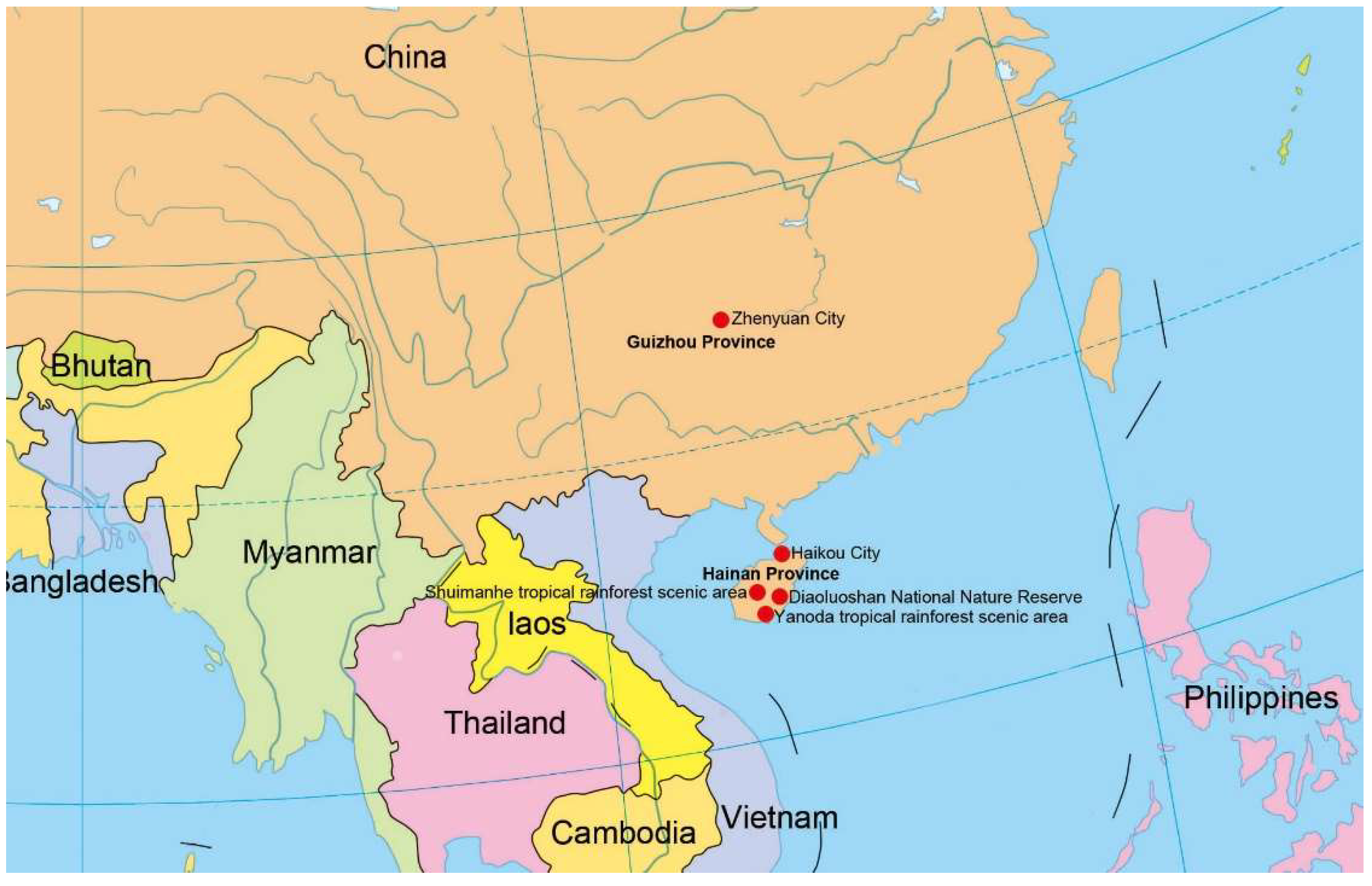
Figure 1. Collecting sites in this study (red dots).
Jof 08 01202 g001
Figure 2. Phylogenetic tree generated from maximum likelihood (ML) analysis based on a combined of LSU, ITS, tef1α, and rpb2 sequence data. Bootstrap support values of maximum likelihood (ML) and Maximum Parsimony (MP) equal to or greater than 75%, and Bayesian posterior probabilities (PP) equal to or greater than 0.95 are given near the nodes as ML/MP/PP. T Pseudostanjehughesia aquitropica (MFLUCC 16-0569) and Ps. lignicola (MFLUCC 15-0352) were used as outgroup taxa. The newly generated sequences are indicated in red bold.
Jof 08 01202 g002
Figure 3. Aquapteridospora hyalina (HKAS 123764, holotype). (a,b) Colonies on the host surface. (ce) Conidiophores, conidiogenous cells with attached conidia. (fh) Conidiogenous cells bearing conidia. (ik) Conidia. (l) Germinated conidium. (m,n) Colonies on PDA, m from above, n from below. Scale bars: (el) 20 μm, (c,d) 10 μm.
Jof 08 01202 g003
Figure 4. Distoseptispora aquisubtropica (GZAAS 22-0080, holotype). (a,b) Colonies on the host surface. (cg) Conidiophores and conidiogenous cells bearing conidia. (hk) Conidia. (l) Germinating conidium. (m,n) Colonies on PDA, m from above, n from below. Scale bars: (cl) 20 μm.
Jof 08 01202 g004
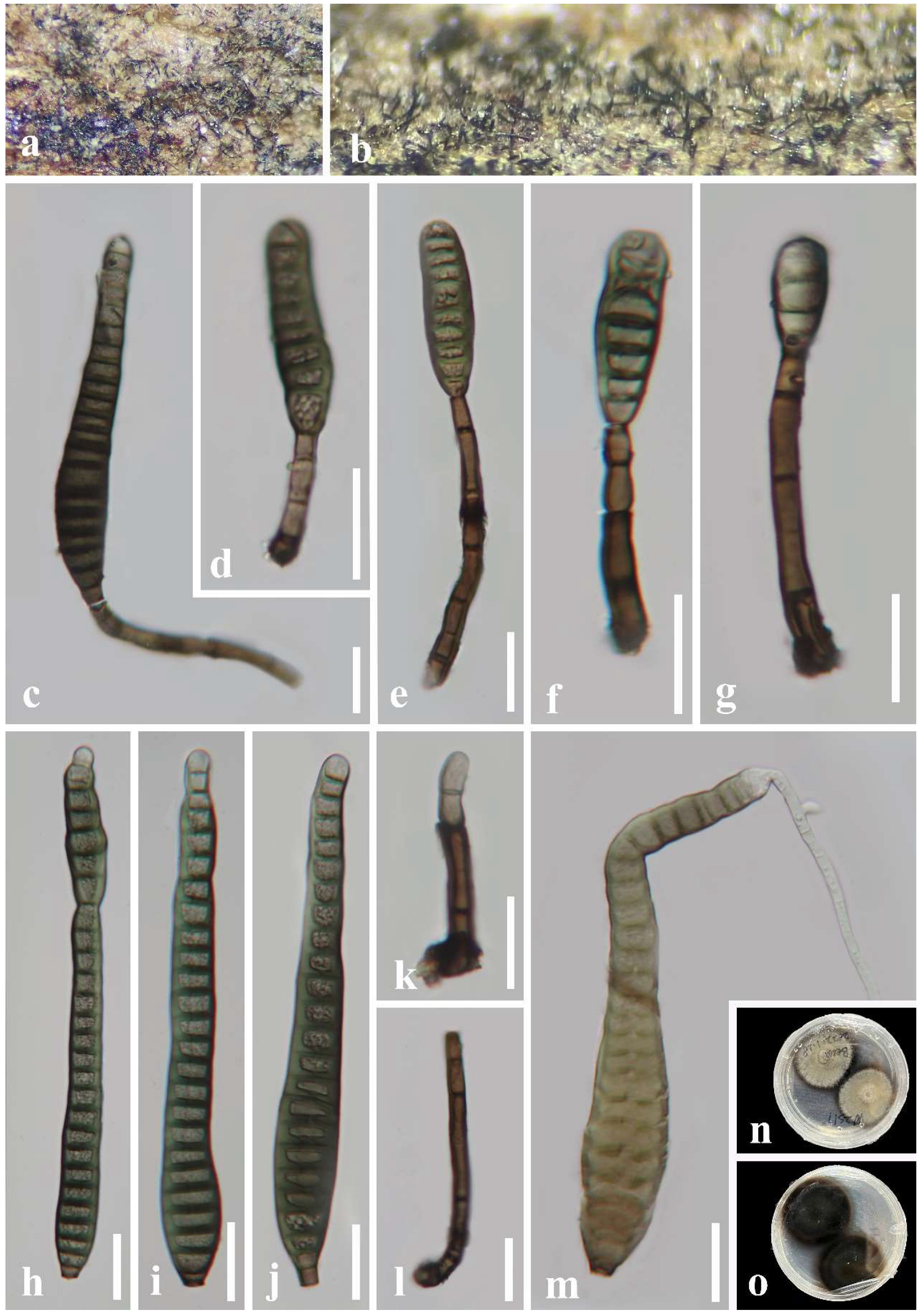
Figure 5. Distoseptispora pachyconidia (HKAS 123754). (a,b) Colonies on the host surface. (cf) Conidiophores and conidiogenous cells bearing conidia. (gl) Conidia. (m) Germinating conidium. (n,o) Colonies on PDA, n from above, o from reverse. Scale bars: (c,eh,jm) 20 μm, (d,i) 10 μm.
Jof 08 01202 g005
Figure 6. Distoseptispora septata (HKAS 123759, holotype). (a,b) Colonies on the host surface. (cg) Conidiophores and conidiogenous cells bearing conidia. (hj) Conidia. (k,l) Conidiophores and conidiogenous cells. (m) Germinating conidium. (n,o) Colonies on PDA, n from above, o from reverse. Scale bars: (cm) 20 μm.
Jof 08 01202 g006
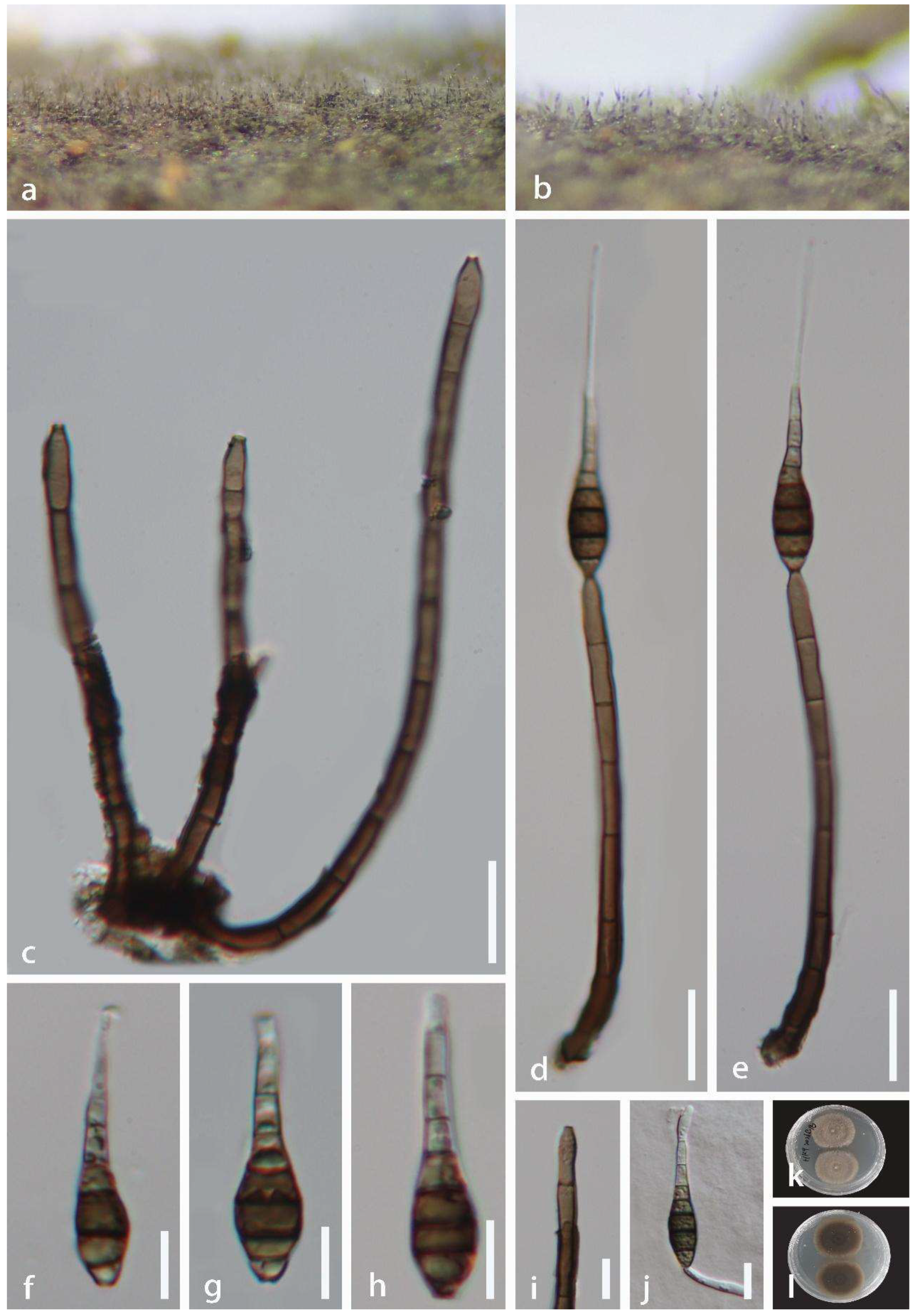
Figure 7. Distoseptispora tropica (HKAS 123761, holotype). (a,b) Colonies on the host surface. (c) Conidiophores and conidiogenous cells. (d,e) Conidiophores with attached conidia. (fh) Conidia. (i) Conidiogenous cell. (j) Germinating conidium. (k,l) Colonies on PDA, k from above, l from reverse. Scale bars: (ce) 20 μm, (fj) 10 μm.
Jof 08 01202 g007
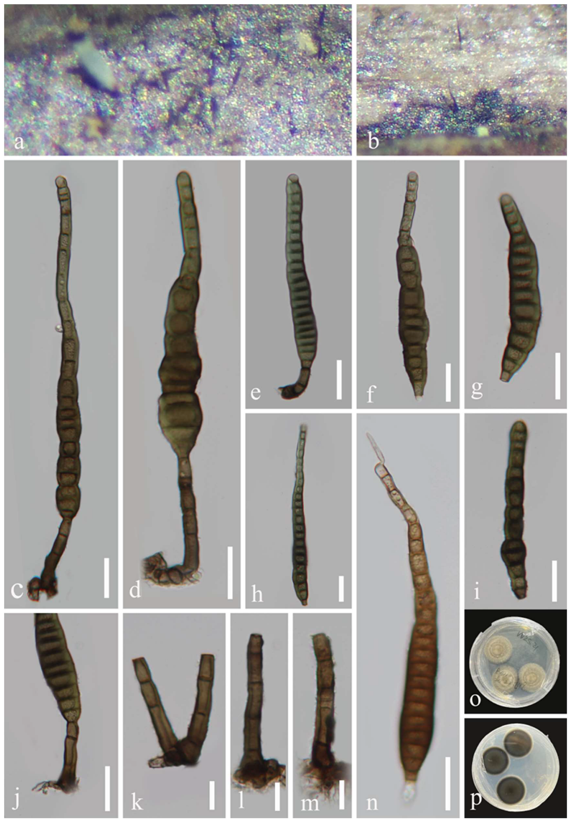
Figure 8. Distoseptispora wuzhishanensis (HKAS 123762, holotype). (a,b) Colonies on the host surface. (ce) Conidiophores and conidia. (fi) Conidia. (jm) Conidiophores and conidiogenous cells. (n) Germinating conidium. (o,p) Colonies on PDA, o from above, p from reverse. Scale bars: (cj,n) 20 μm, (km) 10 μm.
Jof 08 01202 g008
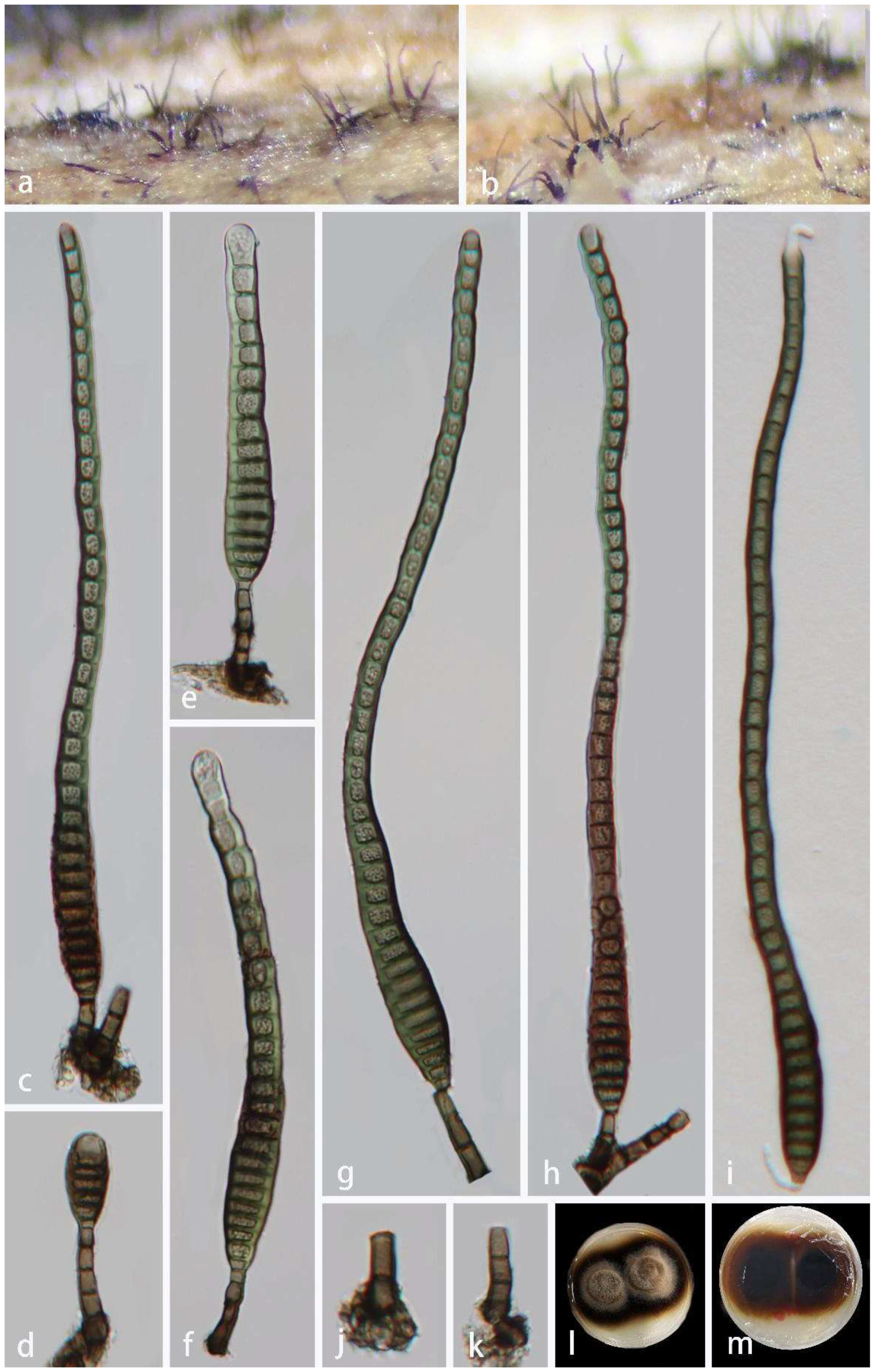
Figure 9. Distoseptispora xishuangbannaensis (HKAS 123760). (a,b) Colonies on the host surface. (ch) Conidiophores, conidiogenous cells with attached conidia. (i) Germinating conidium. (j,k) Conidiophores and conidiogenous cells. (l,m) Colonies on PDA, l from above, m from below. Scale bars: (c,k,ei) 20 μm, (d,j) 10 μm.
Jof 08 01202 g009
Table 1. PCR protocols.
LocusPrimerInitial DenaturationDenaturationAnnealingElongationFinal ExtensionHold
LSUITS5/ITS494 °C/3 min94 °C/45 s56 °C/50 s72 °C/1 min72 °C/10 min4 °C
40 cycles
ITSLR0R/LR594 °C/3 min94 °C/45 s56 °C/50 s72 °C/1 min72 °C/10 min
40 cycles
tef1α983F/2218R94 °C/3 min94 °C/30 s56 °C/50 s72 °C/1 min72 °C/10 min
30 cycles
rpb2frpb2-5f/frpb2-7cr95 °C/5 min95 °C/15 s56 °C/50 s72 °C/1 min72 °C/10 min
35 cycles
Table 2. Taxa used in this study and the GenBank accession numbers of DNA sequences.
TaxonStrainGenBank AccessionsReference
LSUITStef1αrpb2
Aquapteridospora
fusiformis
MFLU 18-1601TMK849798MK828652MN194056Luo et al. (2019)
A. aquaticaMFLUCC 17-2371TMW287767MW286493Dong et al. (2021)
A. bambusinumMFLUCC 12-0850TKU863149KU940161KU940213 Dai et al. (2016)
A. bambusinumMFLUCC 21-0027MZ412526MZ412514MZ442688Bao et al. (2021)
A. hyalinaGZCC 22-0072TON527945ON527937ON533681ON533689This study
A. hyalinaGZCC 22-0073ON527948ON527940ON533684ON533691This study
A. jiangxiensisJAUCC 3008TMZ871501MZ871502MZ855767MZ855768Peng et al. (2022)
A. lignicolaMFLU 15-1172TKU221018MZ868774MZ892980MZ892986Yang et al. (2015)
Distoseptispora
adscendens
HKUCC 10820DQ408561DQ435092Shenoy et al. (2006)
D. amniculiMFLU 21-0138TMZ868761MZ868770MZ892982Yang et al. (2021)
D. appendiculataMFLUCC 18-0259TMN163023MN163009MN174866Luo et al. (2019)
D. aqualignicolaKUNCC 21-10729TON400845OK341186OP413480OP413474Zhang et al. (2022)
D. aquamycesKUNCC 21-10731TOK341199OK341187OP413482OP413476Zhang et al. (2022)
D. aquaticaMFLUCC 15-0374TKU376268MF077552Su et al. (2016)
D. aquaticaMFLUCC 18-0646MK849793MK828648Luo et al. (2019)
D. aquisubtropicaGZCC 22-0075TON527941ON527933ON533677ON533685This study
D. atroviridisHKAS 112616TMZ868763MZ868772MZ892978MZ892984Yang et al. (2021)
D. bambusaeMFLUCC 20-0091TMT232718MT232713MT232880MT232881Sun et al. (2020)
D. bambusaeMFLUCC 14-0583MT232717MT232712MT232882Sun et al. (2020)
D. bambusicolaGZCC 21-0667TMZ474872MZ474873
D. bangkokensisMFLU 21-0110TMZ518206MZ518205Shen et al. (2021)
D. cangshanensisMFLUCC 16-0970TMG979761MG979754MG988419Luo et al. (2018)
D. caricisCPC 36498TMN567632MN562124MN556805Crous et al. (2019)
D. caricisCPC 36442MN562125MN556806Crous et al. (2019)
D. chinensisGZCC 21-0665TMZ474867MZ474871MZ501609Hyde et al. (2021)
D. clematidisMFLUCC 17-2145TMT214617MT310661MT394721Phukhamsakda et al. (2020)
D. crassisporaKUMCC 21-10726TOK341196OK310698OP413479OP413473Zhang et al. (2022)
D. curvulariaKUMCC 21-10725TOK341195OK310697OP413478OP413472Zhang et al. (2022)
D. cylindricosporaHKAS 115796TOK513523OK491122OK524220Phukhamsakda et al. (2022)
D. dehongensisKUMCC 18-0090TMK079662MK085061MK087659Hyde et al. (2019)
D. effusaGZCC 19-0532TMZ227224MW133916Yang et al. (2021)
D. eusptataMFUCC 20-0154TMW081544MW081539MW151860Li et al. (2021)
D. eusptataMFLU 20-0568MW081545MW081540MW084994MW084996Li et al. (2021)
D. fasciculataKUMCC 19-0081TMW287775MW286501MW396656Dong et al. (2021)
D. fluminicolaMFLUCC 15-0417TKU376270MF077553Su et al. (2016)
D. fusiformisHKAS 112617TMZ868764MZ868773MZ892979MZ892985Yang et al. (2021)
D. guizhouensisGZCC 21-0666TMZ474869MZ474868MZ501610MZ501611Hyde et al. (2021)
D. guttulataMFLUCC 16-0183TMF077554MF077543MF135651Yang et al. (2018)
D. guttulataDLUCC B43MN163016MN163011Luo et al. (2019)
D. hyalinaMFLU 21-0137TMZ868760MZ868769MZ892976MZ892981Yang et al. (2021)
D. hydeiMFLUCC 20-0481TMT742830MT734661MT767128Monkai et al. (2020)
D. lancangjiangensisKUN-HKAS 112712TMW879522MW723055MW882260Shen et al. (2021)
D. leonensisHKUCC 10822DQ408566DQ435089Shenoy et al. (2006)
D. lignicolaMFLUCC 18-0198TMK849797MK828651Luo et al. (2019)
D. longisporaHFJAU 0705TMH555357MH555359Yang et al. (2021)
D. martiniiCGMCC 318651TKX033566KU999975Xia et al. (2017)
D. meilingensisJAUCC 4727TOK562396OK562390OK562408Zhai et al. (2022)
D. multiseptataMFLUCC 16-1044MF077555MF077544MF135652MF135644Yang et al. (2018)
D. multiseptataMFLUCC 15-0609TKX710140KX710145MF135659Hyde et al. (2016)
D. neorostrataMFLUCC 18-0376TMN163017MN163008Luo et al. (2019)
D. nonrostrataKUNCC 21-10730TOK341198OK310699OP413481OP413475Zhang et al. (2022)
D. obclavataMFLUCC 18-0329TMN163010MN163012Luo et al. (2019)
D. obpyriformisMFLUCC 17-01694TMG979764MG988422MG988415Luo et al. (2018)
D. obpyriformisDLUCC 0867MG979765MG979757MG988423MG988416Luo et al. (2018)
D. pachyconidiaKUMCC 21-10724TOK341194OK310696OP413477OP413471Zhang et al. (2022)
D. pachyconidiaGZCC 22-0074ON527942ON527934ON533678ON533686This study
D. palmarumMFLUCC 18-1446TMK079663MK085062MK087660MK087670Hyde et al. (2019)
D. phangngaensisMFLUCC 16-0857TMF077556MF077545MF135653Yang et al. (2018)
D. rayongensisMFLUCC 18-0415TMH457137MH457172MH463253MH463255Hyde et al. (2020)
D. rayongensisMFLUCC 18-0417MH457138MH457173MH463254MH463256Hyde et al. (2020)
D. rostrataMFLUCC 16-0969TMG979766MG979758MG988424MG988417Luo et al. (2018)
D. rostrataDLUCC 0885MG979767MG979759MG988425Luo et al. (2018)
D. saprophyticaMFLUCC 18-1238TMW287780MW286506MW396651MW504069Dong et al. (2021)
D. septataGZCC 22-0078TON527947ON527939ON533683ON533690This study
D. songkhlaensisMFLUCC 18-1234TMW287755MW286482MW396642Dong et al. (2021)
D. submersaMFLUCC 16-0946TMG979768MG979760MG988426MG988418Luo et al. (2018)
D. suoluoensisMFLUCC 17-0224TMF077557MF077546MF135654Yang et al. (2018)
D. suoluoensisMFLUCC 17-1305MF077558MF077547Yang et al. (2018)
D. tectonaeMFLUCC 12-0291TKX751713KX751711KX751710KX751708Hyde et al. (2016)
D. tectonaeMFLU 20-0262MT232719MT232714Sun et al. (2020)
D. tectonaeMFLUCC 15-0981MW287763MW286489MW396641Dong et al. (2021)
D. tectonigenaMFLUCC 12-0292TKX751714KX751712KX751709Hyde et al. (2016)
D. thailandicaMFLUCC 16-0270TMH260292MH275060MH412767Tibpromma et al. (2018)
D. thysanolaenaeKUMCC 18-0182TMK064091MK045851MK086031Phukhamsak et al. (2019)
D. tropicaGZCC 22-0076TON527943ON527935ON533679ON533687This study
D. verrucosaHKAS 112652TMZ868762MZ868771MZ892977MZ892983Yang et al. (2021)
D. wuzhishanensisGZCC 22-0077TON527946ON527938ON533682This study
D. xishuangbannaensisKUMCC 17-0290TMH260293MH275061MH412768MH412754Tibpromma et al. (2018)
D. xishuangbannaensisGZCC 22-0079ON527944ON527936ON533680ON533688This study
D. yongxiuensisJAUCC 4725TOK562394OK562388OK562406Zhai et al. (2022)
D. yunjushanensisJAUCC 4723TOK562398OK562392OK562410Zhai et al. (2022)
D. yunnanensisMFLUCC 20-0153TMW081546MW081541MW084995MW151861Li et al. (2021)
Myrmecridium
aquaticum
MFLUCC 15-0366TMK849804Luo et al. (2019)
M. aquaticumS 1158MK849803MK828656MN194061MN124540Luo et al. (2019)
M. banksiaeCBS 132536TJX069855JX069871Crous et al. (2012)
M. montsegurinumJF 13180TKT991664KT991674KT991654Réblová et al. (2016)
M. schulzeriCBS 100.54EU041826EU041769Arzanlou et al. (2007)
Pseudostanjehughesia
aquitropica
MFLUCC 16-0569TMF077559MF077548MF135655Yang et al. (2018)
Ps. lignicolaMFLUCC 15-0352TMK849787MK828643MN194047MN124534Luo et al. (2019)
Sporidesmium
bambusicola
HKUCC 3578TDQ408562Shenoy et al. (2006)
S. dulongenseMFLUCC 17-0116TMH795817MH795812MH801191MH801190Luo et al. (2019)
S. lageniformeDLUCC 0880TMK849782MK828640MN194044MN124533Luo et al. (2019)
S. pyriformatumMFLUCC 15-0620TKX710141KX710146MF135662MF135649Hyde et al. (2016)
S. thailandenseMFLUCC 15-0617MF077561MF077550MF135657Yang et al. (2018)
S. thailandenseMFLUCC 15-0964TMF374370MF374361MF370957MF370955Zhang et al. (2017)
Note: T denote ex-type strain. Newly generated sequences are indicated in bold. “–”means no data available in GenBank.
Publisher’s Note: MDPI stays neutral with regard to jurisdictional claims in published maps and institutional affiliations.