Article

Bacillus velezensis TSA32-1 as a Promising Agent for Biocontrol of Plant Pathogenic Fungi

1
Department of Research and Development, Center for Industrialization of Agricultural and Livestock Microorganisms, Jeongeup-si 56212, Korea
2
Department of Bioactive Material Science, Jeonbuk National University, Jeonju 54896, Korea
3
Department of Molecular Biology, Institute for Molecular Biology and Genetics, Jeonbuk National University, Jeonju 54896, Korea
*
Author to whom correspondence should be addressed.
J. Fungi 2022, 8(10), 1053; https://doi.org/10.3390/jof8101053
Submission received: 22 August 2022 / Revised: 5 October 2022 / Accepted: 6 October 2022 / Published: 8 October 2022
(This article belongs to the Section Fungi in Agriculture and Biotechnology)

Abstract

:
The use of synthetic fungicides has caused major problems such as soil and water pollution and negatively affects non-target species. Microbial biocontrol agents are needed for crop disease management to reduce agrochemical use. Bacillus and related genera produce secondary metabolites with agricultural applications, such as the pathogen-control agent Bacillus velezensis. We isolated B. velezensis TSA32-1 from soil and identified its characteristics by sequencing its 16S rRNA. B. velezensis TSA32-1 showed enzyme activity and antimicrobial effects against phytopathogenic fungi by inhibiting the growth of Fusarium graminearum, F. fujikuroi, Alternatia alternate, and Diaporthe actinidiae. Additionally, B. velezensis TSA32-1 protected diseases in corn and pepper seeds caused by F. graminearum and Pythium ultimum. The complete genome of B. velezensis TSA32-1 was 4.05 Mb with a G+C content of 46.3 mol % and possessed the bacillaene biosynthesis cluster, a polyketide that inhibits protein biosynthesis. We also detected a surfactin synthesis cluster, known as non-ribosomal peptide synthetases, which biosynthesizes the antibacterial substance lipopeptide. Surfactin, and fengycin family compounds, secondary metabolites known as key factors in biological control, also detected B. velezensis TSA32-1 which shows potential as a biocontrol agent for controlling plant pathogens in agriculture.

1. Introduction

In agriculture, biotic stressors such as pathogens and pests cause 20–30% of global crop yield losses [1]. Particularly, soil-borne fungal infections of important grain crops such as corn, rice, and pepper cause large economic losses by reducing yields [2]. Chemicals for protecting crops and promoting growth such as pesticides, herbicides, and fertilizers are widely used in the agricultural sector to ensure sufficient and consistent yields [3]. However, many of these chemicals lead to soil and water pollution and pesticide residues in agricultural products, negatively affecting human health and the environment [4]. Thus, organic products as safe and eco-friendly control technologies, such as biological agents, are needed for disease prevention and management in agriculture [5]. The use of microorganisms as biological agents that can control disease-causing plant pathogens in an environmentally sound manner has been widely reported [6,7].
The biocontrol abilities of these microbial agents are typically related to the secondary metabolite profile of the microorganism. Antagonist microorganisms produce several secondary metabolites that can control various phytopathogens. Representatively, some strains of Bacillus velezensis, B. siamensis, and B. subtilis have been proposed as plant pathogen control agents because they produce numerous antimicrobial compounds, such as surfactin, iturin, bacilysin, fengycin, macrolactin, bacillaene, and difficidin [8,9]. In addition, the Bacillus species have the advantage of growing rapidly under simple trophic conditions and produce endospores that make cells more resistant to environmental stress [10]. B. subtilis SG6 showed antagonistic action against the F. graminearum pathogen, and extraction of secondary metabolites of surfactin and fengycin has been reported [11]. B. subtilis BS8B-1 shows antagonistic effects to the Pythium pathogen under greenhouse conditions; genetic approaches and chemical analysis were used to evaluate the mechanism of volatile substances and antibacterial substances against pathogenic fungi [12]. B. inaquosorum KR2-7 has been proposed as an effective biological control agent against F. oxysporum under greenhouse conditions. Genome mining was performed to explain these antifungal effects, and fengycin, surfactin, and iturin, among others, were detected as secondary metabolites [13]. Li et al. (2021) also reported performing genome mining and mass spectrometry analysis to analyze the various secondary metabolites serving as biological agents of B. velezensis W1 to seek antimicrobial active ingredients [14].
In this study, we aimed to investigate isolates, and identify functional characteristics of B. velezensis TSA32-1 for developing biological control agents against Fusarium and Pythium, pathogens of important crops such as corn and pepper. The antifungal effect of TSA32-1 was detected against soil-borne pathogens by treating corn and pepper seeds with B. velezensis TSA32-1. We provide an understanding of the mechanism of action of B. velezensis TSA32-1 biological agents by detecting secondary metabolites such as surfactin and plipastatin in the B. velezensis TSA32-1 culture medium through chemical analysis. Based on the diverse and effective properties of TSA32-1 for its antifungal action, we propose TSA32-1 as a beneficial biocontrol agent for the eco-friendly agricultural industry.

2. Materials and Methods

2.1. Isolation and Identification of B. velezensis TSA 32-1

B. velezensis TSA32-1 was collected from soil (Muan-gun, Korea) and isolated using the serial dilution method [15]. Soil samples (1 g) were processed by crushing, suspended in physiological saline (9 mL), and homogenized. For enumeration, a 10-fold dilution series of each homogenate was prepared in sterile saline solution, and 0.1 mL of this sample was spread on tropical soy agar plates, which were incubated at 30 °C for 24 h. As reference stains, B. subtilis KCTC3135 and B. velezensis KCTC13012 were obtained from the Korean Collection for Type Cultures (Jeollabuk-do, Korea).

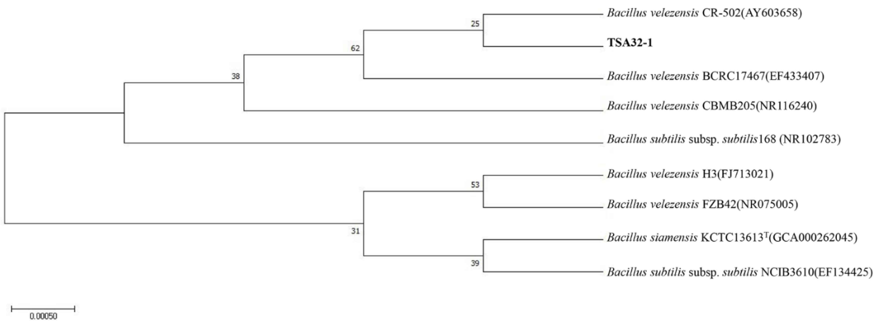
2.2. Phylogenetic and Statistical Analysis

The isolated strains were sequenced and identified using 16S rRNA sequencing. Phylogenetic analysis was conducted using the maximum likelihood method, with the numbers at the branch points indicating the bootstrapping values, using Molecular Evolutionary Genetic Analysis software, version 7.0 [16]. The data were determined as the average value of three independent biological repetitions. Standard deviations represent variation among replicates. One-way analysis of variance (Tukey’s multiple comparison test) was performed using GraphPad Prism 5 software (GraphPad, Inc., La Jolla, CA, USA) to detect statistically significant differences (p < 0.05) between treatments [17].

2.3. Enzymatic Activity Analysis

2.3.1. Cellulase Activity

Cellulase activity was evaluated through a Cellulase Activity Assay Kit (ab189817; Abcam, Cambridge, UK) according to the manufacturer’s instructions. Cellulase activity was measured as the fluorescence at Ex/Em = 550/595 nm (peak Ex/Em = 571/585 nm) in a microplate reader in kinetic mode for 120min at 25 °C. The sample concentration was calculated using a standard curve.

2.3.2. Lysozyme Activity

Microbial resistance to lysosomal hydrolytic enzymes was evaluated using a Lysozyme Assay Kit (E-22013; Thermo Fisher Scientific, Waltham, MA, USA) according to the manufacturer’s instructions with a fluorescent-based microplate reader. Lysosome activity was quantified by measuring the fluorescence intensity at 450 nm.

2.3.3. Protease Activity

Protease activity was evaluated using a Protease Activity Assay Kit (ab111750, Abcam) according to the manufacturer’s instructions. Fluorescence was measured at Ex/Em = 485/530 nm and then re-measured after 30 min. Protease activity was determined according to standard values and Equation (1). In Equation (1), B is the amount of fluorescein isothiocyanate (nmol) from the standard curve, T1 is the time (min) of the first reading (R1), T2 is the time (min) of the second reading (R2), and V is the pretreated sample volume (mL) added to the reaction well.
Protease   activity mU / mL = B   ×   dilution   factor T 2 T 1   ×   V

2.4. Antifungal Analysis

The antifungal ability of TSA32-1 was evaluated using the disk diffusion method as previously described [14] with slight modifications against several phytopathogenic fungi from the Korean Agricultural Culture Collection (KACC, Jeollabuk-do, Korea). Briefly, the fungal pathogen was cultured on a potato dextrose agar plate for 7 days. A pathogenic agar block with a diameter of 8 mm was prepared and placed in the center of the plate. A diffusion disk with a diameter of 8 mm was overlaid on the agar, and 20 µL of 1 × 10⁶ colony forming units/mL of the culture suspension was distributed on the right disk; 20 µL of distilled water was added to the left disk as a control. After culturing at 25 °C for 3–5 days, the antifungal activity of TSA32-1 was assessed by determining the radial mycelial growth of the fungal pathogen. All experiments were performed in triplicate.

2.5. Pathogen Inoculum Production and Disease Evaluation

2.5.1. Preparation of Bacillus Strains

The Bacillus strains were prepared at 1 × 10⁶ colony-forming units/mL or higher by incubation overnight in a 30 °C shaking incubator at 150 rpm in a tryptic soy broth medium. The sample was centrifuged at 4 °C and 10,000 rpm and the pellet was collected. The pellet was resuspended in 250 ppm of Tween 20.

2.5.2. Preparation of Seeds

Corn seeds (Zea mays L.) and pepper seeds (Capsicum annuum L.) were disinfected by immersion in 100% ethanol for 1 min, rinsed three times with water, immersed in 1% sodium hypochlorite for 30 min, and rinsed three times with water [18]. The seeds were dehydrated overnight in a hood.

2.5.3. Preparation of Pathogenic Fungus

F. graminearum KACC41044 was incubated in potato dextrose agar medium at 25 °C for 7 days and then cut into agar blocks. The agar blocks were inoculated with 15 g carboxymethyl cellulose, 1 g NH4NO3, 1 g KH2PO4, 0.5 g MgSO4∙7H2O, 1 g yeast extract, and 1 L distilled water in a medium and incubated at 25 °C in a shaking incubator at 200 rpm for 5 days. After filtering the culture solution through a sterile Miracloth to filter mycelium, the filtrate was centrifuged at 4 °C at 10,000 rpm. The pellet was collected and mixed with 250 ppm of Tween 20, and microconidia were counted using a hemocytometer [19].
Pythium ultimum KACC40705 was prepared by culturing in corn-meal agar medium at 25 °C for 2 days. Several agar blocks of P. ultimum KACC40705 were cut and inoculated onto sterilized rice, with mixing once daily and incubation in the dark at room temperature for 2 weeks [20]. The pathogens were filtered through a sterile Miracloth and resuspended in 250 ppm Tween 20 [21].

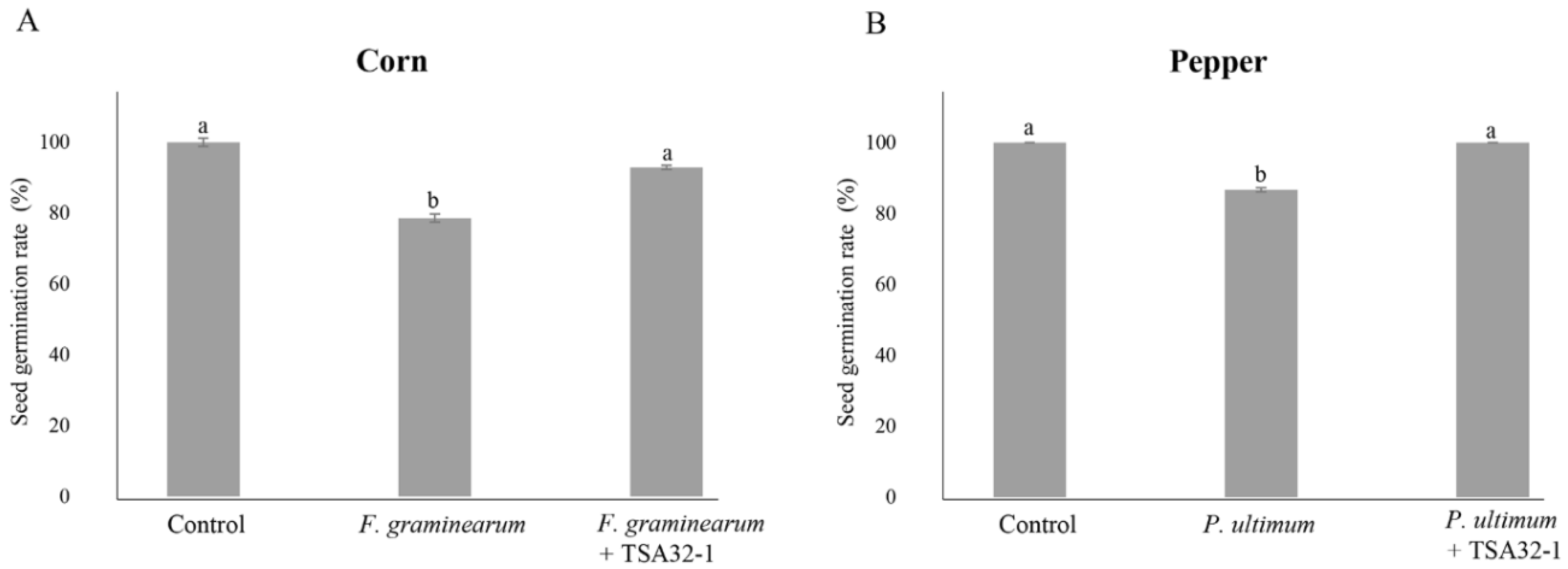
2.5.4. Seed Germination Rate of Treatments

The seed germination rate of corn and pepper was investigated following coating with the TSA32-1 culture. The seed germination rate was estimated as a percentage based on the average percentage of the control seed germination value, which was considered as 100. For pathogen inoculation, corn seeds soaked for 30 min in the prepared Bacillus immersion solution at room temperature were sprayed with the microconidia of F. graminearum KACC41044 pathogen at 1 × 10⁶ microconidia/mL. As a control, untreated corn seeds were placed on filter paper and moistened with distilled water. The pathogen-treated corn seeds were placed on filter paper and directly sprayed with the conidia suspension. The seed germination rate was measured after culturing for 1 week in the dark at 25 °C while maintaining moist conditions.
Pepper seeds, as in the corn seed experiments, were induced to germinate by placing the seeds on a filter paper and supplying moisture as described by Shang et al. (1998) with some modifications. P. ultimum KACC40705 was used as the pathogen [22].

2.5.5. Disease Suppression Effect of Seed Treatments

The ability of TSA32-1 to suppress the disease caused by F. graminearum and P. ulimum on corn and pepper was evaluated in pot tests with autoclaved soil [20,23]. The corn and pepper seeds, either coated or uncoated with TSA32-1 were sowed into the sterilized soil (50 g) and placed in a plastic pot with a diameter of approximately 60 mm and a height of 65 mm. Three seeds were sown in each pot, and the experiment was performed with 10 pots for each configuration. F. graminearum KACC41044 and P. ultimum KACC40705 were inoculated into the soil to induce disease. The conidia suspension of the F. graminearum pathogen was prepared at a concentration of 1 × 10⁶ microconidia/mL and 1 mL was inoculated into each pot of soil. The disease was induced by adding 5 g of the P. ultimum pathogen prepared by infection with rice, to each pot of 50 g of sterile soil [20]. The tests were conducted in a humid greenhouse at 20–25 °C with 12-h photoperiods for 10 days. All tests were performed in triplicate.

2.5.6. DNA Extraction and Whole-Genome Sequencing

Genomic DNA was extracted using a DNeasy UltraClean microbial kit (Qiagen, Hilden, Germany) according to the manufacturer’s instructions. Whole-genome sequencing was performed using the Pacific Biosciences (PacBio) RS II Single-Molecule Real-Time (SMRT) platform with a 20 kb SMRTbell™ template library at ChunLab. The reads were assembled using PacBio SMAR Analysis 2.3.0 and an Illumina Hi-Seq system (San Diego, CA, USA) at Macrogen (Seoul, Korea).

2.5.7. Annotation

The single-molecule real-time sequencing reads were assembled de novo using the hierarchical genome assembly process workflow (HGAP 3.0) in PacBio’s SMRT portal with subreads from PacBio. Paired-end reads from Illumina Hi-Seq 2500 were mapped to the assembled contigs to improve the accuracy of the genome sequences [24]. The sequences were annotated using the combined results of the automated National Center for Biotechnology Information Prokaryotic Genomes Annotation Pipeline and Rapid Annotations Subsystems Technology prokaryotic genome annotation server (http://rast.nmpdr.org/, version 2.0, accessed on 1 August 2022) [25]. The coding genes were predicted according to clusters of orthologous groups using the WebMGA online tool [26].

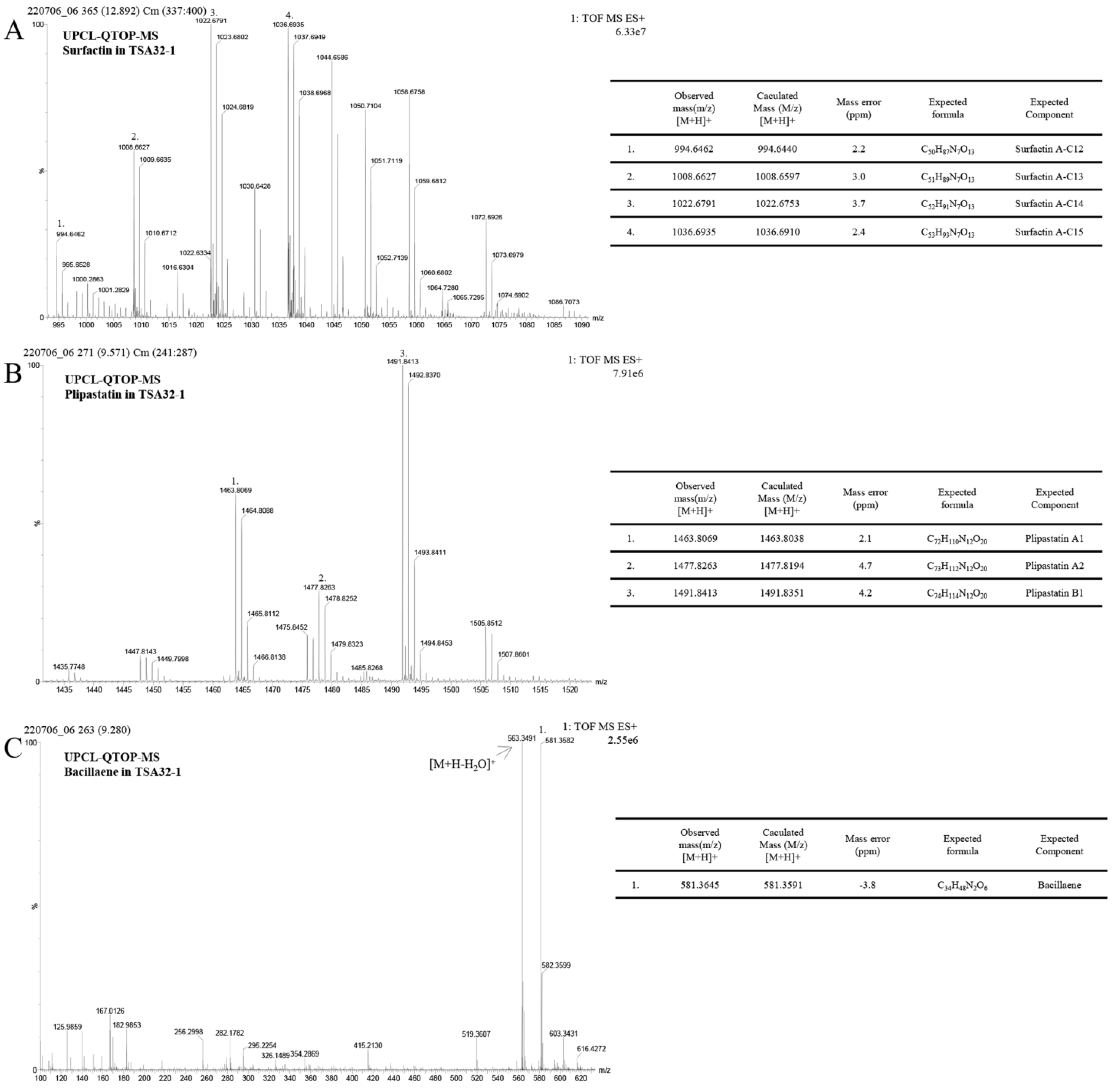
2.6. Antifungal Metabolite Extraction and Analysis

2.6.1. Preparation of Culture Extract

The culture medium used to grow TSA 32-1 and identify secondary metabolites has been described by Kim et al. (2010) [27]. TSA 32-1 was cultured in 1 L tryptic soy broth medium at 30 °C for 48 h and centrifuged to obtain the cell-free extract. HCl (1 N, 40 mL) was added to 1 L of the extract, and the mixture was protonated at 4 °C for 24 h to adjust the pH to 3.1. The mixture was centrifuged, and the pellet was dissolved in methanol and concentrated on a rotary evaporator (R-100, Büchi, Flawil, Switzerland). After filtration through a 0.2 μm filter, liquid chromatography–quantitative time-of-flight (LC–QTOF) analysis was performed. The 24-h culture medium of TSA32-1 was used for bacillaene analysis.

2.6.2. Ultra-High Performance (UP) LC-QTOF-Mass Spectrometry (MS) Analysis of Metabolites

The chemical profile of the metabolites produced by TSA32-1 was analyzed using a Waters Acquity UPLC I-Class PLUS equipped with a Waters ACQUITY UPLC BEH C18 column (2.1 × 100 mm, 1.7 µm) (Milford, MA, USA), maintained at an isothermal temperature of 65 °C. A binary pump delivered the mobile phase at the flow rate of 0.4 mL/min under gradient elution using two mobile phases, LC-MS–grade water + 0.1% v/v formic acid (solvent A) and LC-MS–grade acetonitrile + 0.1% v/v formic acid (solvent B). The auto-sampler was set to an injection volume of 1 µL. MS analysis was performed using a Waters Xevo-G2-XS QTOF LC-MS equipped with an electrospray ionization source. An analysis was conducted in positive-ion mode at a mass range of 100–1600 Da. The source temperature and capillary voltage were set to 150 °C and 3.0 kV, respectively. Ar was used as the collision gas. Standard solutions of surfactin (Santa Cruz Biotechnology, Dallas, TX, USA) and plipastatin (Sigma, St. Louis, MI, USA) were used.

3. Results

3.1. Identification of Bacterial Strains

We isolated TSA32-1 from soil and identified the species based on the 16S rRNA sequences. The genomic DNA sequences of the strains were searched against the available gene sequences in the GenBank database. Strain TSA32-1 is 99% homologous to B. velezensis (AY603658). The phylogenic relationship between the bacterial strains was established based on the maximum likelihood of evolutionary distances of the 16S rRNA sequences, as shown in Figure 1.

3.2. Plant Disease Biocontrol Activity

3.2.1. Enzyme Activity of Isolated Strains

The antifungal effects of cellulolytic, lysosomes, and proteolytic enzymes were evaluated, and TSA32-1 substantially affected the cell wall-related three enzyme treatment as shown in Figure 2. The measured cellulase activity of TSA32-1 was 16.75 μM/mL, which demonstrated marked resistance to cellulose activity as displayed with higher activity than the type strains of B. velezensis KCTC 13012 and B. subtilis KCTC3135 (Figure 2A). The measured lysozyme activity of TSA32-1 was 76.46 U/mL, B. velezensis KCTC 13012 was 63.65 U/mL, and B. subtilis KCTC3135 was 13.73 U/mL, respectively, indicating superior or equivalent lysosomal activity to the type strain (Figure 2B). Similarly, the measured protease activity of TSA32-1 was 2.68 mU/mL, which showed higher activity than the type strain (Figure 2C). TSA32-1 was superior to the type strain in three enzyme activities, cellulase, lysozyme, and protease, which are enzymes that help inhibit fungal growth. These results indicate that increased production and activity of cell wall degrading enzymes of TSA32-1 are associated with decreased invasiveness and pathogenicity.

3.2.2. In Vitro Antifungal Effects against Phytopathogenic Fungi

The antagonistic activity assay showed that TSA32-1 inhibited the filamentous growth of 16 fungal phytopathogens (Figure 3). TSA32-1 strongly inhibited mycelial growth of Alternaria alternate KACC45440 (brown ring spot of kiwi fruit), Collectrichum acutatum KACC40042 (anthracnose of pepper), C. lymphaea GNC1 (anthracnose of kiwi berry), Cylindrocarpon destructans KACC41077 (brown colony of ginseng), Diaporthe actinidiae SN18 (stem-end rot of kiwi fruit), and F. oxysporum f. sp. Raicis-lycopersici O1090 (root rot of tomato) compared to the control. TSA32-1 also inhibited the mycelial growth of Botryosphaeria dothidea KACC45481 (circular growth of crab apple), Botrytis cinereal KACC47009 (gray mold of grape), F. graminearum KACC41044 (head blight of corn), F. graminearum KACC46434 (scab of rice), F. oxysporum KACC41088 (root rot of orchids), F. fugikuroi KACC44002 (bakanae disease of rice), Glomerella cingulate 06-KN-1 (anthracnose of ki-wifruit), and P. ultimum KACC40705 (damping-off of cucumber).

3.2.3. Plant Protection Effects of Seed Treatments with TSA32-1

The germination rate of seeds infected with the phytopathogenic fungus F. graminearum KACC41044 on corn seeds was 78.6%, and seeds coated with TSA32-1 showed a germination rate of 92.9% even after pathogen infection (Figure 4A). When the seeds were treated with TSA32-1, the germination rate was higher than that of corn seeds infected with the pathogen alone. In addition, pepper seeds coated with TSA32-1 showed a germination rate of 100% even after pathogen infection, whereas pepper seeds infected with P. ultimum KACC40705 showed a germination rate of 88.7% (Figure 4B). These results suggest that TSA32-1 antagonizes F. graminearum and P. ultimum and positively affects the seed germination rate, which directly affects crop growth.
The control value was set as 100%, and the data were expressed as percentages relative to this value. The germination rate of TSA32-1-treated and pathogen-infected corn seeds in the soil was 74.6%, whereas the germination rate of corn sprouts was 23.6% when inoculated with the pathogen F. graminearum KACC41044 alone (Figure 5A). Similar in vitro seed germination rates were observed, with TSA32-1 showing antifungal action against F. graminearum pathogens and enabling seedling growth. The germination rate of pepper seeds infected with the P. ultimum KACC40705 pathogen alone was 14.5%, whereas pathogen-infected pepper seeds pre-coated with TSA32-1 showed a germination rate of 41.9% (Figure 5B). The results of measuring sprout germination rate by infecting red pepper seeds treated with TSA32-1 with pathogens tend to show higher values than those treated with pathogens alone.

3.3. Genome Feature of B. velezensis TSA32-1

The complete genome of TSA32-1 was composed of a 4,050,296 base pair circular chromosome with a 46.3% G+C content. The genome contains 3825 protein-coding sequences, 27 rRNAs, and 87 tRNAs (Figure 6). Among the coding sequences, 3512 genes were classified into 20 clusters of orthologous groups’ functional categories (Figure 6B). Many genes were classified into functional categories for amino acid transport and metabolism (n = 291), transcription (n = 261), carbohydrate transport and metabolism (n = 238), inorganic ion transport and metabolism (n = 186), cell wall/membrane/envelope biogenesis (n = 184), energy production and conversion (n = 175), translation (n = 161), signal transduction mechanisms (n = 134), and recombination and repair (n = 127). Interestingly, 15.1% of genes were involved in amino acid transport and metabolism, carbohydrate transport, and metabolism.
The gene clusters related to the antibiotic activity identified in the genome of TSA 32-1 and secondary metabolites synthesized during gene expression are summarized in Table 1. Interestingly, TSA32-1 possesses five non-ribosomal peptide synthetases (NRPS) metabolite clusters within its genome of a large size of over 20 kb, which are conserved in all B. velezensis members. This group of three NRPS clusters comprises surfactin, plipastatin, and bacilysin and two polyketide synthetase-NRPS gene clusters, encoding the anti-bacterial polyketide bacillaene and antifungal polyketide mycosubtilin (Table 1). The whole genome sequences of the isolates have been deposited in GenBank under Bioproject number PRJNA602208 for B. velezensis TSA32-1.

3.4. Antifungal Metabolite of B. velezensis TSA32-1

Lipopeptides produced by TSA32-1 were identified using UPLC–QTOF–MS analysis. Standard solutions of surfactin and plipastatin generated four major peaks. Similarly, crude surfactin gained from the extracts of TSA32-1 culture was further purified via preparative chromatography with a solvent containing methanol, and the partial peak was resolved into four fractions. Each fraction revealed that the sample contained quasi molecular ions at mass-to-charge ratios (m/z) of 994.6462, 1008.6629, 1022.6791, and 1036.6935 ([M + H]+) indicating surfactin (Figure 7A). Another Q-TOF-MS analysis revealed three fractions for plipastatin with m/z of 1463.8069, 1477.8263, and 1491.8413 representing plipastatin A–B, respectively (Figure 7B). There were also well-resolved peaks at a mass range of 400–600. The corresponding compounds were identified as bacillaene ([M + H]+ = 581.3582) by comparison with mass data obtained from B. subtilis XF-1 [28] (Figure 7C).

4. Discussion

Bacillus spp. have been widely studied as microbial biocontrol agents. As a useful microorganism in agriculture, Bacillus can affect plant growth and fruit-bearing by facilitating nutrient absorption and exerting various enzyme activities with antagonistic effects on pathogenic bacteria and fungi [29].
Cellulase enzymes degrade cell walls by cleaving the ß-1,4-D-glycosidic bonds that connect the glucose units containing cellulose and exert antifungal effects by destroying the fungal cell wall and cytoplasmic membrane [30]. Lysosomes and proteolytic enzymes capable of hydrolyzing polysaccharides adversely affect fungal growth and differentiation by dissolving or disturbing polymers in the cell wall of pathogenic fungi [30]. Lysozyme has been widely examined and shown to hydrolyze polymers belonging to the Gram (+) group present in the cell wall of bacteria; its ability to hydrolyze the cell wall of yeast and fungi was also recently reported [31,32]. The established potent activity of cellulase, lysozyme, and protease in TSA32-1 supports its association with the observed growth inhibition of various fungal phytopathogens in the present study (Figure 3). Furthermore, enzymatic lysis of the cell wall can increase chemical uptake into target cells [31]. In Bacillus spp. Vb1, Vb3, and Vb6, the high hydrolytic activity of enzymes, such as amylase, cellulase, and protease, is highly associated with antifungal activity against F. oxysporum pathogens [33]. The synergistic effect of co-production of cell wall hydrolytic enzymes and secondary metabolites useful for biocontrol may enhance the antagonistic effect on phytopathogenic fungi.
The antagonistic activity of TSA32-1 on phytopathogens revealed a broad capacity to inhibit the growth of fungal pathogens, which is a known characteristic of Bacillus species. This inhibitory ability of TSA32-1 may be related to the production of secondary metabolites. Interestingly, TSA32-1 showed antagonistic effects toward various plant pathogens that cause diseases such as root rot, head blight disease, bakanae disease, anthracnose, and damping-off disease. The positive impact of this antagonistic ability on soil-borne fungal pathogens of TSA32-1 is an important function as a microbial agent and increase its biocontrol potential.
Corn and pepper are important and representative food and vegetable crops. The most economically important diseases affecting these crops are blights disease, anthracnose disease, and damping-off disease [34]. Particularly, pathogenic Fusarium fungal species infect corn plants and reduce yields worldwide. The strain of Pythium and Fusarium such as F. graminearum and F. oxysporum are important pathogens of seedling diseases and interfere with seed germination and emergence, thus, affecting seedling development [35]. B. velezensis RC218 showed strong antagonist activity on seeds treated with F. graminearum [36]. In addition, Hyder et al. (2021) showed that the effects of Bacillus sp. on the germination rate of chili pepper seeds and its potential function as a biocontrol agent were evaluated by measuring the antagonistic effect and germination rate against the Pythium pathogen [37]. Analysis of the germination rate of corn and pepper seeds treated with TSA32-1 showed a higher germination rate than when each pathogen was treated alone (Figure 4). Application of TSA32-1 prior to pathogen exposure may induce tolerance in corn and pepper and activate plant defense responses against soil-borne pathogens, thereby limiting disease infection [38]. To complement field studies, pot experiments can allow for direct investigations without the influence of diffusing any environmental factors. Thus, seed or soil treatment pot experiments are performed to identify microbes as effective therapeutic agents against pathogens in agriculture [39]. Kim et al. (2017) showed that B. amyloliquefaciens JCK-12 significantly controlled F. graminearum-induced head blight in wheat under greenhouse and field conditions [40]. B. subtilis BBG100 was tested for its biocontrol potential in seed germination and pot experiments and found to reduce damping-off caused by P. aphanidermatum infection of tomato seedlings [41]. In addition, B. subtilis JA and D1/2 suppressed head blight and ear rot caused by F. graminearum in wheat, barley, and corn in the field [42]. B. velezensis TSA32-1 showed potential as a biocontrol agent by securing the ability to inhibit F. graminearum and Pythium on corn and pepper. Corn and pepper seeds treated with TSA32-1 were infected with soil-borne pathogens, and the sprout germination rate showed a tendency to increase when compared to the values of those infected with pathogens alone. Soil-borne pathogens like F. graminaearum and Pythium infection mainly occur in seeds, seedlings, and young roots, and it is known that it has a significant impact on the growth of seedlings, causing great economic loss and a ripple effect [37]. Against these soil-borne pathogens, TSA32-1 may contribute to the control effect that helps prevent disease in seeds. TSA32-1 exhibits biological control effects, including disease prevention, against several pathogens and crops, which could be a potential biocontrol agent.
Through genome analysis, we predicted the genes and gene clusters involved in the secondary metabolites producing TSA32-1. Bacillus spp. can produce multiple antimicrobials with a variety of chemical structures [43]. Lipopeptides are synthesized by NRPS. Generally, Bacillus strains produce the lipopeptides classified in the three families, iturin, surfactin, and fengycin, including plipastatin [44]. Among them, a cluster consisting of srfAA, srfAB, srfAC, and srfAD encoding the biosynthetic gene cluster of surfactin was detected in the TSA32-1 genome and had a size of 26.1 kb (Table 1). Surfactin is a multifunctional lipopeptide that exerts surfactant and signaling activity necessary for motility and biofilm development [45]. Plipastatin is a potent Bacillus antimicrobial lipopeptide with the potential for replacing conventional antifungal chemicals to control plant pathogens. In the genome of TSA32-1, we detected ppsA, C, D, E (37.6 kb) encoding the subunits of NRPS for plipastatin. The synthesized plipastatin may inhibit phospholipase A2, responsible for structuring pores in the fungal membrane and inducing the morphological change of membrane and cell wall of fungi [46]. In addition, the gene cluster bae of type I polyketide synthetases associated with the biosynthesis of bacillaene [47] were predicted in the TSA32-1 genome. The bae operon encodes an enzymatic macrocomplex that synthesizes bacillaene. Most bae genes appear to consist of large operons (>74 kb in baeC-baeR). Three bae operons were found in the genome of TSA32-1 with sizes of 43.3–71.7 kb. Accordingly, bacillaene of various sizes can be expressed and may improve antibacterial effects. Bacillaene is known as a broad-spectrum antibiotic that inhibits bacterial protein [48]. The gene cluster ywfA-bacABCDE-ywfG-ywfH encoding bacilysin synthesis was detected with a size of 7.9 kb. Bacilysin is known as an antibacterial substance that induces the dissolution of pathogen cell walls by inhibiting glucosamine-6-phosphate synthetase [49]. The mycA-C cluster also exists in the TSA32-1 genome. myc encodes mycosubtilin, which has an antifungal function. B. subtilis Z15, which produces mycosubtilin, applied to a cotton field confirmed its ability to inhibit wilt disease caused by Verticillium dahlia [50].
As described above, bacteria in the Bacillus genus produce a wide variety of antimicrobials. To confirm the genome background of secondary metabolites produced by TSA32-1, the culture medium was analyzed. The surfactin family is constituted by peptides of seven amino acids bound to the beta-hydroxyl fatty acid with 13–15 carbon atoms [51]. The fengycin family is represented by several isomers of fengycin, plipastatin, and maltacine; they consist of 10 amino acids connected to fatty acid with up to 18 carbon atoms [52]. The substances detected in TSA32-1 were similar in molecular weight to the two types of lipopeptides, and molecular ion peaks were detected as surfactin and plipastatin. The peaks show a molecular weight difference of 14 Da, suggesting that the length of the fatty acid chains varies within each group (CH2 = 14 Da). The signal detected in the chromatogram showed Rf value similar to the standard except for bacillaene. A peak similarly detected around an expected core peak was judged to have a water molecule omitted from the molecular formula of bacillaene. Bacillaene is an unstable secondary metabolite that is chemically sensitive to light, oxygen, and temperature [53]. In the 48 h culture of TSA32-1, it was difficult to detect this peak; the peak for bacillaene was detected in the 24 h culture.
Numerous studies and critical reviews have been conducted to determine the impact of Bacillus lipopeptides on plant pathogens [54]. B. subtilis pB2-L produced plipastatin (the fengycin family), which inhibits F. oxysporum mycelium growth [55]. B. velezensis GH1-13 showed high antagonistic activity toward various phytopathogenic fungi, including F. fujikuroi and C. gloeosporioides. Analysis of secondary metabolites revealed the presence of surfactin [56]. Gong et al. showed that B. amyloliquefaciens S76-3 produced plipastatin A and iturin A, which have clear antagonistic effects on F. graminearum [57]. B. subtilis BBG100 which is genetically engineered to overproduce mycosubtilin showed an antifungal effect against Pythium sp. by seed treatment on tomato [41]. Another classic example among other isolates is the plant-associated strain B. amyloliquefaciens FZB42 which produces antimicrobial peptides and secondary metabolites, including lipopeptides, polyketides, and bacteriocins while simultaneously producing substances that promote plant growth [58]. Many Bacillus strains produce more than one family of lipopeptides, and synergistic interactions may occur between different compounds to enhance biological control [59]. B. velezensis TSA32-1 also produces a variety of lipopeptides, such as surfactin, plipastatin, and bacilaene, with biological control effects. The investigation of these various lipopeptides and secondary metabolites provides scientific evidence of the effectiveness of TSA32-1 as a biocontrol agent.

5. Conclusions

B. velezensis TSA32-1 isolated from soil showed cellulase, lysozyme, and protease activities related to polymer degradation in the cell wall of plant pathogens and produced various lipopeptides such as surfactin, plipastatin, and bacillaene as secondary metabolites. B. velezensis TSA32-1 exerts antifungal action that destroys the structural integrity of fungal cells and inhibits the growth of phytopathogenic fungi such as F. graminearum and P. ultimum. Particularly, B. velezensis TSA32-1 reduced the occurrence of plant pathogens via antagonistic effects against F. graminearum and P. ultimum. In conclusion, B. velezensis TSA32-1 showed broad and effective antagonistic activity against various pathogenic fungi, suggesting its potential for use as a biocontrol microorganism in eco-friendly agriculture.

Author Contributions

Conceptualization, Y.K.; methodology, J.-A.K., J.-S.S. and Y.K.; software, J.-A.K. and J.-S.S.; validation data curation, D.-H.K., P.I.K. and J.-A.K.; writing—original draft preparation, J.-A.K. and Y.K.; writing—review and editing, J.-A.K. and Y.K.; supervision, D.-H.K., P.I.K. and Y.K.; funding acquisition, Y.K. All authors have read and agreed to the published version of the manuscript.

Funding

This work was supported by Korea Institute of Planning and Evaluation for Technology in Food, and Agriculture and Forestry (IPET) through Useful Agricultural Life Resources Industry Technology Development Program and Agricultural Machinery/Equipment Localization Technology Development Program, funded by Ministry of Agriculture, Food and Rural Affairs (MAFRA) (321094-2, 122022-3).

Institutional Review Board Statement

Not applicable.

Informed Consent Statement

Not applicable.

Data Availability Statement

All sequences data were submitted to NCBI.

Acknowledgments

We thank the anonymous reviewers for their careful reviewing of this manuscript and providing insightful comments.

Conflicts of Interest

The authors declare no conflict of interest.

References

  1. Savary, S.; Willocquet, L.; Pethybridge, S.J.; Esker, P.; McRoberts, N.; Nelson, A. The global burden of pathogens and pests on major food crops. Nat. Ecol. Evol. 2019, 3, 430–439. [Google Scholar] [CrossRef] [PubMed]
  2. Cheng, X.; Ji, X.; Ge, Y.; Li, J.; Qi, W.; Qiao, K. Characterization of Antagonistic Bacillus methylotrophicus Isolated From Rhizosphere and Its Biocontrol Effects on Maize Stalk Rot. Phytopathology 2019, 109, 571–581. [Google Scholar] [CrossRef] [PubMed] [Green Version]
  3. Legein, M.; Smets, W.; Vandenheuvel, D.; Eilers, T.; Muyshondt, B.; Prinsen, E.; Samson, R.; Lebeer, S. Modes of Action of Microbial Biocontrol in the Phyllosphere. Front. Microbiol. 2020, 11, 1619. [Google Scholar] [CrossRef] [PubMed]
  4. Nishimoto, R. Global trends in the crop protection industry. J. Pestic. Sci. 2019, 44, 141–147. [Google Scholar] [CrossRef] [PubMed] [Green Version]
  5. Muis, S.d.A. Prospect of Bacillus subtilis as a Biological Control Agent of Soilborne Pathogens on Maize. J. Litbang Pert. 2016, 35, 37–45. [Google Scholar]
  6. Foysal, M.J.; Lisa, A.K. Isolation and characterization of Bacillus sp. strain BC01 from soil displaying potent antagonistic activity against plant and fish pathogenic fungi and bacteria. J. Genet. Eng. Biotechnol. 2018, 16, 387–392. [Google Scholar] [CrossRef] [PubMed]
  7. Kim, Y.S.; Lee, Y.; Cheon, W.; Park, J.; Kwon, H.T.; Balaraju, K.; Kim, J.; Yoon, Y.J.; Jeon, Y. Characterization of Bacillus velezensis AK-0 as a biocontrol agent against apple bitter rot caused by Colletotrichum gloeosporioides. Sci. Rep. 2021, 11, 626. [Google Scholar] [CrossRef]
  8. Santoso, I.; Fadhilah, Q.G.; Maryanto, A.E.; Yasman. The Potency of Bacillus siamensis LDR as Biocontrol Agent Against Fungal Phytopathogen. Adv. Biol. Sci. Res. 2020, 14, 463–468. [Google Scholar]
  9. Kuta, F.A. Antifungal effect of Calotropis procera stem bark on Epidermophyton flocosum and Trichophyton gypseum. Afr. J. Biotechnol. 2008, 7, 2116–2118. [Google Scholar]
  10. Mardanova, A.M.; Fanisovna Hadieva, G.; Tafkilevich Lutfullin, M.; Valer’evna Khilyas, I.; Farvazovna Minnullina, L.; Gadelevna Gilyazeva, A.; Bogomolnaya, L.M.; Sharipova, M.R. Bacillus subtilis Strains with Antifungal Activity against the Phytopathogenic Fungi. Agric. Sci. 2017, 8, 1–20. [Google Scholar]
  11. Zhao, Y.; Selvaraj, J.N.; Xing, F.; Zhou, L.; Wang, Y.; Song, H.; Tan, X.; Sun, L.; Sangare, L.; Folly, Y.M.E.; et al. Antagonistic action of Bacillus subtilis strain SG6 on Fusarium graminearum. PLoS ONE 2014, 9, e92486. [Google Scholar] [CrossRef] [PubMed]
  12. Khabbaz, S.E.; Zhang, L.; Cáceres, L.A.; Sumarah, M.; Wang, A.; Abbasi, P.A. Characterisation of antagonistic Bacillus and Pseudomonas strains for biocontrol potential and suppression of damping-off and root rot diseases. Ann. Appl. Biol. 2015, 166, 456–471. [Google Scholar] [CrossRef]
  13. Kamali, M.; Guo, D.; Naeimi, S.; Ahmadi, J. Perception of Biocontrol Potential of Bacillus inaquosorum KR2-7 against Tomato Fusarium Wilt through Merging Genome Mining with Chemical Analysis. Biology 2022, 11, 137. [Google Scholar] [CrossRef]
  14. Li, X.; Munir, S.; Xu, Y.; Wang, Y.; He, Y. Combined mass spectrometry-guided genome mining and virtual screening for acaricidal activity in secondary metabolites of Bacillus velezensis W1. RSC Adv. 2021, 11, 25441–25449. [Google Scholar] [CrossRef] [PubMed]
  15. Jang, H.J.; Son, S.; Kim, J.A.; Jung, M.Y.; Choi, Y.J.; Kim, D.-H.; Lee, H.K.; Shin, D.; Kim, Y. Characterization and Functional Test of Canine Probiotics. Front. Microbiol. 2021, 12, 625562. [Google Scholar] [CrossRef]
  16. Kumar, S.; Stecher, G.; Tamura, K. MEGA7: Molecular Evolutionary Genetics Analysis Version 7.0 for Bigger Datasets. Mol. Biol. Evol. 2016, 33, 1870–1874. [Google Scholar] [CrossRef] [PubMed] [Green Version]
  17. Sung, J.C.; Lai, N.C.; Wu, K.C.; Choi, M.C.; Ma, C.H.; Lin, J.; Kuok, C.-N.; Leong, W.-L.; Lam, W.-K.; Hamied, Y.K.; et al. Safety and Immunogenicity of Inactivated Bacillus subtilis Spores as a Heterologous Antibody Booster for COVID-19 Vaccines. Vaccines 2022, 10, 1014. [Google Scholar] [CrossRef] [PubMed]
  18. Sauer, D.B.; Burroughs, R. Disinfection of Seed Surfaces with Sodium Hypochlorite. Techniques 1986, 76, 745–749. [Google Scholar] [CrossRef]
  19. Leslie, J.F.; Summerell, B.A. The Fusarium Laboratory Manual; Blackwell Publishing: Hoboken, NJ, USA, 2008. [Google Scholar]
  20. Bongiorno, G.; Postma, J.; Bünemann, E.K.; Brussaard, L.; de Goede, R.G.M.; Mäder, P.; Tamm, L.; Thuerig, B. Soil suppressiveness to Pythium ultimum in ten European long-term field experiments and its relation with soil parameters. Soil Biol. Biochem. 2019, 133, 174–187. [Google Scholar] [CrossRef]
  21. St Arnaud, M.; Hamel, C.; Fortin, J.A. Inhibition of Pythium ultimum in roots and growth substrate of mycorrhizal Tagetes patula colonized with Glomus intraradices. Can. J. Plant Pathol. 2009, 16, 187–194. [Google Scholar] [CrossRef]
  22. Shang, H.; Chen, J.; Handelsman, J.; Goodman, R.M. Behavior of Pythium torulosum Zoospores During Their Interaction with Tobacco Roots and Bacillus cereus. Curr. Microbol. 1999, 38, 199–204. [Google Scholar] [CrossRef]
  23. Kaur, K.; Kaur, J.; Puyam, A.; Singh, K. Cultural, Morphological and Molecular Characterization of Fusarium verticillioides causing Maize Ear Rot under Punjab Condition. Int. J. Curr. Microbiol. Appl. Sci. 2020, 9, 1698–1706. [Google Scholar] [CrossRef]
  24. Teng, J.L.L.; Yeung, M.L.; Chan, E.; Jia, L.; Lin, C.H.; Huang, Y.; Tse, H.; Wong, S.S.Y.; Sham, P.C.; Lau, S.K.P.; et al. PacBio But Not Illumina Technology Can Achieve Fast, Accurate and Complete Closure of the High GC, Complex Burkholderia pseudomallei Two-Chromosome Genome. Front. Microbiol. 2017, 8, 1448. [Google Scholar] [CrossRef] [Green Version]
  25. Aziz, R.K.; Bartels, D.; Best, A.A.; DeJongh, M.; Disz, T.; Edwards, R.A.; Formsma, K.; Gerdes, S.; Glass, E.M.; Kubal, M.; et al. The RAST Server: Rapid annotations using subsystems technology. BMC Genom. 2008, 9, 75. [Google Scholar] [CrossRef]
  26. Wu, S.; Zhu, Z.; Fu, L.; Niu, B.; Li, W. WebMGA: A customizable web server for fast metagenomic sequence analysis. BMC Genom. 2011, 12, 444. [Google Scholar] [CrossRef] [Green Version]
  27. Kim, P.I.; Ryu, J.; Kim, Y.H.; Chi, Y.T. Production of Biosurfactant Lipopeptides Iturin A, Fengycin and Surfactin A from Bacillus subtilis CMB32 for Control of Colletotrichum gloeosporioides. J. Microbiol. Biotechnol. 2010, 20, 138–145. [Google Scholar] [CrossRef] [Green Version]
  28. Guo, S.; Li, X.; He, P.; Ho, H.; Wu, Y.; He, Y. Whole-genome sequencing of Bacillus subtilis XF-1 reveals mechanisms for biological control and multiple beneficial properties in plants. J. Ind. Microbiol. Biotechnol. 2015, 42, 925–937. [Google Scholar] [CrossRef]
  29. Fonseca, M.C.D.; Bossolani, J.W.; de Oliveira, S.L.; Moretti, L.G.; Portugal, J.R.; Scudeletti, D.; de Oliveira, E.F.; Crusciol, C.A.C. Bacillus subtilis Inoculation Improves Nutrient Uptake and Physiological Activity in Sugarcane under Drought Stress. Microorganisms 2022, 10, 809. [Google Scholar] [CrossRef]
  30. Poulose, A.J. Biotechnology and fungal control. In Target Sites of Fungicide Action; CRC Press: Boca Raton, FL, USA, 1992; pp. 311–318. [Google Scholar]
  31. Hernández, C. Chitisan/Carrageenan/Lysozyme particles: Synthesis, Characterization and Antifungal activity against Aspergillus parasiticus. Rev. Mex. Ing. Química 2018, 17, 897–912. [Google Scholar] [CrossRef]
  32. Kim, J.A.; Bayo, J.; Cha, J.; Choi, Y.J.; Jung, M.Y.; Kim, D.H.; Kim, Y. Investigating the probiotic characteristics of four microbial strains with potential application in feed industry. PLoS ONE 2019, 14, e0218922. [Google Scholar] [CrossRef] [Green Version]
  33. El-Sersawy, M.M.; Hassan, S.E.; El-Ghamry, A.A.; El-Gwad, A.M.A.; Fouda, A. Implication of plant growth-promoting rhizobacteria of Bacillus spp. as biocontrol agents against wilt disease caused by Fusarium oxysporum Schlecht. in Vicia faba L. Biomol. Concepts 2021, 12, 197–214. [Google Scholar] [CrossRef] [PubMed]
  34. Wang, W.; Wang, B.; Sun, X.; Qi, X.; Zhao, C.; Chang, X.; Khaskheli, M.I.; Gong, G. Symptoms and pathogens diversity of Corn Fusarium sheath rot in Sichuan Province, China. Sci. Rep. 2021, 11, 2835. [Google Scholar] [CrossRef] [PubMed]
  35. Varela, C.P.; Casal, O.A.; Padin, M.C.; Martinez, V.F.; Oses, M.J.S.; Scauflaire, J.; Munaut, F.; Castro, M.J.B.; Vázquez, J.P.M. First Report of Fusarium temperatum Causing Seedling Blight and Stalk Rot on Maize in Spain. Plant Dis. 2013, 97, 1252. [Google Scholar] [CrossRef]
  36. Palazzini, J.M.; Dunlap, C.A.; Bowman, M.J.; Chulze, S.N. Bacillus velezensis RC 218 as a biocontrol agent to reduce Fusarium head blight and deoxynivalenol accumulation: Genome sequencing and secondary metabolite cluster profiles. Microbiol. Res. 2016, 192, 30–36. [Google Scholar] [CrossRef]
  37. Hyder, S.; Gondal, A.S.; Rizvi, Z.F.; Atiq, R.; Haider, M.I.S.; Fatima, N.; Inam-Ul-Haq, M. Biological Control of Chili Damping-Off Disease, Caused by Pythium myriotylum. Front. Microbiol. 2021, 12, 587431. [Google Scholar] [CrossRef]
  38. Conrath, U.; Beckers, G.-J.M.; Flors, V.; Garcia-Agustin, P.; Jakab, G.; Mauch, F.; Newman, M.-A.; Pieterse, C.M.J.; Poinssot, B.; Pozo, M.J.; et al. Priming: Getting Ready for Battle. Am. Phytopathol. Soc. 2006, 19, 1062–1071. [Google Scholar] [CrossRef] [PubMed] [Green Version]
  39. Zhang, J.X.; Xue, A.G.; Tanbong, J.T. Evaluation of Seed and Soil Treatments with Novel Bacillus subtilis Strains for Control of Soybean Root Rot Caused by Fusarium oxysporum and F. graminearum. Plant Dis. 2009, 93, 1317–1323. [Google Scholar] [CrossRef] [Green Version]
  40. Kim, K.; Lee, Y.; Ha, A.; Kim, J.I.; Park, A.R.; Yu, N.H.; Son, H.; Choi, G.J.; Park, H.W.; Lee, C.W.; et al. Chemosensitization of Fusarium graminearum to Chemical Fungicides Using Cyclic Lipopeptides Produced by Bacillus amyloliquefaciens Strain JCK-12. Front. Plant Sci. 2017, 8, 2010. [Google Scholar] [CrossRef] [Green Version]
  41. Leclere, V.; Bechet, M.; Adam, A.; Guez, J.S.; Wathelet, B.; Ongena, M.; Thonart, P.; Gancel, F.; Chollet-Imbert, M.; Jacques, P. Mycosubtilin overproduction by Bacillus subtilis BBG100 enhances the organism’s antagonistic and biocontrol activities. Appl. Environ. Microbiol. 2005, 71, 4577–4584. [Google Scholar] [CrossRef] [PubMed] [Green Version]
  42. Chan, Y.K.; Savard, M.E.; Reid, L.M.; Cyr, T.; McCormick, W.A.; Seguin, C. Identification of lipopeptide antibiotics of a Bacillus subtilis isolate and their control of Fusarium graminearum diseases in maize and wheat. BioControl 2009, 54, 567–574. [Google Scholar] [CrossRef]
  43. Stein, T. Bacillus subtilis antibiotics: Structures, syntheses and specific functions. Mol. Microbiol. 2005, 56, 845–857. [Google Scholar] [CrossRef]
  44. Vahidinasab, M.; Lilge, L.; Reinfurt, A.; Pfannstiel, J.; Henkel, M.; Morabbi Heravi, K.; Hausmann, R. Construction and description of a constitutive plipastatin mono-producing Bacillus subtilis. Microb. Cell Fact. 2020, 19, 205. [Google Scholar] [CrossRef] [PubMed]
  45. Pereira, J.F.B.; Gudiña, E.J.; Costa, R.; Vitorino, R.; Teixeira, J.A.; Coutinho, J.A.P.; Rodrigues, L.R. Optimization and characterization of biosurfactant production by Bacillus subtilis isolates towards microbial enhanced oil recovery applications. Fuel 2013, 111, 259–268. [Google Scholar] [CrossRef] [Green Version]
  46. Harwood, C.R.; Mouillon, J.M.; Pohl, S.; Arnau, J. Secondary metabolite production and the safety of industrially important members of the Bacillus subtilis group. FEMS Microbiol. Rev. 2018, 42, 721–738. [Google Scholar] [CrossRef]
  47. Aleti, G.; Sessitsch, A.; Brader, G. Genome mining: Prediction of lipopeptides and polyketides from Bacillus and related Firmicutes. Comput. Struct. Biotechnol. J. 2015, 13, 192–203. [Google Scholar] [CrossRef]
  48. Patel, P.S.; Huang, S.; Fisher, S.; Pornik, D.; Aklonis, C.; Dean, L.; Meyers, E.; Fernandes, P.; Mayerl, F. Bacillaene, a Novel Inhibitor of Procaryotic Protein Synthesis Produced by Bacillus subtilis: Production, Taxonomy, Isolation, Physico-chemical Characterization and Biological Activity. J. Antibiot. 1995, 48, 997–1003. [Google Scholar] [CrossRef] [PubMed] [Green Version]
  49. Wang, T.; Liu, X.H.; Wu, M.B.; Ge, S. Molecular insights into the antifungal mechanism of bacilysin. J. Mol. Model. 2018, 24, 118. [Google Scholar] [CrossRef] [PubMed]
  50. Lin, R.; Zhang, Q.; Yin, L.; Zhang, Y.; Yang, Q.; Liu, K.; Wang, Y.; Han, S.; Zhao, H.; Zhao, H. Isolation and characterization of a mycosubtilin homologue antagonizing Verticillium dahliae produced by Bacillus subtilis strain Z15. PLoS ONE 2022, 17, e0269861. [Google Scholar] [CrossRef] [PubMed]
  51. Peypoux, F.; Bonmatin, J.M.; Wallach, J. Recent trends in the biochemistry of surfactin. Appl. Microbiol. Biotechnol. 1999, 51, 533–563. [Google Scholar] [CrossRef] [PubMed]
  52. Moyne, A.L.; Shelby, R.; Cleveland, T.E.; Tuzun, S. Bacillomycin D: An iturin with antifungal activity against Aspergillus flavus. J. Appl. Microbiol. 2001, 90, 622–629. [Google Scholar] [CrossRef] [PubMed] [Green Version]
  53. Tsalgatidou, P.C.; Thomloudi, E.E.; Baira, E.; Papadimitriou, K.; Skagia, A.; Venieraki, A.; Katinakis, P. Integrated Genomic and Metabolomic Analysis Illuminates Key Secreted Metabolites Produced by the Novel Endophyte Bacillus halotolerans Cal.l.30 Involved in Diverse Biological Control Activities. Microorganisms 2022, 10, 399. [Google Scholar] [CrossRef] [PubMed]
  54. Fira, D.; Dimkic, I.; Beric, T.; Lozo, J.; Stankovic, S. Biological control of plant pathogens by Bacillus species. J. Biotechnol. 2018, 285, 44–55. [Google Scholar] [CrossRef] [PubMed]
  55. Gao, L.; Han, J.; Liu, H.; Qu, X.; Lu, Z.; Bie, X. Plipastatin and surfactin coproduction by Bacillus subtilis pB2-L and their effects on microorganisms. Antonie Van Leeuwenhoek 2017, 110, 1007–1018. [Google Scholar] [CrossRef] [PubMed]
  56. Park, G.; Nam, J.; Kim, J.; Song, J.; Kim, P.I.; Min, H.J.; Lee, C.W. Structure and Mechanism of Surfactin Peptide from Bacillus velezensis Antagonistic to Fungi Plant Pathogens. Bull. Korean Chem. Soc. 2019, 40, 704–709. [Google Scholar] [CrossRef]
  57. Gong, A.D.; Li, H.P.; Yuan, Q.S.; Song, X.S.; Yao, W.; He, W.J.; Zhang, J.-B.; Liao, Y.-C. Antagonistic mechanism of iturin A and plipastatin A from Bacillus amyloliquefaciens S76-3 from wheat spikes against Fusarium graminearum. PLoS ONE 2015, 10, e0116871. [Google Scholar]
  58. Chowdhury, S.P.; Dietel, K.; Randler, M.; Schmid, M.; Junge, H.; Borriss, R.; Hartmann, A.; Grosch, R. Effects of Bacillus amyloliquefaciens FZB42 on lettuce growth and health under pathogen pressure and its impact on the rhizosphere bacterial community. PLoS ONE 2013, 8, e68818. [Google Scholar] [CrossRef] [PubMed] [Green Version]
  59. Falardeau, J.; Wise, C.; Novitsky, L.; Avis, T.J. Ecological and mechanistic insights into the direct and indirect antimicrobial properties of Bacillus subtilis lipopeptides on plant pathogens. J. Chem. Ecol. 2013, 39, 869–878. [Google Scholar] [CrossRef]
Figure 1. 16S rRNA phylogenetic analysis of selected strain. Phylogenetic tree based on maximum likelihood method of 16S rRNA gene sequences of TSA32-1. The tree was created using the maximum likelihood method and numbers at the branch points are bootstrap values (based on 500 samplings expressed in percentages).
Jof 08 01053 g001
Figure 2. Enzyme activity of Bacillus spp. (A) Cellulase enzymes on the cell wall of TSA32-1. (B) Lysosomal hydrolytic enzymes on the cell wall of TSA32-1. (C) Protease activity of TSA32-1. Data are expressed as the means ± SD. Significant differences are marked with letters (p < 0.05). Reference strains Bacillus subtilis KCTC3135, B. velezensis KCTC13012 were tested as controls.
Jof 08 01053 g002
Figure 3. Antifungal activity of test strains against the indicator strains. Inhibition effect of mycelial growth of Fusarium fujikuroi KACC 44002 (A), F. graminearum KACC 41040 (B), F. graminearum KACC 46434 (C), F. graminearum KACC 41044 (D), F. oxysporum KACC 41088 (E), F. oxysporum f. sp. Raicis-lycopersici O1090 (F), Botryosphaeria dothidea KACC 45481 (G), Botrytis cinerea KACC 47009 (H), Alternaria alternata KACC 45440 (I), Diaporthe actinidiae SN18 (J), Glomerella cingulate 06-KN-1 (K), Colletotrichum nymphaea GNC1 (L), Colletotrichum acutatum KACC 40042 (M), Cylindrocarpon destructans KACC 41077 (N), Pythium ultimum KACC40705 (O), and Rhizoctonia solani AG-1 KACC 40101 (P) by B. velezensis TSA32-1.
Jof 08 01053 g003
Figure 4. The seed germination rate of seed treatments. (A) Treatments of the prototype TSA32-1 for head blight disease in corn caused by Fusarium graminearum KACC41044. (B) Treatments of the prototype TSA32-1 for damping-off disease in pepper caused by Pythium ultimum KACC40705. Data are expressed as the means ± SD. Significant differences are marked with letters (p < 0.05). Control, untreated check; fungal pathogen, infected pathogen; fungal pathogen + TSA32-1, pathogen inoculation after seed dipping.
Jof 08 01053 g004
Figure 5. Plant protection effect of seed treatments. (A) Biocontrol agent effect of treatments of the prototype TSA32-1 for head blight disease in corn caused by Fusarium graminearum. (B) Biocontrol agent effect of treatments of the prototype TSA32-1 for damping-off disease in pepper caused by Pythium ultimum. Data are expressed as the means ± SD. Significant differences are marked with letters (p < 0.05). Control, untreated check; fungal pathogen, infected pathogen; fungal pathogen + TSA32-1, pathogen inoculation after seed dipping.
Jof 08 01053 g005
Figure 6. Genome features of Bacillus velezensis TSA32-1. (A) Circular genome maps of B. velezensis TSA32-1 chromosome. Circles from the outside to the center denote rRNA and tRNA gene, reverse strand coding sequence, forward strand coding sequence, GC skew, and GC content. (B) Genome number of clusters of orthologous groups category.
Jof 08 01053 g006
Figure 7. Identification of signaling metabolites in Bacillus velezensis TSA32-1. Extracts were fractioned using UPLC-QTOF-MS. (A) Mass spectrum of surfactin, (B) plipastatin, (C) bacillaene compounds in TSA32-1 culture extract.
Jof 08 01053 g007
Table 1. Secondary metabolite clusters identified in the genome of Bacillus velezensis TSA32-1.
CompoundSynthetase TypeGenesSize (kb)Bioactivity
SurfactinNRPSsrfAA, AB, AC, AD26.1multiple
BacilysinNRPSywfA, bacA, B, C, D, E, F, ywfH6.7antibacterial
PlipastatinNRPSppsA, ppsC, ppsD, ppsE37.6antifungal
BacillaenePKS-NRPSbaeB, C, D, E, acpK, baeG, H, I, J, L, M, N, R, S
baeC, L, M, ppsC, baeL, M
baeC, G, I, L, M
71.7
43.3
64.6
antibacterial
MycosubtilinPKS-NRPSmycA, B, C35.9antifungal
PKS; Type I polyketide synthetases, NRPS; Non-ribosomal peptide synthetase.
Publisher’s Note: MDPI stays neutral with regard to jurisdictional claims in published maps and institutional affiliations.

