Article

Transcriptional Analysis of Coccidioides immitis Mycelia and Spherules by RNA Sequencing

1
Department of Medicine, Division of Infectious Disease, U.C. San Diego School of Medicine, La Jolla, CA 92093, USA
2
J. Craig Venter Institute, La Jolla, CA 92037, USA
3
Department of Microbiology and Plant Pathology, Institute for Integrative Genome Biology, University of California-Riverside, Riverside, CA 92521, USA
4
Infectious Diseases Section, VA Healthcare San Diego, San Diego, CA 92161, USA
5
Department of Pathology, U.C. San Diego School of Medicine, La Jolla, CA 92093, USA
*
Author to whom correspondence should be addressed.
J. Fungi 2021, 7(5), 366; https://doi.org/10.3390/jof7050366
Submission received: 27 March 2021 / Revised: 13 April 2021 / Accepted: 22 April 2021 / Published: 7 May 2021
(This article belongs to the Special Issue Coccidioides and Coccidioidomycosis 2020)

Abstract

:
Coccidioides immitis and C. posadasii are dimorphic fungi that transform from mycelia with internal arthroconidia in the soil to a tissue form known as a spherule in mammals. This process can be recapitulated in vitro by increasing the temperature, CO2 and changing other culture conditions. In this study, we have analyzed changes in gene expression in mycelia and young and mature spherules. Genes that were highly upregulated in young spherules include a spherule surface protein and iron and copper membrane transporters. Genes that are unique to Coccidioides spp. are also overrepresented in this group, suggesting that they may be important for spherule differentiation. Enriched GO terms in young spherule upregulated genes include oxidation-reduction, response to stress and membrane proteins. Downregulated genes are enriched for transcription factors, especially helix–loop–helix and C2H2 type zinc finger domain-containing proteins, which is consistent with the dramatic change in transcriptional profile. Almost all genes that are upregulated in young spherules remain upregulated in mature spherules, but a small number of genes are differentially expressed in those two stages of spherule development. Mature spherules express more Hsp31 and amylase and less tyrosinase than young spherules. Some expression of transposons was detected and most of the differentially expressed transposons were upregulated in spherules.

1. Introduction

Coccidioides immitis and C. posadasii are primary pathogenic fungi that are endemic in the desert regions of the Western United States, Mexico, and Central and South America [1]. They cause pulmonary infections that range from asymptomatic infections to severe pneumonias and can disseminate beyond the lung [2,3]. The organisms grows as a mold in the soil and produce asexual spores, termed arthroconidia, within the mycelium. When the soil is disturbed, the mycelia can rupture and the arthroconidia are released. If inhaled by a susceptible host, the arthroconidium differentiates into a very different and unique form that is known as a spherule. In tissue, the spherule enlarges and can form many reproductive endospores. Mature spherules rupture and release endospores, which can then differentiate into the next generation of spherules. This form, the spherule, is the disease-associated form of the organism. If the disease is self-limited, the reproduction of spherules is limited, but if the disease is severe, then spherules continue to grow, rupture and give rise to new spherules, while eliciting inflammatory and immune responses. This process goes on in many mammalian hosts, including desert rodents, which may be important in the ecology of the fungus [4].
The morphological transition between mycelia and spherule forms of C. immitis is dependent on sensing the host environment, and this transition can be recapitulated in the laboratory by changing the temperature and other growth conditions. C. immitis grow in saprobic form at 22–30 °C; culturing at 37–42 °C with 5–20% CO2 in Converse media is used to convert arthroconidia to spherules [5,6]. Utilizing these conditions, whole genome-level transcriptional profiling studies of saprobic and parasitic forms have been performed [7]. That work reported that about 1300 genes are upregulated in mycelia, about 1900 genes are upregulated in spherules, and expression of known virulence genes is upregulated in the spherules of both C. immitis and C. posadasii, linking morphology to the virulence traits. For example, the spherule outer wall glycoprotein, which is the outermost layer on the spherule, is expressed only in spherules [8,9]. Another study compared mycelia to young spherules and mature spherules to mycelia and to each other using microarray technology [10]. In the current study, we analyze the RNA obtained from frozen samples of spherules and mycelia obtained from the previous microarray study by strand-specific RNA sequencing. Our results show that there are more than 1500 genes that are differentially regulated in spherules and mycelial phases of C. immitis. We have compared the up- and downregulated genes to previous studies and, where possible, analyzed the function of differentially expressed genes.

2. Materials and Methods

2.1. Culture Conditions

C. immitis RS strain was grown as mycelia or spherules as previously described [10]. To grow the mycelia, 2 × 10⁶ arthroconidia/mL were incubated in 250 mL flat-bottom Erlenmeyer flasks (Corning, Corning, NY, USA) in 50 mL of GYE media. The flasks were cultured in a 30 °C incubator without shaking for 5 days. To grow the spherules, arthroconidia were washed 2 times in modified Converse media. The spores were inoculated at 4 × 10⁶ arthroconidia/mL into a 250 mL baffled Erlenmeyer flask containing 50 mL of modified Converse media. The flasks were set up and grown on a shaker at 160 rpm, in 14% CO2 at 42 °C. Four flasks were harvested 2 days after inoculation and the remaining four flasks after 8 days. Fresh Converse media was not added. The spherules did not rupture and release endospores within that time in this culture system.

2.2. RNA Extraction, Purification and Sequencing

The mycelia and spherule samples were stored in QIAzol (Qiagen) at −70 °C for seven years. Samples were added to 2 mL ZR BashingBead lysis tubes with 0.5 mm beads (Zymo Research, Irvine, CA, USA) and the tubes were arranged in a pre-cooled Tissuelyzer II adapter (Qiagen, Germantown, MD, USA) and disrupted by shaking at 50 Hz for 25 min. Total RNA was purified from mycelia and spherule samples (2 replicates/condition) using chloroform extraction and isopropanol precipitation and quantified using a Qubit 3.0 Fluorometer (Invitrogen, Carlsbad, CA, USA). For RNA-Seq, strand-specific, paired-end libraries were prepared from total RNA by ribosomal depletion using the Yeast Ribo-Zero rRNA Removal Kit (Illumina, San Diego, CA, USA) and then using the TruSeq Stranded total RNA-Seq kit (Illumina) according to manufacturer’s instructions. Then, 100 bases were sequenced from both ends using an Novaseq 6000 instrument according to the manufacturer’s instructions (Illumina, San Diego, CA, USA). A total of 10 million reads per sample were acquired.

2.3. Analysis of RNA-Seq Data

Duplicate sets of paired strand-specific reads were mapped to predicted mRNA sequences and quantified using Kallisto [11]. Read count tables from the Kallisto output were analyzed by DESeq2 in R. The results were filtered for a Benjamini–Hochberg adjusted p value < 0.05; upregulation and downregulation for the gene tables are defined as more than 2log2 or less than −2log2 spherules/mycelia fold change (FC). This cut off is chosen to minimize the false positive identification of differentially regulated genes. All FC in Tables are expressed as log2 values; FC changes in the text are expressed as arithmetic values.

2.4. Functional Analysis of Genes

All differentially expressed genes were evaluated using data from FungiDB version 51 (https://fungidb.org/fungidb/app, accessed on 4 January 2021) [12]. Orthology searches were done using the orthology tool at FungiDB. The FungiDB tool compares two sets of proteins by reciprocal BLASTP and computes the percent match length with a threshold for orthology, where a blast match e value is < 10−5 with a percent match length of ≥ 50%. Finally, the FungiDB tool obtains paralogs, orthologs and ortholog groups using OrthoMCL Pairs release 6.4 (https://orthomcl.org, accessed on 4 January 2021) [13]. C. immitis genes without a BLASTP match with an e value < 10−8 to all fungal species other than Coccidioides spp. and less than ten orthologs were considered genus-specific. Multiple alignment of transcription factors was done with Clustal Omega (https://www.ebi.ac.uk/Tools/msa/clustalo/, accessed on 4 January 2021) [14]. GO enrichment analysis was done using FungiDB (https://fungidb.org/fungidb/app, accessed on 4 January 2021) and FungiFun (https://elbe.hki-jena.de/fungifun/, accessed on 4 January 2021) [15]. GO term enrichment using the FungiFun tool was deemed significant if the Benjamini–Hochberg adjusted p value was < 0.05). GO enrichment analysis using the FungiDB tool was considered significant if the p value was < 0.05. Enrichment was carried determined using a Fisher’s Exact test with the background defined as all genes from the organism being queried. p-values corrected for multiple testing are provided using both the Benjamini – Hochberg false discovery rate method and the Bonferroni method. GO term enrichment are presented in Supplemental Table S2.
The FungiDB pathway tool was used to generate data in Supplemental Table S3. The pathway enrichment analysis of differentially expressed genes was conducted using the tool at FungiDB, which searches for annotation at MetaCyC (https://metacyc.org/, accessed on 4 January 2021) [16] and KEGG (https://www.genome.jp/kegg/, accessed on 4 January 2021) [17].

3. Results and Discussion

The RNA-Seq gene expression from the three conditions tested were compared to each other and the results are shown in Figure 1 and Supplemental Table S1. Comparing young and mature spherules to mycelia, at least 8% of genes were upregulated and 16–18% were downregulated (FC > 2log2 or < −2log2, adjusted p value less than 0.05). The number of differentially expressed genes comparing young spherules to mature spherules was much smaller (1.4–2%).

3.1. Differential Gene Expression in Young Spherules vs. Mycelia

The 20 most differentially expressed genes in young spherules are shown in Table 1.
The most highly upregulated gene is the spherule outer wall glycoprotein (SOWgp). This protein is expressed in very large amounts on the external surface of spherules but not mycelia and is involved in pathogenicity [8,9]. Two other spherule antigenic proteins, expression-library immunization protein-1 and parasitic-phase-specific protein PSP-1, [18,19], are also very highly upregulated, as are three transporters, including a copper transporter. There are only three genes that are predicted to be copper transporters in C. immitis, so this is a substantial increase in transporter expression, which suggests that spherules may have a much higher need for copper than mycelia do. Recent experiments in Paracoccidioides brasiliensis showed that copper deprivation has a major effect on fungal metabolism, but similar experiments have not been done in Coccidioides spp. [20]. In addition, a comparison of gene expression in C. posadasii spherules and an engineered chitinase deletion that does not endosporulate found that a major difference was the upregulation of iron and copper uptake genes, which is further evidence for the importance of iron and copper uptake for spherule and endospore formation [21]. Three of the twenty very highly expressed genes are genus specific. Although this group of genes was highly upregulated, this does not imply that they are functionally related.
Examining all of the 408 upregulated genes in young spherules, there is no correlation between gene length and FC. Those that are genus specific are more common in differentially expressed genes than in the whole genome. A total of 24% of all C. immitis genes are genus specific compared to 36% of differentially expressed genes. This suggests that some genus-specific genes may be important for spherule differentiation (Table 2).
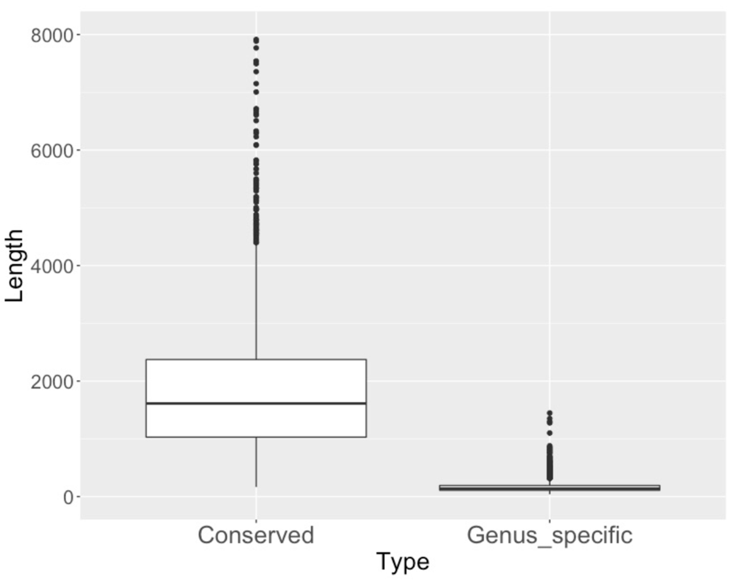
Out of all differentially expressed genus-specific genes, 11% have signal peptides (compared to 5% of all C. immitis genes), 7% have a transmembrane domain (compared to 16% of all genes) and 1.6% have a predicted Pfam domain (compared to 40% of all genes). Differentially expressed genus-specific genes were much shorter than conserved genes (Figure 2).
Enriched GO terms in the upregulated genes (young spherule/mycelia) included oxidation-reduction processes, integral membrane components, transmembrane transport and response to stress (Supplemental Table S2). One of the oxidation/reduction genes is superoxide dismutase, which has been shown to be important for pathogenicity in Histoplasma capsulatum [22]. Changes in expression of oxidation/reduction genes are to be expected since the atmosphere for mycelial culture is air with 0.4% CO2 compared to 14% CO2 in spherule culture conditions. Several metal transporters, including copper and iron transporters are also upregulated. A cluster of genes, including an iron siderophore, are induced by iron-deprivation and are required for pathogenicity in H. capsulatum [23]. Five of the six iron-related genes (including the siderophore biosynthesis cluster) have homologs in C. immitis that are upregulated in young spherules. These genes are tightly clustered (<25 kb) on contig 1 and have the upstream regulatory sites for the GATA transcription factor Sre1 that is identified in identified in H. capsulatum. The Blastomyces dermatitidis homolog of Sre1 has been knocked out and the resulting mutant is unable to differentiate from mold to yeast [24]. However, the C. immitis homolog of Sre1, SreP, is downregulated in young spherules. Taken together, these results show that the organization of iron-related genes in C. immitis and H. capsulatum is very similar and that most of the C. immitis iron acquisition genes are upregulated in both of these dimorphic fungi. Although this may imply the upregulation of spherules in vivo, in this experiment, mycelia and spherules were grown in different media, which may have played a role in the upregulation of iron-regulated genes in this experiment.
Cellular component GO terms associated with cell membranes are dramatically overrepresented in upregulated genes (young spherule/mycelia) (p < 10−4). In addition, 22% of the up-regulated genes have at least one predicted transmembrane domain, compared to 17% of total genes (p < 0.05). However, there is no difference in the proportion of up-regulated genes compared to all genes with a predicted signal peptide.
A pathway enrichment analysis was also conducted (Supplemental Table S3). An analysis of upregulated genes (young spherule/mycelia) found that 24 pathways were enriched with a p value of less 0.01 but none had a Benjamini–Hochberg adjusted p value of less than 0.05. The most highly enriched pathways in upregulated genes were pentose and glucuronate interconversion (see Supplemental Table S3). Other enriched pathways included the degradation of proponate, furfural and benzoate. Two pathways for polyketide synthase were also enriched.
Many genes that have previously been identified to be upregulated in spherules were also found to be upregulated in this study. In addition to SOWgp [8,9] and the parasitic phase specific gene [19], expression of the urease and the ureidoglycolate hydrolase genes was also upregulated [25,26]. One gene that was found to be upregulated in the yeast (or spherule) phase of all dimorphic fungi is 4-hydroxyphenylpyruvate dioxygenase (4-HPPD or HpdA) [27]. This gene is involved in tyrosine catabolism, which plays a role in the synthesis of melanin [28]. Chemical inhibition of 4-HPPD blocks the formation of yeast in P. brasiliensis and deletion of the gene blocks’ differentiation to yeast [29]. There are two genes coding for 4-HPPD in C. immitis—one is upregulated and the other is downregulated. Boyce described a cluster of genes involved in tyrosine catabolism in Pennicillium marneffei and other pathogenic dimorphic fungi. The expression of this cluster of genes is upregulated when tyrosine is the only nitrogen source [28]. However, C. immitis spherules are grown in media containing ammonium salts as the primary nitrogen source.
There are twice as many downregulated genes in young spherules than are up-regulated, so many genes that are expressed well in mycelia are expressed poorly in spherules. Downregulated genes are enriched for the cytochrome p450 superfamily, transcription factors and genes with oxidoreductase GO terms (Table 3). There are two homologs of the cytochrome p450 ERG11 gene in C. immitis: CIMG_00573 and CIMG_07469. ERG11 is involved in azole anti-fungal activity in other fungi, and both of these are downregulated in young spherules.
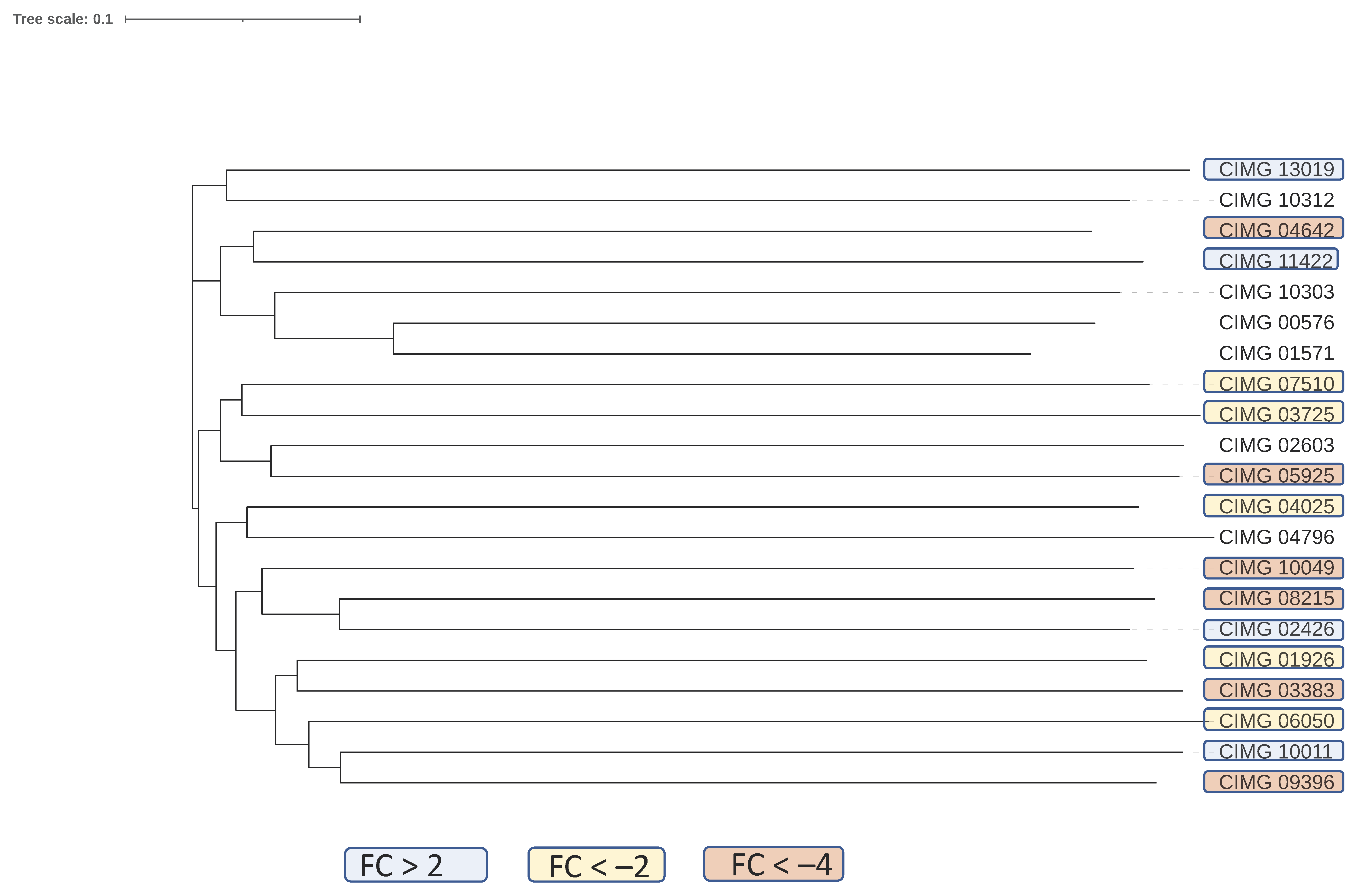
The Stu1 transcription factor was downregulated 5-fold in spherules. This transcription factor was found to be required for optimal hyphal growth in H. capsulatum [30], so the difference between expression of this gene in the mycelial and parasitic form is shared by these two pathogenic, dimorphic fungi. However, direct experiments about the role of Stu1 have not been done in Coccidioides spp. Eleven out of 29 C2H2 type zinc finger domain-encoding proteins were also downregulated more than 2-fold and only four were upregulated. The downregulated genes are clustered together within a phylogenetic tree of the C. immitis C2H2 transcription factors suggesting that they may share functions (Figure 3).
The expression of HLH transcription factors was also downregulated. Both C2H2 and HLH transcription factors influence growth rate and differentiation in Neurospora crassa [31]. Most transcription factors containing the fungal Zn(2)–Cys(6) binuclear cluster domain, the most common class of zinc finger protein in the C. immitis., are not differentially expressed. In H. capsulatum, four transcription factors (Ryp1, Ryp2, Ryp3 and Ryp4) are required for differentiation into yeast [32]. These transcription factors are needed for changes in the transcriptional responses to an increase in temperature that triggers yeast formation [28]. The C. immitis homologs of these proteins, Ryp2 and Ryp4 (also known as FacB), are upregulated 2.7- and 4.72-fold in spherules, but the other transcription factor is not. These factors are critical regulators of yeast morphology and fungal virulence in H. capsulatum [33]. The upregulation of Ryp2 and Ryp4 in C. immitis suggests that this regulator may play a similar role in the Coccidioides spp. as it does in H. capsulatum.
Thirty-four pathways were enriched in downregulated genes (p < 0.01); five had a Benjamini–Hochberg p adjusted value < 0.05. Several pathways involved in detoxification of compounds by glutathione are highly enriched. Other enriched pathways included metabolism of xenobiotics by cytochrome p450, metabolism of chloroalkane and methane. This suggests that the mycelial form may encounter more toxic compounds than spherules. Another interesting observation is that the ergosterol pathway is enriched in downregulated genes, which is consistent with the GO term enrichment results and suggests that mycelia and spherules may have somewhat different susceptibilities to azole antifungal drugs.

3.2. Comparison to of Differential Gene Expression in Young Spherules to Previous Studies in Coccidioides Immitis

The RNA used in this study was derived from mycelia and spherules obtained from frozen samples of spherules and mycelia obtained from the previous microarray analysis [10]. The microarray study found that 2.5% of the total genes were upregulated more than 4-fold in young spherules; the number of differentially expressed genes in the current RNA-Seq analysis was significantly larger (8%). However, 69% of the differentially expressed genes identified in the microarray study were also differentially expressed in this RNA-Seq study. These results suggest that the RNA-Seq is a more sensitive technique for determining differential expression, but differences observed in microarray tend to be found in RNA-Seq too.
GO enrichment analysis of the up- and downregulated genes were similar in the two studies. Upregulated genes in the microarray study were enriched for oxidation-reduction processes and terms for sulfate and sulfite biosynthesis. Enrichment of metal transporter and homeostasis genes was not recognized in the microarray study. However, GO analysis of the down-regulated genes in the microarray study revealed enrichment of transcription factors. One finding of the microarray study was that 25 protein kinase genes were downregulated in spherules. In the current RNA-Seq study, 16 of these genes were also found to be downregulated.
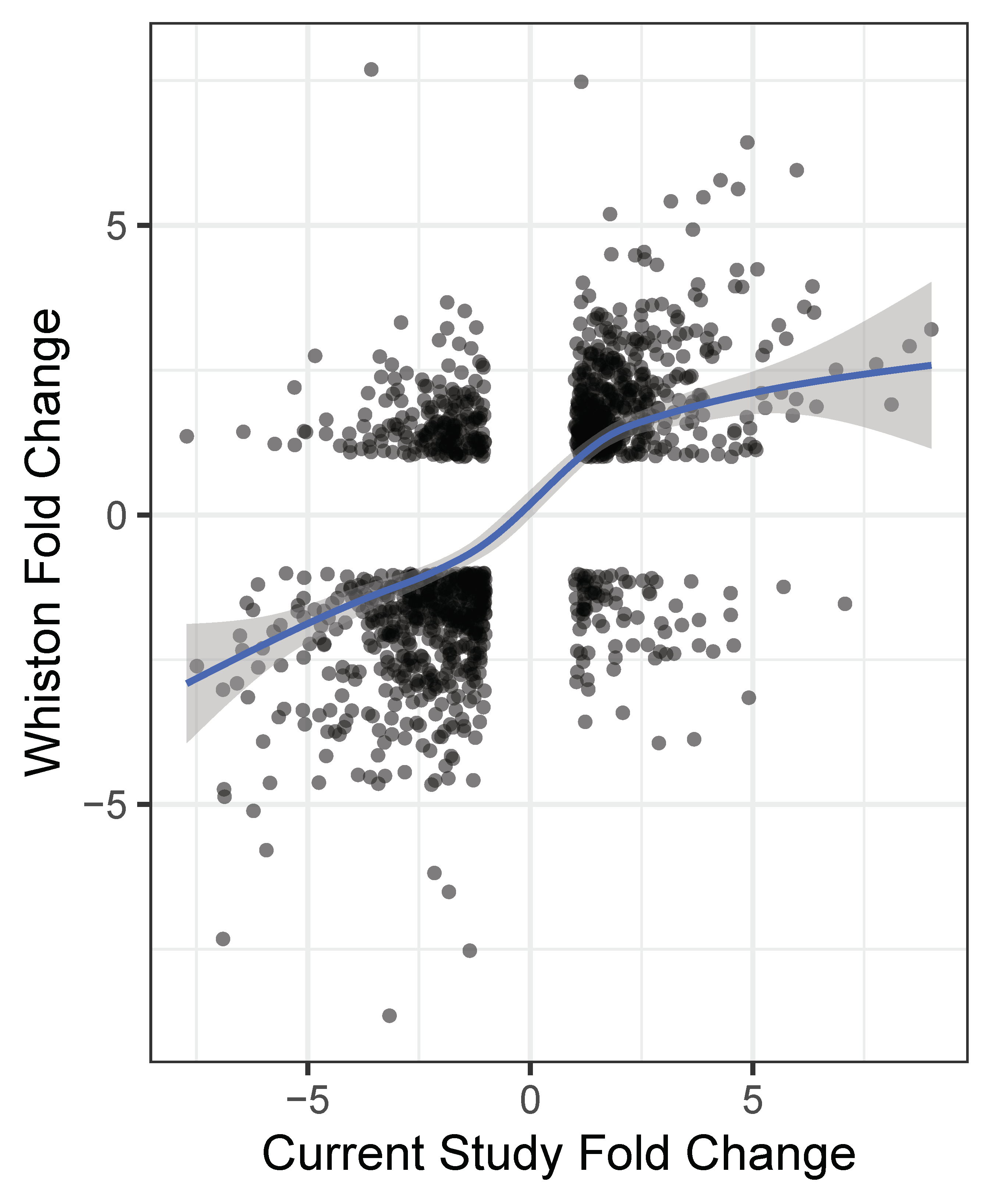
Whiston has previously published a study comparing C. immitis spherules (Day 4 maturity) to mycelia [7]. Reanalyzing Whiston’s data using our Kallisto/DESeq2 pipeline, we found that 902 genes were differentially expressed by at least 2 spherule/mycelia FC in both studies. The FC values for differentially expressed genes in the Whiston study and young spherules/mycelia in this study are compared in Figure 4. There is a positive correlation between the two studies (73% of the genes are in the same quadrants) but there are also obvious exceptions. The difference in spherule maturity may account for some of these disparities.

3.3. Differential Gene Expression in Mature Spherules vs. Mycelia

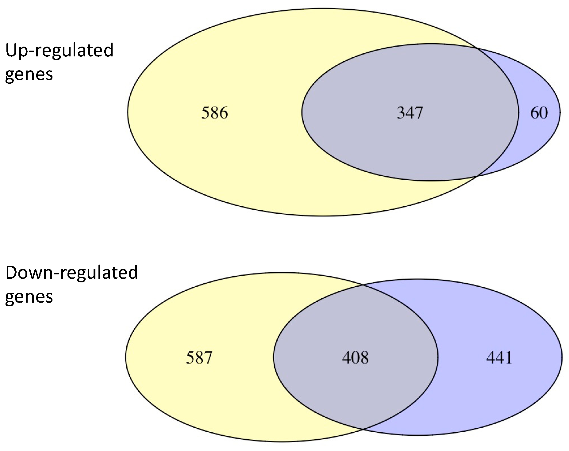
Gene expression in mature spherules was also compared to the gene expression in mycelia (Supplemental Table S1). There were many more genes upregulated in mature spherules (960 genes) than in young ones (408 genes), but the great majority of the genes upregulated in young spherules were also upregulated in mature organisms. In contrast, there were many genes that were downregulated in young spherules but not mature spherules and vice versa (Figure 5). Genes that were differentially expressed in both young and mature spherules had very similar FC values (Supplemental Figure S1). This suggests that upregulation of some genes may be needed for all maturities of the spherule phase.
The enrichment of GO terms in the upregulated genes were similar to the results in young spherules; oxidation-reduction, transmembrane transport and integral membrane component terms were highly enriched (Supplemental Table S2). In contrast, one of the most significantly enriched GO terms in the genes that were downregulated in mature spherules were associated with microtube activity and kinesin. The functional consequences of downregulation of microtubule genes are very difficult to predict, since they play so many different roles in cell biology. The cellular component GO term for the cell wall was also highly enriched in the downregulated genes.

3.4. Mature Spherules Compared to Young Spherules

Gene expression in mature spherules was also compared to that in young spherules. A relatively small number of genes were differentially expressed. The most differentially expressed genes are in Table 4.
The most highly upregulated gene is a homolog of Hsp31, which is a methylglyoxalase, a chaperone stress-response gene in yeast [34]. The expression of this gene increases in response to DNA replication stress [34]. A ferritin-like protein, involved in iron regulation and oxidation reactions is also upregulated, as are genes involved in mRNA splicing, transcription, and meiotic recombination. Expression of alpha-amylase-1 gene was also up-regulated; this gene is required for pathogenicity in H. capsulatum, but its role in the pathogenicity of C. immitis is unknown [35]. The most dramatically downregulated gene in mature spherules was tyrosinase, a gene that plays a central role in the synthesis of melanin. The beta-glucan synthesis-associated protein Kre6 and beta-glucosidase, two genes that influence beta-glucan metabolism were also downregulated, as were two other genes influencing DNA replication (cell division cycle protein Cdc20 and DNA replication helicase Dna2). The downregulation of these genes suggests that remodeling of the cell wall and DNA synthesis decrease in mature spherules.

3.5. Expression of Transposable Elements in Young Spherules Compared to Mycelia

We have previously identified 1309 Gypsy, Copia and TcMar transposons in the C. immitis RS genome and found that proximity to a transposable element (TE) was associated with a lower level of protein-encoding gene expression in C. immitis mycelia [31]. In this study, we compared expression of TE mRNA in mycelia and young spherules. Only 350 TE genes had an adjusted p < 0.05, and the median baseMean of these genes was 20.4, indicating most TEs were poorly expressed. A total of 230 were upregulated in young spherules and only 15 were downregulated. Gypsy TE were the most common upregulated transposons. Analysis of upregulated predicted protein-encoding genes in young spherules showed that a few of them are transposon proteins. A previous proteomic study found one transposon protein in C. posadasii spherules [32]. There was a total of 77 genes that were within 1 kb up- or downstream of the upregulated TE. The median FC of these genes was 3.90, so genes near the up-regulated TE were somewhat overexpressed in spherules.

4. Summary

The transition from the environmental arthroconidia/mycelial form to the spherule is required for the pathogenesis of Coccidioides spp. The morphologic changes are dramatic—the organism changes from a mycelium with internal spores to a round structure that enlarges circumferentially and divides internally to form a large number of endospores [36]; these are released and can differentiate into mature spherules. The importance of spherule maturation and spherule release is demonstrated by the observation that an engineered chitinase deletion mutant that does not form spherules is avirulent [37]. For these reasons, understanding the transcription program of spherules as they mature is clearly important for comprehending the biology of this dimorphic fungus.
Our findings in this study confirm previous experiments in showing that there are substantial differences in gene expression in spherules and mycelia [7,10]. One of the most interesting findings of this study is that differentially expressed genes are more likely to be genus-specific than those that are not. Although the function of these genes in unknown, and they likely have evolved fairly recently, the results in our study suggest that they play an important role in mycelial/spherule differentiation.
The expression of copper and iron transporters is upregulated in spherules, which is consistent with previous studies in C. posadasii [21] and H. capsulatum [23]. Two of the four Ryp transcription factor genes that have been found to be important for yeast formation and virulence in H. capsulatum are also upregulated in C. immitis, suggesting that these two organisms may use similar transcription programs for phase differentiation.
Another interesting group of genes that is downregulated in young spherules are the transcription factors. The APSES family transcription factor Stu1, several HLH and many C2H2 transcription factors are downregulated. Identification of the genetic programs driven by these transcription factors in C. immitis should be useful for understanding the biological significance of these changes.
Analysis of mature spherules shows that almost all the upregulated genes in young spherules remain upregulated. This suggests that this group of genes may be required for life in the spherule phase; examining the transcriptome of spherules of intermediate maturities would address this question. Furthermore, there were a very small number of differentially expressed genes between the two spherule maturities, which also suggests that young and mature spherules have very similar transcription programs. This study confirms that many genes are differentially expressed in mycelia and spherules and suggests that further investigation of the expression profiles of these morphologic forms may be useful for understanding the biology of this organism.

Supplementary Materials

The following are available online at https://www.mdpi.com/article/10.3390/jof7050366/s1, Figure S1: Heat map comparing a sample of FC values of young spherule/mycelia to mature spherules/mycelia, Supplemental Table S1: Relative expression (FC) of young spherules/mycelia, Supplemental Table S2: GO enrichment analysis of differentially expressed young spherule genes/mycelia and mature spherules/mycelia, Supplemental Table S3: Pathway enrichment of differentially expressed young spherules/mycelia, Supplemental Table S4: Relative expression (FC) of mature spherule/mycelia, Supplemental Table S5: Relative expression (FC) of mature spherule/young spherules, Supplemental Table S6: Relative expression (FC) of TE in young spherules/mycelia.

Author Contributions

A.F.C. prepared the RNA and performed the RNA-Seq. S.V. grew and harvested the mycelia and spherules. A.F.C., S.B., J.F.P., J.E.S., S.V., J.F. and T.N.K. all participated in data analysis and preparing the manuscript. All authors have read and agreed to the published version of the manuscript.

Funding

A.F.C. is supported by a Career Award for Medical Scientists from the Burroughs Wellcome Fund and NIH grant (K08 AI130381). A.F.C., S.V., J.F., J.P. and J.E.S. and T.N.K were supported by VFR-19-633952 "Investigating fundamental gaps in Valley Fever knowledge" and MRP-17-454959, “UC Valley Fever Research Initiative”. J.E.S. is a CIFAR Fellow in the program Fungal Kingdom: Threats and Opportunities.

Institutional Review Board Statement

This study did not involve experimental animals.

Informed Consent Statement

This study did not involve human subjects.

Data Availability Statement

The data for this manuscript are available at NCBI GEO, accession number GSE171286.

Conflicts of Interest

The authors declare no conflict of interest.

References

  1. Engelthaler, D.M.; Roe, C.C.; Hepp, C.M.; Teixeira, M.; Driebe, E.M.; Schupp, J.M.; Gade, L.; Waddell, V.; Komatsu, K.; Arathoon, E.; et al. Local Population Structure and Patterns of Western Hemisphere Dispersal for Coccidioides spp., the Fungal Cause of Valley Fever. MBio 2016, 7, e00550-16. [Google Scholar] [CrossRef] [PubMed] [Green Version]
  2. Kirkland, T.N.; Fierer, J. Coccidioides immitis and posadasii; A review of their biology, genomics, pathogenesis, and host immunity. Virulence 2018, 9, 1426–1435. [Google Scholar] [CrossRef] [PubMed] [Green Version]
  3. Nguyen, C.; Barker, B.M.; Hoover, S.; Nix, D.E.; Ampel, N.M.; Frelinger, J.A.; Orbach, M.J.; Galgiani, J.N. Recent advances in our understanding of the environmental, epidemiological, immunological, and clinical dimensions of coccidioidomycosis. Clin. Microbiol. Rev. 2013, 26, 505–525. [Google Scholar] [CrossRef] [Green Version]
  4. Whiston, E.; Taylor, J.W. Genomics in Coccidioides: Insights into evolution, ecology, and pathogenesis. Med. Mycol. 2014, 52, 149–155. [Google Scholar] [CrossRef]
  5. Converse, J.L. Effect of physico-chemical environment of spherulation of Coccidioides immitis in a chemically defined medium. J. Bacteriol. 1956, 72, 784–792. [Google Scholar] [CrossRef] [Green Version]
  6. Mead, H.L.; Van Dyke, M.C.C.; Barker, B.M. Proper Care and Feeding of Coccidioides: A Laboratorian’s Guide to Cultivating the Dimorphic Stages of C. immitis and C. posadasii. Curr. Protoc. Microbiol. 2020, 58, e113. [Google Scholar] [CrossRef]
  7. Whiston, E.; Zhang Wise, H.; Sharpton, T.J.; Jui, G.; Cole, G.T.; Taylor, J.W. Comparative transcriptomics of the saprobic and parasitic growth phases in Coccidioides spp. PLoS ONE 2012, 7, e41034. [Google Scholar] [CrossRef] [PubMed] [Green Version]
  8. Cole, G.T.; Kirkland, T.; Franco, M.; Zhu, S.W.; Yuan, L.; Sun, S.H.; Hearn, V.N. Immunoreactivity of a surface wall fraction produced by spherules of Coccidioides immitis. Infect. Immun. 1988, 56, 2695–2701. [Google Scholar] [CrossRef] [Green Version]
  9. Hung, C.Y.; Yu, J.; Seshan, K.R.; Reichard, U.; Cole, G.T. A parasitic phase-specific adhesin of Coccidioides immitis contributes to the virulence of this respiratory fungal pathogen. Infect. Immun. 2002, 70, 3443–3456. [Google Scholar] [CrossRef] [PubMed] [Green Version]
  10. Viriyakosol, S.; Singhania, A.; Fierer, J.; Goldberg, J.; Kirkland, T.N.; Woelk, C.H. Gene expression in human fungal pathogen Coccidioides immitis changes as arthroconidia differentiate into spherules and mature. BMC Microbiol. 2013, 13, 121. [Google Scholar] [CrossRef] [PubMed] [Green Version]
  11. Bray, N.L.; Pimentel, H.; Melsted, P.; Pachter, L. Near-optimal probabilistic RNA-seq quantification. Nat. Biotechnol. 2016, 34, 525–527. [Google Scholar] [CrossRef] [PubMed]
  12. Basenko, E.Y.; Pulman, J.A.; Shanmugasundram, A.; Harb, O.S.; Crouch, K.; Starns, D.; Warrenfeltz, S.; Aurrecoechea, C.; Stoeckert, C.J.; Kissinger, J.C.; et al. FungiDB: An Integrated Bioinformatic Resource for Fungi and Oomycetes. J. Fungi 2018, 4, 39. [Google Scholar] [CrossRef] [PubMed] [Green Version]
  13. Li, L.; Stoeckert, C.J.; Roos, D.S. OrthoMCL: Identification of ortholog groups for eukaryotic genomes. Genome Res. 2003, 13, 2178–2189. [Google Scholar] [CrossRef] [PubMed] [Green Version]
  14. Sievers, F.; Wilm, A.; Dineen, D.; Gibson, T.J.; Karplus, K.; Li, W.; Lopez, R.; McWilliam, H.; Remmert, M.; Soding, J.; et al. Fast, scalable generation of high-quality protein multiple sequence alignments using Clustal Omega. Mol. Syst. Biol. 2011, 7, 539. [Google Scholar] [CrossRef] [PubMed]
  15. Priebe, S.; Kreisel, C.; Horn, F.; Guthke, R.; Linde, J. FungiFun2: A comprehensive online resource for systematic analysis of gene lists from fungal species. Bioinformatics 2015, 31, 445–446. [Google Scholar] [CrossRef] [PubMed] [Green Version]
  16. Karp, P.D.; Billington, R.; Caspi, R.; Fulcher, C.A.; Latendresse, M.; Kothari, A.; Keseler, I.M.; Krummenacker, M.; Midford, P.E.; Ong, Q.; et al. The BioCyc collection of microbial genomes and metabolic pathways. Brief Bioinform. 2019, 20, 1085–1093. [Google Scholar] [CrossRef] [PubMed]
  17. Kanehisa, M.; Furumichi, M.; Tanabe, M.; Sato, Y.; Morishima, K. KEGG: New perspectives on genomes, pathways, diseases and drugs. Nucleic Acids Res. 2017, 45, D353–D361. [Google Scholar] [CrossRef] [Green Version]
  18. Ivey, F.D.; Magee, D.M.; Woitaske, M.D.; Johnston, S.A.; Cox, R.A. Identification of a protective antigen of Coccidioides immitis by expression library immunization. Vaccine 2003, 21, 4359–4367. [Google Scholar] [CrossRef]
  19. Delgado, N.; Hung, C.Y.; Tarcha, E.; Gardner, M.J.; Cole, G.T. Profiling gene expression in Coccidioides posadasii. Med. Mycol. 2004, 42. [Google Scholar] [CrossRef]
  20. Petito, G.; de Curcio, J.S.; Pereira, M.; Bailão, A.M.; Paccez, J.D.; Tristão, G.B.; de Morais, C.O.B.; de Souza, M.V.; de Castro Moreira Santos, A.; Fontes, W.; et al. Metabolic Adaptation of Paracoccidioides brasiliensis in Response to in vitro Copper Deprivation. Front. Microbiol. 2020, 11. [Google Scholar] [CrossRef]
  21. Mead, H.L.; Roe, C.C.; Higgins Keppler, E.A.; Van Dyke, M.C.C.; Laux, K.L.; Funke, A.L.; Miller, K.J.; Bean, H.D.; Sahl, J.W.; Barker, B.M. Defining Critical Genes During Spherule Remodeling and Endospore Development in the Fungal Pathogen, Coccidioides posadasii. Front. Genet. 2020, 11. [Google Scholar] [CrossRef]
  22. Youseff, B.H.; Holbrook, E.D.; Smolnycki, K.A.; Rappleye, C.A. Extracellular superoxide dismutase protects Histoplasma yeast cells from host-derived oxidative stress. PLoS Pathog. 2012, 8, e1002713. [Google Scholar] [CrossRef] [Green Version]
  23. Hwang, L.H.; Seth, E.; Gilmore, S.A.; Sil, A. SRE1 regulates iron-dependent and -independent pathways in the fungal pathogen Histoplasma capsulatum. Eukaryot. Cell 2012, 11, 16–25. [Google Scholar] [CrossRef] [Green Version]
  24. Gauthier, G.M.; Sullivan, T.D.; Gallardo, S.S.; Brandhorst, T.T.; Vanden Wymelenberg, A.J.; Cuomo, C.A.; Suen, G.; Currie, C.R.; Klein, B.S. SREB, a GATA transcription factor that directs disparate fates in Blastomyces dermatitidis including morphogenesis and siderophore biosynthesis. PLoS Pathog. 2010, 6, e1000846. [Google Scholar] [CrossRef]
  25. Mirbod-Donovan, F.; Schaller, R.; Hung, C.Y.; Xue, J.; Reichard, U.; Cole, G.T. Urease produced by Coccidioides posadasii contributes to the virulence of this respiratory pathogen. Infect. Immun. 2006, 74, 504–515. [Google Scholar] [CrossRef] [PubMed] [Green Version]
  26. Wise, H.Z.; Hung, C.-Y.; Whiston, E.; Taylor, J.W.; Cole, G.T. Extracellular ammonia at sites of pulmonary infection with Coccidioides posadasii contributes to severity of the respiratory disease. Microb. Pathog. 2013, 59–60, 19–28. [Google Scholar] [CrossRef] [PubMed] [Green Version]
  27. Kirkland, T.N. A few shared up-regulated genes may influence conidia to yeast transformation in dimorphic fungal pathogens. Med. Mycol. 2016, 54, 648–653. [Google Scholar] [CrossRef] [Green Version]
  28. Boyce, K.J.; McLauchlan, A.; Schreider, L.; Andrianopoulos, A. Intracellular growth is dependent on tyrosine catabolism in the dimorphic fungal pathogen Penicillium marneffei. PLoS Pathog. 2015, 11, e1004790. [Google Scholar] [CrossRef] [Green Version]
  29. Felipe, M.S.S.; Andrade, R.V.; Arraes, F.B.M.; Nicola, A.M.; Maranhão, A.Q.; Torres, F.A.G.; Silva-Pereira, I.; Poças-Fonseca, M.J.; Campos, E.G.; Moraes, L.M.P.; et al. Transcriptional profiles of the human pathogenic fungus Paracoccidioides brasiliensis in mycelium and yeast cells. J. Biol. Chem. 2005, 280, 24706–24714. [Google Scholar] [CrossRef] [PubMed] [Green Version]
  30. Longo, L.V.G.; Ray, S.C.; Puccia, R.; Rappleye, C.A. Characterization of the APSES-family transcriptional regulators of Histoplasma capsulatum. FEMS Yeast Res. 2018, 18. [Google Scholar] [CrossRef] [PubMed] [Green Version]
  31. Carrillo, A.J.; Schacht, P.; Cabrera, I.E.; Blahut, J.; Prudhomme, L.; Dietrich, S.; Bekman, T.; Mei, J.; Carrera, C.; Chen, V.; et al. Functional Profiling of Transcription Factor Genes in Neurospora crassa. G3 2017, 7, 2945–2956. [Google Scholar] [CrossRef] [PubMed] [Green Version]
  32. Webster, R.H.; Sil, A. Conserved factors Ryp2 and Ryp3 control cell morphology and infectious spore formation in the fungal pathogen Histoplasma capsulatum. Proc. Natl. Acad. Sci. USA 2008, 105, 14573–14578. [Google Scholar] [CrossRef] [Green Version]
  33. Rodriguez, L.; Voorhies, M.; Gilmore, S.; Beyhan, S.; Myint, A.; Sil, A. Opposing signaling pathways regulate morphology in response to temperature in the fungal pathogen Histoplasma capsulatum. PLoS Biol. 2019, 17, e3000168. [Google Scholar] [CrossRef] [Green Version]
  34. Bankapalli, K.; Saladi, S.; Awadia, S.S.; Goswami, A.V.; Samaddar, M.; D’Silva, P. Robust glyoxalase activity of Hsp31, a ThiJ/DJ-1/PfpI family member protein, is critical for oxidative stress resistance in Saccharomyces cerevisiae. J. Biol. Chem. 2015, 290, 26491–26507. [Google Scholar] [CrossRef] [Green Version]
  35. de Vries, R.P.; Riley, R.; Wiebenga, A.; Aguilar-Osorio, G.; Amillis, S.; Uchima, C.A.; Anderluh, G.; Asadollahi, M.; Askin, M.; Barry, K.; et al. Comparative genomics reveals high biological diversity and specific adaptations in the industrially and medically important fungal genus Aspergillus. Genome Biol. 2017, 18, 28. [Google Scholar] [CrossRef] [PubMed] [Green Version]
  36. Cole, G.T.; Pope, L.M.; Huppert, M.; Sun, S.H.; Starr, P. Ultrastructure and composition of conidial wall fractions of Coccidioides immitis. Exp. Mycol. 1983, 7, 297–318. [Google Scholar] [CrossRef]
  37. Xue, J.; Chen, X.; Selby, D.; Hung, C.Y.; Yu, J.J.; Cole, G.T. A genetically engineered live attenuated vaccine of Coccidioides posadasii protects BALB/c mice against coccidioidomycosis. Infect. Immun. 2009, 77. [Google Scholar] [CrossRef] [PubMed] [Green Version]
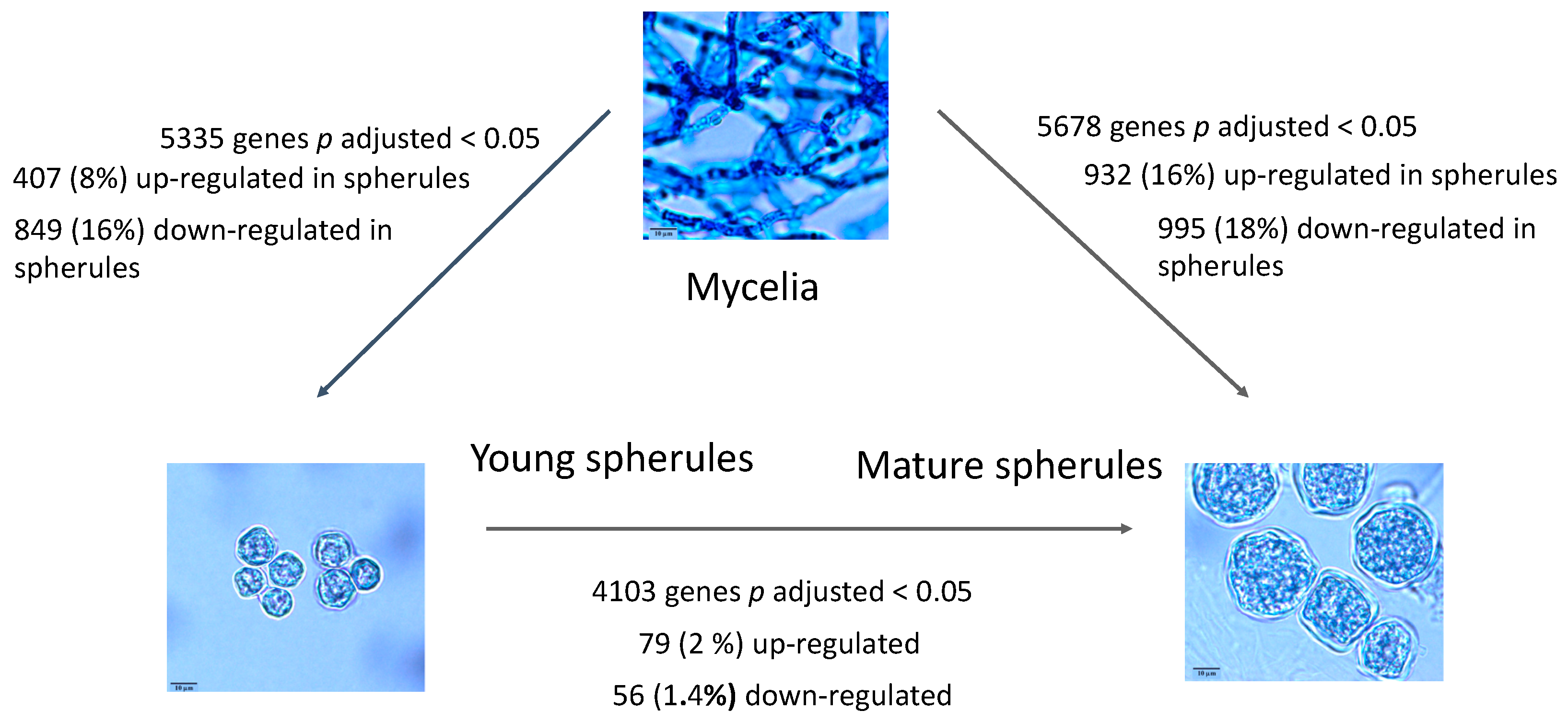
Figure 1. Differential expression of genes in mycelia, young and mature spherules. Legend Figure 1: mRNA was obtained from mycelia, young spherules and mature spherules for RNA-Seq. The morphology of the organism and the number of differentially expressed genes is shown.
Jof 07 00366 g001
Figure 2. Length of genus-specific and conserved genes. Legend Figure 2: Lengths (Y-axis) of conserved and differentially expressed genus-specific proteins.
Jof 07 00366 g002
Figure 3. Phylogenetic tree of C2H2 transcription factors and FC. Legend: Phylogenetic tree was built on protein sequence. FC (young spherule/mycelium) values of C. immitis C2H2 transcription factors. Gene IDs within tan boxes are downregulated more than 4-fold; those that are within yellow boxes are downregulated more than 2-fold but less than 4-fold; those not within boxes are not significantly differentially expressed and those within blue boxes are upregulated more than 2-fold. None of these genes were upregulated more than 4-fold. All values are arithmetic.
Jof 07 00366 g003
Figure 4. Comparison of FC values in this study to a previous study by Whiston et al. Legend: Genes with log2 FC values (young spherules/mycelia) > 2 or < –2 in the current study were selected. FC values of genes in the current study are shown on the X axis and the FC values of matched genes from Whiston study [7] are shown on the Y axis. The regression line was derived from the linear model and the shaded area indicates the 95% confidence limits. The plot was made with ggplot2 R package and the regression line was drawn with the geom smooth option.
Jof 07 00366 g004
Figure 5. Overlap of up- and downregulated genes in young and mature spherules. Legend: Comparison of up- and downregulated genes in young spherules and mature spherules. Blue represents young spherules, yellow represents mature spherules.
Jof 07 00366 g005
Table 1. Most differentially expressed genes in young spherules compared to mycelia.
Gene IDProduct DescriptionOrtholog CountLength# TM DomainsSignalPFC
(log2 Values)
Genus-Specific
Upregulated
CIMG_04613Hypothetical protein—SOWgp33240Y9.012Y
CIMG_10264Aldehyde reductase11763140N8.116N
CIMG_09753ABC multidrug transporter569150116N7.773N
CIMG_10032Expression library immunization antigen 1-surface protein antigen962240Y7.092N
CIMG_10037Copper transporter1211932N7.072N
CIMG_01115Thiamine thiazole synthase, thiamine thiazole synthase, variant1703280N6.901N
CIMG_08103Opsin 12142897N6.870N
CIMG_06250Methyltransferase domain-containing protein513201Y6.431Y
CIMG_11522Hypothetical protein—genus-specific2851N6.376Y
CIMG_10007Hypothetical protein—genus-specific31250N6.341Y
CIMG_02628Arp2/3 complex subunit Arc161993120N6.158N
CIMG_09539Hypothetical1363870N5.989N
CIMG_12484hypothetical protein—genus-specific11240N5.977Y
CIMG_09750Amino acid adenylation domain-containing protein126280320N5.898N
CIMG_05758parasitic phase-specific protein PSP-14582797N5.752N
CIMG_11264Hypothetical protein59761Y5.693N
CIMG_00534Glycerate kinase374371N5.635N
CIMG_10013Hypothetical protein—genus-specific01140N5.586Y
CIMG_01211Putative major facilitator superfamily (MFS) transporter1155313N5.294N
Downregulated
CIMG_03418CBF1-interacting co-repressor CIR domain-containing protein993360N−8.094N
CIMG_013104-hydroxyphenylpyruvate dioxygenase1833990N−7.717N
CIMG_07209Life-span regulatory factor domain-containing protein561510N−7.491N
CIMG_03873Protoglobin domain-containing protein1362450N−7.320N
CIMG_07928SAM and PH domain-containing protein747950N−6.970N
CIMG_03716Heat shock protein 301152380N−6.904N
CIMG_00925Hypothetical protein—cell wall protein821570Y−6.903N
CIMG_00284Hypothetical protein51180Y−6.898Y
CIMG_09232Hypothetical protein595711Y−6.880Y
CIMG_00099C2H2 finger domain-containing protein1165950N−6.868N
CIMG_08613Metalloproteinase 743570Y−6.746N
CIMG_12190Hypothetical protein—genus-specific11370N−6.689Y
CIMG_03848DnaJ domain-containing protein685360N−6.660N
CIMG_13167Beta-glucosidase 5944890N−6.588N
CIMG_13284Hypothetical protein—genus-specific0740N−6.523Y
CIMG_04597GPI anchored serine-threonine rich protein682160Y−6.467Y
CIMG_00348Endochitinase 23178970Y−6.445N
CIMG_10468Hypothetical protein—genus-specific31320N−6.370Y
CIMG_00302Hypothetical protein—genus-specific742280Y−6.345Y
CIMG_07925Hypothetical protein—genus-specific31680N−6.229Y
Legend Table 1: The 20 most up- and downregulated genes in young spherules. The gene ID, product description, ortholog count, length, number of transmembrane domains (TM), presence of a signal peptide Yes (Y) or No (N), fold change log2 expression ratios of young spherules/young spherules and whether the gene is genus-specific Yes (y) No (Y) is shown SOWgp: spherule outer wall glycoprotein.
Table 2. Genus-specific genes.
GroupTotalGenus-Specific%
All genes9759230524
Upregulated40712731a
Downregulated84932939 a
Legend Table 2: Comparison of the number of differentially expressed (young spherule/mycelia) genus-specific genes to all genus-specific genes. a) p value < 0.05 (Chi-square test) comparing the fraction of genus-specific genes in up- or downregulated genes to the fraction in all genes.
Table 3. Relative expression of transcription factors in young spherules and mycelia.
Transcription FactorTotal NumberNumber (Adjusted p < 0.05)PFAMFC (log2 Young Spherule/Mycelium)
>2>1<−1<−2
APSES53PF13637 21
HLH transcription factor83PF00010 21
C2H2 type zinc finger domain-containing protein2921PF00096 1117
fungal Zn(2)–Cys(6) binuclear cluster domain-containing9151PF00172416177
Homeobox-like domain-containing protein66PF002492
Other Regulatory Proteins
ryp family43PF09729; PF11754
PF00172; PF04082
121
Legend: Differential expression of transcription factors in spherules compared to mycelia. All FC values are expressed as log2.
Table 4. Most differentially expressed genes in mature spherules compared to young spherules.
Gene IDProduct DescriptionOrtholog CountLength# TM DomainsSignalPFC
(log2 Values)
Upregulated
CIMG_03805Possible HSP312812430N4.626
CIMG_12285hypothetical protein101510N4.074
CIMG_08736hypothetical protein51560N3.705
CIMG_07646hypothetical protein33500N3.606
CIMG_07274Ferritin-like domain-containing protein1054550Y3.507
CIMG_13581hypothetical protein0710N3.306
CIMG_05456Possible pre-mRNA-splicing factor cef1452370N3.281
CIMG_08524Dynamin family protein4507110N3.186
CIMG_02168Possible transcription factor wer124440N3.140
CIMG_00573Cytochrome P450 512365112N3.064
CIMG_08156Possible meiotic recombination protein dmc1/lim15 homolog914630N3.058
CIMG_04521DUF1399 domain-containing protein1957450N2.984
CIMG_03465hypothetical protein83830N2.907
CIMG_06696Possible dual oxidase5082952Y2.902
CIMG_06262Possible rest corepressor 1371850N2.900
CIMG_08666FK506-binding protein 12021200N2.829
CIMG_13473YjgH family protein452060N2.778
CIMG_04544Alpha-amylase AmyA655820Y2.769
CIMG_01977Hypothetical protein54820Y2.740
CIMG_07685Hypothetical protein81050N2.711
Downregulated
CIMG_07583Tyrosinase416580Y−4.248
CIMG_03173Hypothetical protein51891N−3.863
CIMG_00927Acyl–CoA synthetase1376931N−3.204
CIMG_05421Beta-glucan synthesis-associated protein KRE62226421N−3.046
CIMG_03257Cell division cycle protein Cdc201725990N−3.030
CIMG_03517Hypothetical protein8521370N−2.887
CIMG_01729Aconitate hydratase, mitochondrial3527840N−2.844
CIMG_13686Hypothetical protein0940N−2.806
CIMG_00254GTP-binding protein rhoC1092780N−2.783
CIMG_03888Betaglucosidase11538580Y−2.731
CIMG_01317Dethiobiotin synthase1368070N−2.731
CIMG_01369Myosin rod fragments superfamily protein, putative138712710N−2.703
CIMG_08907Cytochrome b24805040N−2.660
CIMG_12964Hypothetical protein01680Y−2.615
CIMG_00847GTP binding protein10914690N−2.609
CIMG_05523Ca2+ regulator and membrane fusion protein Figure 1, putative1112684Y−2.552
CIMG_13558DNA replication helicase Dna215816590N−2.523
CIMG_08747Putative tetratricopeptide repeat (TPR)-like superfamily protein2327020N−2.493
CIMG_01268MFS phospholipid transporter31749610N−2.492
CIMG_05728L-serine dehydratase1734260N−2.475
CIMG_09628Protein–tyrosine phosphatase1716000N−2.471
Legend: The 20 most up- and downregulated genes in mature spherule compared to young spherules. The gene ID, product description, ortholog count, length, number of transmembrane domains (TM), presence of a signal peptide Yes (Y) or No (N) and fold change log2 expression ratios of mature spherules/young spherules are shown.
Publisher’s Note: MDPI stays neutral with regard to jurisdictional claims in published maps and institutional affiliations.