Article

Daldiniaeschsone A, a Rare Tricyclic Polyketide Having a Chromone Unit Fused to a δ-Lactone and Its Symmetrical Biphenyl Dimer, Daldiniaeschsone B, from an Endophytic Fungus Daldinia eschscholtzii SDBR-CMUNKC745

by
Natnicha Wutthiwong
1,2,†,
Virayu Suthiphasilp
3,†,
Aknarin Pintatum
3,
Nakarin Suwannarach
1,2,
Jaturong Kumla
1,2,
Saisamorn Lumyong
1,2,4,*,
Tharakorn Maneerat
3,5,
Rawiwan Charoensup
5,6,
Sarot Cheenpracha
7,
Thunwadee Limtharakul
8,
Stephen G. Pyne
9 and
Surat Laphookhieo
3,5,*
1
Department of Biology, Faculty of Science, Chiang Mai University, Chiang Mai 50200, Thailand
2
Research Center of Microbial Diversity and Sustainable Utilization, Chiang Mai University, Chiang Mai 50200, Thailand
3
Center of Chemical Innovation for Sustainability (CIS), School of Science, Mae Fah Luang University, Chiang Rai 57100, Thailand
4
Academy of Science, The Royal Society of Thailand, Bangkok 10300, Thailand
5
Medicinal Plants Innovation Center of Mae Fah Luang University, Chiang Rai 57100, Thailand
6
School of Integrative Medicine, Mae Fah Luang University, Chiang Rai 57100, Thailand
7
School of Science, University of Phayao, Phayao 56000, Thailand
8
Department of Chemistry, Faculty of Science and Research Center on Chemistry for Development of Health Promoting Products from Northern Resources, Chiang Mai University, Chiang Mai 50200, Thailand
9
School of Chemistry and Molecular Bioscience, University of Wollongong, Wollongong, NSW 2522, Australia
*
Authors to whom correspondence should be addressed.
These authors have contributed equally to this work.
J. Fungi 2021, 7(5), 358; https://doi.org/10.3390/jof7050358
Submission received: 24 March 2021 / Revised: 29 April 2021 / Accepted: 30 April 2021 / Published: 2 May 2021

Abstract

:
Daldiniaeschsone A (1), a rare tricyclic polyketide having a chromone unit fused to a δ-lactone and its symmetrical 6,6′-biphenyl dimer, daldiniaeschsone B (2), together with three known compounds (35), were isolated from a plant-derived endophytic fungus, Daldinia eschscholtzii SDBR-CMUNKC745. Their structures were elucidated by extensive 1D and 2D NMR spectroscopic data and HRESIMS. All compounds showed α-glucosidase inhibitory activity with IC50 values ranging from 0.16−0.85 mM and compound 1 was the best α-glucosidase inhibitory activity (IC50 = 0.16 mM).

1. Introduction

Natural products from fungi have been observed to inhibit or kill a wide variety of harmful microorganisms, including, but not limited to, phytopathogens and bacteria, fungi, viruses, and protozoans that affect humans and animals [1,2,3]. Similar to plants, many compounds isolated from microorganisms have become well-known drugs such as penicillin (Penicillium notatum), vancomycin (Amycolatopsis orientalis), and erythromycin (Saccharopolyspora erythraea) [4]. Due to the short period to scale up and the diversity production of secondary metabolites, fungi have become the new resource for natural products. The genus Daldinia belonging to the Xylariaceae is a genus of fungi that produces polyketide compounds with diverse structures and impressive biological activities, including immunosuppressive, cytotoxic, antiangiogenesis, anti-inflammatory, and antibacterial activities [1,2,5,6,7,8]. The two most investigated Daldinia species are D. eschscholtzii and D. concentrica which have produced many new compounds having remarkable biological activities including α-glucosidase inhibitory, antiangiogenesis, and anti-HIV activities [1,2,9,10].
Daldinia eschscholtzii is a wood-inhabiting and decaying endophytic fungus distributed in warm tropical and subtropical areas [11]. Previous chemical constituent investigation of D. eschscholtzii resulted in the discovery of various types of secondary metabolites, including chromones, lactones, phenolics, and alkaloids [1,12,13,14,15,16,17] Many of these isolated compounds have demonstrated a wide range of bioactivities, such as α-glucosidase inhibition [1], immunosuppression [12], antimicrobial [6,13,14,15], stem cell differentiation induction [16] and anti-inflammatory [17] activities. In the course of our ongoing search for new compounds and their biological activities from natural resources, the investigation of the broth extract of D. eschscholtzii SDBR-CMUNKC745 resulted in the isolation of two new chromone derivatives and three known compounds. In this study, we report the isolation and structure elucidation of two new compounds (1 and 2). The α-glucosidase and NO production inhibition activities of all compounds are also reported.

2. Materials and Methods

2.1. General Experimental Procedures

The optical rotations were measured with a Jasco P-1010 polarimeter. The UV spectra were recorded with a PerkinElmer UV–Vis spectrophotometer. Infrared (IR) spectra were recorded using a PerkinElmer Frontier Optica FT–IR spectrometer. The 1D and 2D NMR spectra recorded using a 500 MHz Bruker AV-500 spectrometer. Chemical shifts are reported in parts per million (δ) and coupling constants (J) are expressed in hertz. The HRESIMS were carried out on a QTOF 6540 UHD mass spectrometer (Agilent Technologies, Santa Clara, CA, USA). Chiral HPLC was performed on a CHIRALCEL OD-H column of 4.6ϕ—250 mm and attached to Agilent Technologies 1260 Infinity II. Silica gel G60 (60–200 µm, SiliCycle® Inc., Québec, QC G1P 4S6, Canada) was used to perform column chromatography (CC). Precoated plates of silica gel (60F254, Merck, Kenilworth, NJ, USA) were used for analytical purposes.

2.2. Fungal Material

The fungus strain SDBR-CMUNKC745 was isolated from Cinnamomum bejolghota, collected from Chiang Mai, Thailand, in October 2015. This strain was deposited in the Culture Collection of Sustainable Development of Biological Resources (SDBR) Laboratory, Faculty of Science, Chiang Mai University, Thailand. Fungal colonies on potato dextrose agar grew to 80−85 mm at 30 °C in darkness after one week. Colonies were white to pale grey, and the reverse side was whitish-grey and black in the centre (Figure 1A). Sporulation was observed within three weeks. Conidiophores were 0.8–2 μm long × 0.7–1.5 μm wide, hyaline, mononematous or synnematous, nodulisporium-like branching pattern with dichotomous or trichotomous branched (Figure 1B). Conidiogeneous cells were 2.8–3.1 × 2.5–2.9 μm, cylindrical, hyaline, and smooth. Conidia were 4.8–6.4 × 2–3.8 μm, holoblastic, hyaline, ellipsoid to obovoid, aseptate, and smooth to finely roughened with flattened base (Figure 1C). The internal transcribed spacer (ITS) and large subunit (LSU) region of ribosomal DNA, RNA polymerase II second largest subunit (RPB2), and β-tubulin (TUB) genes of this strain were deposited in GenBank under the accession number MW1937612 MW193761, MW202236, and MW202237, respectively. The phylogenetic tree from the combined data set of ITS, LSU, RBP2, and TUB indicated that strain SDBR-CMUNKC745 clustered with D. eschscholtzii with 100% bootstrap and 1.0 posterior probability supports (Figure 2). Moreover, each gene sequence of this strain revealed a similarity greater than 99% to the D. eschscholtzii MUCL 45435.

2.3. Fermentation, Extraction, and Isolation of Secondary Metabolites

Daldinia eschscholtzii SDBR-CMUNKC745 from stock culture was cultivated on potato dextrose agar (Difco™, Becton, Dickinson and Company, Franklin Lakes, NJ, USA) at 30 °C for five days. Then, five fungal mycelial plugs (5 mm in diameter) were inoculated into 500 mL Erlenmeyer flask containing 150 mL of Czapek Dox broth (CDB; 30 g/L sucrose, 2 g/L NaNO2, 1 g/L K2HPO4, 0.5 g/L KCl, 0.5 g/L MgSO4, and 0.01 g/L Fe2SO4, pH 8). Cultivation was performed in the dark at 30 °C with shaking at 100 rpm on a reciprocal shaker. After 10 days of incubation, the cultures were filtrated by Whatman’s No. 1 paper to harvest the supernatant. After that, the culture filtrate of endophytic fungus was extracted twice with an equal volume of EtOAc (1:1 v/v). The upper solvent phase was collected and evaporated using a rotary evaporator. The crude extract was stored at −20 °C for further experiments.
After removing the solvent under reduced pressure, the culture medium was extracted with EtOAc to provide a crude extract (4.4 g), which was subjected to silica gel CC (60–200 µm, 8 × 70 cm) using a gradient of EtOAc-hexanes (0.5:4.5, 1:4, 1.5:3.5, 2:3, 2.5:2.5, 3:1.5, 3.5:1.5, 4:1, 4.5:0.5, 0.5:4.5, and 5:0, v/v, 200 mL each) to afford five fractions (F1–F5). Fraction F2 (1.96 g) was further fractionated by silica gel CC (60–200 µm, 6 × 70 cm) using EtOAc-hexanes (1:4, v/v, 2500 mL) to give nine subfractions (F2A–F2J). Compound 2 (8.0 mg) was obtained from subfraction F2D (80.6 mg) by silica gel CC (60–200 µm, 3 × 30 cm) using EtOAc-CH2Cl2 (1:50, v/v, 1000 mL), while compound 1 (4.4 mg) was isolated from subfraction F2I (132.8 mg) by silica gel CC (60–200 µm, 3 × 30 cm) using EtOAc-CH2Cl2 (1:20, v/v, 1000 mL). Fraction F3 (390.0 mg) was further fractionated by silica gel CC (60–200 µm, 4× 50 cm) using EtOAc-hexanes (1:4, v/v, 1500 mL) to afford 10 subfractions (F3A–F3J). Subfraction F3A (75.8 mg) was further purified by silica gel CC (60–200 µm, 3 × 30 cm) using EtOAc-hexanes (1:4, v/v, 1000 mL) to give compound 3 (8.4 mg). Compound 4 (10.1 mg) was obtained from subfraction F3H (19.0 mg), while compound 5 (3.7 mg) was afforded from subfraction F3J (86.0 mg) by silica gel CC (60–200 µm, 2 × 30 and 3 × 30 cm, respectively) using EtOAc-hexanes (1:4, v/v, 500 mL) and EtOAc-CH2Cl2 (1:20, v/v, 1000 mL), respectively (Supplementary Materials, Figure S22).
Daldiniaeschsone A (1): colourless amorphous solid; [α]21D −1.2 (c 1, MeOH); UV (MeOH) λmax (log ε) 209 (3.44), 272 (3.26), 354 (2.74) nm; IR (neat) νmax 3590, 2960, 1788, 1760, 1651, 1628, 1356, 1224, 1158, 1054, 735 cm−1; 1H and 13C NMR data, see Table 1; HRESITOFMS m/z 321.0972 [M + H]+ (calcd for 321.0969, C16H17O7).
Daldiniaeschsone B (2): yellow amorphous solid; [α]21D −3.9 (c 1, MeOH); UV (MeOH) λmax (log ε) 204 (3.79), 261 (2.78), 368 (3.31) nm; ECD (MeOH, c 0.9 × 10⁻⁴) λmaxε) 317 (+5.26), 273 (+5.40), 225 (−6.10) nm; IR (neat) νmax 3594, 2922, 1790, 1760, 1643, 1628, 1432, 1207, 1062, 734 cm−1; 1H and 13C NMR data, see Table 1; HRESITOFMS m/z 661.1504 [M + Na]+ (calcd for 661.1528, C32H30NaO14).

2.4. Bioassays

2.4.1. α-Glucosidase Inhibitory Activity

A colorimetric α-glucosidase assay was carried out by the previously described procedure [18]. Briefly, the sample solution was dissolved in DMSO after the solutions were diluted with a phosphate buffer (100 mM KH2PO4, pH 6.9) become DMSO (10%, v/v) concentration. The substrate, p-nitrophenyl α-D-glucoside (Sigma, St. Louis, USA, CAS No. N1377), (c 1.5 mM), was prepared by dissolving with phosphate buffer. The tested samples (50 uL) were combined with 100 μL of the α-glucosidase enzyme solution (0.35 U/mL, Sigma, St. Louis, MO, USA, CAS. No. G5003), then the mixture was pre-incubated for 10 min at 37 °C. The enzymatic reaction was started by applying substrate (100 μL, c 1.5 mM) into the mixture and incubated at 37 °C for 20 min. The reaction was subsequently terminated by the addition of Na2CO3 (1 mL, c 1 M). The absorption was immediately measured at 405 nm by determining the quantity of p-nitrophenol released from the substrate. Acarbose was used as a positive control with an IC50 value of 0.08 mM.

2.4.2. Nitric Oxide (NO) Production Inhibitory Activity

The NO inhibitory activity was performed using the same procedure as previously reported [19]. RAW 264.7 (American Type Culture Collection, Manassas, VA, USA) were seeded at 4 × 10⁴ cells/well in 96 well plates suspended in 100 μL DMEM supplemented with 10% FBS and incubated at 37 °C 5% CO2 overnight. Cells were incubated with 1 µg/mL LPS for 1 h and treated with various concentrations of sample compounds and vehicle control (DMSO) for 24 h. After 24 h, the NO production inhibitory was determined in the culture supernatant using the Griess reaction by adding 100 µL of Griess reagent in a 96 well plate for 10 min. The determination of nitric oxide was measured at 570 nm with Biochrom EZ Read 400 ELISA microplate reader (Biochrom Ltd., Cambridge, United Kingdom). Additionally, the data were presented as IC50 which was calculated with GraphPad Prism 6.0 software. Indomethacin was used as a positive control of NO production inhibitor with an IC50 value of 19.61 µM.

2.4.3. Cytotoxicity Assay (MTT Assay)

Cell viability studies were evaluated by the MTT (3-[4,5-dimethylthiazol-2-yl]-2,5-diphenyl tetrazolium bromide) assay [20]. In brief, RAW 264.7 cells (5 × 10⁴ cells/well) were cultured and incubated in 96-well plates for 24 h. The samples were prepared in DMSO at different concentrations (0.01–1000 g/mL). The cells were then treated with samples in DMEM medium (10% FBS, 100 μg/L streptomycin, and 100 IU/mL penicillin at 37 °C in a 5% CO2) for 24 h. The medium was then removed and each well was filled with fresh DMEM containing 0.5 mg/mL MTT solution for 4 h at 37 °C in a 5% CO2. After that, the medium was removed, and the formazan precipitate was dissolved in DMSO [21]. The absorbance was measured at 550 nm on a microplate reader. All processes were performed in triplicate.

3. Results

3.1. Isolated Compounds from Broth Extract of D. eschscholtzii SDBR-CMUNKC745

The broth extract of D. eschscholtzii SDBR-CMUNKC745 (Figure 1) was subjected to various column chromatographic techniques over silica gel or Sephadex LH-20 to afford two new chromone derivatives, daldiniaeschsones A and B (1 and 2), together with three known compounds (35) (Figure 3). The three known compounds were identified as (+)-regiolone (isosclerone) (3) [22], (+)-3,4-dihydro-3,4,8-trihydroxy-1(2H)-naphthalenone (4) [23], and (+)-mellein (5) [24] by comparing their spectroscopic data with reported data.
Daldiniaeschsone A (1), [α]21D −1.3 (c 1, MeOH), was isolated as a colourless amorphous solid. The molecular formula C16H16O7 was determined from the 13C NMR spectroscopic data and HRESITOFMS, which showed an ion peak at m/z 321.0972 [M + H]+ (calcd for C16H17O7, 321.0969). The IR spectrum showed the characteristic bands for hydroxy (3590 cm−1) and carbonyl (1788, 1760, and 1651 cm−1) functionalities, while the UV spectrum showed maxima absorption bands at λmax 209, 272, and 354 nm, indicating the presence of a conjugated system in compound 1 (Supplementary Materials, Figures S8 and S9). The 13C NMR and DEPT spectroscopic data (Table 1) displayed 16 resonances, including those for two methyls (δC 54.0 and 15.2), two methylenes (δC 40.2 and 37.1), five methines (δC 139.1, 110.5, 108.0, 83.1, 33.9), seven quaternary carbons (δC 194.3 (conjugated ketone carbonyl), 175.3 (ester carbonyl), 169.5 (ester carbonyl), 159.3, 162.3, 107.9, and 84.8). The 1H NMR spectroscopic data (Table 1) displayed resonances for the presence of a 1,2,3-trisubstituted benzene ring [δH 7.42 (1H, t, J = 8.3 Hz, H-7), 6.56 (1H, dd, J = 8.3, 0.9 Hz, H-6), and 6.54 (1H, dd, J = 8.3, 0.9 Hz, H-8)], one H-bonded hydroxy proton [δH 11.44 (1H, s, OH-5)], two diastereotopic methylene groups [δH 3.25 (1H, d, J = 17.3 Hz, H-13a), 3.18 (1H, d, J = 17.3 Hz, H-13b), 2.70 (1H, dd, J = 17.3, 8.3 Hz, H-10a) and 2.47 (dd, J = 17.3, 8.0 Hz, H-10b)], two methine protons [δH 4.79 (1H, d, J = 7.5 Hz, H-2) and 2.96 (1H, hept, J = 7.5 Hz, H-9)], and two methyls [δH 3.73 (3H, s, −CO2Me-15) and 1.32 (3H, d, J = 7.5 Hz, H3-12)].
Daldiniaeschsone B (2), [α]21D −3.9 (c 1, MeOH), was isolated as a yellow amorphous solid. The molecular formula C32H30O14 was determined from the 13C NMR spectroscopic data and HRESITOFMS, which showed an ion peak at m/z 661.1504 [M + Na]+ (calcd for 661.1528, C32H30NaO14). The IR spectrum showed the characteristic bands for hydroxy (3594 cm−1) and carbonyl (1790, 1760, and 1643 cm−1) functionalities. The UV spectrum showed maxima absorption bands at λmax 204, 261, and 368 nm. The 13C NMR and DEPT spectroscopic data (Table 1) displayed 16 resonances, including those for two methyls (δC 53.8 and 14.9), two methylenes (δC 39.8 and 36.7), four methines (δC 141.2, 107.4, 82.6, 33.5), eight quaternary carbons (δC 194.1 (conjugated ketone carbonyl), 174.9 (ester carbonyl), 169.1 (ester carbonyl), 159.2, 158.4, 117.7, 107.5, and 84.5). The 1H NMR spectroscopic data (Table 1) displayed resonances for the presence of a 1,2,3,4-tetrasubstituted benzene ring [δH 7.52 (1H, d, J = 8.5 Hz, H-7 and H-7′) and 6.63 (1H, d, J = 8.5 Hz, H-8 and H-8′)], one H-bonded hydroxy proton [δH 11.91 (1H, s, OH-5 and OH-5′)], two diastereotopic methylene groups [δH 3.27 (1H, d, J = 17.3 Hz, H-13a and H-13a′), 3.20 (1H, d, J = 17.3 Hz, H-13b and H-13b′), 2.70 (1H, dd, J = 17.3, 8.3 Hz, H-10a and H-10a′) and 2.48 (dd, J = 17.3, 8.0 Hz, H-10b and H-10b′)], two methine protons [δH 4.81 (1H, d, J = 7.4 Hz, H-2 and H-2′) and 2.98 (1H, hept, J = 7.5 Hz, H-9 and H-9′)], and two methyls [δH 3.77 (3H, s, −CO2Me-15 and CO2Me-15′) and 1.34 (3H, d, J = 7.4 Hz, H3-12 and H-12′)].

3.2. α-Glucosidase and NO Production Inhibitory Activities

The crude extract and all isolated compounds were evaluated for their α-glucosidase and NO production inhibitory activities (Table 2). The crude extract showed α-glucosidase inhibitory activity with an IC50 = 5.69 µg/mL, while compounds 15 also showed α-glucosidase inhibitory activity with IC50 values ranging from 0.16 to 0.85 mM. For the NO production inhibitory activity, the crude extract also showed efficient inhibition with an IC50 value of 19.85 µg/mL. Unfortunately, compounds 1 and 2 were inactive, without toxicity, against RAW 264.7 macrophage cells at 50 µg/mL.

4. Discussion

4.1. Structure Elucidation of Two Novel Compounds Isolated from D. eschscholtzii SDBR-CMUNKC745 Broth Extract

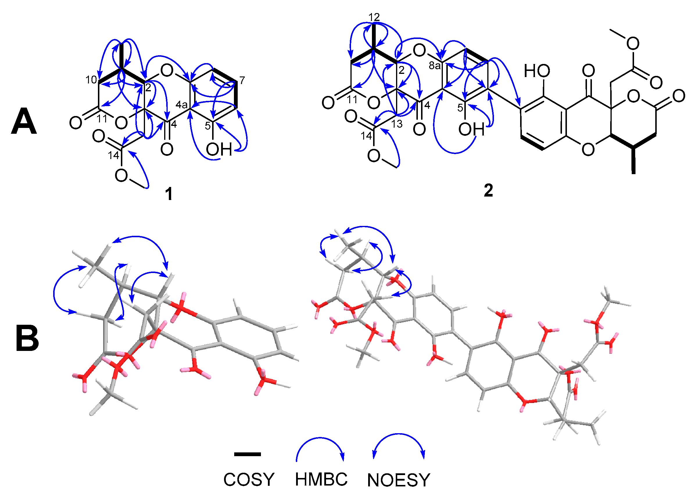
Analysis of the 2D NMR spectra suggested that daldiniaeschsone A (1) had a chromone core structure, which was fused to a δ-lactone. The 1H-1H COSY cross-peaks indicated three coupling systems, CH(6)-CH(7)-CH(8), CH(2)-CH(9)-CH2(10) and CH(9)-CH3(12) (Figure 4). Additionally, the HMBC correlations from H3-12 (δH 1.32) to C-2 (δC 83.1), C-9 (δC 33.9), and C-10 (δC 37.1) revealed the position of the methyl group at C-9. The presence and position of the δ-lactone were established on the basis of the HMBC correlations: H-2 (δH 4.79) with C-3 (δC 84.8), C-4 (δC 194.3), C-10, C-12 (δC 15.2), and C-13 (δC 40.2) and H2-13 (δH 3.25/3.18) with C-2, C-3, C-4, C-14 (δC 169.5), and H-9 (δH 2.96) with C-3, C-11 (δC 175.3), and C-12. The HMBC correlation of Me-15 (δH 3.73) with C-14 supported the identification of the methyl ester at C-14. While the HMBC correlations of H-7 (δH 7.42) with C-5 (δC 162.3), C-6 (δC 110.5), and C-8a (δC 159.3) and OH-5 (δH 11.44) with C-4a (δC 107.9), C-5, and C-6 indicated that the hydroxy group was attached at C-5. The relative configuration of 1 was evident from the NOESY experiment, as shown in Figure 4. NOESY cross peaks were observed between H-2/H2-13, and H-2/H3-12, indicating H-2, H2-13, and H3-12 were located in the same face of the lactone ring. The configuration of the vicinal coupling of H-2 and H-9 were assigned as trans based on the magnitude of the coupling constant (3J) of 7.5 Hz. Thus, the structure of 1 was identified as a daldiniaeschsone A. Compound 1 was subjected to chiral HPLC analysis and the results showed that this compound was a scalemic mixture with an enantiomeric excess (ee) of 15.6% (Supplementary Materials, Figure S19). Due to the small amount of this compound, the separation of its enantiomers was not carried out.
Daldiniaeschsone B (2), [α]21D −3.9 (c 1, MeOH), was isolated as a yellow amorphous solid, and the molecular formula C32H30O14 was determined based on the HRESITOFMS data (m/z 661.1504 [M + Na]+) and 13C NMR spectroscopic data. The UV and IR spectra were almost identical to those of 1 (Supplementary Materials, Figures S17 and S18). Analysis of 1D and 2D NMR spectroscopic data (Table 1) indicated that 2 shared the same core chromone–lactone structure to 1. However, the molecular formula C32H30O14 of 2, along with the observation of only 16 carbon resonances, indicated that this compound was a symmetrical dimer. The 1H and 13C NMR spectroscopic data of compound 2 were similar to those of 1 except compound 2 displayed 1H NMR resonances for an AB aromatic proton system [δH/δC 7.52 (1H, d, J = 8.5 Hz, H-7/7′)/δC 141.2 and 6.63 (1H, d, J = 8.5 Hz, H-8/8′)/δC 107.4] instead of an ABC spin system as in 1. The HMBC correlations of H-7 with C-5 (δC 159.2), C-6/C-6′ (δC 117.7), and C-8a (δC 158.4) and the lack of resonances for H-6/H-6′ in 1H NMR spectral data revealed the C-6/C-6′ carbon–carbon linkage between the two chromone moieties. A detailed assignment of the protons and carbons of 2 is shown in Table 1. Chiral HPLC analysis indicated a scalemic mixture with an ee of 86.2% (Supplementary Materials, Figure S20). Previously, several biphenyl core units have been synthesised by Bringmann et al. [25] and CD spectra have identified their axial/rotameric configuration (aS or aR isomers). The aS isomers of the biphenyls showed Cotton effects (CEs) similar to daldiniaeschsone B, whereas the aR isomers showed opposite signs. Shim et al. [26] also reported experimental and calculated CD spectra established aS-configuration of three new polyketide biphenyl derivatives, phomalevones A–C, isolated from an unidentified Phoma-like fungus. Daldiniaeschsone B displayed a strong negative CE at 225 nm and weak positive CEs at 273 and 317 nm (Supplementary Materials, Figure S21). This was similar to data reported for phomalevones A–C which possess P-helicity. Thus, the chirality of the 6,6′-axis of daldiniaeschsone B was assigned as the aS-configuration.

4.2. Putative Biosynthesis Pathway of Two Novel Compounds

The biosynthetic pathway of monomeric and dimeric polyketide metabolites has been investigated [27]. The putative biosynthetic pathway of compounds 1 and 2 is shown in Figure 5. The multistep condensations of C16-octaketide would produce xanthone intermediate i (ravenelin) [27,28,29]. Oxidative cleavage of intermediate i (ravenelin) could give intermediate ii, and further decarboxylation, followed by cyclisation, would give intermediate iii. Further oxidative cleavage of intermediate iii yields intermediate iv. The addition of one acetyl CoA unit will be followed by lactonisation and would give intermediate v. Compound 1 could obtain from intermediate v via hydrolysis and O-methylation, respectively. Finally, the oxidative coupling of compound 1 at C-6/C-6′ would give compound 2. The scalemic nature of compound 1 would suggest, perhaps, that the oxidative cleavage of i (ravenelin) to ii is not stereospecific. The significantly higher ee of compound 2 might suggest a kinetic preference (kinetic resolution) for dimerization of one enantiomer of 1.

4.3. α-Glucosidase and NO Production Inhibitory Activities

The broth extract of D. eschscholtzii was evaluated for its α-glucosidase inhibitory activity at the concentrations of 100, 150, and 200 µg/mL. At the highest concentration (200 µg/mL), the broth extract showed the highest inhibition of α-glucosidase with the % inhibition of 99.4% and its IC50 value was 5.69 µg/mL. However, at the concentrations of 100 and 150 µg/mL, the broth extract had the % inhibition less than 70%, indicating that at these concentrations (100 and 150 µg/mL), the broth extract was inactive. In the case of the pure compounds, compounds 1–5 showed α-glucosidase inhibitory activity with IC50 values of 0.16, 0.23, 0.85, 0.55, and 0.76 mM, respectively, while acarbose, the positive control had an IC50 = 0.08 mM. The broth extract and the most active compounds (1 and 2) were also evaluated for their cytotoxicities against mammalian cells, RAW 264.7 cells, using the MTT assay at the concentrations of 6.25, 12.5, 25, and 50 μg/mL. At the concentration of 50 μg/mL, the broth extract, and compounds 1 and 2 showed cell viability over 75%, indicating that all testing samples had no cytotoxicity at this concentration. The other concentrations (6.25–25 μg/mL) were cytotoxic. Notably, this is the first publication on the α-glucosidase inhibitory activity of these compounds. For the NO production inhibitory activity, the broth extract showed weak activity with an IC50 value of 19.85 µg/Ml, whereas compounds 1 and 2 were inactive.

5. Conclusions

Previous chemical constituent investigations of D. eschscholtzii, using different fungal collections and different conditions of fermentation yielded different compounds. Various types of compounds have been isolated and identified, but polyketides are found as the major compounds. To the best of our knowledge, two new polyketides (chromones) were isolated and identified for the first time in D. eschscholtzii isolated from the cinnamon plant (Cinnamomum bejolghota) collected from Chiang Mai Province, Thailand. Compounds 1 and 2 are rare tricyclic chromones found in nature, having the chromone unit fused to δ-lactone. Compound 1 was a monomer, whereas compound 2 was a symmetrical dimer with a C-6/C-6′ carbon–carbon linkage between the two chromone moieties. The compounds 4 and 5 were discovered from the Daldinia genus for the first time. Many chromones isolated from D. eschscholtzii have shown good inhibitory activities against α-glucosidase and nitric oxide. Unfortunately, in this study, all of the chromone derivatives showed weak α-glucosidase inhibitory, and compounds 1 and 2 showed no NO production inhibitory activities.

Supplementary Materials

The following are available online at https://www.mdpi.com/article/10.3390/jof7050358/s1, Figure S1: 1H NMR spectrum of compound 1 (500 MHz, CDCl3), Figure S2: 13C, DEPT90/135 NMR spectra of compound 1 (125 MHz, CDCl3), Figure S3: COSY spectrum of compound 1, Figure S4: HMQC spectrum of compound 1, Figure S5: HMBC spectrum of compound 1, Figure S6: NOESY spectrum of compound 1, Figure S7: HRESITOFMS spectrum of compound 1, Figure S8: IR (CH2Cl2) spectrum of compound 1, Figure S9: UV spectrum of compound 1, Figure S10: 1H NMR spectrum of compound 2 (500 MHz, CDCl3), Figure S11: 13C and DEPT NMR spectra of compound 2 (125 MHz, CDCl3), Figure S12: COSY spectrum of compound 2, Figure S13: HMQC spectrum of compound 2, Figure S14: HMBC spectrum of compound 2, Figure S15: NOESY spectrum of compound 2, Figure S16: HRESITOFMS spectrum of compound 2, Figure S17: IR (CH2Cl2) spectrum of compound 2, Figure S18: UV spectrum of compound 2, Figure S19: Chiral HPLC chromatogram of compound 1, Figure S20: Chiral HPLC chromatogram of compound 2, Figure S21: ECD spectrum of compound 2, Figure S22: The isolation and purification of isolated compounds 1–5 from Daldinia eschscholtzii SDBR-CMUNKC745 crude extract.

Author Contributions

Conceptualisation, S.L. (Surat Laphookhieo) and S.L. (Saisamorn Lumyong); methodology, N.W., N.S., V.S., A.P. and J.K.; formal analysis, N.W., V.S., A.P., N.S., J.K., S.L. (Saisamorn Lumyong), T.M., R.C., S.C., T.L., S.G.P. and S.L. (Surat Laphookhieo); investigation, N.W. and V.S.; resources, N.S., S.L. (Saisamorn Lumyong), T.M., S.L. (Surat Laphookhieo) and R.C.; data curation, N.W., V.S., A.P., N.S. and J.K.; writing—original draft preparation, V.S., A.P., T.M., R.C., S.C., T.L., S.G.P. and S.L. (Surat Laphookhieo); writing—review and editing, V.S., T.M., R.C., S.C., T.L., S.G.P. and S.L. (Surat Laphookhieo); funding acquisition, S.L. (Surat Laphookhieo) and S.L. (Saisamorn Lumyong). All authors have read and agreed to the published version of the manuscript.

Funding

Thailand Science Research and Innovation provided this research through the Direct Basic Research Grant (DBG6280007). The Postdoctoral Fellowship from Mae Fah Luang University to V.S., studentship (T.A. and R.A.) from the Graduate School, Chiang Mai University to N.W. and partially supported by Chiang Mai University and Mae Fah Luang University were also acknowledged.

Institutional Review Board Statement

Not applicable.

Informed Consent Statement

Not applicable.

Data Availability Statement

Data are included in the Article/Supplementary Materials/References.

Acknowledgments

Mae Fah Luang University and Chiang Mai University are also acknowledged for laboratory facilities.

Conflicts of Interest

The authors declare no conflict of interest.

References

  1. Liao, H.X.; Zheng, C.J.; Huang, G.L.; Mei, R.Q.; Nong, X.H.; Shao, T.M.; Chen, G.-Y.; Wang, C.-Y. Bioactive polyketide derivatives from the mangrove-derived fungus Daldinia eschscholtzii HJ004. J. Nat. Prod. 2019, 82, 2211–2219. [Google Scholar] [CrossRef]
  2. Kamauchi, H.; Shiraishi, Y.; Kojima, A.; Kawazoe, N.; Kinoshita, K.; Koyama, K. Isoindolinones, phthalides, and a naphthoquinone from the fruiting body of Daldinia concentrica. J. Nat. Prod. 2018, 81, 1290–1294. [Google Scholar] [CrossRef] [PubMed]
  3. Huang, R.H.; Gou, J.Y.; Zhao, D.L.; Wang, D.; Liu, J.; Ma, G.Y.; Lia, Y.-Q.; Zhang, C.-S. Phytotoxicity and anti-phytopathogenic activities of marine-derived fungi and their secondary metabolites. RSC Adv. 2018, 8, 37573–37580. [Google Scholar] [CrossRef] [Green Version]
  4. Dias, D.A.; Urban, S.; Roessner, U.A. A historical overview of natural products in drug discovery. Metabolites 2012, 2, 303–336. [Google Scholar] [CrossRef] [PubMed] [Green Version]
  5. Mishra, R.; Kushveer, J.S.; Khan, M.; Imran, K.; Pagal, S.; Meena, C.K.; Murali, A.; Dhayalan, A.; Sarma, V.V. 2,4-Di-tert-butylphenol isolated from an endophytic fungus, Daldinia eschscholtzii, reduces virulence and quorum sensing in Pseudomonas aeruginosa. Front. Microbiol. 2020, 11, 1668. [Google Scholar] [CrossRef] [PubMed]
  6. Yang, L.J.; Liao, H.X.; Bai, M.; Huang, G.L.; Luo, Y.P.; Niu, Y.Y.; Zheng, C.-J.; Wang, C.-Y. One new cytochalasin metabolite isolated from a mangrove-derived fungus Daldinia eschscholtzii HJ001. Nat. Prod. Res. 2018, 32, 208–213. [Google Scholar] [CrossRef]
  7. Tarman, K.; Palm, G.J.; Porzel, A.; Merzweiler, K.; Arnold, N.; Wessjohann, L.A.; Unterseher, M.; Lindequist, U. Helicascolide C, a new lactone from an Indonesian marine algicolous strain of Daldinia eschscholzii (Xylariaceae, Ascomycota). Phytochem. Lett. 2012, 5, 83–86. [Google Scholar] [CrossRef]
  8. Nagasawa, H.; Nagura, F.; Mohamad, S.B.; Uto, Y.; Zhu, J.W.; Tsukuda, T.; Hashimoto, T.; Asakawa, T.; Hori, H. Apoptosis induction in HCT116 cells by cytochalasins isolated from the fungus Daldinia vernicosa. Phytomedicine 2000, 6, 403–409. [Google Scholar] [CrossRef]
  9. Quang, D.N.; Hashimoto, T.; Tanaka, M.; Baumgartner, M.; Stadler, M.; Asakawa, Y. Chemical constituents of the Ascomycete Daldinia concentrica. J. Nat. Prod. 2002, 65, 1869–1874. [Google Scholar] [CrossRef] [PubMed]
  10. Qin, X.D.; Dong, Z.J.; Liu, J.K.; Yang, L.M.; Wang, R.R.; Zheng, Y.T.; Zheng, Q.T. Concentricolide, an anti-HIV Agent from the Ascomycete Daldinia concentrica. Helv. Chim. Acta 2006, 89, 127–133. [Google Scholar] [CrossRef]
  11. Stadler, M.; Læssøe, T.; Fournier, J.; Decock, C.; Schmieschek, B.; Tichy, H.V.; Peršoh, D.A. polyphasic taxonomy of Daldinia (Xylariaceae). Stud. Mycol. 2014, 77, 1–143. [Google Scholar] [CrossRef] [PubMed] [Green Version]
  12. Zhang, Y.L.; Ge, H.M.; Zhao, W.; Dong, H.; Xu, Q.; Li, S.H.; Li, J.; Zhang, J.; Song, Y.C.; Tan, R.X. Unprecedented immunosuppressive polyketides from Daldinia eschscholzii, a mantis-associated fungus. Angew. Chem. Int. Ed. 2008, 47, 5823–5826. [Google Scholar] [CrossRef] [PubMed]
  13. Liu, H.X.; Tan, H.B.; Li, S.N.; Chen, Y.C.; Li, H.H.; Zhang, W.M. Two new metabolites from Daldinia eschscholtzii, an endophytic fungus derived from Pogostemon cablin. J. Asian Nat. Prod. Res. 2019, 21, 150–156. [Google Scholar] [CrossRef]
  14. Kongyen, W.; Rukachaisirikul, V.; Phongpaichit, S.; Sakayaroj, J. A new hydronaphthalenone from the mangrove-derived Daldinia eschscholtzii PSU-STD57. Nat. Prod. Res. 2015, 29, 1995–1999. [Google Scholar] [CrossRef]
  15. Luo, Y.; Qiu, L.; Deng, Y.; Yuan, X.H.; Gao, P. A new chromone and a new aliphatic ester isolated from Daldinia eschscholtzii. J. Asian Nat. Prod. Res. 2018, 20, 883–888. [Google Scholar] [CrossRef] [PubMed]
  16. Zhang, A.H.; Tan, R.; Jiang, N.; Yusupu, K.; Wang, G.; Wang, X.L.; Tan, R.X. Selesconol, a fungal polyketide that induces stem cell differentiation. Org. Lett. 2016, 18, 5488–5491. [Google Scholar] [CrossRef]
  17. Lin, L.P.; Yuan, P.; Jiang, N.; Mei, Y.N.; Zhang, W.J.; Wu, H.M.; Zhang, A.H.; Cao, J.M.; Xiong, Z.X.; Lu, Y.; et al. Gene-inspired mycosynthesis of skeletally new indole alkaloids. Org. lett. 2015, 17, 2610–2613. [Google Scholar] [CrossRef]
  18. Phukhatmuen, P.; Raksat, A.; Laphookhieo, S.; Charoensup, R.; Duangyod, T.; Maneerat, W. Bioassay-guided isolation and identification of antidiabetic compounds from Garcinia cowa leaf extract. Heliyon 2020, 6, e03625. [Google Scholar] [CrossRef]
  19. Joo, T.; Sowndhararajan, K.; Hong, S.; Lee, J.; Park, S.Y.; Kim, S.; Jhoo, J.W. Inhibition of nitric oxide production in LPS-stimulated RAW 264.7 cells by stem bark of Ulmus pumila L. Saudi. J. Biol. Sci. 2014, 21, 427–435. [Google Scholar] [CrossRef] [Green Version]
  20. Dong, M.; Dan, L.; Li, Y.H.; Xuan, Q.C.; Kaim, L.; Yong, M.Z.; Li, R.T. Naphthoquinones from Onosma paniculatum with potential anti-inflammatory activity. Planta Med. 2016, 83, 631–635. [Google Scholar] [CrossRef]
  21. Polbuppha, I.; Suthiphasilp, V.; Maneerat, T.; Charoensup, R.; Limtharakul, T.; Cheenpracha, S.; Pyne, S.G.; Laphookhieo, S. Macluracochinones AE, antimicrobial flavonoids from Maclura cochinchinensis (Lour.) Corner. Phytochemistry 2021, 187, 112773. [Google Scholar] [CrossRef] [PubMed]
  22. Talapatra, S.K.; Karmacharya, B.; De, S.C.; Talapatra, B. (−)-Regiolone, an α-tetralone from Juglans regia: Structure, stereochemistry and conformation. Phytochemistry 1988, 27, 3929–3932. [Google Scholar] [CrossRef]
  23. Iwasaki, S.; Muro, H.; Sasaki, K.; Nozoe, S.; Okuda, S.; Sato, Z. Isolations of phytotoxic substances produced by Pyricularia oryzae Cavara. Tetrahedron Lett. 1973, 14, 3537–3542. [Google Scholar] [CrossRef]
  24. Chacón-Morales, P.; Amaro-Luis, J.M.; Bahsas, A. Isolation and characterization of (+)-mellein, the first isocoumarin reported in Stevia genus. Av. Quím. 2013, 8, 145–151. [Google Scholar]
  25. Bringmann, G.; Hinrichs, J.; Henschel, P.; Kraus, J.; Peters, K.; Peters, E.M. Atropo-enantioselective synthesis of the natural bicoumarin (+)-isokotanin a via a configurationally stable biaryl lactone. Eur. J. Org. Chem. 2002, 6, 1096–1106. [Google Scholar] [CrossRef]
  26. Shim, S.H.; Baltrusaitis, J.; Gloer, J.B.; Wicklow, D.T. Phomalevones A−C: Dimeric and pseudodimeric polyketides from a fungicolous hawaiiani of Phoma sp. (Cucurbitariaceae). J. Nat. Prod. 2011, 74, 395–401. [Google Scholar] [CrossRef]
  27. Rönsberg, D.; Debbab, A.; Mándi, A.; Vasylyeva, V.; Böhler, P.; Stork, B.; Engelke, L.; Hamacher, A.; Sawadogo, R.; Diederich, M.; et al. Pro-apoptotic and immunostimulatory tetrahydroxanthone dimers from the endophytic fungus Phomopsis longicolla. J. Org. Chem. 2013, 78, 12409–12425. [Google Scholar]
  28. Kurobane, I.; Vining, L.C.; Mcinnes, A.G. Biosynthetic relationships among the secalonic acids isolation of emodin, endocrocin and secalonic Acids from Pyrenochaeta Terrestris and Aspergillus Aculeatus. J. Antibiot. 1979, 32, 1256–1266. [Google Scholar] [CrossRef]
  29. Padhi, S.; Masi, M.; Cimmino, A.; Tuzi, A.; Jena, S.; Tayung, K.; Evidente, A. Funiculosone, a substituted dihydroxanthene-1,9-dione with two of its analogues produced by an endolichenic fungus Talaromyces funiculosus and their antimicrobial activity. Phytochemistry 2019, 157, 175–183. [Google Scholar] [CrossRef]
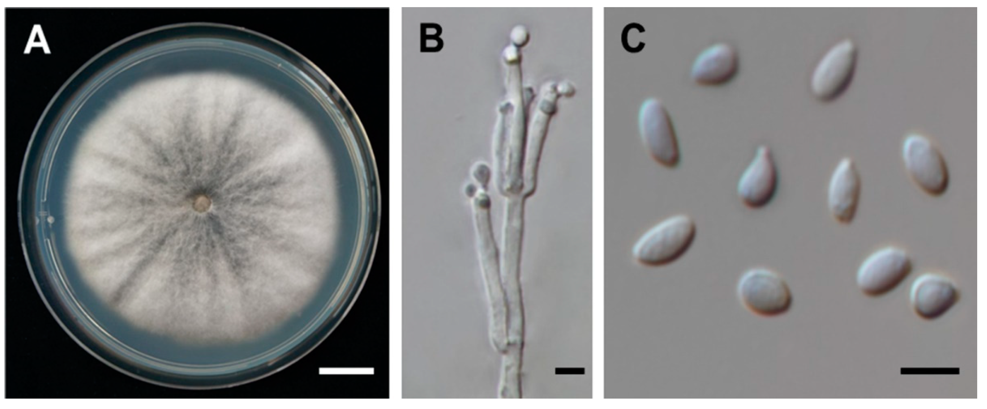
Figure 1. Daldinia eschscholtzii SDBR-CMUNKC745. (A) Colony on PDA at 30 °C for one week. (B) Conidiophores and conidiogeneous cells. (C) Conidia. Scale bars: (A) = 10 mm, (B) = 1 μm, (C) = 5 μm.
Jof 07 00358 g001
Figure 2. Phylogram derived from maximum likelihood analysis of the combined ITS, LSU, RBP2, and TUB genes of 26 fungal taxa. Sequences of Annulohypoxylon annulatum and A. moriforme were used as outgroup. The numbers above branches represent maximum likelihood bootstrap percentages (left) and Bayesian posterior probabilities (right). Only bootstrap values ≥ 50% and Bayesian posterior probabilities ≥ 0.70 are shown. The scale bar represents ten substitutions per nucleotide position. Sequences of fungal species obtained in this study are in bold.
Jof 07 00358 g002
Figure 3. Isolated compounds from D. eschscholtzii SDBR-CMUNKC745 broth extract.
Jof 07 00358 g003
Figure 4. (A) Selected HMBC, COSY, and (B) NOESY correlations of compounds 1 and 2.
Jof 07 00358 g004
Figure 5. Putative biosynthetic pathway for the polyketides 1 and 2.
Jof 07 00358 g005
Table 1. 1H (500 MHz) and 13C (125 MHz) NMR Spectroscopic data of 1 and 2 in CDCl3.
Position1Position2
δcδH (J in Hz)δcδH (J in Hz)
283.1, CH4.79 (d, 7.5)2/2′82.6, CH4.81 (d, 7.4)
384.8, C 3/3′84.5, C
4194.3, C 4/4′194.1, C
4a107.9, C 4a/4a′107.5, C
5162.3, C 5/5′159.2, C
6110.5, CH6.56 (dd, 8.3, 0.9)6/6′117.7, C
7139.1, CH7.42 (t, 8.3)7/7′141.2, CH7.52 (d, 8.5)
8108.0, CH6.54 (dd, 8.3, 0.9)8/8′107.4, CH6.63 (d, 8.5)
8a159.3, C 8a/8a′158.4, C
933.9, CH2.96 (hept, 7.5)9/9′33.5, CH2.98 (hept, 7.4)
10a
10b
37.1, CH22.70 (dd, 17.3, 8.3)
2.47 (dd, 17.3, 8.0)
10a/10a′
10b/10b′
36.7, CH22.70 (dd, 17.3, 8.3)
2.48 (dd, 17.3, 8.0)
11175.3, C 11/11′174.9, C
1215.2, CH31.32 (d, 7.5)12/12′14.9, CH31.34 (d, 7.4)
13a
13b
40.2, CH23.25 (d, 17.3)
3.18 (d, 17.3)
13a/13a′
13b/13b′
39.8, CH23.27 (d, 17.3)
3.20 (d, 17.3)
14169.5, C 14/14′169.1, C
CO2Me-1554.0, CH33.73 (s)CO2Me-15/CO2Me-15′53.8, CH33.77 (s)
OH-5 11.44 (s)OH-5/OH-5′ 11.91 (s)
Table 2. α-Glucosidase inhibitory and NO production inhibitory activities of isolated compounds from D. eschscholtzii SDBR-CMUNKC745.
Compoundsα-Glucosidase Inhibitory ActivityAnti-Inflammatory (NO Production Inhibitory Activity)% Cell Viability at 50 µg/mL
% Inhibition at 200 µg/mLIC50 (mM)% Inhibition at 50 µg/mLIC50 (µM)
Crude extract99.45.96 ± 0.06 µg/mL99.519.85 ± 0.14 µg/mL75.52 ± 1.77
198.70.16 ± 0.1126.5inactive88.52 ± 3.97
296.80.23 ± 0.7636.9inactive100.24 ± 1.75
397.80.85 ± 1.19not testednot testednot tested
497.80.55 ± 1.33not testednot testednot tested
597.50.76 ± 0.09not testednot testednot tested
Acarbose95.60.08 ± 0.04not testednot testednot tested
Indomethacinnot testednot tested71.019.61 ± 0.6284.96 ± 2.92
Publisher’s Note: MDPI stays neutral with regard to jurisdictional claims in published maps and institutional affiliations.