Article

Phenotypic Characterization and Comparative Genomics of the Melanin-Producing Yeast Exophiala lecanii-corni Reveals a Distinct Stress Tolerance Profile and Reduced Ribosomal Genetic Content

1
National Research Council Postdoctoral Research Associate, U.S. Naval Research Laboratory, Washington, DC 20375, USA
2
Center for Biomolecular Sciences and Engineering, U.S. Naval Research Laboratory, Washington, DC 20375, USA
3
Infectious Disease and Microbiome Program, Broad Institute of MIT and Harvard, Cambridge, MA 02142, USA
4
Biotechnology Branch, CCDC Army Research Laboratory, Adelphi, MD 20783, USA
5
American Society for Engineering Education Postdoctoral Research Associate, U.S. Naval Research Laboratory, Washington, DC 20375, USA
*
Author to whom correspondence should be addressed.
J. Fungi 2021, 7(12), 1078; https://doi.org/10.3390/jof7121078
Submission received: 22 October 2021 / Revised: 7 December 2021 / Accepted: 10 December 2021 / Published: 15 December 2021
(This article belongs to the Special Issue Yeast Biorefineries)

Abstract

:
The black yeast Exophiala lecanii-corni of the order Chaetothyriales is notable for its ability to produce abundant quantities of DHN-melanin. While many other Exophiala species are frequent causal agents of human infection, E. lecanii-corni CBS 102400 lacks the thermotolerance requirements that enable pathogenicity, making it appealing for use in targeted functional studies and biotechnological applications. Here, we report the stress tolerance characteristics of E. lecanii-corni, with an emphasis on the influence of melanin on its resistance to various forms of stress. We find that E. lecanii-corni has a distinct stress tolerance profile that includes variation in resistance to temperature, osmotic, and oxidative stress relative to the extremophilic and pathogenic black yeast Exophiala dermatitidis. Notably, the presence of melanin substantially impacts stress resistance in E. lecanii-corni, while this was not found to be the case in E. dermatitidis. The cellular context, therefore, influences the role of melanin in stress protection. In addition, we present a detailed analysis of the E. lecanii-corni genome, revealing key differences in functional genetic content relative to other ascomycetous species, including a significant decrease in abundance of genes encoding ribosomal proteins. In all, this study provides insight into how genetics and physiology may underlie stress tolerance and enhances understanding of the genetic diversity of black yeasts.

1. Introduction

Black yeasts belonging to the ascomycete order Chaetothyriales have been of historic interest due to their frequent characterization as opportunistic pathogens [1]. These organisms, including those within the Exophiala genus, are distinguished by their abundant melanin content, thick cell walls, polymorphic growth, and ability to withstand a range of extreme conditions, including high and low temperatures, high salinity, desiccation, and various forms of non-ionizing and ionizing radiation [2,3,4,5]. Despite this, closely related species of black yeasts can also differ significantly with regard to pathogenicity and other attributes [1,6]. For example, Exophiala lecanii-corni is unable to grow at human physiological temperatures, and therefore is less pathogenic than Exophiala dermatitidis, being isolated from clinical settings approximately four times less frequently [7]. In addition, E. lecanii-corni CBS 102400, which was originally isolated from a bioreactor that aimed to decontaminate a toluene-containing waste gas stream [8], has been characterized as Biosafety Level 1 (BSL1), indicating that it is safe to work with and increasing its feasibility for use in biomanufacturing or other biotechnological applications. In recent decades, E. lecanii-corni has also garnered attention due to its demonstrated capacity to efficiently eliminate volatile organic compounds in vapor-phase, indicating its potential for bioremediation applications [8,9]. Importantly, the genetic tractability of this organism has already been demonstrated [10], which paves the way for targeted functional studies, as well as genetic optimization of production yields for bioproducts of interest.
E. lecanii-corni is notable for its constitutive production of large quantities of melanin via the 1,8-dihydroxynaphthalene (DHN)-melanin pathway, which are embedded in the cell wall and also released in culture filtrate [10]. Melanin has been reported to play a variety of roles in fungal systems, including the conferral of tolerance to various forms of stress, such as temperature, desiccation, and osmotic shock [11,12,13]. In some fungal species, melanin is also required for normal cell development, including accurate assembly of the cell wall and differentiation into secondary hyphae and conidia [14,15]. In addition, melanin contributes to the virulence of fungi by enhancing their ability to elude host immune responses [16,17]. The functional versatility of melanin within biological systems is a reflection of the broad variability of its physico-chemical properties, which are largely dictated by melanin synthesis and polymerization conditions that can lead to differences in oligomer size, molecular scaffold diversity, redox state, and supramolecular structure, resulting in different melanin variants with distinct characteristics [18]. These properties include antioxidant and free radical scavenging [19,20], photoprotection [21], semiconductivity [22], and metal and drug binding [23,24], which has led to the proposed use of melanin in bioelectronics, medical applications, materials science, and environmental remediation [25,26].
Phenotypic characterization coupled with comparative genomic analysis can strengthen our understanding of the genetic underpinnings that define variation among black yeasts. Our research group has previously conducted numerous studies into the black yeast E. dermatitidis that have advanced our understanding of the molecular basis of its extremophilic characteristics, as well as the impact of melanin on such processes [6,27,28,29,30]. In a previous study, we observed that although E. dermatitidis produces substantially less melanin than E. lecanii-corni, E. dermatitidis is far more resistant to high-dose acute ionizing radiation [6]. Here, we expand on this finding to evaluate differences in stress tolerance between these two species, as well as the impact of melanin on those processes. We find that there are substantive differences in resistance to temperature, osmotic stress, and oxidative stress between E. lecanii-corni and E. dermatitidis, suggesting that even among these closely related species of black yeast, the influence of melanin on stress resistance is not uniform. In addition, we report a comparative genomic analysis of E. lecanii-corni in relation to other ascomycetous species, revealing key differences in functional content, including significant underrepresentation of genes encoding ribosomal proteins. Together, this investigation represents the first detailed phenotypic and genomic characterization of E. lecanii-corni and contributes to the growing body of knowledge that continues to illuminate the genetic diversity of black yeast.

2. Materials and Methods

2.1. Cultivation of Fungi

The E. lecanii-corni strains used in this study were derived from the wild-type strain CBS 102400, and the E. dermatitidis strains used in this study were obtained from a previous study and derived from standard laboratory strain 8656 [6]. Unless otherwise noted, all strains were cultured at 25 °C in liquid or solid yeast peptone dextrose (YPD; 10 g/L yeast extract, 20 g/L peptone, 20 g/L glucose), with liquid cultures shaking at 225 RPM.
To identify culture media conditions that optimize melanin production yields, E. lecanii-corni was cultured using a range of standard fungal media recipes, including liquid YPD, yeast extract glucose (YEG; 5 g/L yeast extract, 20 g/L glucose, 1 mL/L Hutner’s trace elements), yeast extract sucrose (YES; 20 g/L yeast extract, 100 g/L sucrose, 1 mL/L Hutner’s trace elements), malt peptone glucose (MPG; 20 g/L malt extract, 1 g/L peptone, 20 g/L glucose), malt broth (MB; 130 g/L malt extract), Czapek (CZ; 30 g/L sucrose, 3 g/L NaNO3, 1 g/L K2HPO4, 0.5 g/L MgSO4, 0.5 g/L KCl, 0.01 g FeSO4), Czapek Yeast (CY; 5 g/L yeast extract, 30 g/L sucrose, 3 g/L NaNO3, 1 g/L K2HPO4, 0.5 g/L MgSO4, 0.5 g/L KCl, 0.01 g FeSO4), glucose minimal media optimized for yeast (GMM; 5.4 g/L glucose, 4 g/L KH2PO4, 1.20 g/L MgSO4, 1 g/L glycine, 0.8 mg/L thiamine, pH 5.5).
To screen cultures for melanin production, freshly cultured E. lecanii-corni cells were collected and inoculated at 104 cells/mL into 250 mL flasks that each contained 100 mL of culture media to be screened. E. lecanii-corni cells were cultured at 25 °C while shaking at 200 RPM for 2 weeks, at which point the cells should be in stationary phase.

2.2. Isolation of Fungal Melanin

Fungal melanin was isolated from cultures screened in various growth conditions following 2 weeks of growth. To extract crude melanin, cells were spun down (10,000× g, 20 min) and the supernatant was transferred to a new Falcon tube. The supernatant was then acidified to a pH of 3 using 0.6 M HCl and kept at room temperature for 24 h, and then autoclaved to fully precipitate the remaining melanin. The precipitated crude melanin was then collected by centrifugation at (10,000× g, 10 min), washed with distilled water, and centrifuged again. This step was repeated until a neutral pH was achieved. Crude melanin was dried using a Savant SpeedVac Concentrator and the dry weight was measured. Isolation and purification of fungal melanin ghosts follows the method described by Dadachova [31].
Melanin ghosts were stained with 1% phosphotungstic acid aqueous solution (Electron Microscopy Sciences) for 2 h, spun down and freeze-dried, and were then imaged using a FEI Quanta 200 F field emission scanning electron microscope (FESEM) at an accelerating voltage of 5 kV.
Melanin ghosts were fixed with 4% paraformaldehyde for 2 h, stained with 1% osmium tetroxide for 1 h, centrifuged, and the pellets were freeze-dried. The dried samples were embedded in epoxy resin (low viscosity embedding kit) (Electron Microscopy Sciences, Hatfield, PA, USA) and cured overnight at 60 °C in an oven. Samples for TEM were prepared using a Leica EM UC7 ultramicrotome with a diamond knife. Sections were cut from the embedded samples at room temperature to a thickness of approximately 100 nm and collected on the TEM copper grids. The sections were imaged with a JEOL JEM-ARM200F transmission electron microscope operated at 200 kV.

2.3. Genetic Manipulation

The E. lecanii-corni Elpks1Δ mutant strain deficient in melanin production was generated using established gene deletion techniques [10,32]. Gene deletion cassettes were created by amplifying approximately 1 kb of the upstream and downstream genetic sequence flanking the target gene, which were fused to each end of the hygromycin resistance maker using the double-joint PCR technique [32]. The resulting construct was then transformed into E. lecanii-corni CBS 102400 using a previously described protocol [10]. In brief, cells were cultured in 300 mL YPD at a concentration of 105 cells/mL for 36 h. Cells were then harvested and hyphae was removed via filtration with a sterile Miracloth. The resulting filtrate was chilled on ice for at least 30 min and cells were collected via centrifugation (4000× g, 8 min). The cell pellet was washed with cold water twice and resuspended in cold water at a concentration of 109 cells/mL. Approximately 100 μL of the competent cells were combined with the DNA construct (3–4 μg DNA in a volume of 10 μL or less), mixed thoroughly, transferred to a chilled 0.2 cm electroporation cuvette, and subjected to electroporation using a Bio-Rad MicroPulser with default settings for fungi. The electroporated cells were transferred to 1 mL of liquid YPD, where they were incubated at 25 °C while shaking at 225 RPM for 3 h. The cells were then spread onto YPD plates containing 100 μg/mL hygromycin, which were grown at 25 °C until transformants were visible. Correct transformants were identified by observing a loss of color (due to lack of melanin production), which then underwent further confirmation via PCR (data not shown). The E. dermatitidis pks1Δ deletion strain was obtained from a previous study [6].

2.4. Cell Survival and Sensitivity Assays

For radiation resistance and stress sensitivity assays, cells were cultured in 2.5 mL YPD in 15 mL culture tubes for 6 days. Following growth, cells were re-suspended to a concentration of 108 cells/mL in cold water and placed on ice. For UV-C exposure, cells were plated on YPD solid media and exposed in triplicate to either 0, 25, 50, or 75 mJ/cm2 of UV-C light within a biosafety cabinet with a dose rate of 0.273 mJ cm−2 s−1. For γ-radiation exposure, 100 μL of cells were aliquoted to PCR tubes, which were exposed to either 0, 500, 1000, or 1500 Gy of γ-radiation by a Cobalt-60 source with a dose rate of approximately 36 Gy/min. For each sample and dose, two biological replicates were exposed to the radiation source and each biological replicate was plated onto two YPD plates following the irradiation. For both UV-C and γ-radiation survival assays, relative survival was evaluated by counting colonies and determining CFU measurements. The standard deviations were generated with Microsoft Excel. For stress sensitivity, 2 μL droplets of cells were spotted in serial 100–105-fold dilutions on YPD plates with the appropriate concentration of stress-inducing agent (0.5 M NaCl or 7.5 mM H2O2). Unless otherwise noted, all culture were grown at 25 °C. Desiccation resistance was evaluated by transferring 100 μL of washed cells at a concentration 108 cells/mL into a well of a sterile 96-well culture plate. The plate was placed in a sealed plastic container that also contained calcium sulfate desiccant and incubated at 30 °C for 1 week. The cells were resuspended in 100 μL of sterile water, diluted 100–105, and spotted in serial dilutions on YPD plates. Each stress assay was performed at least two times with two different biological replicates.

2.5. Phylogenetic and Comparative Genomic Analysis

The genome sequence and assembly of E. lecanii-corni was recently reported by our research group in collaboration with the Broad Institute [33]. Genes were predicted using BRAKER, which executed Genemark-ET with the parameter “fungus” [34,35]. tRNAs were predicted using tRNAscan [36] and rRNAs were predicted using RNAmmer [37]. Genes harboring Pfam domains that are present in repetitive elements or overlapping tRNA/rRNA features were removed. Genes were named and numbered sequentially. For comparative analyses, protein FASTA sequences for E. dermatitidis [27], Exophiala mesophila [38], Exophiala sideris, Exophiala xenobiotica, Exophiala aquamarina, Exophiala oligosperma, Exophiala spinifera, Fonsecaea pedrosoi, Cladophialophora bantiana, Cladophialophora immunda [5], Aspergillus fumigatus [39], Aspergillus nidulans [40], Aspergillus niger [41], Trichophyton rubrum [42], Coccidioides immitis [43], Saccharomyces cerevisiae [44], and Schizosaccharomyces pombe [45] were obtained from the National Center for Biotechnology Information (NCBI) web portal. Using these protein sequences, a rooted species tree was inferred using OrthoFinder [46,47], which involved prediction of orthogroups using the OrthoFinder algorithm, followed by inference of unrooted gene trees for each orthogroup using DendroBLAST [48], inference of an unrooted species tree using the STAG algorithm [49], and inference of the rooted species tree using the STRIDE algorithm [50].
Repeat elements were identified using RepeatModeler version 2.0.1 [51] and RepeatMasker version 4.1.0 [52], followed by classification using a 2017 RepBase RepeatMasker repetitive DNA element library [53]. For functional analysis, Pfam database protein families [54] were identified in all 18 species using the PfamScan Perl script (ftp://ftp.sanger.ac.uk/pub/databases/Pfam/Tools/ accessed on 11 November 2020). OrthoVenn2 was used to identify and annotate orthologous gene clusters and to generate Venn diagrams that display the distribution of shared gene families across selected species [55]. Gene ontology (GO) terms significantly enriched in E. lecanii-corni relative to other fungal species were identified using the GO term enrichment tool on the OrthoVenn2 platform. To evaluate orthologues underrepresented in E. lecanii-corni, orthogroups generated from OrthoFinder were assessed to identify orthogroups that lacked an E. lecanii-corni orthologue relative to either E. dermatitidis or the 17 other species included in this study [46,47]. GO term enrichment analysis was then carried out on E. dermatitidis genes present in these groups using FungiFun2 [56]. Secondary metabolite core biosynthesis genes, including polyketide synthases (PKSs), PKS-like enzymes, nonribosomal peptide synthetases (NRPSs), NRPS-like enzymes, and terpene cyclases (TCs) were predicated using antiSMASH version 5.0 [57].

3. Results

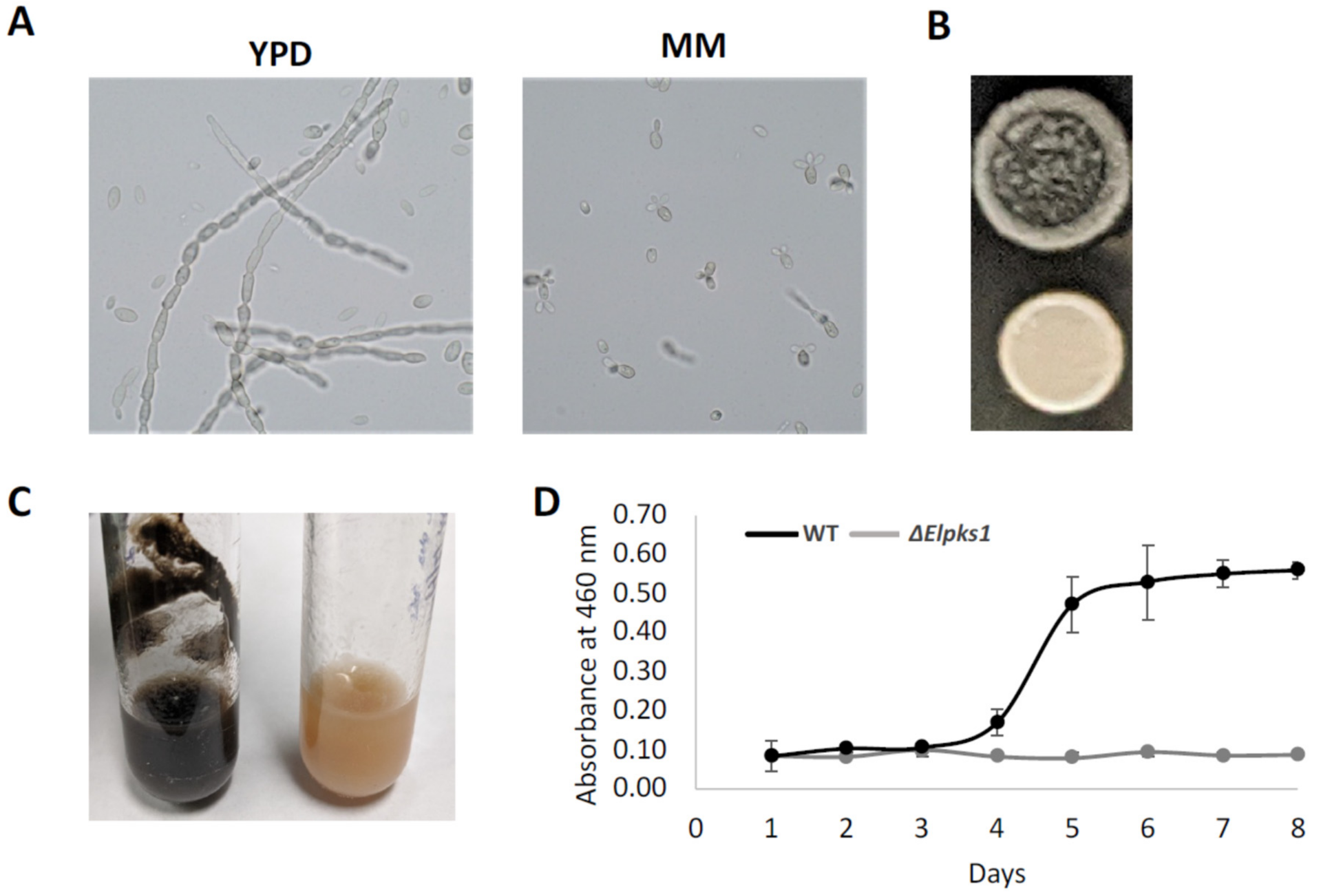
3.1. Phenotypic Analyses, Melanin Biosynthesis and Melanin Characterization in E. lecanii-corni

Like many other black yeasts, the highly melanized E. lecanii-corni is dimorphic, exhibiting both yeast and hyphae forms in rich medium. Interestingly, however, the yeast form appears dominant when grown in minimal medium (Figure 1A). This indicates that the yeast-to-hyphae transition is regulated by environmental conditions. Melanin has been shown to regulate cell morphology [58], and therefore, in characterizing impact of melanin on the phenotypic properties of E. lecanii-corni, we generated a melanin deficient mutant by deleting Elpks1, a gene encoding the polyketide synthase responsible for melanin biosynthesis (Figure 1B). Melanin deficiency did not affect cell dimorphism (data not shown). However, in YPD agar, the macroscopic colony morphology of the wild type appeared strikingly different-more wrinkled and more invasive than that of the Elpks1Δ mutant. In liquid medium, moreover, the wild type strain tended to form biofilms along the surface, which did not occur in the non-melanized strain (Figure 1C). This result suggests that melanin enhanced cellular strength for penetration of substrates and promoted biofilm formation.
Given the broad range of functional properties and proposed applications of fungal melanin [25,26], we were interested in identifying a prolific melanin-producing fungus, and therefore, we next examined growth of E. lecanii-corni in a range of liquid media compositions to identify culture conditions that optimized melanin production yields. Media screened included yeast peptone dextrose (YPD), yeast extract glucose (YEG), yeast extract sucrose (YES), malt peptone glucose (MPG), malt extract (MB), Czapek (CZ), Czapek yeast (CY), and glucose minimal media optimized for yeast (GMM). E. lecanii-corni cells were inoculated into each medium and grown at 25 °C for 2 weeks, followed by extraction of crude melanin from culture supernatants by acidification and autoclaving. Of the conditions tested, melanin production was maximized when E. lecanii-corni cells were cultivated in MB liquid media, which exhibited a melanin production rate of approximately 3.2 g/L. High yields were also observed in MPG and YPD cultures, both of which produced approximately 2 g of melanin per 1 L culture media. Other conditions, including CY, YES, and YEG, were found to produce approximately 0.6 g/L, 0.4 g/L, and 0.1 g/L of melanin, respectively. Minimal melanin production was observed in both MM and CZA. These findings suggest that melanin production is varied in different growth conditions and optimized in malt- or peptone-based culture media. Moreover, measurement of OD460 intensities of the culture supernatants was used to monitor the rate of melanization of E. lecanii-corni in YPD broth at 25 °C over the course of eight days. Melanization exponentially increased between the fourth and fifth day and reached a plateau after the sixth day (Figure 1D).
Melanin synthesized from fungi is typically assembled within the cell wall and plays a significant role in maintaining cell wall integrity [59]. This means that melanin structures associated with the fungal cell walls, so-called “melanin ghosts”, can be generated, usually by treating melanized cells with 4 M guanidinium isothiocyanate followed by 6 M HCl at 100 °C. This protocol was applied to treat E. lecanii-corni wild type and Elpks1Δ mutant cells. The albino cells were completely solubilized but the wild type cells remained as black particulate materials. SEM images showed typical yeast morphologies with defined nano-sized melanin granules on the surface (Figure 2A). TEM revealed the hollow ghost structures and the melanin layer from the cell walls (Figure 2B), the density of which was substantially higher than that observed in melanin ghosts of E. dermatitidis [60], confirming that E. lecanii-corni synthesized significantly more melanin than E. dermatitidis. Additionally, the melanin particles precipitated from the black supernatant of cell culture with acid showed porous structures by SEM (Figure 2C).

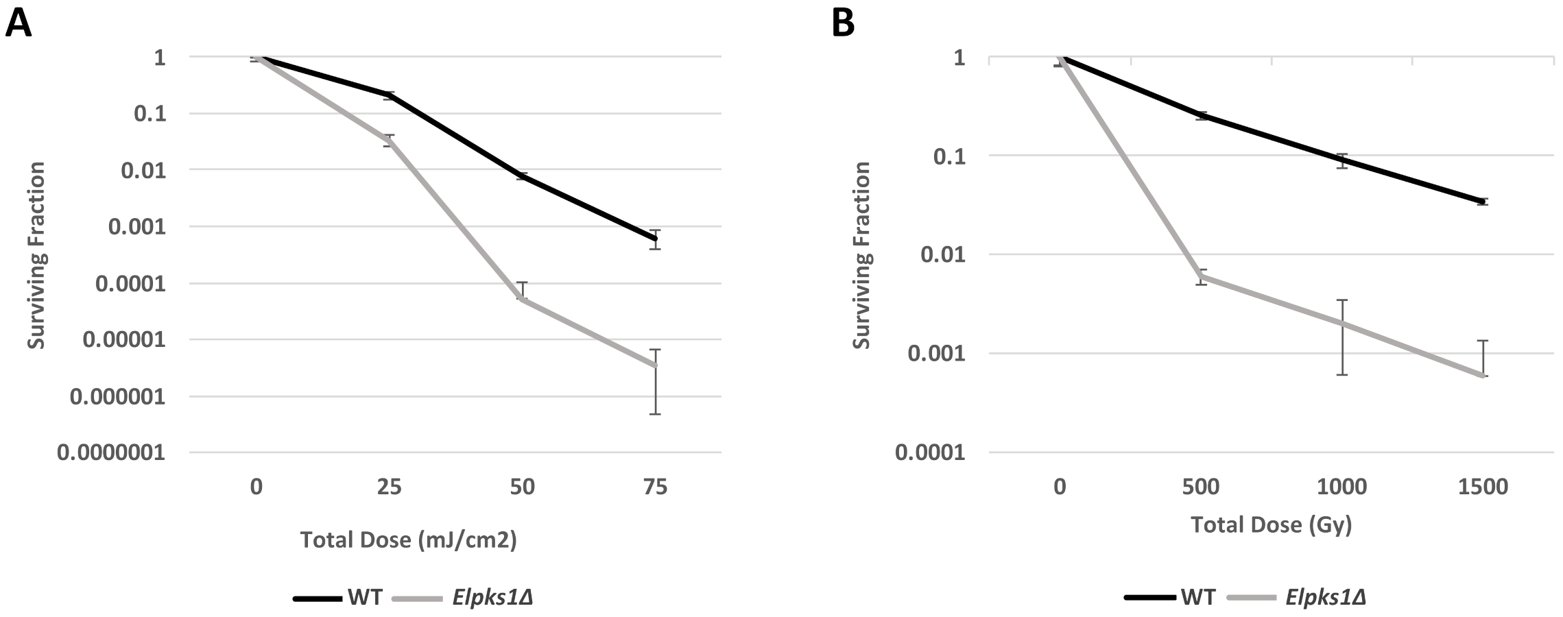
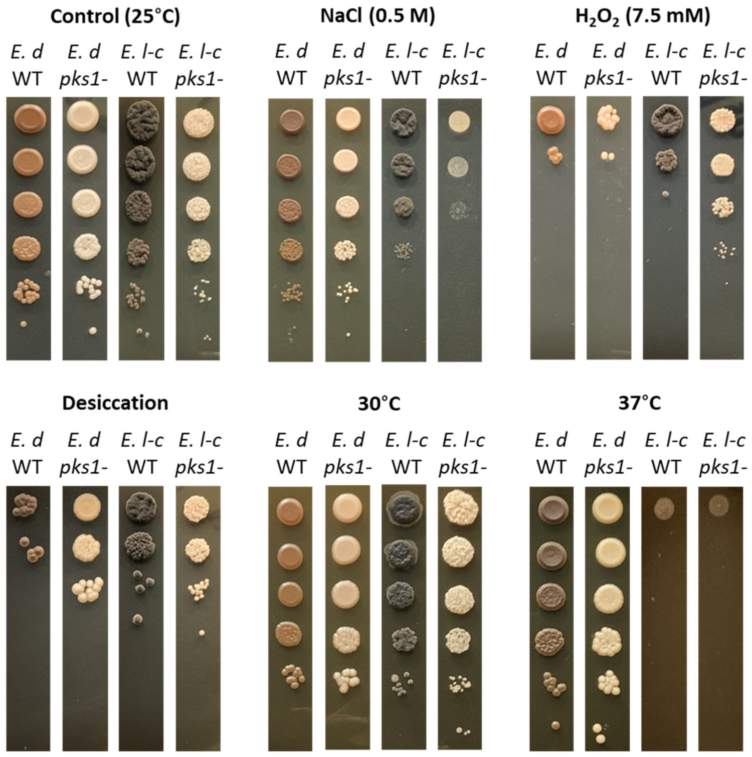
3.2. Effect of Melanin Production on Stress Resistance in E. lecanii-corni

Given the well-established UV-protective properties of melanin in fungi [61,62,63,64], as well as some evidence that melanin may also confer protection to ionizing radiation, we next characterized the impact of melanin on resistance to UV- and γ-radiation in E. lecanii-corni. The melanin-producing wild type strain and melanin-deficient Elpks1Δ mutant strain were exposed to varying doses of UV-C and γ-radiation, ranging from 5–75 mJ/cm2 and 500–1500 Gy, respectively. This experiment yielded two key findings. First, we established that E. lecanii-corni has a D10 (the dose after which only 10% of cells survive) of approximately 1000 Gy (Figure 3A). While this is within the range of reported D10 values for other fungal species (500–2500 Gy) [65], it is still considerably lower than the closely related Exophiala dermatitidis (D10 = ~3000 Gy) [6]. Second, we found that deletion of the melanin biosynthesis gene leads to increased susceptibility to both UV-C and γ-radiation in E. lecanii-corni (Figure 3). Notably, melanin-deficiency resulted in an approximately 40-fold reduction in resistance to γ-radiation at doses of 500 and 1000 Gy, and this effect appears become more pronounced as dose increases, with 1500 Gy resulting in a 57-fold reduction in viability in the non-melanized strain (Figure 3B). These findings suggest that in this particular organism, melanin confers protection against both non-ionizing and ionizing forms of radiation.
We considered that melanin could affect several aspects of the fungal stress response, and so next we investigated the role of melanin in resistance of E. lecanii-corni to other forms of stress. In this case, we also performed a comparison with E. dermatitidis, as this latter organism has been shown to not require melanin for many of its stress responses [6]. This was accomplished by exposing the wild type and pks1Δ mutant strains of E. lecanii-corni and E. dermatitidis to osmotic (NaCl), oxidative (H2O2), desiccation, and temperature stress (30 °C and 37 °C) (Figure 4). The results revealed that E. lecanii-corni is substantially more susceptible to osmotic stress than E. dermatitidis, and that melanin contributes to osmoresistance in E. lecanii-corni. In contrast, E. lecanii-corni was found to be more resistant to oxidative stress than E. dermatitidis, with the effect being more pronounced in the melanin-deficient strain. E. lecanii-corni was also observed to be more resistant to desiccation relative to E. dermatitidis, and while melanin deficiency improved desiccation tolerance in E. dermatitidis, no such effect was observed with E. lecanii-corni (Figure 4). Lastly, while viability of both species is unaltered at 25 °C and 30 °C, growth at 37 °C leads to complete loss of viability in both the E. lecanii-corni wild type and non-melanized strains, despite this temperature being well-tolerated by E. dermatitidis. Notably, both desiccation and higher temperature enhanced melanin biosynthesis in E. dermatitidis.

3.3. Comparison of the E. lecanii-corni and E. dermatitidis Genomes

Having established key differences in the radiation- and stress-resistance phenotypes of the somewhat closely related black yeasts E. lecanii-corni and E. dermatitidis, we next aimed to investigate the genetics underlying these differences. The characteristics of each genome, as well as those of E. mesophila [38], the closest relative of E. lecanii-corni, are presented in Table 1. We recently reported the sequencing and assembly of the E. lecanii-corni genome [33], which revealed a 34.4 Mb genome containing 11,005 protein-coding sequences and 64 tRNAs. This is substantially larger than the genomes of E. mesophila (30.4 Mb, ~10,400 proteins) [38] and E. dermatitidis (26.4 Mb, ~9600 proteins). Similar to E. dermatitidis, and most other fungi, however, only a small fraction of the E. lecanii-corni genome consisted of repeat sequences, with a total of 660 elements comprising 2.7% of the genome (Table 2). Approximately one-third of these sequences were identified as retroelements, which consisted of both Ty1/Copia (63%) and Gypsy/DIRS1 (37%) types. DNA transposons represent 9.0% of the identified repeat sequences, with an approximate 2-to-1 ratio of PiggyBac to Tcl-IS630-Pogo types. Alternatively, while the genome of E. dermatitidis is comprised of a roughly similar proportion of repeat sequences, nearly half of them are retroelements, most of which are Gypsy/DIRS1 type, and just 3.3% of repeat sequences were identified as DNA transposons. Notably, transposable elements have been reported to drive adaptive evolution through genome restructuring in eukaryotic organisms [66], and pathogenic and animal-related fungi generally feature a higher percentage of transposable elements relative to fungi with different lifestyles [67].

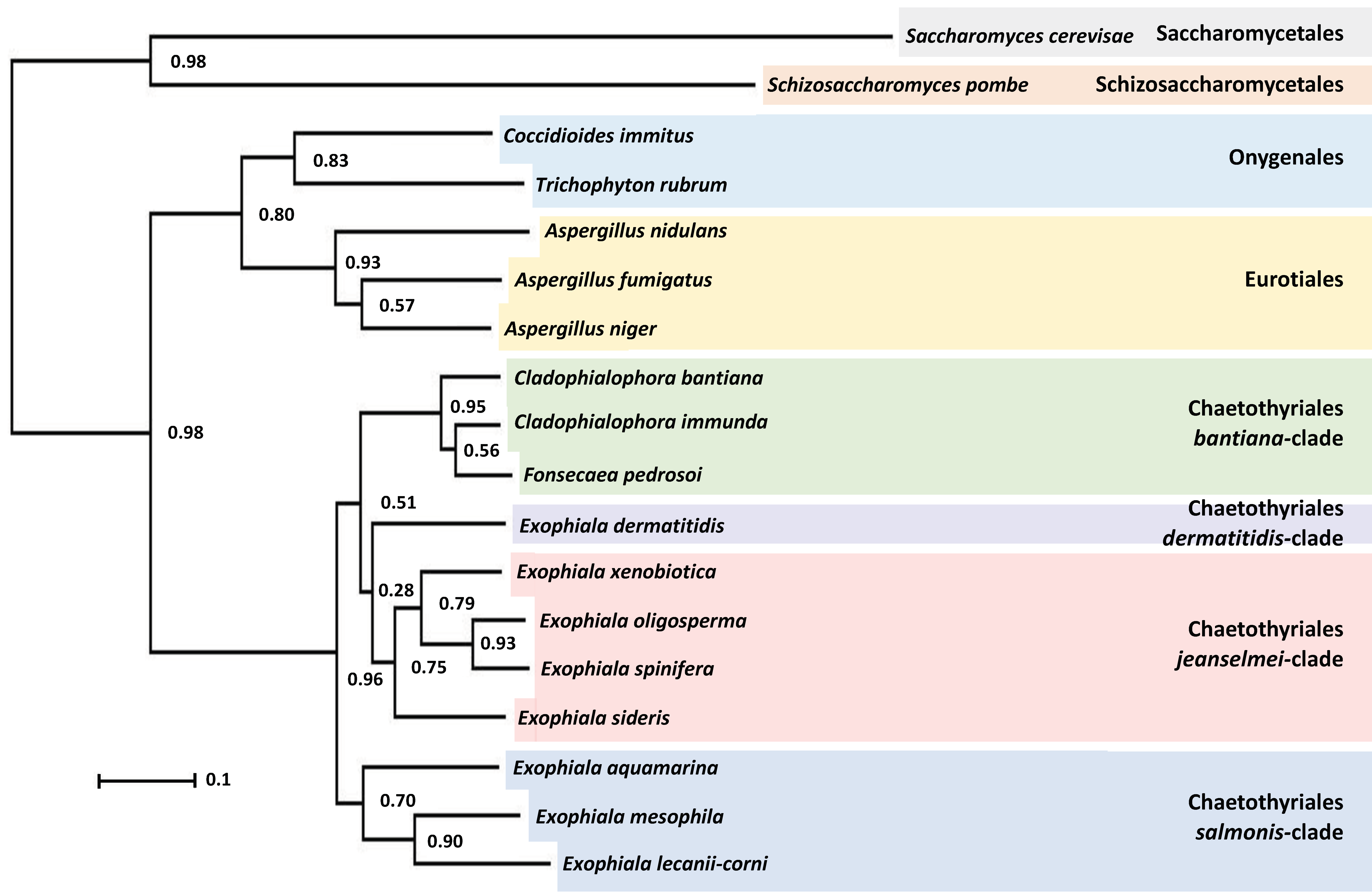
3.4. Phylogenetic Characterization of E. lecanii-corni

To obtain a phylogenetic framework for the comparative genomic analyses, we assessed the phylogenetic relationship of E. lecanii-corni relative to other Chaetothyriales, including those within the bantiana-clade (Cladophialophora bantiana, Cladophialophora immunda, Fonsecaea pedrosoi), the dermatitidis-clade (E. dermatitidis), the jeanselmei-clade (Exophiala xenobiotica, Exophiala oligoosperma, Exophiala spinifera, Exophiala sideris), and the salmonis-clade (Exophiala aquamarina, Exophiala mesophila) [5]. For comparative purposes, we also included three well-characterized Eurotiales (Aspergillus nidulans, Aspergillus fumigatus, Aspergillus niger), two Onygenales (Coccidioides immitis, Trichophyton rubrum), and two other well-studied ascomycete models (Saccharomyces cerevisiae, Schizosaccharomyces pombe). For all the species listed above, OrthoFinder was used to identify a total of 13,838 orthogroups that represented approximately 94% of genes in the genomes (Supplemental Table S1), and from that data infer a rooted species tree (Figure 5) [46,47]. This revealed that E. lecanii-corni is situated in the salmonis-clade of the order Chaetothyriales, which is generally comprised of mesophilic waterborne Exophiala species [68]. Notably, while fungal species within this clade commonly cause infection in aquatic animals, such as fish, turtles, crabs, and frogs, they rarely infect humans, as their maximal growth temperature is approximately 33 °C [68]. Within this clade, E. lecanii-corni displays the closest phylogenetic relationship to E. mesophila, a black yeast with significant bioremediation potential due to its ability to digest alkylbenzene compounds [69].

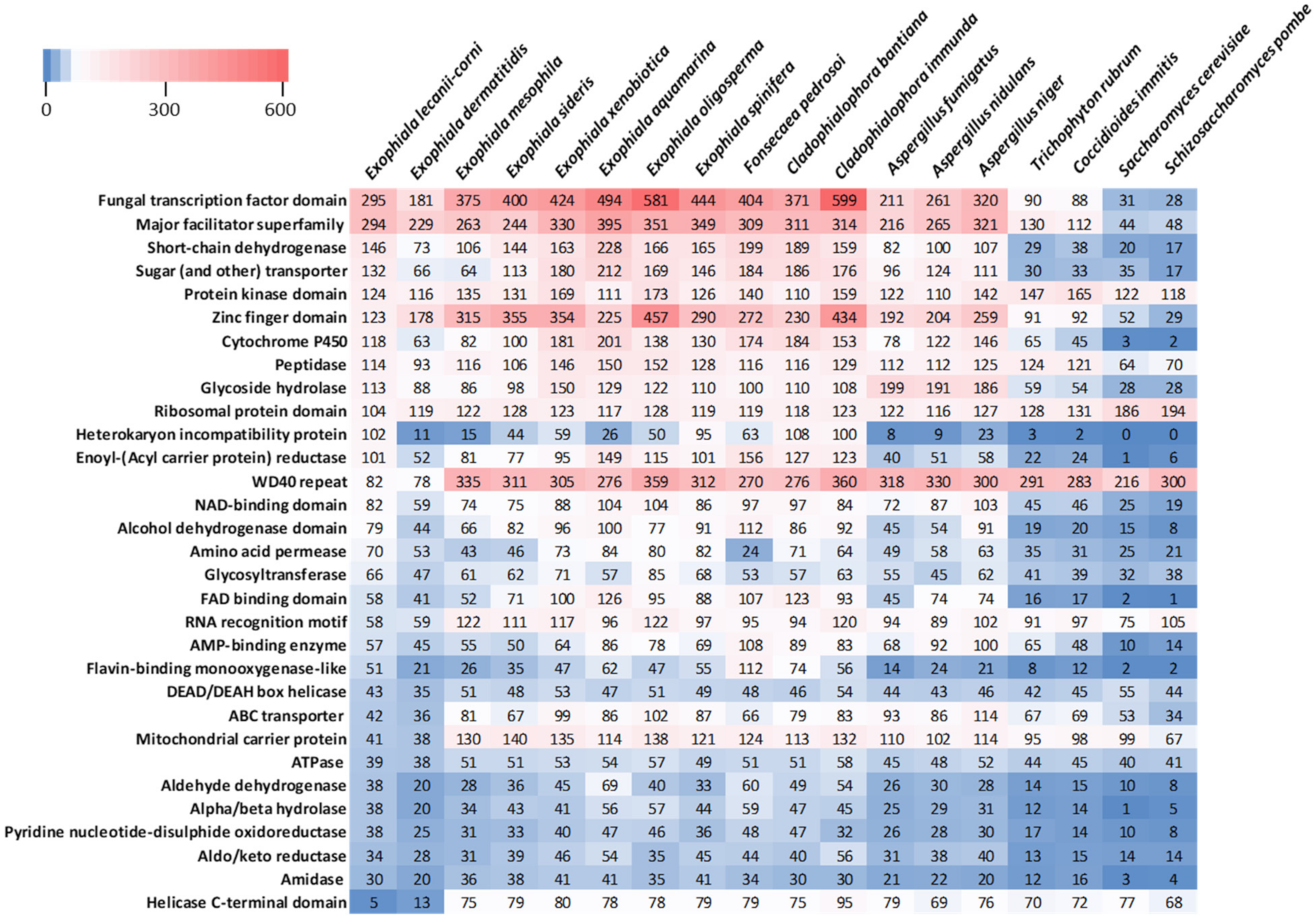
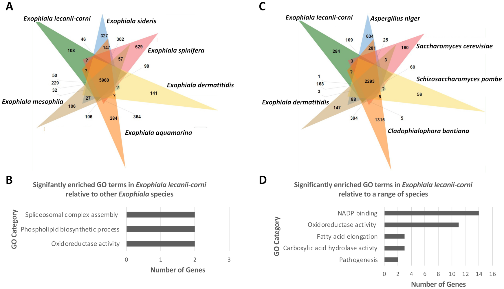
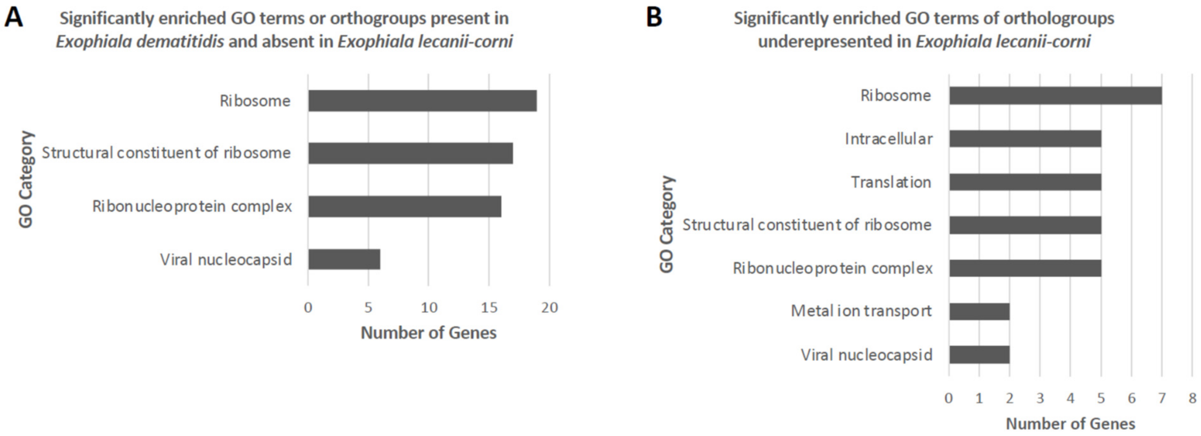
3.5. Comparative Analysis of Predicted Functional Content

Next, we examined variability in the predicted functional proteomic content of the E. lecanii-corni genome relative to the range of species included in the phylogenetic tree. To accomplish this, functional Pfam database protein families were identified for each of the species included in the analysis, and the relative occurrence of each protein domain or family was assessed (Figure 6). In the E. lecanii-corni genome, the most prevalent Pfam domains are for the categories of fungal transcription factor (TF) domain and major facilitator superfamily. However, while the genome of E. lecanii-corni harbors approximately 63% more TF domains than E. dermatitidis, it still generally has less TFs and zinc finger domains compared to other species within the Chaetothyriales order.
Relative to E. dermatitidis, E. lecanii-corni also possesses approximately twice as many short-chain dehydrogenases, sugar transporters, and cytochrome P450 domains, and most notably, encodes a relatively high number of heterokaryon incompatibility proteins compared to nearly all of the other species that were evaluated. On the other hand, the data also revealed that the genomes of E. lecanii-corni and E. dermatitidis both harbor substantially fewer WD40 repeats, RNA recognition motifs, mitochondrial carrier proteins, and helicases, and that E. lecanii-corni has an average of 20% fewer ribosomal protein domains compared to the other species included in the study (Figure 6).
To further investigate differences in the relative functional content of E. lecanii-corni, OrthoVenn2 was first used to comparatively evaluate orthologous gene clusters across species within the Exophiala genus (Figure 7A) [55]. The results indicated that relative to E. dermatitidis, E. sideris, E. spinifera, E. aquamarina, and E. mesophila, the genome of E. lecanii-corni harbors 5960 overlapping and 108 unique orthologous gene clusters. Gene Ontology (GO) enrichment analysis further revealed that among the 108 E. lecanii-corni-specific clusters, the GO categories of spliceosomal complex activity (two genes), phospholipid biosynthetic processes (two genes), and oxidoreductase activity (two genes) were significantly overrepresented (Figure 7B). When compared to a range of other fungal species, including A. niger, S. cerevisiae, S. pombe, C. bantiana, and E. dermatitidis, E. lecanii-corni was found to possess 284 unique orthologous gene clusters (Figure 7C) that feature significant overrepresentation of genes involved in NADP binding (14 genes) and oxidoreductase activity (11 genes) (Figure 7D). Furthermore, GO enrichment analysis conducted on orthogroups (Supplemental Table S1) (predicted by OrthoFinder [47]) that are absent in E. lecanii-corni but present in either E. dermatitidis (Figure 8A) or in all the species included in the previous analyses (Figure 8B) resulted in ribosomal constituents being significantly enriched, indicating that ribosomal functional activities are significantly underrepresented in E. lecanii-corni relative to a range of other ascomycetous fungal species.

3.6. Toluene Degradation Pathway Comparison

Given the potential for E. lecanii-corni to be utilized as a bioremediation agent for the degradation of volatile organic compounds [8], we next examined its genome for genes involved in toluene degradation [70]. The putative genes involved in the fungal toluene degradation pathway were previously identified in the toluene-degrading black yeast C. immunda based on sequence homology and regulation in response to toluene exposure [71]. Using pBLAST [72], we identified E. lecanii-corni homologs for all of the putative C. immunda toluene degradation pathway genes, displayed in Table 3. While many of the genes involved in the fungal toluene degradation pathway are clustered in C. immunda, this trend was substantially less pronounced in E. lecanii-corni. In C. immunda, four of the putative genes encoding benzylalcohol dehydrogenase (BADH) and (benzaldehyde dehydrogenase) BZDH enzymes are grouped in two separate clusters and seven of the genes involved in protocatechuate degradation are grouped in two additional clusters [71]. In contrast, in the E. lecanii-corni genome, only two genes, EXLC_001487T0 (carboxy-muconolactone decarboxylase (CMD)) and EXLC_001488T0 (β-carboxy-muconate lactonizing enzyme (CMLE)), were observed to be clustered.

3.7. Melanin Biosynthesis and Regulation Pathway Comparison

We next examined the genome of E. lecanii-corni for genes known to be involved in the production of melanin, as it is distinguished by its high capacity for melanin production. Most fungi produce either 1,8-dihydroxynaphthalene (DHN)-melanin or 3,4-dihydroxyphenylalanine (DOPA)-melanin, with ascomycetous fungi generally utilizing the DHN pathway and basidiomycetous fungi commonly using the DOPA pathway [73]. Biosynthesis of DHN-melanin initiates with the catalyzed formation of 1,3,6,8-tetrahydroxynaphthalene (1,3,6,8-THN) from malonyl-CoA and acetyl-CoA by a polyketide synthase (PKS). Next, a series of enzymatic reactions catalyzed by scytalone dehydratase, 1,3,6,8-tetrahydroxynaphthalene reductase, alpha/beta hydrolases, multicopper oxidases, ferroxidase led to the formation of DHN. Finally, polymerization of DHN, which may be catalyzed by a laccase, generates DHN-melanin [74,75]. We identified E. lecanii corni homologs for all the enzymes involved in melanin biosynthesis, which are displayed in Table 4 alongside E. dermatitidis homologs [27]. The genome of E. lecanii-corni harbors two homologs of the E. dermatitidis melanin-producing PKS, along with a homolog of alpha/beta hydrolase Ayg1, both of which are required for DHN-melanin biosynthesis in E. dermatitidis [76,77]. However, it was previously reported that only one of these E. lecanii-corni PKSs, EXLC_009455T0 (the gene that results in a non-melanized phenotype when deleted), is involved in melanin production, while the function of the other PKS remains unknown [10]. Similar to the E. dermatitidis genome, E. lecanii-corni possesses a single homolog of scytalone dehydratase Arp1 and alpha/beta hydrolase HMPREF1120_02312, along with two homologs laccase Abr2 and L-ascorbate oxidase. In contrast, we identified two homologs of 1,3,6,8-tetrahydroxynaphthalene reductase Arp2, relative to the single homolog present in E. dermatitidis. We also noted that ferrooxidoreductase is underrepresented in E. lecanii-corni relative to E. dermatitidis, which harbor three homologs. Lastly, similar to E. dermatitidis, the genes involved in melanin biosynthesis in E. lecanii-corni are not clustered together in the genome as they are in many other fungal species [78].
Ascomycetous fungi typically produce DHN-melanin, but several members have been reported to produce DOPA-melanin, including Aspergillus nidulans, Hypoxylon archeri, Penicillium marneffei, and Sporothrix schenckii [79,80,81,82]. Other fungi, such as the edible mushroom Auricularia auricula, have been reported to produce a mixture of DHN- and DOPA-melanin [83]. Biosynthesis of DOPA-melanin in fungi resembles human melanogenesis, initiating with the catalyzed oxidation of either DOPA to dopaquinone by laccase, or tyrosine to DOPA to dopaquinone by tyrosinase [58,74]. Although DHN-melanin is the form of melanin constitutively produced by E. lecanii-corni [10], its genome also contains genes required for the production of DOPA-melanin (Table 4). We identified five tyrosinase homologs and seven multicopper oxidases in the E. lecanii-corni genome, which is comparable to the numbers present in E. dermatitidis, as well as other melanized fungal species, such as A. niger [27].
Finally, the genes involved in the production of a third type of melanin, pyomelanin, are also conserved in E. lecanii-corni (Table 4). Pyomelanin production involves polymerization of homogentisic acid of the tyrosine degradation pathway, and is conserved among other ascomycetous fungi, including E. dermatitidis, A. fumigatus, and Penicillium chrysogenum [84,85]. More specifically, its biosynthesis involves the catalyzed formation of 4-hydroxyphenyl pyruvate from tyrosine-by-tyrosine aminotransferase Tat, followed by conversion to homogentisate by 4-hydroxyphenylpyruvate dioxygenase HppD, and subsequent oxidation and polymerization to form pyomelanin. Alternatively, homogentisate can be degraded by homogentisate dioxygenase HmgA, maleylacetoacetate isomerase MaiA, and fumarylacetoacetate hydrolase FahA to yield acetoacetate and fumarate [84]. Our analysis revealed that the genome of E. lecanii-corni harbors single homologs of Tat, HppD, and MaiA, along with six and three homologs of HmgA and FahA, respectively, which is in contrast to the single homolog present in E. dermatitidis (Table 4).

3.8. Potential for Secondary Metabolite Production

Many fungal species are potent producers of secondary metabolites (SMs), which are small bioactive molecules that confer selective advantage while not being directly required for survival [86]. These small molecules possess a broad range of bioactivities that have resulted in their widespread use in medicine and industry [87,88]. The genes involved in the biosynthesis of SMs are typically clustered together in the genome and include a core biosynthesis enzyme, such as a PKS or PKS-like enzyme, a nonribosomal peptide synthetase (NRPS) or NRPS-like enzyme, or a terpene cyclase (TC), along with various tailoring enzymes. To evaluate the relative potential for SM production in E. lecanii-corni, we identified core SM biosynthesis enzymes in all species included in this study using antiSMASH [57]. This resulted in the identification of five PKS or PKS-like enzymes, seven NRPS of NRPS-like enzymes, and three TCs, or a total of fifteen SM core enzymes in the genome of E. lecanii-corni (Table 5). While this number is significantly higher than the number of SMs produced by some yeast species, such as S. cerevisiae and S. pombe, and comparable to other Exophiala species, it is considerably less than other ascomycetous fungi, including many within the Aspergillus genus [89] (Table 5). Of the fifteen SM core synthase enzymes identified in E. lecanii corni, only four possessed a high percentage of genes that have significant BLAST hits to genes present in characterized clusters with known SM products [57]. These clusters are involved in the production of nonribosomal peptides dimethylcoprogen (100% cluster similarity) and acetylaranotin (80% cluster similarity), the terpene clavaric acid (100% cluster similarity), and the polyketide 1,3,6,8-tetrahydroxynaphthalene involved in DHN-melanin production, indicating that these SM biosynthesis pathways are conserved in E. lecanii-corni.

4. Discussion

In this study, we conducted a phenotypic and comparative genomic analysis of the melanin-producing yeast E. lecanii-corni to acquire insight into the biological basis for differences in stress tolerance and pathogenicity among black yeast. Toward this goal, we have successfully identified key differences in stress tolerance of E. lecanii-corni relative to the well-characterized E. dermatitidis, as well as the impact of melanin on these phenotypes. Further, we uncovered variability in genetic functional content that may explain some observed differences in extremophilic characteristics. These studies have contributed to our understanding of the biology of black yeast and provide a framework for targeted functional studies that will advance our understanding of this unique organism.
Our investigation revealed that the presence of melanin significantly increased the resistance of E. lecanii-corni to acute doses of UV-C and γ-radiation. This finding was expected for UV-C radiation, as the capacity of melanin to absorb radiation at a broad range of wavelengths within the UV and visible electromagnetic spectrum is well understood [21], and several studies have consistently found that melanin confers protection against UV radiation in fungi [61,62,63,64]. On the other hand, the involvement of melanin in resistance to ionizing radiation is less clear, which makes our finding, that melanin confers protection against γ-radiation as well, particularly notable. While some studies have observed a protective effect of melanin against ionizing radiation, those findings have been mixed, with any impact often being marginal [90,91] or entirely absent [6,64,92]. Other investigations have revealed a more complicated role for melanin in the fungal response to ionizing radiation. For example, one study found that in E. dermatitidis, the presence of melanin altered the regulation of specific types of genes, including those encoding transporters and ribosomal biosynthesis enzymes, in response to low-dose ionizing radiation [93]. In contrast, a different study that examined the response of Cryptococcus neoformans to ionizing radiation found that melanin did not have a substantive impact on gene expression patterns [92]. Together, these findings suggest that the impact of melanin on fungal radiation resistance is likely species-specific and can depend on a range of other factors, including total dose, dose rate, the physiological conditions of the fungi prior to exposure, and the distinct physico-chemical properties of the melanin variant being produced. Further, it is unclear the extent to which the presence or absence of melanin alters other physiological parameters that can impact the ability of fungi to survive and recover from ionizing radiation exposure. Since the presence of melanin on gene expression appears to vary substantively between organisms [6,92], studies that aim to quantify the true ionizing radiation protective properties of melanin should consider conducting such experiments outside the constraints of biological systems, which can convolute the findings. Still, the effect is substantial for E. lecanii-corni melanin and should be explored further.
Similarly, our study also revealed that the impact of melanin on tolerance to other forms of stress can also vary drastically, even among closely related species. For example, we observed that while melanin deficiency does not alter oxidative stress resistance in E. dermatitidis, E. lecanii-corni strains lacking melanin production are substantially more resistant to oxidative stress due to H2O2 than their melanized counterparts. Although the antioxidative properties of melanin make this finding somewhat counterintuitive, it may be explained by the fact that melanin production represents a significant metabolic cost to the cell. It is therefore possible that the saved energy expenditures from lack of melanin production can be redirected to activate biological pathways that are critical for ionizing radiation resistance, such as those involved in oxidative stress protection and DNA repair. This phenomenon may be more pronounced in E. lecanii-corni than in E. dermatitidis because E. lecanii-corni produces a relatively larger quantity of melanin, and is therefore, burdened by a higher metabolic load. In contrast, melanin was found to enhance resistance to osmotic stress in E. lecanii-corni, which is in agreement with other studies that have examined the impact of cell wall DHN-melanin on osmotic stress resistance in other ascomycetous fungi [11]. Rigid structure and high-density melanin nanoparticles enriched in cell wall as demonstrated in the melanin ghosts (Figure 2) may contribute to resistance of melanized cells to osmotic stress. Interestingly, no such impact was observed in E. dermatitidis, which also may be due to its decreased abundance of cell wall melanin relative to E. lecanii-corni.
We were particularly interested in understanding the biological basis underlying the reduced human pathogenicity of E. lecanii-corni, as this characteristic makes this organism attractive for functional laboratory studies or production host capabilities in industry. In order for a fungus to successfully invade the human body, it must be capable of thriving in a range of extreme environments, including the high temperatures associated with vertebrate endothermy [94], low pH and water if penetration occurs through the skin [95], and reactive oxygen species produced by phagocytes [96]. We observed that E. lecanii-corni CBS 102400 is entirely unable to grow at the physiological temperature of 37 °C, which distinguishes it from other Exophiala species, most of which grow at 37–40 °C [68]. This is considered to be a key virulence factor within the order Chaetothyriales [1], and helps to explain why this strain of E. lecanii-corni was categorized as BSL1. Notably, other Exophiala species that lack thermotolerance have been isolated, and many of these isolates are capable of causing disease in cold-blooded animals [68].
This investigation revealed distinct differences in functional genomic content that may play a role in stress tolerance variation. In particular, our most statistically significant observation was that the genome of E. lecanii-corni exhibited an approximate 20% reduction in genes encoding ribosomal proteins relative to the other ascomycetous species included in this study. Ribosomal proteins are the structural constituents of ribosomes, which are the cellular machinery responsible for the translation of mRNA to protein synthesis. Ribosomal gene expression is closely correlated with fungal growth and proliferation, as species harboring higher ribosome content typically exhibit increased growth rates [97,98]. Accordingly, we observed that E. lecanii-corni exhibits a slower growth rate than other black yeast, such as E. dermatitidis (data not shown). Interestingly, several studies have indicated that some ribosomal proteins have additional functions that are independent of their primary role of protein biosynthesis, also referred to as “moonlighting”, that can include regulation of cell proliferation, induction of apoptosis, cell signaling, and DNA repair [99,100]. More recently, the moonlighting functions of ribosomal proteins in fungi have been extended to include the conferral of tolerance to various forms of abiotic stress, including those imposed by salt, drought, and heavy metals [101]. In a similar vein, we previously observed that increased levels of ribosomal proteins were associated with the acquisition of ionizing radiation resistance in the black yeast E. dermatitidis [29]. This may be due to the fact that some proteins required for cellular recovery from radiation exposure are not synthesized until after exposure [6], and therefore, a strain with higher ribosomal content will have an increased capacity to maintain synthesis of key proteins required for survival following irradiation. On the other hand, this observation may also be another example of a novel ribosomal protein moonlighting function that involved the conferral of resistance to ionizing radiation. Either way, there is sufficient evidence to suggest that the significantly reduced ribosomal protein content in the genome of E. lecanii-corni may contribute to differences in its stress tolerance phenotype, including increased susceptibility to osmotic stress and ionizing radiation, which will be a topic of investigation in future studies.
Given the reduced pathogenicity of E. lecanii-corni CBS 102400 relative to other black yeast, a goal of this study was to provide a genetic framework that will advance the utilization of E. lecanii-corni for biotechnological applications. Toward this goal, we identified E. lecanii-corni homologues for the 13 putative genes belonging to the toluene degradation pathway in fungi. Notably, E. lecanii-corni has demonstrated bioremediation potential, as it is capable of using toluene as its sole carbon source and can eliminate toluene at a rate that is two to seven times greater than those reported for bacterial biofiltration systems [8]. This is a significant finding, as the capacity to degrade toluene is quite rare in fungi and is often strain-specific rather than species-specific [102,103]. Future efforts that aim to further develop E. lecanii-corni for bioremediation applications, including the degradation of volatile organic compounds, should consider targeting the putative toluene degradation pathway genes identified in this study.
Finally, this study illuminated the suitability for E. lecanii-corni to be utilized as a melanin production host for biotechnological applications. As expected, we identified homologs for all the genes involved in the biosynthesis of DHN-melanin, and in addition, we found that E. lecanii-corni also harbors the genes required for production of DOPA-melanin and pyomelanin. Similar to E. dermatitidis, the genes involved in DHN-melanin production are not clustered within the E. lecanii-corni genome as they are in other fungal species, reiterating previous findings that the coordinated regulation required for abundant melanin production can be achieved without gene clustering. DHN-melanin production was maximized when E. lecanii-corni was cultured in malt-based media, with a production yield of approximately 3.2 g/L, which represents a remarkable production rate. A number of native and recombinant microorganisms have been reported to produce melanin at relatively high yields, but they all produce eumelanin (DOPA-melanin) and require supplying tyrosine as the precursor in growth media [104]. There still remains significant potential for enhancing melanin production efficiency in E. lecanii-corni, either through optimization of fermentation conditions or engineering of biosynthetic pathways [105]. Future endeavors that require large quantities of DHN-melanin for characterization studies or other biotechnological applications should consider the utilization and development of E. lecanii-corni for such purposes.

5. Conclusions

Taken together, this study highlights the effectiveness of combining phenotypic characterization with comparative genomic analysis to identify candidate genetic signatures that may contribute to a particular stress tolerance phenotype in fungi. Using this approach, we have uncovered the significant underrepresentation of ribosomal genes within the E. lecanii-corni genome, providing insight into the biological basis for its distinct stress tolerance profile, as well as a starting point for future studies investigating how ribosomal content alters the characteristics of a particular organism. Further, we have provided a detailed account of the functional genomic content within the genome of E. lecanii-corni, paving the way for targeted functional studies and its use as a production host for melanin or other useful biomolecules.

Supplementary Materials

The following are available online at https://www.mdpi.com/article/10.3390/jof7121078/s1, Supplemental Table S1. Orthologous gene clusters identified in this study, as predicted using OrthoFinder.

Author Contributions

Conceptualization, J.R. and Z.W.; methodology, J.R., Z.S., C.A.C., H.D.; software, J.R. and C.A.C. and W.J.H.IV; formal analysis, J.R., Z.S. and Z.W.; investigation, J.R., Z.S., H.D., H.A.-P. and Z.W.; data curation, J.R., Z.S. and C.A.C.; writing—original draft preparation, J.R. and Z.W.; writing—review and editing, Z.S., C.A.C.; supervision, Z.W.; project administration, Z.W.; funding acquisition, Z.W. All authors have read and agreed to the published version of the manuscript.

Funding

This work was supported by the Defense Threat Reduction Agency Grant HDTRA-17-1-002 and NASA Grant NNK190B09A.

Institutional Review Board Statement

Not applicable.

Data Availability Statement

The raw genome sequencing reads for E. lecanii-corni CBS 102400 are available in NCBI SRA under Accession Number SRX5145602, as previously described [33].

Acknowledgments

We thank the Department of Defense’s High Performance Computing Modernization Program (HPCMP) for the allocation of computational resources used in this work to J.H. The opinions and assertions contained herein are those of the authors and are not to be construed as those of the U.S. Navy, military service at large, or the U.S. Government.

Conflicts of Interest

The authors declare no conflict of interest.

References

  1. Seyedmousavi, S.; Netea, M.G.; Mouton, J.W.; Melchers, W.J.G.; Verweij, P.E.; de Hoog, G.S. Black Yeasts and Their Filamentous Relatives: Principles of Pathogenesis and Host Defense. Clin. Microbiol. Rev. 2014, 27, 527–542. [Google Scholar] [CrossRef] [PubMed] [Green Version]
  2. Onofri, S.; Barreca, D.; Selbmann, L.; Isola, D.; Rabbow, E.; Horneck, G.; de Vera, J.P.P.; Hatton, J.; Zucconi, L. Resistance of Antarctic Black Fungi and Cryptoendolithic Communities to Simulated Space and Martian Conditions. Stud. Mycol. 2008, 61, 99–109. [Google Scholar] [CrossRef] [PubMed]
  3. Plemenitaš, A.; Vaupotič, T.; Lenassi, M.; Kogej, T.; Gunde-Cimerman, N. Adaptation of Extremely Halotolerant Black Yeast Hortaea Werneckii to Increased Osmolarity: A Molecular Perspective at a Glance. Stud. Mycol. 2008, 61, 67–75. [Google Scholar] [CrossRef] [PubMed]
  4. Selbmann, L.; Zucconi, L.; Isola, D.; Onofri, S. Rock Black Fungi: Excellence in the Extremes, from the Antarctic to Space. Curr. Genet. 2015, 61, 335–345. [Google Scholar] [CrossRef]
  5. Teixeira, M.M.; Moreno, L.F.; Stielow, B.J.; Muszewska, A.; Hainaut, M.; Gonzaga, L.; Abouelleil, A.; Patané, J.S.L.; Priest, M.; Souza, R.; et al. Exploring the Genomic Diversity of Black Yeasts and Relatives (Chaetothyriales, Ascomycota). Stud. Mycol. 2017, 86, 1–28. [Google Scholar] [CrossRef] [Green Version]
  6. Schultzhaus, Z.; Romsdahl, J.; Chen, A.; Tschirhart, T.; Kim, S.; Leary, D.; Wang, Z. The Response of the Melanized Yeast Exophiala Dermatitidis to Gamma Radiation Exposure. Environ. Microbiol. 2020, 22, 1310–1326. [Google Scholar] [CrossRef]
  7. Zeng, J.S.; Sutton, D.A.; Fothergill, A.W.; Rinaldi, M.G.; Harrak, M.J.; de Hoog, G.S. Spectrum of Clinically Relevant Exophiala Species in the United States. J. Clin. Microbiol. 2007, 45, 3713–3720. [Google Scholar] [CrossRef] [Green Version]
  8. Woertz, J.R.; Kinney, K.A.; McIntosh, N.D.P.; Szaniszlo, P.J. Removal of Toluene in a Vapor-Phase Bioreactor Containing a Strain of the Dimorphic Black YeastExophiala Lecanii-Corni. Biotechnol. Bioeng. 2001, 75, 550–558. [Google Scholar] [CrossRef]
  9. Qi, W.; Moe, K.; Kinney, B. Biodegradation of Volatile Organic Compounds by Five Fungal Species. Appl. Microbiol. Biotechnol. 2002, 58, 684–689. [Google Scholar] [CrossRef]
  10. Cheng, Q.; Kinney, K.A.; Whitman, C.P.; Szaniszlo, P.J. Characterization of Two Polyketide Synthase Genes in Exophiala Lecanii-Corni, a Melanized Fungus with Bioremediation Potential. Bioorg. Chem. 2004, 32, 92–108. [Google Scholar] [CrossRef]
  11. Fernandez, C.W.; Koide, R.T. The Function of Melanin in the Ectomycorrhizal Fungus Cenococcum Geophilum under Water Stress. Fungal Ecol. 2013, 6, 479–486. [Google Scholar] [CrossRef]
  12. Cordero, R.J.B.; Casadevall, A. Functions of Fungal Melanin beyond Virulence. Fungal Biol. Rev. 2017, 31, 99–112. [Google Scholar] [CrossRef]
  13. Cordero, R.J.B.; Robert, V.; Cardinali, G.; Arinze, E.S.; Thon, S.M.; Casadevall, A. Impact of Yeast Pigmentation on Heat Capture and Latitudinal Distribution. Curr. Biol. 2018, 28, 2657–2664.e3. [Google Scholar] [CrossRef] [Green Version]
  14. Pihet, M.; Vandeputte, P.; Tronchin, G.; Renier, G.; Saulnier, P.; Georgeault, S.; Mallet, R.; Chabasse, D.; Symoens, F.; Bouchara, J.-P. Melanin Is an Essential Component for the Integrity of the Cell Wall of Aspergillus Fumigatus Conidia. BMC Microbiol. 2009, 9, 177. [Google Scholar] [CrossRef]
  15. Bashyal, B.M.; Chand, R.; Kushwaha, C.; Sen, D.; Prasad, L.C.; Joshi, A.K. Association of Melanin Content with Conidiogenesis in Bipolaris Sorokiniana of Barley (Hordeum Vulgare L.). World J. Microbiol. Biotechnol. 2010, 26, 309–316. [Google Scholar] [CrossRef]
  16. Nosanchuk, J.D.; Casadevall, A. The Contribution of Melanin to Microbial Pathogenesis. Cell Microbiol. 2003, 5, 203–223. [Google Scholar] [CrossRef]
  17. Revankar, S.G.; Sutton, D.A. Melanized Fungi in Human Disease. CMR 2010, 23, 884–928. [Google Scholar] [CrossRef] [Green Version]
  18. d’Ischia, M.; Napolitano, A.; Ball, V.; Chen, C.-T.; Buehler, M.J. Polydopamine and Eumelanin: From Structure–Property Relationships to a Unified Tailoring Strategy. Acc. Chem. Res. 2014, 47, 3541–3550. [Google Scholar] [CrossRef]
  19. Ju, K.-Y.; Lee, Y.; Lee, S.; Park, S.B.; Lee, J.-K. Bioinspired Polymerization of Dopamine to Generate Melanin-Like Nanoparticles Having an Excellent Free-Radical-Scavenging Property. Biomacromolecules 2011, 12, 625–632. [Google Scholar] [CrossRef]
  20. Le Na, N.T.; Duc Loc, S.; Minh Tri, N.L.; Bich Loan, N.T.; Anh Son, H.; Linh Toan, N.; Phuong Thu, H.; My Nhung, H.T.; Lai Thanh, N.; Van Anh, N.T.; et al. Nanomelanin Potentially Protects the Spleen from Radiotherapy-Associated Damage and Enhances Immunoactivity in Tumor-Bearing Mice. Materials 2019, 12, 1725. [Google Scholar] [CrossRef] [Green Version]
  21. Meredith, P.; Sarna, T. The Physical and Chemical Properties of Eumelanin. Pigment Cell Res. 2006, 19, 572–594. [Google Scholar] [CrossRef]
  22. Bothma, J.P.; de Boor, J.; Divakar, U.; Schwenn, P.E.; Meredith, P. Device-Quality Electrically Conducting Melanin Thin Films. Adv. Mater. 2008, 20, 3539–3542. [Google Scholar] [CrossRef]
  23. Karlsson, O.; Lindquist, N.G. Melanin and Neuromelanin Binding of Drugs and Chemicals: Toxicological Implications. Arch. Toxicol. 2016, 90, 1883–1891. [Google Scholar] [CrossRef]
  24. Tran-Ly, A.N.; Ribera, J.; Schwarze, F.W.M.R.; Brunelli, M.; Fortunato, G. Fungal Melanin-Based Electrospun Membranes for Heavy Metal Detoxification of Water. Sustain. Mater. Technol. 2020, 23, e00146. [Google Scholar] [CrossRef]
  25. Pombeiro-Sponchiado, S.R.; Sousa, G.S.; Andrade, J.C.R.; Lisboa, H.F.; Gonçalves, R.C.R. Production of Melanin Pigment by Fungi and Its Biotechnological Applications. In Melanin; Blumenberg, M., Ed.; InTech: London, UK, 2017; ISBN 978-953-51-2979-0. [Google Scholar]
  26. Tran-Ly, A.N.; Reyes, C.; Schwarze, F.W.M.R.; Ribera, J. Microbial Production of Melanin and Its Various Applications. World J. Microbiol. Biotechnol. 2020, 36, 170. [Google Scholar] [CrossRef]
  27. Chen, Z.; Martinez, D.A.; Gujja, S.; Sykes, S.M.; Zeng, Q.; Szaniszlo, P.J.; Wang, Z.; Cuomo, C.A. Comparative Genomic and Transcriptomic Analysis of Wangiella Dermatitidis, A Major Cause of Phaeohyphomycosis and a Model Black Yeast Human Pathogen. G3 Genes|Genomes|Genet. 2014, 4, 561–578. [Google Scholar] [CrossRef] [Green Version]
  28. Romsdahl, J.; Schultzhaus, Z.; Chen, A.; Liu, J.; Ewing, A.; Hervey, J.; Wang, Z. Adaptive Evolution of a Melanized Fungus Reveals Robust Augmentation of Radiation Resistance by Abrogating Non-Homologous End-Joining. Environ. Microbiol. 2020, 23, 3627–3645. [Google Scholar] [CrossRef]
  29. Schultzhaus, Z.; Schultzhaus, J.N.; Romsdahl, J.; Chen, A.; Hervey IV, W.J.; Leary, D.H.; Wang, Z. Proteomics Reveals Distinct Changes Associated with Increased Gamma Radiation Resistance in the Black Yeast Exophiala Dermatitidis. Genes 2020, 11, 1128. [Google Scholar] [CrossRef]
  30. Malo, M.E.; Schultzhaus, Z.; Frank, C.; Romsdahl, J.; Wang, Z.; Dadachova, E. Transcriptomic and Genomic Changes Associated with Radioadaptation in Exophiala Dermatitidis. Comput. Struct. Biotechnol. J. 2021, 19, 196–205. [Google Scholar] [CrossRef] [PubMed]
  31. Dadachova, E.; Bryan, R.A.; Huang, X.; Moadel, T.; Schweitzer, A.D.; Aisen, P.; Nosanchuk, J.D.; Casadevall, A. Ionizing Radiation Changes the Electronic Properties of Melanin and Enhances the Growth of Melanized Fungi. PLoS ONE 2007, 2, e457. [Google Scholar] [CrossRef] [PubMed] [Green Version]
  32. Yu, J.-H.; Hamari, Z.; Han, K.-H.; Seo, J.-A.; Reyes-Domínguez, Y.; Scazzocchio, C. Double-Joint PCR: A PCR-Based Molecular Tool for Gene Manipulations in Filamentous Fungi. Fungal Genet. Biol. 2004, 41, 973–981. [Google Scholar] [CrossRef] [PubMed]
  33. Schultzhaus, Z.; Cuomo, C.A.; Wang, Z. Genome Sequence of the Black Yeast Exophiala Lecanii-Corni. Microbiol. Resour. Announc. 2019, 8, 2. [Google Scholar] [CrossRef] [PubMed] [Green Version]
  34. Hoff, K.J.; Lange, S.; Lomsadze, A.; Borodovsky, M.; Stanke, M. BRAKER1: Unsupervised RNA-Seq-Based Genome Annotation with GeneMark-ET and AUGUSTUS. Bioinformatics 2016, 32, 767–769. [Google Scholar] [CrossRef] [PubMed]
  35. Hoff, K.J.; Lomsadze, A.; Borodovsky, M.; Stanke, M. Whole-Genome Annotation with BRAKER. In Gene Prediction; Kollmar, M., Ed.; Springer: New York, NY, USA, 2019; Volume 1962, pp. 65–95. ISBN 978-1-4939-9172-3. [Google Scholar]
  36. Lowe, T.M.; Eddy, S.R. TRNAscan-SE: A Program for Improved Detection of Transfer RNA Genes in Genomic Sequence. Nucleic Acids Res. 1997, 25, 955–964. [Google Scholar] [CrossRef]
  37. Lagesen, K.; Hallin, P.; Rødland, E.A.; Stærfeldt, H.-H.; Rognes, T.; Ussery, D.W. RNAmmer: Consistent and Rapid Annotation of Ribosomal RNA Genes. Nucleic Acids Res. 2007, 35, 3100–3108. [Google Scholar] [CrossRef]
  38. Coleine, C.; Selbmann, L.; Masonjones, S.; Onofri, S.; Zucconi, L.; Stajich, J.E. Draft Genome Sequence of an Antarctic Isolate of the Black Yeast Fungus Exophiala Mesophila. Microbiol. Resour. Announc. 2019, 8, e00142-19. [Google Scholar] [CrossRef] [Green Version]
  39. Nierman, W.C.; Pain, A.; Anderson, M.J.; Wortman, J.R.; Kim, H.S.; Arroyo, J.; Berriman, M.; Abe, K.; Archer, D.B.; Bermejo, C.; et al. Genomic Sequence of the Pathogenic and Allergenic Filamentous Fungus Aspergillus Fumigatus. Nature 2005, 438, 1151–1156. [Google Scholar] [CrossRef]
  40. Galagan, J.E.; Calvo, S.E.; Cuomo, C.; Ma, L.-J.; Wortman, J.R.; Batzoglou, S.; Lee, S.-I.; Baştürkmen, M.; Spevak, C.C.; Clutterbuck, J.; et al. Sequencing of Aspergillus Nidulans and Comparative Analysis with A. Fumigatus and A. Oryzae. Nature 2005, 438, 1105–1115. [Google Scholar] [CrossRef]
  41. Pel, H.J.; de Winde, J.H.; Archer, D.B.; Dyer, P.S.; Hofmann, G.; Schaap, P.J.; Turner, G.; de Vries, R.P.; Albang, R.; Albermann, K.; et al. Genome Sequencing and Analysis of the Versatile Cell Factory Aspergillus Niger CBS 513.88. Nat. Biotechnol. 2007, 25, 11. [Google Scholar] [CrossRef] [Green Version]
  42. Martinez, D.A.; Oliver, B.G.; Gräser, Y.; Goldberg, J.M.; Li, W.; Martinez-Rossi, N.M.; Monod, M.; Shelest, E.; Barton, R.C.; Birch, E.; et al. Comparative Genome Analysis of Trichophyton Rubrum and Related Dermatophytes Reveals Candidate Genes Involved in Infection. mBio 2012, 3, e00259-12. [Google Scholar] [CrossRef] [Green Version]
  43. Sharpton, T.J.; Stajich, J.E.; Rounsley, S.D.; Gardner, M.J.; Wortman, J.R.; Jordar, V.S.; Maiti, R.; Kodira, C.D.; Neafsey, D.E.; Zeng, Q.; et al. Comparative Genomic Analyses of the Human Fungal Pathogens Coccidioides and Their Relatives. Genome Res. 2009, 19, 1722–1731. [Google Scholar] [CrossRef] [Green Version]
  44. Goffeau, A.; Barrell, B.G.; Bussey, H.; Davis, R.W.; Dujon, B.; Feldmann, H.; Galibert, F.; Hoheisel, J.D.; Jacq, C.; Johnston, M.; et al. Life with 6000 Genes. Science 1996, 274, 546–567. [Google Scholar] [CrossRef] [Green Version]
  45. Wood, V.; Gwilliam, R.; Rajandream, M.-A.; Lyne, M.; Lyne, R.; Stewart, A.; Sgouros, J.; Peat, N.; Hayles, J.; Baker, S.; et al. The Genome Sequence of Schizosaccharomyces Pombe. Nature 2002, 415, 871–880. [Google Scholar] [CrossRef] [Green Version]
  46. Emms, D.M.; Kelly, S. OrthoFinder: Solving Fundamental Biases in Whole Genome Comparisons Dramatically Improves Orthogroup Inference Accuracy. Genome Biol. 2015, 16, 157. [Google Scholar] [CrossRef] [Green Version]
  47. Emms, D.M.; Kelly, S. OrthoFinder: Phylogenetic Orthology Inference for Comparative Genomics. Genome Biol. 2019, 20, 238. [Google Scholar] [CrossRef] [Green Version]
  48. Kelly, S.; Maini, P.K. DendroBLAST: Approximate Phylogenetic Trees in the Absence of Multiple Sequence Alignments. PLoS ONE 2013, 8, e58537. [Google Scholar] [CrossRef] [Green Version]
  49. Emms, D.M.; Kelly, S. STAG: Species Tree Inference from All Genes. BioRxiv 2018, 267914. [Google Scholar]
  50. Emms, D.M.; Kelly, S. STRIDE: Species Tree Root Inference from Gene Duplication Events. Mol. Biol. Evol. 2017, 34, 3267–3278. [Google Scholar] [CrossRef]
  51. Smit, A.; Hubley, R.; Green, P. RepeatModeler Open-1.0 (2008–2015). Institute for Systems Biology: Seattle, WA, USA, 2015. Available online: www.repeatmasker.org (accessed on 20 November 2020).
  52. Smit, A.; Hubley, R.; Green, P. RepeatMasker Open-4.0. 2013–2015. 2015. Available online: www.repeatmasker.org (accessed on 20 November 2020).
  53. Bao, W.; Kojima, K.K.; Kohany, O. Repbase Update, a Database of Repetitive Elements in Eukaryotic Genomes. Mob. DNA 2015, 6, 11. [Google Scholar] [CrossRef] [Green Version]
  54. Mistry, J.; Chuguransky, S.; Williams, L.; Qureshi, M.; Salazar, G.A.; Sonnhammer, E.L.L.; Tosatto, S.C.E.; Paladin, L.; Raj, S.; Richardson, L.J.; et al. Pfam: The Protein Families Database in 2021. Nucleic Acids Res. 2021, 49, D412–D419. [Google Scholar] [CrossRef]
  55. Xu, L.; Dong, Z.; Fang, L.; Luo, Y.; Wei, Z.; Guo, H.; Zhang, G.; Gu, Y.Q.; Coleman-Derr, D.; Xia, Q.; et al. OrthoVenn2: A Web Server for Whole-Genome Comparison and Annotation of Orthologous Clusters across Multiple Species. Nucleic Acids Res. 2019, 47, W52–W58. [Google Scholar] [CrossRef] [PubMed] [Green Version]
  56. Priebe, S.; Kreisel, C.; Horn, F.; Guthke, R.; Linde, J. FungiFun2: A Comprehensive Online Resource for Systematic Analysis of Gene Lists from Fungal Species. Bioinformatics 2015, 31, 445–446. [Google Scholar] [CrossRef] [PubMed] [Green Version]
  57. Blin, K.; Shaw, S.; Steinke, K.; Villebro, R.; Ziemert, N.; Lee, S.Y.; Medema, M.H.; Weber, T. AntiSMASH 5.0: Updates to the Secondary Metabolite Genome Mining Pipeline. Nucleic Acids Res. 2019, 47, W81–W87. [Google Scholar] [CrossRef] [PubMed] [Green Version]
  58. Eisenman, H.C.; Casadevall, A. Synthesis and Assembly of Fungal Melanin. Appl. Microbiol. Biotechnol. 2012, 93, 931–940. [Google Scholar] [CrossRef] [Green Version]
  59. Wang, Y.; Aisen, P.; Casadevall, A. Melanin, Melanin “Ghosts,” and Melanin Composition in Cryptococcus Neoformans. Infect. Immun. 1996, 64, 2420–2424. [Google Scholar] [CrossRef] [Green Version]
  60. Kim, E.; Kang, M.; Tschirhart, T.; Malo, M.; Dadachova, E.; Cao, G.; Yin, J.-J.; Bentley, W.E.; Wang, Z.; Payne, G.F. Spectroelectrochemical Reverse Engineering DemonstratesThat Melanin’s Redox and Radical Scavenging Activities Are Linked. Biomacromolecules 2017, 18, 4084–4098. [Google Scholar] [CrossRef]
  61. Wang, Y.; Casadevall, A. Decreased Susceptibility of Melanized Cryptococcus Neoformans to UV Light. Appl. Environ. Microbiol. 1994, 60, 3864–3866. [Google Scholar] [CrossRef] [Green Version]
  62. Shang, Y.; Duan, Z.; Huang, W.; Gao, Q.; Wang, C. Improving UV Resistance and Virulence of Beauveria Bassiana by Genetic Engineering with an Exogenous Tyrosinase Gene. J. Invertebr. Pathol. 2012, 109, 105–109. [Google Scholar] [CrossRef]
  63. Blachowicz, A.; Raffa, N.; Bok, J.W.; Choera, T.; Knox, B.; Lim, F.Y.; Huttenlocher, A.; Wang, C.C.C.; Venkateswaran, K.; Keller, N.P. Contributions of Spore Secondary Metabolites to UV-C Protection and Virulence Vary in Different Aspergillus Fumigatus Strains. mBio 2020, 11, e03415-19. [Google Scholar] [CrossRef] [Green Version]
  64. Cortesão, M.; de Haas, A.; Unterbusch, R.; Fujimori, A.; Schütze, T.; Meyer, V.; Moeller, R. Aspergillus Niger Spores Are Highly Resistant to Space Radiation. Front. Microbiol. 2020, 11, 560. [Google Scholar] [CrossRef] [Green Version]
  65. Maity, J.P.; Kar, S.; Banerjee, S.; Sudershan, M.; Chakraborty, A.; Santra, S.C. Effects of Gamma Radiation on Fungi Infected Rice (in Vitro). Int. J. Radiat. Biol. 2011, 87, 1097–1102. [Google Scholar] [CrossRef]
  66. Seidl, M.F.; Thomma, B.P. Sex or No Sex: Evolutionary Adaptation Occurs Regardless. Bioessays 2014, 36, 335–345. [Google Scholar] [CrossRef] [Green Version]
  67. Muszewska, A.; Steczkiewicz, K.; Stepniewska-Dziubinska, M.; Ginalski, K. Transposable Elements Contribute to Fungal Genes and Impact Fungal Lifestyle. Sci. Rep. 2019, 9, 1–10. [Google Scholar] [CrossRef]
  68. de Hoog, G.S.; Vicente, V.A.; Najafzadeh, M.J.; Harrak, M.J.; Badali, H.; Seyedmousavi, S. Waterborne Exophiala Species Causing Disease in Cold-Blooded Animals. Pers. Mol. Phylogeny Evol. Fungi 2011, 27, 46–72. [Google Scholar] [CrossRef] [Green Version]
  69. Blasi, B.; Poyntner, C.; Rudavsky, T.; Prenafeta-Boldú, F.X.; de Hoog, S.; Tafer, H.; Sterflinger, K. Pathogenic Yet Environmentally Friendly? Black Fungal Candidates for Bioremediation of Pollutants. Geomicrobiol. J. 2016, 33, 308–317. [Google Scholar] [CrossRef]
  70. Parales, R.E.; Parales, J.V.; Pelletier, D.A.; Ditty, J.L. Chapter 1 Diversity of Microbial Toluene Degradation Pathways. In Advances in Applied Microbiology; Elsevier: Amsterdam, The Netherlands, 2008; Volume 64, pp. 1–73. ISBN 978-0-12-374338-1. [Google Scholar]
  71. Blasi, B.; Tafer, H.; Kustor, C.; Poyntner, C.; Lopandic, K.; Sterflinger, K. Genomic and Transcriptomic Analysis of the Toluene Degrading Black Yeast Cladophialophora Immunda. Sci. Rep. 2017, 7, 11436. [Google Scholar] [CrossRef] [Green Version]
  72. Johnson, M.; Zaretskaya, I.; Raytselis, Y.; Merezhuk, Y.; McGinnis, S.; Madden, T.L. NCBI BLAST: A Better Web Interface. Nucleic Acids Res. 2008, 36, W5–W9. [Google Scholar] [CrossRef]
  73. Toledo, A.V.; Franco, M.E.E.; Yanil Lopez, S.M.; Troncozo, M.I.; Saparrat, M.C.N.; Balatti, P.A. Melanins in Fungi: Types, Localization and Putative Biological Roles. Physiol. Mol. Plant Pathol. 2017, 99, 2–6. [Google Scholar] [CrossRef]
  74. Bell, A.A.; Wheeler, M.H. Biosynthesis and Functions of Fungal Melanins. Annu. Rev. Phytopathol. 1986, 24, 411–451. [Google Scholar] [CrossRef]
  75. Langfelder, K.; Streibel, M.; Jahn, B.; Haase, G.; Brakhage, A.A. Biosynthesis of Fungal Melanins and Their Importance for Human Pathogenic Fungi. Fungal Genet. Biol. 2003, 38, 143–158. [Google Scholar] [CrossRef]
  76. Feng, B.; Wang, X.; Hauser, M.; Kaufmann, S.; Jentsch, S.; Haase, G.; Becker, J.M.; Szaniszlo, P.J. Molecular Cloning and Characterization of WdPKS1, a Gene Involved in Dihydroxynaphthalene Melanin Biosynthesis and Virulence in Wangiella (Exophiala) dermatitidis. Infect. Immun. 2001, 69, 1781–1794. [Google Scholar] [CrossRef] [PubMed] [Green Version]
  77. Wheeler, M.H.; Abramczyk, D.; Puckhaber, L.S.; Naruse, M.; Ebizuka, Y.; Fujii, I.; Szaniszlo, P.J. New Biosynthetic Step in the Melanin Pathway of Wangiella (Exophiala) dermatitidis: Evidence for 2-Acetyl-1,3,6,8-Tetrahydroxynaphthalene as a Novel Precursor. Eukaryot. Cell 2008, 7, 1699–1711. [Google Scholar] [CrossRef] [PubMed] [Green Version]
  78. Jia, S.; Chi, Z.; Chen, L.; Liu, G.-L.; Hu, Z.; Chi, Z.-M. Molecular Evolution and Regulation of DHN Melanin-Related Gene Clusters Are Closely Related to Adaptation of Different Melanin-Producing Fungi. Genomics 2021, 113, 1962–1975. [Google Scholar] [CrossRef] [PubMed]
  79. Wu, Y.; Shan, L.; Yang, S.; Ma, A. Identification and Antioxidant Activity of Melanin Isolated FromHypoxylon Archeri, a Companion Fungus OfTremella Fuciformis. J. Basic Microbiol. 2008, 48, 217–221. [Google Scholar] [CrossRef] [PubMed]
  80. Gonçalves, R.C.R.; Lisboa, H.C.F.; Pombeiro-Sponchiado, S.R. Characterization of Melanin Pigment Produced by Aspergillus Nidulans. World J. Microbiol. Biotechnol. 2012, 28, 1467–1474. [Google Scholar] [CrossRef]
  81. Liu, D.; Wei, L.; Guo, T.; Tan, W. Detection of DOPA-Melanin in the Dimorphic Fungal Pathogen Penicillium Marneffei and Its Effect on Macrophage Phagocytosis In Vitro. PLoS ONE 2014, 9, e92610. [Google Scholar] [CrossRef] [PubMed]
  82. Almeida-Paes, R.; Figueiredo-Carvalho, M.H.G.; Brito-Santos, F.; Almeida-Silva, F.; Oliveira, M.M.E.; Zancopé-Oliveira, R.M. Melanins Protect Sporothrix Brasiliensis and Sporothrix Schenckii from the Antifungal Effects of Terbinafine. PLoS ONE 2016, 11, e0152796. [Google Scholar] [CrossRef]
  83. Prados-Rosales, R.; Toriola, S.; Nakouzi, A.; Chatterjee, S.; Stark, R.; Gerfen, G.; Tumpowsky, P.; Dadachova, E.; Casadevall, A. Structural Characterization of Melanin Pigments from Commercial Preparations of the Edible Mushroom Auricularia Auricula. J. Agric. Food Chem. 2015, 63, 7326–7332. [Google Scholar] [CrossRef] [Green Version]
  84. Schmaler-Ripcke, J.; Sugareva, V.; Gebhardt, P.; Winkler, R.; Kniemeyer, O.; Heinekamp, T.; Brakhage, A.A. Production of Pyomelanin, a Second Type of Melanin, via the Tyrosine Degradation Pathway in Aspergillus Fumigatus. AEM 2009, 75, 493–503. [Google Scholar] [CrossRef] [Green Version]
  85. Vasanthakumar, A.; DeAraujo, M.; DeAraujo, A.; Schilling, M.; Mitchell, R. Pyomelanin Production in Penicillium Chrysogenum Is Stimulated by L-Tyrosine. Microbiology 2015, 161, 1211–1218. [Google Scholar] [CrossRef]
  86. Demain, A.; Fang, A. The Natural Functions of Secondary Metabolites. In History of Modern Biotechnology I; Springer: Singapore, 2000; pp. 1–39. [Google Scholar]
  87. Newman, D.J.; Cragg, G.M. Natural Products as Sources of New Drugs from 1981 to 2014. J. Nat. Prod. 2016, 79, 629–661. [Google Scholar] [CrossRef] [Green Version]
  88. Narsing Rao, M.P.; Xiao, M.; Li, W.-J. Fungal and Bacterial Pigments: Secondary Metabolites with Wide Applications. Front. Microbiol. 2017, 8, 1113. [Google Scholar] [CrossRef]
  89. Romsdahl, J.; Wang, C.C.C. Recent Advances in the Genome Mining of Aspergillus Secondary Metabolites (Covering 2012–2018). Med. Chem. Commun. 2019, 10, 840–866. [Google Scholar] [CrossRef]
  90. Dadachova, E.; Bryan, R.A.; Howell, R.C.; Schweitzer, A.D.; Aisen, P.; Nosanchuk, J.D.; Casadevall, A. The Radioprotective Properties of Fungal Melanin Are a Function of Its Chemical Composition, Stable Radical Presence and Spatial Arrangement: Radioprotective Properties of Fungal Melanin. Pigment Cell Melanoma Res. 2007, 21, 192–199. [Google Scholar] [CrossRef]
  91. Pacelli, C.; Bryan, R.A.; Onofri, S.; Selbmann, L.; Shuryak, I.; Dadachova, E. Melanin Is Effective in Protecting Fast and Slow Growing Fungi from Various Types of Ionizing Radiation. Environ. Microbiol. 2017, 19, 1612–1624. [Google Scholar] [CrossRef]
  92. Schultzhaus, Z.; Chen, A.; Kim, S.; Shuryak, I.; Chang, M.; Wang, Z. Transcriptomic Analysis Reveals the Relationship of Melanization to Growth and Resistance to Gamma Radiation in Cryptococcus Neoformans. Environ. Microbiol. 2019, 21, 2613–2628. [Google Scholar] [CrossRef]
  93. Robertson, K.L.; Mostaghim, A.; Cuomo, C.A.; Soto, C.M.; Lebedev, N.; Bailey, R.F.; Wang, Z. Adaptation of the Black Yeast Wangiella Dermatitidis to Ionizing Radiation: Molecular and Cellular Mechanisms. PLoS ONE 2012, 7, e48674. [Google Scholar] [CrossRef] [Green Version]
  94. Robert, V.A.; Casadevall, A. Vertebrate Endothermy Restricts Most Fungi as Potential Pathogens. J. Infect. Dis. 2009, 200, 1623–1626. [Google Scholar] [CrossRef]
  95. Elias, P.M. The Skin Barrier as an Innate Immune Element. Semin. Immunopathol. 2007, 29, 3–14. [Google Scholar] [CrossRef]
  96. Dantas, A.; Day, A.; Ikeh, M.; Kos, I.; Achan, B.; Quinn, J. Oxidative Stress Responses in the Human Fungal Pathogen, Candida Albicans. Biomolecules 2015, 5, 142–165. [Google Scholar] [CrossRef] [Green Version]
  97. Nomura, M. Regulation of Ribosome Biosynthesis in Escherichia Coli and Saccharomyces Cerevisiae: Diversity and Common Principles. J. Bacteriol. 1999, 181, 6857–6864. [Google Scholar] [CrossRef] [PubMed] [Green Version]
  98. Brauer, M.J.; Huttenhower, C.; Airoldi, E.M.; Rosenstein, R.; Matese, J.C.; Gresham, D.; Boer, V.M.; Troyanskaya, O.G.; Botstein, D. Coordination of Growth Rate, Cell Cycle, Stress Response, and Metabolic Activity in Yeast. MBoC 2008, 19, 352–367. [Google Scholar] [CrossRef] [PubMed] [Green Version]
  99. Wool, I.G. Extraribosomal Functions of Ribosomal Proteins. Trends Biochem. Sci. 1996, 21, 164–165. [Google Scholar] [CrossRef]
  100. Warner, J.R.; McIntosh, K.B. How Common Are Extraribosomal Functions of Ribosomal Proteins? Mol. Cell 2009, 34, 3–11. [Google Scholar] [CrossRef] [Green Version]
  101. Liu, X.-D.; Xie, L.; Wei, Y.; Zhou, X.; Jia, B.; Liu, J.; Zhang, S. Abiotic Stress Resistance, a Novel Moonlighting Function of Ribosomal Protein RPL44 in the Halophilic Fungus Aspergillus Glaucus. Appl. Environ. Microbiol. 2014, 80, 4294–4300. [Google Scholar] [CrossRef] [Green Version]
  102. Prenafeta-Boldú, F.X.; Kuhn, A.; Luykx, D.M.A.M.; Anke, H.; van Groenestijn, J.W.; de Bont, J.A.M. Isolation and Characterisation of Fungi Growing on Volatile Aromatic Hydrocarbons as Their Sole Carbon and Energy Source. Mycol. Res. 2001, 105, 477–484. [Google Scholar] [CrossRef]
  103. Prenafeta-Boldú, F.X.; Summerbell, R.; Sybren de Hoog, G. Fungi Growing on Aromatic Hydrocarbons: Biotechnology’s Unexpected Encounter with Biohazard? FEMS Microbiol. Rev. 2006, 30, 109–130. [Google Scholar] [CrossRef] [Green Version]
  104. Pavan, M.E.; López, N.I.; Pettinari, M.J. Melanin Biosynthesis in Bacteria, Regulation and Production Perspectives. Appl. Microbiol. Biotechnol. 2020, 104, 1357–1370. [Google Scholar] [CrossRef]
  105. Pham, J.V.; Yilma, M.A.; Feliz, A.; Majid, M.T.; Maffetone, N.; Walker, J.R.; Kim, E.; Cho, H.J.; Reynolds, J.M.; Song, M.C.; et al. A Review of the Microbial Production of Bioactive Natural Products and Biologics. Front. Microbiol. 2019, 10, 1404. [Google Scholar] [CrossRef] [Green Version]
Figure 1. Phenotypes of E. lecanii-cornii melanin-producing WT and melanin-deficient mutant Elpks1Δ: (A) microscopic imaging dimorphic phenotypes of WT in YPD and MM; (B) colony morphology in agar plate; (C) biofilm formation in liquid YPD media; (D) melanization process in liquid YPD media.
Jof 07 01078 g001
Figure 2. (A) FESEM and (B) TEM imaging melanin ghosts and (C) SEM imaging melanin particles.
Jof 07 01078 g002
Figure 3. Survival rate of E. lecanii-corni WT and Elpks1Δ cells following exposure varying total doses of (A) UV-C radiation with dose rate of 0.273 mJ/cm2, and (B) γ-radiation with a dose rate of 36 Gy/min.
Jof 07 01078 g003
Figure 4. Relative survival of E. dermatitidis WT and pks1Δ cells and E. lecanii-corni WT and Elpks1Δ cells following exposure to osmotic (NaCl), oxidative (H2O2), desiccation, or temperature (30 °C and 37 °C) stress. Each stress assay was performed at least two times with two different biological replicates. Control refers to untreated sample.
Jof 07 01078 g004
Figure 5. Rooted species tree inferred using OrthoFinder displaying the relative placement of E. lecanii-corni among the various clades of Chaetothyriales and other well-characterized ascomycetous species. Numbers at internal nodes refer to bootstrap support values.
Jof 07 01078 g005
Figure 6. Pfam database protein domain count, as predicted using the PfamScan Perl script, in the E. lecanii-corni relative to other fungi included in this analysis.
Jof 07 01078 g006
Figure 7. Venn diagrams displaying the distribution of shared orthologous gene clusters among (A) E. lecanii-corni and other Exophiala species, and (C) E. lecanii-corni, E. dermatitidis, C. bantiana, S. cerevisiae, S. pombe, and A. niger, as predicted using OrthoVenn. Significantly enriched GO categories associated with (B) the 108 gene cluster families that are specific to E. lecanii corni relative to E. dermatitidis, E. sideris, E. spinifera, E. aquamarina, and E. mesophila, and (D) the 284 gene cluster families that are specific to E. lecanii corni relative to E. dermatitidis, C. bantiana, S. cerevisiae, S. pombe, and A. niger (p < 0.05).
Jof 07 01078 g007
Figure 8. Significantly enriched (p < 0.05) GO categories of orthologues identified using OrthoFinder that are absent in E. lecanii-corni relative to (A) all species including in this analysis, and (B) E. dermatitidis.
Jof 07 01078 g008
Table 1. General genome characteristics and assembly features of E. lecanii-corni CSB 102400 compared to E. mesophila CCFEE 6314 [38] and E. dermatitidis NIH/UT8656.
Genome FeatureExophiala lecanii-corniExophiala mesophilaExophiala dermatitidis
CBS 102400CCFEE 6314 [38]NIH/UT8656
Size (Mb)34.4230.4326.38
GC content (%)48.945051.47
Scaffolds1320711
N50 (Mb)2.950.433.62
tRNA (#)643873
Protein-coding genes (#)11,00510,3559562
Table 2. Distribution of classified repeat elements within the genomes of E. lecanii-corni and E. dermatitidis.
Exophiala lecanii-corniExophiala dermatitidis
Number of Elements% of Genome Sequences% of Repeat SequencesNumber of Elements% of Genome Sequences% of Repeat Sequences
Retro-
elements
LINEs00%0%140.21%5.8%
Ty1/Copia (LTR)760.78%20.8%530.30%8.4%
Gypsy/DIRS1 (LTR)590.45%12.0%1281.09%30.4%
Total1351.23%32.8%1951.60%44.7%
DNA transposonsTcl-IS630-Pogo1030.12%3.2%300.12%3.3%
PiggyBac390.22%5.90%00.00%0.00%
Total1420.34%9.0%300.12%3.3%
Unclassified 3831.13%30.0%8110.95%26.7%
Total 6602.70%71.70%10362.67%74.70%
Table 3. Toluene degradation pathway genes present in the genome of E. lecanii-corni relative to C. immunda.
Gene AnnotationE. lecanii-corniC. immunda CBS 110551C. immunda CBS 834.96BLAST Score (Bits)E-Value
Cytochrome P450EXLC_010870T0CLAIMM_07379XP_016251043.17230.0
Cytochrome P450EXLC_003659T0CLAIMM_09044XP_016247671.15650.0
Cytochrome P450EXLC_001047T0CLAIMM_11603 5190.0
Cytochrome P450EXLC_007222T0CLAIMM_10796XP_016250278.16680.0
Benzylalcohol dehydrogenase (BADH)EXLC_009515T0CLAIMM_14566 3171 × 10−70
Benzylalcohol dehydrogenase (BADH)EXLC_000716T0CLAIMM_12646 2904 × 10−96
Benzylalcohol dehydrogenase (BADH)EXLC_010898T0CLAIMM_02069 3502 × 10−119
Benzaldehyde dehydrogenase (BZDH)EXLC_003682T0CLAIMM_14573XP_016255229.15360.0
Benzaldehyde dehydrogenase (BZDH)EXLC_007394T0CLAIMM_12645 4104 × 10−140
Benzaldehyde dehydrogenase (BZDH)EXLC_003083T0CLAIMM_07604 3104 × 10−101
Benzoate hydroxylase (BH)EXLC_000444T0CLAIMM_00094XP_016243286.19360.0
p-hydroxybenzoate hydroxylase (PHBH)EXLC_002510T0CLAIMM_03385 10210.0
Decarboxylase (PCAD)EXLC_008460T0CLAIMM_02205 3392 × 10−111
Dioxygenase (P34O)EXLC_010602T0CLAIMM_02325XP_016246009.15170.0
Catechol 1,2-dioxygenase (C12O)EXLC_006633T0CLAIMM_07423 2341 × 10−75
Catechol 1,2-dioxygenase (C12O)EXLC_005860T0CLAIMM_03467XP_016253603.13361 × 10−115
β-carboxy-muconate lactonizing enzyme (CMLE)EXLC_001488T0CLAIMM_03466 2856 × 10−96
β-carboxy-muconate lactonizing enzyme (CMLE)EXLC_010962T0CLAIMM_08374 1121 × 10−32
Carboxy-muconolactone decarboxylase (CMD)EXLC_001487T0CLAIMM_03468XP_016253602.12032 × 10−67
Carboxy-muconolactone decarboxylase (CMD)EXLC_004832T0CLAIMM_07415XP_016251077.173.96 × 10−15
Muconate lactonizing enzyme (MLE)EXLC_006601T0CLAIMM_14446XP_016251156.14496 × 10−157
β-ketoadipate-enol-actone hydrolase (ELH)EXLC_008892T0CLAIMM_05933 4171 × 10−149
β-ketoadipate succinyl-CoA transferase (TR)EXLC_006182T0CLAIMM_07426 6450.0
β-ketoadipyl-CoA thiolase (TH)EXLC_000315T0CLAIMM_07421 4272 × 10−148
Table 4. Melanin biosynthesis pathway genes present in the genome of E. lecanii-corni compared to E. dermatitidis.
GeneAnnotationExophiala lecanii-corniExophiala dermatitidis
DHN-melanin biosynthetic pathway
Pks1Polyketide synthaseEXLC_004613T0, EXLC_009455T0HMPREF1120_03173
Arp1Scytalone dehydrataseEXLC_004054T0HMPREF1120_07724
Arp21,3,6,8-tetrahydroxynaphthalene reductaseEXLC_001201T0, EXLC_006778T0HMPREF1120_05939
Ayg1Alpha/beta hydrolaseEXLC_008577T0HMPREF1120_00377
Alpha/beta hydrolaseEXLC_003727T0HMPREF1120_02312
Abr2LaccaseEXLC_004610T0, EXLC_010027T0HMPREF1120_02828, HMPREF1120_05645
Abr1FerrooxidoreductaseEXLC_006706T0HMPREF1120_00173, HMPREF1120_01590, HMPREF1120_04510
L-ascorbate oxidaseEXLC_003729T0, EXLC_006667T0HMPREF1120_03706, HMPREF1120_04536
DOPA-melanin biosynthetic pathway
MelC2TyrosinaseEXLC_006713T0, EXLC_009460T0, EXLC_010702T0HMPREF1120_03345, HMPREF1120_04514, HMPREF1120_05316
MelOTyrosinaseEXLC_000450T0, EXLC_009626T0HMPREF1120_07692
MelOMulticopper oxidaseEXLC_005401T0, EXLC_010629T0HMPREF1120_05865
MelOMulticopper oxidaseEXLC_001274T0HMPREF1120_00199
MelOMulticopper oxidaseEXLC_002340T0, EXLC_008951T0HMPREF1120_08116
MelOMulticopper oxidaseEXLC_009732T0, EXLC_009741T0HMPREF1120_02754, HMPREF1120_04578, HMPREF1120_08564
L-tyrosine degradation pathway
TatTyrosine aminotransferaseEXLC_000526T0HMPREF1120_02164
HppD4-Hydroxyphenylpyruvate dioxygenaseEXLC_000706T0HMPREF1120_05584
HmgAHomogentisate dioxygenaseEXLC_000997T0, EXLC_002037T0, EXLC_006635T0, EXLC_007332T0, EXLC_008087T0, EXLC_010843T0HMPREF1120_03827
FahAFumarylacetoacetate hydrolaseEXLC_000996T0, EXLC_002038T0, EXLC_007334T0HMPREF1120_03825
MaiAMaleylacetoacetate isomeraseEXLC_009316T0HMPREF1120_03438
Table 5. Count of secondary metabolite core synthase genes in the genomes of E. lecanii-corni and other ascomycetous fungal species.
PKS/PKS-LikeNRPS/NRPS-LikeTCTotal
Aspergillus niger4635788
Aspergillus nidulans3325260
Coccidioides immitis1011425
Exophiala aquamarina712625
Fonsecaea pedrosoi411520
Cladophialophora bantiana410519
Exophiala spinifera511319
Exophiala dermatitidis38415
Exophiala lecanii-corni57315
Exophiala mesophila27413
Schizosaccharomyces pombe0314
Saccharomyces cerevisiae0112
Publisher’s Note: MDPI stays neutral with regard to jurisdictional claims in published maps and institutional affiliations.

