Quick-Freeze, Deep-Etch Electron Microscopy Reveals the Characteristic Architecture of the Fission Yeast Spore


Abstract
:1. Introduction
2. Materials and Methods
2.1. Yeast Strains, Media, and Culture Conditions
2.2. Isolation of Spores by Density Gradient Centrifugation
2.3. Quick-Freeze Deep-Etch Electron Microscopy
3. Results
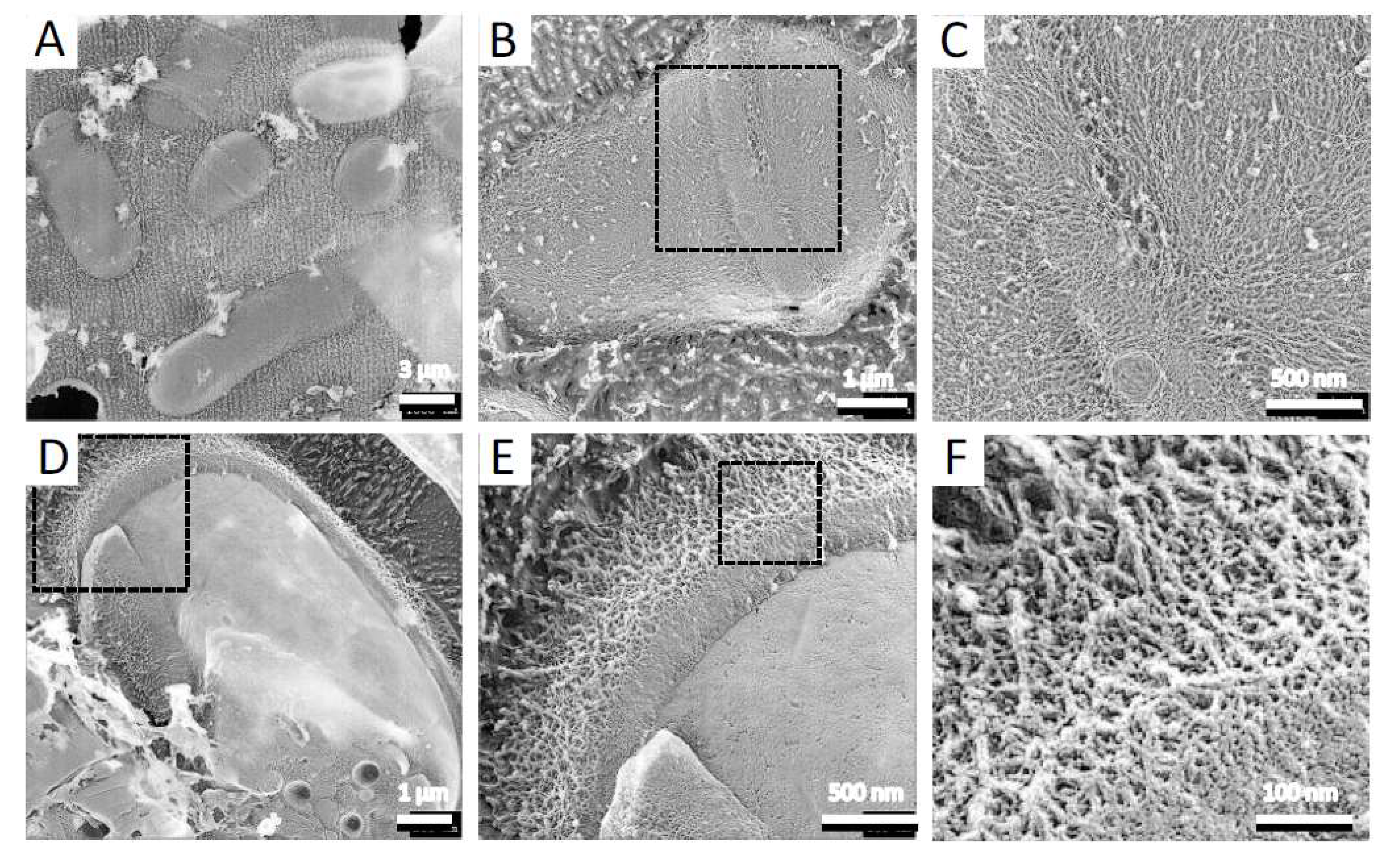
3.1. The S. pombe Spore Is Covered by a Fibrillar Structure of Isp3
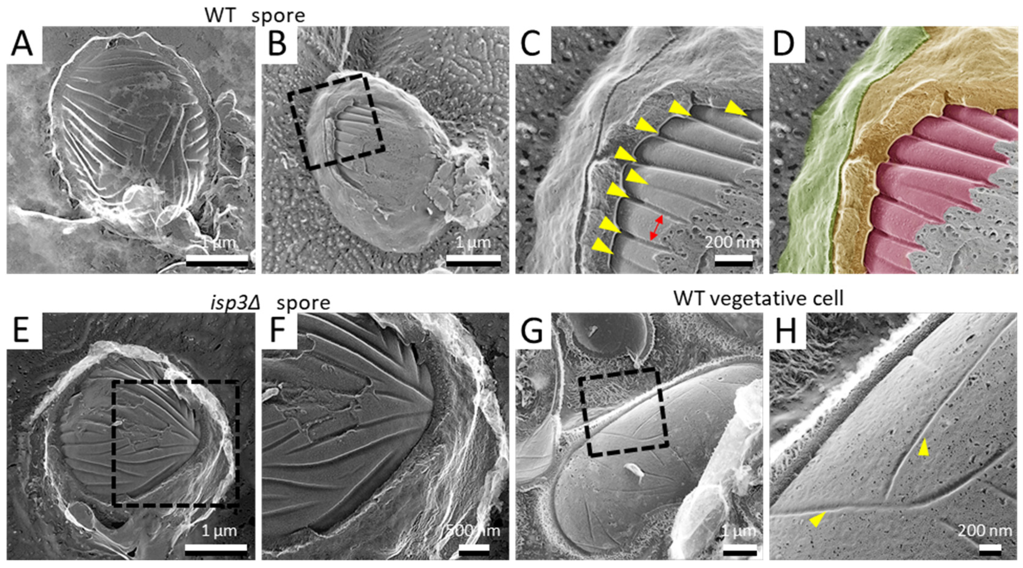
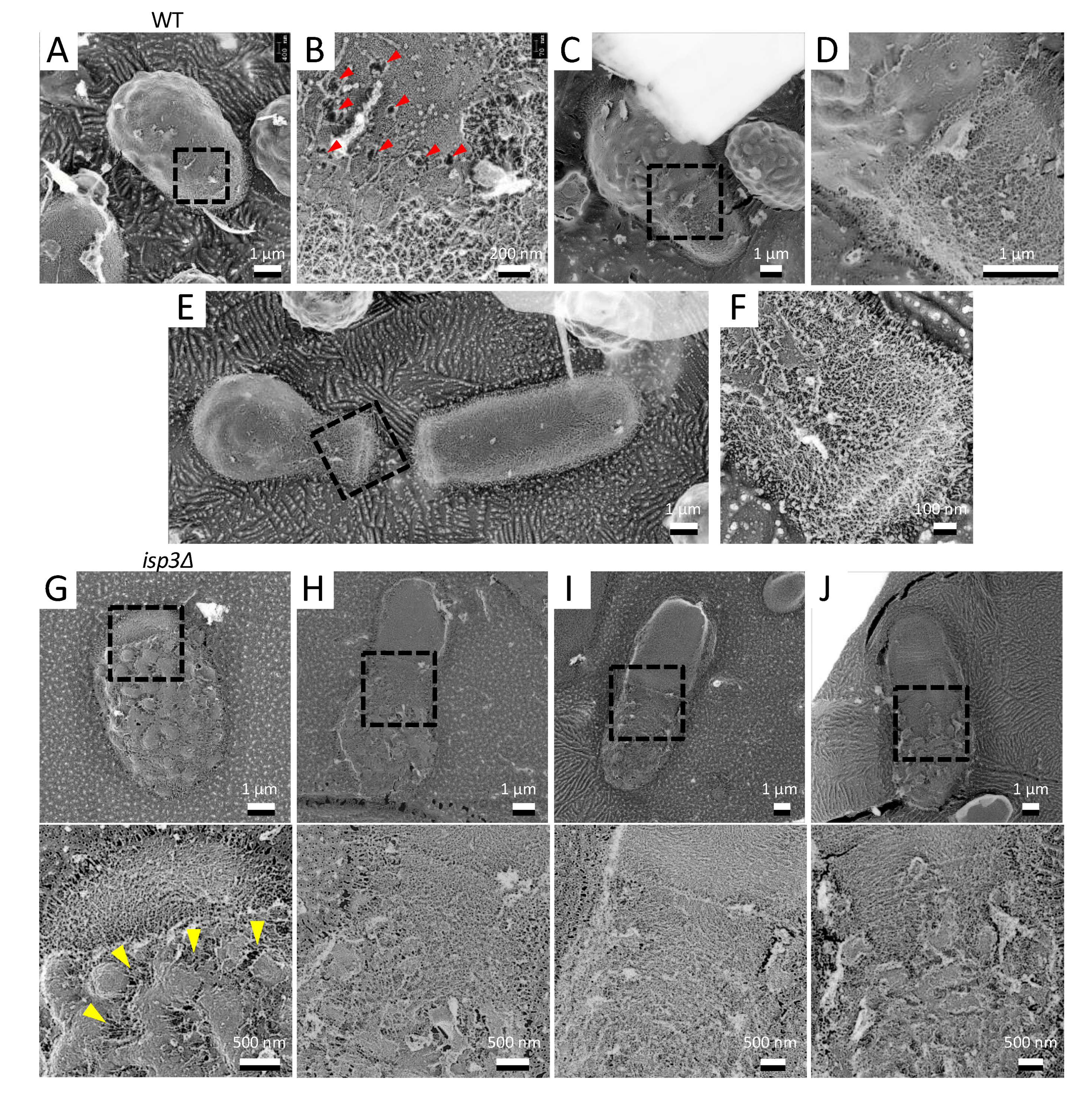
3.2. Deep Invaginations Are Aligned in Parallel on the Spore Cell Membrane
3.3. Spore Germination
4. Discussion
4.1. The Surface of the S. pombe Spore
4.2. Invaginations on the Spore Cell Membrane
Author Contributions
Funding
Acknowledgments
Conflicts of Interest
References
- Egel, R. Selective spore survival during replica-plating of fission yeast. Arch. Microbiol. 1977, 112, 109–110. [Google Scholar] [CrossRef]
- Kupiec, M.; Byers, B.; Esposito, B. Meiosis and sporulation in Saccharomyces cerevisiae. In The molecular and Cellular Biology of the Yeast Saccharomyces; Jones, E.W., Pringle, J.R., Broach, J.R., Eds.; Cold Spring Harbor Laboratory Press: Cold Spring Harbor, NY, USA, 1997; pp. 889–1036. [Google Scholar]
- Yoo, B.Y.; Calleja, G.B.; Johnson, B.F. Ultrastructural changes of the fission yeast (Schizosaccharomyces pombe) during ascospore formation. Arch. Microbiol. 1973, 91, 1–10. [Google Scholar] [CrossRef] [PubMed]
- Tanaka, K.; Hirata, A. Ascospore development in the fission yeasts Schizosaccharomyces pombe and S. japonicus. J. Cell Sci. 1982, 56, 263–279. [Google Scholar] [PubMed]
- Hirata, A.; Shimoda, C. Electron microscopic examination of sporulation-deficient mutants of the fission yeast Schizosaccharomyces pombe. Arch. Microbiol. 1992, 158, 249–255. [Google Scholar] [CrossRef]
- Osumi, M. Development of methods for observing of bacteria and fungi by electron microscopy. QJM Int. J. Med. 1989, 38, S150. [Google Scholar]
- Humbel, B.M.; Konomi, M.; Takagi, T.; Kamasawa, N.; Ishijima, S.A.; Osumi, M. In situ localization of β-glucans in the cell wall of Schizosaccharomyces pombe. Yeast 2001, 18, 433–444. [Google Scholar] [CrossRef] [PubMed] [Green Version]
- Duran, A.; Perez, P. Cell wall synthesis. In Molecular Biology of Schizosaccharomyces pombe; Springer: Berlin/Heidelberg, Germany, 2004; pp. 269–280. [Google Scholar]
- Shimoda, C. Existence of a major spore wall protein (23K) in the fission yeast Schizosaccharomyces pombe: A possible sporulation-specific protein. Arch. Microbiol. 1983, 136, 26–27. [Google Scholar] [CrossRef]
- Martin, V.; Ribas, J.C.; Carnero, E.; Duran, A.; Sánchez, Y. bgs2+, a sporulation-specific glucan synthase homologue is required for proper ascospore wall maturation in fission yeast. Mol. Microbiol. 2000, 38, 308–321. [Google Scholar] [CrossRef] [Green Version]
- Liu, J.; Tang, X.; Wang, H.; Balasubramanian, M. Bgs2p, a 1,3-β-glucan synthase subunit, is essential for maturation of ascospore wall in Schizosaccharomyces pombe. FEBS Lett. 2000, 478, 105–108. [Google Scholar] [CrossRef] [Green Version]
- Arellano, M.; Cartagena-Lirola, H.; Hajibagheri, M.A.N.; Duran, A.; Valdivieso, M.-H. Proper ascospore maturation requires the chs1+ chitin synthase gene in Schizosaccharomyces pombe. Mol. Microbiol. 2000, 35, 79–89. [Google Scholar] [CrossRef] [Green Version]
- Matsuo, Y.; Tanaka, K.; Matsuda, H.; Kawamukai, M. cda1+, encoding chitin deacetylase is required for proper spore formation in Schizosaccharomyces pombe. FEBS Lett. 2005, 579, 2737–2743. [Google Scholar] [CrossRef] [Green Version]
- Garcia, I.; Tajadura, V.; Martín, Y.S.; Toda, T.; Sanchez, Y. Synthesis of α-glucans in fission yeast spores is carried out by three α-glucan synthase paralogues, Mok12p, Mok13p and Mok14p. Mol. Microbiol. 2006, 59, 836–853. [Google Scholar] [CrossRef] [Green Version]
- Fukunishi, K.; Miyakubi, K.; Hatanaka, M.; Otsuru, N.; Hirata, A.; Shimoda, C.; Nakamura, T. The fission yeast spore is coated by a proteinaceous surface layer comprising mainly Isp3. Mol. Biol. Cell 2014, 25, 1549–1559. [Google Scholar] [CrossRef] [PubMed]
- Nakamura, T.; Abe, H.; Hirata, A.; Shimoda, C. ADAM family protein Mde10 is essential for development of spore envelopes in the fission yeast Schizosaccharomyces pombe. Eukaryot. Cell 2004, 3, 27–39. [Google Scholar] [CrossRef] [PubMed] [Green Version]
- Heuser, J.E.; Reese, T.S.; Dennis, M.J.; Jan, Y.; Jan, L.; Evans, L. Synaptic vesicle exocytosis captured by quick freezing and correlated with quantal transmitter release. J. Cell Biol. 1979, 81, 275–300. [Google Scholar] [CrossRef] [PubMed] [Green Version]
- Heuser, J.E. The origins and evolution of freeze-etch electron microscopy. Microbiology 2011, 60, S3–S29. [Google Scholar] [CrossRef] [PubMed] [Green Version]
- Nakamura-Kubo, M.; Nakamura, T.; Hirata, A.; Shimoda, C. The fission yeast spo14+ gene encoding a functional homologue of budding yeast Sec12 is required for the development of forespore membranes. Mol. Biol. Cell 2003, 14, 1109–1124. [Google Scholar] [CrossRef] [Green Version]
- Egel, R.; Egel-Mitani, M. Premeiotic DNA synthesis in fission yeast. Exp. Cell Res. 1974, 88, 127–134. [Google Scholar] [CrossRef]
- Moreno, S.; Klar, A.; Nurse, P. Molecular genetic analysis of fission yeast Schizosaccharomyces pombe. Methods Enzymol. 1991, 194, 795–823. [Google Scholar] [CrossRef]
- Nishi, K.; Shimoda, C.; Hayashibe, M. Germination and outgrowth of Schizosaccharomyces pombe ascospores isolated by Urografin density gradient centrifugation. Can. J. Microbiol. 1978, 24, 893–897. [Google Scholar] [CrossRef]
- Miyata, M.; Petersen, J.D. Spike structure at the interface between gliding Mycoplasma mobile cells and glass surfaces visualized by rapid-freeze-and-fracture electron microscopy. J. Bacteriol. 2004, 186, 4382–4386. [Google Scholar] [CrossRef] [PubMed] [Green Version]
- Tulum, I.; Tahara, Y.O.; Miyata, M. Peptidoglycan layer and disruption processes in Bacillus subtilis cells visualized using quick-freeze, deep-etch electron microscopy. Microbiology 2019, 68, 441–449. [Google Scholar] [CrossRef] [Green Version]
- Hatanaka, M.; Shimoda, C. The cyclic AMP/PKA signal pathway is required for initiation of spore germination in Schizosaccharomyces pombe. Yeast 2001, 18, 207–217. [Google Scholar] [CrossRef]
- Hess, W.M.; Sassen, M.M.; Remsen, C.C. Surface characteristics of Penicillum conidia. Microbiology 1968, 60, 290–303. [Google Scholar]
- Wessels, J.G. Hydrophobins: Proteins that change the nature of the fungal surface. Adv. Microbial. Physiol. 1996, 38, 1–45. [Google Scholar] [CrossRef]
- Wosten, H.A.; de Vocht, M.L. Hydrophobins, the fungal coat unravelled. Biochim. Biiophys. Acta 2000, 1469, 79–86. [Google Scholar] [CrossRef]
- Linder, M.B.; Szilvay, G.R.; Nakari-Setälä, T.; Penttilä, M.E. Hydrophobins: The protein-amphiphiles of filamentous fungi. FEMS Microbiol. Rev. 2005, 29, 877–896. [Google Scholar] [CrossRef] [Green Version]
- Coluccio, A.; Neiman, A.M. Interspore bridges: A new feature of the Saccharomyces cerevisiae spore wall. Microbiology 2004, 150 Pt 10, 3189–3196. [Google Scholar] [CrossRef] [Green Version]
- Coluccio, A.; Bogengruber, E.; Conrad, M.N.; Dresser, M.E.; Briza, P.; Neiman, A.M. Morphogenetic pathway of spore wall assembly in Saccharomyces cerevisiae. Eukaryot. Cell 2004, 3, 1464–1475. [Google Scholar] [CrossRef] [Green Version]
- Neiman, A.M. Sporulation in the budding yeast Saccharomyces cerevisiae. Genetics 2011, 189, 737–765. [Google Scholar] [CrossRef] [Green Version]
- Briza, P.; Kalchhauser, H.; Pittenauer, E.; Allmaier, G.; Breitenbach, M. N,N’ Bisformyl Dityrosine is an in vivo precursor of the yeast ascospore wall. J. Biol. Inorg. Chem. 1996, 239, 124–131. [Google Scholar] [CrossRef] [PubMed]
- Bush, D.A.; Horisberger, M.; Horman, I.; Würsch, P. The wall structure of Schizosaccharomyces pombe. Microbiol. 1974, 81, 199–206. [Google Scholar] [CrossRef] [PubMed] [Green Version]
- Osumi, M. Visualization of yeast cells by electron microscopy. Microbiology 2012, 61, 343–365. [Google Scholar] [CrossRef] [PubMed] [Green Version]
- Takeo, K. Lack of invaginations of the plasma membrane during budding and cell division of Saccharomyces cerevisiae and Schizosaccharomyces pombe. FEMS Microbiol. Lett. 1984, 22, 97–100. [Google Scholar] [CrossRef]
- Walther, T.C.; Brickner, J.H.; Aguilar, P.S.; Bernales, S.; Pantoja, C.; Walter, P. Eisosomes mark static sites of endocytosis. Nat. Cell Biol. 2006, 439, 998–1003. [Google Scholar] [CrossRef] [PubMed]
- Strádalová, V.; Stahlschmidt, W.; Grossmann, G.; Blažíková, M.; Rachel, R.; Tanner, W.; Malinsky, J. Furrow-like invaginations of the yeast plasma membrane correspond to membrane compartment of Can1. J. Cell Sci. 2009, 122, 2887–2894. [Google Scholar] [CrossRef] [Green Version]
- Lee, J.-H.; Heuser, J.E.; Roth, R.; Goodenough, U. Eisosome ultrastructure and evolution in fungi, microalgae, and lichens. Eukaryot. Cell 2015, 14, 1017–1042. [Google Scholar] [CrossRef] [Green Version]


Publisher’s Note: MDPI stays neutral with regard to jurisdictional claims in published maps and institutional affiliations. |
© 2020 by the authors. Licensee MDPI, Basel, Switzerland. This article is an open access article distributed under the terms and conditions of the Creative Commons Attribution (CC BY) license (http://creativecommons.org/licenses/by/4.0/).
Share and Cite
Tahara, Y.O.; Miyata, M.; Nakamura, T. Quick-Freeze, Deep-Etch Electron Microscopy Reveals the Characteristic Architecture of the Fission Yeast Spore. J. Fungi 2021, 7, 7. https://doi.org/10.3390/jof7010007
Tahara YO, Miyata M, Nakamura T. Quick-Freeze, Deep-Etch Electron Microscopy Reveals the Characteristic Architecture of the Fission Yeast Spore. Journal of Fungi. 2021; 7(1):7. https://doi.org/10.3390/jof7010007
Chicago/Turabian StyleTahara, Yuhei O., Makoto Miyata, and Taro Nakamura. 2021. "Quick-Freeze, Deep-Etch Electron Microscopy Reveals the Characteristic Architecture of the Fission Yeast Spore" Journal of Fungi 7, no. 1: 7. https://doi.org/10.3390/jof7010007
APA StyleTahara, Y. O., Miyata, M., & Nakamura, T. (2021). Quick-Freeze, Deep-Etch Electron Microscopy Reveals the Characteristic Architecture of the Fission Yeast Spore. Journal of Fungi, 7(1), 7. https://doi.org/10.3390/jof7010007