1. Introduction
Colombia is a country rich in biodiversity, particularly in higher plant and animal species. However, current research has largely focused on flora and fauna, with relatively few studies addressing the chemical exploration and biological potential of fungi [
1]. These organisms play a vital role in natural ecological cycles, often in association with other species, and are key contributors to soil formation. Fungi can be classified as macromycetes or micromycetes, with the ability to degrade various types of natural organic materials through parasitic, symbiotic, or saprophytic interactions [
2]. Thus, mycological flora represents an indispensable component of global biodiversity. Although fungal diversity has been estimated to comprise several million species worldwide, only a small proportion has been formally described to date, highlighting the substantial taxonomic and biochemical knowledge gaps that remain for this kingdom [
1].
Macromycetes are a group of fungal organisms distinguished by the production of visible fruiting bodies, such as mushrooms, brackets, or sclerotia, predominantly belonging to the phyla Basidiomycota and Ascomycota. These fungi play essential ecological roles as saprotrophic decomposers, mycorrhizal symbionts, and occasionally as parasites, contributing significantly to organic matter recycling and the balance of natural ecosystems [
3]. Their ability to degrade complex polymers such as lignin, cellulose, and chitin is due to the production of a wide array of specialized extracellular enzymes, including laccases, peroxidases, and cellulases, positioning them as key players in soil formation and nutrient cycling [
4,
5]. Moreover, macromycetes have attracted considerable scientific interest for their ability to produce bioactive secondary metabolites, including terpenoids, phenolic compounds, polysaccharides, and volatile compounds, which possess antimicrobial, antioxidant, immunomodulatory, and even antitumor properties [
6]. These attributes highlight their relevance not only in ecological contexts but also in biotechnology, medicine, and sustainable agriculture. Despite this growing interest, only a relatively small fraction of fungal diversity has been taxonomically and chemically characterized, underscoring the enormous potential of macromycetes as a source of novel natural products and structurally diverse metabolites [
7].
Additionally, macromycetes have become a subject of increasing interest in the emerging field of volatilomics, a discipline focused on the study of volatile organic compounds (VOCs) emitted by living organisms [
8]. Fungal VOCs (FVOCs) generated by macromycetes represent a unique class of secondary metabolites with potential applications as chemical biomarkers, species identification tools, and bioactive agents in food, pharmaceutical, and environmental industries [
9]. These volatile emissions play not only functional roles in fungal ecology (insect attraction or defense against other microorganisms), but also contribute to chemotaxonomic profiling, enabling differentiation of metabolomic patterns among edible, toxic, and environmentally significant fungi. Furthermore, fungal volatilomics has emerged as a complementary strategy for investigating fungal biodiversity because VOC profiles frequently reflect species-specific metabolic characteristics and ecological adaptations.
The volatilome of a fungal fruiting body is strongly influenced by its physiological state and surrounding environmental conditions. Under natural in situ conditions, volatile organic compounds are continuously released as products of primary and secondary metabolism while the fungus remains attached to its original substrate and maintains interactions with neighboring microorganisms, plants, insects, and abiotic environmental factors [
10]. Consequently, VOCs collected directly in the field are considered representative of the natural volatilomic state of the organism. In contrast, detachment of fruiting bodies from their natural substrate for laboratory analysis interrupts water and nutrient exchange, modifies oxygen availability and humidity, and exposes tissues to mechanical manipulation during sampling and transport. These changes may disrupt cellular redox homeostasis, promoting the accumulation of reactive oxygen species (ROS) and triggering oxidative stress responses that include lipid peroxidation, activation of antioxidant enzymes, and remodeling of secondary metabolism. Such physiological alterations have been associated with qualitative and quantitative modifications in fungal volatile emissions, particularly in compounds derived from fatty acid oxidation and terpenoid metabolism [
11]. Therefore, differences observed between VOC profiles obtained in situ and after laboratory handling may reflect metabolic changes associated with post-harvest physiological stress. However, because oxidative stress was not directly evaluated through biochemical or molecular assays in the present study, these interpretations are based on metabolite annotation and previous experimental evidence reported in the literature and should not be interpreted as direct experimental demonstration.
Various analytical techniques have been employed to identify secondary metabolites in fungi, including high-performance liquid chromatography (HPLC–DAD/MS), gas chromatography–mass spectrometry (GC–MS), and nuclear magnetic resonance (NMR) spectroscopy. These techniques enable the identification of novel fungal species by comparing metabolite profiles in conjunction with molecular phylogeny [
12]. Thus, it is crucial to implement strategies aimed at the discovery of novel natural products (NPs) that facilitate differentiation between fungal species. GC–MS analysis, in particular, has proven effective in distinguishing edible, toxic, and potentially toxic species through the identification of amino acids, fatty acids, and sterols via derivatization, as well as a wide range of volatile organic compounds (VOCs) [
13]. Fungi generate a diverse array of VOCs, with approximately 250 fungal-derived compounds biochemically synthesized through primary and secondary metabolic pathways. These compounds fulfill multiple functions, including roles in medical diagnostics and ecological signaling, as VOC-mediated interactions are crucial in natural environments [
14].
The diversity of FVOCs holds significant biotechnological potential for applications such as biofuel development, mycofumigation, and biocontrol strategies, representing a novel approach for bioprospecting and the discovery of new NPs [
14]. Moreover, fungal-bacterial interactions contribute to the generation of a broad spectrum of microbial volatile organic compounds (MVOCs) [
15], including FVOCs [
16]. The variation in VOCs is influenced by fungal species, developmental stage, and environmental conditions that modulate their production [
17]. FVOCs are characterized by different combinations of organic functional groups, producing aromas typically associated with molds, mushrooms, and yeasts. Among these, 1-octen-3-ol (commonly referred to as mushroom alcohol or matsutake alcohol) stands out for its involvement in communication, germination inhibition, plant growth promotion, and semiochemical signaling in arthropod interactions [
17]. The identification of FVOCs has been instrumental in developing fungal chemotaxonomy, facilitating the detection of candidate volatile biomarkers to characterize both environmentally beneficial fungi and those hazardous to human health [
18].
VOCs are produced by all living microorganisms, which express their biological potential through active interactions within natural biological cycles and cross-kingdom activities involving fungi and other ecosystem organisms [
19]. The genetic basis for the biosynthesis of VOCs and the enzymes involved in the assembly of secondary metabolites is currently under investigation [
20]. Research on fungal VOC biosynthesis has reported insights into key flavor components, such as sulfur-containing compounds, C
8 structures, and certain aldehydes, linked to specific functional groups [
21]. Nonetheless, the underlying biochemical mechanisms remain an active area of study. Consequently, studies integrating analytical chemistry, fungal taxonomy, and multivariate statistical analyses are essential to expand current knowledge of fungal volatilomes and to generate hypotheses regarding the metabolic origin and ecological relevance of these compounds.
In tropical countries like Colombia, biodiversity is vast due to geographic location, climate, and the abundance of natural resources, among other factors. Within this context, forests and ecosystems located in various regions of the country—such as those found in the department of Caldas—hold great potential for the bioprospecting of the diverse groups of organisms inhabiting these environments, with fungi being among the least studied kingdoms. Furthermore, there is a lack of both basic and applied research on the fungal biota of Colombian forests, particularly concerning taxonomic identification and biotechnological potential [
22]. To address this knowledge gap, the present study aimed to characterize the volatile organic compounds (VOCs) emitted by different genera of wild macromycetes under their natural in situ conditions and following laboratory sampling, using complementary HS-SPME and DHS-SPME extraction strategies coupled with GC–MS analysis. In addition, multivariate statistical analyses and metabolic pathway annotation were employed to compare volatilomic profiles among fungal species and extraction methodologies, providing a comprehensive chemical characterization of fungal VOC diversity. The inferred metabolic pathways and ecological implications discussed herein are based on metabolite annotation and literature-supported interpretation and should therefore be considered as hypotheses requiring further biological validation.
2. Materials and Methods
2.1. Sampling and Sample Preparation
Fungal samples were randomly collected from different areas of the Botanical Garden of the University of Caldas, located in Manizales, Colombia (5°03′25.5″ N, 75°29′27.2″ W; approximately 2150 m above sea level). According to Holdridge’s life zone classification, the study area corresponds to a Lower Montane Wet Forest (LMWF), characterized by annual precipitation exceeding 1800 mm, a mean annual temperature of 17.5 °C, and persistently high relative humidity, providing favorable environmental conditions for the development of diverse macromycete communities. During the sampling campaigns, the soil remained highly moist due to recent rainfall and was covered by a leaf litter layer approximately 3 cm thick, creating an organic substrate that promotes fungal growth and fruiting body development. The sampled fungi were collected from naturally occurring substrates, including decaying wood, leaf litter, and soil-associated organic matter. Specimen collection was conducted under the Framework Permit for Biological Collections granted through Resolution No. 1166 of 9 October 2014, issued by the National Environmental Licensing Authority (ANLA), and its subsequent amendments contained in Resolutions No. 2497 of 2018, No. 854 of 2019, No. 1396 of 2021, No. 519 of 2022, No. 26 of 2024, No. 002547 of 2024, and No. 000680 of 2025.
Volatile organic compounds (VOCs) were extracted both in situ and under laboratory conditions using small portions of each fungal specimen. For the in situ analyses, VOCs were collected immediately after locating the fruiting bodies while they remained attached to their natural substrate, thereby preserving their physiological state and minimizing environmental disturbance. In contrast, laboratory analyses were performed after transporting the specimens from the field, where the interruption of water and nutrient exchange together with sample manipulation could promote post-harvest physiological responses associated with oxidative stress. Consequently, the in situ extractions were considered representative of the naturally emitted fungal volatilome, whereas laboratory extractions were regarded as reflecting VOC profiles potentially influenced by collection-induced physiological changes rather than experimentally induced oxidative stress. Depending on specimen availability and field conditions, individual fungal samples were analyzed using one, two, or all three extraction approaches (SHS-SPME, DHS-SPME, and laboratory HS-SPME), providing complementary volatilomic information for each taxonomically identified specimen. Solid-phase microextraction (SPME) was performed using fibers coated with polydimethylsiloxane/divinylbenzene/carboxen (PDMS/DVB/CARB).
2.2. Extraction Procedure of Volatile Organic Compounds (VOCs) in Fungi
2.2.1. Dynamic Headspace Solid-Phase Microextraction (DHS-SPME) for Field VOC Collection
For this DHS method, field extractions were performed for 60 min using a silanized glass tube packed with Tenax TA adsorbent (Sigma-Aldrich, St. Louis, MO, USA) connected to a 9 V portable diaphragm vacuum pump (NMP 03 Series, KNF Neuberger Inc., Trenton, NJ, USA) operating at its maximum flow rate. After sampling, the tube was transported to the laboratory for preparation and subsequent GC–MS analysis. A total of 300 µL of HPLC-grade hexane (Merck KGaA, Darmstadt, Germany) was used to elute the VOCs adsorbed onto the Tenax TA into a glass vial, from which 1 µL of the extract was injected into the chromatographic system. The extraction was performed at ambient temperature using a 50/30 µm DVB/CAR/PDMS StableFlex™ SPME fiber (Supelco, Bellefonte, PA, USA).
Dynamic headspace extraction continuously draws the surrounding air through the adsorbent, allowing the collection of volatile compounds emitted by fungal fruiting bodies under natural environmental conditions. This approach favors the recovery of highly volatile compounds present at low concentrations by continuously concentrating VOCs throughout the sampling period. Because the fruiting bodies remained attached to their natural substrate during sampling, the extracted VOCs were considered representative of the natural fungal volatilome under field conditions.
2.2.2. Static Headspace Solid-Phase Microextraction (SHS-SPME) for Field VOC Collection
SHS sampling was conducted in the field by positioning the SPME fiber near the air intake of the DHS setup. The exposure time was 30 min. Sampling was performed inside a polyester sampling bag using a 50/30 µm DVB/CAR/PDMS StableFlex™ fiber (Supelco, Bellefonte, PA, USA) under ambient temperature conditions. Unlike DHS-SPME, SHS-SPME does not involve active airflow. Instead, volatile compounds accumulate within the enclosed headspace surrounding the fungal specimen and are passively adsorbed onto the SPME fiber. This methodology preserves the equilibrium composition of the volatilome under natural (in situ) conditions while minimizing disturbance of the fungal microenvironment. Similar to DHS-SPME, fruiting bodies remained attached to their original substrate throughout sampling, minimizing physiological alterations associated with specimen manipulation.
2.2.3. Headspace Solid-Phase Microextraction (HS-SPME) for Laboratory VOC Extraction
Field-collected fungal samples were placed into 20 mL glass headspace vials (Supelco, Bellefonte, PA, USA) for laboratory analysis. VOC extraction was conducted under static headspace conditions at room temperature for 30 min using a 50/30 µm DVB/CAR/PDMS StableFlex™ fiber (Supelco, Bellefonte, PA, USA). Following exposure, the fiber was thermally desorbed in the injection port of the GC–MS system.
Laboratory HS-SPME analysis was performed under controlled experimental conditions to minimize environmental variability associated with temperature, air movement, and humidity. Consequently, this approach provides a complementary volatilomic profile that can be directly compared with the in situ extractions (DHS-SPME and SHS-SPME). Unlike the field-based extractions, laboratory analyses were performed after the fruiting bodies had been detached from their natural substrate and transported to the laboratory. This procedure interrupts water and nutrient exchange and exposes fungal tissues to handling and modified environmental conditions. Therefore, laboratory VOC profiles were interpreted as representing post-collection volatilomic changes potentially associated with physiological responses reported in the literature for fungal oxidative stress. Oxidative stress was not experimentally induced nor directly measured in this study; consequently, any stress-related interpretation is based on comparative metabolomic analysis and literature-supported biological inference.
The combined application of these three extraction strategies was intended to maximize VOC coverage, since each methodology exhibits different extraction efficiencies depending on compound volatility, polarity, and environmental conditions. Rather than representing experimentally induced biological treatments, these complementary extraction approaches provide distinct analytical windows for characterizing the fungal volatilome under field and post-collection conditions.
2.2.4. Rationale for the Comparison Between In Situ and Laboratory VOC Profiles
The experimental design was based on comparing VOCs collected directly from fruiting bodies under natural field conditions with VOCs obtained after specimen collection and laboratory processing. The in situ extraction approaches (DHS-SPME and SHS-SPME) were performed while the fruiting bodies remained attached to their natural substrate, thereby representing the natural volatilomic profile under environmental conditions. In contrast, laboratory HS-SPME analysis was conducted after specimen removal and transport to the laboratory. Because detachment from the substrate interrupts water and nutrient exchange and exposes fungal tissues to handling and changes in environmental conditions, laboratory VOC profiles may reflect physiological responses associated with post-harvest stress, including metabolic alterations previously associated with oxidative stress in fungi. It should be emphasized that oxidative stress was not experimentally induced nor directly quantified through biochemical or molecular assays in this study. Therefore, any discussion regarding oxidative stress is based on comparative metabolomic observations and previous literature describing fungal physiological responses following tissue removal and environmental perturbation.
2.3. Gas Chromatography–Mass Spectrometry (GC–MS) Analysis
Analysis was performed using a Shimadzu GCMS-QP2010 Plus gas chromatograph coupled to a quadrupole mass spectrometer (Shimadzu Corporation, Kyoto, Japan). A ZB-5 fused-silica capillary column (Phenomenex Inc., Torrance, CA, USA) (30 m length × 0.25 mm internal diameter × 0.25 µm film thickness) was employed. The SPME fiber was thermally desorbed in the injector port at 250 °C in splitless mode prior to chromatographic separation. The injector temperature was set at 250 °C, while the ion source and interface were maintained at 300 °C [
8]. The temperature program began at 40 °C, increased to 280 °C at a rate of 6 °C min
−1, followed by a rapid increase to 300 °C at 30 °C min
−1 and held for 1 min, resulting in a total run time of 41.67 min. Helium (99.999% purity) was used as the carrier gas at a constant flow rate of 1.0 mL min
−1 in SPME injection mode.
Mass spectra were acquired under electron ionization (EI) at 70 eV. Data acquisition was performed using GCMSsolution software version 4.30 (Shimadzu Corporation, Kyoto, Japan) in full-scan mode over an m/z range of 40–500, enabling the qualitative characterization of fungal volatile organic compounds. Instrument performance and chromatographic stability were periodically verified through the analysis of quality control (QC) samples interspersed throughout the analytical sequence, as described in
Section 2.6.
2.4. Molecular Identification of Fungal Species
The molecular identification of fungal species was carried out by CorpoGen Research and Biotechnology S.A.S. (Bogotá, Colombia; NIT 830.009.610-5). Mycelial material obtained from the selected fungal specimens was submitted for genomic DNA extraction and purification. The internal transcribed spacer (ITS) region of the ribosomal DNA was amplified by polymerase chain reaction (PCR) using the universal fungal primers ITS5 (5′-GGAAGTAAAAGTCGTAACAAGG-3′) and ITS4 (5′-TCCTCCGCTTATTGATATGC-3′). PCR products were subsequently purified and sequenced using the Sanger chain-termination method. Following sequencing, sequence editing and assembly were performed to generate consensus sequences for each specimen prior to taxonomic assignment.
Taxonomic identification was conducted using the BLASTn algorithm, version 2.17.0, available through the National Center for Biotechnology Information (NCBI), complemented with comparisons against the UNITE fungal database, GenBank, EMBL (European Molecular Biology Laboratory), DDBJ (DNA Data Bank of Japan), RefSeq, and the MycoID platform (MycoBank/BioloMICS). The final molecular identification was established according to the highest sequence identity obtained among the consulted databases. For several specimens, molecular identification could not be completed because the extracted DNA was insufficient or did not meet the quality requirements for successful PCR amplification and sequencing. Consequently, these specimens were maintained at the level of morphological identification and are reported in the results section.
2.5. VOC Identification
VOC identification was performed using GCMSsolution software version 4.30 (Shimadzu Corporation, Kyoto, Japan) coupled to the Shimadzu GCMS-QP2010 Plus system. Compound annotation was based on mass spectral matching against the NIST14, NIST14S, Adams Essential Oil Library, Essential Oils Library, and FFNSC (Flavor and Fragrance Natural and Synthetic Compounds) libraries using a minimum similarity index (SI) threshold of 80%. When available, metabolite annotations were further supported by comparison with PubChem, the Kyoto Encyclopedia of Genes and Genomes (KEGG), and the Human Metabolome Database (HMDB), allowing the assignment of unique chemical identifiers for subsequent structural classification and metabolic pathway annotation.
The software performed automated baseline correction, noise smoothing, peak deconvolution, and chromatographic peak integration based on total ion chromatogram (TIC) peak areas. Identified VOCs were annotated according to their retention time (RT), mass-to-charge ratio (m/z), similarity index (SI), peak area, adduct information, PubChem identifier, and metabolomics identification level, according to the analytical capabilities of the GC–MS platform. Since compound identities were assigned exclusively by spectral library matching without confirmation using authentic reference standards or retention index verification, all metabolite annotations were considered putative (Metabolomics Standards Initiative, MSI Level 2). Consequently, all subsequent chemical classifications, metabolic pathway assignments, and biological interpretations presented in this study should be regarded as annotation-based inferences rather than experimentally confirmed metabolite identifications.
2.6. Quality Assurance and Quality Control (QA/QC)
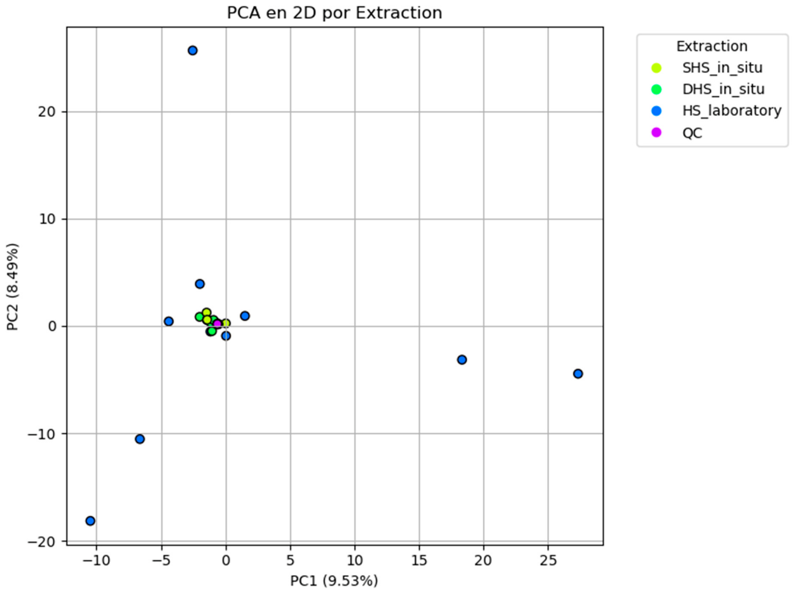
To ensure the reliability and reproducibility of the analytical workflow, quality assurance (QA) and quality control (QC) procedures were implemented throughout sample preparation, chromatographic analysis, and data processing. System quality control (SQC) injections were performed using solvent blanks and quality control samples to evaluate instrument stability, chromatographic reproducibility, and mass spectrometer performance during the analytical sequence. A total of six QC injections were distributed throughout the batch, with one QC sample analyzed after every five experimental samples to continuously monitor analytical performance. QC injections were used to verify signal stability, retention time reproducibility, and instrument response, as well as to detect background noise, potential carryover, and signal drift during the analytical sequence. Procedural blanks consisting of extraction materials and solvents processed under the same analytical conditions were also included to identify contaminants originating from laboratory consumables, reagents, or the analytical system. Features consistently detected in blank samples were removed from the final data matrix prior to multivariate statistical analysis. Instrument performance was considered acceptable when QC samples exhibited highly consistent chromatographic profiles and clustered closely during principal component analysis (PCA), confirming the stability and reproducibility of the analytical platform throughout the study.
2.7. Structural Analysis of Metabolites
Identified metabolites were annotated, whenever possible, using the Human Metabolome Database (HMDB;
https://www.hmdb.ca/ accessed on 19 August 2026) and the Kyoto Encyclopedia of Genes and Genomes (KEGG;
https://www.genome.jp/kegg/ accessed on 19 August 2026). Depending on the availability of information, individual metabolites could be assigned HMDB identifiers, KEGG identifiers, both identifiers, or remain without database annotation. The complementary use of these databases increased the chemical coverage of the detected volatile organic compounds by providing structural, physicochemical, and biochemical information that supported subsequent metabolite classification.
Chemical enrichment analysis was performed using the MetaboAnalyst 6.0 platform [
23], allowing the classification of metabolites into major chemical families and the statistical evaluation of enriched compound classes across the fungal volatilome. The enrichment analysis was performed exclusively from the annotated chemical identities of the detected VOCs and therefore reflects the statistical overrepresentation of chemical classes rather than direct evidence of metabolic activity. HMDB and KEGG annotations were subsequently employed to associate the identified metabolites with previously reported biochemical pathways for visualization and functional interpretation. However, these pathway assignments were based exclusively on database annotations and published biochemical knowledge. Therefore, the metabolic networks and pathway enrichments presented in this study should be interpreted as annotation-based hypotheses that provide a biochemical framework for discussion, rather than as experimental demonstrations of pathway activity in the analyzed fungal species.
2.8. Multivariate Analysis
A multivariate statistical analysis was performed using the Pythonversion 3.14.6 programming language through custom-developed scripts for data importation, quality verification, missing value imputation, scaling, normalization, and statistical analysis. Prior to multivariate analysis, VOC peak areas were standardized using Z-score normalization to minimize differences in signal intensity among variables and improve comparability across samples. Multivariate normality was assessed using the Henze–Zirkler (HZ) test to determine whether the dataset satisfied the assumptions required for parametric multivariate analyses. The outcome of this assessment was subsequently used to guide the selection of the most appropriate statistical methods. Exploratory analysis was performed using Principal Component Analysis (PCA) to evaluate the overall structure of the dataset, visualize sample distribution, identify potential clustering patterns, and assess analytical reproducibility through the inclusion of quality control (QC) samples. Scree plots and cumulative variance analyses were subsequently generated to determine the contribution of each principal component and evaluate the dimensionality of the volatilomic dataset. Sample relationships were further investigated by Hierarchical Cluster Analysis (HCA).
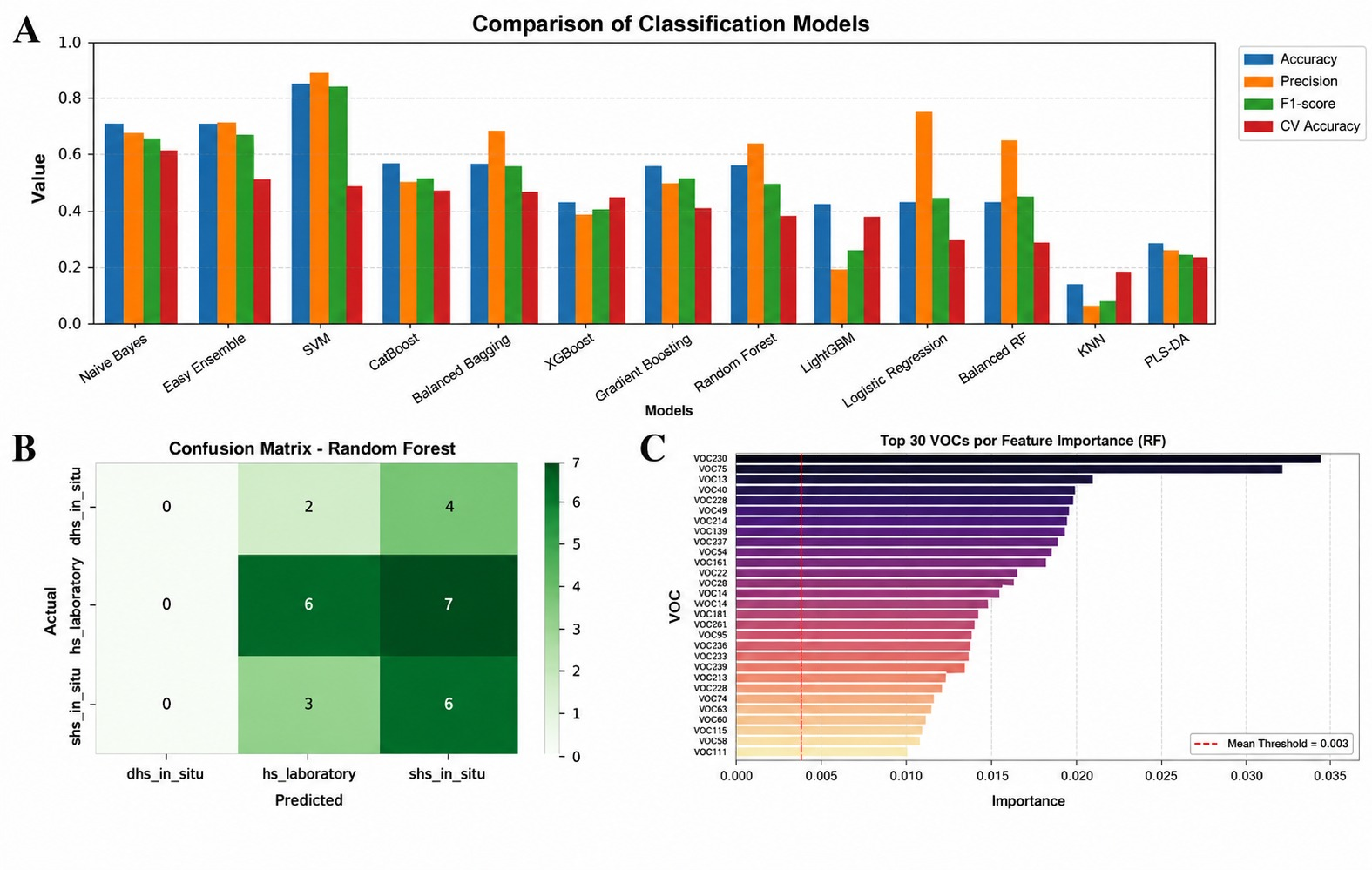
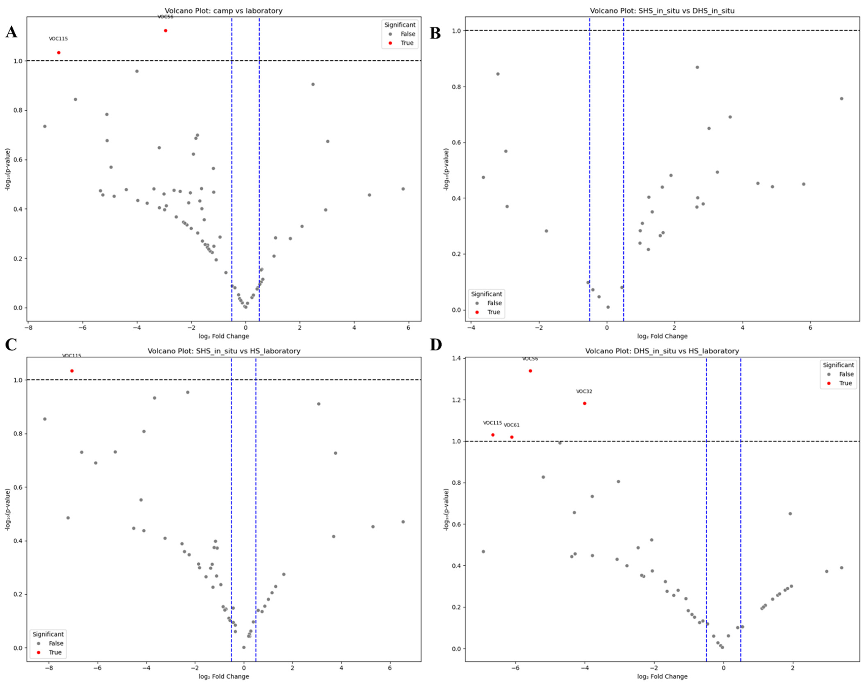
To evaluate the discrimination of samples according to extraction methodology, several supervised machine-learning algorithms were compared, including Naïve Bayes, Support Vector Machine (SVM), CatBoost, Balanced Bagging, XGBoost, Gradient Boosting, Logistic Regression, Balanced Random Forest, K-Nearest Neighbors (KNN), Partial Least Squares Discriminant Analysis (PLS-DA), and Random Forest (RF). Model performance was assessed using accuracy, precision, F1-score, and cross-validation accuracy, and the best-performing algorithms were subsequently compared. Random Forest was selected for downstream interpretation because it provided robust classification performance while allowing estimation of variable importance for sample discrimination. Volcano plot analysis was additionally performed to identify VOCs exhibiting statistically significant differential abundance between extraction methods. Variable importance scores and multivariate classifications were interpreted exclusively as statistical indicators of sample discrimination and should not be regarded as direct evidence of biological function, metabolic regulation, or causal biochemical mechanisms. Instead, these analyses were used to identify candidate discriminatory VOCs for subsequent interpretation in the context of published literature and metabolite annotation databases.
2.9. Construction of the Biosynthetic Pathway
Metabolite identifiers from the Human Metabolome Database (HMDB), MetaCyc [
24], and the Kyoto Encyclopedia of Genes and Genomes (KEGG) [
25] were retrieved, whenever available, through the Chemical Entities of Biological Interest (ChEBI) database (
https://www.ebi.ac.uk/chebi/ accessed on 19 August 2026), which was used to harmonize metabolite annotations among databases. These identifiers were subsequently queried using a custom Python script to establish metabolite–pathway associations based on curated biochemical information available in the referenced databases. The resulting annotations enabled the construction of integrated metabolite–pathway networks, allowing visualization of the relationships between the identified VOCs and previously reported fungal metabolic pathways, as well as the recognition of major biosynthetic classes and potential metabolic precursors. The pathway reconstruction was based exclusively on bioinformatic annotation and database integration and did not involve metabolomic flux analysis, transcriptomic profiling, enzymatic assays, or gene expression experiments. Consequently, the reconstructed networks represent putative biochemical associations inferred from metabolite annotation rather than experimentally validated biosynthetic pathways or active metabolic fluxes in the analyzed fungal specimens. Accordingly, these analyses were intended to provide a functional biochemical framework for interpreting the detected VOCs and for comparison with previously reported fungal metabolic pathways, thereby facilitating hypothesis generation for future experimental validation.
4. Discussion
4.1. Metabolite Identification in Fungi
The use of multiple extraction techniques (DHS-SPME, SHS-SPME, and HS-SPME) highlights the methodological complexity required to achieve a comprehensive characterization of fungal volatile organic compounds (VOCs). Each extraction approach is based on different physicochemical principles and therefore exhibits distinct affinities for volatile metabolites according to their molecular weight, polarity, vapor pressure, and abundance. Dynamic headspace extraction (DHS-SPME) continuously concentrates highly volatile compounds under natural environmental conditions, whereas SHS-SPME performed in situ preserves the equilibrium composition of the surrounding headspace. In contrast, laboratory HS-SPME provides controlled analytical conditions that minimize environmental variability during extraction. Consequently, the differences observed in the number and composition of VOCs recovered by each methodology are expected and indicate that these techniques provide complementary rather than redundant analytical information for fungal volatilomic studies.
The variability in the number of detected VOCs among fungal species also indicates substantial chemical diversity within the analyzed fungal specimens. Species such as Lentinus sp., Crepidotus sp., Agaricales sp., and Annulohypoxylon stygium yielded considerably richer volatilomic profiles than Auricularia nigricans, suggesting substantial interspecific differences in the detected VOC profiles. Such variability has been widely reported in fungal volatilomics and is commonly associated with differences in phylogeny, developmental stage, substrate composition, nutritional status, and environmental conditions that may influence secondary metabolite production. Therefore, the detected VOC diversity likely reflects the combined influence of taxonomic identity, physiological condition, substrate characteristics, and local environmental factors, rather than a single biological factor.
It is important to note that, according to the molecular identification results, samples VCQ12 and VCQ17 belong to the same fungal genus (Laetiporus). However, these specimens were collected from different locations within the Botanical Garden of the University of Caldas and were therefore exposed to potentially different local environmental conditions. Although they share a common taxonomic affiliation, differences in their volatilomic profiles may be associated with variations in microhabitat characteristics, including substrate composition, moisture availability, light exposure, microbial interactions, and other local environmental factors that have been reported to influence fungal secondary metabolism. These observations suggest that fungal VOC profiles may vary among specimens within the same taxonomic group and highlight the potential contribution of local environmental conditions to this variability.
Environmental conditions are well recognized as important factors associated with variation in fungal volatilomes. Factors such as humidity, temperature, nutrient availability, oxygen concentration, and interactions with surrounding microorganisms have been reported to influence VOC production and emission. Likewise, physiological responses associated with sample handling and transfer from natural conditions to laboratory environments may alter the abundance of certain volatile metabolites. Consequently, the complementary use of in situ and laboratory-based extraction strategies provides a broader representation of the fungal volatilome than would be obtained using a single analytical approach, allowing both environmentally emitted VOCs and metabolites detected under standardized analytical conditions to be incorporated into the overall chemical characterization. However, differences between field and laboratory profiles cannot be attributed specifically to oxidative stress in the present study, because no dedicated physiological, biochemical, or molecular measurements of oxidative stress were performed.
Overall, the combined analytical strategy adopted in the present study substantially increased metabolite coverage and indicated that the detected fungal volatilomic composition varied according to both biological characteristics of the specimens and extraction methodology. Rather than representing methodological bias, the differences observed among extraction techniques provide complementary information that improves the chemical characterization of fungal VOCs and may facilitate the identification of candidate discriminatory compounds for future biomarker studies and the investigation of VOCs of potential ecological or biotechnological interest.
4.2. Structural Analysis of the Fungal Volatilome
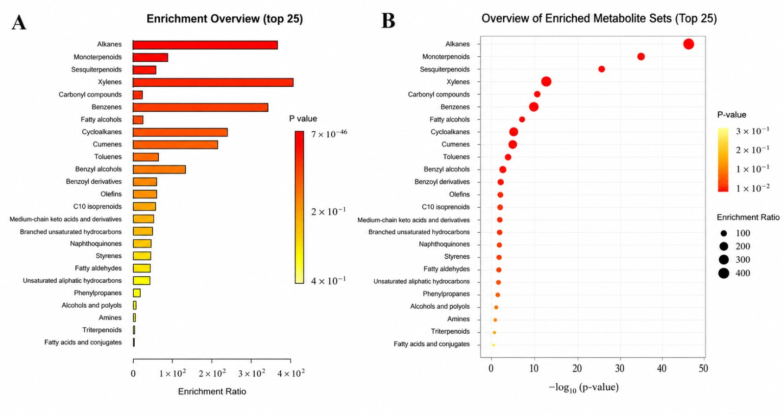
The structural enrichment analysis revealed that the fungal volatilome was characterized predominantly by hydrocarbons, terpenoids, carbonyl compounds, and aromatic metabolites. The predominance of these chemical classes is consistent with previous volatilomic studies reporting that fungal VOCs are largely composed of relatively apolar compounds with high volatility, particularly lipid-derived metabolites and terpenoids, which can be readily released into the surrounding environment. Although the enrichment analysis performed in this study was based on chemical annotation rather than direct metabolic measurements, the observed distribution of metabolite classes is consistent with the general chemical composition previously described for fungal volatilomes.
The enrichment of alkanes and carbonyl compounds may reflect the substantial contribution of lipid metabolism to fungal volatile production. Previous studies have shown that fatty acid degradation and oxidation pathways can generate a wide variety of volatile aldehydes, ketones, alcohols, and hydrocarbons during fungal growth and development. Consequently, the predominance of these compound classes observed in the present study is consistent with the important contribution reported for lipid-derived metabolites in fungal volatilomes. However, because no enzymatic activity or gene expression analyses were performed, these biochemical associations should be interpreted as literature-supported hypotheses derived from metabolite annotation rather than direct evidence of pathway activation.
Likewise, the strong enrichment of monoterpenoids and sesquiterpenoids is consistent with the widespread occurrence of terpene metabolism in fungi. Terpenoid biosynthesis has been extensively described as an important source of fungal secondary metabolites, and individual fungal terpenoids have been associated in the literature with ecological processes such as chemical communication, antimicrobial activity, competition with neighboring microorganisms, and interactions with plants and insects [
30]. The recurrent detection of these metabolite classes across multiple fungal specimens therefore suggests that terpenoid-derived compounds contribute substantially to the chemical diversity observed in the analyzed macromycetes, although the activity of the corresponding biosynthetic pathways and their specific ecological functions were not experimentally evaluated in the present study.
Aromatic metabolites, including xylenes, benzene derivatives, and related compounds, were also represented among the enriched chemical classes. These metabolites have previously been associated with the metabolism of aromatic amino acids, polyketide biosynthesis, and the transformation of complex aromatic substrates. Nevertheless, caution should be exercised when interpreting these annotations because the analyzed fruiting bodies were collected directly from their natural environment. Consequently, some aromatic compounds may reflect interactions between fungal metabolism and the surrounding substrate or environmental matrix rather than being exclusively synthesized by the fungi themselves. Additional experimental approaches, such as stable-isotope labeling, transcriptomic analyses, or controlled cultivation experiments, would be required to distinguish endogenous fungal biosynthesis from environmental contributions.
The bubble plot further demonstrated that several chemical classes accounted for most of the statistical enrichment observed in the volatilomic dataset. The concentration of large, highly significant metabolite groups in the upper-right region of the plot indicates that these compound classes showed strong enrichment within the annotated dataset. Such enrichment patterns identify these chemical families as prominent components of the observed volatilome and suggest their potential value for future chemotaxonomic and comparative studies, although their consistency across fungal taxa and their biological significance would require validation using larger and independently replicated datasets.
Overall, the structural enrichment analysis provides a comprehensive overview of the chemical composition of the fungal volatilome and demonstrates that fungal VOCs encompass multiple metabolite classes commonly associated in the literature with lipid metabolism, terpene biosynthesis, and aromatic compound transformation. Rather than demonstrating the activation of specific metabolic pathways, these findings establish a chemical framework for interpreting fungal VOC diversity and identify metabolite classes that may serve as targets for future biochemical, ecological, chemotaxonomic, and functional investigations.
4.3. Multivariate Analysis
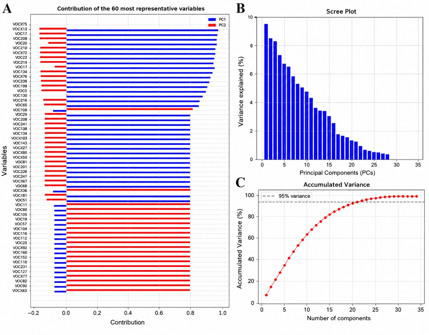
The multivariate analyses demonstrate the remarkable complexity of the fungal volatilome, as reflected by the relatively low proportion of variance explained by the first two principal components. Although PC1 and PC2 together accounted for only 18.02% of the total variance, this behavior is commonly observed in untargeted metabolomic and volatilomic datasets, where hundreds of chemically diverse metabolites contribute simultaneously to biological variability. Rather than indicating poor model performance, the dispersion of variance across multiple components reflects the intrinsic chemical heterogeneity of fungal VOC profiles. Consequently, PCA fulfilled its primary objective as an exploratory method by revealing the overall structure of the dataset, identifying clustering tendencies, and assessing analytical reproducibility through the quality control samples.
The scree plot and cumulative variance analyses further support this interpretation. Approximately twenty principal components were required to explain nearly 95% of the total variance, demonstrating that the volatilomic information is distributed across numerous independent dimensions rather than concentrated within only a few variables. This behavior is consistent with the highly diverse chemical composition of fungal volatilomes, in which multiple metabolite classes—including lipid-derived compounds, terpenoids, aromatic compounds, alcohols, ketones, and hydrocarbons—collectively contribute to sample variability. Therefore, the relatively low variance explained by the first two principal components should not be interpreted as a limitation of the PCA itself but rather as a reflection of the inherent complexity of the analyzed metabolomic dataset. In this context, the PCA served as an exploratory visualization tool, whereas subsequent supervised analyses were employed to improve sample discrimination and identify the metabolites contributing most strongly to class separation.
The variable contribution analysis revealed that only a limited subset of VOCs exerted a major influence on the first principal components. Several of these metabolites correspond to compounds previously reported in fungal volatilomes, including terpenoids such as D-limonene and caryophyllene, lipid-derived compounds including 1-octen-3-ol, 3-octanone, and 3-octanol, together with several aromatic hydrocarbons. Their high contribution to the multivariate models suggests that these compounds represent major sources of variability within the analyzed volatilomic dataset. Nevertheless, their statistical importance should be interpreted as an indicator of discriminatory capacity rather than direct evidence of biological function.
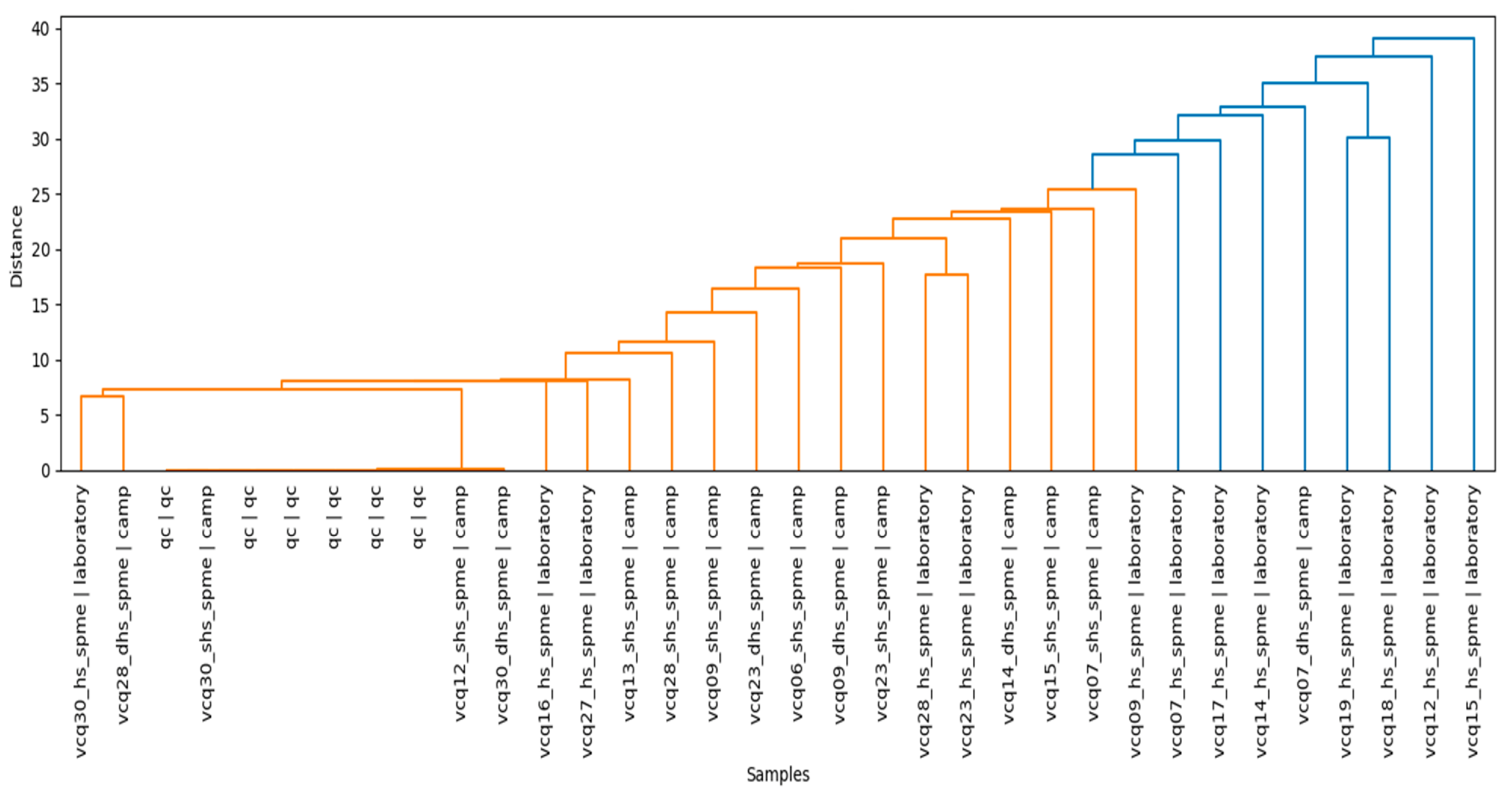
Hierarchical Cluster Analysis (HCA) demonstrated that the extraction methodology strongly influenced the organization of samples within the multivariate space. Samples obtained using the two in situ extraction techniques (DHS-SPME and SHS-SPME) exhibited greater similarity to one another than to samples analyzed by laboratory HS-SPME, indicating that extraction conditions contributed substantially to the observed volatilomic variation. This clustering pattern suggests that environmental sampling conditions and analytical methodology influence the composition of the detected VOC profiles. Because the laboratory analyses were performed under controlled conditions following specimen transport, differences between field and laboratory profiles may reflect changes associated with sample handling, environmental exposure, or physiological responses occurring after collection. However, the present study did not directly evaluate oxidative stress biomarkers, gene expression, or enzymatic activity, and therefore the observed volatilomic differences cannot be unequivocally attributed to oxidative stress alone.
The supervised classification analyses further demonstrated that fungal volatilomic profiles contain sufficient chemical information to discriminate samples according to extraction methodology. Although the Support Vector Machine (SVM) model achieved slightly higher predictive performance, the Random Forest classifier was selected for biological interpretation because it provides quantitative estimates of variable importance while maintaining excellent predictive accuracy and robustness. The Variable Importance analysis identified several VOCs that consistently contributed to sample discrimination, including 3-octanone, 1-octen-3-ol [
32], D-limonene, caryophyllene, m-xylene, longifolene, and 3-octanol. These compounds therefore represent candidate discriminatory metabolites within the analyzed fungal volatilome.
The volcano plot analyses provided an independent statistical approach for evaluating differential metabolite abundance among extraction methodologies. Several metabolites identified as important by the Random Forest model, including 3-octanone, linalool, m-xylene, and 3-octanol, also exhibited significant fold changes in pairwise comparisons between extraction conditions. The convergence of these independent analytical approaches increases confidence in the reproducibility of the statistical findings and suggests that these VOCs consistently contribute to the observed differences among volatilomic profiles. Nevertheless, additional biochemical and physiological studies will be required to determine the biological mechanisms responsible for these differences.
Overall, the integration of unsupervised multivariate analysis, supervised machine-learning classification, variable importance analysis, and differential abundance testing provides a comprehensive statistical framework for characterizing fungal volatilomes. Collectively, these complementary approaches reveal that fungal VOC profiles are highly multidimensional and are influenced by both biological variability and extraction methodology. Rather than demonstrating causal relationships between specific metabolites and biological processes, the present analyses identify statistically robust candidate biomarkers that constitute promising targets for future functional, biochemical, and ecological validation studies.
4.4. Metabolic Pathway Analysis
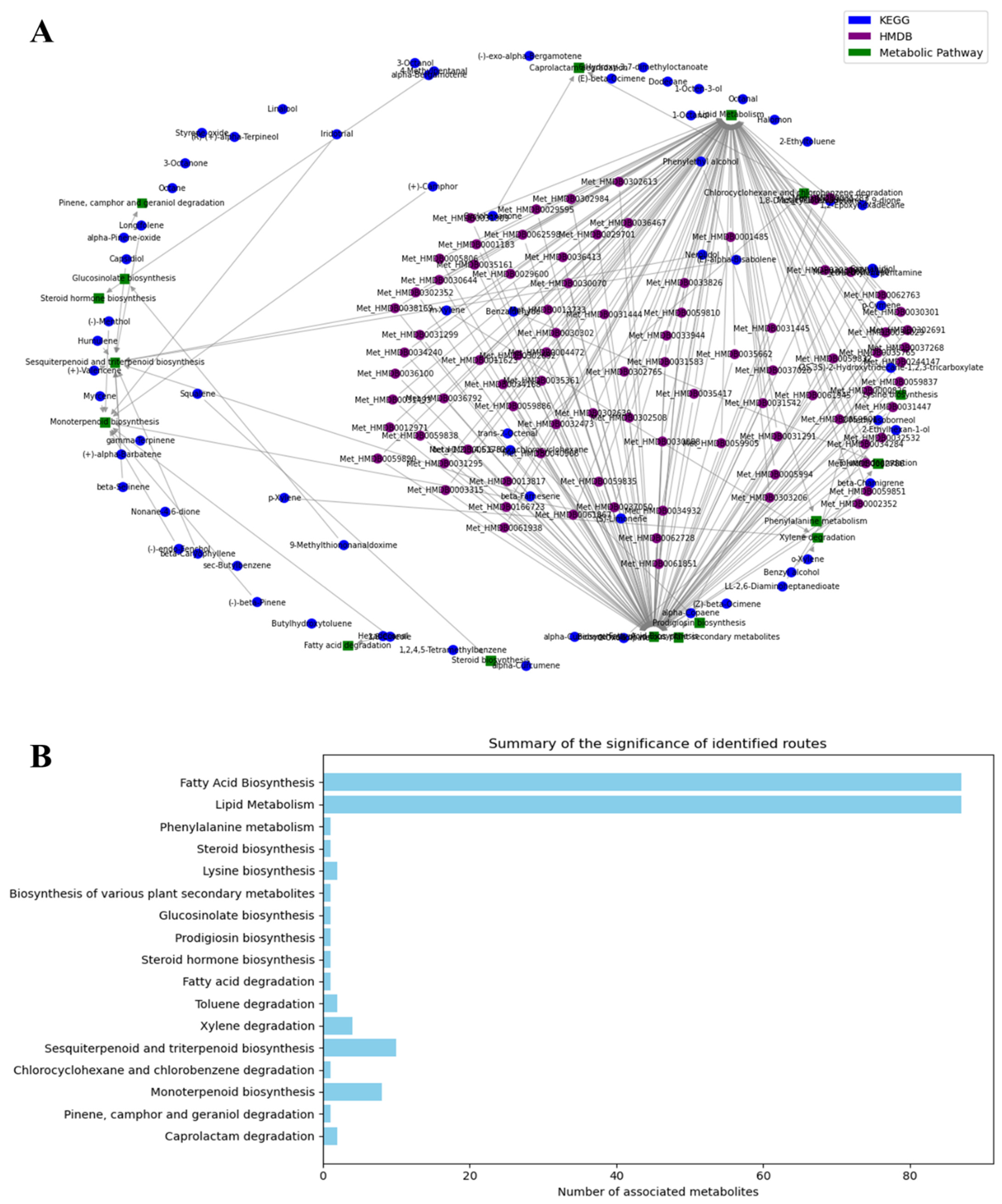
The pathway enrichment analysis provides a comprehensive overview of the biochemical framework potentially associated with the fungal volatilome. The integration of KEGG and HMDB annotations allowed the detected volatile organic compounds (VOCs) to be associated with previously described metabolic pathways, facilitating the interpretation of the chemical diversity observed among the analyzed fungal species. Because these pathway assignments were generated through bioinformatic annotation rather than direct biochemical measurements, they should be interpreted as putative metabolic associations supported by existing databases and published literature rather than as experimental evidence of active metabolic fluxes. Nevertheless, the metabolite-pathway network illustrates the high degree of connectivity among primary and secondary metabolic processes that have previously been reported in fungi.
The enrichment analysis identified Fatty Acid Biosynthesis and Lipid Metabolism as the pathways containing the largest number of annotated metabolites. This observation is consistent with the predominance of lipid-derived VOCs identified throughout the dataset, including several C
8 compounds that are widely recognized as characteristic fungal volatiles. Previous studies have shown that fatty acid oxidation and lipid turnover constitute major biochemical sources of volatile aldehydes, alcohols, ketones, and hydrocarbons in fungi [
32,
33]. Consequently, the pathway enrichment obtained in the present study is consistent with the well-established contribution of lipid metabolism to fungal volatilomes. However, the present work did not evaluate enzyme activities, metabolite fluxes, or gene expression; therefore, these pathway assignments should be regarded as functional hypotheses derived from metabolite annotation.
Similarly, the enrichment of Monoterpenoid Biosynthesis and Sesquiterpenoid and Triterpenoid Biosynthesis agrees with the identification of numerous terpene-derived VOCs in the analyzed samples. Terpenoids have been extensively described as important fungal secondary metabolites and have been associated in the literature with ecological interactions, including chemical communication, antimicrobial activity, defense against competitors, and interactions with plants and insects. The presence of these annotated pathways therefore is consistent with the possibility that terpenoid metabolism represents an important component of the fungal volatilome, although the activation of these biosynthetic pathways was not directly demonstrated in the present study.
The pathway analysis also associated several metabolites with xylene degradation, toluene degradation, and other aromatic compound transformation pathways [
34,
35]. These metabolic annotations are consistent with previous reports describing the remarkable capacity of many fungi to transform aromatic molecules through oxidative and catabolic enzymatic systems, a property that has attracted considerable interest for environmental bioremediation applications. However, these findings should be interpreted with caution. Because the analyzed fruiting bodies were collected from the Botanical Garden of the University of Caldas, an open natural environment influenced by surrounding vegetation, soil organic matter, associated microorganisms, and potential atmospheric inputs from adjacent urban areas, the detected aromatic compounds cannot be unequivocally attributed to endogenous fungal metabolism. Some of these VOCs may have originated from the surrounding substrate or environmental exposure and subsequently been adsorbed or accumulated by the fungal tissues. Consequently, the present study cannot exclude the possibility that part of the detected aromatic profile reflects environmental uptake rather than de novo fungal biosynthesis. Therefore, although the annotated pathways are compatible with metabolic capabilities previously reported for fungi, additional controlled laboratory experiments, isotope-labeling approaches, or enzymatic and transcriptomic analyses will be required to demonstrate active degradation of aromatic compounds and to distinguish endogenous metabolism from exogenous environmental contamination.
The identification of Lysine Biosynthesis among the enriched pathways further illustrates the diversity of metabolic processes represented in the volatilomic dataset. Amino acid metabolism has previously been associated with the formation of several nitrogen-containing volatile compounds and with metabolic interactions connecting primary and secondary metabolism. Although relatively few nitrogen-containing VOCs were detected compared with lipid-derived metabolites and terpenoids, the enrichment analysis suggests that amino acid metabolism may also contribute to the overall chemical diversity of the fungal volatilome.
The combined use of HMDB and KEGG substantially improved metabolite annotation by increasing the number of compounds that could be associated with known biochemical pathways. Owing to the evolutionary conservation of numerous metabolic processes among eukaryotes, databases originally developed for human metabolism can provide valuable complementary information for fungal metabolomics, particularly for non-model organisms whose metabolic pathways remain incompletely characterized. The integration of both databases therefore expanded metabolite coverage and provided additional context for the interpretation of the identified VOCs.
Overall, the pathway enrichment analysis indicates that the fungal volatilome is predominantly associated with biochemical processes related to lipid metabolism, terpenoid biosynthesis, amino acid metabolism, and aromatic compound transformation. Rather than demonstrating the activation of specific metabolic pathways, these analyses provide a bioinformatic framework for interpreting the detected VOCs and identifying metabolic processes that are consistent with previous knowledge of fungal biochemistry. Future investigations integrating transcriptomics, proteomics, enzyme activity assays, or stable-isotope labeling will be necessary to experimentally validate the biosynthetic origin and metabolic regulation of the volatile compounds identified in this study.
4.5. Biochemical Pathways in Fungi
The biochemical pathway annotation performed in this study provides a functional framework for interpreting the fungal volatilome and illustrates the remarkable metabolic versatility previously described for fungi. The association of the identified volatile organic compounds (VOCs) with pathways related to terpenoid biosynthesis, lipid metabolism, and aromatic compound transformation is consistent with the integration of both primary and secondary metabolism that has been widely reported in fungal systems. Because these pathway assignments were generated through metabolite annotation using KEGG and HMDB databases, they should be interpreted as putative biochemical associations supported by previous literature rather than as direct evidence of active biosynthetic pathways in the analyzed specimens.
Among the annotated metabolites, several terpenoids—including D-limonene (VOC75), caryophyllene (VOC209), longifolene (VOC207), and dauca-4(11),8-diene (VOC236)—have previously been reported to originate from the fungal mevalonate pathway through the action of terpene synthases that catalyze the cyclization of isoprenoid precursors [
35]. These compounds have been associated with ecological processes such as chemical communication, antimicrobial defense, competition with neighboring microorganisms, and interactions with plants and insects. The detection of these metabolites in the analyzed fungal species is therefore consistent with previous reports describing terpenoid metabolism as an important source of fungal volatile compounds, although the activity of these biosynthetic pathways was not experimentally evaluated in the present study.
Lipid-derived VOCs constituted another major group of annotated metabolites. Compounds such as VOC250 (Heneicosane), VOC253 (2-Methyloctacosane), VOC261 (Tetratetracontane), VOC199 (4-Ethyl-2-methylhexane), VOC95 (2,3,3-Trimethyloctane), VOC28 (2,3,4-Trimethylhexane), VOC56 (3-Octanone), VOC54 (1-Octen-3-ol), and VOC61 (3-Octanol) have previously been associated with fatty acid metabolism through the elongation of acetyl-CoA and malonyl-CoA by the Fatty Acid Synthase (FAS) complex, followed by oxidative reactions that generate volatile aldehydes, alcohols, ketones, and hydrocarbons [
29,
30]. In particular, 1-octen-3-ol is widely recognized as the characteristic “mushroom alcohol” and represents one of the most extensively studied fungal volatile metabolites [
35].
Previous biochemical investigations have demonstrated that linoleic acid may be converted into 10-hydroperoxide (10-HPOD) through dioxygenase-mediated oxidation [
33]. Hydroperoxide lyases subsequently cleave this intermediate to generate 1-octen-3-ol together with 10-oxodecanoic acid, whereas additional oxidation-reduction reactions may produce 3-octanone and 3-octanol [
33,
34]. The identification of these metabolites in the present study is therefore consistent with lipid oxidation pathways previously described in fungi. However, no enzymatic assays, transcriptomic analyses, or isotope-labeling experiments were performed; consequently, these biosynthetic relationships should be regarded as literature-supported interpretations rather than experimentally validated metabolic processes.
Several aromatic and polycyclic compounds identified among the discriminatory VOCs—including VOC31 (
o-Xylene), VOC32 (
m-Xylene), VOC4 (Methyl
N-hydroxybenzenecarboximidate), VOC234 (2,6-Di-
tert-butyl-4-methylphenol), VOC220 (2,6-Di-
tert-butyl-
p-benzoquinone), and VOC41 (Cyclohexanone)—have previously been associated with the metabolism of aromatic amino acids and with secondary metabolism mediated by fungal polyketide synthases (PKSs) [
31]. PKS enzymes have been reported to catalyze the sequential condensation of malonyl-CoA units to generate structurally diverse aromatic metabolites that frequently exhibit ecological and biological activities. Nevertheless, because the analyzed fruiting bodies were collected directly from the Botanical Garden of the University of Caldas under natural environmental conditions, the present study cannot unequivocally distinguish whether all detected aromatic compounds originated exclusively from endogenous fungal metabolism or whether some were influenced by the surrounding substrate, associated microorganisms, vegetation, soil organic matter, or atmospheric inputs characteristic of an open environment. Consequently, environmental adsorption of some aromatic VOCs cannot be completely excluded.
Likewise, the annotation of pathways related to xylene degradation and toluene degradation is consistent with previous reports describing the metabolic versatility of fungi for transforming aromatic compounds and environmental pollutants [
36,
37]. Such metabolic capabilities have attracted considerable interest because of their potential applications in environmental bioremediation. However, the present results should not be interpreted as demonstrating active degradation of these compounds by the analyzed fungi. Rather, the detected aromatic VOCs should be considered compatible with metabolic pathways previously described in fungi, while acknowledging that part of the aromatic profile may also reflect environmental exposure associated with the sampling site. Therefore, additional physiological experiments, controlled cultivation under axenic conditions, stable-isotope labeling, and transcriptomic or enzymatic analyses will be necessary to distinguish endogenous fungal metabolism from exogenous environmental contamination and to experimentally confirm these metabolic activities.
Taken together, the pathway annotation indicates that the fungal volatilome is compatible with the coordinated contribution of lipid metabolism, terpenoid biosynthesis, amino acid metabolism, and aromatic compound transformation. Rather than demonstrating the activation of specific biosynthetic routes, the present analyses provide a biologically plausible framework for interpreting the detected VOCs and identifying candidate metabolic processes that may explain the observed chemical diversity. Future studies integrating transcriptomics, proteomics, enzyme activity measurements, and stable-isotope tracing will be necessary to validate these hypotheses and to establish direct links between fungal metabolism and volatile compound production.
5. Conclusions
The present study provides one of the first comprehensive volatilomic characterizations of wild macromycetes collected from the Botanical Garden of the University of Caldas (Manizales, Colombia), revealing the remarkable chemical diversity of fungal volatile organic compounds (VOCs). A total of 22 discriminant metabolites were identified through multivariate analysis, including 20 annotated compounds and two unknown metabolites, highlighting the potential of fungal volatilomics for identifying candidate chemical biomarkers associated with differences in fungal diversity and extraction methodology.
The combined application of DHS-SPME, SHS-SPME, and laboratory HS-SPME coupled with GC–MS substantially expanded VOC coverage by providing complementary analytical windows into the fungal volatilome. The multivariate analyses indicated that fungal VOC profiles are highly multidimensional and are influenced by both biological variability and extraction methodology. Although laboratory HS-SPME recovered a broader range of volatile metabolites, the complementary use of in situ and laboratory-based approaches enabled a more comprehensive characterization of fungal volatile emissions under different sampling conditions.
Metabolite annotation and pathway enrichment analyses indicated that many of the detected VOCs are associated with biochemical processes previously reported for fungi, particularly lipid metabolism, fatty acid biosynthesis, terpenoid biosynthesis, amino acid metabolism, and aromatic compound transformation. Likewise, several characteristic fungal VOCs, including 1-octen-3-ol, 3-octanone, 3-octanol, D-limonene, caryophyllene, and longifolene, were identified among the most important discriminatory metabolites. These metabolic associations are based on database annotation and previously published literature and therefore should be regarded as biologically plausible hypotheses rather than experimentally validated biosynthetic pathways.
The comparison between in situ and laboratory extractions demonstrated that sampling conditions were associated with differences in the detected volatilomic profiles, emphasizing the importance of considering analytical methodology during experimental design and data interpretation. Although differences between field and laboratory profiles were observed, the present study did not directly evaluate oxidative stress biomarkers or physiological responses; consequently, the mechanisms responsible for these changes remain to be experimentally investigated.
Overall, this work establishes a robust analytical framework for fungal volatilomics by integrating complementary VOC extraction strategies, GC–MS profiling, multivariate statistics, machine-learning approaches, and metabolite pathway annotation. The resulting dataset contributes to the chemical characterization of understudied tropical macromycetes and provides a valuable resource for future investigations focused on fungal chemical ecology, chemotaxonomy, environmental monitoring, natural product discovery, and the experimental validation of the metabolic pathways inferred in the present study.



 ,
,