Article

Aflatoxin B1 Exposure and Hepatocellular Carcinoma in South America: A Multinational Cross-Sectional Analysis


1
Departamento de Bioquímica Clínica-Centro de Investigación en Bioquímica Clínica e Inmunología (CIBICI), Facultad de Ciencias Químicas, Universidad Nacional de Córdoba-Consejo Nacional de Investigaciones Científicas y Técnicas (CONICET), Córdoba 5000, Argentina
2
Centro de Investigación en Medicina Traslacional “Severo R. Amuchástegui” (CIMETSA-CONICET), Instituto Universitario de Ciencias Biomédicas de Córdoba (IUCBC), Córdoba 5000, Argentina
3
Hospital Privado Centro Medico de Córdoba S.A. (HPCMC), Córdoba 5000, Argentina
4
Department of Medicine, University of Minnesota, Minneapolis, MN 55455, USA
5
Centro de Enfermedades Hepáticas y Digestivas (CEHYD), Bogotá 110111, Colombia
6
Department of Gastroenterology and Hepatology, Erasmus University Medical Center, 3000 CA Rotterdam, The Netherlands
*
Authors to whom correspondence should be addressed.
J. Fungi 2026, 12(8), 560; https://doi.org/10.3390/jof12080560
Submission received: 9 June 2026 / Revised: 25 July 2026 / Accepted: 28 July 2026 / Published: 31 July 2026

Abstract

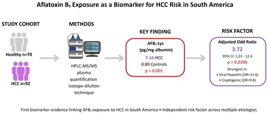
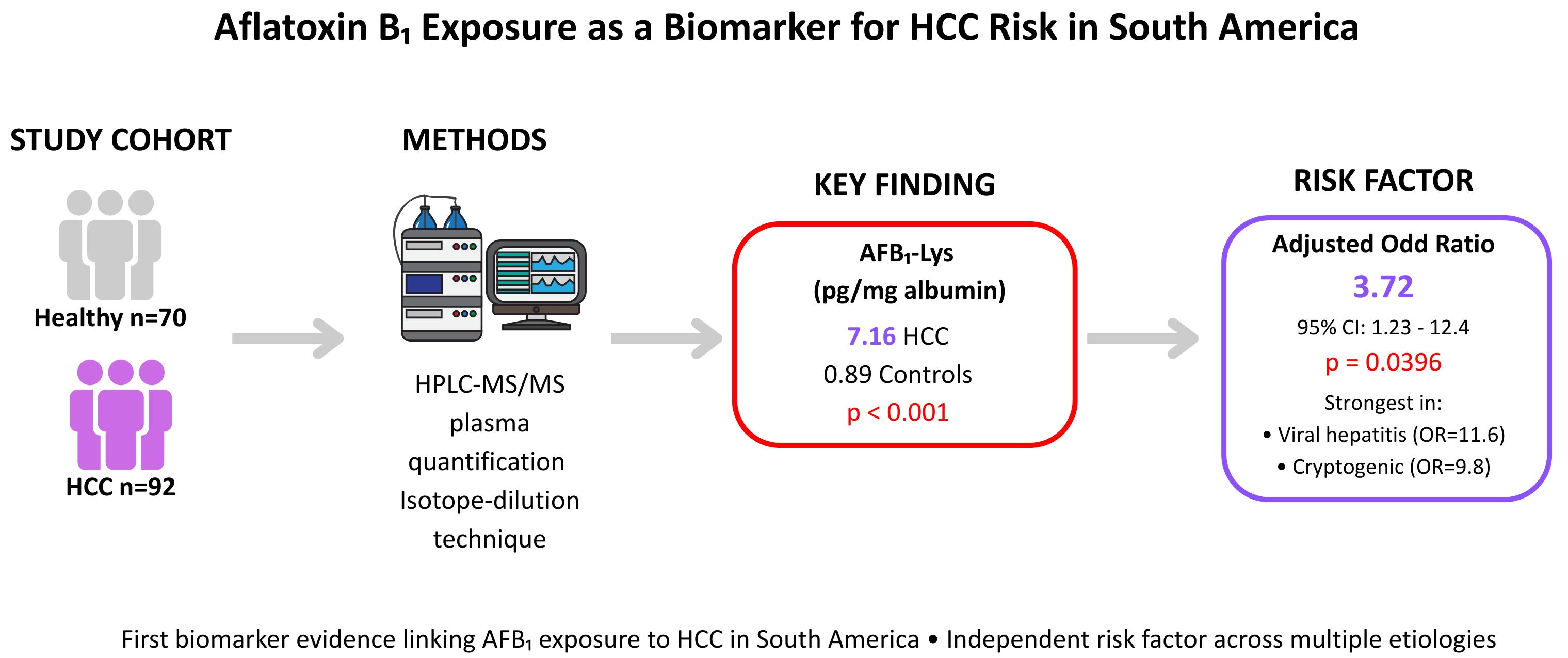
Aflatoxin B1 (AFB1), a dietary mycotoxin classified as a Group 1 carcinogen and a risk factor for hepatocellular carcinoma (HCC), has seen limited biomarker-based evidence linking it to HCC in South America despite favorable regional contamination conditions. Utilizing a newly validated isotope-dilution HPLC–MS/MS method to quantify AFB1-Lysine (AFB1-Lys) adducts, we conducted a cross-sectional study involving 92 HCC patients and 70 healthy controls across six South American nations. The primary case–control analysis, focusing on 64 HCC patients and 70 controls from Argentina and Colombia, revealed that AFB1-Lys concentrations and positivity rates were significantly higher in HCC cases compared to controls (7.16 vs. 0.89 pg/mg albumin; 43% vs. 10%). Multivariable logistic regression demonstrated that detectable AFB1-Lys was significantly and independently associated with HCC (adjusted OR = 3.72), with associations most pronounced, though based on small subgroups, in viral hepatitis-related and cryptogenic HCC. Furthermore, broader regional analysis indicated higher AFB1-Lys positivity rates in HBV-positive patients than in HCV-positive or non-viral HCC cases. Ultimately, chronic dietary aflatoxin exposure shows a consistent, statistically significant association with hepatocarcinogenesis across diverse etiological backgrounds in South America, highlighting an urgent need for integrated regional food safety surveillance and expanded prospective studies.

Graphical Abstract

1. Introduction

Aflatoxin B1 (AFB1), a mycotoxin produced by Aspergillus species that readily contaminates numerous staple agricultural crops, represents a significant global public health concern due to its role in promoting liver cancer and its classification as a Group 1 carcinogen by the International Agency for Research on Cancer [1]. Dietary exposure to AFB1 is a major contributor to hepatocellular carcinoma (HCC) in sub-Saharan Africa and Asia [2,3]. Chronic AFB1 exposure acts synergistically with hepatitis B (HBV) and hepatitis C (HCV) viruses, substantially increasing HCC risk in endemic regions [4,5].
Human exposure to mycotoxins has traditionally been estimated by combining data on mycotoxin occurrence in food commodities with information on food consumption patterns [6]. However, accurate assessment based on food analysis alone is challenging due to the heterogeneous distribution of mycotoxins within food matrices, their interactions with food components, and chemical modifications occurring during food processing or storage [7]. The wide range of susceptible food commodities further complicates the generation of reliable dietary intake data, particularly in low- and middle-income countries. Critically, the presence of mycotoxins in food does not necessarily indicate actual human exposure at the biological level. As an alternative, measuring mycotoxin biomarkers in human biological samples overcomes key limitations of food sampling, dietary recall, and source attribution, providing a more integrated and biologically relevant estimate of population-level exposure [6,8].
The aflatoxin B1-Lysine adduct (AFB1-Lys) is formed when the reactive AFB1-8,9-epoxide metabolite covalently binds to the N-terminal lysine of serum albumin. Because circulating albumin has a half-life of approximately 20–25 days, the adduct accumulates and persists over this period, offering pre-analytical stability and an integrated measure of average dietary exposure over the preceding one to two months. This contrasts with short-lived urinary biomarkers such as aflatoxin M1 or the AFB1-N7-guanine adduct, which reflect only very recent (approximately 24–48 h) exposure and are more susceptible to day-to-day variability driven by transient dietary intake. This longer exposure window, combined with a demonstrated correlation with dietary intake and downstream carcinogenic risk in prior prospective cohorts, has made AFB1-Lys a widely used biomarker in molecular epidemiology studies, particularly suited to cross-sectional designs where repeated sampling or dietary recall is impractical [9,10]. Epidemiological studies utilizing AFB1-Lys have established robust associations between aflatoxin exposure and HCC incidence, particularly in sub-Saharan Africa and parts of Asia [11,12,13]. This association is especially pronounced in young men, where HBV co-infection and aflatoxin exposure act synergistically to accelerate HCC development [11,13]. However, no validated biomarker-based study of AFB1 exposure has been conducted specifically in South American HCC patients.
Agricultural conditions in countries such as Argentina, Colombia, Peru, Ecuador, Brazil, and Chile favor fungal contamination of staple crops and animal feed [14,15,16,17,18], making the absence of biomarker-based HCC studies in the region particularly notable. This gap is especially relevant given rising regional HCC incidence [19] and a rapidly shifting etiological landscape: while HCV and alcohol-related cirrhosis were historically dominant [20], metabolic dysfunction-associated steatotic liver disease (MASLD) now represents the leading cause of HCC in South America. HBV-related cases continue to present at younger ages than other etiologies, with approximately 30% occurring in patients under 40 years of age [21,22,23,24]. Whether aflatoxin exposure contributes to HCC across these diverse etiologies, and how it interacts with metabolic and viral risk factors, remains unknown.
In this study, we validated a sensitive isotope-dilution HPLC-MS/MS method for quantifying AFB1-Lys adducts and applied it in an exploratory case–control study of HCC patients and healthy controls from Argentina and Colombia, with additional samples from Brazil, Ecuador, Chile, and Peru. By integrating biomarker-based exposure data with demographic and clinical risk factors, we provide the first systematic evidence characterizing aflatoxin’s contribution to HCC risk in the region and its relationship with viral, metabolic, and alcohol-related HCC etiologies. Our findings also contribute directly to ongoing efforts to characterize the food safety threat posed by mycotoxins in South America.

2. Materials and Methods

2.1. Chemicals and Reagents

HPLC methanol (Carlo Erba, Val de Reuil, France), HPLC water, PRONASE® Protease 25KU (Roche, Mannheim, Germany), ammonium hydroxide (Sintorgan, Buenos Aires, Argentine), HPLC formic acid (Sintorgan, Buenos Aires, Argentine), Aflatoxin B1 (Sigma-Aldrich, Steinheim, Germany), L-Lysine hydrochloride (Sigma-Aldrich, St. Louis, MO, USA), and L-Lysine:2HCl (13C6, 15N2, 99%) (Cambridge Isotope Laboratories, Inc., Tewksbury, MA, USA) were used in this study.

2.2. AFB1-Lysine Adduct Synthesis and Quantification

AFB1-Lys and its isotopically labeled analogue were synthesized and purified according to a previously published protocol [25], which includes full spectroscopic (including NMR) confirmation of adduct identity. The synthesis involves three sequential steps: AFB2a production, AFB2a-Lysine conjugation, and subsequent conversion to AFB1-Lysine. Adduct purification was performed using an Agilent 1220 HPLC system (Agilent Technologies, Santa Clara, CA, USA) equipped with a Discovery C18 column (4.6 × 15 mm, 5 µm) maintained at 24 °C. Separation was achieved with a 20-min gradient of water (0.1% TFA) and acetonitrile (0.1% TFA) at a flow rate of 1 mL/min. Fractions were collected based on UV detection at 399 nm and further characterized by HPLC-MS/MS under the same analytical conditions. Purified adducts were quantified spectrophotometrically at pH 7.4 (ε400 = 30,866 M−1 cm−1) [26]. The present validation was fit-for-purpose for quantitative bioanalysis rather than de novo structural elucidation, and relied on chromatographic retention-time matching, MS/MS fragmentation consistent with previously reported spectra [9,10,25], and isotopic co-elution with the labeled internal standard—together with in vivo detection in the mouse model—as converging evidence of adduct identity. We did not independently reconfirm structure by NMR in this study, which we note as a limitation relative to full structural re-characterization.
To confirm the chromatographic identity and resolution of the synthesized standard, and as a proof-of-concept demonstration of in vivo detectability (rather than a formal reproducibility study), two C57BL/6 mice were administered three daily oral doses of 1 µg AFB1, while one mouse received a single oral dose of 20 µg AFB1. Serum samples were collected 24 h post-treatment and analyzed for AFB1-Lys. This minimal cohort was sufficient to confirm that the assay could detect the expected in vivo-generated adduct peak, but was not designed or powered to characterize inter-animal precision or reproducibility, which we note as a limitation of this validation step.
Sample processing was performed according to established methods [27]. Briefly, serum samples or calibration standards (250 μL) were mixed with 10 μL of the internal standard AFB1-Lys-13C6 15N2 (150 ng/mL) and 250 μL of Pronase solution in PBS (6.5 mg/mL). After vortexing, samples were incubated at 37 °C for 4 h to allow enzymatic digestion, followed by addition of 500 μL deionized water. Solid-phase extraction was performed on preconditioned Welchrom® P-SAX cartridges (1 cm3, 30 mg) (Shanghai, China). Samples were loaded by gravity, washed twice with 1 mL water, once with 1 mL 70% MeOH, 1 mL 1% ammonium hydroxide in MeOH, and 0.5 mL MeOH, then eluted with 2 mL 2% formic acid in MeOH. Eluates were evaporated to dryness at 35 °C, reconstituted in 150 μL 25% MeOH, briefly vortexed, and transferred to HPLC vials with silanized glass inserts.
Final extracts were analyzed using an Acquity UPLC H-Class system (Waters, Milford, MA, USA) equipped with a BEH C18 column (50 × 2.1 mm, 1.7 µm) and coupled to a Xevo TQ-S mass spectrometer (Waters, Milford, MA, USA). The column was maintained at 35 °C and the autosampler at 15 °C. A 50 μL aliquot of each extract or standard was injected. Gradient elution was performed with water (eluent A) and acetonitrile (eluent B), both containing 0.1% formic acid. The gradient started at 97.5% A for 1.6 min, increased linearly to 100% B over 3.4 min, held for 2 min, then returned to 2.5% B within 0.5 min, followed by re-equilibration for 0.5 min (total run time: 8 min; flow rate: 0.3 mL/min).
The mass spectrometer was operated in multiple reaction monitoring (MRM) mode with positive-mode electrospray ionization. Settings included a capillary voltage of 4 kV, source temperature of 150 °C, desolvation temperature of 350 °C, desolvation gas flow of 800 L/h, and cone gas flow of 150 L/h. Cone voltage and collision energy were set to 20 V and 30 eV, respectively. Quantification and confirmation MRM transitions for AFB1-Lysine were m/z 457 → 394 and 457 → 328, respectively. Calibration curves were generated using blank serum samples spiked with AFB1-Lys and the internal standard. Limits of detection (LOD) and quantification (LOQ) were calculated based on signal-to-noise ratios of 3:1 and 10:1, respectively. Linearity was evaluated by verifying the coefficient of determination (r2 ≥ 0.99) and visual inspection of residuals. Recovery and precision (RSD) values were evaluated at three concentration levels (1, 7.5, and 15 ng/mL), both before and after internal standard correction.

2.3. Study Design and Population

A cross-sectional study was conducted to investigate the association between AFB1 exposure and HCC. A total of 92 HCC patients and 70 healthy controls were enrolled between 2019 and 2023 from referral hepatology and oncology centers in Argentina, Brazil, Colombia, Chile, Ecuador, and Peru participating in the ESCALON Consortium (escalon.eu). Of these, 64 HCC patients and all 70 controls from Argentina and Colombia formed the primary case–control cohort, while the remaining 28 patients from Brazil, Chile, Ecuador, and Peru contributed to the broader cross-sectional exposure analysis. These countries were selected given the high HCC burden in South America and the availability of established hepatology networks, as described in previous multicenter epidemiological studies from the region [20,22].
HCC diagnosis was confirmed by histopathology, imaging criteria (contrast-enhanced CT or MRI consistent with international guidelines), or a combination of clinical, laboratory, and radiological findings [22]. Controls were recruited from the same hospitals and communities in Argentina and Colombia and comprised individuals without a history of chronic liver disease, malignancy, or significant alcohol intake. Controls in Colombia were recruited from the same hospital network and surrounding communities as HCC cases, rather than through deliberate age/sex matching at enrollment; this recruitment strategy, common in exploratory case–control studies, produced a control group skewed toward younger, predominantly female volunteers. While the nearest-neighbor matched subsets (stratified by AFB1 exposure or by HCC status) and the propensity-score-adjusted model all preserved the association between AFB1-Lys and HCC, neither approach can fully exclude residual confounding by unmeasured factors that differ systematically between the recruitment sources of cases and controls. All participants provided demographic and clinical information through structured questionnaires and medical record review via REDCap (https://project-redcap.org). Blood samples were obtained at enrollment throughout the study period (2019–2023); serum was separated within 2 h of collection and stored continuously at −80 °C from collection until analysis, with no freeze–thaw cycles prior to processing. AFB1-Lys adducts have previously been shown to be stable in serum stored at −80 °C over multi-year periods [10]; however, we did not independently re-verify adduct stability in a subset of our archived samples, and we note this as a limitation of the present analysis. AFB1-Lys was quantified using the validated isotope-dilution HPLC-MS/MS method described above. Adduct concentrations were normalized to serum albumin levels, measured using an automated immunoturbidimetric method (ALBT-2 Tina-quant Albumin Gen.2 kit; Roche Diagnostics, Rotkreuz, Switzerland) on the same Cobas clinical analyzer (Roche Diagnostics, Basel, Switzerland) at the central laboratory (Hospital Privado Centro Medico de Córdoba S.A.), and expressed as pg/mg albumin. This assay was applied consistently across all participants, minimizing inter-batch variability.

2.4. Statistical Analysis

Descriptive statistics were used to summarize demographic and clinical characteristics. Continuous variables were expressed as means with ranges or means with standard deviations as appropriate, and compared between groups using Student’s t-test or the Mann–Whitney U test. Categorical variables were expressed as frequencies and percentages and compared using Fisher’s exact test.
AFB1-Lys was analyzed dichotomously (detectable vs. non-detectable relative to the LOQ of 0.3 ng/mL, ≈4.5 pg/mg albumin) and as a continuous variable, with serum concentrations below the LOQ assigned a value of LOD/2 prior to statistical analysis. Associations between HCC and AFB1-Lys (binary detection and continuous concentration) were evaluated using logistic regression to estimate odds ratios (ORs) and 95% confidence intervals (CIs). Unadjusted (crude) ORs were estimated from univariate models; adjusted ORs were obtained from multivariable models. Given the limited sample size of the primary case–control cohort (n = 134), the multivariable model was restricted to age, sex and Body Mass Index (BMI) to avoid overfitting; a model incorporating diabetes, smoking, alcohol consumption, HBV/HCV status, liver enzymes, and cirrhosis status simultaneously was not statistically supportable at this sample size. Etiology-related factors were instead examined through etiology-stratified analyses.
To evaluate whether age, sex, and BMI confounded the association between AFB1-Lys exposure and HCC, we performed three sensitivity analyses. First, we constructed a 1:1 nearest-neighbor matched subset by pairing AFB1-Lys-positive and -negative individuals on age and sex, using a propensity score-based algorithm without replacement and a caliper width of 0.2 standard deviations of the logit of the propensity score. Second, we applied a propensity score adjustment model across the full sample, entering the propensity of AFB1-Lys levels and detection (modeled on age, sex, and BMI) as a continuous covariate in the final multivariable model. Third, to directly test whether the case–control demographic imbalance could account for the observed association, we constructed an additional 1:1 nearest-neighbor matched subset on HCC status, matching cases exactly on sex and to the nearest available control on age. Etiology-specific subgroup analyses (MASLD, alcohol-related, viral hepatitis, and cryptogenic) were conducted using stratified logistic regression.
All statistical analyses were performed using GraphPad Prism (version 10, GraphPad Software, San Diego, CA, USA). Sensitivity analyses were performed using Python (version 3.10.16), utilizing the statsmodels package (version 0.14.0) for regression and weighting models and the scikit-learn package (version 1.4.0) for the nearest-neighbor matching algorithm.

3. Results

3.1. Method Validation

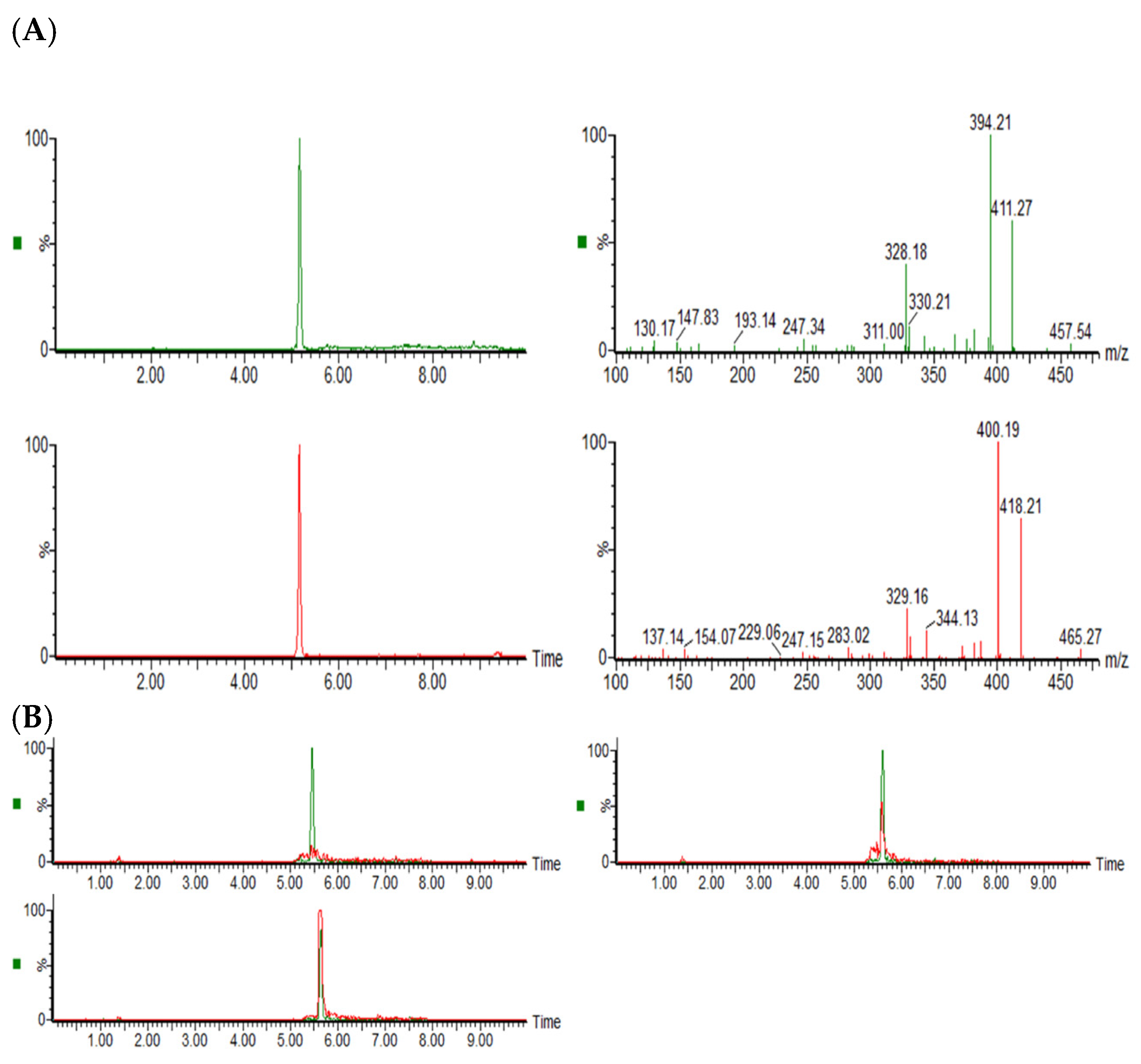
We successfully synthesized the AFB1-Lys adduct and its isotopically labeled analogue, AFB1-Lys-13C6 15N2, using a recently reported procedure [25]. Reaction products were isolated by HPLC-UV detection at 399 nm and characterized by HPLC-MS/MS. Representative chromatograms and mass spectra for both adducts are shown in Figure 1A. Both eluted at a retention time of 5.3 min, with MS/MS fragmentation patterns consistent with those previously reported for AFB1-εLys and AFB1-εLys-13C6 15N2 [9,10,25].
The method demonstrated linearity over the concentration range 0.25–17 ng/mL. The LOD was 0.1 ng/mL and the LOQ was 0.3 ng/mL (equivalent to approximately 1.5 and 4.5 pg/mg albumin, respectively). The mean absolute recovery was 50 ± 11% (recovery at 1, 7.5 and 15 ng/mL was 41.5%, 47.4% and 62. 5%, respectively). This improved to a mean relative recovery of 102 ± 10% (recovery at 1, 7.5 and 15 ng/mL was 89%, 110% and 106%, respectively) when corrected with the isotopically labeled internal standard. The average relative standard deviation (RSD) was 15% (RSD at 1, 7.5 and 15 ng/mL was 22%, 14% and 9%, respectively).
To validate the synthesized standards and the analytical procedure as a proof of concept in vivo, mice were orally administered three doses of 1 µg AFB1 within one week (n = 2) or a single dose of 20 µg AFB1 (n = 1). Serum analysis revealed a chromatographic peak with a retention time identical to that of the AFB1-Lys standard (Figure 1B), with measured serum concentrations of 60 and 732 pg/mg albumin, respectively. This minimal animal cohort verified that the mass spectrometer could reliably resolve and identify the in vivo-generated adduct peak compared with the synthetic isotope standard.

3.2. Demographic and Clinical Characteristics

A total of 64 patients with HCC and 70 healthy controls were included in the primary case–control study, comprising 35 HCC cases and 39 controls from Argentina, and 29 HCC cases and 31 controls from Colombia (Table 1). The HCC and control groups exhibited marked baseline imbalances in age, sex, and underlying liver disease status. The overall mean age of HCC patients was significantly higher than that of controls (65.8 vs. 54.3 years; p < 0.0001). This imbalance was most pronounced in the Colombian cohort, where healthy controls were considerably younger and predominantly female compared with HCC cases (mean age 41.5 vs. 68.1 years; female sex 90.3% vs. 31.0%; p < 0.0001). Conversely, the Argentinian cohort was well-balanced in age (63.9 vs. 64.5 years; p = 0.7997). HCC patients also had significantly higher BMI values compared with controls (28.7 vs. 24.1 kg/m2; p < 0.0001). This difference in BMI values was evident in both Argentina (29.1 vs. 26.2 kg/m2; p = 0.046) and Colombia (28.2 vs. 22.7 kg/m2; p < 0.0001).
Among HCC patients, 95% presented with underlying cirrhosis. MASLD was the predominant etiology in Colombian cases (50%), whereas alcohol-related liver disease was similarly prevalent in both countries (approximately 40%). Hepatitis virus-related etiologies (HBV and HCV) were observed exclusively among Argentinian patients, where they accounted for 25% of cases. Cryptogenic HCC was identified in 12.5% of the overall HCC population, primarily among Argentinian patients.
To further investigate the relationship between aflatoxin exposure and viral hepatitis, additional HCC cases were recruited from four South American countries: 15 from Peru, 6 from Ecuador, 4 from Brazil, and 3 from Chile. The mean age of the overall 92-patient cohort was 62.07 years, with a male predominance of 67.8% (Table S1).

3.3. AFB1 Exposure Is Higher in HCC Patients than in Controls

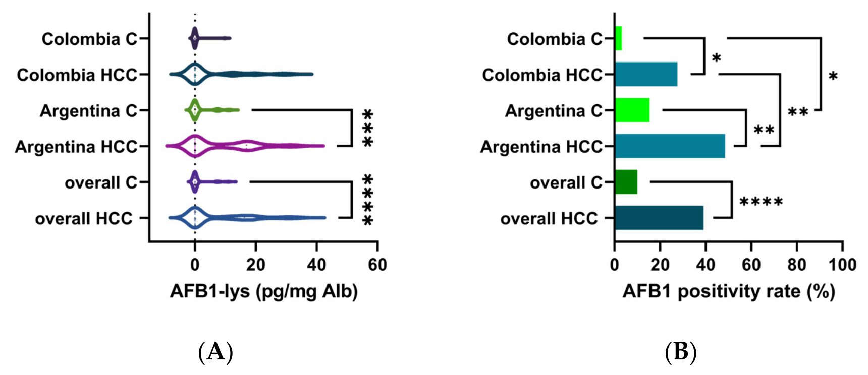
Mean AFB1-Lys levels were significantly higher in HCC patients than in controls across the combined Argentina and Colombia cohort (Figure 2A). Mean adduct concentrations were 7.16 pg/mg albumin (range: 0–34.5) in HCC cases versus 0.89 pg/mg albumin (range: 0–11.3) in controls (p < 0.001). When stratified by country, HCC cases from Argentina exhibited the highest AFB1-Lys levels (8.44 pg/mg albumin), whereas concentrations in Colombian HCC cases were comparatively lower (5.11 pg/mg albumin) but still elevated relative to their respective controls.
The proportion of samples positive for AFB1-Lys followed a similar pattern (Figure 2B). The overall positivity rate was significantly higher among HCC cases (43%) than among controls (10%; p < 0.001). AFB1 detection was most frequent in Argentinian HCC patients (48.6%) compared with Colombian HCC cases (27.6%), whereas positivity among controls remained low in both countries (Argentina: 15.4%; Colombia: 3.2%).
In the broader cross-sectional analysis including all six countries, HCC cases from Peru exhibited the highest AFB1-Lys levels (8.15 pg/mg albumin), while concentrations in Ecuador, Brazil, and Chile were comparatively lower (5.36, 6.34, and 1.47 pg/mg albumin, respectively) (Figure S1). AFB1-Lys positivity rates were ≥50% across all countries except Chile. Collectively, these findings demonstrate a consistent and statistically significant association between aflatoxin exposure and HCC status across populations, with the highest burden observed in Argentina and Peru.

3.4. Analysis of HCC Risk Factors

Logistic regression analysis evaluated the association between AFB1-Lys detection and HCC, adjusting for age, sex and BMI (Table 2). In univariate analyses, older age, male sex, higher BMI and detectable AFB1 exposure were each significantly associated with increased HCC odds. After multivariable adjustment, all three associations remained significant. Age and BMI were positively associated with HCC (Age adjusted OR = 1.05 per year, p = 0.0129; BMI adjusted OR: 1.29, p = 0.0004). Male sex was strongly associated with HCC (adjusted OR = 5.04, p = 0.0004). AFB1-Lys detection and levels were independently associated with HCC (adjusted OR = 3.72, p = 0.00244; OR = 1.41, p = 0.0002). The model showed good overall fit (likelihood ratio test vs. intercept-only model, p < 0.0001) and adequate discrimination (AUC = 0.87, 95% CI: 0.81–0.93); using a probability cutoff of 0.5, the model correctly classified 79.1% of individuals.
Sensitivity analyses confirmed that the observed association was not an artifact of demographic imbalances. In the unmatched full cohort (n = 134), substantial demographic imbalance was present across both groupings: AFB1-exposed individuals were older on average (65.6 ± 12.1 vs. 58.0 ± 15.9 years) and predominantly male (78.1% vs. 37.3%) compared to unexposed individuals, and HCC cases were similarly older (65.8 ± 8.0 vs. 54.3 ± 18.2 years) and more frequently male (71.9% vs. 24.3%) than controls (Table 1 and Table S2). Matching substantially reduced these imbalances in both approaches: in the exposure-matched subset (n = 38), age was comparable (66.4 ± 9.3% vs. 66.4 ± 9.1%) and sex was perfectly balanced (68.4% male in both groups); in the case–control-matched subset (n = 66), age difference was minimized (63.16 ± 7.9 years for HCC cases vs. 60.68 ± 14.5 years for controls) and sex was perfectly balanced (48.5% male in both groups). The association between AFB1-Lys exposure and HCC (Tables S3 and S4) remained statistically significant in the exposure-matched subset (AFB1-Lys detection: OR = 4.17, 95% CI: 1.00–17.31, p = 0.049; AFB1-Lys levels: OR = 1.98, 95% CI: 1.2–4, p = 0.0221), in the propensity-adjusted full sample (AFB1-Lys detection: adjusted OR = 3.03, 95% CI: 1.07–8.59, p = 0.037; AFB1-Lys levels: adjusted OR = 1.12, 95% CI: 1–1.2, p = 0.0151), and in the case–control-matched subset (AFB1-Lys detection: adjusted OR = 11.3, 95% CI: 1.9–107, p = 0.0136; AFB1-Lys levels: adjusted OR = 1.85, 95% CI: 1.2–3.2, p = 0.0126), confirming that the observed association is not an artifact of demographic imbalance in either direction.
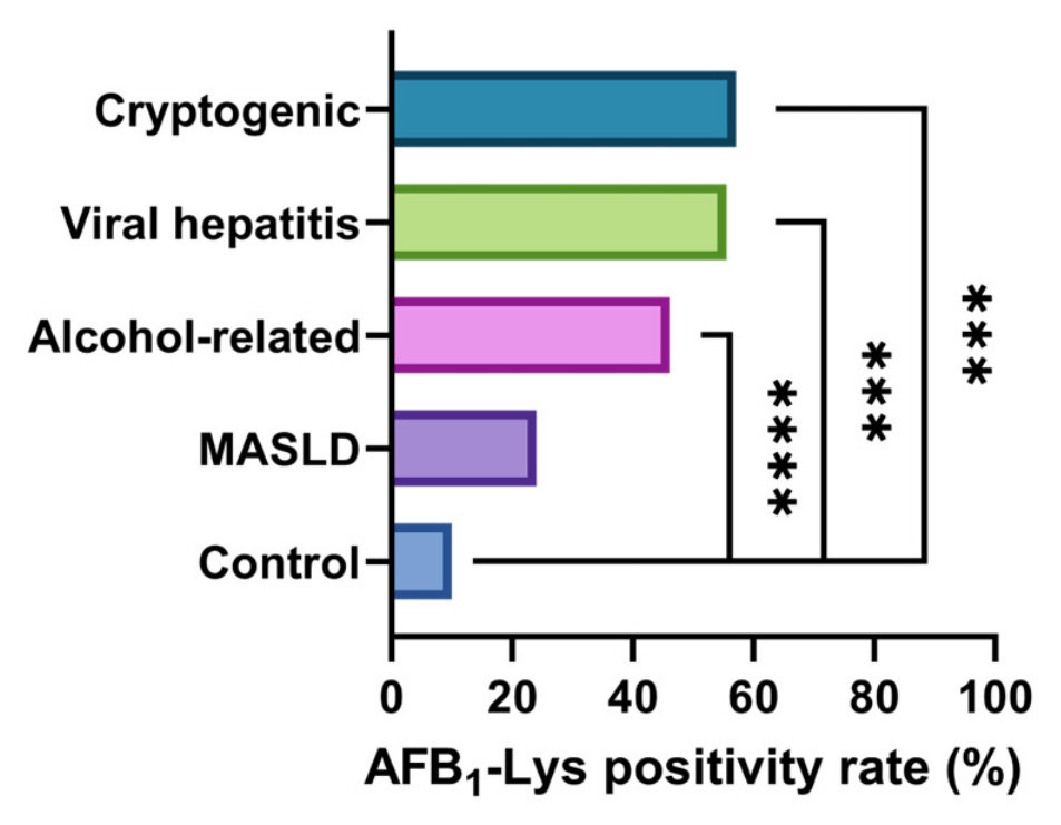
When analyses were stratified by country, sex, BMI and AFB1-Lys levels remained significantly associated with HCC in the Argentinian cohort, while only age, sex, and BMI remained significant in the Colombian cohort (Table S5). HCC cases from both countries were analyzed according to the frequency of AFB1 exposure across primary etiological groups (Figure 3); AFB1 was detected across all etiology groups at higher frequencies than in controls, and was particularly prevalent among patients with cryptogenic and viral-related HCC.
Stratified logistic regression analyses by etiology demonstrated positive trends across all subgroups (Table 3). However, statistical power was constrained by small subgroup sample sizes, and formal interaction testing across etiologies was not possible due to the absence of etiology classifications in the control group; thus, these subgroup-specific findings should be interpreted with caution. The strongest associations of AFB1-Lys detection and levels with HCC were observed in viral hepatitis-related HCC (adjusted OR = 11.6, p = 0.005 and OR = 1.3, p = 0.0046, respectively) and cryptogenic HCC (adjusted OR = 9.8, p = 0.0128 and OR = 1.3, p = 0.0045, respectively). The association for MASLD-related HCC and alcohol related HCC did not reach statistical significance.
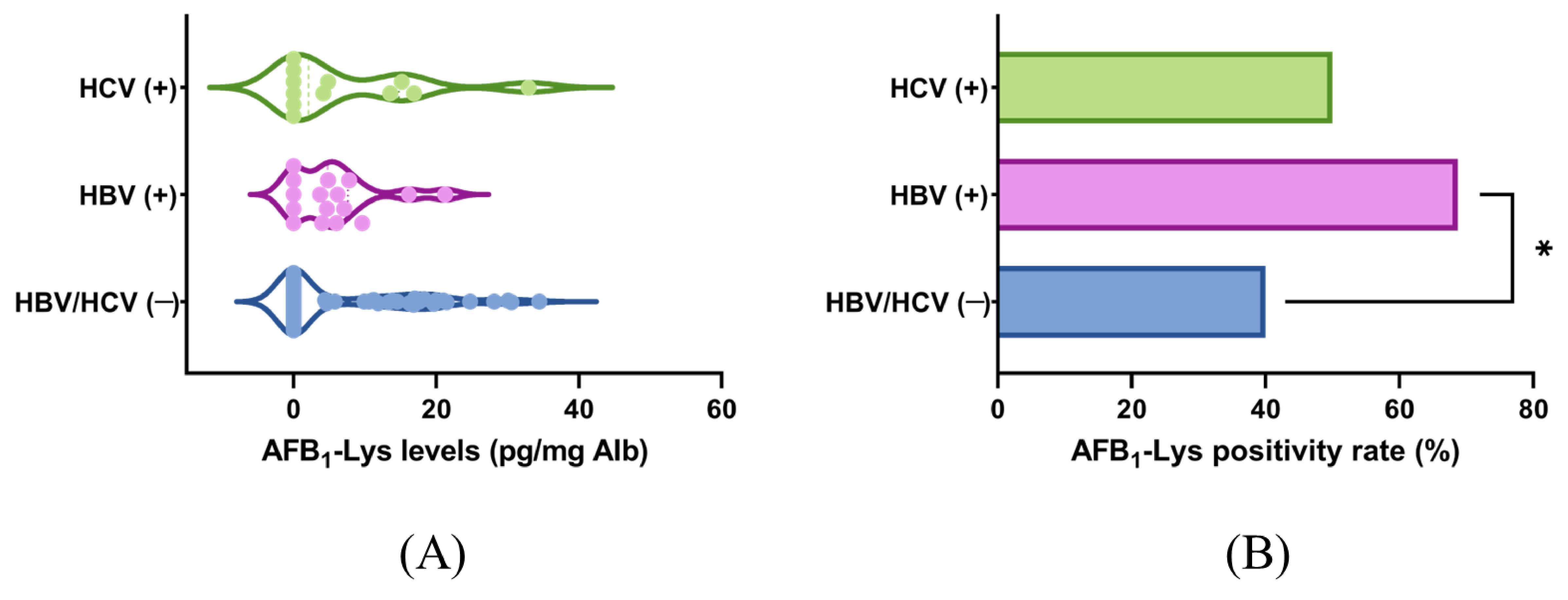
A broader six-country analysis was conducted to characterize aflatoxin exposure according to viral etiology. AFB1-Lys levels and positivity rates were compared among HCC cases with hepatitis B virus (HBV), hepatitis C virus (HCV), or no viral hepatitis. No significant difference in AFB1-Lys levels was observed between HCC cases with or without viral hepatitis (Figure 4A). However, AFB1-Lys positivity rates were significantly higher in HBV-positive patients than in HCV-positive or non-viral HCC cases (Figure 4B), reflecting a high co-occurrence of these two exposures. While this pattern is consistent with a potential synergistic interaction between HBV and aflatoxin exposure previously described in high-incidence regions [3,5], the absence of viral load and inflammatory marker data in our cohort precludes a mechanistic conclusion from these findings alone.
Overall, detectable AFB1 exposure, male sex, advancing age, and high BMI were independently associated with HCC, with the effect of aflatoxin most pronounced in viral hepatitis-related and cryptogenic HCC.

4. Discussion

We successfully synthesized and validated AFB1-Lys and its isotopically labeled analogue (AFB1-Lys-13C6 15N2). The analytical method showed high sensitivity, accuracy, and reproducibility; its biological relevance was confirmed through in vivo detection of AFB1-Lys adducts in mouse serum following controlled oral exposure. These performance characteristics are consistent with previous validations of AFB1-Lys as a reliable biomarker of aflatoxin exposure [9,10] and extend the method’s applicability to South American populations where direct measurement tools are scarce.
Applying this approach to human samples revealed significantly higher AFB1-Lys concentrations and positivity rates in patients with HCC than in healthy controls. Although the absolute adduct concentrations were lower than those typically observed in high-incidence regions of Africa and Asia [4,10], a significant positive association between AFB1-Lys and HCC was found. These findings provide the first biomarker-based description of aflatoxin exposure in these South American cohorts. Several, non-mutually exclusive factors may explain how a significant association can persist at these comparatively lower exposure levels. First, differences in staple food composition and post-harvest storage conditions likely result in lower per capita consumption of the specific high-risk commodities most implicated in Africa and Asia (e.g., certain maize varieties, groundnuts) [10]. Second, existing regional food-safety regulations may limit the most extreme contamination events even where enforcement is incomplete. Third, individual susceptibility to aflatoxin-induced hepatocarcinogenesis is known to be modulated by polymorphisms in xenobiotic-metabolizing enzymes (e.g., glutathione S-transferases) and DNA-repair genes, which could amplify carcinogenic risk independent of absolute adduct concentration [28]. Fourth, the effective carcinogenic threshold of aflatoxin is substantially lowered when it acts synergistically with endemic co-factors such as chronic HBV infection [29], which remained prevalent among our HCC cases. Distinguishing among these possibilities will require dietary, genetic, and virological data beyond the scope of the present study, and we identify this as a priority for future prospective, multi-omic investigation.
The public health relevance of this study is underscored by mounting evidence of widespread aflatoxin contamination throughout South American food chains. Multiple mycotoxins, including aflatoxins, have been detected in staple foods from Argentina, Brazil, and Peru [17,30]. In Argentina, aflatoxin M1 has been detected in milk and dairy products at levels approaching safety limits, with children identified as the most vulnerable population [15]. In Peru, carcinogenic aflatoxins have been quantified in unprocessed peanuts and chili peppers sold in Lima markets at concentrations up to 150 ppb—well above the EU regulatory limit of 4 ppb [31]. Biomarker-based evidence further supports ongoing exposure: urinary aflatoxin M1 was detected in 59% of Chilean adults [18], 41.7% of Colombian children [32], and 65% of Brazilian adults [33]. These findings underscore that the contamination burden in the region is not merely theoretical but translates into measurable human exposure, providing biological plausibility for our results and pointing to a clear need for integrated food safety monitoring and regulatory action.
Consistent with prior studies [34], male sex was a strong independent risk factor for HCC (adjusted OR = 5.5), reflecting both hormonal mechanisms—estrogen’s hepatoprotective effects and testosterone’s role in promoting hepatocyte proliferation [35]—and differential exposure to environmental risk factors including alcohol, smoking, occupational hazards, and viral hepatitis in men [36]. Elevated BMI also emerged as a significant association with a 29% increase in HCC risk in agreement with previous reports [34].
Stratified analyses indicated that aflatoxin exposure frequently co-occurred with viral hepatitis, a pattern that aligns with, but does not confirm, prior mechanistic evidence of synergy between AFB1 and HBV in hepatocarcinogenesis [3,5]. The strong association observed for cryptogenic HCC may be partially explained by its recognized link to occult hepatitis B infection [37]. However, we did not measure viral load, HBV DNA integration status, or hepatic inflammatory markers, and cannot establish a mechanistic interaction from these cross-sectional data; this remains an important hypothesis for dedicated mechanistic and longitudinal studies. Similarly, the association in alcohol-related HCC, showed a trend (p = 0.058) consistent with evidence that alcohol consumption increases susceptibility to AFB1-related hepatotoxicity and HCC risk [38]. In contrast, the association between AFB1-Lys and MASLD-related HCC was weaker and did not reach statistical significance, though elevated AFB1-Lys levels in this subgroup relative to controls suggest some degree of aflatoxin involvement in metabolic hepatocarcinogenesis. Although the interaction between AFB1 and metabolic liver disease is less well characterized than its interaction with viral hepatitis, emerging evidence supports biological plausibility. While AFB1 exposure by itself can lead to HCC and accelerate occurrence of cirrhosis [39,40], AFB1 has been shown to aggravate diet-induced non-alcoholic steatohepatitis through TLR4-mediated hepatocyte necroptosis and inflammation [41], to induce a toxicant-associated fatty liver disease phenotype with steatosis and inflammatory foci in chronic low-dose rodent exposure [42], and to disrupt hepatic lipid and carbohydrate metabolism through mitochondrial dysfunction and oxidative stress, mechanisms proposed to contribute to insulin resistance and lipid accumulation in hepatocytes [43,44]. These pathways—oxidative stress, mitochondrial dysfunction, and disrupted lipid/glucose handling—overlap substantially with the core pathogenic mechanisms of MASLD itself, raising the possibility that AFB1 exposure could act as a second hit accelerating steatosis-to-HCC progression in metabolically susceptible individuals. This hypothesis warrants dedicated mechanistic and longitudinal investigation given that MASLD is now the leading cause of HCC in South America [22] and understanding these potential interactions is of increasing public health importance.
To place these findings in a global context, global burden of disease estimates suggest that aflatoxin-attributable HCC causes between 25,200 and 155,000 cases annually worldwide, primarily through synergy with viral hepatitis [2]. However, mycotoxin surveillance in South America remains incomplete, and the accelerating impact of climate change on fungal proliferation suggests that current exposure estimates underestimate future risk [30]. Our exploratory data indicate that South American populations may face similarly unrecognized risks to those documented in sub-Saharan Africa and Southeast Asia. From a food safety perspective, these findings support the implementation of stricter regulatory limits for aflatoxin in staple foods, improved post-harvest management practices, and biomarker-based surveillance programs that could identify the most exposed populations for targeted intervention.
This exploratory study was not powered a priori for etiology-specific subgroup comparisons, as no regional estimate of the expected effect size was available at the time of design; the resulting wide confidence intervals in small subgroups should be interpreted in this light, and we treat these estimates explicitly as hypothesis-generating rather than confirmatory. A formal sample-size calculation, informed by the effect sizes observed here, should be planned for a prospective, multicenter follow-up study. The cross-sectional design and exploratory nature of the study preclude causal inference.
Because controls were selected to be free of chronic liver disease, malignancy, and significant alcohol use, the reported odds ratios likely represent an upper-bound estimate of the association between AFB1 exposure and HCC specifically, rather than an estimate isolated from AFB1’s broader association with chronic liver injury; a disease-control group (e.g., patients with cirrhosis or chronic viral hepatitis without HCC), matched on etiology, would allow this distinction to be made and is an important next step for the region. Substantial demographic imbalances between HCC cases and controls—particularly the age and sex disparity in the Colombian cohort—were evaluated through three complementary sensitivity analyses, including a matched subset constructed directly on case–control status, which confirmed that the AFB1-Lys–HCC association was robust both to age/sex confounding of exposure status and to the underlying case–control demographic imbalance itself; however, residual confounding from unmeasured clinical and environmental covariates cannot be excluded. Given the limited sample size of the primary case–control cohort, the multivariable model was restricted to age and sex to avoid overfitting, and larger cohorts are needed to support a model adjusted for the fuller set of clinical covariates (e.g., BMI, diabetes, smoking, HBV/HCV status, liver enzymes, cirrhosis status).
Etiology-stratified odds ratios should also be interpreted with caution: subgroup sizes were small (as few as 8–9 cases in the cryptogenic and viral hepatitis groups), the resulting confidence intervals are correspondingly wide, and these point estimates are therefore unstable and hypothesis-generating rather than precise or confirmatory; replication in larger, adequately powered etiology-specific cohorts is required before these subgroup associations can be considered established. Relatedly, we did not formally test for a statistical interaction between AFB1-Lys detection and HCC etiology, and the etiology-stratified odds ratios should be interpreted as descriptive comparisons across small strata rather than as evidence of formal effect modification; a properly powered interaction analysis will require a substantially larger, ideally multi-center, cohort. Finally, this study relied exclusively on a serum biomarker of AFB1 exposure—no dietary consumption data (e.g., intake of maize, groundnuts, spices, or other AFB1-prone staples) were collected, which limits our ability to attribute exposure to specific dietary sources or to corroborate biomarker findings against independent intake estimates. Together, these factors underscore the need for larger, well-balanced, multicenter prospective investigations that integrate longitudinal biomarker monitoring, dietary and food-frequency data, and a broader covariate set with the etiology- and country-specific granularity established here.

5. Conclusions

This study validates a highly sensitive isotope-dilution HPLC–MS/MS method for AFB1-Lys quantification and demonstrates that detectable biomarker exposure is significantly associated with HCC status in South American cohorts. Although the exploratory and cross-sectional design precludes causal inference, the associations were notably pronounced in viral hepatitis-related and cryptogenic HCC, suggesting etiology-specific interactions that warrant corroboration in larger studies. Importantly, these findings identify a food safety-related carcinogen as a potentially underrecognized correlate of liver cancer in South America, warranting further investigation into a possible causal role. However, the lack of direct dietary data restricts our ability to identify precise exposure sources. We call for coordinated research and regulatory actions to mitigate aflatoxin exposure in at-risk populations, including the adoption of robust food safety monitoring systems, harmonized regulatory frameworks across the region, and larger demographically balanced prospective cohorts to validate and extend our findings across Latin America.

Supplementary Materials

The following supporting information can be downloaded at: https://www.mdpi.com/article/10.3390/jof12080560/s1, Table S1: Baseline characteristics of HCC cases from South America countries; Table S2: Age, Sex and BMI composition of the full cohort and Adjusted subsets. Table S3: Logistic Regression Analysis of Risk Factors for Hepatocellular Carcinoma (HCC) Adjusted for Age, BMI and Sex in AFB1-Exposed and Unexposed Subsets; Table S4: Logistic Regression Analysis of Risk Factors for Hepatocellular Carcinoma (HCC) Adjusted for Age, BMI and Sex in HCC and HC Subsets; Table S5: Country-stratified logistic regression analysis of risk factors for HCC; Figure S1: Analysis of AFB1-Lys in individuals with hepatocellular carcinoma (HCC) in South American countries.

Author Contributions

Conceptualization, R.A., J.P., A.B., D.C.B. and P.A.R.; methodology, R.A. and G.B.; validation, R.A. and P.A.R.; formal analysis, M.L.F.; investigation, R.A. and G.B.; resources, M.L.F., J.D., D.C.B. and P.A.R.; data curation, R.A. and P.A.R.; writing—original draft preparation, R.A.; writing—review and editing, R.A. and P.A.R.; supervision, D.C.B. and P.A.R.; project administration, R.A. and D.C.B.; funding acquisition, R.A., P.A.R., J.P., A.B. and D.C.B. All authors have read and agreed to the published version of the manuscript.

Funding

This research was funded by the IAEA CRP project “Nuclear Techniques to Support Risk Assessment of Biotoxins and Pathogen Detection in Food and Related Matrices,” D52044, Contract Number 25058, and by the European–Latin American ESCALON consortium funded by the European Union Horizon 2020 program, grant number 825510.

Institutional Review Board Statement

All research was conducted in accordance with the Declaration of Helsinki. Ethical approval was obtained from the institutional review boards of all participating centers, and written informed consent was provided by all participants. Animal studies were approved by the Institutional Animal Care and Use Committee of the Chemical Sciences School at Universidad Nacional de Córdoba under protocol RD-2025-2214-E-UNC-DEC#FCQ (14 November 2025). The study was conducted in strict adherence to the principles of the Helsinki Declaration, and was reviewed and approved by the local Ethics Committees: Protocol codes HP4-315 (HPCMC, Argentina; 13 June 2019) and R00781-19 (CEHD, Colombia; 12 April 2019).

Informed Consent Statement

Informed consent was obtained from all subjects involved in this study.

Data Availability Statement

The original contributions presented in the study are included in this article and Supplementary Materials. Further inquiries can be directed to the corresponding author.

Acknowledgments

We acknowledge the support of clinicians, researchers, and health professionals participating in the South American Liver Research Network (SALRN) and the international ESCALON multidisciplinary consortium. We acknowledge the valuable technical support of Guillermo Aldo Blanco and Ezequiel Alberto Faichi during the HPLC–MS analyses. We acknowledge the support of Cambridge Isotope Laboratories, Inc., which provided L-lysine·2HCl (13C6, 99%; 15N2, 99%) through the CIL Research Grant Program. We also appreciate the support provided by all participating institutions, in particular Raúl Capra from HPCMC for his support of the ESCALON consortium.

Conflicts of Interest

The authors declare no conflicts of interest.

References

  1. IARC. Some traditional herbal medicines, some mycotoxins, naphthalene and styrene. In IARC Monographs on the Evaluation of Carcinogenic Risks to Humans; International Agency for Research on Cancer: Lyon, France, 2002; Volume 82, pp. 1–556. Available online: https://www.ncbi.nlm.nih.gov/books/NBK326619/ (accessed on 3 March 2026).
  2. Liu, Y.; Wu, F. Global burden of aflatoxin-induced hepatocellular carcinoma: A risk assessment. Environ. Health Perspect. 2010, 118, 818–824. [Google Scholar] [CrossRef] [Scilit] [PubMed]
  3. Kew, M.C. Aflatoxins as a cause of hepatocellular carcinoma. J. Gastrointestin Liver Dis. 2013, 22, 305–310. [Google Scholar] [PubMed]
  4. Wild, C.P.; Gong, Y.Y. Mycotoxins and human disease: A largely ignored global health issue. Carcinogenesis 2009, 31, 71–82. [Google Scholar] [CrossRef] [Scilit] [PubMed]
  5. McGlynn, K.A.; London, W.T. The global epidemiology of hepatocellular carcinoma: Present and future. Clin. Liver Dis. 2011, 15, 223–243. [Google Scholar] [CrossRef] [Scilit] [PubMed]
  6. Arce-López, B.; Lizarraga, E.; Vettorazzi, A.; González-Peñas, E. Human biomonitoring of mycotoxins in blood, plasma and serum in recent years: A review. Toxins 2020, 12, 147. [Google Scholar] [CrossRef] [Scilit] [PubMed]
  7. Rychlik, M.; Humpf, H.U.; Marko, D.; Dänicke, S.; Mally, A.; Berthiller, F.; Klaffke, H.; Lorenz, N. Proposal of a comprehensive definition of modified and other forms of mycotoxins including “masked” mycotoxins. Mycotoxin Res. 2014, 30, 197–205. [Google Scholar] [CrossRef] [Scilit] [PubMed]
  8. Vidal, A.; Claeys, L.; Mengelers, M.; Vanhoorne, V.; Vervaet, C.; Huybrechts, B.; De Saeger, S.; De Boevre, M. Humans significantly metabolize and excrete the mycotoxin deoxynivalenol and its modified form deoxynivalenol-3-glucoside within 24 hours. Sci. Rep. 2018, 8, 5255. [Google Scholar] [CrossRef] [Scilit] [PubMed]
  9. McMillan, A.; Renaud, J.B.; Burgess, K.M.N.; Orimadegun, A.E.; Akinyinka, O.O.; Allen, S.J.; Miller, J.D.; Reid, G.; Sumarah, M.W. Aflatoxin exposure in Nigerian children with severe acute malnutrition. Food Chem. Toxicol. 2018, 111, 356–362. [Google Scholar] [CrossRef] [Scilit] [PubMed]
  10. Scholl, P.F.; Groopman, J.D. Long-term stability of human aflatoxin B1 albumin adducts assessed by isotope dilution mass spectrometry and high-performance liquid chromatography-fluorescence. Cancer Epidemiol. Biomark. Prev. 2008, 17, 1436–1439. [Google Scholar] [CrossRef] [Scilit] [PubMed]
  11. Zitomer, N.C.; Awuor, A.O.; Widdowson, M.A.; Daniel, J.H.; Sternberg, M.R.; Rybak, M.E.; Mbidde, E.K. Human aflatoxin exposure in Uganda: Estimates from a subset of the 2011 Uganda AIDS indicator survey. Food Addit. Contam. Part A 2021, 38, 136–147. [Google Scholar] [CrossRef] [Scilit] [PubMed]
  12. Wang, J.S.; Qian, G.S.; Zarba, A.; He, X.; Zhu, Y.R.; Zhang, B.C.; Jacobson, L.; Gange, S.J.; Muñoz, A.; Kensler, T.W.; et al. Temporal patterns of aflatoxin-albumin adducts in hepatitis B surface antigen-positive and antigen-negative residents of Daxin, Qidong County, People’s Republic of China. Cancer Epidemiol. Biomark. Prev. 1996, 5, 253–261. [Google Scholar]
  13. Smith, J.W.; Kroker-Lobos, M.F.; Lazo, M.; Rivera-Andrade, A.; Egner, P.A.; Wedemeyer, H.; Torres, O.; Freedman, N.D.; McGlynn, K.A.; Guallar, E.; et al. Aflatoxin and viral hepatitis exposures in Guatemala: Molecular biomarkers reveal a unique profile of risk factors in a region of high liver cancer incidence. PLoS ONE 2017, 12, e0189255. [Google Scholar] [CrossRef] [Scilit] [PubMed]
  14. Blanco-Lizarazo, C.M.; Gamboa-Marín, A.; Vega, S.; Jiménez-Rodríguez, L.P.; Sánchez, B.I.C. Assessment of dietary exposure to aflatoxin B1 from corn arepas in Colombia. Food Addit. Contam. Part A 2019, 36, 1109–1117. [Google Scholar] [CrossRef] [Scilit] [PubMed]
  15. Costamagna, D.; Gaggiotti, M.; Signorini, M.L. Quantitative risk assessment for aflatoxin M1 associated with the consumption of milk and traditional dairy products in Argentina. Mycotoxin Res. 2021, 37, 315–325. [Google Scholar] [CrossRef] [Scilit] [PubMed]
  16. Puga-Torres, B.; Ron, L.; Gómez, C. Dietary aflatoxin B1-related risk factors for the presence of aflatoxin M1 in raw milk of cows from Ecuador. Open Vet. J. 2022, 12, 129–137. [Google Scholar] [CrossRef] [Scilit] [PubMed]
  17. Ribeiro da Silva, A.; Sataque Ono, E.Y.; Massahiro de Souza Suguiura, I.; Pomini, T.M.; Itano, E.N.; Kawamura, O.; Maciel, L.F.; Hirooka, E.Y. Risk assessment of aflatoxin exposure for consumers of in natura and blanched peanuts from Brazil from 2016 to 2019. Food Chem. Toxicol. 2025, 201, 115437. [Google Scholar] [CrossRef] [Scilit] [PubMed]
  18. Foerster, C.; Monsalve, L.; Maldonado, C.; Cortés, S.; Ferreccio, C. A preliminary study on aflatoxin exposure by urine biomonitoring in Chile. Mycotoxin Res. 2022, 38, 185–191. [Google Scholar] [CrossRef] [Scilit] [PubMed]
  19. Bray, F.; Ferlay, J.; Soerjomataram, I.; Siegel, R.L.; Torre, L.A.; Jemal, A. Global cancer statistics 2018: GLOBOCAN estimates of incidence and mortality worldwide for 36 cancers in 185 countries. CA Cancer J. Clin. 2018, 68, 394–424. [Google Scholar] [CrossRef] [Scilit] [PubMed]
  20. Debes, J.D.; Chan, A.J.; Balderramo, D.; Kikuchi, L.; Gonzalez Ballerga, E.; Prieto, J.E.; Tapias, M.; Idrovo, V.; Davalos, M.B.; Cairo, F.; et al. Hepatocellular carcinoma in South America: Evaluation of risk factors, demographics and therapy. Liver Int. 2018, 38, 136–143. [Google Scholar] [CrossRef] [Scilit] [PubMed]
  21. Carrilho, F.J.; Paranaguá-Vezozzo, D.C.; Chagas, A.L.; Alencar, R.S.S.M.; da Fonseca, L.G. Epidemiology of liver cancer in Latin America: Current and future trends. Semin. Liver Dis. 2020, 40, 101–110. [Google Scholar] [CrossRef] [Scilit] [PubMed]
  22. Farah, M.; Anugwom, C.; Ferrer, J.D.; Baca, E.L.; Mattos, A.Z.; Possebon, J.P.P.; Arrese, M.; Prieto, J.; Balderramo, D.; Carrera, E.; et al. Changing epidemiology of hepatocellular carcinoma in South America: A report from the South American Liver Research Network. Ann. Hepatol. 2023, 28, 100876. [Google Scholar] [CrossRef] [Scilit] [PubMed]
  23. de Mattos, A.Z.; Bombassaro, I.Z.; Vogel, A.; Debes, J.D. Hepatocellular carcinoma—The role of the underlying liver disease in clinical practice. World J. Gastroenterol. 2024, 30, 2488–2495. [Google Scholar] [CrossRef] [Scilit] [PubMed]
  24. Younossi, Z.M.; Koenig, A.B.; Abdelatif, D.; Fazel, Y.; Henry, L.; Wymer, M. Global epidemiology of nonalcoholic fatty liver disease—Meta-analytic assessment of prevalence, incidence, and outcomes. Hepatology 2016, 64, 73–84. [Google Scholar] [CrossRef] [Scilit] [PubMed]
  25. Renaud, J.B.; Walsh, J.P.; Sumarah, M.W. Simplified synthesis and stability assessment of aflatoxin B1-lysine and aflatoxin G1-lysine. Toxins 2022, 14, 56. [Google Scholar] [CrossRef] [Scilit] [PubMed]
  26. Sabbioni, G. Chemical and physical properties of the major serum albumin adduct of aflatoxin B1 and their implications for the quantification in biological samples. Chem. Biol. Interact. 1990, 75, 1–15. [Google Scholar] [CrossRef] [Scilit] [PubMed]
  27. McCoy, L.F.; Scholl, P.F.; Schleicher, R.L.; Groopman, J.D.; Powers, C.D.; Pfeiffer, C.M. Analysis of aflatoxin B1-lysine adduct in serum using isotope-dilution liquid chromatography/tandem mass spectrometry. Rapid Commun. Mass. Spectrom. 2005, 19, 2203–2210. [Google Scholar] [CrossRef] [Scilit] [PubMed]
  28. Wu, H.C.; Santella, R. The role of aflatoxins in hepatocellular carcinoma. Hepat. Mon. 2012, 12, e7238. [Google Scholar] [CrossRef] [Scilit] [PubMed]
  29. Montesano, R.; Hainaut, P.; Wild, C.P. Hepatocellular carcinoma: From gene to public health. J. Natl. Cancer Inst. 1997, 89, 1844–1851. [Google Scholar] [CrossRef] [Scilit] [PubMed]
  30. Foerster, C.; Müller-Sepúlveda, A.; Copetti, M.V.; Arrúa, A.A.; Monsalve, L.; Ramirez, M.L.; Torres, A.M. A mini review of mycotoxin’s occurrence in food in South America in the last 5 years: Research gaps and challenges in a climate change era. Front. Chem. Biol. 2024, 3, 1400481. [Google Scholar] [CrossRef] [Scilit]
  31. Rojas Jaimes, J.; Chacón-Cruzado, M.; Castañeda-Peláez, L.; Díaz-Tello, A. Quantification of carcinogenic aflatoxins in unprocessed foods and their implication for consumption in Lima, Peru. Nutr. Hosp. 2021, 38, 146–151. [Google Scholar] [CrossRef] [Scilit] [PubMed]
  32. Sánchez, E.M.; Diaz, G.J. Frequency and levels of aflatoxin M1 in urine of children in Bogota, Colombia. Mycotoxin Res. 2019, 35, 271–278. [Google Scholar] [CrossRef] [Scilit] [PubMed]
  33. Jager, A.V.; Tonin, F.G.; Baptista, G.Z.; Souto, P.C.; Oliveira, C.A. Assessment of aflatoxin exposure using serum and urinary biomarkers in São Paulo, Brazil: A pilot study. Int. J. Hyg. Environ. Health 2016, 219, 294–300. [Google Scholar] [CrossRef] [Scilit] [PubMed]
  34. Lampimukhi, M.; Qassim, T.; Venu, R.; Pakhala, N.; Mylavarapu, S.; Perera, T.; Sathar, B.S.; Nair, A. A review of incidence and related risk factors in the development of hepatocellular carcinoma. Cureus 2023, 15, e49429. [Google Scholar] [CrossRef] [Scilit] [PubMed]
  35. Yang, C.; Hu, J.; Yao, L.; Yu, Y.; Dong, X.; Chen, J.; Wang, A.; Li, H. Comprehensive phosphoproteomic profiling reveals sex-specific regulatory mechanisms in HrasG12V-driven hepatocarcinogenesis. Cancer Cell Int. 2025, 25, 367. [Google Scholar] [CrossRef] [Scilit] [PubMed]
  36. Nevola, R.; Tortorella, G.; Rosato, V.; Rinaldi, L.; Imbriani, S.; Perillo, P.; Mastrocinque, D.; La Montagna, M.; Russo, A.; Di Lorenzo, G.; et al. Gender differences in the pathogenesis and risk factors of hepatocellular carcinoma. Biology 2023, 12, 984. [Google Scholar] [CrossRef] [Scilit] [PubMed]
  37. Mak, L.Y.; Wong, D.K.; Pollicino, T.; Raimondo, G.; Hollinger, F.B.; Yuen, M.F. Occult hepatitis B infection and hepatocellular carcinoma: Epidemiology, virology, hepatocarcinogenesis and clinical significance. J. Hepatol. 2020, 73, 952–964. [Google Scholar] [CrossRef] [Scilit] [PubMed]
  38. Chu, Y.J.; Yang, H.I.; Wu, H.C.; Lee, M.H.; Liu, J.; Wang, L.Y.; Lu, S.N.; Jen, C.L.; You, S.L.; Santella, R.M.; et al. Aflatoxin B1 exposure increases the risk of hepatocellular carcinoma associated with hepatitis C virus infection or alcohol consumption. Eur. J. Cancer 2018, 94, 37–46. [Google Scholar] [CrossRef] [Scilit] [PubMed]
  39. Njombwa, C.A.; Moreira, V.; Williams, C.; Aryana, K.; Matumba, L. Aflatoxin M1 in raw cow milk and associated hepatocellular carcinoma risk among dairy farming households in Malawi. Mycotoxin Res. 2021, 37, 89–96. [Google Scholar] [CrossRef] [Scilit] [PubMed]
  40. Mekuria, A.N.; Routledge, M.N.; Gong, Y.Y.; Sisay, M. Aflatoxins as a risk factor for liver cirrhosis: A systematic review and meta-analysis. BMC Pharmacol. Toxicol. 2020, 21, 39. [Google Scholar] [CrossRef] [Scilit] [PubMed]
  41. Zeng, J.; Chen, L.; Tang, J.; Xue, J.; Wang, Q.; Feng, X.; Chen, X.; Huang, K.; Gan, F. Short-term exposure to low doses of aflatoxin B1 aggravates nonalcoholic steatohepatitis by TLR4-mediated necroptosis. Free Radic. Biol. Med. 2025, 226, 129–142. [Google Scholar] [CrossRef] [Scilit] [PubMed]
  42. Vornoli, A.; Tibaldi, E.; Gnudi, F.; Sgargi, D.; Manservisi, F.; Belpoggi, F.; Tovoli, F.; Mandrioli, D. Evaluation of Toxicant-Associated Fatty Liver Disease and Liver Neoplastic Progress in Sprague-Dawley Rats Treated with Low Doses of Aflatoxin B1 Alone or in Combination with Extremely Low Frequency Electromagnetic Fields. Toxins 2022, 14, 325. [Google Scholar] [CrossRef] [Scilit] [PubMed]
  43. Moloi, T.P.; Ziqubu, K.; Mazibuko-Mbeje, S.E.; Mabaso, N.H.; Ndlovu, Z. Aflatoxin B1-induced hepatotoxicity through mitochondrial dysfunction, oxidative stress, and inflammation as central pathological mechanisms: A review of experimental evidence. Toxicology 2024, 509, 153983. [Google Scholar] [CrossRef] [Scilit] [PubMed]
  44. James, A.S.; Ugwor, E.I.; Adebiyi, V.A.; Ezenandu, E.O.; Ugbaja, V.C. Aflatoxin and Disruption of Energy Metabolism. In Aflatoxins–Occurrence, Detoxification, Determination and Health Risks; IntechOpen: London, UK, 2022. [Google Scholar] [CrossRef] [Scilit]
Figure 1. AFB1-Lys isotope characterization and in vivo validation. (A) Left: Representative chromatograms of selected ions, m/z 457 for AFB1-Lys (green) and m/z 465 for AFB1-Lys-13C615N2 (red). Right: MS/MS fragmentation spectrum of daughter ions from m/z 457 (green) and m/z 465 (red). (B) Chromatograms of AFB1-Lys (red) and AFB1-Lys isotopic standard (green) from mouse serum: top-left: control, top-right: 3 × 1 µg AFB1; bottom-left: 1 × 20 µg AFB1.
Jof 12 00560 g001
Figure 2. AFB1-Lys levels in individuals with hepatocellular carcinoma (HCC) and healthy controls (C): (A) mean serum AFB1-Lys concentrations (pg/mg albumin) by group and country; (B) proportion of individuals with detectable AFB1-Lys (positivity rate) by group and country. Significance indicated depending on the p-value: *: 0.01 < p ≤ 0.05, **: 0.001 < p ≤ 0.01, ***: 0.0001 < p ≤ 0.001, ****: p ≤ 0.0001.
Jof 12 00560 g002
Figure 3. Prevalence of AFB1 exposure within primary liver disease etiologies among HCC cases in Argentina and Colombia. Bar graph shows the percentage of HCC cases with detectable AFB1-Lys by etiology, compared with the overall control positivity rate. Significance indicated depending on the p-value: ***: 0.0001 < p ≤ 0.001, ****: p ≤ 0.0001.
Jof 12 00560 g003
Figure 4. AFB1-Lys levels and positivity rates in HCC patients across all six countries, stratified by viral hepatitis status. (A) mean AFB1-Lys concentrations (pg/mg albumin) in HBV-positive, HCV-positive, and non-viral HCC patients; (B) positivity rates for AFB1-Lys by viral hepatitis subgroup. Significance indicated depending on the p-value: *: 0.01 < p ≤ 0.05.
Jof 12 00560 g004
Table 1. Baseline characteristics of HCC cases and non-HCC controls.
Overallp-ValueArgentinep-ValueColombiap-Value
HCC
(n = 64)
C
(n = 70)
HCC
(n = 35)
C
(n = 39)
HCC
(n = 29)
C
(n = 31)
Age (years)
  Mean65.854.3<0.000163.964.50.8068.141.5<0.0001
  Range46–8218–83 46–7820–83 55–8218–75
Sex, n (%)
  Male46
(71.9%)
17
(24.3%)
<0.000126
(74.3%)
14
(35.9%)
0.001220
(69%)
3
(9.7%)
<0.0001
  Female18
(28.1%)
53
(75.7%)
9
(25.7%)
25
(64.1%)
9
(31%)
28
(90.3%)
BMI (Kg/m2)
  Mean28.6624.09<0.000129.1526.240.045828.2322.73<0.0001
  Range21–38.517.9–35.2 22.9–37.619.5–30.3 21–37.117.9–28.9
Albumin (g/dL)3.44.20.00023.44.20.02333.54.30.0142
Cirrhosis, n (%)61
(95.3%)
0<0.000133
(94.3%)
0<0.000128
(96.5%)
0<0.0001
MASLD, n (%)25
(39%)
0<0.00019
(25.7%)
00.000716
(55.2%)
0<0.0001
Alcohol-related, n
(%)
26
(40.6%)
0<0.000114
(40%)
0<0.000112
(41.4%)
0<0.0001
Viral hepatitis, n
(%)
9
(14%)
00.00129
(25.7%)
00.000700
Cryptogenic, n (%)8
(12.5%)
00.00237
(20%)
00.00331
(3.4%)
0
Table 2. Logistic regression analysis of risk factors for HCC.
VariableCrude OR
(95% CI)
p-ValueAdjusted OR 1
(95% CI)
p-Value
Age (per year)1.06
(1.03–1.1)
<0.00011.05
(1.01–1.09)
0.0129
Sex (male)7.97
(3.76–17.7)
<0.00015.04
(2.09–12.8)
0.0004
BMI5.77
(2.38–15.62)
0.00011.29
(1.13–1.5)
0.0004
AFB1-Lys (pg/mg Alb)1.57
(1.26–2)
0.00011.41
(1.07–1.9)
0.0002
AFB1-Lys (detectable)5.77
(2.38–15.62)
0.00013.72
(1.23–12.4)
0.0244
1 Adjusted for age (years) and sex (male/female) in multivariable logistic regression. AFB1-Lys exposure coded dichotomously (1 = detected, 0 = not detected). Crude ORs from univariate logistic regression models.
Table 3. Crude and adjusted odds ratios (OR) for HCC according to detection and levels of AFB1-Lys, by liver disease etiology.
EtiologyCrude OR
(95% CI)
p-ValueAdjusted OR 1
(95% CI)
p-ValueAFB1-Lys (+)AFB1-Lys (−)
MASLD2.8 (0.8–9.6)0.08932.6 (0.5–11)0.21594 217 2
1.1 (1–1.3)0.02361.1 (1.1–1.3)0.06135.6 30.9 3
Alcohol-related7.7 (2.6–24.3)0.00034.1 (0.9–25)0.090911 215 2
1.2 (1–1.3)0.00011.1 (1–1.3)0.05859.5 30.9 3
Viral hepatitis11.2 (2.5–56.1)0.001911.6 (2.0–75)0.0057 22 2
1.2 (1–1.5)0.00441.3 (1–1.6)0.00468.4 30.9 3
Cryptogenic15 (3.0–87)0.00119.8 (1.6–68)0.01286 22 2
1.3 (1.1–1.6)0.00031.3 (1.1–1.6)0.004510.2 30.9 3
1 Adjusted for age (years), BMI (kg/m2) and sex (male/female) in multivariable logistic regression. AFB1-Lys exposure coded dichotomously (1 = detected, 0 = not detected). Crude ORs from univariate logistic regression models. 2 Number of HCC cases with (+) or without (−) detectable AFB1-Lys exposure. 3 Mean AFB1-Lys levels (pg/mg albumin).
Disclaimer/Publisher’s Note: The statements, opinions and data contained in all publications are solely those of the individual author(s) and contributor(s) and not of MDPI and/or the editor(s). MDPI and/or the editor(s) disclaim responsibility for any injury to people or property resulting from any ideas, methods, instructions or products referred to in the content.