Multilocus Phylogenetic Identification and Fruit Pathogenicity of Lasiodiplodia Isolates Obtained from Mango Branches with Dieback and Fruits with Stem-End Rot in Mexico
 , , , , , , and
Abstract
1. Introduction
2. Materials and Methods
2.1. Field Surveys
2.2. Isolation, Purification, and Preservation of Fungi
2.3. DNA Extraction, PCR Amplification, and Sequencing
2.4. Phylogenetic Analysis
2.5. Pathogenicity and Aggressiveness on Detached Fruits
3. Results
3.1. Fungal Isolates
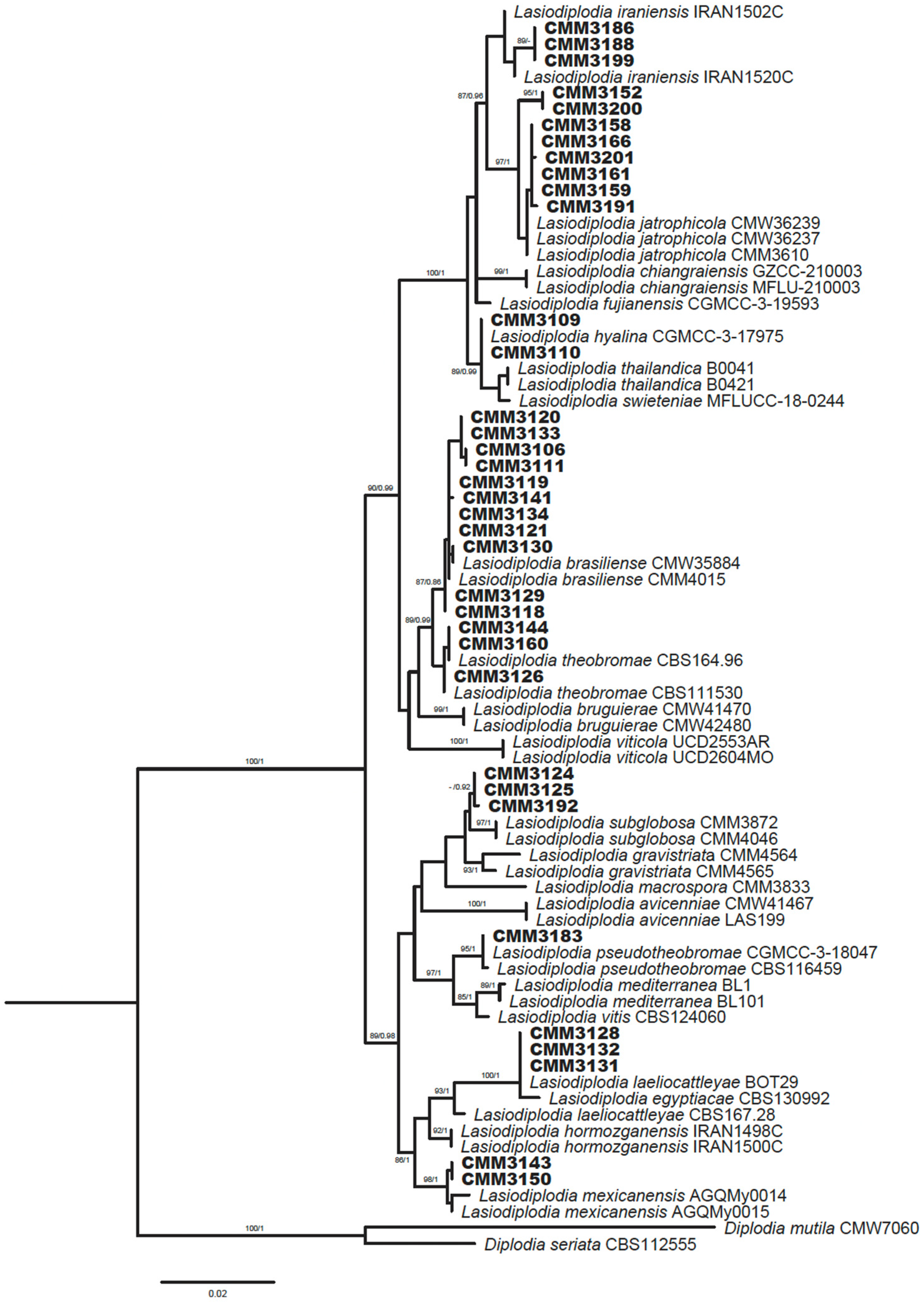
3.2. Phylogenetic Analysis
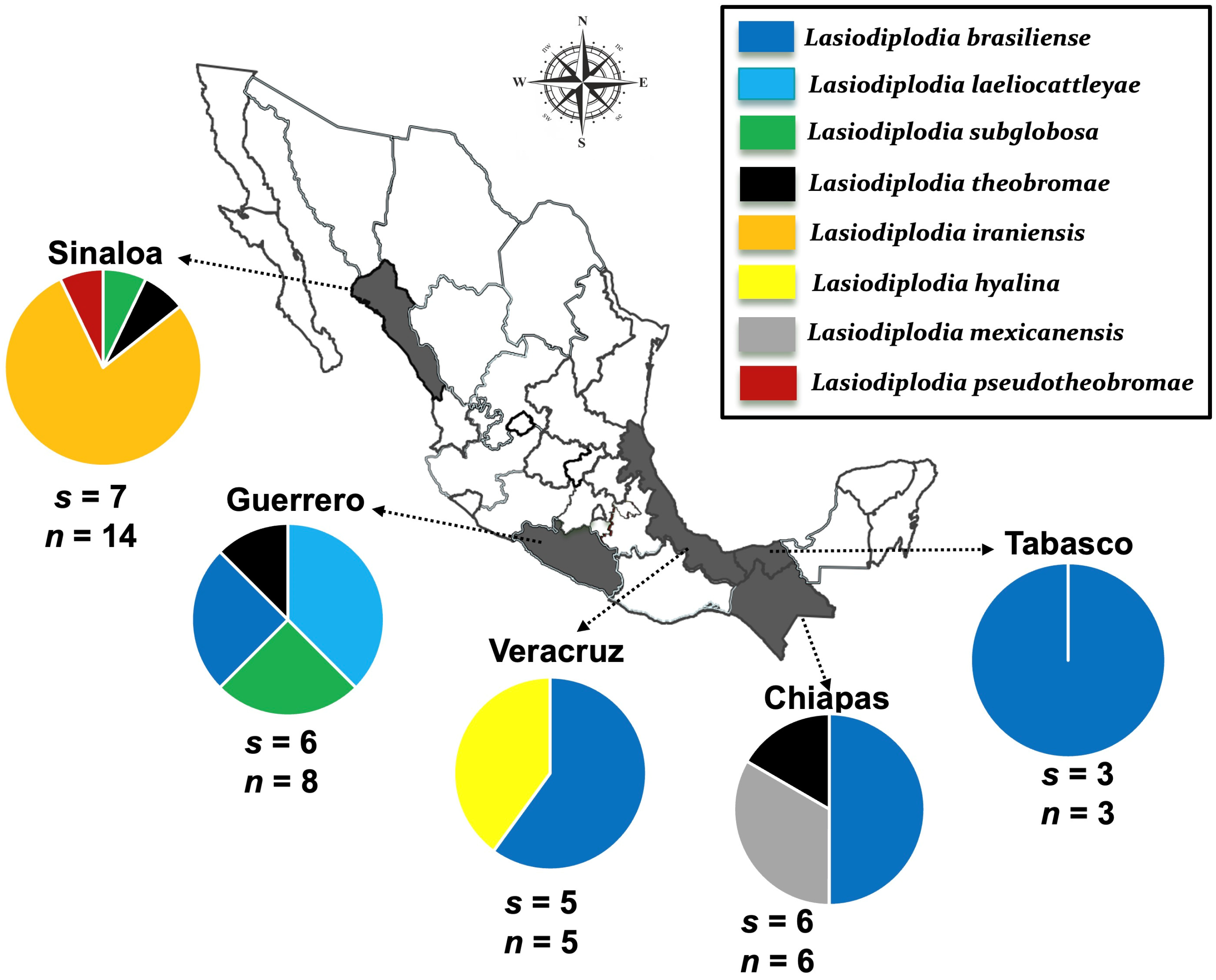
3.3. Distribution of Lasiodiplodia Species
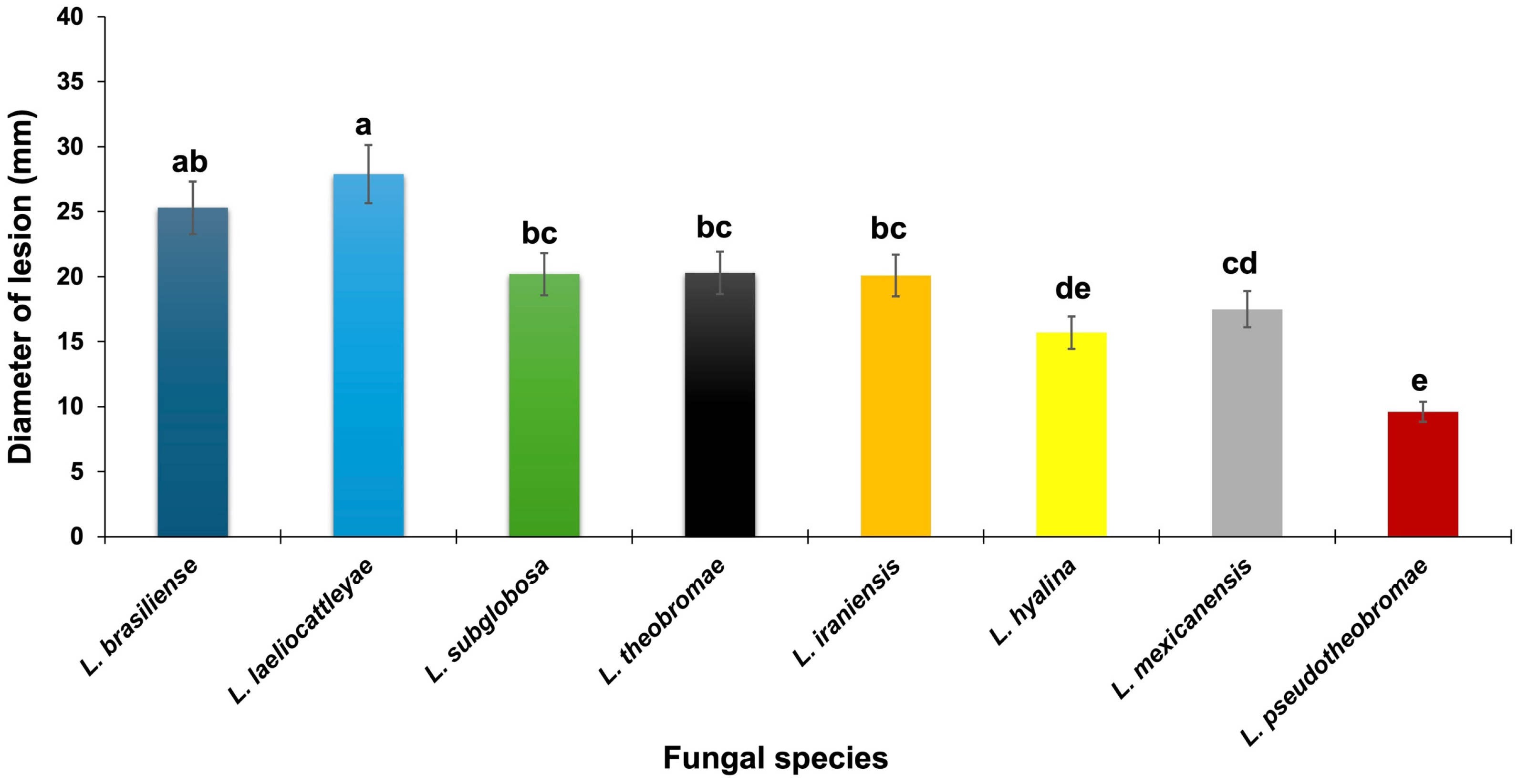
3.4. Pathogenicity and Aggressiveness on Fruits
4. Discussion
Author Contributions
Funding
Institutional Review Board Statement
Informed Consent Statement
Data Availability Statement
Acknowledgments
Conflicts of Interest
References
- Mukherjee, S.K.; Litz, R.E. Introduction: Botany and importance. In The Mango: Botany, Production and Uses; CABI: Wallingford, UK, 2009; pp. 1–18. [Google Scholar] [CrossRef] [Scilit]
- Paul, R.E.; Duarte, O. Tropical Fruits, 2nd ed.; CABI: Wallingford, UK, 2011; Volume 1. [Google Scholar]
- Servicio de Información Agroalimentaria y Pesquera. Panorama Agroalimentario. Available online: https://www.gob.mx/siap (accessed on 10 January 2026).
- Hyde, K.D.; Noorabadi, M.T.; Thiyagaraja, V.; He, M.Q.; Johnston, P.R.; Wijesinghe, S.N.; Armand, A.; Biketova, A.Y.; Chethana, K.W.T.; Erdoğdu, M.; et al. The 2024 outline of fungi and fungus-like taxa. Mycosphere 2024, 15, 5142–6239. [Google Scholar] [CrossRef] [Scilit]
- Thiyagaraja, V.; Hyde, K.D.; Piepenbring, M.; Davydov, E.A.; Dai, D.Q.; Abdollahzadeh, J.; Bundhun, D.; Chethana, K.W.T.; Crous, P.W.; Gajanayake, A.J.; et al. Orders of Ascomycota. Mycosphere 2025, 16, 536–1411. [Google Scholar] [CrossRef] [Scilit]
- Garcia, J.F.; Lawrence, D.P.; Morales-Cruz, A.; Travadon, R.; Minio, A.; Hernandez-Martinez, R.; Rolshausen, P.E.; Baumgartner, K.; Cantu, D. Phylogenomics of Plant-Associated Botryosphaeriaceae Species. Front. Microbiol. 2021, 12, 652802. [Google Scholar] [CrossRef] [Scilit]
- Batista, E.; Lopes, A.; Alves, A. What Do We Know about Botryosphaeriaceae? An Overview of a Worldwide Cured Dataset. Forests 2021, 12, 313. [Google Scholar] [CrossRef] [Scilit]
- Slippers, B.; Wingfield, M.J. Botryosphaeriaceae as endophytes and latent pathogens of woody plants: Diversity, ecology and impact. Fungal Biol. Rev. 2007, 21, 90–106. [Google Scholar] [CrossRef] [Scilit]
- Osorio, J.A.; Crous, C.J.; de Beer, Z.W.; Wingfield, M.J.; Roux, J. Endophytic Botryosphaeriaceae, including five new species, associated with mangrove trees in South Africa. Fungal Biol. 2017, 121, 361–393. [Google Scholar] [CrossRef] [Scilit] [PubMed]
- Laurent, B.; Marchand, M.; Chancerel, E.; Saint-Jean, G.; Capdevielle, X.; Poeydebat, C.; Bellée, A.; Comont, G.; Villate, L.; Desprez-Loustau, M.-L. A Richer Community of Botryosphaeriaceae Within a Less Diverse Community of Fungal Endophytes in Grapevines than in Adjacent Forest Trees Revealed by a Mixed Metabarcoding Strategy. Phytobiomes J. 2020, 4, 252–267. [Google Scholar] [CrossRef] [Scilit]
- Sakalidis, M.L.; Ray, J.D.; Lanoiselet, V.; Hardy, G.E.S.; Burgess, T.I. Pathogenic Botryosphaeriaceae associated with Mangifera indica in the Kimberley Region of Western Australia. Eur. J. Plant Pathol. 2011, 130, 379–391. [Google Scholar] [CrossRef] [Scilit]
- Abdollahzadeh, J.; Zare, R.; Phillips, A.J.L. Phylogeny and taxonomy of Botryosphaeria and Neofusicoccum species in Iran, with description of Botryosphaeria scharifii sp. nov. Mycologia 2013, 105, 210–220. [Google Scholar] [CrossRef] [Scilit]
- Marques, M.W.; Lima, N.B.; de Morais, M.A.; Michereff, S.J.; Phillips, A.J.L.; Câmara, M.P.S. Botryosphaeria, Neofusicoccum, Neoscytalidium and Pseudofusicoccum species associated with mango in Brazil. Fungal Divers. 2013, 61, 195–208. [Google Scholar] [CrossRef] [Scilit]
- de Oliveira Costa, V.S.; Michereff, S.J.; Martins, R.B.; Gava, C.A.T.; Mizubuti, E.S.G.; Câmara, M.P.S. Species of Botryosphaeriaceae associated on mango in Brazil. Eur. J. Plant Pathol. 2010, 127, 509–519. [Google Scholar] [CrossRef] [Scilit]
- Ismail, A.M.; Cirvilleri, G.; Polizzi, G.; Crous, P.W.; Groenewald, J.Z.; Lombard, L. Lasiodiplodia species associated with dieback disease of mango (Mangifera indica) in Egypt. Australas. Plant Pathol. 2012, 41, 649–660. [Google Scholar] [CrossRef] [Scilit]
- Sandoval-Sánchez, M.; Nieto-Ángel, D.; Sandoval-Islas, J.S.; Téliz-Ortiz, D.; Orozco-Santos, M.; Silva-Rojas, H.V. Hongos asociados a pudrición del pedúnculo y muerte descendente del mango (Mangifera indica L.). Agrociencia 2013, 47, 61–73. [Google Scholar]
- de Silva, N.I.; Phillips, A.J.L.; Liu, J.-K.; Lumyong, S.; Hyde, K.D. Phylogeny and morphology of Lasiodiplodia species associated with Magnolia forest plants. Sci. Rep. 2019, 9, 14355. [Google Scholar] [CrossRef] [Scilit] [PubMed]
- Al-Sadi, A.M.; Al-Wehaibi, A.N.; Al-Shariqi, R.M.; Al-Hammadi, M.S.; Al-Hosni, I.A.; Al-Mahmooli, I.H.; Al-Ghaithi, A.G. Population Genetic Analysis Reveals Diversity in Lasiodiplodia Species Infecting Date Palm, Citrus, and Mango in Oman and the UAE. Plant Dis. 2013, 97, 1363–1369. [Google Scholar] [CrossRef] [Scilit]
- Rodríguez-Gálvez, E.; Guerrero, P.; Barradas, C.; Crous, P.W.; Alves, A. Phylogeny and pathogenicity of Lasiodiplodia species associated with dieback of mango in Peru. Fungal Biol. 2017, 121, 452–465. [Google Scholar] [CrossRef] [Scilit]
- Munirah, M.; Azmi, A.; Syc, Y.; NurAinIzzati, M. Characterization of Lasiodiplodia theobromae and L. pseudotheobromae causing fruit rot on pre-harvest mango in Malaysia. Plant Pathol. Quar. 2017, 7, 202–213. [Google Scholar] [CrossRef] [Scilit]
- Sharma, G.; Elazar, M.; Maymon, M.; Meshram, V.; Freeman, S. Identification and pathogenicity of Lasiodiplodia and Neoscytalidium species associated with mango (Mangifera indica) dieback disease in Israel. Phytoparasitica 2024, 52, 8. [Google Scholar] [CrossRef] [Scilit]
- Marques, M.W.; Lima, N.B.; de Morais, M.A., Jr.; Barbosa, M.A.G.; Souza, B.O.; Michereff, S.J.; Phillips, A.J.; Câmara, M.P. Species of Lasiodiplodia associated with mango in Brazil. Fungal Divers. 2013, 61, 181–193. [Google Scholar] [CrossRef] [Scilit]
- Nuñez-García, P.R.; Rebollar-Alviter, A.; García-Estrada, R.S.; Correia, K.C.; Mora-Romero, G.A.; Ramírez-Gil, J.G.; Solano-Báez, A.R.; Léón-Félix, J.; Tovar-Pedraza, J.M. Unraveling the Diversity and Pathogenicity of Botryosphaeriales Species Causing Stem Blight and Dieback of Blueberries in Sinaloa, Mexico. Plant Dis. 2026, 110, 604–616. [Google Scholar] [CrossRef] [Scilit]
- Phillips, A.J.L.; Alves, A.; Abdollahzadeh, J.; Slippers, B.; Wingfield, M.J.; Groenewald, J.Z.; Crous, P.W. The Botryosphaeriaceae: Genera and species known from culture. Stud. Mycol. 2013, 76, 51–167. [Google Scholar] [CrossRef] [Scilit] [PubMed]
- Machado, A.R.; Pinho, D.B.; Pereira, O.L. Phylogeny, identification and pathogenicity of the Botryosphaeriaceae associated with collar and root rot of the biofuel plant Jatropha curcas in Brazil, with a description of new species of Lasiodiplodia. Fungal Divers. 2014, 67, 231–247. [Google Scholar] [CrossRef] [Scilit]
- Slippers, B.; Roux, J.; Wingfield, M.J.; van der Walt, F.J.; Jami, F.; Mehl, J.W.; Marais, G.J. Confronting the constraints of morphological taxonomy in the Botryosphaeriales. Persoonia 2014, 33, 155–168. [Google Scholar] [CrossRef] [Scilit]
- Rosado, A.W.C.; Machado, A.R.; Freire, F.d.C.O.; Pereira, O.L. Phylogeny, Identification, and Pathogenicity of Lasiodiplodia Associated with Postharvest Stem-End Rot of Coconut in Brazil. Plant Dis. 2016, 100, 561–568. [Google Scholar] [CrossRef] [Scilit]
- Coutinho, I.B.L.; Freire, F.C.O.; Lima, C.S.; Lima, J.S.; Gonçalves, F.J.T.; Machado, A.R.; Silva, A.M.S.; Cardoso, J.E. Diversity of genus Lasiodiplodia associated with perennial tropical fruit plants in northeastern Brazil. Plant Pathol. 2017, 66, 90–104. [Google Scholar] [CrossRef] [Scilit]
- Bautista-Cruz, M.A.; Almaguer-Vargas, G.; Leyva-Mir, S.G.; Colinas-León, M.T.; Correia, K.C.; Camacho-Tapia, M.; Robles-Yerena, L.; Michereff, S.J.; Tovar-Pedraza, J.M. Phylogeny, Distribution, and Pathogenicity of Lasiodiplodia Species Associated With Cankers and Dieback Symptoms of Persian Lime in Mexico. Plant Dis. 2018, 103, 1156–1165. [Google Scholar] [CrossRef] [Scilit]
- Farr, D.F.; Rossman, A.Y.; Castlebury, L.A. United States National Fungal Databases. Available online: https://fungi.ars.usda.gov (accessed on 10 January 2026).
- Alves, A.; Crous, P.W.; Correia, A.; Phillips, A.J.L. Morphological and molecular data reveal cryptic speciation in Lasiodiplodia theobromae. Fungal Divers. 2008, 28, 13. [Google Scholar]
- White, T.J.; Bruns, T.D.; Lee, S.B.; Taylor, J.W. Amplification and Direct Sequencing of Fungal Ribosomal RNA Genes for Phylogenetics: PCR Protocols: A Guide to Methods and Applications; Innis, M.A., Gelfand, D.H., Sninsky, J.J., White, T.J., Eds.; Academic Press, Inc.: New York, NY, USA, 1990. [Google Scholar]
- Glass, N.L.; Donaldson, G.C. Development of primer sets designed for use with the PCR to amplify conserved genes from filamentous ascomycetes. Appl. Environ. Microbiol. 1995, 61, 1323–1330. [Google Scholar] [CrossRef] [Scilit]
- Staden, R.; Beal, K.F.; Bonfield, J.K. The Staden package. Methods Mol. Biol. 1998, 132, 115–130. [Google Scholar] [CrossRef] [Scilit]
- Thompson, J.D.; Gibson, T.J.; Plewniak, F.; Jeanmougin, F.; Higgins, D.G. The CLUSTAL_X Windows Interface: Flexible Strategies for Multiple Sequence Alignment Aided by Quality Analysis Tools. Nucleic Acids Res. 1997, 25, 4876–4882. [Google Scholar] [CrossRef] [Scilit] [PubMed]
- Kumar, S.; Stecher, G.; Tamura, K. MEGA7: Molecular Evolutionary Genetics Analysis Version 7.0 for Bigger Datasets. Mol. Biol. Evol. 2016, 33, 1870–1874. [Google Scholar] [CrossRef] [Scilit]
- Nguyen, L.-T.; Schmidt, H.A.; von Haeseler, A.; Minh, B.Q. IQ-TREE: A Fast and Effective Stochastic Algorithm for Estimating Maximum-Likelihood Phylogenies. Mol. Biol. Evol. 2014, 32, 268–274. [Google Scholar] [CrossRef] [Scilit] [PubMed]
- Kalyaanamoorthy, S.; Minh, B.Q.; Wong, T.K.F.; von Haeseler, A.; Jermiin, L.S. ModelFinder: Fast model selection for accurate phylogenetic estimates. Nat. Methods 2017, 14, 587–589. [Google Scholar] [CrossRef] [Scilit]
- Minh, B.Q.; Schmidt, H.A.; Chernomor, O.; Schrempf, D.; Woodhams, M.D.; von Haeseler, A.; Lanfear, R. IQ-TREE 2: New Models and Efficient Methods for Phylogenetic Inference in the Genomic Era. Mol. Biol. Evol. 2020, 37, 1530–1534. [Google Scholar] [CrossRef] [Scilit] [PubMed]
- Ronquist, F.; Huelsenbeck, J.P. MrBayes 3: Bayesian phylogenetic inference under mixed models. Bioinformatics 2003, 19, 1572–1574. [Google Scholar] [CrossRef] [Scilit]
- Slippers, B.; Boissin, E.; Phillips, A.J.; Groenewald, J.Z.; Lombard, L.; Wingfield, M.J.; Postma, A.; Burgess, T.; Crous, P.W. Phylogenetic lineages in the Botryosphaeriales: A systematic and evolutionary framework. Stud. Mycol. 2013, 76, 31–49. [Google Scholar] [CrossRef] [Scilit]
- Correia, K.C.; Silva, M.A.; de Morais, M.A., Jr.; Armengol, J.; Phillips, A.J.L.; Câmara, M.P.S.; Michereff, S.J. Phylogeny, distribution and pathogenicity of Lasiodiplodia species associated with dieback of table grape in the main Brazilian exporting region. Plant Pathol. 2016, 65, 92–103. [Google Scholar] [CrossRef] [Scilit]
- Dissanayake, A.J.; Phillips, A.J.L.; Li, X.H.; Hyde, K.D. Botryosphaeriaceae: Current status of genera and species. Mycosphere 2016, 7, 1001–1073. [Google Scholar] [CrossRef] [Scilit]
- Zhang, H.; Wei, Y.; Yanxiang, Q.; Pu, J.; Liu, X. First report of Lasiodiplodia brasiliense associated with stem-end rot of mango in China. Plant Dis. 2018, 102, 679. [Google Scholar] [CrossRef] [Scilit]
- Dianda, O.Z.; Wonni, I.; Ouédraogo, L.; Sankara, P.; Tollenaere, C.; Del Ponte, E.M.; Fernandez, D. Lasiodiplodia species associated with mango (Mangifera indica L.) decline in Burkina Faso and influence of climatic factors on the disease distribution. Physiol. Mol. Plant Pathol. 2023, 126, 102041. [Google Scholar] [CrossRef] [Scilit]
- Yéo, Y.S.; Dembélé, D.D.; Camara, B.; Amari, E.G.L.D.; Rey, J.Y.; Koné, D.; Fernandez, D. First report of Lasiodiplodia brasiliense causing mango dieback in Côte d’Ivoire. New Dis. Rep. 2024, 50, e12299. [Google Scholar] [CrossRef] [Scilit]
- Trakunyingcharoen, T.; Cheewangkoon, R.; To-anun, C.; Crous, P.W.; van Niekerk, J.M.; Lombard, L. Botryosphaeriaceae associated with diseases of mango (Mangifera indica). Australas. Plant Pathol. 2014, 43, 425–438. [Google Scholar] [CrossRef] [Scilit]
- Alam, M.W.; Malik, A.; Rehman, A.; Sarwar, M.; Ahmad, S.; Shafeeq, T.; Hameed, A.; Rajput, N.A.; Atiq, M. First Report of Lasiodiplodia pseudotheobromae Causing Stem End Rot of Mango Fruit in Pakistan. Plant Dis. 2021, 105, 2249. [Google Scholar] [CrossRef] [Scilit]



| Species | Isolate | Mango Cultivar | Organ | Locality | GenBank Accession No. | ||
|---|---|---|---|---|---|---|---|
| ITS | Tef1α | Tub2 | |||||
| L. brasiliense | CMM3106 | Manila | Stem | Cazones, Veracruz | PV590619 | PV575859 | PV5954651 |
| CMM3111 | Manila | Fruit | Cazones, Veracruz | PV590620 | PV575860 | PV5954652 | |
| CMM3118 | Manila | Stem | Cazones, Veracruz | PV590621 | PV575861 | PV5954653 | |
| CMM3119 | Haden | Fruit | Frontera, Tabasco | PV590622 | PV575862 | PV5954654 | |
| CMM3120 | Haden | Stem | Frontera, Tabasco | PV590623 | PV575863 | PV5954655 | |
| CMM3121 | Tommy Atkins | Stem | Frontera, Tabasco | PV590624 | PV575864 | PV5954656 | |
| CMM3129 | Tommy Atkins | Fruit | Cutzamala, Guerrero | PV590629 | PV575865 | PV5954649 | |
| CMM3130 | Tommy Atkins | Stem | Cutzamala, Guerrero | PV590630 | PV575866 | PV5954650 | |
| CMM3133 | Ataulfo | Stem | Tonala, Chiapas | PV590633 | PV575867 | PV5954646 | |
| CMM3134 | Ataulfo | Stem | Metapa, Chiapas | PV590634 | PV575868 | PV5954647 | |
| CMM3141 | Ataulfo | Stem | Tonala, Chiapas | PV590635 | PV575869 | PV5954648 | |
| L. hyalina | CMM3109 | Manila | Fruit | Cazones, Veracruz | PV590617 | PV582999 | PV5954644 |
| CMM3110 | Manila | Stem | Cazones, Veracruz | PV590618 | PV583000 | PV5954645 | |
| L. iraniensis | CMM3152 | Kent | Stem | Ahome, Sinaloa | PV590639 | PV583001 | PV5954674 |
| CMM3158 | Kent | Stem | Ahome, Sinaloa | PV590640 | PV583006 | PV5954675 | |
| CMM3159 | Kent | Stem | Ahome, Sinaloa | PV590641 | PV583003 | PV5954676 | |
| CMM3161 | Kent | Stem | Ahome, Sinaloa | PV590643 | PV583007 | PV5954677 | |
| CMM3166 | Kent | Stem | Ahome, Sinaloa | PV590644 | PV583008 | PV5954678 | |
| CMM3186 | Keitt | Stem | Ahome, Sinaloa | PV590646 | PV583010 | PV5954668 | |
| CMM3188 | Keitt | Stem | Ahome, Sinaloa | PV590647 | PV583009 | PV5954669 | |
| CMM3191 | Keitt | Stem | Ahome, Sinaloa | PV590648 | PV583004 | PV5954673 | |
| CMM3199 | Keitt | Stem | Ahome, Sinaloa | PV590650 | PV583011 | PV5954670 | |
| CMM3200 | Keitt | Stem | Ahome, Sinaloa | PV590651 | PV583002 | PV5954671 | |
| CMM3201 | Keitt | Stem | Ahome, Sinaloa | PV590652 | PV583005 | PV5954672 | |
| L. laeliocattleyae | CMM3128 | Tommy Atkins | Stem | Cutzamala, Guerrero | PV590628 | PV583015 | PV5954663 |
| CMM3131 | Tommy Atkins | Fruit | Cutzamala, Guerrero | PV590631 | PV583016 | PV5954664 | |
| CMM3132 | Tommy Atkins | Stem | Cutzamala, Guerrero | PV590632 | PV583017 | PV5954665 | |
| L. mexicanensis | CMM3143 | Ataulfo | Fruit | Tonala, Chiapas | PV590636 | PV583018 | PV5954666 |
| CMM3150 | Ataulfo | Stem | Metapa, Chiapas | PV590638 | PV583019 | PV5954667 | |
| L. pseudotheobromae | CMM3183 | Keitt | Stem | Ahome, Sinaloa | PV590645 | PV583020 | PV5954679 |
| L. subglobosa | CMM3124 | Tommy Atkins | Stem | Cutzamala, Guerrero | PV590625 | PV583012 | PV5954658 |
| CMM3125 | Tommy Atkins | Fruit | Cutzamala, Guerrero | PV590626 | PV583013 | PV5954655 | |
| CMM3192 | Keitt | Stem | Ahome, Sinaloa | PV590649 | PV583014 | PV5954657 | |
| L. theobromae | CMM3126 | Tommy Atkins | Fruit | Cutzamala, Guerrero | PV590627 | PV582996 | PV5954661 |
| CMM3144 | Manililla | Stem | Pijijiapan, Chiapas | PV590637 | PV582997 | PV5954662 | |
| CMM3160 | Kent | Stem | Ahome, Sinaloa | PV590642 | PV58298 | PV5954660 | |
| Species | Isolate Code | Host | Origin | GenBank Accession Number | ||
|---|---|---|---|---|---|---|
| ITS | tef1-α | tub2 | ||||
| Diplodia mutila | CMW7060 | Fraxinus excelsior | Netherlands | CMW7060 | AY236955 | AY236904 |
| D. seriata | CBS112555 | Vitis vinifera | Portugal | AY259094 | AY573220 | ------ |
| Lasiodiplodia avicenniae | CMW41467 | Avicennia marina | South Africa | KP860835 | KP860680 | KP860758 |
| L. avicenniae | LAS199 | Avicennia marina | South Africa | KU587957 | KU587947 | KU587868 |
| L. brasiliense | CMM4015 | Mangifera indica | Brazil | JX464063 | JX464049 | ------ |
| L. brasiliense | CMW35884 | A. madagascariensis | Madagascar | KU887094 | KU886972 | KU887466 |
| L. bruguierae | CMW41470 | Bruguiera gymnorrhiza | South Africa | KP860832 | KP860677 | KP860755 |
| L. bruguierae | CMW42480 | Bruguiera gymnorrhiza | South Africa | KP860834 | KP860679 | KP860757 |
| L. chiangraiensis | MFLUCC-21-0003 | ------ | Thailand | MW760854 | MW815630 | MW815628 |
| L. chiangraiensis | GZCC-21-0003 | ------ | Thailand | MW760853 | MW815629 | MW815627 |
| L. fujianensis | CGMCC-3-19593 | Vaccinium uliginosum | China | MK802164 | MK887178 | MK816337 |
| L. gravistriata | CMM4564 | Anacardium humile | Brazil | KT250949 | KT250950 | ------ |
| L. gravistriata | CMM4565 | Anacardium sp. | Brazil | KT250947 | KT266812 | ------ |
| L. hormozganensis | IRAN1500C | Olea sp. | Iran | GU945355 | GU945343 | KU887515 |
| L. hormozganensis | IRAN1498C | Mangifera indica | Iran | GU945355 | GU945344 | KU887514 |
| L. hyalina | CGMCC3.17975 | Acacia confusa | China | KX499879 | KX499917 | KX499992 |
| L. iraniensis | IRAN1502C | Juglans sp. | Iran | GU945347 | GU945335 | KU887517 |
| L. iraniensis | IRAN1520C | Salvadora persica | Iran | GU945348 | GU945336 | KU887516 |
| L. iraniensis | CMM3610 | Jatropha curcas | Brazil | KF234544 | KF226690 | KF254927 |
| L. iraniensis | CMW36237 | Adansonia digitata | Mozambique | KU887121 | KU886998 | KU887499 |
| L. iraniensis | CMW36239 | Adansonia digitata | Mozambique | KU887123 | KU887000 | KU887501 |
| L. laeliocattleyae | CBS 167.28 | Laeliocattleyae sp. | Italy | KU507487 | KU507454 | ------ |
| L. laeliocattleyae | BOT 29 | Mangifera indica | Egypt | JN814401 | JN814428 | ------ |
| L. laeliocattleyae | CBS130992 | Mangifera indica | Egypt | JN814397 | JN814424 | KU887508 |
| L. macrospora | CMM3833 | Jatropha curcas | Brazil | KF234557 | KF226718 | KF254941 |
| L. mediterranea | BL1 | Quercus ilex | Italy | KJ638312 | KJ638331 | ------ |
| L. mediterranea | BL101 | Vitis sp. | Italy | KJ638311 | KJ638330 | ------ |
| L. mexicanensis | AGQMy 0014 | Chamaedorea seifrizii | Mexico | MW274151 | MW604234 | MW604243 |
| L. mexicanensis | AGQMy0015 | Chamaedorea seifrizii | Mexico | MW274150 | MW604233 | MW604242 |
| L. pseudotheobromae | CBS116459 | Gmelina arborea | Costa Rica | EF622077 | EF622057 | EU673111 |
| L. pseudotheobromae | CGMCC-3-18047 | Pteridium sp. | China | KX499876 | KX499914 | KX499989 |
| L. subglobosa | CMM3872 | Jatropha curcas | Brazil | KF234558 | KF226721 | KF254942 |
| L. subglobosa | CMM4046 | Jatropha curcas | Brazil | KF234560 | KF226723 | KF254944 |
| L. swieteniae | MFLUCC-18-0244 | Swietenia mahagoni | Thailand | MK347789 | MK340870 | MK412877 |
| L. thailandica | B0041 | Phyllanthus acidus | Thailand | KM006433 | KM006464 | ------ |
| L. thailandica | B0421 | Mangifera indica | Thailand | KJ193637 | KJ193681 | ------ |
| L. theobromae | CBS111530 | Unknown | Unknown | EF622074 | EF622054 | KU887531 |
| L. theobromae | CBS164.96 | Unknown | New Guinea | AY640255 | AY640258 | KU887532 |
| L. viticola | UCD2553AR | Vitis vinifera | USA | HQ288227 | HQ288269 | HQ288306 |
| L. viticola | USD2604MO | Vitis vinifera | USA | HQ288228 | HQ288270 | HQ288307 |
| L. vitis | CBS 124060 | Vitis vinifera | Italy | KX464148 | KX464642 | KX464917 |
Disclaimer/Publisher’s Note: The statements, opinions and data contained in all publications are solely those of the individual author(s) and contributor(s) and not of MDPI and/or the editor(s). MDPI and/or the editor(s) disclaim responsibility for any injury to people or property resulting from any ideas, methods, instructions or products referred to in the content. |
© 2026 by the authors. Licensee MDPI, Basel, Switzerland. This article is an open access article distributed under the terms and conditions of the Creative Commons Attribution (CC BY) license.
Share and Cite
Tovar-Pedraza, J.M.; Estrada-Arroyo, G.; Macedo-Arzate, R.; Michereff, S.J.; Correia, K.C.; Leyva-Mir, S.G.; Mora-Aguilera, J.A.; Camacho-Tapia, M.; Márquez-Licona, G.; Solano-Báez, A.R. Multilocus Phylogenetic Identification and Fruit Pathogenicity of Lasiodiplodia Isolates Obtained from Mango Branches with Dieback and Fruits with Stem-End Rot in Mexico. J. Fungi 2026, 12, 370. https://doi.org/10.3390/jof12050370
Tovar-Pedraza JM, Estrada-Arroyo G, Macedo-Arzate R, Michereff SJ, Correia KC, Leyva-Mir SG, Mora-Aguilera JA, Camacho-Tapia M, Márquez-Licona G, Solano-Báez AR. Multilocus Phylogenetic Identification and Fruit Pathogenicity of Lasiodiplodia Isolates Obtained from Mango Branches with Dieback and Fruits with Stem-End Rot in Mexico. Journal of Fungi. 2026; 12(5):370. https://doi.org/10.3390/jof12050370
Chicago/Turabian StyleTovar-Pedraza, Juan Manuel, Guillermo Estrada-Arroyo, Rafael Macedo-Arzate, Sami J. Michereff, Kamila C. Correia, Santos Gerardo Leyva-Mir, José Antonio Mora-Aguilera, Moisés Camacho-Tapia, Guillermo Márquez-Licona, and Alma Rosa Solano-Báez. 2026. "Multilocus Phylogenetic Identification and Fruit Pathogenicity of Lasiodiplodia Isolates Obtained from Mango Branches with Dieback and Fruits with Stem-End Rot in Mexico" Journal of Fungi 12, no. 5: 370. https://doi.org/10.3390/jof12050370
APA StyleTovar-Pedraza, J. M., Estrada-Arroyo, G., Macedo-Arzate, R., Michereff, S. J., Correia, K. C., Leyva-Mir, S. G., Mora-Aguilera, J. A., Camacho-Tapia, M., Márquez-Licona, G., & Solano-Báez, A. R. (2026). Multilocus Phylogenetic Identification and Fruit Pathogenicity of Lasiodiplodia Isolates Obtained from Mango Branches with Dieback and Fruits with Stem-End Rot in Mexico. Journal of Fungi, 12(5), 370. https://doi.org/10.3390/jof12050370