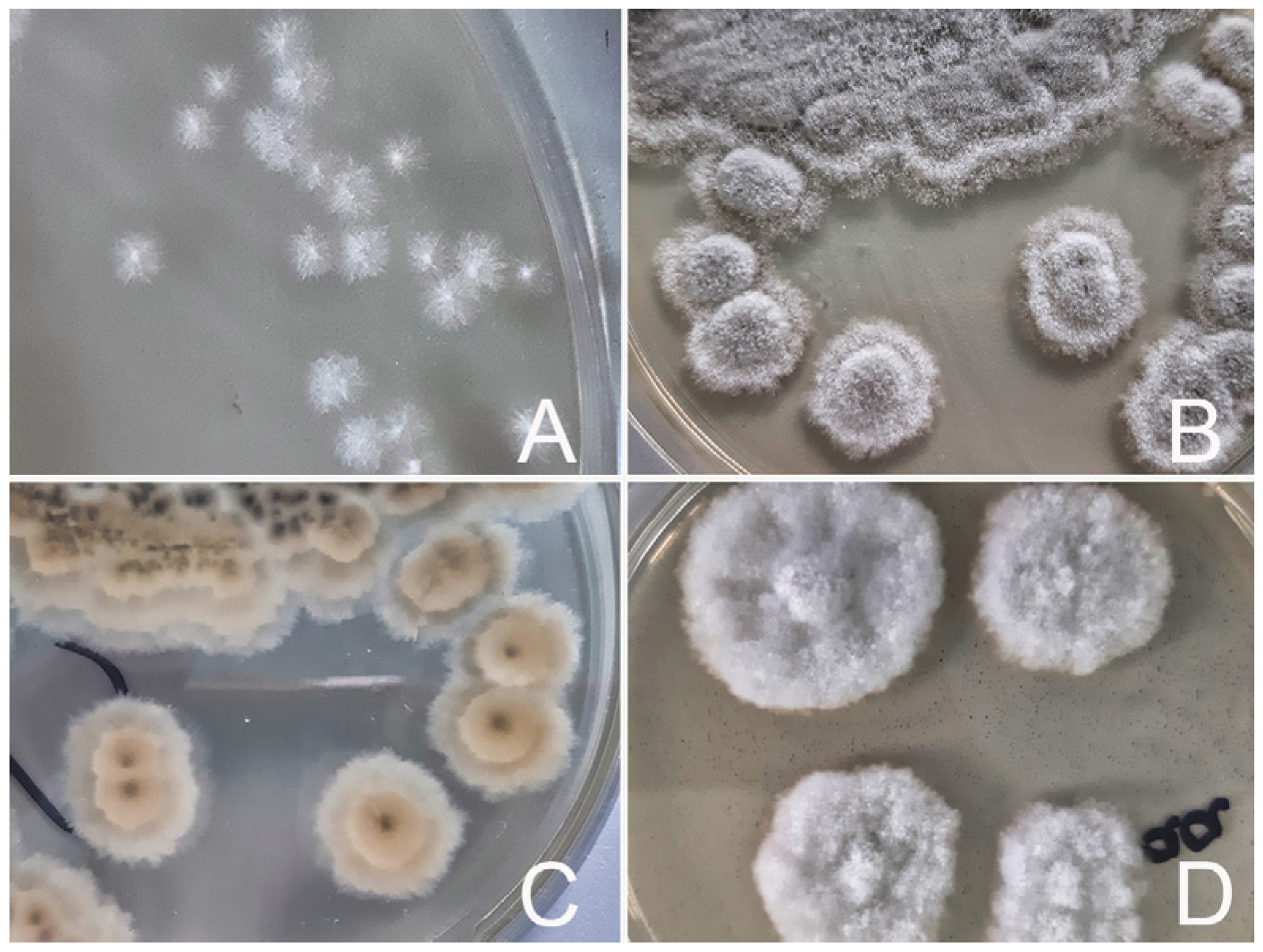
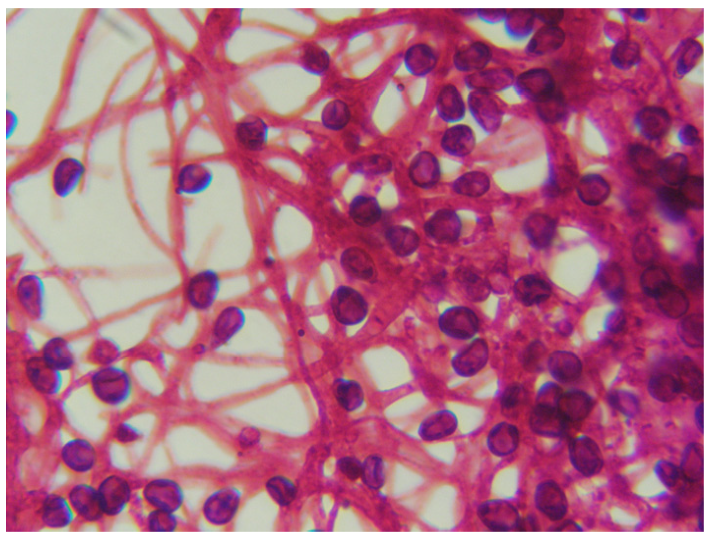
Scedosporium apiospermum: An Emerging yet Overlooked Fungal Pathogen in Veterinary Medicine—A Case-Based Review




Abstract
1. Introduction
1.1. The Genus Scedosporium: Taxonomy and History
1.2. Distribution in the Environment
1.3. Scedosporium apiospermum as a Causative Agent of Animal Diseases

| Country | Age, Sex and Breed | Anamnesis, Disease Symptoms, and Clinical Findings | Treatment and Outcome | Causative Agent Identification | Reference |
|---|---|---|---|---|---|
| (a) | |||||
| Dogs | |||||
| Spain | 10-month-old Male American Staffordshire terrier | -6-month history of mucopurulent bilateral nasal discharge and some sneezing; unresponsive to antibiotics; -rhinoscopic examination revealed destruction of the vomer bone and a large mass completely obstructing the nasal cavity; biopsy specimens revealed the presence of granules containing numerous septate hyphae. | -oral ketoconazole and amoxicillin; -general improvement of the lesions was observed. | Culture S. apiospermum | [24] |
| New Zealand | 2-year-old Female Siberian Husky | -6-month history of sneezing and mucous discharge from the right nostril; reduced airflow; -radiographs demonstrated a subtle loss of detail of turbinates within the right nasal chamber; histological examination of the white mass revealed a mixture of fungal hyphae and spores; dg: fungal rhinitis. | -no treatment; -the apparent spontaneous resolution of this case is an interesting finding. | Culture S. apiospermum | [25] |
| Spain | 2-year old intact Female Labrador retriever | -8-month history of bilateral mucopurulent nasal discharge; -previous several courses of antimicrobials without positive response; -histopathological diagnosis was mycotic rhinitis. | -surgical debridement with topical clotrimazole treatment; -complete remission after 4 months. | Culture S. apiospermum | [26] |
| USA | 9-year-old Male castrated Australian shepherd mixed-breed dog | -3-month history of intermittent, right-sided epistaxis; previously treatment with enrofloxacin, prednisone and amoxicillin/clavulanate. -CT findings revealed right-sided destructive rhinitis and sinusitis, which were thought to be most likely associated with infection with Aspergillus; rhinoscopy revealed marked turbinate destruction. | -debridement, frontal sinus trephination and clotrimazole therapy; -3 months after the dog remained free of clinical signs. | Culture, DNA sequencing P. boydii anamorph S. apiospermum | [27] |
| Italy | 4-year-old Female neutered Bull Terrier | -history of partially hemorrhagic, unilateral, mucopurulent left-sided nasal discharge and reverse sneezing; failure to respond to antibiotic therapy; -yellowish white/brown material of rubbery consistency was found in dorsal meatus of the left nasal cavity; the histological samples of the nasal mucosa revealed a marked inflammatory pyogranulomatous process; the mucous surface presented large clusters of fungal hyphae. | -nasal cavities infused with miconazole; -2 weeks after the start of the therapy, no clinical symptoms of rhinitis found. | Culture MALDI TOF S. apiospermum | [16] |
| Australia | 8-year-old Male neutered Golden Retriever | -history of violent sneezing and licking at the nasal planum; -rhinoscopic examination revealed inflamed nasal mucosa and blood in the right nasal passage; CT scan: a large rotting grass seed in the right middle meatus and turbinate destruction of the right dorsal nasal cavity; biopsy dg: fungal rhinitis. | -the grass seed was removed; -oral itraconazole for 3 months; -complete resolution of clinical disease. | Culture, PCR and DNA sequencing S. apiospermum | [9] |
| UK | 10-year-old Male neutered Irish Terrier | -unilateral nasal discharge; long-term treatment with prednisolone; -computed tomography showed severe chronic erosive rhinitis and severe periodontal disease; rhinoscopy: multiple plaque-like lesions throughout the nasal cavity with turbinate destruction. | -3-month treatment with itraconazole, followed by topical treatment with clotrimazole without surgical debridement; -dog was euthanized. | Culture S. apiospermum | [28] |
| Cat | |||||
| France | 3-year-old neutered Male Bengal cat | -history of mucopurulent bilateral nasal discharge and chronic sneezing; several courses of antibiotics without any improvement; -CT: chronic severe inflammatory lesion; bilateral mucopurulent nasal discharge associated with a facial asymmetry due to an apparent firm deformation of the right nasal cavity; the histological diagnosis was pyogranulomatous chronic rhinosinusitis with multifocal ulceration of the mucosa. | -surgical debridement; -topical and systemic therapy with enilconazole and itraconazole for 2 months; -the owners refused further treatments; after 12 months the symptoms returned. | Culture S. apiospermum the asexual form of Pseudallescheria boydii | [29] |
| Cattle | |||||
| USA | 2-year-old Hereford cow | -chronic upper respiratory dysfunction, increased respiratory noise, and bloody nasal discharge; prior empiric therapy with parenteral oxytetracycline and sodium iodide; -clinically: nodular rhinitis (multiple polypoid masses in both nares), mild tachypnea and inspiratory stridor; nasal biopsy revealed fungal-induced granulomatous and eosinophilic inflammation. | -parenteral antihistamine -no improvement; -the animal was euthanized due to financial constraints. | Culture Pseudallescheria boydii species complex -anamorph S. apiospermum | [30] |
| Horses | |||||
| UK | 8-year-old mare | -for two years several episodes of left-sided purulent to mucopurulent nasal discharge; treated with sulphonamides; -on examination there was a left-sided purulent nasal discharge; endoscopic examination of the nasal chambers revealed plaques of material resembling mycotic hyphae. | -mare was euthanized at the owner’s request. | Culture Pseudallescheria boydii | [31] |
| USA | 18-year-old American Quarter Horse | -bilateral, mucopurulent nasal discharge; empirical judgment with penicillin G and trimethoprim-sulfamethoxazole; -purulent discharge from the right nostril; large white plaques in the nasal cavity; cytologic evaluation of the samples of the plaques revealed numerous fungal hyphae and conidia; a presumptive diagnosis: fungal sinusitis. | -topical treatment with miconazole and systemic treatment with sodium iodide and potassium iodide; -infection resolved. | Culture Pseudallescheria boydii | [32] |
| (b) | |||||
| Dogs | |||||
| USA | 8-year-old West Highland White Terrier | -bilateral chronic keratoconjunctivitis sicca and corneal ulceration in the right eye; a history of chronic topic ocular therapy with antibiotics and corticosteroids. | -the owner chose to proceed with orbital exenteration due to the uncertain prognosis and the prospect of long-term treatment. | Culture Pseudallescheria boydii | [33] |
| UK | 6-year-old dog Male castrated Norfolk Terrier | -21-day history of an increasingly painful eye; the dog had been on systemic steroids for inflammatory bowel disease for the previous 3 months and 21-day period of antibiotic use; -ulcerative keratitis of left eye, blepharospasm, photophobia, purulent ocular discharge; conjunctival hyperemia; cytology slides from the corneal scrape demonstrated a high number of branching septate hyphae. | -keratectomy; -topical voriconazole; -after thirty-five days, the ulcer had healed. | PCR (DNA was extracted from the corneal scrape). S. apiospermum | [34] |
| Horses | |||||
| USA | Quarter Horse | Keratomycosis. Despite topical treatment with miconazole and natamycin, the cornea developed a stromal abscess. Orbital exenteration was performed after 3 weeks. Full-text not available. | [35] | ||
| USA | 11-year-old American Saddlebred gelding | -2 years earlier a painless white corneal opacity in the left eye;1 month previously noted mass involving the left eye and had subsequently increased in size; -ophthalmic examination revealed a firm mass under the palpebral conjunctiva of the left eye; fine-needle aspiration of the mass and smears were submitted for cytologic examination; the cytologic diagnosis was pyogranulomatous inflammation with intralesional fungal organisms. | -initial: neomycin–bacitracin–polymyxin; -based on cytologic evaluation, therapy was changed to neomycin–polymyxin gramicidin solution and voriconazole 1% ophthalmic solution; -the patient recovered after surgical excision of the mass. | Culture S. apiospermum | [20] |
| Poultry | |||||
| Australia | 42-day-old birds | -42-day-old birds in a layer pullet flock have ocular abnormalities, unilateral keratoconjunctivitis and apparent exophthalmos; thick, cream apparently fibrinous ocular discharge; -pathohistological diagnosis: severe chronic mycotic keratitis and severe chronic active iritis or iridocyclitis. | -not treated. | Culture Scedosporium apiospermum | [23] |
| (c) | |||||
| Dogs | |||||
| Australia | 2y Rhodesian Ridgeback | -immune-mediated polyarthritis and immune-mediated hemolytic anemia; immunosuppressant therapy; -lymphocutaneous infection (rump, lateral front leg, and right cranial shoulder). | -successful treatment with itraconazole, azathioprine and terbinafine. | Culture S. apiospermum | [36] |
| India | age and sex-not specified Pug | -a history of dogfight, which had caused a bite wound on its right front leg, -wound was infected and the lesion started spreading to nearby areas and became dark in color. | -successful oral treatment with itraconazole and voriconazole for 4 weeks and another two months to avoid relapse. | Culture and molecular confirmation by gene sequencing S. apiospermum | [37] |
| Horse | |||||
| USA | 11-year-old Quarter Horse gelding | -a cutaneous mass adjacent to the medial canthus of the left eye, present for approximately six months; -histological examination: infiltrate of neutrophils, macrophages, lymphocytes and plasma cells around fungal grains or microcolonies; dg: eumycotic mycetoma. | -successful treatment: curative excision. | Immunofluorescent examination of formalin -fixed tissue Pseudallescheria boydii the ascocarpic form of S. apiospermum | [38] |
| Country | Age, Sex and Breed | Anamnesis, Disease Symptoms, and Clinical Findings | Treatment and Outcome | Pathological Findings | Causative Agent Identification | Reference |
|---|---|---|---|---|---|---|
| Dogs | ||||||
| USA | 6-year-old spayed Miniature Poodle | -previous rupture of the uterus and ovariohysterectomy with stainless steel sutures, antibiotics treatment; -gastrointestinal signs (vomiting and had diarrhea), weight loss, anorexia; non-movable mass was palpated in the ventral abdomen; radiographs revealed an irregular mass caudal to the liver and two stainless sutures free in the abdomen; -histologic examination of biopsy specimen consistent with eumycotic mycetoma. | -surgical excision of the mass; -propantheline bromide and chloramphenicol; -the dog died. | -acute peritonitis; -surgical site was not intact, resulting in fecal leakage into the abdominal cavity. | Culture Monosporium apiospermum (asexual stage of Allescheria boydii) | [39] |
| USA | 2-year-old spayed Golden Retriever-type | -at six months of age, the dog underwent an ovariohysterectomy, but recovery was complicated by abdominal incision dehiscence and subsequent evisceration; previously antibiotic treatment for six weeks; chronic weight loss, fever, diarrhea, vomiting; -dog was cachectic, icteric and dehydrated; palpable midabdominal mass; hematuria, proteinuria, glucosuria. | -supportive therapy with intravenous fluids and antibiotic; -dog died before surgery could be performed. | -pathological diagnosis: pyogranulomatous peritonitis, hepatitis, pneumonia, vasculitis and thrombosis. | Culture Scedosporium apiospermum (the anamorphic phase of Pseudallescheria boydii) | [40] |
| USA | 5-year-old castrated Male German Shepherd | -3-week history of lethargy, intermittent fever; left forelimb lameness; -soft tissue swelling of the left elbow; bilateral exudative chorioretinitis and retinal detachment; radiographically: periosteal proliferation of the left distal humerus and a cranial mediastinal mass; aspirates from the bone lesion contained macrophages, neutrophils, and fungal elements; presumptive diagnosis: disseminated mycotic disease. | -the dog was euthanized. | -large cranial mediastinal mass; generalized lymphadenomegaly, -disseminated granulomas in the visceral organs and bone (left distal humerus); -granulomatous inflammation with necrotizing vasculitis. | Immunofluorescence (using fluorescein-conjugated immunoglobulins specific for P. boydii in deparaffinized tissue) Pseudallescheria boydii | [41] |
| USA | 3-year-old Male Siberian Husky | -1 month history of weight loss and signs of depression, fever, vomiting; previous treatment: penicillin and dexamethasone parenterally; -caudal portion of abdomen was sensitive to palpation; testicular swelling; | -orchiectomy and ketoconazole; -dog died one month later. | -purulent peritonitis; -pyogranulomatous periorchitis, enteritis, pancreatitis; -the duodenal mucosa was ulcerated and its wall contained multiple small craters or tracts, filled with caseous yellow exudate. | Culture and immunofluorescence (using fluorescein isothiocyanate-conjugated immunoglobulins specific for P. boydii on formalin-fixed tissue) Monosporium apiospermum | [42] |
| France | 6-year-old Female German Shepherd Dog | -progressive rear limb paresis/paralysis; -radiographs revealed narrowed vertebral lesions consistent with osteomyelitis and discospondylitis. | -enrofloxacin and intravenous corticosteroid; -dog was euthanized. | -chronic, severe, pyogranulomatous fungal T13-L1 vertebral osteomyelitis and discospondylitis. | PCR and sequencing on formalin-fixed, paraffin- embedded samples Scedosporium apiospermum | [43] |
| Germany | 4-year-old mixed-breed | -chronic gastrointestinal signs (vomiting, lethargy anorexia); polydipsia; previously: antibiotic therapy; -upon palpation, the abdomen was tense and painful; the radiological findings suggested gastric outflow obstruction with pyloric dilatation and focal peritonitis; histologic examination revealed septic-purulent to pyogranulomatous inflammation, focally numerous lymphocites, plasma cells and numerous fungal hyphae. | -surgical resection (hepatic lobectomy and enterectomy of parts of the descending duodenum); -systemic antifungal therapy for several months with itraconazole. -nine months after surgery the dog was presented in undisturbed general condition. | NA | DNA sequencing of the β tubulin gene Scedosporium apiospermum | [17] |
| Italy | 10-month-old Female Maremmano-Abruzzese sheepdog | -weakness, lethargy, lateral decubitus, miosis and muscular rigidity; -an episode of diarrhea, vomiting and anorexia was reported 24–36 h before referral to the veterinary clinic; -the right inguinal mammary gland and the surrounding subcutaneous tissues were moderately swollen. | -supportive therapy with intravenous fluids and amoxicillin-clavulanic acid; -dog died. | -Severe multifocal fungal pyogranulomas in kidney, mesentery, lymph nodes, and mammary gland. | Culture and nucleotide sequence-based analysis Scedosporium apiospermum | [18] |
| USA | 10-year-old intact Male Border Collie | -prior history of septic peritonitis caused by gastrointestinal perforation related to the use of nonsteroidal anti-inflammatory drugs, as well as a right femoral head and neck ostectomy; -two-month history of stranguria, tenesmus, and weight loss; -abdominal ultrasound revealed a large caudal abdominal mass and marked medial iliac lymphadenopathy; -histologic examination showing chronic pyogranulomatous cystitis and focal peritonitis with intralesional fungal hyphae. | -itraconazole: 5 mg/kg for 6 months; -partial cystectomy; -successful outcome. | NA | Culture, MALDI-TOF, PCR, and DNA sequencing Scedosporium apiospermum | [22] |
| USA | 5-year-old, spayed Female, Basset Hound | -hematuria, stranguria, and urinary incontinence over a period of 4 months; -ultrasonography: mass in the urinary bladder (not reveal any metastatic disease); histologic examination of surgical specimens revealed pyogranulomatous cystitis and ureteritis; ureteral obstruction by fungal granuloma and hydronephrosis. | -surgical excision of the mass; -oral voriconazole for several months; -8 months later the mass was no longer visible. | NA | Culture and DNA sequencing Scedosporium apiospermum | [44] |
| Japan | 6-year-old castrated Male Golden Retriever | -history of gastrointestinal anastomosis under laparotomy 2 years earlier; -ultrasonography revealed multiple large masses in the abdominal cavity; fine-needle aspiration (FNA) of the masses revealed numerous neutrophils, macrophages, and septate hyphae; -dg: fungal granuloma attached to the jejunum, pancreas, main portal vein, and other abdominal organs. | -surgical removal of the masses; -variconazole p.o., 5 mg/kg; -after 4 months fungal granuloma has reduced; -continue treatment until the lesions completely resolve. | NA | Culture and and nucleotide sequence-based analysis Scedosporium apiospermum | [45] |
| USA | 2-year-old intact Female mixed-breed dog | -a chronic history of nonspecific gastrointestinal signs: vomiting, hyporexia and progressive weight loss; -unsuccessfully managed with famotidine; -a large peripancreatic mass and several other masses and effusions were found during an abdominal ultrasound; fine-needle aspiration of the largest mass revealed numerous degenerate neutrophils and epithelioid macrophages surrounding dense mats of fungal organisms; exploratory laparotomy confirmed widespread peritoneal granulomas. | -due to the extensive nature of the lesions and the poor prognosis associated with intra-abdominal fungal infection, the owner elected euthanasia. | -the peritoneum was diffusely thickened, exhibiting dark-red discoloration and extensive fibrous adhesions involving the liver, pancreas, intestines, stomach, and mesentery. | Culture and PCR Scedosporium apiospermum (coinfection with Nocardia spp.) | [46] |
| Cattle | ||||||
| India | 45 days old dead calf | NA | NA | -severe pneumonic granulomatous lesions containing septate, pleomorphic hyphae were observed in the central caseated core, which had a bright eosinophilic periphery surrounded by polymorphonuclear cells and macrophages, followed by a zone of epithelioid cells mixed with lymphocytes. | Culture P. boydii (is a sexual form of Scedosporium apiospermum) | [47] |
2. Overall Observations
2.1. Routes of Infection and Predisposing Factors
2.2. Diagnosis
2.3. Microbiological Diagnosis
2.4. Antifungal Susceptibility Testing
2.5. Therapy
3. Conclusions
Author Contributions
Funding
Institutional Review Board Statement
Informed Consent Statement
Data Availability Statement
Conflicts of Interest
References
- Bouchara, J.-P.; Papon, N. Scedosporium apiospermum. Trends Microbiol. 2019, 27, 1045–1046. [Google Scholar] [CrossRef]
- Chen, S.C.; Halliday, C.L.; Hoenigl, M.; Cornely, O.A.; Meyer, W. Scedosporium and Lomentospora infections: Contemporary microbiological tools for the diagnosis of invasive disease. J. Fungi 2021, 7, 23. [Google Scholar] [CrossRef] [PubMed]
- Abrantes, R.A.; Refojo, N.; Hevia, A.I.; Fernández, J.; Isla, G.; Córdoba, S.; Dávalos, M.F.; Lubovich, S.; Maldonado, I.; Davel, G.O.; et al. Scedosporium spp. from clinical setting in Argentina, with the proposal of the new pathogenic species Scedosporium americanum. J. Fungi 2021, 7, 160. [Google Scholar] [CrossRef] [PubMed]
- Bronnimann, D.; Garcia-Hermoso, D.; Dromer, F.; Lanternier, F.; French Mycoses Study Group. Scedosporiosis/lomentosporiosis observational study (SOS): Clinical significance of Scedosporium species identification. Med. Mycol. 2021, 59, 486–497. [Google Scholar] [CrossRef] [PubMed]
- Guarro, J.; Kantarcioglu, A.S.; Horré, R.; Rodriguez-Tudela, J.L.; Cuenca Estrella, M.; Berenguer, J.; de Hoog, G.S. Scedosporium apiospermum: Changing clinical spectrum of a therapy-refractory opportunist. Med. Mycol. 2006, 44, 295–327. [Google Scholar] [CrossRef]
- Luplertlop, N. Pseudallescheria/Scedosporium complex species: From saprobic to pathogenic fungus. J. Mycol. Med. 2018, 28, 249–256. [Google Scholar] [CrossRef]
- Cortez, K.J.; Roilides, E.; Quiroz-Telles, F.; Meletiadis, J.; Antachopoulos, C.; Knudsen, T.; Buchanan, W.; Milanovich, J.; Sutton, D.A.; Fothergill, A.; et al. Infections caused by Scedosporium spp. Clin. Microbiol. Rev. 2008, 21, 157–197. [Google Scholar] [CrossRef] [PubMed]
- Luna-Rodríguez, C.E.; Treviño-Rangel, R.J.; Montoya, A.M.; Becerril-García, M.A.; Andrade, A.; González, G.M. Scedosporium spp.: Chronicle of an emerging pathogen. Med. Univ. 2019, 21, 4–13. [Google Scholar] [CrossRef]
- Smith, C.G.; Woolford, L.; Talbot, J.J.; Lee, K.C.; Crocker, P. Canine rhinitis caused by an uncommonly-diagnosed fungus, Scedosporium apiospermum. Med. Mycol. Case Rep. 2018, 22, 38–41. [Google Scholar] [CrossRef]
- Chen, M.; Zeng, J.; de Hoog, G.S.; Stielow, B.; van den Ende, A.H.G.; Liao, W.; Lackner, M. The “species complex” issue in clinically relevant fungi: A case study in Scedosporium apiospermum. Fungal Biol. 2016, 120, 137–146. [Google Scholar] [CrossRef]
- Nweze, E.I.; Okafor, J.I. Comparative virulence of Scedosporium species in animal models. Braz. J. Infect. Dis. 2010, 14, 271–276. [Google Scholar] [CrossRef] [PubMed]
- Kaltseis, J.; Rainer, J.; de Hoog, G.S. Ecology of Pseudallescheria and Scedosporium species in human-dominated and natural environments and their distribution in clinical samples. Med. Mycol. 2009, 47, 398–405. [Google Scholar] [CrossRef] [PubMed]
- Elizondo-Zertuche, M.; de J. Treviño-Rangel, R.; Robledo-Leal, E.; Luna-Rodríguez, C.E.; Martínez-Fierro, M.L.; Rodríguez-Sánchez, I.P.; González, G.M. Molecular identification and in vitro antifungal susceptibility of Scedosporium complex isolates from high-human-activity sites in Mexico. Mycologia 2017, 109, 874–881. [Google Scholar] [CrossRef] [PubMed]
- Mina, S.; Yaakoub, H.; Razafimandimby, B.; Dwars, E.; Wery, M.; Papon, N.; Meyer, W.; Bouchara, J.-P. First environmental survey of Scedosporium species in Lebanon. Front. Cell. Infect. Microbiol. 2025, 15, 1547800. [Google Scholar] [CrossRef] [PubMed]
- Elad, D. Infections caused by fungi of the Scedosporium/Pseudallescheria complex in veterinary species. Vet. J. 2011, 187, 33–41. [Google Scholar] [CrossRef]
- Matteucci, G.; Lovari, L.; Binanti, D. Mycotic rhinitis caused by Scedosporium apiospermum in a dog: First report in Italy. Veterinaria 2017, 31, 333–337. [Google Scholar]
- Thiel, C.; Scheufen, S.; Urban, C.; Kramer, M.; Bauer, N.; Malberg, S.; Günther, C. Intraabdominelle Infektion mit zwei Isolaten aus dem Scedosporium apiospermum-Komplex bei einem Hund [Intraabdominal infection with two isolates of the Scedosporium apiospermum complex in a bitch]. Wien. Tierärztl. Monatsschr. 2018, 105, 13–24. (In German) [Google Scholar]
- Di Teodoro, G.; Averaimo, D.; Primavera, M.; Santoleri, D.; Giovannini, G.; Cocco, A.; Di Francesco, G.; Malatesta, D.; Defourny, S.; D’Alterio, N.; et al. Disseminated Scedosporium apiospermum infection in a Maremmano-Abruzzese sheepdog. BMC Vet. Res. 2020, 16, 372. [Google Scholar] [CrossRef]
- Mhmoud, A.N. Mycetoma in Animals: A Review of Cases Reported from 1925–2022; Epidemiology and Management Strategies. Qeios 2024, 6, e12345. [Google Scholar] [CrossRef]
- Berzina, I.; Trumble, N.S.; Novicki, T.; Sharkey, L.C. Subconjunctival mycetoma caused by Scedosporium apiospermum infection in a horse. Vet. Clin. Pathol. 2011, 40, 84–88. [Google Scholar] [CrossRef]
- Mello, T.P.; Barcellos, I.C.; Aor, A.C.; Branquinha, M.H.; Santos, A.L.S. Extracellularly released molecules by multidrug-resistant fungal pathogens belonging to the Scedosporium genus: An overview focused on their ecological significance and pathogenic relevance. J. Fungi 2022, 8, 1172. [Google Scholar] [CrossRef] [PubMed]
- Tsoi, M.F.; Kline, M.A.; Conkling, A.; Mani, R.; Corner, S.M. Scedosporium apiospermum infection presenting as a mural urinary bladder mass and focal peritonitis in a Border Collie. Med. Mycol. Case Rep. 2021, 33, 9–13. [Google Scholar] [CrossRef] [PubMed]
- McCowan, C.; Bibby, S.; Scott, P.C. Mycotic keratitis due to Scedosporium apiospermum in layer pullets. Vet. Ophthalmol. 2014, 17, 63–66. [Google Scholar] [CrossRef] [PubMed]
- Cabañes, F.J.; Roura, X.; García, F.; Domingo, M.; Abarca, M.L.; Pastor, J. Nasal granuloma caused by Scedosporium apiospermum in a dog. J. Clin. Microbiol. 1998, 36, 2755–2758. [Google Scholar] [CrossRef]
- Coleman, M.G.; Robson, M.C. Nasal infection with Scedosporium apiospermum in a dog. N. Z. Vet. J. 2005, 53, 81–83. [Google Scholar] [CrossRef] [PubMed]
- Caro-Vadillo, A.; García-Real, I.; Payá-Vicens, M.J.; Sainz-Rodríguez, A.; Rodríguez-Franco, F.; Rodríguez-Bertos, A. Fungal rhinitis caused by Scedosporium apiospermum in a Labrador retriever. Vet. Rec. 2005, 157, 175–177. [Google Scholar] [CrossRef]
- Bruskiewicz, K.; Crawford-Jakubiak, M. Pseudallescheria boydii species complex fungal rhinitis and sinusitis in a dog. J. Am. Anim. Hosp. Assoc. 2011, 47, 365–369. [Google Scholar] [CrossRef] [PubMed]
- Natsiopoulos, T.; Rasotto, R.; Housley, D.; Sanchez, F.V. Fungal rhinosinusitis caused by Scedosporium apiospermum in a dog in the United Kingdom. Vet. Rec. 2022, 190, e438. [Google Scholar] [CrossRef]
- Leperlier, D.; Vallefuoco, R.; Laloy, E.; Debeaupuits, J.; De Fornel Thibaud, P.; Crespeau, F.L.; Guillot, J. Fungal rhinosinusitis caused by Scedosporium apiospermum in a cat. J. Feline Med. Surg. 2010, 12, 967–971. [Google Scholar] [CrossRef]
- Singh, K.; Boileau, M.J.; Streeter, R.N.; Welsh, R.D.; Meier, W.A.; Ritchey, J.W. Granulomatous and eosinophilic rhinitis in a cow caused by Pseudallescheria boydii species complex (anamorph Scedosporium apiospermum). Vet. Pathol. 2007, 44, 917–920. [Google Scholar] [CrossRef]
- Brearley, J.C.; McCandlish, I.A.; Sullivan, M.; Dawson, C.O. Nasal granuloma caused by Pseudallescheria boydii. Equine Vet. J. 1986, 18, 151–153. [Google Scholar] [CrossRef]
- Davis, P.R.; Meyer, G.A.; Hanson, R.R.; Stringfellow, J.S. Pseudallescheria boydii infection of the nasal cavity of a horse. J. Am. Vet. Med. Assoc. 2000, 217, 707–709. [Google Scholar] [CrossRef]
- Smedes, S.L.; Miller, P.E.; Dubielzig, R.R. Pseudallescheria boydii keratomycosis in a dog. J. Am. Vet. Med. Assoc. 1992, 200, 199–202. [Google Scholar] [CrossRef]
- Newton, E.J. Scedosporium apiospermum keratomycosis in a dog. Vet. Ophthalmol. 2012, 15, 417–420. [Google Scholar] [CrossRef]
- Friedman, D.S.; Schoster, J.V.; Pickett, J.P.; Dubielzig, R.R.; Czuprynski, C.; Knoll, J.S.; Wolfgram, L.J. Pseudallescheria boydii keratomycosis in a horse. J. Am. Vet. Med. Assoc. 1989, 195, 616–618. [Google Scholar] [CrossRef]
- Brown, J.S.; Benet, P.; Labuc, R.H.; Merrett, D. Lymphocutaneous infection with Scedosporium apiospermum in a dog on immuno-suppressant therapy. Aust. Vet. Pract. 2009, 39, 50–54. [Google Scholar]
- Singh, A.D.; Debnath, C.; Biswas, R.; Barua, R.; Gurung, N. Scedosporium apiospermum isolated from a skin wound in a dog and in vitro activity of eleven antifungal drugs. Vlaams Diergeneeskd. Tijdschr. 2022, 91, 268–272. [Google Scholar] [CrossRef]
- McEntee, M. Eumycotic mycetoma: Review and report of a cutaneous lesion caused by Pseudallescheria boydii in a horse. J. Am. Vet. Med. Assoc. 1987, 191, 1459–1461. [Google Scholar] [CrossRef]
- Jang, S.S.; Popp, J.A. Eumycotic mycetoma in a dog caused by Allescheria boydii. J. Am. Vet. Med. Assoc. 1970, 157, 1071–1076. [Google Scholar] [CrossRef]
- Walker, R.L.; Monticello, T.M.; Ford, R.B.; English, R.V. Eumycotic mycetoma caused by Pseudallescheria boydii in the abdominal cavity of a dog. J. Am. Vet. Med. Assoc. 1988, 192, 67–70. [Google Scholar] [CrossRef] [PubMed]
- Baszler, T.; Chandler, F.W.; Bertoy, R.W.; Smith, C.W.; Whiteley, H.E. Disseminated pseudallescheriasis in a dog. Vet. Pathol. 1988, 25, 95–97. [Google Scholar] [CrossRef]
- Allison, N.; McDonald, R.K.; Guist, S.R.; Bentinck-Smith, J. Eumycotic mycetoma caused by Pseudallescheria boydii in a dog. J. Am. Vet. Med. Assoc. 1989, 194, 797–799. [Google Scholar] [CrossRef] [PubMed]
- Hugnet, C.; Marrou, B.; Dally, C.; Guillot, J. Osteomyelitis and discospondylitis due to Scedosporium apiospermum in a dog. J. Vet. Diagn. Investig. 2009, 21, 120–123. [Google Scholar] [CrossRef] [PubMed]
- Kochenburger, J.; Eriksson, C.; Greenberg, M.; Hoyt, L. Ultrasonography of a ureteral and bladder fungal granuloma caused by Scedosporium apiospermum in a Basset Hound. Vet. Radiol. Ultrasound 2019, 60, E6–E9. [Google Scholar] [CrossRef]
- Yoshimura, A.; Fukushima, R.; Michishita, M.; Omura, M.; Makimura, K.; Azakami, D. Multiple intra-abdominal fungal granulomas caused by Scedosporium apiospermum effectively treated with voriconazole in a Golden Retriever. Med. Mycol. Case Rep. 2023, 42, 100611. [Google Scholar] [CrossRef]
- Lambert, J.R.; Cheng, A.C.; Lee, L.M.; Raiford, D.; Zuber, E.; Kilbane, E.; Fish, E.J.; Królak, E.; Hlusko, K.C.; McMichael, M.; et al. Intra-abdominal nocardiosis and scedosporiosis in a dog: Case report and literature review. J. Vet. Diagn. Investig. 2025, 37, 189–198. [Google Scholar] [CrossRef]
- Pawaiya, R.V.; Charan, K.; Sikdar, A.; Parihar, N.S. Invasive pulmonary pseudallescheriosis in a cross-breed calf. Mycopathologia 1994, 128, 9–11. [Google Scholar] [CrossRef] [PubMed]
- Mahaffey, L.W.; Rossdale, P.D. An abortion due to Allescheria boydii and general observations concerning mycotic abortions of mares. Vet. Rec. 1965, 77, 541–545. [Google Scholar]
- Knudtson, W.U.; Wohlgemuth, K.; Kirkbride, C.A.; Robl, M.G.; Vorhies, M.W.; McAdaragh, J.P. Mycologic, serologic and histologic findings in bovine abortion associated with Allescheria boydii. Sabouraudia 1974, 12, 81–86. [Google Scholar] [CrossRef] [PubMed]
- Carter, M.E.; di Menna, M.E. Petriellidium boydii from the reproductive tracts in mares. N. Z. Vet. J. 1975, 23, 13. [Google Scholar] [CrossRef] [PubMed]
- Lagneau, P.E.; Bierny, M.; Vercouter, M. Isolation of Scedosporium apiospermum/Pseudallescheria boydii from a bovine mastitis case. J. Mycol. Med. 2000, 10, 43–45. [Google Scholar]
- Kurtz, H.J.; Finco, D.R.; Perman, V. Maduromycosis (Allescheria boydii) in a dog. J. Am. Vet. Med. Assoc. 1970, 157, 917–921. [Google Scholar] [CrossRef]
- Yadav, K.K.; Nimonkar, Y.; Green, S.J.; Dewala, S.; Dhanorkar, M.N.; Sharma, R.; Rale, V.R.; Prakash, O. Anaerobic growth and drug susceptibility of versatile fungal pathogen Scedosporium apiospermum. iScience 2023, 26, 108304. [Google Scholar] [CrossRef]
- Milanov, D.; Stevanović, O.; Đilas, M.; Aleksić, N. Identification of a mould of the Scedosporium genus isolated from milk of a cow with mastitis. Vet. Glas. 2025, 79, 148–155. [Google Scholar] [CrossRef]
- Hoenigl, M.; Levitz, S.M.; Schuetz, A.N.; Zhang, S.X.; Cornely, O.A. All you need to know and more about the diagnosis and management of rare mold infections. mBio 2021, 12, e02920-20. [Google Scholar] [CrossRef] [PubMed]
- Prošić, I.; Radalj, A.; Gajdov, V.; Milanov, D.; Mićić, J.; Stevanović, O. Detection of Scedosporium apiospermum as a cause of bovine mastitis: A case report. In Proceedings of the XXVII Symposium of Epizootiologists and Epidemiologists; Serbian Veterinary Society: Belgrade, Serbia, 2025; p. 203. [Google Scholar]
- Elad, D.; Perl, S.; Yamin, G.; Blum, S.; David, D. Disseminated pseudallescheriosis in a dog. Med. Mycol. 2010, 48, 635–638. [Google Scholar] [CrossRef]
- Hoenigl, M.; Salmanton-García, J.; Walsh, T.J.; Nucci, M.; Neoh, C.F.; Jenks, J.D.; Lackner, M.; Sprute, R.; Al-Hatmi, A.M.S.; Bassetti, M.; et al. Global guideline for the diagnosis and management of rare mould infections: An initiative of the European Confederation of Medical Mycology in cooperation with the International Society for Human and Animal Mycology and the American Society for Microbiology. Lancet Infect. Dis. 2021, 21, e246–e257. [Google Scholar] [CrossRef]


| Antifungal Agents | No. of Susceptible Isolates | MIC (μg/mL) for Susceptible | References | No. of Resistant Isolates | MIC (μg/mL) for Resistant | References |
|---|---|---|---|---|---|---|
| Amphotericin B | - | - | - | 7 | 16 μg/mL | [16,24,26,33], [17,37,45] * |
| Clotrimazole | 3 | NR | [9,25,32] | 1 | NR | [33] |
| Econazole | 1 | NR | [16] | - | - | - |
| Fluconazole | 1 | NR | [26] | 5 | >16 μg/mL | [16,24], [22,37,45] * |
| Flucytosine | 0 | - | - | 6 | >64 μg/mL | [22,37,45] *, [24,29,33] |
| Itraconazole | 4 | 0.12–2 μg/mL | [17,22] *, [25,29] | 4 | >8 μg/mL | [16,24,26], [45] * |
| Ketoconazole | 7 | 1.0 μg/mL | [9,24,25,26,32,33], [37] * | 1 | NR | [16] |
| Miconazole | 3 | NR | [16,25,32] | - | - | - |
| Natamycin | 3 | NR | [25,32,33] | - | - | - |
| Nystatin | - | - | - | 1 | NR | [33] |
| Posaconazole | 4 | 0.12–4 μg/mL | [17,22,37] *, [44] | - | - | - |
| Terbinafine | 1 | NR | [44] | 2 | NR | [25,29] |
| Voriconazole | 6 | 0.12–1 μg/mL | [17,22,37,45] *, [26,44] | - | - | - |
Disclaimer/Publisher’s Note: The statements, opinions and data contained in all publications are solely those of the individual author(s) and contributor(s) and not of MDPI and/or the editor(s). MDPI and/or the editor(s) disclaim responsibility for any injury to people or property resulting from any ideas, methods, instructions or products referred to in the content. |
© 2026 by the authors. Licensee MDPI, Basel, Switzerland. This article is an open access article distributed under the terms and conditions of the Creative Commons Attribution (CC BY) license.
Share and Cite
Milanov, D.; Vidaković-Knežević, S.; Polaček, V.; Pajić, M. Scedosporium apiospermum: An Emerging yet Overlooked Fungal Pathogen in Veterinary Medicine—A Case-Based Review. J. Fungi 2026, 12, 195. https://doi.org/10.3390/jof12030195
Milanov D, Vidaković-Knežević S, Polaček V, Pajić M. Scedosporium apiospermum: An Emerging yet Overlooked Fungal Pathogen in Veterinary Medicine—A Case-Based Review. Journal of Fungi. 2026; 12(3):195. https://doi.org/10.3390/jof12030195
Chicago/Turabian StyleMilanov, Dubravka, Suzana Vidaković-Knežević, Vladimir Polaček, and Marko Pajić. 2026. "Scedosporium apiospermum: An Emerging yet Overlooked Fungal Pathogen in Veterinary Medicine—A Case-Based Review" Journal of Fungi 12, no. 3: 195. https://doi.org/10.3390/jof12030195
APA StyleMilanov, D., Vidaković-Knežević, S., Polaček, V., & Pajić, M. (2026). Scedosporium apiospermum: An Emerging yet Overlooked Fungal Pathogen in Veterinary Medicine—A Case-Based Review. Journal of Fungi, 12(3), 195. https://doi.org/10.3390/jof12030195