Article

The Transformation and Protein Expression of the Edible Mushroom Stropharia rugosoannulata Protoplasts by Agrobacterium-tumefaciens-Mediated Transformation

College of Life Sciences, South-Central Minzu University, Wuhan 430074, China
*
Author to whom correspondence should be addressed.
J. Fungi 2025, 11(9), 674; https://doi.org/10.3390/jof11090674
Submission received: 7 August 2025 / Revised: 7 September 2025 / Accepted: 8 September 2025 / Published: 12 September 2025
(This article belongs to the Section Fungal Genomics, Genetics and Molecular Biology)

Abstract

Stropharia rugosoannulata is a cultivated edible mushroom characterized by its nutritional composition and efficient cellulolytic enzymatic systems. However, the lack of genetic tools has significantly impeded the investigation of its molecular mechanisms, severely constraining the study of functional genomic and precision breeding in S. rugosoannulata. It was demonstrated in this study that the Agrobacterium-tumefaciens-mediated genetic transformation (ATMT) system is applicable for the transformation of S. rugosoannulata protoplasts. Through this proposal, we successfully achieved the expression of exogenous genes (mCherry gene encoding red fluorescent protein, hph gene encoding hygromycin B phosphotransferase, and GUS gene encoding β-glucuronidase) and the endogenous mutant gene SDI encoding the iron-sulfur protein subunit of succinate dehydrogenase in S. rugosoannulata. Furthermore, this study employed endogenous promoters of GPD encoding glyceraldehyde-3-phosphate dehydrogenase and SDI to enhance transformation efficiency and drive target gene expression. This study establishes the feasibility of ATMT in S. rugosoannulata systems, while achieving stable expression of a panel of selectable marker genes and reporter genes critical for genetic research in S. rugosoannulata.

1. Introduction

Stropharia rugosoannulata (Stropharia rugosoannulata Farl. ex Murrill), an edible cultivated fungus of Basidiomycota, Agaricales, Strophariaceae, is a straw-rotting species with a wide geographical distribution [1]. In field trials, S. rugosoannulata achieved a lignocellulose degradation rate of 40–60% within 90 days when cultivated on rice/wheat straw substrates, significantly enhancing nutrient availability for subsequent crop rotations. As a result of its high yield, S. rugosoannulata has become one of the edible fungi recommended by Food and Agriculture Organization of the United Nations (FAO) for cultivation in developing countries, and it also has a large trading volume in the international edible mushroom market [2]. The fruiting body of S. rugosoannulata is a kind of high-quality edible fungus with high protein, low fat, and rich carbohydrates [3]. The bioactive components of S. rugosoannulata mainly include polysaccharides, lectins, small peptides, and sterols [4]. It exhibits various pharmacological activities, such as immunomodulation, antitumor, anticancer, and antioxidant effects [3,5], and it has preventive effects on hyperglycemia, coronary heart disease [6,7], etc. Zhang et al. isolated a lectin glycoprotein from the fruiting bodies of S. rugosoannulata. The experimental results indicated that the lectin exhibited inhibitory activity on the proliferation of hepatocellular carcinoma Hep G2 cells and leukemia L1210 cells while also inhibiting the activity of HIV-1 reverse transcriptase [8]. Other small molecules such as flavonoids and sterols were also shown to possess pharmacological activities including antifungal effects [5,9]. However, the specific pathways involved in the growth and development of S. rugosoannulata and the synthesis of its bioactive components remain unclear, which has severely hindered related research on gene function and breeding of S. rugosoannulata.
To systematically investigate the functions of genes involved in the growth and development of fruiting bodies and the biosynthesis of secondary metabolites in S. rugosoannulata, a range of molecular genetic tools, such as genetic transformation, target-gene functional analysis, selectable markers, and reporter genes, is essential. To date, multiple DNA-genetic transformation methods have been successfully established in fungi, including protoplast-mediated transformation (PMT) [10], electroporation transformation [11], Agrobacterium-mediated transformation (AMT) [12], biolistic transformation [13], and shock-wave-mediated transformation [14]. The Agrobacterium-tumefaciens-mediated transformation (ATMT) has been extensively employed in commercially valuable edible and/or medicinal fungi, including Agrocybe aegerita [15], Ophiocordyceps sinensis [16], Flammulina velutipes [17], and Tremella fuciformis [18], owing to its high transformation efficiency, minimal equipment requirements, and low-copy integration of target genes. Nevertheless, the genetic transformation system for S. rugosoannulata remains underdeveloped, with no experimental evidence currently available to substantiate the in vivo expression of either homologous or heterologous genes in S. rugosoannulata.
The rational selection of screening marker genes and reporter genes is crucial for establishing a stable genetic transformation system. To date, numerous studies have provided a wide range of screening marker genes and reporter genes. Common screening markers can be categorized into nutritional deficiency type markers (such as argB [19], pyrG [20], trpC [21], and ura5 [22]) and resistance markers (hph [23], bar [24], nat1 [25], and ble [26]). However, the use of auxotrophic markers requires obtaining auxotrophic strains first, which complicates the experimental process. Moreover, studies have shown that exogenous resistance markers often exhibit low expression levels, are prone to loss, and pose biosafety concerns. Therefore, developing stable and reliable homologous resistance genes as selection markers for target strains is of paramount importance. Reporter genes play a significant role in assessing promoter strength, gene expression patterns, and protein localization. Commonly used reporter genes in fungal genetic transformation include GUS [12,27] and fluorescent protein genes, such as DsRed, eGFP, and mCherry [28,29,30].
In this study, we successfully established a stable genetic transformation system for S. rugosoannulata protoplasts mediated by Agrobacterium tumefaciens EHA105. Additionally, the endogenous promoter of glyceraldehyde-3-phosphate dehydrogenase (GPD) and the iron-sulfur protein subunit of succinate dehydrogenase (SDI) in S. rugosoannulata were identified and designated as PSrGPD and PSrSDI, respectively. These promoters were used to drive the expression of target genes in S. rugosoannulata. Through ATMT systems, we endowed S. rugosoannulata with two resistance selection markers (the endogenous mutated SDI gene and the exogenous hph gene) and achieved the expression of two exogenous reporter genes (mCherry and GUS). The development of the S. rugosoannulata ATMT systems and the expression of marker genes and reporter genes provide genetic tools for subsequent research on the functions of other genes and precision breeding in S. rugosoannulata.

2. Results

2.1. Characterization of the Wild-Type GPD and SDI Gene from S. rugosoannulata

In fungi, the GPD gene exhibits high expression levels, and its promoter has been selected to drive the expression of target genes [31,32]. Following the annotation analysis of the S. rugosoannulata genome (Figure 1a), the gene encoding the GPD protein was predicted to be SrGPD. The predicted coding sequence (CDS) of SrGPD is 1017 bp long and spans nine exons (Figure 1b). To validate the accuracy of the SrGPD gene prediction, the total RNA from the mycelium of S. rugosoannulata (Figure 1c) was extracted, PCR was performed using cDNA synthesized from mRNA of S. rugosoannulata with specific primers, and the PCR products were sequenced (Figure S1). Additionally, full-length transcriptome sequencing was conducted on S. rugosoannulata mRNA, and the alignment results of SrGPD against the reference sequence are shown in Figure 1d. At the amino acid level, GPD shares 85.92% identity with SrGPD and clusters closely with other fungal GPDs, indicating a high level of evolutionary conservation (Figure S2). SrGPD is a member of the PLN02272 superfamily, and its sequence alignment results are presented in Figure S3. Phylogenetic analysis further confirmed the orthology of SrGPD (Figure S4, Table S4). By integrating these findings, the 1500 bp non-coding region upstream of the SrGPD start codon was defined as the promoter region, the potential promoter elements that may exist were predicted (Figures S5 and S6), and the 1000 bp non-coding region downstream of the stop codon was defined as the terminator region (Figure S7). Consequently, the complete gene structure of SrGPD, as depicted in Figure 1e, includes a coding region of 1464 bp with eight introns, confirming the predicted CDS length.
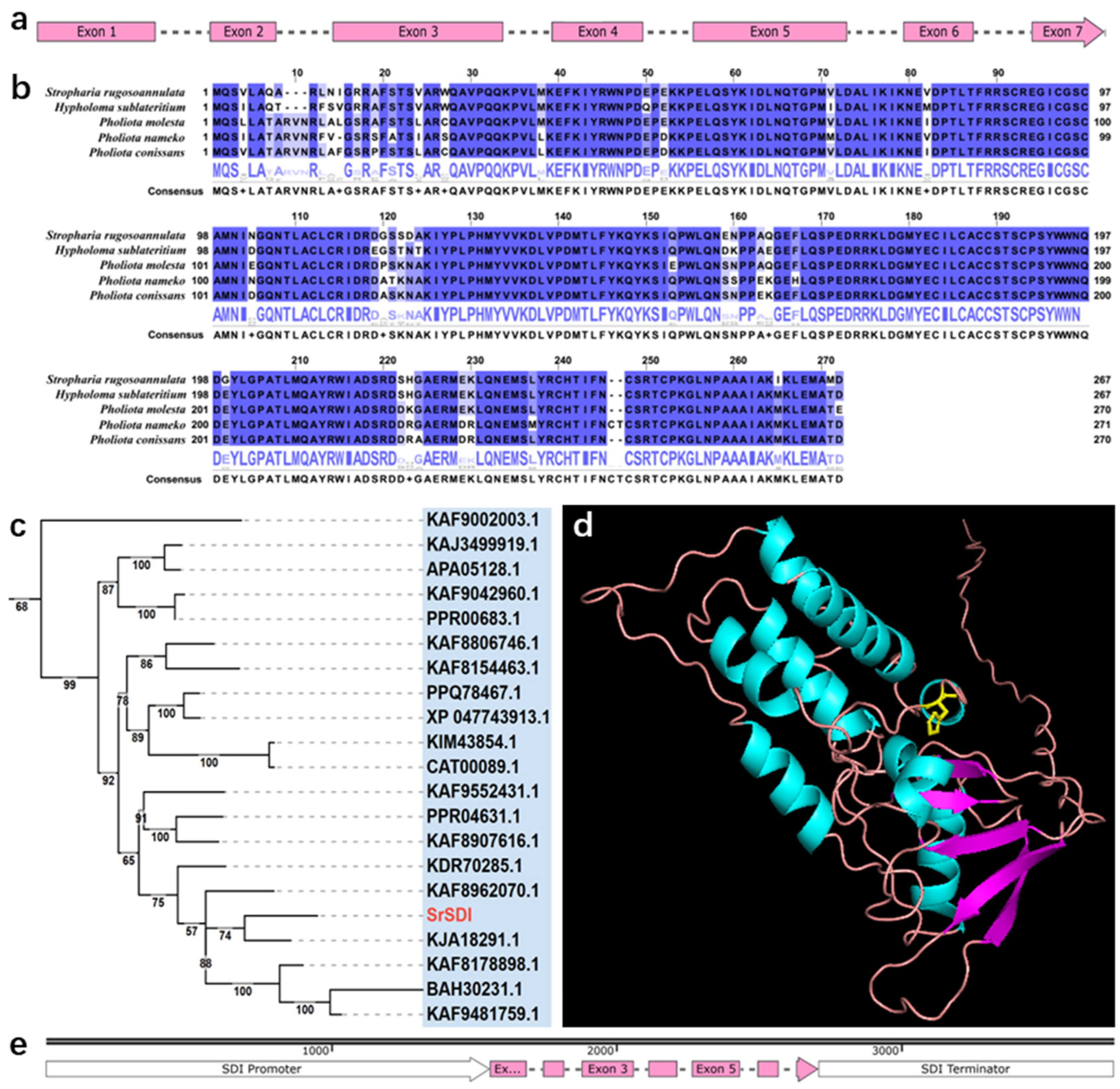
Similarly, the SrSDI gene in S. rugosoannulata was characterized. By integrating gene prediction, full-length transcriptome sequencing, and Sanger sequencing results, the coding region of SrSDI was found to be 1151 bp long, comprising 7 exons and 6 introns (Figure S8), with a coding sequence length of 804 bp. The gene structure is depicted in Figure 2a. Additionally, the full-length transcriptome alignment results of SrSDI against the reference sequence are shown in Figure S9. To identify conserved amino acid positions conferring carboxin resistance in SrSDI, the SrSDI amino acid sequence was aligned with those from various fungal species (Figure 2b). A phylogenetic tree of the SDI gene across different fungi was also constructed (Figure 2c and Figure S10, Table S5). The SrSDI is a member of the PLN00129 superfamily, and its sequence alignment results are presented in Figure S10. Based on previous reports in the literature [15,33], the histidine codon CAT at position 238 of the SrSDI amino acid sequence was replaced with the leucine codon CTT to generate the putative carboxin-resistant allele SrSDIR (Figure 2d and Figure S11). Subsequently, SrSDIR was ligated with the native SrSDI promoter (Figure S12) and terminator (Figure S13) and inserted into a transformation vector to serve as an element conferring carboxin resistance to S. rugosoannulata (Figure 2e).

2.2. The Monokaryon Mycelium of S. rugosoannulata Exhibits Sensitivity to Hygromycin B and Carboxin

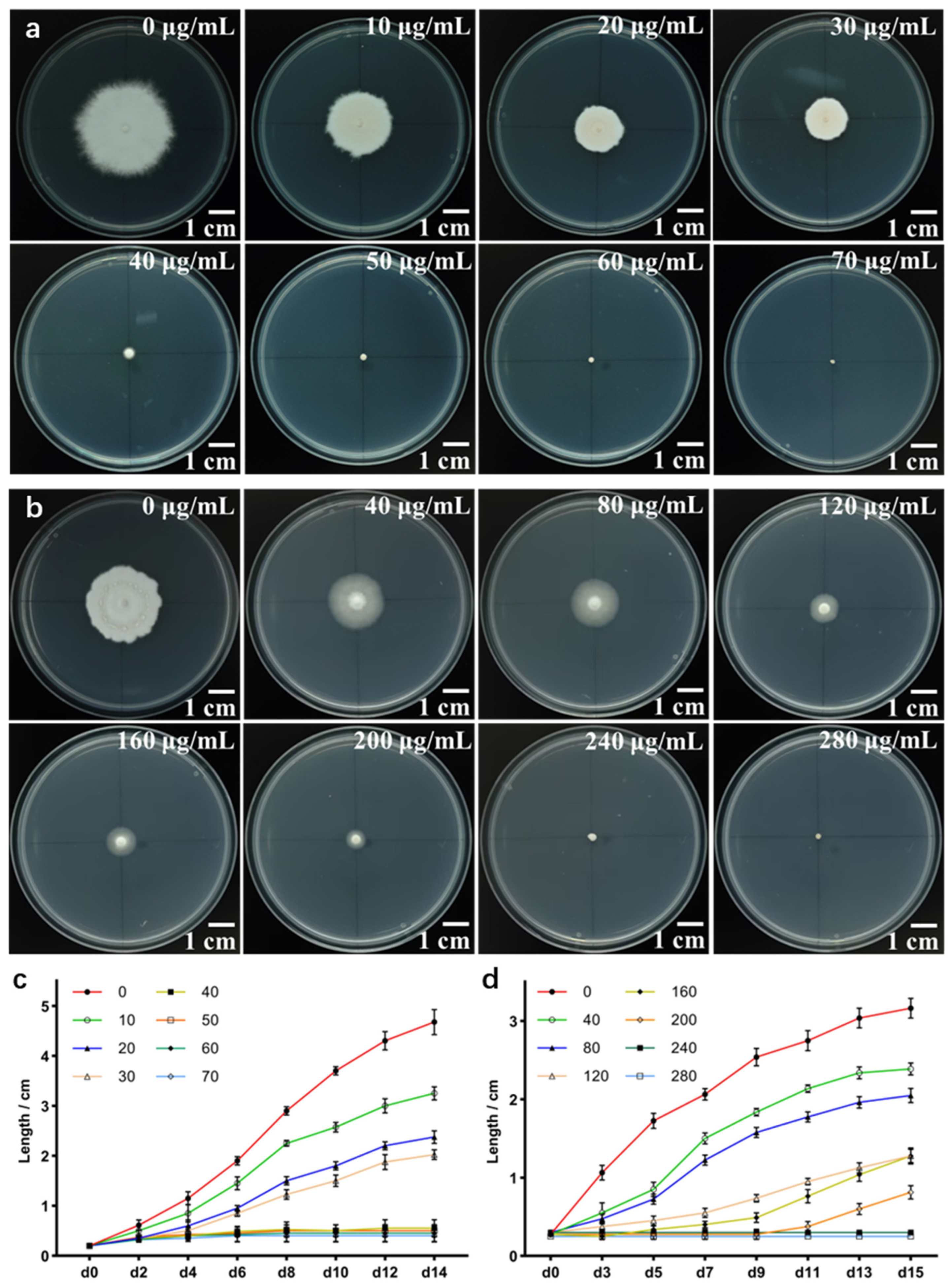
The hygromycin resistance gene, initially isolated from Escherichia coli, has been widely applied in genetic transformation studies of plants and various fungi [34,35]. In this study, the monokaryon mycelium of S. rugosoannulata (HC7) was inoculated on mPDA medium supplemented with different concentrations of hygromycin B. It was found that a concentration of 50 μg/mL hygromycin B significantly inhibited the growth of HC7 mycelia on the modified PDA plates (Figure 3a,c).
Mutations at key sites such as C_S42 or B_H216 of the SDI gene in fungi decreases the strength of the water-bridged hydrogen-bond network between the mitochondrial succinate-ubiquinone oxidoreductase (SQR) and the NH group of the amide bond of carboxin. This significantly reduces the binding affinity of carboxin for the SQR target, thereby conferring resistance to the fungicide in fungi. In contrast, the wild-type enzyme maintains an intact hydrogen-bonding network that allows for strong and effective binding of carboxin, resulting in no resistance [36]. Generally, heterologous genes containing non-native components exhibit relatively low expression efficiency in basidiomycetes. For example, the hygromycin resistance gene, which originates from Escherichia coli, often shows limited stability and expression efficiency in fungi. In contrast, the endogenous mutated SDI gene demonstrates higher stability and stronger expression efficiency, making it a widely applied marker in fungal genetic transformation studies [37,38]. The experimental results showed that when the concentration of carboxin was 240 μg/mL, the growth of HC7 mycelia was completely inhibited (Figure 3b,d). These findings indicate that both hygromycin B and carboxin can serve as effective selection agents for transformants in S. rugosoannulata genetic transformation experiments.

2.3. Establishment of a Transformation Protocol for HC7

Resistant transformants were selected using regeneration medium supplemented with 50 μg/mL hygromycin B for HC7 protoplasts transformed with pCM-GHT-GMT and pCM-GHT-GGT or 240 μg/mL carboxin for HC7 protoplasts transformed with pCM-mSDI-GMT and pCM-mSDI-GGT (Figure 4a and Figure S14). Colonies grown on the regeneration medium (RM) were transferred to mPDA plates containing either 50 μg/mL hygromycin B or 240 μg/mL carboxin for re-screening (Figure 4b and Figure S15). Vigorously growing transformants from the re-screening plates were individually expanded, and their genomic DNA was extracted. PCR amplification of T-DNA fragments (such as hph, mCherry, GUS, and mSDI) confirmed the presence of the transferred sequences (Figure 4c and Figure S16). Additionally, PCR was used to detect potential contamination by A. tumefaciens DNA outside the T-DNA borders in the genomic samples, as shown in Figure S17. Statistical analysis revealed that the regeneration efficiency of HC7 protoplasts was measured to be 1.07% (Figure S18). Among these, the transformation efficiency of S. rugosoannulata protoplasts mediated by the resistance gene of hph was relatively low, yielding approximately 13 transformants per 106 protoplasts, with a transformation efficiency of about 1.17‰. In contrast, in transformations mediated by mSDI as the resistance gene, approximately 17 transformants per 106 protoplasts were obtained, resulting in a transformation efficiency of about 1.56‰ (Figure 4d).

2.4. Identification of the Inserted T-DNA Flanking Sequences in Transformants

The flanking sequences of the T-DNA inserted in the positive transformants were identified using mhiTAIL-PCR. The amplified products ranged from 0.5 kb to 3 kb (Figure 5a). The T-DNA flanking sequences of the transformants were aligned against the S. rugosoannulata reference genome to confirm the insertion sites of T-DNA in the HC7 genome (Figure 5b). Among the selected positive transformants, the right end of the T-DNA inserted into shc-5 was truncated 39 bases away from the RB border sequence; the left end of the T-DNA inserted into shg-2 was truncated 20 bases away from the LB border sequence; and the truncation position of the right end of the T-DNA inserted into smg-13 was located on RB. Sequence analysis near the T-DNA insertion sites revealed that the insertion site in transformant sch-5 corresponded to the predicted gene S003.1G004910.1, which encodes a natural resistance-associated macrophage protein. In contrast, the insertion sites in transformants shg-2 and smg-13 were not located within predicted genes.

2.5. Detection and Visualization of mCherry

The fluorescence microscopy was used to observe multiple-passage positive transformants (Figure 6a) generated by ATMT with the recombinant plasmid pCM-GHT-GMT to detect mCherry expression. The results are shown in Figure 6b. PCR analysis was conducted on the transformants exhibiting stronger fluorescence obtained from the pCM-GHT-GMT-mediated transformation (Figure 6c). Subsequently, laser confocal microscopy was used to further examine the distribution of fluorescence within hyphae, as shown in Figure 6d. No fluorescence was detected in the control group, while the experimental group showed uniform distribution of mCherry fluorescence signals within the hyphae, indicating stable expression of the mCherry protein in the transformants.

2.6. Detection of GUS Gene Expression

The positive transformants generated by ATMT with the recombinant plasmid pCM-mSDI-GGT (Figure 7a) were selected. After PCR verification and sequencing (Figure 7b,c), these transformants were stained using the X-gluc staining solution, and the results are presented in Figure 7d. The blue precipitates were observed in the EP tubes containing the mycelia of positive transformants, indicating successful expression and functional activity of the GUS, which catalyzed the conversion of X-gluc into blue precipitates. After decolorization, the mycelia were examined under an inverted microscope (Figure 7e). The mycelia of the transformants were uniformly stained blue, while the untransformed mycelia remained colorless, further confirming the catalytic activity of GUS in the transformants.

3. Discussion

This study demonstrated that both the exogenous hygromycin resistance gene and the homologous mutant SDI gene can serve as effective genetic transformation screening markers for S. rugosoannulata. By making appropriate modifications to the ATMT system originally developed for Morchella importuna [39] and optimizing the protoplast preparation method for S. rugosoannulata, we successfully established an ATMT system suitable for genetic transformation of S. rugosoannulata HC7 protoplasts. After co-cultivating the induced A. tumefaciens EHA105 with HC7 protoplasts for 36 h, the mixture was transferred to regeneration medium plates containing the corresponding antibiotics. Through multiple rounds of selection, a significant number of transformants were obtained. These results indicate the feasibility and effectiveness of the ATMT system in S. rugosoannulata.
Overall, fungal ATMT transformation is influenced by multiple factors, including Agrobacterium strain, fungal recipient, co-culture conditions, and selection markers [40]. To achieve optimal transformation efficiency and obtain the maximum number of transformants, it is essential to optimize these influencing factors [41,42,43]. The optimal temperature for S. rugosoannulata mycelial growth is generally 24–27 °C, with an optimal pH range of 5–7 [44]. Transformation efficiencies mediated by different A. tumefaciens strains can vary significantly. For example, LBA4404 can produce hygromycin-resistant transformants in Flammulina velutipes, whereas no transformants were obtained using AGL-1 [45]. In Pleurotus ostreatus, Agrobacterium strains GV3101, EHA105, and LBA4404 exhibit varying transformation efficiencies [46]. Our studies have shown that co-culturing HC7 protoplasts with EHA105 at 25 °C and pH 5.5 for 36 h yields a substantial number of transformants.
The T-DNA flanking sequences were analyzed using the mhiTAIL-PCR technique. It was inferred that the genes potentially affected by the insertion site, such as S003.1G004910.1, encode a natural resistance-associated macrophage protein. Disruption of the functional sequences of these related genes may influence the morphology, growth and development, and stress tolerance of the transformants. In the absence of selection pressure, resistance gene loss was observed in 11% of positive transformants. This phenomenon has been reported in various fungi, including Mucor miehei [47], Aspergillus sojae [48], and Ophiocordyceps sinensis [16]. Research suggests that transposons present in the genome may contribute to reduced T-DNA stability [49,50,51]. It is hypothesized that T-DNA loss in S. rugosoannulata may be associated with specific characteristics of its genome.
As a straw-rotting fungus, S. rugosoannulata is typically cultivated in agricultural settings using rice straw, wheat straw, and other crop residues as substrates, with a biological conversion efficiency of approximately 45%. For optimal harvest, S. rugosoannulata should be collected when the mycelial membrane has not ruptured and the pileus remains curled and unexpanded. However, under unfavorable environmental conditions, such as high temperatures, S. rugosoannulata tends to prematurely expansion its pileus. The low biological conversion efficiency and the tendency to open prematurely under unsuitable conditions pose significant challenges for agricultural cultivation. Current research indicates that genes such as exg2 [52] and exp1 [53] play crucial roles in the pileus expansion of basidiomycetes, suggesting that the pileus expansion mechanism of S. rugosoannulata may also be regulated by similar genes. This hypothesis should be verified through genomic analysis. Different strains of S. rugosoannulata exhibit varying abilities to degrade substrate such as straw [54,55], and the underlying mechanisms require further investigation. Through this ATMT system, studies on the deletion, overexpression, or silencing of genes related to cellulose degradation and fruiting body development can provide valuable insights into improving agricultural practices. Such research can ultimately enhance the economic benefits of S. rugosoannulata cultivation by optimizing both yield and quality.
The ATMT system developed in this study facilitates more in-depth molecular genetics research on S. rugosoannulata. By fusing functional protein genes with reporter genes such as mCherry, the localization of gene expression products of interest can be precisely studied [56,57]. In a recent study on Arthrobotrys flagrans, the GprC gene was fused to a C-terminal green fluorescent protein (GFP) reporter, and its distribution in the apical and subapical regions of A. flagrans hyphae was observed using fluorescence microscopy, enabling the subcellular localization of the GprC protein [56]. Furthermore, by replacing components on plasmids like pCM-mSDI-GMT constructed in this study to build new recombinant vectors and utilizing the ATMT system for transformation, overexpression or silencing of target genes becomes more feasible, similar to approaches used in Grifola frondosa [58] and Cordyceps militaris [59]. Additionally, CRISPR/Cas9 technology has advanced precise genome editing in many fungi, including gene insertion, deletion, base conversion, and transcriptional activation [12,60,61]. In particular, Kamiya et al. demonstrated that transient expression of a CRISPR/Cas9 cassette enabled efficient homologous recombination in Lentinula edodes, overcoming the low natural recombination efficiency in this species [61]. Integrating CRISPR/Cas9 with the ATMT system will enable precise genome editing, gene knockout, and pathway regulation studies in S. rugosoannulata.

4. Conclusions

In this research, a series of recombinant vectors were constructed, in which the HphR cassette (composed of PSrGPD, hph, and TAnTrpC) or CbxR cassette (composed of PSrSDI, mutSDI, and TSrSDI) served as resistance screening markers, and mCherry or GUS acted as a reporter gene. Based on the aforementioned plasmids, the ATMT-based S. rugosoannulata HC7 protoplast transformation method was established, and the aforementioned screening markers and reporter genes were successfully functionalized. The achievement of the S. rugosoannulata protoplast ATMT system makes it possible to investigate the functions of target genes of interest in S. rugosoannulata and provides an essential genetic tool for subsequent studies on the molecular mechanism and gene function of S. rugosoannulata.

5. Materials and Methods

5.1. Strains and Culture Conditions

The S. rugosoannulata monokaryon strain (HC7) was obtained from our laboratory’s previous screening and utilized in all experiments of this study. The purified mycelium was maintained on mPDA composed of 8.0 g/L potato extract powder, 20.0 g/L dextrose, 0.5 g/L MgSO4·7H2O, 1.0 g/L KH2PO4, 2.0 g/L peptone, and 18.0 g/L agar at 25 °C. The Escherichia coli DH5α, used for plasmid transformation, amplification, and preservation, was cultured on Luria-Bertani (LB) medium supplemented with ampicillin or kanamycin at 37 °C. A. tumefaciens EHA105, which serves as the donor of T-DNA, was employed for fungal transformation. The induction medium (IM) [62] was used to induce A. tumefaciens virulence factors. Co-cultivation of EHA105 and S. rugosoannulata protoplasts was conducted using the co-culture medium (CIM) [62]. For regeneration and selection of transformed protoplasts, the RM composed of 8.0 g/L potato extract powder, 20.0 g/L sucrose, 1.5 g/L MgSO4·7H2O, 1.5 g/L KH2PO4, 1.5 g/L K2HPO4·3H2O, 2.0 g/L yeast extract, 2.0 g/L peptone, 0.1 g/L vitamin B1, 0.1 g/L vitamin B6, 109.3 g/L mannitol, and 15.0 g/L agar containing 200 μg/mL cefotaxime sodium salt (Solarbio, Beijing, China) was supplemented with either 50 μg/mL hygromycin B or 240 μg/mL carboxin.

5.2. Identification of Candidate Genes and Their Regulatory Sequences

To identify the candidate genes encoding GPD and SDI in HC7, we retrieved the published S. rugosoannulata genome sequence from the NCBI database, as well as GPD and/or SDI gene sequences from closely related species, such as Flammulina velutipes, Pleurotus ostreatus, Hypsizygus marmoreus, and Lentinula edodes. BLASTP was employed to compare these sequences against the genome of S. rugosoannulata to obtain homologous sequences for the gene of GPD and SDI. Multiple sequence alignment was performed using Muscle, and phylogenetic analysis was conducted using IQ-TREE. To avoid potential errors in genome annotation, the full-length RNA sequencing of HC7 mycelia was carried out using Oxford Nanopore Technologies (ONT). Primers were designed based on the reference genome sequence, and gene annotation results of S. rugosoannulata, and PCR amplification followed by Sanger sequencing was used to confirm the gene sequences of GPD and SDI in HC7.

5.3. Extraction of Genomic DNA and Long-Read RNA-Seq

Genomic DNA was extracted from the mycelium by employing the Fungal Genomic DNA Extraction Kit (Sangon Biotech, Beijing, China). Total RNA was isolated from the mycelium with TRIzolTM Reagent (Thermo Fisher Scientific, Waltham, MA, USA), and cDNA was synthesized using PrimeScriptTM IV 1st strand cDNA Synthesis Mix (TaKaRa, Kyoto, Japan). The Oxford Nanopore long-read RNA-Seq was conducted on the PromethION platform at Biomarker Technology Company (Beijing, China).

5.4. Construction of Plasmid

The plasmid pMD19-T (Takara, Kyoto, Japan) was used for the preservation of plasmid elements, while pCAMBIA1300 served as the backbone vector for genetic transformation. The hph, mCherry, and GUS genes were obtained from the laboratory-preserved plasmids pGH-hph, pGH-mCherry, and pGH-GUS, respectively. The TrpC terminator (TAnTrpC) sequence was amplified from plasmid pAN7-1. The sequences of GPD promoter, including the first exon, the first intron, and the initial 9 base pairs of the second exon (PSrGPD), SDI gene encoding SDI, SDI gene promoter (PSrSDI), and SDI gene terminator (TSrSDI), were all amplified from the HC7 genome.
The primer pairs hph-F/hph-R, mCherry-F/mCherry-R, GUS-F/GUS-R, and TtrpC-F/TtrpC-R were used to amplify the hph, mCherry, GUS, and TAnTrpC sequences from plasmids pGH-hph, pGH-mCherry, pGH-GUS, and pAN7-1, respectively. The GPDi-F/GPDi-R primer pair was used to amplify the PSrGPD promoter from the HC7 genome. The SDI-F/SDI-R primer pair was used to amplify the SDI cassette, including the promoter (PSrSDI) and terminator (TSrSDI) of the SDI gene, from the HC7 genome, and the amplified product was designated as oriSDI. To generate a mutant version of SDI (mutSDI), primers SDI-F/mut-SDI-R and mut-SDI-F/SDI-R were used to amplify oriSDI, followed by gene splicing by overlap extension PCR (SOE PCR). Both the oriSDI (oSDI) and mutSDI (mSDI) sequences were ligated into the pMD18-T vector, resulting in recombinant vectors pMD-oSDI and pMD-mSDI, respectively (Figure S19). The pCM-F/pCM-R and SP-F/SP-R primer pairs were used to amplify the backbone sequence (pCM) and spacer sequence (SP) from the pCAMBIA1300 plasmid. Homologous arms were added to the hph, mCherry, and GUS fragments using the primer pairs homo-GH/homo-HT, homo-GM/homo-MT, and homo-GG/homo-GT, respectively. The SOE PCR was then performed to connect the PSrGPD promoter, the homologous-arm-modified hph, mCherry, and GUS fragments, and the TAnTrpC terminator. The resulting constructs were cloned into the pMD18-T vector via TA cloning, yielding the recombinant vectors pMD-GHT, pMD-GMT, and pMD-GGT (Figure S19).
To construct the binary vector pCM-GHT-GMT, PCR amplification was performed on the recombinant vectors pMD-GHT and pMD-GMT using primers homo-GTS/homo-BGT and homo-SGT/homo-GTB, respectively. The amplified fragments were then ligated with the backbone sequence pCM and the spacer sequence SP via homologous recombination. Similarly, the binary vector pCM-GHT-GGT was constructed using the same primer pairs for PCR amplification of pMD-GHT and pMD-GGT. For the construction of the binary vector pCM-mSDI-GMT, PCR amplification was conducted on the recombinant vectors pMD-mSDI and pMD-GMT using primers homo-BMS/homo-MSS and homo-SGT/homo-GTB, respectively. The resulting fragments were ligated with the backbone sequence pCM via homologous recombination. Likewise, the binary vector pCM-mSDI-GGT was obtained by performing PCR amplification on pMD-mSDI and pMD-GGT using the same primer pairs and subsequent ligation with pCM via homologous recombination. The primers used for PCR amplification and plasmid construction are listed in Table S1.

5.5. Test of Fungal Sensitivity Toward Hygromycin B and Carboxin

The mycelial blocks of HC7 were inoculated onto mPDA medium supplemented with different concentrations of hygromycin (0, 10, 20, 30, 40, 50, 60, and 70 μg/mL) or carboxin (0, 40, 80, 120, 160, 200, 240, and 280 μg/mL). Cultures were incubated in the dark at 25 °C for 2 weeks. Mycelial growth was recorded to evaluate the sensitivity of HC7 to hygromycin and validamycin.

5.6. Protoplast Preparation

The method for preparing protoplasts of S. rugosoannulata was adapted primarily from the protocol described in previous studies [63,64], with several modifications. Specifically, mycelial blocks of HC7 were harvested from mPDA plates using a 0.5 cm diameter circular punch. These blocks were then homogenized and transferred to 250 mL of liquid mPDA medium (without agar) and incubated in darkness at 25 °C with shaking at 180 rpm for 6 days. Following this, the mixture was centrifuged at 3000× g for 10 min, after which the supernatant was discarded. The mycelia were subsequently washed twice with 0.6 M sorbitol solution, discarding the supernatant each time. For every 200 mg of mycelia, 1 mL of freshly prepared 1.5% (m/v) lywallzyme (Guangdong microbial culture collection center) solution was added, and the suspension was incubated in darkness at 25 °C for 3 h. The resulting mixture was filtered through a 10 μm cell strainer, and the filtrate was collected in a 50 mL centrifuge tube and centrifuged again at 3000× g for 10 min. The protoplast pellet was then gathered, washed twice with 0.6 M mannitol, and resuspended in an appropriate volume of 0.6 M mannitol to yield purified protoplasts.

5.7. Agrobacterium-tumefaciens-Mediated Transformation of S. rugosoannulata

The A. tumefaciens EHA105 harboring different plasmids was inoculated into LB medium supplemented with 20 μg/mL rifampicin and 50 μg/mL kanamycin and cultured at 28 °C with shaking at 200 rpm until the OD600 value reached 0.6 to 0.8. After centrifugation, the bacterial pellet was resuspended in IM medium containing 200 μM acetosyringone (AS) and adjusted to an OD600 of approximately 0.2. The culture was then incubated under dark conditions at 28 °C with shaking at 200 rpm until the OD600 value reached 0.6 to 0.8. The induced EHA105 suspension was mixed in equal volume with S. rugosoannulata protoplasts and incubated statically at 25 °C for 30 min before being transferred onto a CIM plate (containing 200 μM AS) covered with nitrocellulose membrane. Co-cultivation was performed in the dark at 25 °C for 48 h. Subsequently, the nitrocellulose membrane was transferred to an RM plate containing appropriate antibiotics and cultured at 25 °C until transformants emerged. All experiments were conducted in triplicate.

5.8. PCR Analysis of Putative Transformants

The transformants that grew on the regeneration medium were transferred to mPDA medium supplemented with the corresponding antibiotics (50 μg/mL hygromycin B or 240 μg/mL carboxin) for secondary screening and incubated in the dark at 25 °C for 7 days. Genomic DNA was extracted from the transformants, and PCR identification of the transformants obtained by ATMT was performed using specific primers (Table S1) and ITS primers (Table S2). The amplification products were analyzed by agarose gel electrophoresis and Sanger sequencing. Positive transformants were confirmed based on the position of the amplification product bands and the Sanger sequencing results. PCR was performed on the transformed genome using the primers listed in Table S2 to exclude potential contamination from bacterial DNA. Furthermore, mhiTAIL-PCR [65] was employed to clone the flanking sequences of the left or right T-DNA borders from the selected transformants. The primers used for mhiTAIL-PCR are presented in Table S3. The resulting T-DNA flanking sequences were aligned against the reference genome of S. rugosoannulata to verify the precise insertion site and assess whether the functional genes of HC7 were disrupted.

5.9. Fluorescence Observation and GUS Activity Analysis

To observe mCherry expression in transformants, the mycelia were cultivated on mPDA medium without antibiotics and cultured for 7 days. The mycelia were placed on a glass slide for preliminary examination under an inverted fluorescence microscope (Olympus, IX73P2F, Tokyo, Japan) at 400x magnification. Hyphae exhibiting mCherry fluorescence were selected and examined using a laser confocal microscope (Leica, Stellaris 5, Wetzlar, Germany) with excitation at 587 nm and peak emission at 610 nm, corresponding to its characteristic fluorescence peak. Images were acquired using a 400× objective lens. To assess the activity of GUS in the positive transformants, mycelia were scraped from fresh plates and resuspended in the GUS staining solution (0.5 mg/mL X-Gluc, 100 mM phosphate buffer [pH 7.0], 0.5 mM K3[Fe (CN)6], 0.5 mM K4[Fe (CN)6], 10 mM EDTA, and 1% Triton X-100). The suspension was incubated in the dark at room temperature for 1 h. Following incubation, the mycelia were washed three times with 10mM PBS buffer (pH 7.2) to remove residual staining solution, and the mycelia were observed under a under an inverted microscope at 400× magnification in bright-field.

5.10. Statistical Analysis

Data are presented as the mean ± standard error of measurement (SEM). In the line graphs, SEM values are indicated by error bars. Statistical analysis was carried out using Student’s t-tests when applicable. The statistical results were derived from an independent and randomized experiment with three replicates, and a p-value < 0.05 was considered significant.

Supplementary Materials

The following supporting information can be downloaded at: https://www.mdpi.com/article/10.3390/jof11090674/s1. Figure S1: The coding region sequence of the glycerol-3-phosphate dehydrogenase gene (SrGPD) from S. rugosoannulata HC7. Figure S2: Multiple sequence alignment of GPD protein sequences. Figure S3: Sequence alignment of SrGPD with the PLN02272 superfamily. Figure S4: Phylogenetic tree of GPD protein sequences from S. rugosoannulata and other selected fungi. Figure S5: The putative promoter sequence of the GPD in S. rugosoannulata HC7. Figure S6: Prediction of the GPD promoter elements in S. rugosoannulata. Figure S7: The putative terminator sequence of the GPD in S. rugosoannulata HC7. Figure S8: The coding region sequence of the succinate dehydrogenase [ubiquinone] iron-sulfur subunit (SrSDI) from S. rugosoannulata HC7. Figure S9: Alignment of transcripts obtained via full-length transcriptome sequencing against the reference sequence of the SrSDI. Figure S10: Phylogenetic tree of SDI protein sequences from S. rugosoannulata and other selected fungi. Figure S11: Sequence alignment of SrSDI with the PLN00129 superfamily. Figure S12: The putative promoter sequence of the SDI in S. rugosoannulata HC7. Figure S13: The putative terminator sequence of the SDI in S. rugosoannulata HC7. Figure S14: Schematic diagrams of the recombinant plasmids used for protoplast transformation in S. rugosoannulata HC7. Figure S15: The re-screening plates for S. rugosoannulata transformants obtained through Agrobacterium-mediated transformation with plasmid pCM-GHT-GGT (a), pCM-mSDI-GMT (b), or pCM-mSDI-GGT (c). Figure S16: PCR-based identification of the insertion fragments in S. rugosoannulata transformants generated using plasmids pCM-GHT-GGT (a), pCM-mSDI-GMT (b), and pCM-mSDI-GGT (c). Figure S17: Verification of exogenous DNA contamination in S. rugosoannulata transformants generated using the recombinant vectors pCM-GHT-GMT. Figure S18: Regeneration of S. rugosoannulata protoplasts. Figure S19: Schematic diagrams of recombinant plasmids used for constructing binary vectors. Table S1: The sequences of primers used in plasmids construction. Table S2: The primer sequences for the identification of transformants. Table S3: The primer sequences for mhiTAIL-PCR. Table S4: The GPD sequence information from 82 species was used for constructing the evolutionary tree in S. rugosoannulata. Table S5: The SDI sequence information from 87 species was used for constructing the evolutionary tree in S. rugosoannulata.

Author Contributions

D.Y.: Investigation, Visualization, Writing—Original Draft, Writing—Review & Editing, Methodology, Formal analysis, and Validation. Formal analysis. H.X.: Conceptualization, Resources, Writing—Review & Editing, Supervision, Project administration, and Funding acquisition. All authors have read and agreed to the published version of the manuscript.

Funding

This research was supported by the Fund for Academic Innovation Teams of South-Central Minzu University (Grant Number: PTZ25027, XTZ24020 & CZD24002).

Data Availability Statement

Data will be made available upon request.

Conflicts of Interest

The authors declare that they have no known competing financial interest or personal relationships that could have appeared to influence the work reported in this paper.

References

  1. Liu, Y.; Hu, C.-F.; Feng, X.; Cheng, L.; Ibrahim, S.A.; Wang, C.-T.; Huang, W. Isolation, characterization and antioxidant of polysaccharides from Stropharia rugosoannulata. Int. J. Biol. Macromol. 2020, 155, 883–889. [Google Scholar] [CrossRef]
  2. Huang, L.; He, C.; Si, C.; Shi, H.; Duan, J. Nutritional, bioactive, and flavor components of Giant Stropharia (Stropharia rugoso-annulata): A Review. J. Fungi 2023, 9, 792. [Google Scholar] [CrossRef]
  3. Liu, Y.-T.; Sun, J.; Luo, Z.-Y.; Rao, S.-Q.; Su, Y.-J.; Xu, R.-R.; Yang, Y.-J. Chemical composition of five wild edible mushrooms collected from Southwest China and their antihyperglycemic and antioxidant activity. Food Chem. Toxicol. 2012, 50, 1238–1244. [Google Scholar] [CrossRef]
  4. Weng, M. Extraction, Structure and Bioactivity of Polysaccharides from Stropharia rugodo-annulata. Master’s Thesis, Fujian Agriculture and Forestry University, Fuzhou, China, 2010. [Google Scholar]
  5. Sato, S.; Fukuda, Y.; Ogura, Y.; Kwon, E.; Kuwahara, S. Synthesis of the epimeric secosteroids strophasterols A and B. Angew. Chem. 2017, 129, 11051–11054. [Google Scholar] [CrossRef]
  6. Kodama, N.; Kakuno, T.; Nanba, H. Nanba Stimulation of the natural immune system in normal mice by polysaccharide from maitake mushroom. Mycoscience 2003, 44, 257–261. [Google Scholar] [CrossRef]
  7. Jiang, L. The Research on Preparation, Structural Identification and Biological Activity of Polysaccharide from Stropharia rugosoannulata (SR-1) and Polysaccharide from Tricholoma lascivum (Fr.) Gillet (TLG-1). Master’s Thesis, China West Normal University, Nanchong, China, 2019. [Google Scholar]
  8. Zhang, W.; Tian, G.; Geng, X.; Zhao, Y.; Ng, T.B.; Zhao, L.; Wang, H. Isolation and characterization of a novel lectin from the edible mushroom Stropharia rugosoannulata. Molecules 2014, 19, 19880–19891. [Google Scholar] [CrossRef] [PubMed]
  9. Duan, Y. Nutritional Quality Analyse, Extraction and Application of Flavonoids of Stropharia rugoso-annulata Farlow. Master’s Thesis, Fujian Agriculture and Forestry University, Fuzhou, China, 2010. [Google Scholar]
  10. Meyer, V.; Mueller, D.; Strowig, T.; Stahl, U. Comparison of different transformation methods for Aspergillus giganteus. Curr. Genet. 2003, 43, 371–377. [Google Scholar] [CrossRef]
  11. Zhang, B.; Sun, Q.; Li, H. Advances in genetic modification technologies. Chin. J. Biotechnol. 2015, 31, 1162–1174. [Google Scholar]
  12. Chen, B.-X.; Wei, T.; Ye, Z.-W.; Yun, F.; Kang, L.-Z.; Tang, H.-B.; Guo, L.-Q.; Lin, J.-F. Efficient CRISPR-Cas9 gene disruption system in edible-medicinal mushroom Cordyceps militaris. Front. Microbiol. 2018, 9, 1157. [Google Scholar] [CrossRef] [PubMed]
  13. Barcellos, F.G.; Fungaro, M.H.P.; Furlaneto, M.C.; Lejeune, B.; Pizzirani-Kleiner, A.A.; Lúcio de Azevedo, J. Genetic analysis of Aspergillus nidulans unstable transformants obtained by the biolistic process. Can. J. Microbiol. 1998, 44, 1137–1141. [Google Scholar] [CrossRef]
  14. Magaña-Ortíz, D.; Coconi-Linares, N.; Ortiz-Vazquez, E.; Fernández, F.; Loske, A.M.; Gómez-Lim, M.A. A novel and highly efficient method for genetic transformation of fungi employing shock waves. Fungal Genet. Biol. 2013, 56, 9–16. [Google Scholar] [CrossRef]
  15. Herzog, R.; Solovyeva, I.; Bölker, M.; Lugones, L.G.; Hennicke, F. Exploring molecular tools for transformation and gene expression in the cultivated edible mushroom Agrocybe aegerita. Mol. Genet. Genom. 2019, 294, 663–677. [Google Scholar] [CrossRef]
  16. Liu, G.; Cao, L.; Rao, Z.; Qiu, X.; Han, R. Identification of the genes involved in growth characters of medicinal fungus Ophiocordyceps sinensis based on Agrobacterium tumefaciens–mediated transformation. Appl. Microbiol. Biotechnol. 2020, 104, 2663–2674. [Google Scholar] [CrossRef]
  17. Lin, Y.-J.; Liu, W.-T.; Stark, H.; Huang, C.-T. Expression of enterovirus 71 virus-like particles in transgenic enoki (Flammulina velutipes). Appl. Microbiol. Biotechnol. 2015, 99, 6765–6774. [Google Scholar] [CrossRef] [PubMed]
  18. Zhu, H.; Liu, D.; Wang, Y.; Ren, D.; Zheng, L.; Chen, L.; Ma, A. Use of the yeast-like cells of Tremella fuciformis as a cell factory to produce a Pleurotus ostreatus hydrophobin. Biotechnol. Lett. 2017, 39, 1167–1173. [Google Scholar] [CrossRef] [PubMed]
  19. John, M.A.; Peberdy, J.F. Transformation of Aspergillus nidulans using the argB gene. Enzym. Microb. Technol. 1984, 6, 386–389. [Google Scholar] [CrossRef]
  20. Tran, V.-T.; Thai, H.-D.; Vu, T.X.; Vu, H.H.; Nguyen, G.T.; Trinh, M.T.; Tran, H.T.T.; Pham, H.T.T.; Le, N.T.H. An efficient Agrobacterium-mediated system based on the pyrG auxotrophic marker for recombinant expression in the filamentous fungus Penicillium rubens. Biotechnol. Lett. 2023, 45, 689–702. [Google Scholar] [CrossRef]
  21. Yelton, M.M.; Hamer, J.E.; Timberlake, W.E. Transformation of Aspergillus nidulans by using a trpC plasmid. Proc. Natl. Acad. Sci. USA 1984, 81, 1470–1474. [Google Scholar] [CrossRef] [PubMed]
  22. Narasipura, S.D.; Ren, P.; Dyavaiah, M.; Auger, I.; Chaturvedi, V.; Chaturvedi, S. An efficient method for homologous gene reconstitution in Cryptococcus gattii using URA5 auxotrophic marker. Mycopathologia 2006, 162, 401–409. [Google Scholar] [CrossRef]
  23. Balabanova, L.A.; Shkryl, Y.N.; Slepchenko, L.V.; Yugay, Y.A.; Gorpenchenko, T.Y.; Kirichuk, N.N.; Khudyakova, Y.V.; Bakunina, I.Y.; Podvolotskaya, A.B.; Bulgakov, V.P. Development of host strains and vector system for an efficient genetic transformation of filamentous fungi. Plasmid 2019, 101, 1–9. [Google Scholar] [CrossRef]
  24. Ahuja, M.; Punekar, N.S. Phosphinothricin resistance in Aspergillus niger and its utility as a selectable transformation marker. Fungal Genet. Biol. 2008, 45, 1103–1110. [Google Scholar] [CrossRef]
  25. Yang, X.; Peng, J.; Pan, J. Nourseothricin N-acetyl transferase (NAT), a new selectable marker for nuclear gene expression in Chlamydomonas. Plant Methods 2019, 15, 140. [Google Scholar] [CrossRef]
  26. Arentshorst, M.; Niu, J.; Ram, A.F.J. Efficient generation of Aspergillus niger knock out strains by combining NHEJ mutants and a split marker approach. In Genetic Transformation Systems in Fungi; van den Berg, M.A., Maruthachalam, K., Eds.; Springer International Publishing: Cham, Switzerland, 2015; Volume 1, pp. 263–272. [Google Scholar]
  27. Djulic, A.; Schmid, A.; Lenz, H.; Sharma, P.; Koch, C.; Wirsel, S.G.; Voegele, R.T. Transient transformation of the obligate biotrophic rust fungus Uromyces fabae using biolistics. Fungal Biol. 2011, 115, 633–642. [Google Scholar] [CrossRef]
  28. Crespo-Sempere, A.; López-Pérez, M.; Martínez-Culebras, P.V.; González-Candelas, L. Development of a green fluorescent tagged strain of Aspergillus carbonarius to monitor fungal colonization in grapes. Int. J. Food Microbiol. 2011, 148, 135–140. [Google Scholar] [CrossRef]
  29. Wu, L.; Conner, R.L.; Wang, X.; Xu, R.; Li, H. Variation in growth, colonization of maize, and metabolic parameters of GFP-and DsRed-labeled Fusarium verticillioides strains. Phytopathology 2016, 106, 890–899. [Google Scholar] [CrossRef]
  30. Wang, Q.; Coleman, J.J. CRISPR/Cas9-mediated endogenous gene tagging in Fusarium oxysporum. Fungal Genet. Biol. 2019, 126, 17–24. [Google Scholar] [CrossRef]
  31. Liu, D.; Garrigues, S.; de Vries, R.P. Heterologous protein production in filamentous fungi. Appl. Microbiol. Biotechnol. 2023, 107, 5019–5033. [Google Scholar] [CrossRef] [PubMed]
  32. Su, Z.; Li, H.; Xu, Y.; Zhang, C.; Wu, J.; Lei, Y. Establishment of an efficient Agrobacterium tumefaciens-mediated transformation system for an Armillaria species, a host of the fully mycoheterotrophic plant Gastrodia elata. Folia Microbiol. 2024, 69, 1–13. [Google Scholar] [CrossRef] [PubMed]
  33. Bao, D.; Huang, Z.; Li, Y.; Zhou, C.; Wan, J.; Tang, L.; Mao, W.; Wang, Y.; Gong, M.; Zou, G.; et al. Agrobacterium-mediated transformation of arthroconidia obtained from the edible mushroom Hypsizygus marmoreus. J. Microbiol. Methods 2020, 171, 105878. [Google Scholar] [CrossRef]
  34. Punt, P.J.; Oliver, R.P.; Dingemanse, M.A.; Pouwels, P.H.; van den Hondel, C.A. Transformation of Aspergillus based on the hygromycin B resistance marker from Escherichia coli. Gene 1987, 56, 117–124. [Google Scholar] [CrossRef] [PubMed]
  35. Van De Rhee, M.D.; Graça, P.M.A.; Huizing, H.J.; Mooibroek, H. Transformation of the cultivated mushroom, Agaricus bisporus, to hygromycin B resistance. Mol. Genet. Genom. 1996, 250, 252–258. [Google Scholar] [CrossRef]
  36. Zhu, X.; Xiong, L.; Li, H.; Song, X.; Liu, J.; Yang, G. Computational and experimental insight into the molecular mechanism of carboxamide inhibitors of Succinate-ubquinone oxidoreductase. ChemMedChem 2014, 9, 1512–1521. [Google Scholar] [CrossRef] [PubMed]
  37. Honda, Y.T.; Matsuyama, T.; Irie, T.; Watanabe, T.; Kuwahara, M. Carboxin resistance transformation of the homobasidiomycete fungus Pleurotus ostreatus. Curr. Genet. 2000, 37, 209–212. [Google Scholar] [CrossRef]
  38. Guo, M.; Zhu, X.; Li, H.; Tan, L.; Pan, Y. Development of a novel strategy for fungal transformation based on a mutant locus conferring carboxin-resistance in Magnaporthe oryzae. AMB Express 2016, 6, 57. [Google Scholar] [CrossRef] [PubMed]
  39. Lv, S.; Chen, X.; Mou, C.; Dai, S.; Bian, Y.; Kang, H. Agrobacterium-mediated transformation of the ascomycete mushroom Morchella importuna using polyubiquitin and glyceraldehyde-3-phosphate dehydrogenase promoter-based binary vectors. World J. Microbiol. Biotechnol. 2018, 34, 148. [Google Scholar] [CrossRef]
  40. Lichius, A.; Ruiz, D.M.; Zeilinger, S. Genetic transformation of filamentous fungi: Achievements and challenges. In Grand Challenges in Fungal Biotechnology; Springer: Cham, Switzerland, 2020; pp. 123–164. [Google Scholar]
  41. Wang, S.; Chen, H.; Wang, Y.; Tang, X.; Zhang, H.; Chen, W.; Chen, Y.Q. Optimization of Agrobacterium tumefaciens-mediated transformation method of oleaginous filamentous fungus Mortierella alpina on co-cultivation materials choice. J. Microbiol. Methods 2018, 152, 179–185. [Google Scholar] [CrossRef]
  42. Fu, J.; Brockman, N.E.; Wickes, B.L. Optimizing transformation frequency of Cryptococcus neoformans and Cryptococcus gattii using Agrobacterium tumefaciens. J. Fungi 2021, 7, 520. [Google Scholar] [CrossRef] [PubMed]
  43. D’spain, S.; Andrade, P.I.; Brockman, N.E.; Fu, J.; Wickes, B.L. Agrobacterium tumefaciens-Mediated Transformation of Candida glabrata. J. Fungi 2022, 8, 596. [Google Scholar] [CrossRef]
  44. Bruhn, J.N.; Abright, N.; Mihail, J.D. Forest farming of wine-cap Stropharia mushrooms. Agrofor. Syst. 2010, 79, 267–275. [Google Scholar] [CrossRef]
  45. Okamoto, T.; Yamada, M.; Sekiya, S.; Okuhara, T.; Taguchi, G.; Inatomi, S.; Shimosaka, M. Agrobacterium tumefaciens-mediated transformation of the vegetative dikaryotic mycelium of the cultivated mushroom Flammulina velutipes. Biosci. Biotechnol. Biochem. 2010, 74, 2327–2329. [Google Scholar] [CrossRef]
  46. Lei, M.; Wu, X.; Zhang, J.; Wang, H.; Huang, C. Establishment of an efficient transformation system for Pleurotus ostreatus. World J. Microbiol. Biotechnol. 2017, 33, 214. [Google Scholar] [CrossRef]
  47. Martínez-Cruz, J.; Romero, D.; de Vicente, A.; Pérez-García, A. Transformation of the cucurbit powdery mildew pathogen Podosphaera xanthii by Agrobacterium tumefaciens. New Phytol. 2017, 213, 1961–1973. [Google Scholar] [CrossRef]
  48. Mora-Lugo, R.; Zimmermann, J.; Rizk, A.M.; Fernandez-Lahore, M. Development of a transformation system for Aspergillus sojae based on the Agrobacterium tumefaciens-mediated approach. BMC Microbiol. 2014, 14, 247. [Google Scholar] [CrossRef]
  49. Spanu, P.D.; Abbott, J.C.; Amselem, J.; Burgis, T.A.; Soanes, D.M.; Stüber, K.; van Themaat, E.V.L.; Brown, J.K.M.; Butcher, S.A.; Gurr, S.J.; et al. Genome expansion and gene loss in powdery mildew fungi reveal tradeoffs in extreme parasitism. Science 2010, 330, 1543–1546. [Google Scholar] [CrossRef] [PubMed]
  50. Betancourt, A.J.; Wei, K.H.-C.; Huang, Y.; Lee, Y.C.G. Causes and consequences of varying transposable element activity: An evolutionary perspective. Annu. Rev. Genom. Hum. Genet. 2024, 25, 1–25. [Google Scholar] [CrossRef] [PubMed]
  51. Hassan, A.H.; Mokhtar, M.M.; El Allali, A. Transposable elements: Multifunctional players in the plant genome. Front. Plant Sci. 2024, 14, 1330127. [Google Scholar] [CrossRef]
  52. Tao, Y.; Xie, B.; Yang, Z.; Chen, Z.; Chen, B.; Deng, Y.; Jiang, Y.; van Peer, A.F. Identification and expression analysis of a new glycoside hydrolase family 55 exo-β-1,3-glucanase-encoding gene in Volvariella volvacea suggests a role in fruiting body development. Gene 2013, 527, 154–160. [Google Scholar] [CrossRef] [PubMed]
  53. Muraguchi, H.; Fujita, T.; Kishibe, Y.; Konno, K.; Ueda, N.; Nakahori, K.; Yanagi, S.O.; Kamada, T. The exp1 gene essential for pileus expansion and autolysis of the inky cap mushroom Coprinopsis cinerea (Coprinus cinereus) encodes an HMG protein. Fungal Genet. Biol. 2008, 45, 890–896. [Google Scholar] [CrossRef] [PubMed]
  54. Gong, W.; Zeng, Y.; Li, X.; Zhao, Z.; Shen, N.; Zhou, Y.; Bian, Y.; Xiao, Y. Molecular profiling of rice straw degradability discrepancy in Stropharia rugosoannulata core germplasm. J. Agric. Food Chem. 2024, 72, 25379–25390. [Google Scholar] [CrossRef]
  55. Gu, M.; Chen, Q.; Zhang, Y.; Zhao, Y.; Wang, L.; Wu, X.; Zhao, M.; Gao, W. Evaluation of Genetic Diversity and Agronomic Traits of Germplasm Resources of Stropharia rugosoannulata. Horticulturae 2024, 10, 213. [Google Scholar] [CrossRef]
  56. Hu, X.; Hoffmann, D.S.; Wang, M.; Schuhmacher, L.; Stroe, M.C.; Schreckenberger, B.; Elstner, M.; Fischer, R. GprC of the nematode-trapping fungus Arthrobotrys flagrans activates mitochondria and reprograms fungal cells for nematode hunting. Nat. Microbiol. 2024, 9, 1752–1763. [Google Scholar] [CrossRef]
  57. Tsai, M.-C.; Barati, M.T.; Kuppireddy, V.S.; Beckerson, W.C.; Long, G.; Perlin, M.H. Characterization of Microbotryum lychnidis-dioicae secreted effector proteins, their potential host targets, and localization in a heterologous host plant. J. Fungi 2024, 10, 262. [Google Scholar] [CrossRef]
  58. Sato, M.; Kurahashi, A.; Nishibori, K.; Fujimori, F. Development of a transformation system for the edible mushroom Grifola frondosa: Demonstrating heterologous gene expression and RNAi-mediated gene silencing. Mycoscience 2015, 56, 364–372. [Google Scholar] [CrossRef]
  59. Liu, Q.; Meng, G.; Wang, M.; Li, X.; Liu, M.; Wang, F.; Yang, Y.; Dong, C. Safe-harbor-targeted CRISPR/Cas9 system and Cmhyd1 overexpression enhances disease resistance in Cordyceps militaris. J. Agric. Food Chem. 2023, 71, 15249–15260. [Google Scholar] [CrossRef] [PubMed]
  60. Moon, S.; An, J.Y.; Choi, Y.-J.; Oh, Y.-L.; Ro, H.-S.; Ryu, H. Construction of a CRISPR/Cas9-Mediated Genome Editing System in Lentinula edodes. Mycobiology 2021, 49, 599–603. [Google Scholar] [CrossRef]
  61. Kamiya, A.; Ueshima, H.; Nishida, S.; Honda, Y.; Kamitsuji, H.; Sato, T.; Miyamoto, H.; Sumita, T.; Izumitsu, K.; Irie, T. Development of a gene-targeting system using CRISPR/Cas9 and utilization of pyrG as a novel selectable marker in Lentinula edodes. FEMS Microbiol. Lett. 2023, 370, fnad042. [Google Scholar] [CrossRef] [PubMed]
  62. Dai, C.; Yang, H.; Tang, T.; Ma, C.; Kang, C. An effective CRISPR-Cas9 technology for efficiently isolating transgene-free mutants in Arabidopsis, Brassica napus, Strawberry, and Soybean. In CRISPR-Cas Methods; Islam, M.T., Bhowmik, P.K., Molla, K.A., Eds.; Springer: New York, NY, USA, 2020; pp. 99–115. [Google Scholar]
  63. Yan, P.-S.; Jiang, J.-H.; Cui, W.-S. Characterization of protoplasts prepared from the edible fungus, Stropharia rugoso-annulata. World J. Microbiol. Biotechnol. 2004, 20, 173–177. [Google Scholar] [CrossRef]
  64. Zhou, F.; Yang, H.-L.; Zhao, Y.; Jiang, S.-X.; Li, Z.-P.; Zha, L.; Li, Y. Characteristics of mononuclear properties and the protoplast monokaryogenesis of different varieties of Stropharia rugosoannulata. Acta Agric. Shanghai 2023, 39, 33–37. [Google Scholar]
  65. Tan, J.; Gong, Q.; Yu, S.; Hou, Y.; Zeng, D.; Zhu, Q.; Liu, Y.-G. A modified high-efficiency thermal asymmetric interlaced PCR method for amplifying long unknown flanking sequences. J. Genet. Genom. 2019, 46, 363–366. [Google Scholar] [CrossRef]
Figure 1. Characterization of the gpd in S. rugosoannulata. (a) mature fruiting bodies of S. rugosoannulata; (b) linear model of the predicted coding region for SrGPD; (c) morphology of S. rugosoannulata mycelium grown on modified potato dextrose agar (mPDA) medium; (d) alignment of transcripts obtained via full-length transcriptome sequencing against the reference sequence of SrGPD; (e) comprehensive structural model of SrGPD, including the promoter, coding region, and terminator.
Jof 11 00674 g001
Figure 2. Characterization of the SDI in S. rugosoannulata. (a) linear model of the predicted coding region for the SrSDI gene; (b) multiple sequence alignment results analyzing the conservation differences between the SrSDI protein and those from other species; (c) partial phylogenetic tree based on the SrSDI protein sequence, with all species shown belonging to the Agaricineae suborder; (d) schematic diagram of the three-dimensional structure of the SrSDI protein, obtained via homology modeling using Swiss-Model; (e) comprehensive structural model of SrSDI, including the promoter, coding region, and terminator.
Jof 11 00674 g002
Figure 3. Effects of different concentrations of hygromycin B and carboxin on mycelial growth inhibition of S. rugosoannulata. (a) growth status of S. rugosoannulata mycelia on mPDA plates containing varying concentrations of hygromycin B; (b) growth status of S. rugosoannulata mycelia on mPDA plates containing varying concentrations of carboxin; (c) changes in mycelial diameter under different concentrations of hygromycin B treatment; (d) changes in mycelial diameter under different concentrations of validamycin treatment. The x-axis indicates the number of days of mycelial growth on the medium, while the y-axis shows the elongation of mycelial diameter measured using the cross-measurement method in (c,d) (three replicates were set for each group).
Jof 11 00674 g003
Figure 4. Protoplast transformation of HC7 and identification of transformants. (a) Schematic representation of the structure of plasmid pCM-GHT-GMT. Primers hph-F and hph-R were used to verify the presence of the hph in the transformants, while primers mCherry-F and mCherry-R were used to confirm the presence of the mCherry in the transformants. (b) Growth status of S. rugosoannulata transformants using plasmid pCM-GHT-GMT on re-screening plates. (c) Identification of insertion fragments from plasmid pCM-GHT-GMT in S. rugosoannulata transformants using the primer pairs described in panel (a). PC, plasmid positive control; WT, wild type; 1–6, transformants after re-screening; M, DNA molecular marker. (d) Transformation efficiency of S. rugosoannulata protoplasts using different plasmids. ns, no significant.
Jof 11 00674 g004
Figure 5. Identification of flanking sequences in transformants. (a) Agarose gel electrophoresis results of mhiTAIL-PCR products. Three positive transformants were selected from each of the four plasmid-transformed lines for mhiTAIL-PCR analysis. Specifically, mg, mc, hg, and hc refers to transformants harboring T-DNA from plasmid pCM-mSDI-GGT, pCM-mSDI-GMT, pCM-GHT-GGT, and pCM-GHT-GMT, respectively. M, DNA molecular weight marker. (b) Schematic representation of the sequence at which T-DNA is joined to its flanking regions. The base sequences in black capital letters originate from the recombinant plasmid DNA fragments, while those in red capital letters represent genomic DNA sequences of S. rugosoannulata. The position indicated by the black arrow denotes the insertion site of T-DNA within the S. rugosoannulata genome.
Jof 11 00674 g005
Figure 6. Expression and fluorescence detection of mCherry in S. rugosoannulata. (a) Growth comparison of wild-type (upper images) and mCherry-expressing transformant shc-5 (lower images) on mPDA medium with (left images) or without (right images) hygromycin B. (b) Fluorescence imaging analysis of mycelia from wild-type and transformant shc-5. The images display the mycelia of wild-type (upper images) and transformant (lower images) under bright-field and fluorescence modes. BF, bright field; FF, fluorescence mode, showing DAPI staining and mCherry expression. (c) Agarose gel electrophoresis analysis of transformants. PCR amplification of genomic DNA from positive transformant shc-5 using primer pairs hph-F/hph-R (lane 1), mCherry-F/mCherry-R (lane 2), and ITS1/ITS4 (lane 3). M, DNA molecular marker. (d) Laser confocal microscopy imaging analysis of mycelia from wild-type and transformant shc-5. The images depict the mycelia of wild-type (upper images) and transformant (lower images) under bright-field and fluorescence modes, as well as the merged images.
Jof 11 00674 g006
Figure 7. Expression and fluorescence detection of GUS in S. rugosoannulata. (a) Growth comparison of wild-type (left images) and mCherry-expressing transformant smg-13 (right images) on mPDA medium with (upper images) or without (lower images) carboxin. (b) Agarose gel electrophoresis analysis of transformants. PCR amplification of genomic DNA from positive transformant smg-13 using primer pairs SDI-F/SDI-R (lane 2–4), GUS-F/GUS-R (lane 5–7), and ITS1/ITS4 (lane 8–10). M, DNA molecular marker. (c) Sanger sequencing results of transformants obtained using plasmid pCM-mSDI-GGT. The 238th histidine (CAT) in the wild-type SrSDI was mutated to leucine (CTT), highlighted with blue background, to confer carboxin resistance to the transformants, and this mutation was detected in the transformants (bases highlighted with red background). (d) GUS staining results of different transformants. Tubes 1–3 display the GUS staining situation for three different transformants. CK, wild type. (e) Microscopic images of hyphae from positive transformants. The images depict the hyphae of the wild type (left) and the transformant smg-13 (right) before and after GUS staining.
Jof 11 00674 g007
Disclaimer/Publisher’s Note: The statements, opinions and data contained in all publications are solely those of the individual author(s) and contributor(s) and not of MDPI and/or the editor(s). MDPI and/or the editor(s) disclaim responsibility for any injury to people or property resulting from any ideas, methods, instructions or products referred to in the content.