Article

Seven New Series and Four New Species in Sections Subinflati and Trachyspermi of Talaromyces (Trichocomaceae, Eurotiales)

1
State Key Laboratory of Microbial Diversity and Innovative Utilization, Institute of Microbiology, Chinese Academy of Sciences, Beijing 100101, China
2
University of Chinese Academy of Sciences, Beijing 100049, China
3
Central Asian Center for Development Studies, New Uzbekistan University, Tashkent 100007, Uzbekistan
4
Department of Ecology, Faculty of Biology and Ecology, National University of Uzbekistan, Tashkent 100174, Uzbekistan
*
Author to whom correspondence should be addressed.
J. Fungi 2025, 11(7), 508; https://doi.org/10.3390/jof11070508
Submission received: 4 June 2025 / Revised: 30 June 2025 / Accepted: 3 July 2025 / Published: 4 July 2025
(This article belongs to the Special Issue Ascomycota: Diversity, Taxonomy and Phylogeny, 3rd Edition)

Abstract

Species of Talaromyces C.R. Benj. are valuable biological resources for human beings as competent producers of enzymes, antibiotics, antifungal agents and biopigments, but a comprehensive taxonomic system at the series level has not been fully provided for this genus. In this study, three new series, Palmarum, Resedani and Subinflati, are proposed in section Subinflati. Section Trachyspermi is also restructured to include five series, in which Diversi, Erythromelles, Miniolutei and Resinarum are newly erected, and Trachyspermi is emended. Additionally, four new species are discovered: T. elephas, T. sinensis and T. xishuangbannaensis isolated from rotten fruit husk in Yunnan Province, China, belonging to the series Erythromelles, Subinflati and Miniolutei, respectively, and T. tianshanicus from soil in Uzbekistan, located in ser. Diversi. Morphological distinctions, including colony characteristics, conidiophore structures, and conidial morphologies, along with phylogenetic analyses based on multi-locus datasets (ITS, BenA, CaM and RPB2), confirm their novelty to science. Detailed descriptions and illustrations of the new species are given. The proposed classification of Talaromyces at the series level provides a refined infrageneric framework and facilitates taxonomic stability and future biodiversity studies.

1. Introduction

Species of Talaromyces C.R. Benj. are valuable biological resources for human beings. Talaromyces minioluteus (Dierckx) Samson et al. produces dextranases [1]. Stipitatic acid is an antibiotic discovered from T. stipitatus (Thom ex C.W. Emmons) C.R. Benj. [2], and the antifungal agents wortmannin and talcarpones were isolated from T. wortmannii (Klöcker) C.R. Benj. [2] and T. johnpittii E. Lacey et al. [3]. Many natural biopigments are produced by T. albobiverticillius (H.M. Hsieh et al.) Samson et al., T. atroroseus N. Yilmaz et al., T. purpureogenus (Stoll) Samson et al. and T. ruber (Stoll) N. Yilmaz et al. [4]. Nevertheless, T. marneffei (Segretain et al.) Samson et al. causes talaromycosis, an invasive mycosis that is endemic in tropical and subtropical Asia and results in more than 17,000 infections annually [5]. Related research on the infection mechanisms and human immune responses has been gradually improved.
Talaromyces was established by C.R. Benj. in 1955, and 171 species were accepted in 2020 following a monographic treatment [6]. Until 2022, 199 species were known in this genus [7]. Afterwards, 44 new species were further added: 20 from Asia, including 15 from China (T. albidus L. Wang, T. cystophila Y.X. Mo & H.Y. Wu, T. disparis Y. Ruan & L. Wang, T. ellipsoideus M. Li & L. Cai, T. funiformis Y. Ruan & L. Wang, T. guiyangensis Zhi Y. Zhang et al., T. hainanensis K. Hong & L. Liu, T. jianfengicus Y. Ruan & L. Wang, T. jiangxiensis Zhi Y. Zhang et al., T. longistipes Zhi Y. Zhang & Y.F. Han, T. paecilomycetoides Zhi Y. Zhang et al., T. parapalmae Zhi Y. Zhang & Y.F. Han, T. phialiformis M. Li & L. Cai, T. rubidus L. Wang, T. virens C. Liu et al.), two from Thailand (T. phuphaphetensis Nuankaew et al. and T. satunensis Nuankaew et al.) and one each from Japan (T. mellisjaponici A. Okubo & D. Hirose), South Korea (T. echinulatus Hyang B. Lee & T.T.T. Nguyen) and Isreal (T. cupressi Meshram et al.); three from Europe, including two from Portugal (T. benedictus D.S. Paiva and T. saxoxalicus J. Trovão et al.) and one from Czechia (T. clematidis Spetik & Houbraken); four from Africa (South Africa), namely T. gautengensis Visagie & N. Yilmaz, T. podocarpi Visagie & N. Yilmaz, T. macrodendroideus Visagie et al. and T. mzansiensis Visagie et al.; one from North America (USA), namely T. apricus Y.P. Tan et al.; four from South America, including three from Brazil (T. cattleyae Condé et al., T. cavernicola V.C.S. Alves et al. and T. potiguarorum J.M.S. Lima et al.) and one from Colombia (T. santanderensis B.E. Guerra-Sierra & L.A. Arteaga-Figueroa); 11 from Oceania (Australia), namely T. atkinsoniae Y.P. Tan et al., T. hallidayae Y.P. Tan et al., T. johnpittii and so on; and another one from the Mariana Trench (T. sedimenticola Y. Wang & H. Zhou). In total, 243 species are listed in this genus.
Infrageneric ranks may be helpful in managing complicated speciose genera, e.g., Aspergillus, Penicillium and Talaromyces. Seven sections were recognized in Talaromyces in 2014 [8]—Bacillispori, Helici, Islandici, Purpurei, Subinflati, Talaromyces and Trachyspermi—and section Tenues was subsequently proposed [9]. Sections were originally divided based on morphological (conidiophore structure) or physiological (optimum growth temperature) characteristics [10], but, nowadays, they are erected mainly based on multigene phylogeny [8]. Houbraken and Frisvad [6] established classifications at the rank of series in Aspergillus and Penicillium, which resulted in 75 series in the former and 89 in the latter. However, a comprehensive taxonomic system at the series level has not been provided for Talaromyces.
During our investigations of Talaromyces diversity in plant or soil samples collected from China and Uzbekistan, four new species were discovered based on phylogenetic analyses and morphological comparisons: one belonging to sect. Subinflati and the other three in sect. Trachyspermi. Series divisions were proposed for the two sections.

2. Materials and Methods

2.1. Fungal Materials

Cultures were isolated from plant debris material collected from Yunnan Province, China or soil samples from Tashkent Province, Uzbekistan in 2024. Dried cultures were preserved in the Herbarium Mycologicum Academiae Sinicae (HMAS, Beijing, China), and the living ex-type strains were deposited in the China General Microbiological Culture Collection Center (CGMCC, Beijing, China).

2.2. Morphological Observations

Morphological characteristics were observed and recorded according to standardized methods [11]. Four standard growth media were adopted: Czapek yeast autolysate agar (CYA, yeast extract Oxoid, Hampshire, UK), malt extract agar (MEA, Amresco, Solon, OH, USA), yeast extract agar (YES) and potato dextrose agar (PDA). The methods for colonial inoculation, incubation (7 days), macroscopic and microscopic examinations and digital capture followed our previous studies [7,12,13,14,15].

2.3. DNA Extraction, PCR Amplification and Sequencing

DNA was extracted from living cultures grown on PDA for 7 days using the Plant Genomic DNA Kit (DP305, TIANGEN Biotech, Beijing, China). Polymerase chain reaction (PCR) amplifications of four gene partitions—internal transcribed spacer (ITS), beta-tubulin (BenA), calmodulin (CaM) and RNA polymerase II second-largest subunit (RPB2)—were conducted with routine methods [11]. Specifically, the ITS was amplified by the primers ITS5 and ITS4 [16] and BenA by Bt2a and Bt2b [17], while cmd5 and cmd6 were used for CaM [18] and the pairs 5F and 7CR for RPB2 [19]. The products were sequenced on an ABI 3730 DNA Sequencer (Applied Biosystems, Foster, CA, USA).

2.4. Phylogenetic Analyses

The newly generated forward and reverse sequences in this research were assembled by Seqman v. 7.1.0 (DNASTAR Inc., Madison, WI, USA). The assembled sequences were deposited at GenBank with the given accessions in bold (Table 1 and Table 2). The additional sequences used for phylogenetic analyses are also listed. Sequences were aligned using MAFFT v. 7.221 [20], either from each of the three single-gene datasets (BenA, CaM and RPB2) or from the concatenated ones. Then, they were manually edited and concatenated in BioEdit v. 7.1.10 [21] and MEGA v. 11.0.13 [22]. Maximum likelihood (ML) analyses were performed using the IQ-TREE web server [23] with the default Auto substitution model and bootstrap (BP) iteration settings. Bayesian inference (BI) analyses were conducted with MrBayes v. 3.2.7 [24]. Modeltest v. 3.7 [25] was adopted to determine appropriate nucleotide substitution models and parameters. Four MCMC chains (three heated ones and one cold chain) were run for at least 1 million generations, and posterior probability (PP) values were calculated based on the remaining 75% of trees after the burn-in phase. The consensus trees were viewed using FigTree v. 1.4.4 (http://tree.bio.ed.ac.uk/software/figtree (accessed on 28 December 2023)).

3. Results

To reconstruct the phylogenies of Talaromyces sections Subinflati and Trachyspermi, the single-gene datasets (ITS, BenA, CaM and RPB2) and the concatenated three-locus (BenA+CaM+RPB2) ones were compiled and analyzed. The detailed characteristics of the datasets are listed in Table 3.
Three clades were clearly separated with strong statistic support in the phylogenetic tree of Talaromyces sect. Subinflati inferred from the multi-gene dataset (Figure 1), which was treated as the classification at the series level. Strain XCW_SN562, as a new species, was grouped with T. guizhouensis and T. subinflatus in the series. The phylogenies based on individual genes are given in Figure S1.
Five highly supported clades were revealed in the phylogeny of Talaromyces sect. Trachyspermi inferred from the combined dataset (Figure 2). Three proposed new species were located in different series. Two of them in series Erythromelles and Miniolutei were clearly differentiated from their relatives, with obvious sequence divergences, while the other one in series Diversi was clustered with T. cystophila. The phylogenetic trees based on each single locus are shown in Figures S2–S5.

4. Taxonomy

4.1. New Series

Talaromyces C.R. Benj., Mycologia 47: 681, 1955.
Section Subinflati N. Yilmaz, Frisvad & Samson, Stud. Mycol. 78: 192, 2014.
Series Palmarum X.C. Wang & W.Y. Zhuang, ser. nov.
Fungal Names: FN572365
Etymology: Named after the type species of the series, Talaromyces palmae.
Type species: Talaromyces palmae (Samson, Stolk & Frisvad) Samson, N. Yilmaz, Frisvad & Seifert, Stud. Mycol. 70: 176, 2011.
Penicillium palmae Samson, Stolk & Frisvad, Stud. Mycol. 31: 135, 1989.
Accepted species: Talaromyces paecilomycetoides, T. palmae, T. parapalmae.
Notes: Series Palmarum is sister to ser. Subinflati in sect. Subinflati (Figure 1) and monophyletic in each individual gene phylogeny with strong support (Figure S1). The type species of the series usually produces synnemata in its natural habitats, collected from the Netherlands, the USA and Brazil and ecologically related to the seeds of Chrysalidocarpus lutescens. The other two members are from soil samples in China.
Series Resedani X.C. Wang & W.Y. Zhuang, ser. nov.
Fungal Names: FN572368
Etymology: Named after the type species of the series, Talaromyces resedanus.
Type species: Talaromyces resedanus (McLennan & Ducker) A.J. Chen, Houbraken & Samson, MycoKeys 68: 96, 2020.
Penicillium resedanum McLennan & Ducker, Aust. J. Bot. 2(3): 360, 1954.
Accepted species: Talaromyces resedanus.
Notes: This series is located at the basal position of the section (Figure 1 and Figure S1) and currently contains only one species. It produces monoverticillate conidiophores, different from other members of the section. It was reported from Australia and Sweden.
Series Subinflati X.C. Wang & W.Y. Zhuang, ser. nov.
Fungal Names: FN572369
Etymology: Named after the type species of the series, Talaromyces subinflatus.
Type species: Talaromyces subinflatus Yaguchi & Udagawa, Trans. Mycol. Soc. Japan 34(2): 249, 1993.
Accepted species: Talaromyces guizhouensis, T. jiangxiensis, T. sinensis, T. subinflatus, T. tzapotlensis.
Notes: Series Subinflati is always monophyletic in both the combined phylogeny (Figure 1) and each individual gene tree (Figure S1) with strong support and contains more than half of the species of the section, and most of them are from Asia.
Section Trachyspermi Yaguchi & Udagawa, Mycoscience 37(1): 57, 1996.
Series Diversi X.C. Wang & W.Y. Zhuang, ser. nov.
Fungal Names: FN572370
Etymology: Named after the type species of the series, Talaromyces diversus.
Type species: Talaromyces diversus (Raper & Fennell) Samson, N. Yilmaz & Frisvad, Stud. Mycol. 70: 175, 2011.
Penicillium diversum Raper & Fennell, Mycologia 40(5): 539, 1948.
Accepted species: Talaromyces albisclerotius, T. clemensii, T. cystophila, T. diversus, T. peaticola, T. tianshanicus.
Notes: This series received strong support not only from the combined three-locus dataset (MLBP = 98, BIPP = 1.00, Figure 2) but also from the single-gene datasets (MLBP = 98 for BenA, MLBP = 93 for CaM and MLBP = 76 for RPB2, Figures S3–S5). Four members are from Asia, and the others are from Africa (T. clemensii) and North America (T. diversus).
Series Erythromelles X.C. Wang & W.Y. Zhuang, ser. nov.
Fungal Names: FN572371
Etymology: Named after the type species of the series, Talaromyces erythromellis.
Type species: Talaromyces erythromellis (A.D. Hocking) Samson, N. Yilmaz, Frisvad & Seifert, Stud. Mycol. 70: 175, 2011.
Penicillium erythromellis A.D. Hocking, The genus Penicillium and its teleomorph states Eupenicillium and Talaromyces: 459, 1979.
Accepted species: Talaromyces aerius, T. albobiverticillius, T. amyrossmaniae, T. austrocalifornicus, T. catalonicus, T. convolutus, T. elephas, T. erythromellis, T. heiheensis, T. pernambucoensis, T. rubidus, T. rubrifaciens, T. solicola.
Notes: This series formed the basal clade in the section. It received strong support in the combined three-locus tree (MLBP = 93, BIPP = 1.00, Figure 2) and RPB2 analysis (MLBP = 94, Figure S5). It was also monophyletic in the BenA tree (Figure S3) but not in the CaM phylogeny (Figure S4). This series can be divided into three parts: the first one consists of T. austrocalifornicus and T. convolutes, discovered from North America (USA) and Asia (Nepal), respectively; the second is T. amyrossmaniae from Asia (India); and the third, as the core part of the series, is composed of the remaining species. It is notable that many of the members in the third part are from China: T. aerius, T. albobiverticillius, T. elephas, T. heiheensis, T. rubidus and T. rubrifaciens. As shown in the phylogeny (Figure 2), T. halophytorum was proven to be a later synonym of T. pernambucoensis.
Series Miniolutei X.C. Wang & W.Y. Zhuang, ser. nov.
Fungal Names: FN572372
Etymology: Named after the type species of the series, Talaromyces minioluteus.
Type species: Talaromyces minioluteus (Dierckx) Samson, N. Yilmaz, Frisvad & Seifert, Stud. Mycol. 70: 176, 2011.
Penicillium minioluteum Dierckx, Ann. Soc. Sci. Bruxelles 25: 87, 1901.
Accepted species: Talaromyces africanus, T. calidominioluteus, T. chongqingensis, T. gaditanus, T. germanicus, T. minioluteus, T. minnesotensis, T. samsonii, T. udagawae, T. xishuangbannaensis.
Notes: Ser. Miniolutei is sister to ser. Trachyspermi and currently includes 10 species (Figure 2). It is always monophyletic in both the combined phylogeny (Figure 2) and each individual gene tree (Figures S3–S5). Talaromyces minioluteus was first reported as a Penicillium in 1901 and was transferred to Talaromyces in 2011. Talaromyces udagawae was the second species of the series and was described in 1972 [10]. The third one, T. minnesotensis, was of clinical origin and isolated from the human ear [26]. In 2021, the series experienced a “population explosion” and six additional species were added: T. africanus, T. calidominioluteus, T. chongqingensis, T. gaditanus, T. germanicus and T. samsonii [12,27]. In this work, we introduce another new member, T. xishuangbannaensis.
Series Resinarum X.C. Wang & W.Y. Zhuang, ser. nov.
Fungal Names: FN572373
Etymology: Named after the type species of the series, Talaromyces resinae.
Type species: Talaromyces resinae (Z.T. Qi & H.Z. Kong) Houbraken & X.C. Wang, Stud. Mycol. 95: 91, 2020.
Penicillium resinae Z.T. Qi & H.Z. Kong, Acta Mycol. Sin. 1(2): 103, 1982.
Accepted species: Talaromyces brasiliensis, T. longistipes, T. phuphaphetensis, T. resinae, T. satunensis, T. subericola.
Notes: Ser. Resinarum is sister to ser. Miniolutei and ser. Trachyspermi with strong support (Figure 2). It is monophyletic in the BenA (MLBP = 95, Figure S3) and RPB2 (MLBP = 86, Figure S5) phylogenies, but not in the CaM tree (Figure S4). Six members are currently included in the series: four from Asia and one each from Europe and South America.

4.2. Emended Series

Series Trachyspermi Pitt: The genus Penicillium and its teleomorph states Eupenicillium and Talaromyces: 497, 1979, emend.
Type species: Talaromyces trachyspermus (Shear) Stolk & Samson, Stud. Mycol. 2: 32, 1973.
Arachniotus trachyspermus Shear, Science 16: 138, 1902.
Accepted species: Talaromyces albidus, T. affinitatimellis, T. assiutensis (= T. gossypii Pitt), T. atroroseus, T. basipetosporus, T. ellipsoideus, T. guatemalensis, T. hallidayae, T. mellisjaponici, T. phialiformis, T. speluncarum, T. systylus, T. trachyspermus, T. ucrainicus (= T. ohiensis Pitt).
Excluded species: T. galapagensis Samson & Mahoney (currently in sect. Talaromyces), T. intermedius (Apinis) Stolk & Samson (sect. Talaromyces), T. mimosinus A.D. Hocking (sect. Bacillispori).
Notes: Pitt [28] accepted six species in this series. Half of them were excluded in this study. Ser. Trachyspermi is the sister of ser. Miniolutei with strong support (Figure 2). It is monophyletic in the RPB2 phylogeny (MLBP = 99, Figure S5) but not in the BenA or CaM trees (Figures S3 and S4). Four parts can be divided in the series: the first contains T. affinitatimellis, T. basipetosporus and T. mellisjaponici from honey and T. speluncarum from sparkling wine; the second is composed of T. atroroseus from house dust and T. guatemalensis from soil; the third includes T. phialiformis from mangrove sediment and T. hallidayae and T. ucrainicus from soil; and the fourth hosts the rest of the species. Talaromyces sedimenticola from sediment in the Mariana Trench was conspecific with T. ellipsoideus, also from sediment, in the phylogeny (Figure 2), and it could be treated as a later synonym. However, morphological differences between them are noted: the conidia are 2.5–3.5 × 1.5–2.5 µm in T. sedimenticola, while they are 1.5–2.5 × 1.0–1.5 µm in T. ellipsoideus [29,30].

4.3. New Species

Talaromyces elephas X.C. Wang, L.Y. Peng & W.Y. Zhuang, sp. nov. Figure 3.
Fungal Names: FN572376
Etymology: The specific epithet refers to the Asian elephant Elephas maximus.
In Talaromyces sect. Trachyspermi ser. Erythromelles.
Typification: CHINA. Yunnan Province, Xishuangbanna Dai Autonomous Prefecture, Jinghong City, Mengyang Town, Mengyang National Nature Reserve, Wild Elephant Valley, 22°10′55″ N 100°51′38″ E, on the rotten husk of an unknown fruit, 26 May 2024, Xin-Cun Wang, culture, Lu-Yao Peng, XCW_SN 532 (holotype HMAS 353507, ex-type strain CGMCC 3.28742).
DNA barcodes: ITS PV085756, BenA PV102706, CaM PV102719, RPB2 PV102727.
Colony diam., 7 days, 25 °C (unless stated otherwise): CYA 3–7 mm; CYA 37 °C no growth; CYA 5 °C no growth; MEA 15–19 mm; YES 8–11 mm; PDA 17–18 mm.
Colony characteristics: On CYA 25 °C, 7 days: Colonies nearly circular or irregular; margins narrow, entire or fimbriate; mycelia white; texture tight or funiculose; sporulation absent; soluble pigments absent; exudates absent; reverse white, buff or pink.
On MEA 25 °C, 7 days: Colonies nearly circular or irregular, plain; margins narrow, entire or fimbriate; mycelia white; texture velutinous or floccose; sporulation dense; conidia en masse dull green; soluble pigments reddish; exudates absent; reverse yellow or red brown.
On YES 25 °C, 7 days: Colonies nearly irregular, radially sulcate, concave at centers; margins narrow, restricted; mycelia white; texture velutinous; sporulation moderately dense; conidia en masse dark green; soluble pigments absent; exudates absent; reverse yellow brown to red brown.
On PDA 25 °C, 7 days: Colonies nearly circular, plain; margins narrow, fimbriate or irregular; mycelia white; texture velutinous; sporulation dense; conidia en masse bluish green; soluble pigments reddish; exudates hyaline, clear; reverse red brown.
Micromorphology: Conidiophores biverticillate, occasionally terverticillate; stipes smooth-walled, 135–250 × 2.0–4.0 µm; metulae 5–7, 10.5–17.5 × 2.0–3.5 µm; phialides acerose, tapering into very thin neck, 5–7 per metula, 10–13 × 2.0–3.0 µm; conidia ellipsoidal, smooth-walled, 3.0–3.5 (–4.0) × 2.0–3.0 µm.
Additional strains examined: CHINA. Yunnan Province, Xishuangbanna Dai Autonomous Prefecture, Jinghong City, Mengyang Town, Mengyang National Nature Reserve, Wild Elephant Valley, 22°10′55″ N 100°51′38″ E, on the rotten husk of an unknown fruit, 26 May 2024, Xin-Cun Wang, culture, Lu-Yao Peng, XCW_SN 527; ibid., XCW_SN 558.
Notes: This species is sister to T. rubidus (Figure 2, Figures S3 and S4), and they are both from Xishuangbanna, Yunnan Province, China. Talaromyces elephas differs from the latter by 8 bp for BenA (97.91% sequence identity), 12 bp for CaM (97.47%) and 19 bp for RPB2 (97.70%); morphologically, it differs in terms of a faster growth rate on YES (8–11 vs. 6–7 mm), less metulae per stipe (5–7 vs. 6–10), longer metulae (10.5–17.5 vs. 9–11 µm) and smooth and larger conidia (Table 4). Intraspecific variations were also observed: strain XCW_SN 527 differs from the others by 2 bp for BenA, 2 bp for CaM and 1 bp for RPB2.
Talaromyces sinensis X.C. Wang, L.Y. Peng & W.Y. Zhuang, sp. nov. Figure 4.
Fungal Names: FN572374
Etymology: The specific epithet refers to China, where the fungus was discovered.
In Talaromyces sect. Subinflati ser. Subinflati
Typification: CHINA. Yunnan Province, Xishuangbanna Dai Autonomous Prefecture, Jinghong City, Mengyang Town, Mengyang National Nature Reserve, Wild Elephant Valley, 22°10′55″ N 100°51′38″ E, on the rotten husk of an unknown fruit, 26 May 2024, Xin-Cun Wang, culture, Lu-Yao Peng, XCW_SN 562 (holotype HMAS 353505, ex-type strain CGMCC 3.28744).
DNA barcodes: ITS PV085755, BenA PV102705, CaM PV102718, RPB2 PV102726.
Colony diam., 7 days, 25 °C (unless stated otherwise): CYA no growth; CYA 37 °C no growth; CYA 5 °C no growth; MEA 13–16 mm; YES no growth; PDA 16–18 mm.
On MEA 25 °C, 7 days: Colonies nearly circular to irregular, protuberant; margins moderately wide, entire; mycelia white; texture velutinous; sporulation sparse; conidia en masse cream to light grey; soluble pigments absent; exudates absent; reverse yellow brown, white at margins.
On PDA 25 °C, 7 days: Colonies nearly circular, protuberant; margins wide, entire; mycelia white; texture velutinous; sporulation sparse; conidia en masse light grey to greenish grey; soluble pigments absent; exudates absent; reverse yellow to olive green, white at margins.
Micromorphology: Conidiophores biverticillate; stipes smooth-walled, 75–250 × 2.0–4.0 µm; metulae 3–6, 11–22 × 2.0–3.0 µm; phialides acerose, tapering into very thin neck, 4–6 per metula, 8.5–14 × 2.0–2.5 µm; conidia ellipsoidal, smooth-walled, 3.0–4.0 × 2.0–2.5 µm.
Notes: This species was sister to T. subinflatus and T. guizhouensis in the series Subinflati (Figure 1). It differs from T. subinflatus by 13 bp for ITS (97.43% sequence identity), 18 bp for BenA (94.59%), 26 bp for CaM (94.75%) and 38 bp for RPB2 (94.74%) and from T. guizhouensis by 16 bp for ITS (96.83%), 20 bp for BenA (93.98%), 53 bp for CaM (89.48%) and 44 bp for RPB2 (95.64%). Morphologically, it differs from T. subinflatus by no growth on CYA and YES at 25 °C, longer metulae (11–22 vs. 7.5–10.5 µm) and longer phialides (8.5–14 vs. 7.5–11 µm) and from T. guizhouensis by no growth on CYA and YES at 25 °C, longer metulae (11–22 vs. 11–13 µm) and longer phialides (8.5–14 vs. 9–10 µm). Additional morphological comparisons are listed in Table 4.
Talaromyces tianshanicus X.C. Wang, L.Y. Peng, Gafforov & W.Y. Zhuang, sp. nov. Figure 5.
Fungal Names: FN572375
Etymology: The specific epithet refers to the type locality of the fungus.
In Talaromyces sect. Trachyspermi ser. Diversi.
Typification: Uzbekistan. Tashkent Province, Parkent District, Chatkal State Biosphere Nature Reserve, Bashkizilsay area, Western Tian Shan (Tien Shan) Mountains, 41°10′32″ N 69°49′10″ E, in soil, 22 Jan. 2024, Yusufjon Gafforov, culture, Lu-Yao Peng, UZ16-22 (holotype HMAS 353506, ex-type strain CGMCC 3.28741).
DNA barcodes: ITS PV085759, BenA PV102709, CaM n.a. for ex type/PV102721 from UZ08-27, RPB2 PV102730.
Colony diam., 7 days, 25 °C (unless stated otherwise): CYA 16–19 mm; CYA 37 °C 10–14 mm; CYA 5 °C no growth; MEA 21–23 mm; YES 11–15 mm; PDA 19–21 mm.
Colony characteristics: On CYA 25 °C, 7 days: Colonies nearly circular, slightly protuberant; margins moderately wide, entire; mycelia white; texture velutinous; sporulation moderately dense; conidia en masse yellowish green; soluble pigments absent; exudates absent; reverse yellow, white at margins.
On CYA 37 °C, 7 days: Colonies nearly circular or ovoidal, plain; margins moderately wide, entire; mycelia white; texture velutinous; sporulation moderately dense; conidia en masse greyish green; soluble pigments absent; exudates absent; reverse yellow brown.
On MEA 25 °C, 7 days: Colonies nearly circular, protuberant at centers; margins wide, entire; mycelia white; texture floccose, funiculose at centers; sporulation moderately dense; conidia en masse greenish yellow; soluble pigments absent; exudates absent; reverse yellow.
On YES 25 °C, 7 days: Colonies nearly circular, protuberant at centers; margins moderately wide, entire; mycelia white; texture velutinous, funiculose at centers; sporulation moderately dense; conidia en masse blackish grey; soluble pigments absent; exudates absent; reverse yellow brown.
On PDA 25 °C, 7 days: Colonies nearly circular, slightly protuberant; margins moderately wide, entire; mycelia white; texture floccose, funiculose at centers; sporulation moderately dense; conidia en masse greenish yellow; soluble pigments absent; exudates absent; reverse buff to yellow.
Micromorphology: Conidiophores irregularly biverticillate, terverticillate or quaterverticillate; stipes smooth-walled, 110–210 × 2.5–3.5 µm; branches 2–3, 6–26 × 2.5–4.5 µm; rami 2–5, 8–19.5 × 2.0–3.5 µm; metulae 2–6, 7–13 × 2.5–3.5 µm; phialides ampulliform to acerose, tapering into very thin neck, 1–5 per metula, 6.5–12 × 2.0–3.0 µm; conidia subglobose to ellipsoidal, smooth-walled, 2.0–2.5 × 1.7–2.0 µm.
Additional strain examined: Uzbekistan. Tashkent Province, Parkent District, Zarkent Village, Ugam Chatl National State Nature Park, Western Tian Shan (Tien Shan) Mountains, 41°15′53″ N 69°48′15″ E, in soil, 23 Jan. 2024, Yusufjon Gafforov, culture, Lu-Yao Peng, UZ08-27.
Notes: This species was closely related to T. cystophila in ser. Diversi (Figure 2), but they have entirely different CaM sequences (no significant similarity, Figure S4). Additionally, they have five bp differences in the ITS region (99.10% sequence identity, Figure S2), which is not common for the same species in this genus. Morphologically, it differs from the latter in its slower growth rates on all media at 25 °C or 37 °C, denser sporulation, blackish grey conidia en masse on YES, terverticillate or quaterverticillate conidiophores and smaller conidia (Table 4).
Talaromyces xishuangbannaensis X.C. Wang, L.Y. Peng & W.Y. Zhuang, sp. nov. Figure 6.
Fungal Names: FN572377
Etymology: The specific epithet refers to the type locality of the fungus.
In Talaromyces sect. Trachyspermi ser. Miniolutei.
Typification: CHINA. Yunnan Province, Xishuangbanna Dai Autonomous Prefecture, Jinghong City, Mengyang Town, Mengyang National Nature Reserve, Wild Elephant Valley, 22°10′55″ N 100°51′38″ E, on the rotten husk of an unknown fruit, 26 May 2024, Xin-Cun Wang, culture, Lu-Yao Peng, XCW_SN 561 (holotype HMAS 353508, ex-type strain CGMCC 3.28743).
DNA barcodes: ITS PV085761, BenA PV102711, CaM PV102722, RPB2 PV102731.
Colony diam., 7 days, 25 °C (unless stated otherwise): CYA 4–5 mm; CYA 37 °C no growth; CYA 5 °C no growth; MEA 15–16 mm; YES 10–12 mm; PDA 15–16 mm.
Colony characteristics: On CYA 25 °C, 7 days: Colonies irregular, restricted; margins narrow, irregular; mycelia white; texture velutinous; sporulation sparse to moderately dense; conidia en masse dull green; soluble pigments absent; exudates absent; reverse red brown.
On MEA 25 °C, 7 days: Colonies nearly circular or irregular, plain; margins narrow, entire; mycelia white and yellow; texture velutinous; sporulation dense; conidia en masse dull green; soluble pigments absent; exudates absent; reverse yellow brown, orange brown to red brown.
On YES 25 °C, 7 days: Colonies nearly circular to irregular, protuberant, radially sulcate; margins narrow, entire; mycelia white; texture velutinous; sporulation moderately dense; conidia en masse dull green; soluble pigments reddish; exudates absent; reverse red brown.
On PDA 25 °C, 7 days: Colonies nearly circular, slightly protuberant at centers; margins narrow to moderately wide, irregular or irregular; mycelia white; texture velutinous; sporulation moderately dense; conidia en masse dull green; soluble pigments yellow brown; exudates abundant, tiny, yellow, clear; reverse yellow brown, orange brown to red brown.
Micromorphology: Conidiophores biverticillate; stipes smooth-walled, 125–285 × 2.0–4.0 µm; metulae 5, occasionally 3–4, 11–17 × 2.5–4.0 µm; phialides acerose or ampulliform, tapering into very thin neck, 4–6 per metula, 8.5–14 × 2.0–4.5 µm; conidia subglobose to ellipsoidal, smooth-walled, 3.0–4.0 × 2.0–3.0 µm.
Additional strains examined: CHINA. Yunnan Province, Xishuangbanna Dai Autonomous Prefecture, Jinghong City, Mengyang Town, Mengyang National Nature Reserve, Wild Elephant Valley, 22°10′55″N 100°51′38″E, on the rotten husk of an unknown fruit, 26 May 2024, Xin-Cun Wang, culture, Lu-Yao Peng, XCW_SN 525; ibid., XCW_SN 529; ibid., XCW_SN 554; ibid., XCW_SN 556; ibid., XCW_SN 559; ibid., XCW_SN 560.
Notes: This species is a member of ser. Miniolutei and sister to T. germanicus and T. minnesotensis with strong statistic support (Figure 2, Figures S2 and S4). It differs from T. germanicus by 17 bp for BenA (95.53% sequence identity), 39 bp for CaM (91.86%) and 16 bp for RPB2 (98.26%) and from T. minnesotensis by 15 bp for BenA (96.01%), 39 bp for CaM (91.86%) and 18 bp for RPB2 (98.04%). Morphologically, it can be distinguished from T. germanicus by its much slower growth rates on CYA, MEA and YES (Table 4), absence of cherry red pigments on CYA, white mycelia on YES and longer stipes (125–285 vs. 90–150 µm) and from T. minnesotensis by its much slower growth rates on CYA and YES and the production of reddish pigments on YES. Intraspecific variations were observed: three strains (XCW_SN 525, XCW_SN 554 and XCW_SN 560) differ from the ex type by 1 bp for ITS, 2 bp for BenA and 3 bp for RPB2 (lacking for CaM); strain XCW_SN 556 differs from the ex type by 2 bp for CaM.

5. Discussion

The classification of large speciose genera at the rank of series, e.g., Aspergillus and Penicillium, is helpful for both the scientific community and the public. Pitt [31] erected 29 series in Penicillium and most of them are currently accepted, with the exception of the following: Arenicola, Dendritica, Duclauxiorum, Implicata, Islandica, Javanica, Megaspora and Miniolutea. Stolk and Samson [33] established 10 series in this genus; four of them are no longer in use: Dupontii, Inflata, Purpurea and Verruculosa. Houbraken et al. [6] introduced a renewed series classification in Penicillium, including 89 series, with 57 of them published as new. Then, a new series, Penicillium ser. Simianshanica X.C. Wang & W.Y. Zhuang, was later added [14]. Similarly, Aspergillus has also undergone systematic restructuring: 75 series were classified by Houbraken et al. [6] and almost all of them were newly erected. Afterwards, another two new series were introduced, i.e., Aspergillus ser. Hainanici X.C. Wang & W.Y. Zhuang [13] and Aspergillus ser. Annuorum J.J. Silva et al. [34].
Some series in Talaromyces were proposed but are no longer accepted today. Pitt [28] erected five series: Flavi, Lutei, Purpurei, Thermophili and Trachyspermi. Five species were placed in ser. Flavi by him, but they are actually affiliated with different sections or genera: T. flavus (Klöcker) Stolk & Samson and T. stipitatus (Thom ex C.W. Emmons) C.R. Benj. in sect. Talaromyces; T. helicus (Raper & Fennell) C.R. Benj. in sect. Helici; T. panasenkoi Pitt (= T. ucrainicus) in sect. Trachyspermi; and T. striatus (Raper & Fennell) C.R. Benj. in Pseudohamigera Houbraken et al. Three species were included in ser. Lutei but they are also currently in different genera: T. rotundus (Raper & Fennell) C.R. Benj. and T. wortmannii (Klöcker) C.R. Benj. in sect. Islandici and T. luteus (Zukal) C.R. Benj. in Ascospirella Houbraken et al. Series Purpurei in sect. Purpurei contained only T. purpureus (E. Müll. & Pacha-Aue) Stolk & Samson. Talaromyces thermophilus Stolk, the only one member in ser. Thermophili, has been transferred to Thermomyces Tsikl. Series Trachyspermi has since been emended, as outlined above in the Taxonomy section.
The section classification was accepted by Houbraken et al. [6] for Talaromyces in their monographic work, but no series system was proposed. There might be many reasons for this treatment, but one notable difficulty concerns how to divide the largest section, Talaromyces. Unlike the sections Subinflati and Trachyspermi, the internal relationships among the lineages of sect. Talaromyces are not stable. In our previous research [7,12], when a new taxon was added, all existing members of the section were included, analyzed and compared. However, the species number of this section has exhibited fast growth at a very high speed, from 36 in 2014 [8] to 86 in 2022 [7]. For such a large dataset, it is not easy to analyze or clarify the phylogenetic relationships. Recently, some researchers [3,35,36] have focused only on a small clade of sect. Talaromyces, in which the new species were affiliated, to exhibit the topologies in the phylogenetic trees; this reflects the demand for a more refined infrageneric classification. The application of genome-wide sequencing and ecological niche modeling will aid in refining the species delineations and understanding their evolutionary trajectories.
As more new taxa have been discovered in the genus Talaromyces and the number of known species has reached 243, there are still many unexpected discoveries for taxonomists working on this group. The three new species being established, T. elephas, T. sinensis and T. xishuangbannaensis, originated from the same piece of rotten husk of an unknown fruit, which suggests that the species biodiversity of this genus is extremely high in tropical China. This also suggests that the new taxa might represent important ecological functions in the tropical environment as decomposers, symbionts, or potential plant pathogens. The documentation of T. tianshanicus expands the known geographic distribution of the genus into Central Asia. Further extensive surveys are strongly needed, especially for the underexplored areas and habitats.

6. Conclusions

Four new species were discovered based on the polyphasic taxonomy method, including morphological comparisons, physiological experiments and multi-gene phylogenetic analyses. Talaromyces elephas, T. tianshanicus and T. xishuangbannaensis, isolated from rotten fruit husk in China or soil in Uzbekistan, belong to section Trachyspermi, while T. sinensis is located in section Subinflati. Three new series, Palmarum, Resedani and Subinflati, are proposed in section Subinflati, and four new series, Diversi, Erythromelles, Miniolutei and Resinarum, are erected in section Trachyspermi, with ser. Trachyspermi emended. The proposed series classification enhances the systematic organization of Talaromyces. This work provides a foundation for future studies on fungal biodiversity, systematics and biotechnological potential.
Nevertheless, a comprehensive taxonomic system at the series level is still in need for the whole genus Talaromyces. In view of the challenges in the division of section Talaromyces, next-generation sequencing approaches and phylogenomic analyses are expected to be adopted to solve this problem in the future.

Supplementary Materials

The following supporting information can be downloaded at: https://www.mdpi.com/article/10.3390/jof11070508/s1, Figure S1: Maximum likelihood phylogenies of Talaromyces sect. Subinflati inferred from the single ITS, BenA, CaM, and RPB2 datasets. Figure S2: Maximum likelihood phylogeny of Talaromyces sect. Trachyspermi inferred from ITS dataset. Figure S3: Maximum likelihood phylogeny of Talaromyces sect. Trachyspermi inferred from BenA dataset. Figure S4: Maximum likelihood phylogeny of Talaromyces sect. Trachyspermi inferred from CaM dataset. Figure S5: Maximum likelihood phylogeny of Talaromyces sect. Trachyspermi inferred from RPB2 dataset.

Author Contributions

Conceptualization, X.-C.W.; Methodology, X.-C.W.; Software, X.-C.W.; Validation, X.-C.W. and W.-Y.Z.; Formal analysis, L.-Y.P. and X.-C.W.; Investigation, X.-C.W. and Y.G.; Resources, X.-C.W. and Y.G.; Data curation, X.-C.W. and W.-Y.Z.; Writing – original draft, X.-C.W.; Writing – review & editing, X.-C.W., Y.G. and W.-Y.Z.; Visualization, L.-Y.P. and X.-C.W.; Supervision, X.-C.W. and W.-Y.Z.; Project administration, X.-C.W.; Funding acquisition, X.-C.W. and W.-Y.Z. All authors have read and agreed to the published version of the manuscript.

Funding

This project was supported by the National Natural Science Foundation of China (32270008). Yusufjon Gafforov acknowledges the support of the Agency for Innovative Development under the Ministry of Higher Education, Science, and Innovation (Project No. AL-8724052922) and the National Key R&D Program of China (Project No. 2025YFE0104500) for his research.

Institutional Review Board Statement

Not applicable.

Informed Consent Statement

Not applicable.

Data Availability Statement

The newly generated sequences were deposited in the GenBank database.

Conflicts of Interest

The authors declare no conflict of interest.

References

  1. Chen, Z.; Chen, J.; Ni, D.; Xu, W.; Zhang, W.; Mu, W. Microbial dextran-hydrolyzing enzyme: Properties, structural features, and versatile applications. Food Chem. 2024, 437, 137951. [Google Scholar] [CrossRef]
  2. Zhgun, A.A. Industrial Production of Antibiotics in Fungi: Current state, deciphering the molecular basis of classical strain improvement and increasing the production of high-yielding strains by the addition of low-molecular weight inducers. Fermentation 2023, 9, 1027. [Google Scholar] [CrossRef]
  3. Lacey, A.E.; Minns, S.A.; Chen, R.; Vuong, D.; Lacey, E.; Kalaitzis, J.A.; Tan, Y.P.; Shivas, R.G.; Butler, M.S.; Piggott, A.M. Talcarpones A and B: Bisnaphthazarin-derived metabolites from the Australian fungus Talaromyces johnpittii sp. nov. MST-FP2594. J. Antibiot. 2024, 77, 147–155. [Google Scholar] [CrossRef]
  4. Umar, A.; Darwish, D.B.E.; Alenezi, M.A. Fungal pigments: Secondary metabolites and their application. In Fungal Secondary Metabolites: Synthesis and Applications in Agroecosystem; Abd-Elsalam, K.A., Mohamed, H.I., Eds.; Elsevier: Amsterdam, The Netherlands, 2024; pp. 173–195. [Google Scholar]
  5. Wang, F.; Han, R.; Chen, S. An overlooked and underrated endemic mycosis—Talaromycosis and the pathogenic fungus Talaromyces marneffei. Clin. Microbiol. Rev. 2023, 36, e0005122. [Google Scholar] [CrossRef]
  6. Houbraken, J.; Kocsube, S.; Visagie, C.M.; Yilmaz, N.; Wang, X.C.; Meijer, M.; Kraak, B.; Hubka, V.; Bensch, K.; Samson, R.A.; et al. Classification of Aspergillus, Penicillium, Talaromyces and related genera (Eurotiales): An overview of families, genera, subgenera, sections, series and species. Stud. Mycol. 2020, 95, 5–169. [Google Scholar]
  7. Wang, X.C.; Zhuang, W.Y. New species of Talaromyces (Trichocomaceae, Eurotiales) from southwestern China. J. Fungi 2022, 8, 647. [Google Scholar] [CrossRef]
  8. Yilmaz, N.; Visagie, C.M.; Houbraken, J.; Frisvad, J.C.; Samson, R.A. Polyphasic taxonomy of the genus Talaromyces. Stud. Mycol. 2014, 78, 175–341. [Google Scholar] [CrossRef]
  9. Sun, B.D.; Chen, A.J.; Houbraken, J.; Frisvad, J.C.; Wu, W.P.; Wei, H.L.; Zhou, Y.G.; Jiang, X.Z.; Samson, R.A. New section and species in Talaromyces. MycoKeys 2020, 68, 75–113. [Google Scholar] [CrossRef]
  10. Stolk, A.C.; Samson, R.A. The genus Talaromyces. Studies on Talaromyces and related genera. II. Stud. Mycol. 1972, 2, 1–65. [Google Scholar]
  11. Visagie, C.M.; Houbraken, J.; Frisvad, J.C.; Hong, S.B.; Klaassen, C.H.; Perrone, G.; Seifert, K.A.; Varga, J.; Yaguchi, T.; Samson, R.A. Identification and nomenclature of the genus Penicillium. Stud. Mycol. 2014, 78, 343–371. [Google Scholar] [CrossRef]
  12. Zhang, Z.K.; Wang, X.C.; Zhuang, W.Y.; Cheng, X.H.; Zhao, P. New species of Talaromyces (Fungi) isolated from soil in southwestern China. Biology 2021, 10, 745. [Google Scholar] [CrossRef]
  13. Wang, X.C.; Zhuang, W.Y. New species of Aspergillus (Aspergillaceae) from tropical islands of China. J. Fungi 2022, 8, 225. [Google Scholar] [CrossRef]
  14. Wang, X.C.; Zhang, Z.K.; Zhuang, W.Y. Species diversity of Penicillium in Southwest China with discovery of forty-three new species. J. Fungi 2023, 9, 1150. [Google Scholar] [CrossRef]
  15. Song, H.; Ding, Y.J.; Zhuang, W.Y.; Ding, G.Z.; Wang, X.C. Three new species of Penicillium from East and Northeast China. J. Fungi 2024, 10, 342. [Google Scholar] [CrossRef]
  16. White, T.J.; Bruns, T.D.; Lee, S.B.; Taylor, J.W. Amplification and direct sequencing of fungal ribosomal RNA genes for phylogenetics. In PCR Protocols: A Guide to Methods and Applications; Innis, M.A., Gelfand, D.H., Sninsky, J.J., White, T.J., Eds.; Academic Press: New York, NY, USA, 1990; pp. 315–322. [Google Scholar]
  17. Glass, N.L.; Donaldson, G.C. Development of primer sets designed for use with the PCR to amplify conserved genes from filamentous ascomycetes. Appl. Environ. Microbiol. 1995, 61, 1323–1330. [Google Scholar] [CrossRef]
  18. Hong, S.B.; Cho, H.S.; Shin, H.D.; Frisvad, J.C.; Samson, R.A. Novel Neosartorya species isolated from soil in Korea. Int. J. Syst. Evol. Microbiol. 2006, 56, 477–486. [Google Scholar] [CrossRef]
  19. Liu, Y.J.; Whelen, S.; Hall, B.D. Phylogenetic relationships among ascomycetes: Evidence from an RNA polymerse II subunit. Mol. Biol. Evol. 1999, 16, 1799–1808. [Google Scholar] [CrossRef]
  20. Katoh, K.; Standley, D.M. MAFFT multiple sequence alignment software version 7: Improvements in performance and usability. Mol. Biol. Evol. 2013, 30, 772–780. [Google Scholar] [CrossRef]
  21. Hall, T.A. BioEdit: A user-friendly biological sequence alignment editor and analysis program for Windows 95/98/NT. Nucl. Acids Symp. Ser. 1999, 41, 95–98. [Google Scholar]
  22. Tamura, K.; Stecher, G.; Kumar, S. MEGA11: Molecular Evolutionary Genetics Analysis Version 11. Mol. Biol. Evol. 2021, 38, 3022–3027. [Google Scholar] [CrossRef]
  23. Trifinopoulos, J.; Nguyen, L.T.; von Haeseler, A.; Minh, B.Q. W-IQ-TREE: A fast online phylogenetic tool for maximum likelihood analysis. Nucl. Acids Res. 2016, 44, W232–W235. [Google Scholar] [CrossRef]
  24. Ronquist, F.; Teslenko, M.; van der Mark, P.; Ayres, D.L.; Darling, A.; Hohna, S.; Larget, B.; Liu, L.; Suchard, M.A.; Huelsenbeck, J.P. MrBayes 3.2: Efficient Bayesian phylogenetic inference and model choice across a large model space. Syst. Biol. 2012, 61, 539–542. [Google Scholar] [CrossRef]
  25. Posada, D.; Crandall, K.A. MODELTEST: Testing the model of DNA substitution. Bioinformatics 1998, 14, 817–818. [Google Scholar] [CrossRef]
  26. Guevara-Suarez, M.; Sutton, D.A.; Gene, J.; Garcia, D.; Wiederhold, N.; Guarro, J.; Cano-Lira, J.F. Four new species of Talaromyces from clinical sources. Mycoses 2017, 60, 651–662. [Google Scholar] [CrossRef]
  27. Pyrri, I.; Visagie, C.M.; Soccio, P.; Houbraken, J. Re-evaluation of the taxonomy of Talaromyces minioluteus. J. Fungi 2021, 7, 993. [Google Scholar] [CrossRef]
  28. Pitt, J.I. The Genus Penicillium and Its Teleomorphic States Eupenicillium and Talaromyces; Academic Press Inc: London, UK, 1979; p. 634. [Google Scholar]
  29. Li, M.; Raza, M.; Song, S.; Hou, L.; Zhang, Z.F.; Gao, M.; Huang, J.E.; Liu, F.; Cai, L. Application of culturomics in fungal isolation from mangrove sediments. Microbiome 2023, 11, 272. [Google Scholar] [CrossRef]
  30. Zhou, H.; Xu, L.; Liu, W.; Ta, K.; Wang, X.; Guo, J.; Luo, W.; Peng, Z.; Huang, Q.; Wang, Y. Talaromyces sedimenticola sp. nov., isolated from the Mariana Trench. Antonie Van Leeuwenhoek 2024, 117, 44. [Google Scholar] [CrossRef]
  31. Mo, Y.X.; Kan, Y.Z.; Jia, L.M.; Cao, X.T.; Sikandar, A.; Wu, H.Y. Characterization and effect of a nematophagous fungus Talaromyces cystophila sp. nov. for the biological control of corn cyst nematode. Phytopathology 2024, 114, 618–629. [Google Scholar] [CrossRef]
  32. Zang, W.; Li, M.; Sun, J.; Gao, C.; Wang, L. Two new species of Talaromyces sect. Trachyspermi discovered in China. Mycopathologia 2023, 188, 793–804. [Google Scholar] [CrossRef]
  33. Stolk, A.C.; Samson, R.A. A new taxonomic scheme for Penicillium anamorphs. In Advances in Penicillium and Aspergillus Systematics; Samson, R.A., Pitt, J.I., Eds.; Springer: New York, NY, USA, 1986; pp. 163–192. [Google Scholar]
  34. Visagie, C.M.; Yilmaz, N.; Kocsubé, S.; Frisvad, J.C.; Hubka, V.; Samson, R.A.; Houbraken, J. A review of recently introduced Aspergillus, Penicillium, Talaromyces and other Eurotiales species. Stud. Mycol. 2024, 107, 1–66. [Google Scholar] [CrossRef]
  35. Lima, J.M.S.; Barbosa, R.N.; Bento, D.M.; Barbier, E.; Bernard, E.; Bezerra, J.D.P.; Souza-Motta, C.M. Aspergillus, Penicillium, and Talaromyces (Eurotiales) in Brazilian caves, with the description of four new species. Fung. Syst. Evol. 2024, 14, 89–107. [Google Scholar] [CrossRef]
  36. Zárate-Ortiz, A.I.; Villarruel-Ordaz, J.L.; Sánchez-Espinosa, A.C.; Maldonado-Bonilla, L.D. Talaromyces oaxaquensis sp. nov. section Talaromyces (Trichocomaceae, Eurotiales) isolated from pseudostems of banana plants in Mexico. Sci. Fung. 2024, 55, e1457. [Google Scholar] [CrossRef]
Figure 1. Maximum likelihood phylogeny of Talaromyces sect. Subinflati inferred from the combined BenA, CaM and RPB2 dataset. Bootstrap values ≥ 70% (left) or posterior probability values ≥ 0.95 (right) are indicated at nodes. Asterisk denotes 100% bootstrap or 1.00 posterior probability.
Jof 11 00508 g001
Figure 2. Maximum likelihood phylogeny of Talaromyces sect. Trachyspermi inferred from the combined BenA, CaM and RPB2 dataset. Bootstrap values ≥ 70% (left) or posterior probability values ≥ 0.95 (right) are indicated at nodes. Asterisk denotes 100% bootstrap or 1.00 posterior probability.
Jof 11 00508 g002
Figure 3. Talaromyces elephas (XCW_SN 532). (A) Colonies: top row left to right, obverse CYA, MEA, YES and PDA; bottom row left to right, reverse CYA, MEA, YES and PDA; (BF) conidiophores; (G) conidia. Bars: (B) = 17.5 µm; (C) = 10 µm, also for (DG).
Jof 11 00508 g003
Figure 4. Talaromyces sinensis (XCW_SN 562). (A) Colonies: top row left to right, obverse CYA, MEA, YES and PDA; bottom row left to right, reverse CYA, MEA, YES and PDA; (BF) conidiophores; (G) conidia. Bars: (B) = 12.5 µm, also for (C); (D) = 10 µm, also for (EG).
Jof 11 00508 g004
Figure 5. Talaromyces tianshanicus (UZ16-22). (A) Colonies: top row left to right, obverse CYA, MEA, YES and PDA; bottom row left to right, reverse CYA, MEA, YES and PDA; (BF) conidiophores; (G) conidia. Bars: (B) = 12.5 µm, also for (C); (D) = 10 µm, also for (EG).
Jof 11 00508 g005
Figure 6. Talaromyces xishuangbannaensis (XCW_SN 561). (A) Colonies: top row left to right, obverse CYA, MEA, YES and PDA; bottom row left to right, reverse CYA, MEA, YES and PDA; (BF) conidiophores; (G) conidia. Bars: (B) = 20 µm; (C) = 12.5 µm, also for (D); (E) = 10 µm, also for (F,G).
Jof 11 00508 g006
Table 1. Species and sequences of Talaromyces sect. Subinflati used in phylogenetic analyses.
SpeciesStrainCountrySubstrateITSBenACaMRPB2
T. guizhouensis B.D. Sun et al. 2020CBS 141837 TChina: GuizhouSoilMN864277MN863346MN863323MN863335
T. jiangxiensis Zhi Y. Zhang et al. 2023CGMCC 3.20783 TChina: JiangxiSoilOL897029ON569044ON568888ON568963
T. paecilomycetoides Zhi Y. Zhang et al. 2023CGMCC 3.20785 TChina: YunnanSoilOL897033ON569040ON568890ON568959
T. palmae (Samson et al.) Samson et al. 2011CBS 442.88 TNetherlandsSeeds of Chrysalidocarpus lutescensJN899396HQ156947KJ885291KM023300
T. parapalmae Zhi Y. Zhang & Y.F. Han 2024CGMCC 3.25510 TChina: GuizhouSoilOR680520OR843225OR828456OR842937
T. resedanus (McLennan & Ducker) A.J. Chen et al. 2020CBS 181.71 TAustraliaAcid, sandy soilMN431413MN969436MN969355MN969214
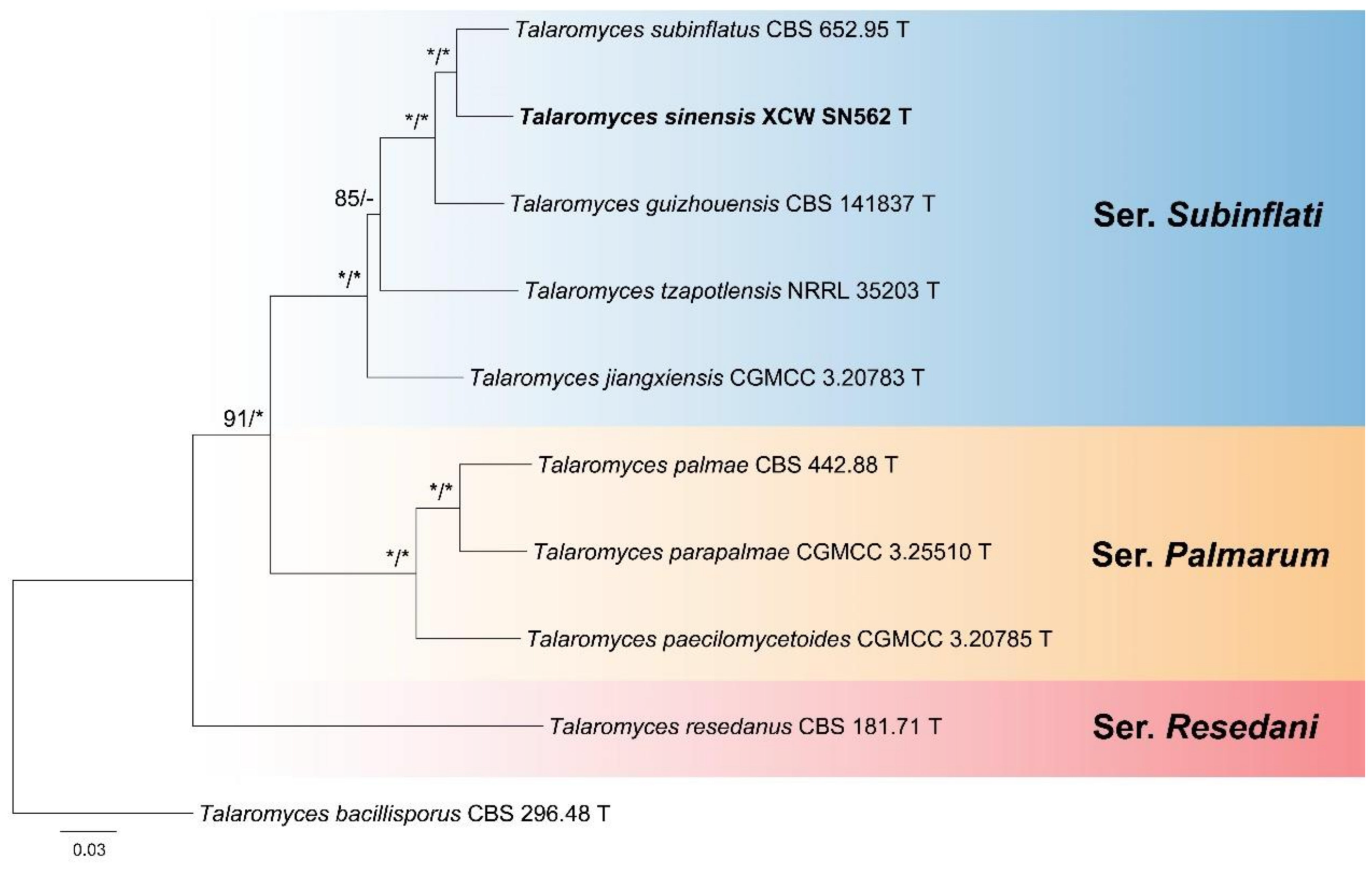
T. sinensis X.C. Wang, L.Y. Peng & W.Y. Zhuang, sp. nov.XCW_SN 562 = CGMCC 3.28744 TChina: YunnanRotten husk of an unknown fruitPV085755PV102705PV102718PV102726
T. subinflatus Yaguchi & Udagawa 1993CBS 652.95 TJapanSoilJN899397MK450890KJ885280KM023308
T. tzapotlensis Jurjević & S.W. Peterson 2017NRRL 35203 TMexicoHypothenemus hampeiKX946902KX946884KX946893KX946922
T. bacillisporus (Swift) C.R. Benj. 1955CBS 296.48 TNorth AmericaLeaves of BegoniaKM066182AY753368KJ885262JF417425
GenBank accession numbers in bold indicate newly generated sequences.
Table 2. Species and sequences of Talaromyces sect. Trachyspermi used in phylogenetic analyses.
SpeciesStrainCountrySubstrateITSBenACaMRPB2
T. aerius A.J. Chen et al. 2016CGMCC 3.18197 TChina: BeijingIndoor airKU866647KU866835KU866731KU866991
T. affinitatimellis Rodr.-Andr. et al. 2019CBS 143840 TSpainHoneyLT906543LT906552LT906549LT906546
T. africanus Houbraken et al. 2021CBS 147340 TSouth AfricaHouse dustOK339610OK338782OK338808OK338833
T. albidus L. Wang 2023CGMCC 3.26143 TChina: ShanghaiSoilOQ746343OQ746324OQ746326OQ746328
T. albisclerotius B.D. Sun et al. 2020CBS 141839 TChina: XinjiangSoilMN864276MN863345MN863322MN863334
T. albobiverticillius (H.M. Hsieh et al.) Samson et al. 2011CBS 133440 TChina: TaiwanFallen decaying leavesHQ605705KF114778KJ885258KM023310
T. amyrossmaniae Rajeshkumar et al. 2019NFCCI 1919 TIndiaDecaying fruits of Terminalia bellericaMH909062MH909064MH909068MH909066
T. assiutensis Samson & Abdel-Fattah 1978CBS 147.78 TEgyptSoilJN899323KJ865720KJ885260KM023305
T. atroroseus N. Yilmaz et al. 2013CBS 133442 TSouth AfricaHouse dustKF114747KF114789KJ775418KM023288
T. austrocalifornicus Yaguchi & Udagawa 1993CBS 644.95 TUSASoilJN899357KJ865732KJ885261MN969147
T. basipetosporus Stchigel et al. 2019CBS 143836 TArgentinaHoneyLT906542LT906563n.a.LT906545
T. brasiliensis R.N. Barbosa et al. 2018CBS 142493 TBrazilHoneyMF278323LT855560LT855563MN969198
T. calidominioluteus Houbraken & Pyrri 2021CBS 147313 TNetherlandsMelonOK339612OK338786OK338817OK338837
T. catalonicus Guevara-Suarez et al. 2020CBS 143039 TSpainHerbivore dungLT899793LT898318LT899775LT899811
T. chongqingensis X.C. Wang & W.Y. Zhuang 2021CGMCC 3.20482 TChina: ChongqingSoilMZ358001MZ361343MZ361350MZ361357
T. clemensii Visagie & N. Yilmaz 2019PPRI 26753 TSouth AfricaRotting wood in mineMK951940MK951833MK951906MN418451
T. convolutus Udawaga 1993CBS 100537 TNepalSoilJN899330KF114773MN969316JN121414
T. cystophila Y.X. Mo & H.Y. Wu 2024CGMCC 40002 TChina: GuangxiHeterodera zeae cystOM835900ON164851ON164853ON164852
T. diversus (Raper & Fennell) Samson et al. 2011CBS 320.48 TUSAMoldy leatherKJ865740KJ865723KJ885268KM023285
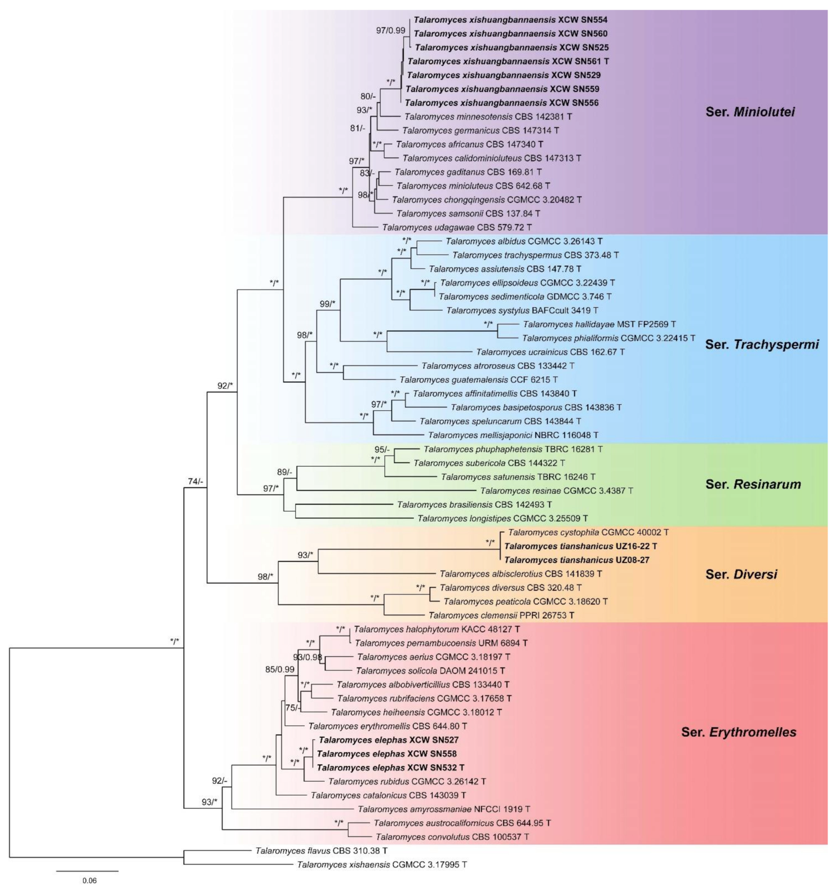
T. elephas X.C. Wang, L.Y. Peng & W.Y. Zhuang, sp. nov.XCW_SN 532 = CGMCC 3.28742 TChina: YunnanRotten husk of an unknown fruitPV085756PV102706PV102719PV102727
XCW_SN 527China: YunnanRotten husk of an unknown fruitPV085757PV102707PV102720PV102728
XCW_SN 558China: YunnanRotten husk of an unknown fruitPV085758PV102708n.a.PV102729
T. ellipsoideus M. Li & L. Cai 2023CGMCC 3.22439 TChina: GuangdongSedimentOQ798985OQ808981OQ808994OQ809036
T. erythromellis (A.D. Hocking) Samson et al. 2011CBS 644.80 TAustraliaSoilJN899383HQ156945KJ885270KM023290
T. gaditanus (C. Ramírez & A.T. Martínez) Houbraken & Soccio 2021CBS 169.81 TSpainAirMH861318OK338775OK338802OK338827
T. germanicus Houbraken & Pyrri 2021CBS 147314 TGermanyWallboardOK339619OK338799OK338812OK338845
T. guatemalensis A. Nováková et al. 2019CCF 6215 TGuatemalaSoilMN322789MN329687MN329688MN329689
T. hallidayae Y.P. Tan et al. 2024MST FP2569 TAustraliaSoil under turf grassPP665728PP682580PP682551PP682567
T. halophytorum Y.H. You & S.B. Hong 2020KACC 48127 TSouth KoreaRoots of Limonium tetragonumMH725786MH729367MK111426MK111427
T. heiheensis X.C. Wang & W.Y. Zhuang 2017CGMCC 3.18012 TChina: HeilongjiangRotten woodKX447526KX447525KX447532KX447529
T. longistipes Zhi Y. Zhang & Y.F. Han 2024CGMCC 3.25509 TChina: GuizhouSoilOR680518OR843223OR828454OR842935
T. mellisjaponici A. Okubo & D. Hirose 2024NBRC 116048 TJapanHoneyLC763421LC763430LC763439LC763448
T. minioluteus (Dierckx) Samson et al. 2011CBS 642.68 TUnknownUnknownJN899346MN969409KJ885273JF417443
T. minnesotensis Guevara-Suarez et al. 2017CBS 142381 TUSAHuman earLT558966LT559083LT795604LT795605
T. peaticola J.Q. Tian & J.Z. Sun 2021CGMCC 3.18620 TChina: SichuanPeaty soil of wetlandMF135613MF284705MF284703MF284704
T. pernambucoensis R. Cruz et al. 2019URM 6894 TBrazilSoilLR535947LR535945LR535946LR535948
T. phialiformis M. Li & L. Cai 2023CGMCC 3.22415 TChina: GuangdongMangrove sedimentOQ798986OQ808982OQ808995n.a.
T. phuphaphetensis Nuankaew et al. 2022TBRC 16281 TThailandSoil in caveON692803ON706960ON706962ON706964
T. resinae (Z.T. Qi & H.Z. Kong) Houbraken & X.C. Wang 2020CGMCC 3.4387 TChina: GuizhouResin of Eucalyptus tereticornisMT079858MN969442MT066184MN969221
T. rubidus L. Wang 2023CGMCC 3.26142 TChina: YunnanSoilOQ746342OQ746323OQ746325OQ746327
T. rubrifaciens W.W. Gao 2016CGMCC 3.17658 TChina: BeijingHospital airKR855658KR855648KR855653KR855663
T. samsonii (Quintan.) Houbraken & Pyrri 2021CBS 137.84 TSpainInsect-damaged fruit of Malus pumilaMH861709OK338798OK338824OK338844
T. satunensis Nuankaew et al. 2022TBRC 16246 TThailandSoil in caveON692804ON706961ON706963n.a.
T. sedimenticola Y. Wang & H. Zhou 2024GDMCC 3.746 TMariana TrenchSedimentON000284ON384002ON326484ON000277
T. solicola Visagie & K. Jacobs 2012DAOM 241015 TSouth AfricaFynbos soilFJ160264GU385731KJ885279KM023295
T. speluncarum Rodr.-Andr. et al. 2020CBS 143844 TSpainSparkling wineLT985890LT985901LT985906LT985911
T. subericola Rodr.-Andr. et al. 2020CBS 144322 TSpainSparkling wineLT985888LT985899LT985904LT985909
T. systylus S.M.Romero et al. 2016BAFCcult 3419 TArgentinaSoilKP026917KR233838KR233837n.a.
T. tianshanicus X.C. Wang, L.Y. Peng, Gafforov & W.Y. Zhuang, sp. nov.UZ16-22 = CGMCC 3.28741 TUzbekistanSoilPV085759PV102709n.a.PV102730
UZ08-27UzbekistanSoilPV085760PV102710PV102721n.a.
T. trachyspermus (Shear) Stolk & Samson 1972CBS 373.48 TUSAUnknownJN899354KF114803KJ885281JF417432
T. ucrainicus (Panas.) Udagawa 1966CBS 162.67 TJapanSoilJN899394KF114771KJ885282KM023289
T. udagawae Stolk & Samson 1972CBS 579.72 TJapanSoilJN899350OK338783KX961260MN969148
T. xishuangbannaensis X.C. Wang, L.Y. Peng & W.Y. Zhuang, sp. nov.XCW_SN 561 = CGMCC 3.28743 TChina: YunnanRotten husk of an unknown fruitPV085761PV102711PV102722PV102731
XCW_SN 525China: YunnanRotten husk of an unknown fruitPV085762PV102712n.a.PV102732
XCW_SN 529China: YunnanRotten husk of an unknown fruitPV085763PV102713PV102723PV102733
XCW_SN 554China: YunnanRotten husk of an unknown fruitPV085764PV102714n.a.PV102734
XCW_SN 556China: YunnanRotten husk of an unknown fruitPV085765PV102715PV102724PV102735
XCW_SN 559China: YunnanRotten husk of an unknown fruitPV085766PV102716PV102725PV102736
XCW_SN 560China: YunnanRotten husk of an unknown fruitPV085767PV102717n.a.PV102737
T. flavus (Klöcker) Stolk & Samson 1972CBS 310.38 TNew ZealandUnknownJN899360JX494302KF741949JF417426
T. xishaensis X.C. Wang et al. 2016CGMCC 3.17995 TChina: HainanSoilKU644580KU644581KU644582MZ361364
GenBank accession numbers in bold indicate newly generated sequences. ‘n.a.’ is the abbreviation of ‘not available’.
Table 3. Detailed characteristics of the datasets.
DatasetGene FragmentNo. of Seq.Length of Alignment (bp)No. of Variable SitesNo. of
Parsimony-Informative Sites
Model for BI
SubinflatiITS1051812968
BenA1038313165
CaM10532234141
RPB2101008287166
BenA + CaM + RPB2101923652372TrNef + I + G
TrachyspermiITS62626218162
BenA62590290221
CaM56658398311
RPB258917349314
BenA + CaM + RPB26221651037846GTR + I + G
Abbreviations of the model: GTR + I + G (general time-reversible model with invariant sites and Gamma distribution); TrNef + I + G (equal-frequency Tamura–Nei model with invariant sites and Gamma distribution).
Table 4. Morphological comparisons of new species and their closely related species.
SeriesSpeciesCYA 25 °C (mm)CYA 37 °C (mm)MEA (mm)YES (mm)ConidiophoreConidia ShapeConidia WallConidia Size (µm)Reference
DiversiT. tianshanicus16–1910–1421–2311–15Irregularly biverticillate to quaterverticillateSubglobose to ellipsoidalSmooth2.0–2.5 × 1.7–2.0This study
T. cystophila21–2618–2132–3319–24BiverticillateSubglobose to ellipsoidalSmooth2.6–3.0 × 2.5–3.0[31]
ErythromellesT. elephas3–7No growth15–198–11BiverticillateEllipsoidalSmooth3.0–3.5 × 2.0–3.0This study
T. rubidus5–6No growth17–186–7BiverticillateOvoid to spheroidalEchinulate2.5–3.0[32]
MinioluteiT. xishuangbannaensis4–5No growth15–1610–12BiverticillateSubglobose to ellipsoidalSmooth3.0–4.0 × 2.0–3.0This study
T. germanicus20–22No growth23–2522–24BiverticillateNarrow ellipsoidal to fusiformSmooth2.5–3.5 × 1.5–2.5[27]
T. minnesotensis24–26No growth13–1521–24BiverticillateEllipsoidalSmooth2.5–3.5 × 2.0–3.0[26]
SubinflatiT. sinensisNo growthNo growth13–16no growthBiverticillateEllipsoidalSmooth3.0–4.0 × 2.0–2.5This study
T. subinflatus3–4No growth14–153–4BiverticillateEllipsoidal to fusiformSmooth2.5–4.0 × 1.5–2.0[8]
T. guizhouensis8–9No growth24–2712–13BiverticillateSubglobose to fusiformFinely rough2.5–4.5 × 2.5–3.0[9]
Disclaimer/Publisher’s Note: The statements, opinions and data contained in all publications are solely those of the individual author(s) and contributor(s) and not of MDPI and/or the editor(s). MDPI and/or the editor(s) disclaim responsibility for any injury to people or property resulting from any ideas, methods, instructions or products referred to in the content.

