Article

The Antifungal Effects of Equol Against Candida albicans Involve Mitochondrial Dysfunction

1
School of Basic Medical Sciences, Southwest Medical University, Luzhou 646000, China
2
The Public Platform of Molecular Biotechnology, Public Center of Experimental Technology, Southwest Medical University, Luzhou 646000, China
3
Hemodynamics and Medical Engineering Combination Key Laboratory of Luzhou, Southwest Medical University, Luzhou 646000, China
*
Authors to whom correspondence should be addressed.
J. Fungi 2025, 11(5), 339; https://doi.org/10.3390/jof11050339
Submission received: 17 March 2025 / Revised: 25 April 2025 / Accepted: 25 April 2025 / Published: 27 April 2025
(This article belongs to the Topic Natural Products to Fight Fungal Infections)

Abstract

:
Novel antifungal agents are urgently needed because of the increasing number of drug-resistant Candida strains encountered in clinical practice and the limited variety of available antifungal drugs. Equol, a metabolite of soy isoflavone glycosides, exhibits antifungal activities. In this study, Equol had good inhibitory activity against Candida species. The lowest inhibitory concentration of 125–500 μg/mL was confirmed by the gradient dilution method. In addition, transmission electron microscopy and the relative content assay showed that Equol altered the cell wall and membrane of Candida albicans. Further studies found that Equol treatment increased the intracellular levels of reactive oxygen species and Ca2+. Subsequent experiments suggested that Equol treatment depolarized the membrane potential of C. albicans and up-regulated the expression of the apoptosis-inducing factor gene. These results confirmed that Equol damaged the cell wall and membrane, dysregulated the intracellular components, induced oxidative stress and Ca2+ accumulation, and ultimately resulted in mitochondrial dysfunction. Collectively, these findings demonstrated that Equol is a potential antifungal agent.

1. Introduction

In 2022, the World Health Organization included C. albicans and Candidozyma auris among the four key priority fungal pathogens because of the increasing incidence of drug resistance [1,2]. In 2023, the proposed guidelines for the Joint Plan of Action for Health stated that antimicrobial drug resistance is the leading cause of death due to microbial infections [3]. C. albicans, a component of the normal human microbiota, is a diploid polymorphic yeast that resides on the skin and mucosal surfaces. The pathogenic potential of C. albicans has been linked to a state of compromised immunity due to critical illnesses, implanted medical devices, or during long-term use of broad-spectrum antibiotics [4]. C. albicans is considered an opportunistic pathogen that causes a range of diseases, including mucocutaneous candidiasis and recurrent candidal vaginitis, which accounts for 103–172 million cases annually [5]. Moreover, the incidence of systemic candidiasis usually ranges from about 2 to 21 cases per 100,000 population, with about 1.6 million cases of systemic infection and 99,500 deaths per year [6]. Typically, C. albicans accounts for about 40–60% of Candida isolates from hospitalized patients [7,8].
Due to the lack of new antifungal treatment options and related biocompatibility issues, antifungal resistance has further increased, limiting medical applications [9]. Therefore, there is an urgent need to develop novel, highly effective antifungal agents with low toxicity for treatment of diseases associated with pathogenic Candida. In fungi, cell walls and membranes are the primary line of protection against environmental stressors and medication harm [10,11,12]. These structures may experience adaptive changes or even destruction when subjected to such loads, which could have several negative effects, such as intracellular oxidative stress and calcium overload [13,14]. The excessive production of reactive oxygen species (ROS) can damage nucleic acids, proteins, and lipids, and increase the permeability of the cell membrane, ultimately resulting in mitochondrial-dependent programmed cell death [15,16,17]. Besides ROS, the excessive accumulation of intracytoplasmic Ca2+ disrupts mitochondrial membrane potential (MMP), causing mitochondrial dysfunction and the release of pro-apoptotic factors, which initiate mitochondria-dependent programmed cell death [18]. Thus, damage to cell walls and membranes by pathogenic fungi, along with substances that induce excessive ROS production and intracytoplasmic Ca2+ overaccumulation, are potential therapeutic approaches against fungal infections.
Equol is a metabolite of soy isoflavones under the action of the intestinal microflora [19]; it is easily absorbed by the colon wall and cleared more slowly in the plasma [20], and is most commonly taken orally at a dose of 10 mg per day by adults according to a US-based online medical information platform (https://www.webmd.com, accessed on 13 February 2025). It has been shown to exhibit pharmacological activities against inflammation, tumor cell proliferation, and bacterial infection [21,22,23]. For instance, Equol was reported to inhibit biofilm formation and decrease the motility of Yersinia enterocolitica [24], inhibit the growth and sporulation of Clostridioides difficile in a concentration-dependent manner [23], and block the production of carbapenemases and virulence factors of carbapenem-resistant Escherichia coli when used in combination with meropenem [22]. Moreover, Equol can effectively inhibit mycelial growth, conidial production, and the germination of the plant pathogenic fungus Magnaporthe oryzae and reduce leaf virulence of rice and barley [25]. However, although Equol activity against C. albicans has been investigated [26,27], the specific antifungal mechanism remains unclear. In view of the wide range of biological effects mediated by Equol and previous work by our group to clarify the efficacy against C. albicans infection in vivo and ex vivo [27], the aim of the present in-depth investigation was to elucidate the antifungal mechanism of Equol against C. albicans as a novel antifungal agent.

2. Materials and Methods

2.1. Chemicals and Reagents

Equol and amphotericin B (AmB) were purchased from Shanghai Yuanye Bio-Technology Co., Ltd. (Shanghai, China). Roswell Park Memorial Institute (RPMI) 1640 medium was purchased from HyClone Laboratories, Inc. (South Logan, UT, USA). Calcofluor white (CFW) and Congo red dye were purchased from Sigma-Aldrich Corporation (St. Louis, MO, USA). Prior to each experiment, a stock solution of Equol dissolved in dimethyl sulfoxide at 50 mg/mL was prepared and diluted with YPD medium (1% yeast extract, 2% peptone, 2% glucose) to the target concentration.

2.2. Strains and Culture Conditions

Standard strains purchased from the American Type Culture Collection (ATCC; Manassas, VA, USA) included Candida albicans ATCC MYA-2876, Candida parapsilosis ATCC 22019, and Cryptococcus neoformans ATCC 208821. In addition, clinical isolate strains of Candidozyma auris (named swcau and swcau1) and Candidozyma duobushaemulonii (named swcd) were obtained by routine hospital microbiology isolation procedures for experimental studies. All strains were stored in 15% glycerol at −80 °C and incubated overnight in YPD medium at 37 °C and 200 rpm before use.

2.3. Determination of Minimal Inhibitory Concentration (MIC)

The MIC of Equol against the tested strains was determined in reference to the American Society for Clinical and Laboratory Standard M27-A4. Briefly, the fungi were activated and resuspended in RPMI-1640 medium to a final concentration of 0.25–5 × 10³ cells/mL. Equol at 1–1024 μg/mL was added to the drug-treated wells (CTWs), positive control wells (PCWs) with no drug, and sterile blank control wells (BCWs) of 96-well plates, with three replicates/group. The plates were incubated for 24 h at 35 °C, and C. neoformans was incubated for 72 h. Afterward, the optical density (OD) of each well was measured at 600 nm using a multifunctional microplate reader. The inhibition rate was calculated as 100 × [1 − (ODCTW − ODBCW)/(ODPCW − ODBCW)]. The MIC was defined as the concentration that resulted in 90% growth inhibition as compared to the control.

2.4. Growth Inhibition Kinetics

Growth curves were generated as described in a previous study [28]. Briefly, C. albicans ATCC MYA-2876 cells cultured overnight were diluted to 5 × 10⁵ cells/mL in YPD medium containing either 250 µg/mL Equol or 2 µg/mL AmB. Controls were free of Equol and AmB. Cells were incubated at 37 °C and 200 rpm. The OD at 600 nm was measured during co-cultivation using a multifunctional microplate reader. Growth kinetics were recorded for 48 h.

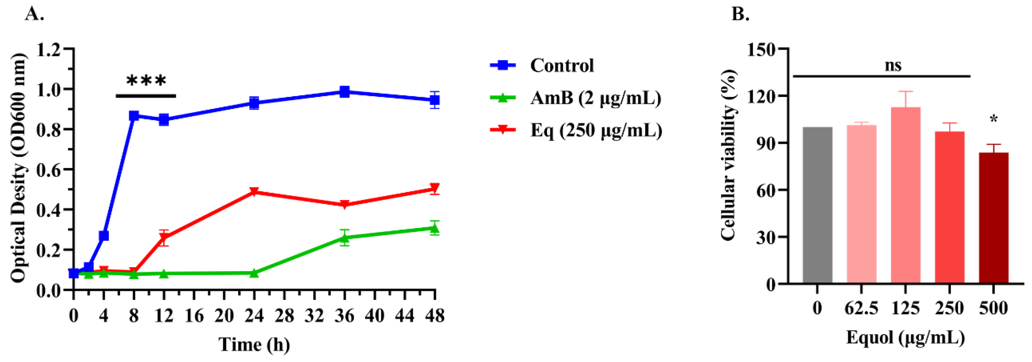
2.5. Effects of Equol on Proliferation of Ana-1 Macrophages Cell

The Ana-1 cell line was counted to 1 × 10⁵ cells/mL, and 100 μL of the cell suspension was added to each well of a 96-well cell culture plate. The plate was incubated overnight at 37 °C in a 5% CO2 incubator. After discarding the culture medium, 0–500 μg/mL of Equol was added. Following incubation, cells were washed twice with phosphate-buffered saline (PBS), and 100 μL of 10% Cell Counting Kit-8 reagent (APExBIO Technology, Houston, TX, USA) was added. The plate was incubated for 2 h, and the absorbance at 450 nm was measured using a microplate reader. The cell survival rate (%) was calculated as (OD administration − OD blank)/(OD control − OD blank) × 100%. The experiment was repeated three independent times.

2.6. Measurement of Cell Membrane Permeability

The membrane permeability of C. albicans ATCC MYA-2876 cells was quantified and visualized using the fluorescence intensity method and fluorescence microscopy, respectively. Membrane integrity was assessed by staining with propidium iodide (PI). Overnight cultured cells were diluted to 1 × 10⁶ cells/mL in YPD medium and incubated for 8 or 12 h with 250 µg/mL Equol at 37 °C with 200 rpm. Untreated cells were used as a negative control. After incubation, the fungal cells were adjusted to 1 × 10⁶ cells/mL with PBS, followed by the addition of 10 mM PI (Sigma-Aldrich Corporation) solution and incubation for 15 min at 37 °C. After washing with PBS, membrane permeability was evaluated by measuring the fluorescence intensity of PI at excitation and emission wavelengths of 525 and 590 nm, respectively, using a multimode microplate reader (Varioskan™ LUX; Thermo Fisher Scientific, Waltham, MA, USA) [29]. The samples were collected and observed under a fluorescence microscope at 40× magnification.

2.7. Cell Wall Staining and Microscopic Examination

The fluorescent brightener and aniline blue staining methods have been applied to assay the cell walls of C. albicans and C. neoformans. Based on these established methods [29,30], the effect of Equol on the cell wall of C. albicans ATCC MYA-2876 was determined. Briefly, fungal cells were incubated at 200 rpm at 37 °C for 8 or 12 h in the presence or absence of 250 μg/mL Equol, then washed with PBS, resuspended to 5 × 10⁶ cells/mL, fixed with 4% paraformaldehyde for 15 min at room temperature, washed again with PBS, and incubated with 5 μg/mL CFW at 37 °C in the dark for 10 min for the staining of chitin. Afterward, the cells were washed twice with PBS and the total chitin content was quantified by measuring fluorescence with a microplate reader at excitation and emission wavelengths of 365 and 435 nm, respectively. In addition, the cells were observed under a fluorescence microscope at 40× magnification (BX63; Olympus Corporation, Tokyo, Japan). For quantification of total β-1,3-glucan, the cells were incubated with 0.1% aniline blue (Wako Pure Chemical Industries, Ltd., Osaka, Japan) at 80 °C for 15 min. Subsequently, fluorescence was measured at excitation and emission wavelengths of 400 and 460 nm, respectively. Change in fluorescence (∆F) was calculated as [F(experimental group) − F(blank group)], where F(experimental group) is the fluorescence of the test samples, F(blank group) is the fluorescence of the test samples, and F(blank group) is the fluorescence of the test group without the dye solution.

2.8. Reverse Transcription Quantitative Polymerase Chain Reaction (RT-qPCR)

Overnight cultures of fungal cells (106 cells/mL) were incubated with or without Equol at 37 °C and 200 rpm and collected by centrifugation at 12,000 rpm for 3 min at 4 °C. Total RNA was isolated using a Yeast Processing Reagent (TaKaRa, Dalian, China) and reverse-transcribed into complementary DNA using a PrimeScript™ RT kit with a gDNA Eraser (TaKaRa). RT-qPCR was performed using TB Green Premix Ex Taq™ II Master Mix (TaKaRa). qPCR data were first normalized with β-actin expression as an internal reference, and then relative expression levels were calculated using 2−ΔΔCT at each time point with reference to the untreated group [27].

2.9. Measurement of ROS Concentrations

Dichlorodihydrofluorescein diacetate (DCFH-DA) was used to determine intracellular ROS levels. Overnight cultured fungal cells were diluted to 1 × 10⁶ cells/mL and cultured in 250 µg/mL of Equol in YPD medium for 8 or 12 h at 37 °C and 200 rpm. Untreated fungal cells were used as a negative control. After incubation, the test cells were resuspended in PBS to 1 × 10⁶ cells/mL, incubated with 40 μmol/L DCFH-DA for 30 min at 37 °C, and washed twice with PBS. Fluorescence intensity (FI) values were detected with an enzyme labeling instrument at excitation and emission wavelengths of 490 and 530 nm, respectively. The FI values, reflecting intracellular ROS levels, were calculated by subtracting the FI values of the stained cells from those of the unstained cells [29]. The cells were collected and observed under a fluorescence microscope at 40× magnification.

2.10. Measurement of Cytosolic Ca2+ Concentrations

The Fluo-3/4 AM probe method for measuring intracellular Ca2+ concentration has been widely used in C. albicans and C. neoformans [17,31]. Additionally, Fluo-4 AM (Beyotime Biotechnology, Shanghai, China), an analog of Fluo-3 AM, has been extensively utilized for measuring intracellular calcium ion concentrations in mammalian cells [32,33]. Based on these established methods, the intracellular calcium ion concentration of C. albicans ATCC MYA-2876 was examined. Briefly, overnight cultured fungal cells were diluted to 1 × 10⁶ cells/mL in YPD medium and 250 µg/mL Equol at 37 °C for 8 or 12 h with shaking at 200 rpm, collected by centrifugation, washed twice in sterile PBS, resuspended in 500 µL PBS, incubated with Fluo-4 AM (final concentration of 2 µM) for 40 min at 37 °C in the dark, and then washed three times with PBS. The fluorescence intensity of the Fluo-4 AM probe was measured after another 20 min of incubation at 37 °C in the absence of Fluo-4 AM. Untreated fungal cells were used as a negative control.

2.11. Measurement of MMP

Changes to the MMP of C. albicans cells after Equol treatment were assessed using a JC-1 MMP assay kit (Beyotime Biotechnology) in accordance with the manufacturer’s protocol [34,35]. The depolarization of the MMP after Equol treatment was assessed using the molecular probe JC-1. The treated fungal cells were washed with PBS and then incubated with the JC-1 probe for 20 min at 37 °C. After incubation, the cells were washed twice with JC-1 buffer, and the green fluorescence intensity of the JC-1 probe was measured at excitation and emission wavelengths of 490 and 530 nm, while red fluorescence was measured at excitation and emission wavelengths of 525 and 590 nm using a multifunctional microplate reader and fluorescence microscopy, respectively. Untreated fungal cells were used as a negative control and carbonyl cyanide m-chlorophenyl hydrazone (CCCP) was used to decrease the MMP as a positive control. Red fluorescence (JC-1 aggregates) indicates healthy mitochondria with a high MMP and green fluorescence (JC-1 monomer) indicates mitochondria with a low MMP.

2.12. Transmission Electron Microscopy (TEM)

Morphological changes to Equol-treated cells were observed by TEM [36]. Briefly, fungal cell suspensions (1 × 10⁶ cells/mL) were exposed to 250 μg/mL Equol and incubated at 37 °C and 200 rpm for 8 or 12 h. Untreated fungal cells were used as a negative control. The cells were collected by centrifugation at 12,000× g for 10 min, prefixed with 3% glutaraldehyde, postfixed with 1% osmium tetroxide, dehydrated, embedded in epoxy resin, and cut into semi-thin sections, which were stained with methylene blue, or ultrathin sections with a diamond knife and stained with uranyl acetate and lead citrate, and examined by TEM (JEM-1400-FLASH; JEOL, Ltd., Tokyo, Japan).

2.13. Statistical Analysis

All statistical analyses were performed using Prism 8 software (GraphPad Software, San Diego, CA, USA). A two-sided Student’s t-test and a one-way analysis of variance (ANOVA) were used to compare differences between and among groups, respectively. The data are presented as the mean ± standard error. A probability (p) value ≤ 0.05 was considered statistically significant. All experiments were repeated in triplicate.

3. Results

3.1. Equol Inhibited Growth of Fungal Cells

As shown in Table 1, Equol showed antifungal activity against C. parapsilosis, C. neoformans, C. auris, and C. duobushaemulonii with MIC values ranging from 125 to 500 µg/mL. A growth curve analysis showed that untreated C. albicans entered the logarithmic phase within 4 h of incubation and reached the plateau phase at 8 h, while C. albicans treated with Equol (250 μg/mL) entered the logarithmic phase only at 12 h after incubation and reached the plateau phase at 24 h (Figure 1A). Moreover, the results indicated that less than 250 μg/mL of Equol was non-cytotoxic (Figure 1B).

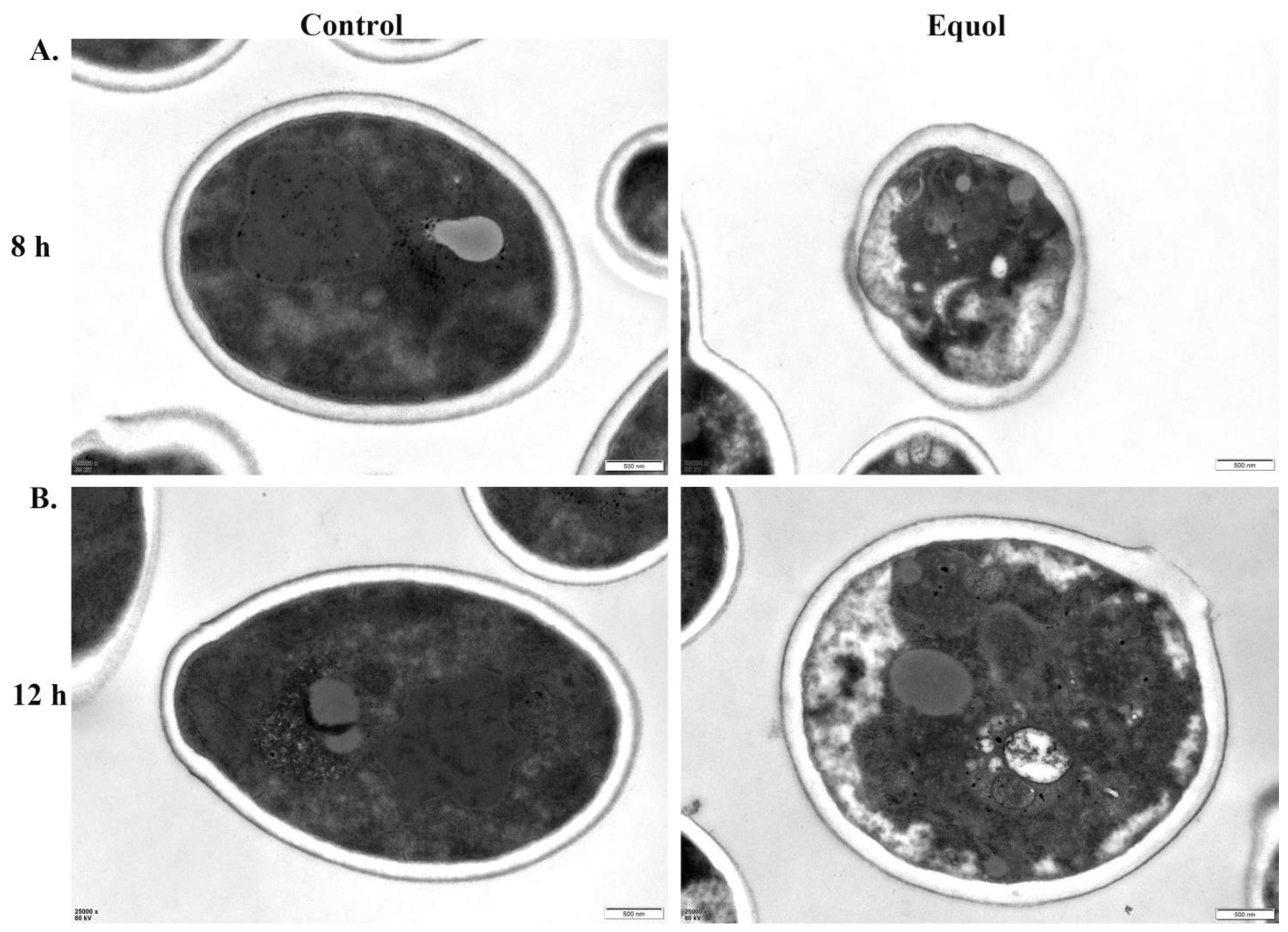
3.2. Equol Caused Ultrastructural Changes to Fungal Cells

The TEM analysis showed that the untreated cell wall, cell membrane, and cytoplasm were dense with complete structures and boundaries of the organelles. In addition, the fungal cells were round or oval with a uniform size. However, after exposure to Equol at 250 μg/mL for 8 and 12 h, the fungal cytoplasm was loose, the organelle structures were disorganized, the number of vacuoles was increased, the cell membrane was zigzagged and uneven, and the cell wall was destroyed, allowing for leakage of intracellular substances. These findings suggest that Equol caused morphological changes to the fungal cells and destroyed the surface structure (Figure 2), resulting in intracellular imbalance and inhibition fungal cell growth.

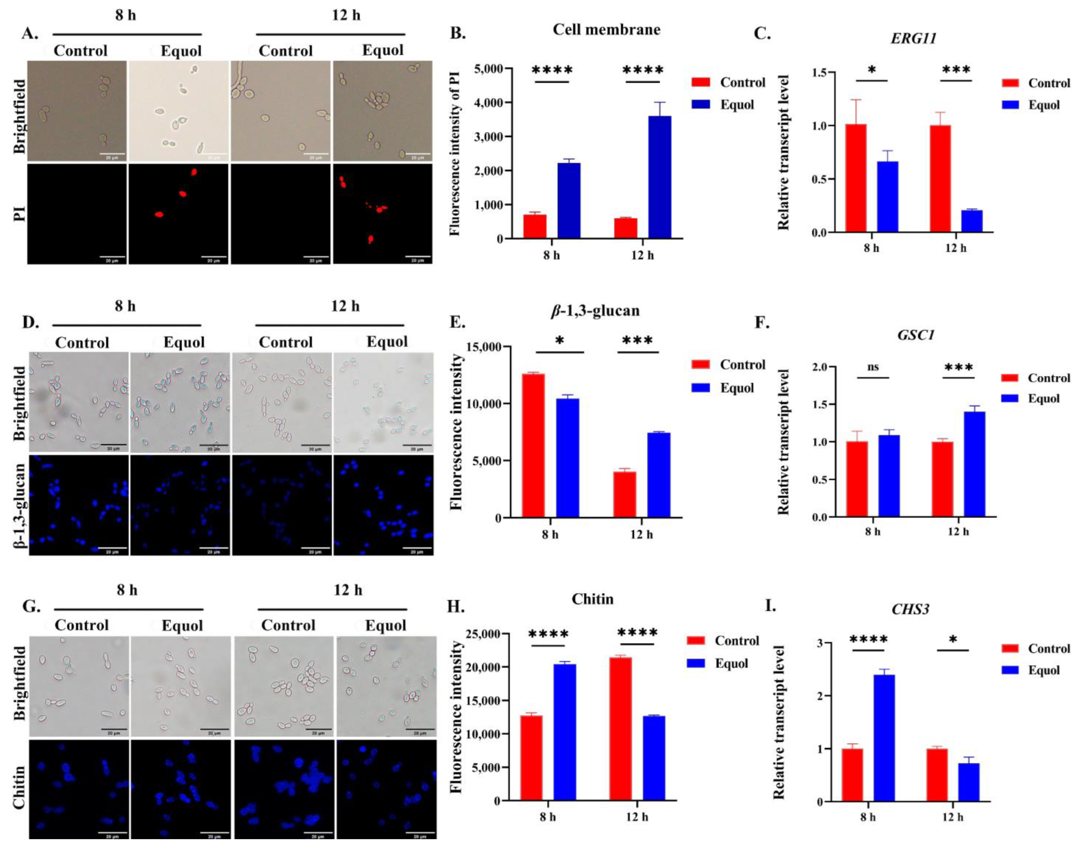
3.3. Equol Damaged the Fungal Cell Wall and Membrane

PI enters the cell and binds to DNA with red fluorescence after damage to membrane integrity [28]. The PI staining results showed that the fluorescence intensity was stronger after Equol treatment as compared to the control group (Figure 3A,B). Further RT-qPCR analysis of ERG11, a key synthesis gene for ergosterol [37], revealed that Equol decreased the expression of ERG11 (Figure 3C).
Meanwhile, fluorescence staining of the important components of the fungal cell wall, including chitin and β-1,3-glucan [29], confirmed that the cell wall components had undergone changes over different time periods after Equol treatment. Specifically, the content of the content of β-1,3-glucan had decreased (Figure 3D,E), while the chitin of the cell wall had increased (Figure 3G,H) after 8 h of Equol treatment. However, opposite results were observed after 12 h of Equol treatment. Further RT-qPCR analysis showed that the β-glucan synthesis gene GSC1 (Figure 3F) and chitin synthesis gene CHS3 (Figure 3I) were consistent with these changes. Specifically, GSC1 expression was upregulated after drug treatment for 12 h, while CHS3 expression was upregulated after drug treatment for 8 h. These findings demonstrate that Equol remodeled and disrupted the cell wall and impaired cell membrane integrity.

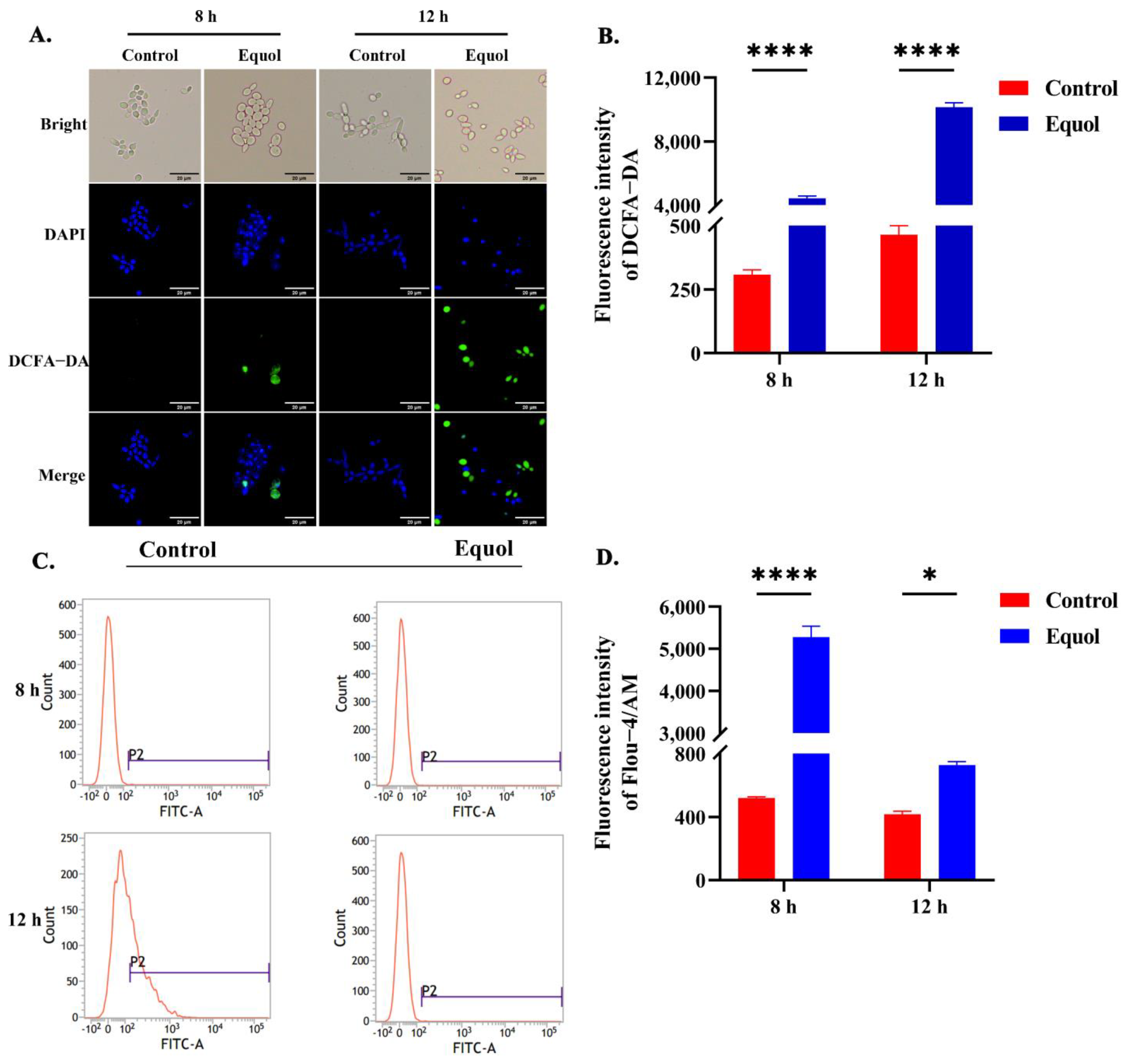
3.4. Equol Caused Intracellular ROS and Ca2+ Overload

An imbalance in intracellular ROS leads to apoptosis [38]. Therefore, alterations to intracellular ROS contents were detected. As expected, the intracellular ROS content of fungal cells had progressively increased after Equol treatment, as demonstrated by enhanced green fluorescence (Figure 4A,B). Moreover, ROS and Ca2+ complement each other and are considered to be crucial mediators of apoptosis [39,40]. In this study, the accumulation of intracytoplasmic Ca2+ was significantly increased after 8 h of Equol treatment as compared to the control group. In addition, as compared to the control group, Ca2+ after 12 h of Equol treatment was increased and significantly increased after 8 h (Figure 4C,D). These data indicate that the culture of C. albicans with Equol leads to the generation of ROS and the accumulation of Ca2+, suggesting that Equol causes oxidative damage to fungal cells.

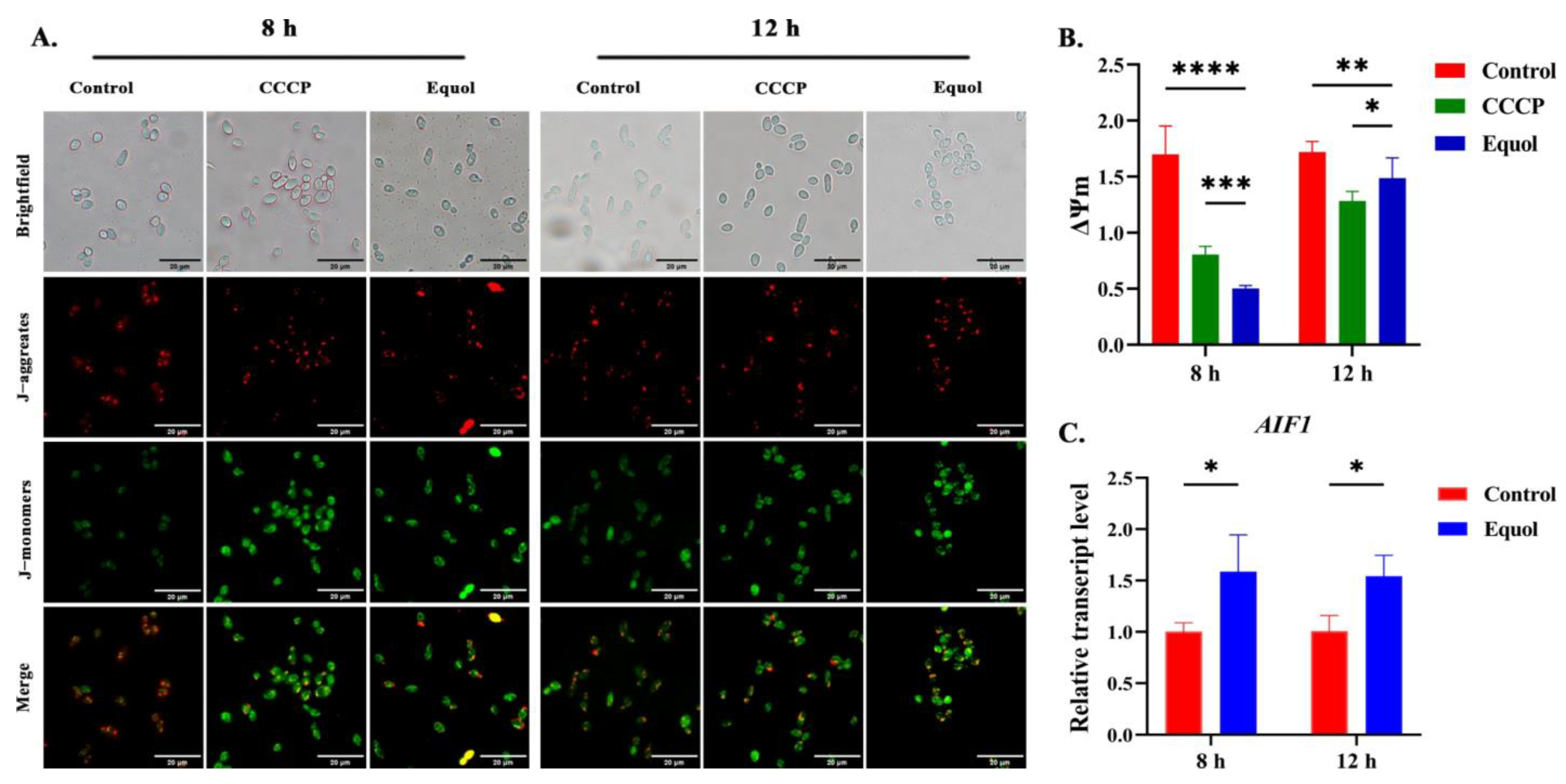
3.5. Equol Induced Mitochondrial Dysfunction
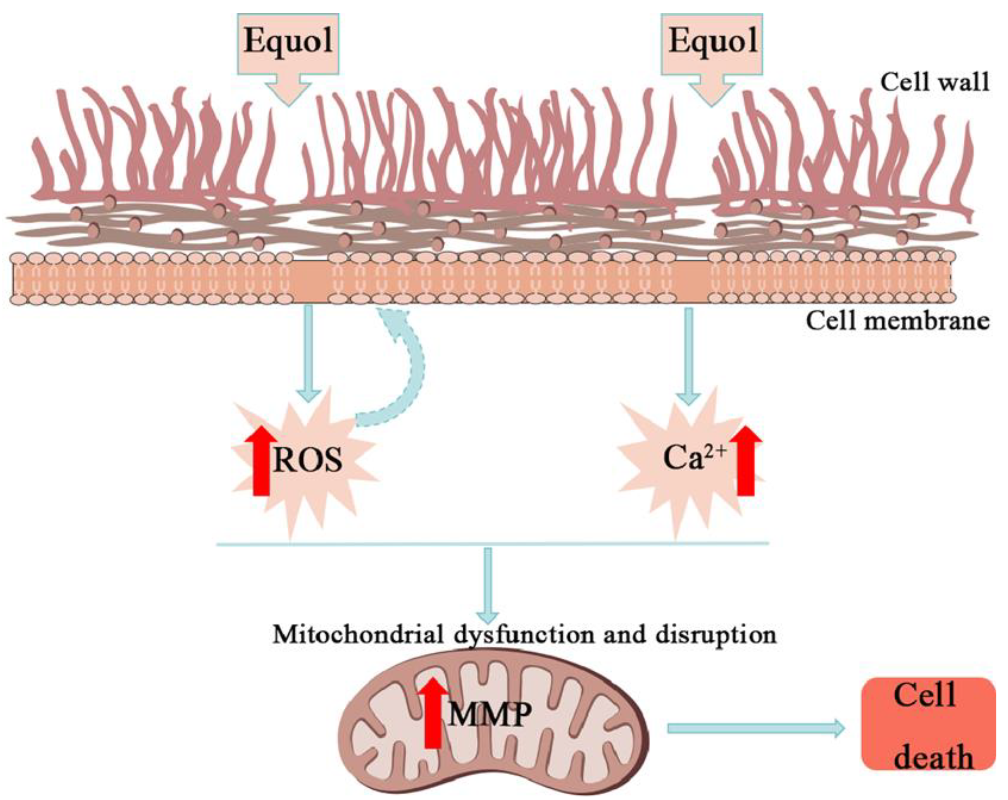
The ROS overproduction and disruption of Ca2+ homeostasis induce mitochondrial damage [15]. In order to evaluate the effects of Equol on mitochondrial function of C. albicans, changes in MMP and the expression of related apoptosis genes were detected. The detection of MMP by a JC-1 fluorescence probe showed that compared with the control group, both the CCCP (a mitochondrial uncoupling agent)- and Equol-treated groups showed enhanced green fluorescence and weakened red fluorescence at 8 and 12 h, and the red/green fluorescence ratio of JC-1 was decreased, indicating mitochondrial membrane potential depolarization (Figure 5A,B). These results indicated that Equol damaged mitochondrial membrane potential and mitochondrial homeostasis was unbalanced. The expression level of gene-encoding mitochondrial-related apoptosis factor AIF1 showed that the transcription level of AIF1 was significantly upregulated after 12 h of Equol treatment, suggesting that Equol may induce apoptosis of C. albicans cells by activating the mitochondrial pathway (Figure 5C).

4. Discussion

The incidence of invasive Candida infections continues to increase yearly due to the rise in immunocompromised populations, as well as the misuse of antibiotics and antifungal medications, and the impact of the coronavirus disease pandemic in 2019, which has generated a large number of susceptible populations [41,42,43]. However, inappropriate antifungal drugs and the emergence of multidrug-resistant strains have led to a decline in the effectiveness of existing antifungal treatments [44]. Therefore, novel targeted antifungal drugs are urgently needed. Previous investigations by our group confirmed that Equol exerts antifungal effects against C. albicans by inhibiting mycelial transformation and biofilm formation, as well as destroying formed biofilms. However, the intracellular antifungal mechanism of Equol has not been thoroughly investigated [27]. The results of the present study demonstrated that Equol also has antifungal activity against non-C. albicans species. Further investigations on the intracellular antifungal molecular mechanism revealed that Equol can effectively destroy the fungal cell wall and membrane integrity and cause the depolarization of MMP, which collectively ultimately induce apoptosis.
The cell wall not only plays an important role in the maintenance of cell morphology and polarized growth, but also in adhesion with host cells and phagocytosis [45,46]. Previous investigations have demonstrated that antifungal drugs, such as echinocandins and β-glucan derivatives, target with the cell wall and enhance intracellular ROS levels, thereby achieving an inhibitory effect against fungal cells [47,48]. In this study, observations by TEM showed that treatment with Equol severely damaged the integrity of the fungal cell wall (Figure 2 and Figure 3). In addition to the cell wall, the cell membrane also plays an important role in maintaining and regulating the cellular structure and function [49,50,51]. The fluorescence staining results indicated that Equol also disrupted the integrity of the cell membrane (Figure 3). Therefore, the disruption of the cell wall and membrane by Equol would cause changes to fungal cell permeability and the intracellular environment. However, changes to the intracellular environment must be further confirmed.
ROS play an important role in biological processes, such as proliferation and the regulation of the cell cycle. An imbalance in intracellular ROS has been shown to lead to apoptosis in fungal cells [38]. In this study, intracellular ROS were significantly increased after Equol treatment (Figure 4). The accumulation of ROS damages cells and mediates mitochondrial dysfunction. Moreover, oxidative stress as well as mitochondrial dysfunction are important molecular targets for the action of antifungal agents. For example, the antifungal activity of kalopanaxsaponin A is due to increased intracellular ROS production [52,53]. Moreover, the antifungal effects of allotoxin A1 are due to increased intracellular ROS accumulation and mitochondrial dysfunction [54]. In addition, the antifungal effects of the peptide LL-37 have been linked to the dysregulation of Ca2+ homeostasis and increased ROS production in mitochondria [55]. Therefore, agents that target the fungal mitochondria provide a promising antifungal strategy.
As an important second messenger in the developmental and stress signaling pathways of fungal cells, Ca2+ plays an important role in cation homeostasis, morphogenesis, virulence traits, and antifungal resistance [56,57,58]. Elevated cytoplasmic Ca2+ content has been shown to be the main cause of pheromone-induced apoptosis in Saccharomyces cerevisiae [39,40,53]. Meanwhile, the Ca2+ and ROS signaling pathways overlap and influence each other, as both are involved in cellular stress, injury, and even apoptosis in fungal cells [59]. Therefore, both pathways are considered key mediators of apoptosis [38,60,61]. In this study, Equol treatment increased intracellular concentration of both ROS and Ca2+ (Figure 4). These complementary effects were mainly manifested by the accumulation of ROS, which leads to the release of Ca2+ from the endoplasmic reticulum or other organelles, and Ca2+ overload, which disrupts Ca2+ homeostasis in the mitochondria and increases the production of ROS [38,60,61].
Oxidative stress and Ca2+ overload also increase the permeability of the mitochondrial membrane and subsequent depolarization [39]. Increased permeability of the inner mitochondrial membrane also causes the release of protons, resulting in the depolarization of the MMP, which is one of the earliest stages of apoptosis [62,63]. As potential targets for the development of antifungal agents, the mitochondria are responsible for cellular adaptation to oxidative stress and Ca2+ overload and play an important role in the production of adenosine triphosphate, the control of lipid homeostasis, and the regulation of programmed fungal cell death [18,64]. Under normal conditions, the intracellular MMP is in a steady state, as determined by the red fluorescence of the JC-1 probe. Conversely, in a state of apoptosis, the MMP is depolarized, and the JC-1 probe emits green fluorescence. In the present study, the intracellular monomeric form of JC-1 (green fluorescence) had increased after Equol treatment (Figure 5). AIF1 codes for a conserved flavoprotein located in mitochondria that acts as an apoptosis-inducing factor that leads to nuclear cohesion and DNA disassembly after translocation to the nucleus, ultimately inducing apoptosis [65,66]. AIF1 expression was significantly increased after Equol treatment (Figure 5), providing favorable evidence for mitochondrial membrane depolarization and subsequent apoptosis. Moreover, it had confirmed that even within the same class of drugs, the drug sensitivity and response varied among various strains [67]. Therefore, the effect of Equol on other clinically relevant strains, such as C. auris or C. parapsilosis, still needs to be further validated.

5. Conclusions

The results of this study confirmed the antifungal effect of Equol against Candida species. The mechanism underlying the antifungal effect of Equol against C. albicans involves significant damage to the cell membrane, cell wall, and mitochondrial membrane, along with changes to the intracellular environment, resulting in the apoptosis of C. albicans (Figure 6). Collectively, these findings highlight the potential clinical use of Equol as an antifungal agent.

Author Contributions

A.G.: conceptualization, methodology, investigation, writing—original draft, writing—review and editing. H.Z.: formal analysis and validation. X.Y.: resources, visualization, and methodology. C.Z.: resources and visualization. C.X.: supervision, writing—review and editing, and funding acquisition. Z.S.: supervision, writing—review and editing, funding acquisition, and project administration. All authors have read and agreed to the published version of the manuscript.

Funding

This research was supported by the Sichuan Science and Technology Program (25NSFSC0850), Science and Technology Strategic Cooperation Programs of Luzhou Municipal People’s Government and Southwest Medical University (2023LZXNYDJ034), and the Technology Strategic Cooperation Project of Pengzhou County People’s Hospital-Southwest Medical University (2024PZXNYD03).

Institutional Review Board Statement

This is an observational study. The Southwest Medical University Institutional Animal Care and Use Committee Research confirmed that no ethical approval was required.

Informed Consent Statement

Not applicable.

Data Availability Statement

All data generated or analyzed during this study are included in the article materials, which are available from the corresponding author upon reasonable request.

Conflicts of Interest

The authors have no relevant financial or non-financial interests to disclose.

References

  1. Katsipoulaki, M.; Stappers, M.H.T.; Malavia-Jones, D.; Brunke, S.; Hube, B.; Gow, N.A.R. Candida albicans and Candida glabrata: Global priority pathogens. Microbiol. Mol. Biol. Rev. 2024, 88, e0002123. [Google Scholar] [CrossRef] [PubMed]
  2. World Health Organization. WHO Fungal Priority Pathogens List to Guide Research, Development and Public Health Action; World Health Organization: Geneva, Switzerland, 2022. [Google Scholar]
  3. Fisher, M.C.; Denning, D.W. The WHO fungal priority pathogens list as a game-changer. Nat. Rev. Microbiol. 2023, 21, 211–212. [Google Scholar] [CrossRef] [PubMed]
  4. Lopes, J.P.; Stylianou, M.; Nilsson, G.; Urban, C.F. Opportunistic pathogen Candida albicans elicits a temporal response in primary human mast cells. Sci. Rep. 2015, 5, 12287. [Google Scholar] [CrossRef] [PubMed]
  5. Denning, D.W.; Kneale, M.; Sobel, J.D.; Rautemaa-Richardson, R. Global burden of recurrent vulvovaginal candidiasis: A systematic review. Lancet Infect. Dis. 2018, 18, e339–e347. [Google Scholar] [CrossRef]
  6. Denning, D.W. Global incidence and mortality of severe fungal disease. Lancet Infect. Dis. 2024, 24, e428–e438. [Google Scholar] [CrossRef]
  7. Bays, D.J.; Jenkins, E.N.; Lyman, M.; Chiller, T.; Strong, N.; Ostrosky-Zeichner, L.; Hoenigl, M.; Pappas, P.G.; Thompson, G.R., III. Epidemiology of Invasive Candidiasis. Clin. Epidemiol. 2024, 16, 549–566. [Google Scholar] [CrossRef]
  8. Ferngren, G.; Yu, D.; Unalan-Altintop, T.; Dinnétz, P.; Özenci, V. Epidemiological patterns of candidaemia: A comprehensive analysis over a decade. Mycoses 2024, 67, e13729. [Google Scholar] [CrossRef]
  9. Garvey, M.; Rowan, N.J. Pathogenic Drug Resistant Fungi: A Review of Mitigation Strategies. Int. J. Mol. Sci. 2023, 24, 1584. [Google Scholar] [CrossRef]
  10. Garcia-Rubio, R.; de Oliveira, H.C.; Rivera, J.; Trevijano-Contador, N. The Fungal Cell Wall: Candida, Cryptococcus, and Aspergillus Species. Front. Microbiol. 2019, 10, 2993. [Google Scholar] [CrossRef]
  11. Lima, S.L.; Colombo, A.L.; de Almeida Junior, J.N. Fungal Cell Wall: Emerging Antifungals and Drug Resistance. Front. Microbiol. 2019, 10, 2573. [Google Scholar] [CrossRef]
  12. Douglas, L.M.; Konopka, J.B. Plasma membrane organization promotes virulence of the human fungal pathogen Candida albicans. J. Microbiol. 2016, 54, 178–191. [Google Scholar] [CrossRef] [PubMed]
  13. Yu, Q.; Zhang, B.; Li, J.; Zhang, B.; Wang, H.; Li, M. Endoplasmic reticulum-derived reactive oxygen species (ROS) is involved in toxicity of cell wall stress to Candida albicans. Free Radic. Biol. Med. 2016, 99, 572–583. [Google Scholar] [CrossRef] [PubMed]
  14. Dias, L.P.; Santos, A.L.E.; Araújo, N.M.S.; Silva, R.R.S.; Santos, M.H.C.; Roma, R.R.; Rocha, B.A.M.; Oliveira, J.T.A.; Teixeira, C.S. Machaerium acutifolium lectin alters membrane structure and induces ROS production in Candida parapsilosis. Int. J. Biol. Macromol. 2020, 163, 19–25. [Google Scholar] [CrossRef] [PubMed]
  15. Kim, H.; Lee, D.G. Naringin-generated ROS promotes mitochondria-mediated apoptosis in Candida albicans. IUBMB Life 2021, 73, 953–967. [Google Scholar] [CrossRef]
  16. Seyedjavadi, S.S.; Khani, S.; Eslamifar, A.; Ajdary, S.; Goudarzi, M.; Halabian, R.; Akbari, R.; Zare-Zardini, H.; Imani Fooladi, A.A.; Amani, J.; et al. The Antifungal Peptide MCh-AMP1 Derived From Matricaria chamomilla Inhibits Candida albicans Growth via Inducing ROS Generation and Altering Fungal Cell Membrane Permeability. Front. Microbiol. 2019, 10, 3150. [Google Scholar] [CrossRef]
  17. Sun, C.Q.; Peng, J.; Yang, L.B.; Jiao, Z.L.; Zhou, L.X.; Tao, R.Y.; Zhu, L.J.; Tian, Z.Q.; Huang, M.J.; Guo, G. A Cecropin-4 Derived Peptide C18 Inhibits Candida albicans by Disturbing Mitochondrial Function. Front. Microbiol. 2022, 13, 872322. [Google Scholar] [CrossRef]
  18. Carraro, M.; Bernardi, P. Calcium and reactive oxygen species in regulation of the mitochondrial permeability transition and of programmed cell death in yeast. Cell Calcium 2016, 60, 102–107. [Google Scholar] [CrossRef]
  19. Setchell, K.D.; Clerici, C. Equol: History, chemistry, and formation. J. Nutr. 2010, 140, 1355s–1362s. [Google Scholar] [CrossRef]
  20. Mayo, B.; Vázquez, L.; Flórez, A.B. Equol: A Bacterial Metabolite from The Daidzein Isoflavone and Its Presumed Beneficial Health Effects. Nutrients 2019, 11, 2231. [Google Scholar] [CrossRef]
  21. Fatima, A.; Khan, M.S.; Ahmad, M.W. Therapeutic Potential of Equol: A Comprehensive Review. Curr. Pharm. Des. 2020, 26, 5837–5843. [Google Scholar] [CrossRef]
  22. Kim, H.R.; Eom, Y.B. Synergistic Activity of Equol and Meropenem against Carbapenem-Resistant Escherichia coli. Antibiotics 2021, 10, 161. [Google Scholar] [CrossRef] [PubMed]
  23. Tanaka, Y.; Kimura, S.; Ishii, Y.; Tateda, K. Equol inhibits growth and spore formation of Clostridioides difficile. J. Appl. Microbiol. 2019, 127, 932–940. [Google Scholar] [CrossRef]
  24. Kim, H.R.; Han, M.S.; Eom, Y.B. Anti-bacterial and Anti-biofilm Effects of Equol on Yersinia enterocolitica. Indian. J. Microbiol. 2022, 62, 401–410. [Google Scholar] [CrossRef]
  25. Wang, J.; Li, L.; Yin, Y.; Gu, Z.; Chai, R.; Wang, Y.; Sun, G. Equol, a Clinically Important Metabolite, Inhibits the Development and Pathogenicity of Magnaporthe oryzae, the Causal Agent of Rice Blast Disease. Molecules 2017, 22, 1799. [Google Scholar] [CrossRef]
  26. Lee, J.A.; Chee, H.Y. In Vitro Antifungal Activity of Equol against Candida albicans. Mycobiology 2010, 38, 328–330. [Google Scholar] [CrossRef] [PubMed]
  27. Wang, F.; Zhang, J.; Zhang, Q.; Song, Z.; Xin, C. Antifungal activities of Equol against Candida albicans in vitro and in vivo. Virulence 2024, 15, 2404256. [Google Scholar] [CrossRef] [PubMed]
  28. Lei, J.; Xiao, W.; Zhang, J.; Liu, F.; Xin, C.; Zhou, B.; Chen, W.; Song, Z. Antifungal activity of vitamin D(3) against Candida albicans in vitro and in vivo. Microbiol. Res. 2022, 265, 127200. [Google Scholar] [CrossRef]
  29. Huang, J.; Lei, J.; Ge, A.; Xiao, W.; Xin, C.; Song, Z.; Zhang, J. Antifungal Effect of Vitamin D(3) against Cryptococcus neoformans Coincides with Reduced Biofilm Formation, Compromised Cell Wall Integrity, and Increased Generation of Reactive Oxygen Species. J. Fungi 2023, 9, 772. [Google Scholar] [CrossRef]
  30. Lei, J.; Huang, J.; Xin, C.; Liu, F.; Zhang, J.; Xie, Y.; Mao, Y.; Chen, W.; Song, Z. Riboflavin Targets the Cellular Metabolic and Ribosomal Pathways of Candida albicans In Vitro and Exhibits Efficacy against Oropharyngeal Candidiasis. Microbiol. Spectr. 2023, 11, e0380122. [Google Scholar] [CrossRef]
  31. Stempinski, P.R.; Goughenour, K.D.; du Plooy, L.M.; Alspaugh, J.A.; Olszewski, M.A.; Kozubowski, L. The Cryptococcus neoformans Flc1 Homologue Controls Calcium Homeostasis and Confers Fungal Pathogenicity in the Infected Hosts. mBio 2022, 13, e0225322. [Google Scholar] [CrossRef]
  32. Zhou, R.; Chen, Y.; Li, S.; Wei, X.; Hu, W.; Tang, S.; Ding, J.; Fu, W.; Zhang, H.; Chen, F.; et al. TRPM7 channel inhibition attenuates rheumatoid arthritis articular chondrocyte ferroptosis by suppression of the PKCα-NOX4 axis. Redox Biol. 2022, 55, 102411. [Google Scholar] [CrossRef] [PubMed]
  33. Wang, Q.; Song, Y.; Chen, J.; Li, Q.; Gao, J.; Tan, H.; Zhu, Y.; Wang, Z.; Li, M.; Yang, H.; et al. Direct in vivo reprogramming with non-viral sequential targeting nanoparticles promotes cardiac regeneration. Biomaterials 2021, 276, 121028. [Google Scholar] [CrossRef] [PubMed]
  34. Xu, D.; Liu, L.; Zhao, Y.; Yang, L.; Cheng, J.; Hua, R.; Zhang, Z.; Li, Q. Melatonin protects mouse testes from palmitic acid-induced lipotoxicity by attenuating oxidative stress and DNA damage in a SIRT1-dependent manner. J. Pineal Res. 2020, 69, e12690. [Google Scholar] [CrossRef] [PubMed]
  35. Du, L.; Lin, L.; Li, Q.; Liu, K.; Huang, Y.; Wang, X.; Cao, K.; Chen, X.; Cao, W.; Li, F.; et al. IGF-2 Preprograms Maturing Macrophages to Acquire Oxidative Phosphorylation-Dependent Anti-inflammatory Properties. Cell Metab. 2019, 29, 1363–1375.e1368. [Google Scholar] [CrossRef]
  36. Ma, H.; Yang, L.; Tian, Z.; Zhu, L.; Peng, J.; Fu, P.; Xiu, J.; Guo, G. Antimicrobial peptide AMP-17 exerts anti-Candida albicans effects through ROS-mediated apoptosis and necrosis. Int. Microbiol. 2023, 26, 81–90. [Google Scholar] [CrossRef]
  37. Derkacz, D.; Bernat, P.; Krasowska, A. K143R Amino Acid Substitution in 14-α-Demethylase (Erg11p) Changes Plasma Membrane and Cell Wall Structure of Candida albicans. Int. J. Mol. Sci. 2022, 23, 1631. [Google Scholar] [CrossRef]
  38. Tian, H.; Qu, S.; Wang, Y.; Lu, Z.; Zhang, M.; Gan, Y.; Zhang, P.; Tian, J. Calcium and oxidative stress mediate perillaldehyde-induced apoptosis in Candida albicans. Appl. Microbiol. Biotechnol. 2017, 101, 3335–3345. [Google Scholar] [CrossRef]
  39. Yun, D.G.; Lee, D.G. Silibinin triggers yeast apoptosis related to mitochondrial Ca2+ influx in Candida albicans. Int. J. Biochem. Cell Biol. 2016, 80, 1–9. [Google Scholar] [CrossRef]
  40. Lu, H.; Zhu, Z.; Dong, L.; Jia, X.; Sun, X.; Yan, L.; Chai, Y.; Jiang, Y.; Cao, Y. Lack of trehalose accelerates H2O2-induced Candida albicans apoptosis through regulating Ca2+ signaling pathway and caspase activity. PLoS ONE 2011, 6, e15808. [Google Scholar] [CrossRef]
  41. Ikuta, K.S.; Meštrović, T.; Naghavi, M. Global incidence and mortality of severe fungal disease. Lancet Infect. Dis. 2024, 24, e268. [Google Scholar] [CrossRef]
  42. Thomas-Rüddel, D.O.; Schlattmann, P.; Pletz, M.; Kurzai, O.; Bloos, F. Risk Factors for Invasive Candida Infection in Critically Ill Patients: A Systematic Review and Meta-analysis. Chest 2022, 161, 345–355. [Google Scholar] [CrossRef]
  43. Gold, J.A.W.; Adjei, S.; Gundlapalli, A.V.; Huang, Y.A.; Chiller, T.; Benedict, K.; Toda, M. Increased Hospitalizations Involving Fungal Infections During COVID-19 Pandemic, United States, January 2020–December 2021. Emerg. Infect. Dis. 2023, 29, 1433–1437. [Google Scholar] [CrossRef] [PubMed]
  44. Lockhart, S.R.; Chowdhary, A.; Gold, J.A.W. The rapid emergence of antifungal-resistant human-pathogenic fungi. Nat. Rev. Microbiol. 2023, 21, 818–832. [Google Scholar] [CrossRef] [PubMed]
  45. Arita, G.S.; Faria, D.R.; Capoci, I.R.G.; Kioshima, E.S.; Bonfim-Mendonça, P.S.; Svidzinski, T.I.E. Cell wall associated proteins involved in filamentation with impact on the virulence of Candida albicans. Microbiol. Res. 2022, 258, 126996. [Google Scholar] [CrossRef] [PubMed]
  46. Razmi, M.; Kim, J.; Chinnici, J.; Busarajan, S.; Vuppalapaty, H.; Lankipalli, D.; Li, R.; Maddi, A. Candida albicans Mannosidases, Dfg5 and Dcw1, Are Required for Cell Wall Integrity and Pathogenesis. J. Fungi 2024, 10, 525. [Google Scholar] [CrossRef]
  47. Szymański, M.; Chmielewska, S.; Czyżewska, U.; Malinowska, M.; Tylicki, A. Echinocandins—Structure, mechanism of action and use in antifungal therapy. J. Enzyme Inhib. Med. Chem. 2022, 37, 876–894. [Google Scholar] [CrossRef]
  48. Ramzan, A.; Padder, S.A.; Masoodi, K.Z.; Shafi, S.; Tahir, I.; Rehman, R.U.; Prasad, R.; Shah, A.H. β-Nitrostyrene derivatives as broad range potential antifungal agents targeting fungal cell wall. Eur. J. Med. Chem. 2022, 240, 114609. [Google Scholar] [CrossRef]
  49. Parks, L.W.; Casey, W.M. Physiological implications of sterol biosynthesis in yeast. Annu. Rev. Microbiol. 1995, 49, 95–116. [Google Scholar] [CrossRef]
  50. Mukherjee, P.K.; Chandra, J. Candida biofilm resistance. Drug Resist. Updat. 2004, 7, 301–309. [Google Scholar] [CrossRef]
  51. Shahi, G.; Kumar, M.; Kumari, S.; Rudramurthy, S.M.; Chakrabarti, A.; Gaur, N.A.; Singh, A.; Prasad, R. A detailed lipidomic study of human pathogenic fungi Candida auris. FEMS Yeast Res. 2020, 20, foaa045. [Google Scholar] [CrossRef]
  52. Li, Y.; Shan, M.; Zhu, Y.; Yao, H.; Li, H.; Gu, B.; Zhu, Z. Kalopanaxsaponin A induces reactive oxygen species mediated mitochondrial dysfunction and cell membrane destruction in Candida albicans. PLoS ONE 2020, 15, e0243066. [Google Scholar] [CrossRef] [PubMed]
  53. Guirao-Abad, J.P.; Sánchez-Fresneda, R.; Alburquerque, B.; Hernández, J.A.; Argüelles, J.C. ROS formation is a differential contributory factor to the fungicidal action of Amphotericin B and Micafungin in Candida albicans. Int. J. Med. Microbiol. 2017, 307, 241–248. [Google Scholar] [CrossRef]
  54. Liao, M.; Xia, X.; Meng, Q.; Zhu, C.; Liao, B.; Wang, J.; Gou, L.; Zhou, X.; Yuan, W.; Cheng, L.; et al. Holotoxin A(1) from Apostichopus japonicus inhibited oropharyngeal and intra-abdominal candidiasis by inducing oxidative damage in Candida albicans. Br. J. Pharmacol. 2024, 181, 1857–1873. [Google Scholar] [CrossRef] [PubMed]
  55. Chen, S.Y.; Chang, C.K.; Lan, C.Y. Antimicrobial peptide LL-37 disrupts plasma membrane and calcium homeostasis in Candida albicans via the Rim101 pathway. Microbiol. Spectr. 2023, 11, e0255123. [Google Scholar] [CrossRef]
  56. Pinton, P.; Giorgi, C.; Siviero, R.; Zecchini, E.; Rizzuto, R. Calcium and apoptosis: ER-mitochondria Ca2+ transfer in the control of apoptosis. Oncogene 2008, 27, 6407–6418. [Google Scholar] [CrossRef] [PubMed]
  57. King, M.M.; Kayastha, B.B.; Franklin, M.J.; Patrauchan, M.A. Calcium Regulation of Bacterial Virulence. Adv. Exp. Med. Biol. 2020, 1131, 827–855. [Google Scholar]
  58. Kong, F.; You, H.; Zheng, K.; Tang, R.; Zheng, C. The crosstalk between pattern-recognition receptor signaling and calcium signaling. Int. J. Biol. Macromol. 2021, 192, 745–756. [Google Scholar] [CrossRef]
  59. Zhang, S.; Zhou, X.; Ou, M.; Fu, X.; Lin, Q.; Tao, X.; Wang, Z.; Liu, A.; Li, G.; Xu, Y.; et al. Berbamine promotes macrophage autophagy to clear Mycobacterium tuberculosis by regulating the ROS/Ca2+ axis. mBio 2023, 14, e0027223. [Google Scholar] [CrossRef]
  60. Niu, C.; Wang, C.; Yang, Y.; Chen, R.; Zhang, J.; Chen, H.; Zhuge, Y.; Li, J.; Cheng, J.; Xu, K.; et al. Carvacrol Induces Candida albicans Apoptosis Associated with Ca2+/Calcineurin Pathway. Front. Cell Infect. Microbiol. 2020, 10, 192. [Google Scholar] [CrossRef]
  61. Han, G.; Lee, D.G. Indole propionic acid induced Ca2+-dependent apoptosis in Candida albicans. IUBMB Life 2022, 74, 235–244. [Google Scholar] [CrossRef]
  62. Zou, K.; Yin, K.; Ren, S.; Zhang, R.; Zhang, L.; Zhao, Y.; Li, R. Activity and mechanism of action of antimicrobial peptide ACPs against Candida albicans. Life Sci. 2024, 350, 122767. [Google Scholar] [CrossRef] [PubMed]
  63. Eisenberg, T.; Büttner, S.; Kroemer, G.; Madeo, F. The mitochondrial pathway in yeast apoptosis. Apoptosis 2007, 12, 1011–1023. [Google Scholar] [CrossRef] [PubMed]
  64. Schofield, J.H.; Schafer, Z.T. Mitochondrial Reactive Oxygen Species and Mitophagy: A Complex and Nuanced Relationship. Antioxid. Redox Signal 2021, 34, 517–530. [Google Scholar] [CrossRef] [PubMed]
  65. Ma, F.; Zhang, Y.; Wang, Y.; Wan, Y.; Miao, Y.; Ma, T.; Yu, Q.; Li, M. Role of Aif1 in regulation of cell death under environmental stress in Candida albicans. Yeast 2016, 33, 493–506. [Google Scholar] [CrossRef]
  66. Reinhardt, C.; Arena, G.; Nedara, K.; Edwards, R.; Brenner, C.; Tokatlidis, K.; Modjtahedi, N. AIF meets the CHCHD4/Mia40-dependent mitochondrial import pathway. Biochim. Biophys. Acta Mol. Basis Dis. 2020, 1866, 165746. [Google Scholar] [CrossRef]
  67. Costa, I.; Lopes, I.; Morais, M.; Silva, R.; Remião, F.; Medeiros, R.; Alves, L.G.; Pinto, E.; Cerqueira, F. Disclosing the Antifungal Mechanisms of the Cyclam Salt H4[H2(4-CF3PhCH2)2Cyclam]Cl4 against Candida albicans and Candida krusei. Int. J. Mol. Sci. 2024, 25, 5209. [Google Scholar] [CrossRef]
Figure 1. Growth curves of C. albicans ATCC MYA-2876 and Macrophage toxicity. (A) C. albicans ATCC MYA-2876 (5 × 10⁵ CFU/mL) treated with Equol or amphotericin B. AmB, amphotericin B; Eq, Equol. (B) The effect of various concentrations of Equol on the 12 h survival rate of Ana-1 macrophages was detected by using the Cell Counting Kit-8. Data were analyzed by one-way ANOVA (ns: p > 0.05, * p < 0.05, *** p < 0.001).
Jof 11 00339 g001
Figure 2. TEM results of C. albicans ATCC MYA-2876. C. albicans ATCC MYA-2876 treated with Equol (250 μg/mL) for 8 h (A) and 12 h (B). Scale bar: 500 nm.
Jof 11 00339 g002
Figure 3. Effect of Equol on the cell wall and membrane. (A) C. albicans ATCC MYA-2876 was stained with PI to determine the extent of cell membrane damage. Observe the extent of cell membrane damage after treatment with Equol using an inverted fluorescence microscope. Bar: 20 μm. (B) The degree of membrane damage was measured by zymography. (C) Expression of the cell membrane synthesis-related gene ERG11. (D) Determination of β-1,3-glucan content using the aniline blue fluorescence staining method and photographic observation under a fluorescence microscope. Bar: 20 μm. (E) The content of β-1,3-gluacn was determined by zymography. (F) Quantitative expression of β-1,3 glucan synthase (GSC1). (G) Chitin (stained with CFW) of C. albicans ATCC MYA-2876 after Equol treatment for 8 and 12 h was detected with the use of fluorescence microscopy (bar: 20 μm) and by zymography (H). (I) Quantitative expression of chitin synthase (CHS3). Data were analyzed by one-way ANOVA and the t-test (ns, p > 0.05; * p < 0.05; *** p < 0.001; **** p < 0.0001).
Jof 11 00339 g003
Figure 4. Effect of Equol on intracellular ROS and Ca2+ levels. Cells were incubated in the presence or absence of Equol for 8 and 12 h and then incubated with DCFH-DA (40 μmol/L). Intracellular ROS levels were assessed by (A) fluorescence microscopy (bar: 20 μm) and (B) zymography. The Fluo−4 AM probe was used to detect changes to intracellular Ca2+; the levels were assessed by (C) flow cytometry and (D) zymography. Data were analyzed by one-way ANOVA and the t-test (* p < 0.05; **** p < 0.0001).
Jof 11 00339 g004
Figure 5. Effect of Equol on fungal MMP. (A) Determination of intracellular mitochondrial membrane potential levels in Candida albicans using the JC-1 probe. JC-1 aggregates emitted red fluorescence, while JC-1 monomers emitted green fluorescence. Merged images showing the extent of the loss of MMP. Scale bar: 20 µm. Carbonyl cyanide m-chlorophenyl hydrazine (CCCP) is a mitochondrial uncoupling agent as a positive control. (B) The bar chart shows that intracellular MMP levels were assessed by zymography. (C) Expression changes of the mitochondrial-related apoptosis-inducing factor gene AIF1. Data were analyzed by one-way ANOVA and the t-test (* p < 0.05; ** p < 0.01; *** p < 0.001; **** p < 0.0001).
Jof 11 00339 g005
Figure 6. Model of the anti-C. albicans mechanism. Disruption of the fungal cell wall and membrane integrity by Equol caused an increase in intracellular ROS and Ca2+, leading to mitochondrial dysfunction. MMP: mitochondrial membrane potential; ROS: reactive oxygen species.
Jof 11 00339 g006
Table 1. Drug susceptibility of different fungal species.
StrainsMIC90 (µg/mL)
EquolAmB
C. albicans ATCC MYA-28762501
C. parapsilosis ATCC 220192500.5
C. neoformans ATCC 2088212500.5
C. auris swcau1250.5
C. auris swcau11250.5
C. duobushaemulonii swcd5000.5
Note: MIC90, minimal drug concentration that inhibits the growth by 90%; AmB, amphotericin B.
Disclaimer/Publisher’s Note: The statements, opinions and data contained in all publications are solely those of the individual author(s) and contributor(s) and not of MDPI and/or the editor(s). MDPI and/or the editor(s) disclaim responsibility for any injury to people or property resulting from any ideas, methods, instructions or products referred to in the content.

