Article

Functional Analysis of FoCrpA in Fusarium oxysporum Causing Rice Seedling Blight

1
College of Plant Protection, Northeast Agricultural University, Harbin 150030, China
2
Institute of Plant Protection, Heilongjiang Academy of Agricultural Sciences/Scientific Observing and Experimental Station of Crop Pests in Harbin, Ministry of Agriculture and Rural Affairs, Harbin 150086, China
*
Author to whom correspondence should be addressed.
These authors contributed equally to this work.
J. Fungi 2025, 11(4), 317; https://doi.org/10.3390/jof11040317
Submission received: 7 March 2025 / Revised: 4 April 2025 / Accepted: 15 April 2025 / Published: 17 April 2025
(This article belongs to the Special Issue Plant Pathogens and Mycotoxins)

Abstract

:
Fusarium oxysporum is one of the main pathogens causing rice seedling blight disease. Revealing its pathogenic mechanism is of great significance for formulating prevention and control strategies for rice seedling blight disease. Copper transporting P-type ATPases (Cu-ATPase) is a large class of proteins located on the plasma membrane that utilize the energy provided by ATP hydrolysis phosphorylation to transport substrates across the membrane. It plays a crucial role in signal transduction, the maintenance of cell membrane stability, and material transport. The main function of Cu-ATPase is to maintain the homeostasis of copper in cells, which is essential for the normal growth and development of organisms. This study utilized the ATMT-mediated gene knockout method to obtain the knockout mutant ∆FoCrpA and the complementation strain ∆FoCrpA-C, which are highly homologous to the P-type heavy metal transport ATPase family in F. oxysporum. The results showed that, compared with the wild-type strain, the knockout mutant ∆FoCrpA had a lighter colony color; a reduced tolerance to copper ion, osmotic, and oxidative stress; a weakened ability to penetrate glass paper; and decreased pathogenicity. However, there was no significant difference in pathogenicity and other biological phenotypes between the complementation strain ∆FoCrpA-C and the wild-type strain. In summary, the FoCrpA gene is involved in osmotic and oxidative stress, affecting the invasion and penetration ability and pathogenicity of F. oxysporum, laying a theoretical foundation for understanding the development and pathogenic mechanism of F. oxysporum.

1. Introduction

Rice seedling blight poses a significant threat to global rice production, resulting in substantial losses in yield and quality [1,2]. This disease, which is among the most important rice seedling diseases, is caused by a diverse array of pathogens, including Fusarium spp. [3,4], Burkholderia plantarii [5], Rhizoctonia solani [6], Marasmius graminum [7], Pythium aristosporum [8], and Cochliobolus carbonum [9]. In Northeast China, Fusarium oxysporum has emerged as the predominant pathogen responsible for rice seedling blight [10]. Given the scarcity of effective resistant rice varieties, chemical fungicides have become one of the most widely used methods for controlling this disease [11]. However, the prolonged application of chemical fungicides has led to the development of drug resistance in F. oxysporum [12,13,14,15]. Consequently, investigating the functions of pathogenesis-related genes in F. oxysporum is essential for elucidating its pathogenic mechanisms and developing sustainable control strategies.
Copper is an essential micronutrient for the normal physiological activities of living organisms. It serves as a component and active site in various redox reaction proteins, such as superoxide dismutase and cytochrome C oxidase, and is involved in numerous biological processes and metabolic activities, including photosynthesis, electron transfer, protein synthesis, and the alleviation of oxidative stress [16]. Copper ions play a crucial role in maintaining the metabolism and normal development of organisms [17]. Both copper deficiency and excess can be detrimental; copper ion deficiency can impair the activity of enzymes that rely on copper ions as a cofactor, thereby affecting cell survival [18]. In contrast, excessive copper ions can cause lipid and protein peroxidation, generating large amounts of reactive oxygen species that disrupt cell structure, impair cell division, and damage enzymatic systems [19]. Additionally, excess copper ions can react with cellular thiols, displacing other metals in proteins and causing oxidative damage at the nucleic acid level [20]. Cu-ATPase, a large class of membrane proteins located on the plasma membrane, utilizes the energy from ATP hydrolysis to transport substrates across the membrane. It plays a vital role in signal transduction, maintaining cell membrane stability, and facilitating substance transport [21]. Cu-ATPase comprises five subfamilies that transport different substrates. Among them, the P1B-type ATPase (Heavy Metal ATPase, HMA) is a subfamily of P-type ATPase that selectively absorbs and transports essential metal ions such as Cu+, Cu2+, Zn2+, Co2+, and Cd2+ [22]. The primary function of Cu-ATPase in HMA is to maintain cellular copper homeostasis, which is critical for normal growth and development [23]. In some pathogenic fungi, Cu-ATPase is required for normal growth, reproduction, and effective host invasion. For instance, in the filamentous plant pathogen Cochliobolus heterospira, mutation of the ChCcc2 gene significantly affects its growth rate, pigmentation, spore count, and colony morphology [24]. In the pathogen Colletotrichum lindemuthianum, a knockout mutant of the clap1 gene exhibited similar nutritional growth and spore formation as the wild type but displayed lighter pigmentation, fewer appressoria, and no disease symptoms [25]. In the ubiquitous fungus Aspergillus flavus, the deletion of CrpA and CrpB resulted in reduced toxicity to mice and a diminished colonization ability on corn seeds treated with copper fungicides [26]. Despite these findings, the role of Cu-ATPase in the pathogenicity of F. oxysporum remains poorly understood, necessitating further investigation into its specific mechanisms and impact on virulence.
Previously, we identified multiple pathogenesis-related genes, including FoCrpA, using a T-DNA insertion mutant library of the rice seedling blight fungus F. oxysporum. Through integrated bioinformatics characterization of the FoCrpA-encoded protein and functional validation via Agrobacterium tumefasciens-mediated transformation (ATMT)-based homologous recombination, this study systematically investigated the biological role of FoCrpA in F. oxysporum. Our approach enabled comprehensive evaluation of FoCrpA influence on fungal adaptive mechanisms, particularly focusing on stress response pathways and virulence regulation. These findings advance our understanding of F. oxysporum pathogenicity determinants, reveal promising molecular targets for next-generation antifungal agents, and provide theoretical foundation for developing novel disease management strategies against rice seedling blight.

2. Materials and Methods

2.1. Fungal Strains, Rice Variety, Plasmids

The F. oxysporum wild-type strain Fo21, originally isolated from symptomatic rice seedlings exhibiting rice seedling blight in Heilongjiang Province, China, served as the progenitor for genetic manipulation. All fungal strains including WT Fo21, ∆FoCrpA mutants, and complemented strain ∆FoCrpA-C were maintained on potato dextrose agar (PDA: 200 g/L potato, 20 g/L glucose, 15 g/L agar). The rice cultivar Longjing 31 (O. sativa subsp. japonica), provided as pathologically certified seeds by the Phytopathology Laboratory of Northeast Agricultural University (Harbin, China). Agrobacterium tumefaciens AGL-1 was gifted by Professor Zhang Shihong of Jilin University. Escherichia coli DH5α was gifted by Professor Qin Qingming of Jilin University. The plasmid pXEH was gifted by Professor Li Guihua from Jilin University, which contains the hygromycin B resistance gene and is used for gene knockout. The plasmid pSUL was gifted by Professor Zhang Shihong from Jilin University, which contains the resistance gene Chlorpyriprone and is used for gene complementation.

2.2. Protein Sequence Analysis and Phylogenetic Reconstruction

The coding sequence of FoCrpA (GenBank accession: XP_018237293.1) was retrieved from the NCBI RefSeq database. Functional domain architecture was resolved through complementary analyses usingNCBI Conserved Domain Database (CDD) and SMART. Orthologous CrpA sequences from 15 taxonomically diverse Ascomycete fungi were downloaded from GenBank. Multiple sequence alignment and the maximum-likelihood phylogenetic tree was reconstructed in MEGA7.0 with 1000 bootstrap replications.

2.3. Targeted Gene Disruption and Complementation

Gene knockout and complementation were performed via Agrobacterium tumefaciens-mediated transformation (ATMT) using the binary vector system [27,28]. FoCrpA disruption mutant was generated by amplifying upstream and downstream flanking regions of FoCrpA from wild-type Fo21 genomic DNA using primers FoCrpA-UP-F/R and FoCrpA-DN-F/R. The plasmid pXEH was linearized with EcoRI, and the upstream fragment was ligated into the EcoRI site via ligation-independent cloning (Takara Bio). Recombinant plasmid pXEH-CUP was transformed into E. coli DH5α and validated by colony PCR (primers FoCrpA-UP-F/Hup-R). The downstream fragment was subsequently inserted into the SalI site of pXEH-CUP, yielding the final knockout vector pXEH-C. Following Agrobacterium tumefaciens AGL-1-mediated transformation with Fo21 conidia (1 × 10⁶ spores/mL), transformants were selected on PDA containing 80 µg/mL hygromycin B, 300 µg/mL carbenicillin, and 150 µg/mL cefotaxime. Putative mutants were subcultured thrice and validated via multiplex PCR: HPH integration (Hyg-F/R, 1.2 kb), homologous recombination junctions (Cup-F/Hup-R: 800 bp; Hdn-F/Cdn-R: 750 bp), and FoCrpA knockout confirmation (FoCrpA-F/R, no amplification).
The complementation fragment C-FoCrpA (4880 bp), encompassing the full-length FoCrpA gene, was amplified using primers C-FoCrpA-F/R and purified. Plasmid pSUL was digested with BamHI, and the C-FoCrpA fragment was ligated into the BamHI site. The resultant complementation vector pSUL-C was validated through colony PCR, BamHI restriction digestion, and sequencing. pSUL-C was introduced into Agrobacterium tumefaciens AGL-1 and co-cultivated with ∆FoCrpA conidia. Transformants were selected on chlorimuron-ethyl-supplemented medium and verified by PCR amplification using primers FoCrpA-F/R. Successfully complemented strains, designated ∆FoCrpA-C, were confirmed by restored FoCrpA amplification. Mutants were verified by PCR and qRT-PCR. PCR amplification was performed in a 10 μL reaction volume containing: 5 µL 2 × Taq PCR Master Mix, 0.4 µL FoCrpA-F, 0.4 µL FoCrpA-R, 0.4 µL strains DNA, 3.8 µL ddH2O to adjust the final volume. PCR (Bio-rad T100, Hercules, CA, USA) amplification was conducted under the following thermocycling conditions: initial denaturation at 94 °C for 5 min, followed by 30 cycles of denaturation (94 °C, 30 s), annealing (55 °C, 30 s), and extension (72 °C, 1 min), with a final elongation step at 72 °C for 10 min. The ChamQ Universal SYBR qPCR Master Mix (Vazyme, Nanjing, China) was used to perform quantitative real-time PCR (qRT-PCR), and qRT-PCR assays ran on a QuantStudioTM5 real-time PCR system. qRT-PCR reaction system was performed in a 20 μL reaction volume containing: 2xFast qPCR Master Mixture(Green) 10 µL, Actin-F/FoCrpA-F 0.5 µL, Actin-R/FoCrpA-R 0.5 µL, DNA template 1 µL, DEPC-ddH2O 8 µL. qPCR amplification was conducted under the following thermocycling conditions: initial denaturation at 94 °C for 2 min, followed by 40 cycles of denaturation (94 °C, 15 s), annealing (60 °C, 15 s), and extension (72 °C, 45 s). Data analysis was carried out using the delta delta-CT (2−ΔΔCt) method outlined by Livak and Schmittgen [29]. The primer sequences used are shown in Table 1.

2.4. Phenotype Analysis

Mycelial plugs (5 mm diameter) from Fo21, ∆FoCrpA, and ∆FoCrpA-C were placed on PDA plates. For colony morphology, plates were incubated in the dark at 25 °C for 7 days, and then colony diameter and morphology were measured [11]. For mycelial dry weight and culture color, mycelial plugs were inoculated into 100 mL PDB medium and incubated at 180 rpm at 25 °C for 2 days. Culture color was observed, and hyphae were collected by centrifugation at 10,000 rpm for 15 min, dried at 80 °C to constant weight, and weighed. For copper ion stress, mycelial plugs were incubated on PDA plates with 0 mM, 0.25 mM, 0.5 mM, and 1 mM CuSO4 in the dark at 25 °C for 5 days. For conidial production and germination, mycelial plugs were inoculated into 50 mL PDB medium incubated at 180 rpm at 25 °C for 2 days. Then, 100 µL suspension (1 × 10⁸ spores/mL) was inoculated into 40 mL germination medium (GM: 20 g Sucrose, 1 g NaNO3, 0.5 g KH2PO4, 0.5 g NaCl, and 0.5 g MgSO4 in 1 L of distilled water) in a shaker at 180 rpm at 25 °C for 6 h, 9 h, and 12 h to calculate percentage of conidial germination. For stress response, mycelial plugs were placed on PDA plates amended with 1 M sorbitol, 0.5 M NaCl, 0.03% SDS, 1 mM Congo red, and 10 mM H2O2 and incubated in the dark at 25 °C for 5 days. For penetration capacity evaluation, cellophane membranes were overlaid on PDA plates. Post 48 h colonization, membranes were aseptically removed to assess substrate penetration through residual colony development during extended incubation (24 h). The colony diameters were measured after being incubated and the inhibition rates of various stress factors were calculated. Each experiment was repeated three times.

2.5. Toxicity Assays with Culture Filtrates and Pathogenicity Assay

Next, 5 mm mycelial plugs from Fo21, ∆FoCrpA, and ∆FoCrpA-C strains were cultured in 100 mL PDB medium (25 °C, 100 rpm, 7 d). The culture was filtered, centrifuged (4000 rpm, 15 min), and the supernatant filtered through 0.22 µm membranes to obtain crude toxin extract. The surface-sterilized (70% ethanol, immersed for 1 min) rice seeds were incubated with the extract (PDB medium as control) under 25 °C light for 5 d. Germination rate and shoot lengths were determined using three biological replicates, each containing fifty rice seeds. For germination assessment, seeds with radicle emergence ≥ 2 mm were considered germinated. Shoot length was measured from coleoptile base to tip using digital calipers.
Rice seeds were surface-sterilized with 70% ethanol for 1 min, rinsed 3 times with sterile water, and subjected to germination pretreatment on sterile filter paper at 25 °C for 2 days. Spore suspensions of Fo21, ∆FoCrpA, and ∆FoCrpA-C strain were prepared, and conidial concentrations were adjusted to 1 × 10⁶ conidia/mL. Germinated seeds were soaked in the conidial suspensions for 30 min, with sterile water as the control, then sown in pots on top of potting soil (vermiculite/soil ratio of 1:2) and covered with autoclaved sterile soil. Disease progression was observed after 14 days of incubation at 25 °C. Each treatment consisted of 50 rice plants with three replicates. Disease index was calculated based on rice seedling blight grading standards from previous research using a scale from 0 to 4, defined as follows [30]: 0 = no symptoms; 1 = a few small lesions below 1/4 of the stem circumference; 2 = large lesions occupying 1/4–1/2 of stem circumference; 3 = large lesions occupying 1/2–3/4 of stem circumference; 4 = lesions occupying all stem circumference and plant die. The disease index was calculated as follows: ∑(No.of diseased plants × relative grade) × 100/(total No. of investigated plants × the highest grade).

2.6. Statistical Analysis

All experiments were performed at least thrice using independent assays. One-way analysis of variance (ANOVA) was conducted using SPSS (version 22.0 for windows, 2013, IBM, Armonk, NY, USA). The statistical significance of data comparisons was performed between the wild type Fo21 and the deletion mutants with one-way analysis of variance (ANOVA), followed by Duncan’s multiple range test. Values of p < 0.05 were labeled as statistically significant.

3. Results

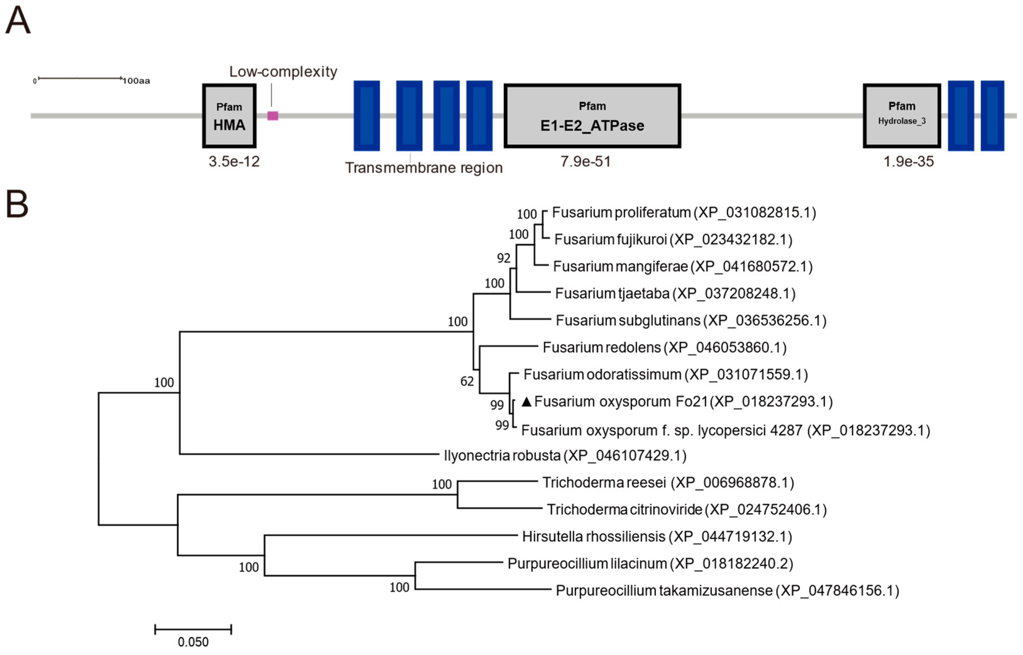
3.1. Identification of FoCrpA from F. oxysporum

Bioinformatic analysis of FoCrpA using the NCBI Conserved Domain Database (CDD) identified its C-terminal region (residues 371–1081) as a canonical member of the P-type ATPase superfamily (PF00122), specifically classified as a heavy metal-transporting ATPase (HM-ATPase). Subsequent SMART analysis further resolved three conserved functional modules: (i) N-terminal HMA domain (PF00403, aa 45–86): A ββαβ metal-binding fold critical for heavy metal ion recognition and chelation. (ii) Central E1-E2_ATPase domain (PF00122, aa 210–450): Characteristic catalytic core of P-type ATPases, comprising E1 ATP-binding subdomain (aa 210–320) with conserved DKTGT phosphorylation motif and E2 transmembrane ion translocation subdomain (aa 350–450) containing CPC metal coordination residues; (iii) C-terminal Hydrolase_3 domain (PF13343, aa 500–620): A HAD-family phosphatase module potentially regulating ATPase autoinhibition (Figure 1A). Phylogenetic reconstruction using maximum-likelihood methods (MEGA7.0) of 15 ascomycete orthologs revealed strong clustering of F. oxysporum FoCrpA with F. oxysporum f. sp. lycopersici (XP_018237293.1, 99% identity), forming a distinct clade from Fusarium odoratissimum (XP_031071559.1) and Fusarium redolens (XP_046053860.1), while the protein from other fungi displays low similarity with homologous proteins (Figure 1B). This phyletic pattern suggests vertical inheritance of FoCrpA within the Fusarium genus, with functional conservation in metal homeostasis.

3.2. Generation of FoCrpA Deletion Mutants and Complementation Assay

FoCrpA was deleted using a homologous recombination strategy (Figure 2A). The gene knockout vector pXEH-C was transferred into Agrobacterium tumefaciens AGL-1 to transform the wild-type Fo21 strain. The FoCrpA gene in F. oxysporum was replaced with the hygromycin resistance gene via homologous recombination (Figure 2A). Four primer pairs (Hyg-F/Hyg-R, Cup-Hup-R, Hdn-F/Cdn, FoCrpA-F/FoCrpA-R) were used to validate the knockout mutant over three generations (Figure 2B). The results showed that two mutants successfully integrated the hygromycin resistance gene (detected by Hyg-F/Hyg-R). The upstream (1531 bp) and downstream (1102 bp) fragments of FoCrpA were correctly integrated in two mutants (validated by Cup-Hup-R and Hdn-F/Cdn). Three mutants lacked the FoCrpA gene (confirmed by FoCrpA-F/FoCrpA-R). These results confirmed the successful knockout of the FoCrpA gene, and the mutant was named ∆FoCrpA.The FoCrpA locus (4880 bp) was cloned from Fo21 into pSUL via BamHI, generating complementation vector pSUL-C (PCR/restriction-verified). Agrobacterium tumefasciens-mediated transformation of pSUL-C into ΔFoCrpA restored the target fragment (867 bp PCR-amplified in complemented strain ∆FoCrpA-C and WT, absent in ∆FoCrpA), confirming genetic complementation (Figure 2C). Transcriptional validation via qRT-PCR using the internal reference gene β-actin as a control demonstrated complete abolition of FoCrpA expression in ∆FoCrpA (expression level <0.01% of wild-type Fo21, normalized to 1), with full transcriptional restoration observed in the complemented strain ∆FoCrpA-C (Figure 2D). This indicates that FoCrpA was disrupted successfully in ΔFoCrpA and reintegrated in ΔFoCrpA-C.

3.3. Effects of FoCrpA on Vegetative Growth and Conidiogenesis

Colony phenotypic characterization of FoCrpA and its mutant strains demonstrated that wild-type Fo21 and its mutant strains exhibited similar spreading growth patterns with comparable radial expansion rates, while ∆FoCrpA colonies displayed significant pigmentation attenuation compared to the wild-type Fo21 and complemented strains, which showed indistinguishable colony coloration (Figure 3A). For the color of the liquid culture, ∆FoCrpA also produced visibly lighter extracellular pigments than wild-type and complemented strains (Figure 3B). Liquid cultures grown in PDB medium revealed no significant differences in mycelial biomass accumulation among strains (Figure 3C), demonstrating that FoCrpA deletion does not impair hyphal growth under standard nutrient conditions. Tolerance assays on CuSO4-supplemented PDA (0–1 mM) demonstrated that ∆FoCrpA exhibited significant growth suppression at 0.5 mM and complete growth arrest at 1 mM CuSO4, whereas wild-type and complemented strains maintained partial viability (Figure 3D). Conidial production and germination were similar between WT Fo21 and its mutant strains (Figure 3E,F). These results demonstrate that FoCrpA specifically modulates pigment biosynthesis and copper ion homeostasis in F. oxysporum, while exhibiting no significant regulatory effects on mycelial biomass accumulation, conidiogenesis, or conidial germination.

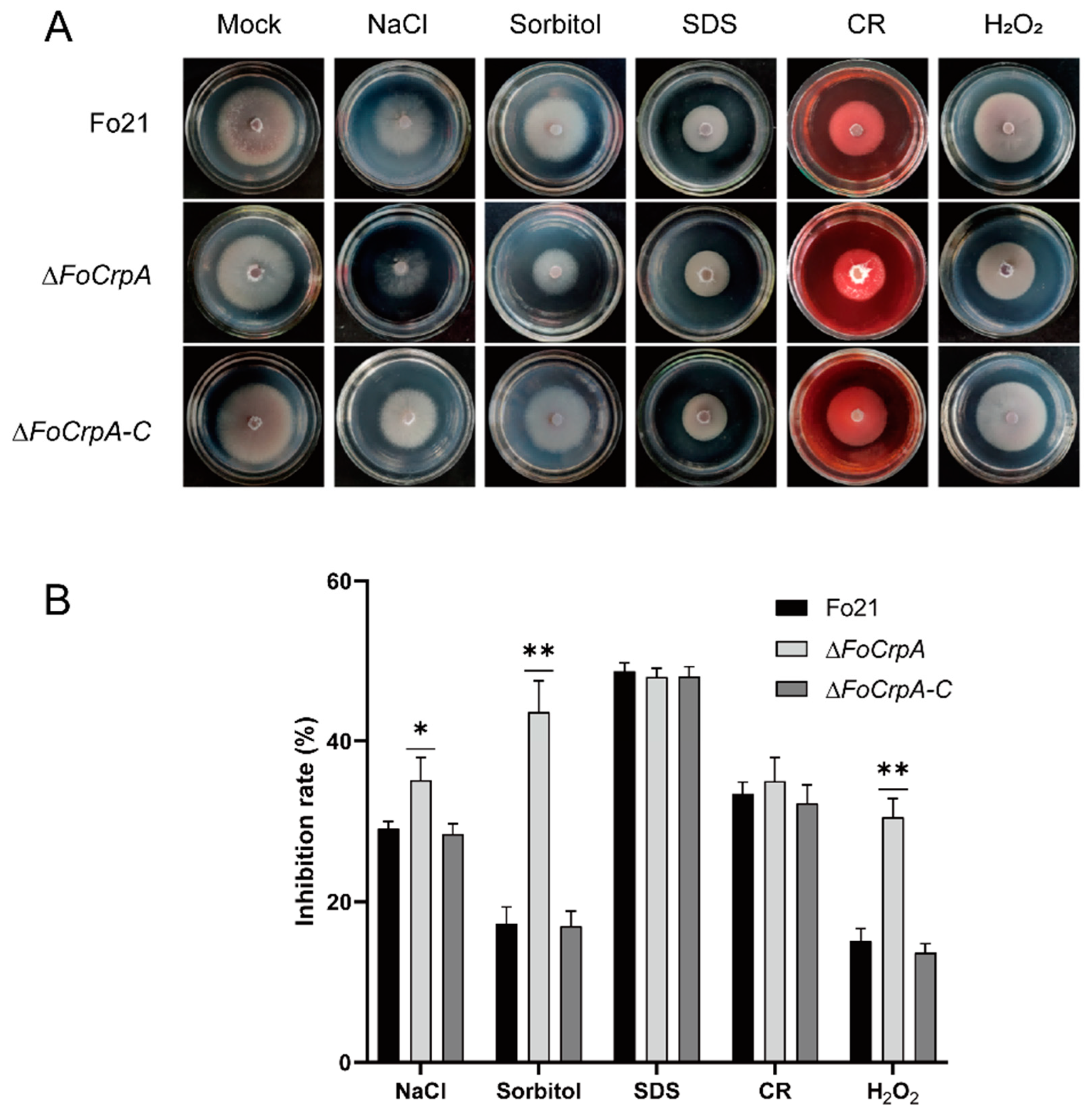
3.4. Response of FoCrpA to Stress Agents

To investigate the regulatory role of FoCrpA in environmental stress adaptation of F. oxysporum, comparative phenotypic analyses were performed using wild-type Fo21, ∆FoCrpA, and ∆FoCrpA-C on PDA plates supplemented with different stress agents, including NaCl and sorbitol (osmotic stresses), SDS and CR (cell wall stresses), and H2O2 (oxidative stress). After five-day incubation at 25 °C, the ∆FoCrpA mutant displayed significantly impaired growth under osmotic and oxidative stress conditions compared to the wild-type Fo21 and complemented strain ∆FoCrpA-C, while maintaining wild-type-level sensitivity to cell wall stressors (Figure 4A,B). These data collectively demonstrate that FoCrpA specifically modulates F. oxysporum’s tolerance mechanisms against osmotic and oxidative challenges.

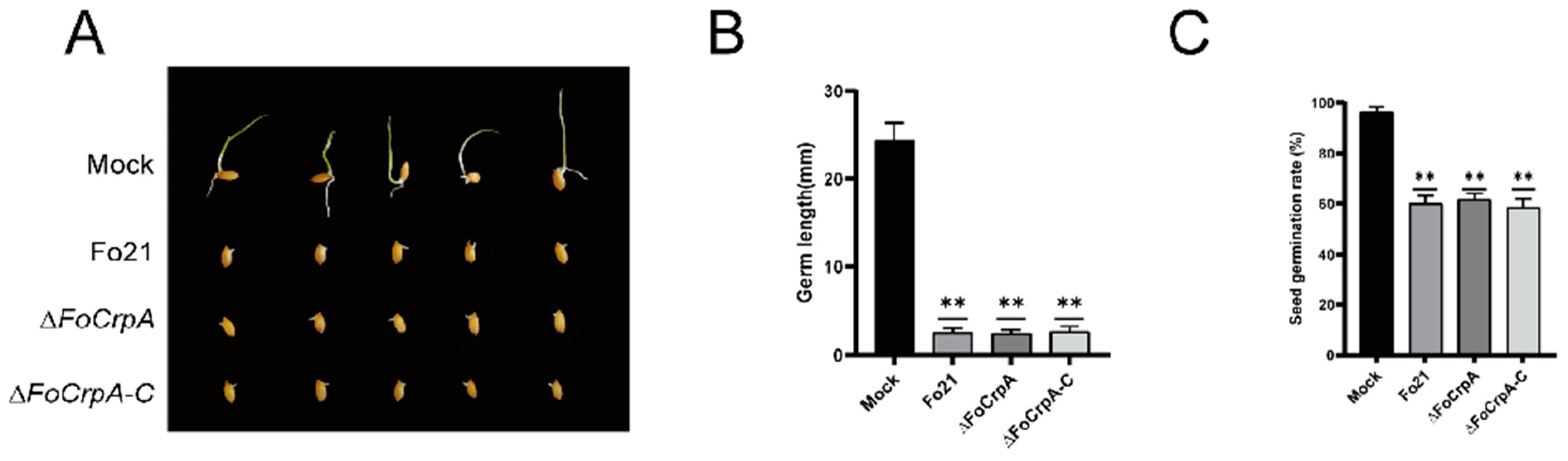
3.5. Culture Filtrates Toxicity from FoCrpA Mutants

Toxicity assessment using culture filtrates from Fo21 and its mutants revealed significant suppression of rice seed germination and germ length compared to the PDB blank control (Figure 5A). Notably, no statistically significant differences in these germination parameters were observed among filtrate-treated groups (Figure 5B,C). These findings demonstrate that F. oxysporum secretes germination-inhibitory metabolites into the extracellular milieu, while crucially establishing that FoCrpA does not participate in the biosynthesis or secretion of these toxic compounds.

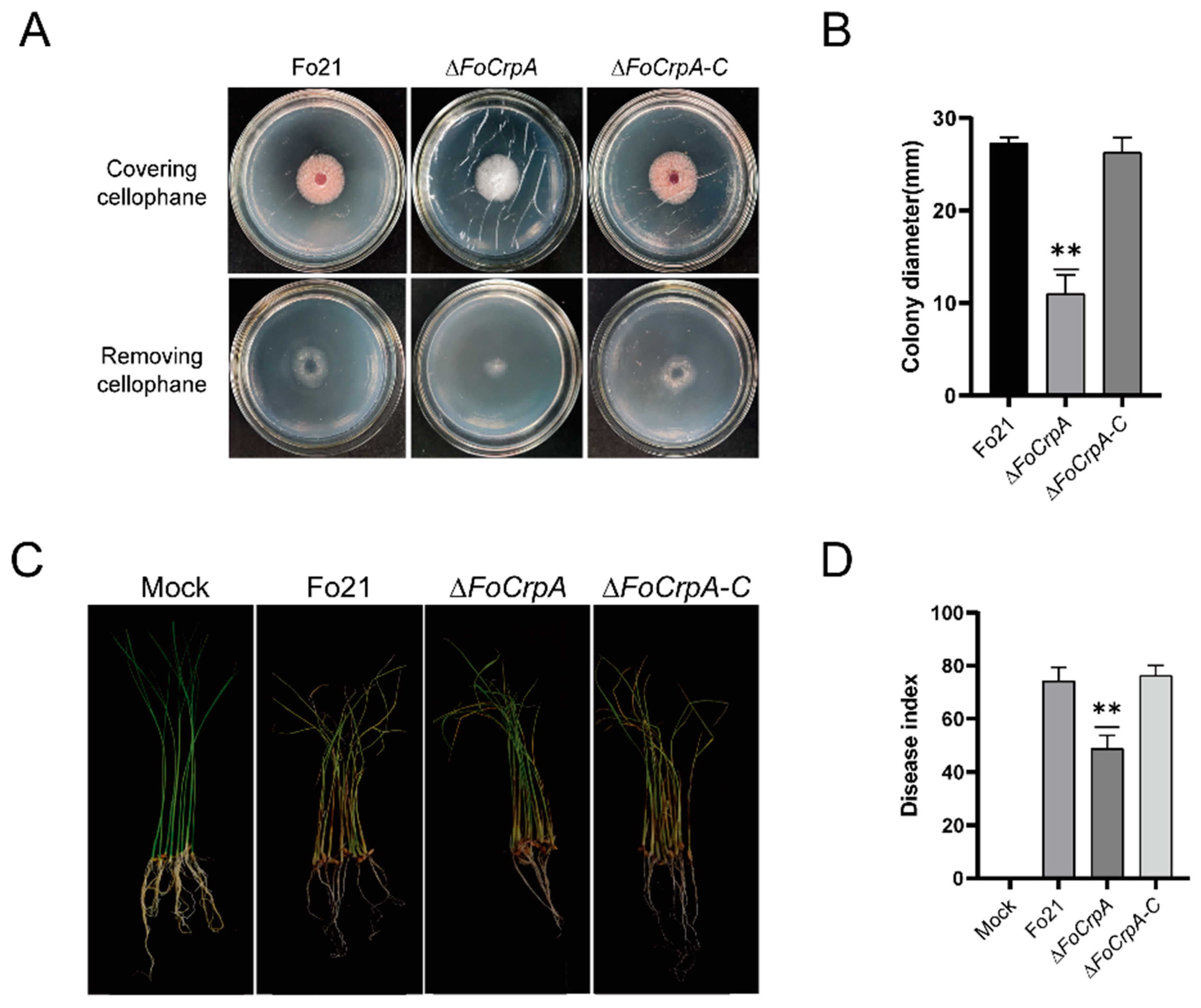
3.6. Effect of FoCrpA Mutants on Pathogenicity

The infection–penetration assay conducted with Fo21, ∆FoCrpA, and ∆FoCrpA-C strains demonstrated comparable growth patterns across all strains when cultured on PDA plates overlaid with cellophane membranes. However, upon removal of the cellophane barrier followed by 24 h incubation, Fo21 and ∆FoCrpA-C strains exhibited normal tissue penetration capabilities, and ∆FoCrpA displayed significantly impaired colonization efficiency, as evidenced by reduced colony diameter (Figure 6A). This phenotypic divergence strongly suggests that FoCrpA plays a critical role in mediating host tissue invasion and penetration processes in F. oxysporum.
Pathogenicity assay results showed that most of the stems of rice seedlings inoculated with Fo21 and ∆FoCrpA-C strain turned brown, and some leaves turned yellow and wilted with a disease index of 74.30 and 76.37, respectively (Table 2). However, rice seedlings inoculated with ∆FoCrpA strain showed milder symptoms than those with Fo21 and ∆FoCrpA-C (Figure 6B,C). The results suggest that the deletion of FoCrpA reduces the pathogenicity of F. oxysporum.

4. Discussion

Copper performs a comprehensive and vital function in biological systems. It orchestrates the conformational and catalytic properties of numerous metalloproteins and enzymes, as typified by cytochrome oxidase and superoxide dismutase. Additionally, due to the propensity of copper to react with non-specific proteins and engender toxic manifestations, a highly intricate and elaborate system is requisite for the accomplishment of processes such as its assimilation, concentration regulation, translocation to specific protein-binding sites, and expulsion. This complex assemblage comprises small carriers, molecular chaperones, and active transporters [31]. From our established mutant library of F. oxysporum, a Cu-ATPase belonging to the HMA subfamily was discovered. It is speculated that it plays a role in the pathogenic process of F. oxysporum. The functional domain analysis of the encoded protein showed that the FoCrpA gene encoded protein belongs to the P-type heavy metal transport ATPase family, which is located on the plasma membrane. In Aspergillus nidulans, gene CrpA coordinates the transport of copper ions between the plasma membrane, endoplasmic reticulum, Golgi apparatus, and other organelles [32]. In this study, by analyzing the biological functions of wild-type strain Fo21, knockout mutant ∆FoCrpA, and complementation mutant ∆FoCrpA-C, we investigated the function of the FoCrpA gene in F. oxysporum. It was found that ∆FoCrpA had no significant differences in mycelial dry weight, spore production, spore germination rate, and tolerance to cell membrane stress and cell wall stress compared to wild-type strains. However, compared with the wild-type strain, the colony and liquid color of ∆FoCrpA and its tolerance to high osmotic stress and oxidative stress decreased.
Cu-ATPase is involved in regulating the dynamic balance of copper metabolism, and copper is a cofactor for various copper-containing proteins, such as Cu/Zn superoxide dismutase (Cu/Zn SOD), laccase, etc. [33,34]. These enzymes are key enzymes in many biological reactions, such as Cu/Zn SOD, which can catalyze the decomposition of superoxide anions (O2-) into less toxic hydrogen peroxide [35,36]. When pathogens invade host plants, it helps them resist the extracellular ROS produced by the host plants [37,38,39]. Moreover, laccase is involved in various biological processes such as fungal morphogenesis, pathogen infection, pigment synthesis, and stress response [40]. In this study, we found that compared with the wild-type strain Fo21, the colony and liquid color of the knockout mutant ∆FoCrpA and its tolerance to high osmotic stress and oxidative stress decreased, indicating that the FoCrpA gene can affect the pigment accumulation of F. oxysporum and its tolerance to exogenous osmotic stress and oxidative stress. Together with previous studies performed in other pathogenic fungi, such as in Verticillium dahliae, the knockout mutant ΔVdSOD1 of the Cu/Zn SOD gene has reduced tolerance to oxidative stress [41]. In Colletotrichum gloeosporiides, the colony color of deletion mutant ΔA2LAC1 changed from gray-black to nearly white, significantly reducing its pathogenicity to mangoes [42]. These results indicated that Cu-ATPase and other copper-containing proteins play a significant role in the growth and development of plant pathogenic fungi.
Our study also found that the deletion of the FoCrpA gene can lead to a decrease in the penetration ability of F. oxysporum, indicating that the FoCrpA gene plays an important regulatory role in the penetration ability of F. oxysporum. When F. oxysporum infects colonized host plants, it produces infection structures such as infection mats and attachment cells. In Botrytis cinerea, the deletion of the BcCcc2 gene resulted in a diminished capacity to form attachment cells and infection mats, consequently impairing the fungus’s ability to invade plant epidermal tissues [43]. It is hypothesized that the functionality of infection structures, including infection pads and attachment cells, might depend on unidentified and essential copper-containing proteins. Cu-ATPase potentially modulates the activity of these proteins. Similarly, the deletion of the FoCrpA gene can reduce the pathogenicity of F. oxysporum, indicating that the FoCrpA gene may affect the pathogenicity of F. oxysporum by reducing its invasion and penetration ability.
Excessive intracellular copper ions can affect cell structure, leading to disruptions in cell division and enzymatic systems, thereby affecting fungal growth. Cu-ATPase can eliminate excess copper ions from the cell and alleviate the inhibitory effect of excessive copper ions on fungal growth. For example, after knocking out Cu-ATPase CrpA in Aspergillus fumigatus, the strain is more sensitive to high concentrations of copper ions [44]. After knocking out the FgCrpA gene of F. graminearum, the knockout mutant showed inhibition of hyphal growth, decreased spore production and germination rate, reduced tolerance to reactive oxygen species, and decreased production of toxin deoxynivalenol under copper ion stress compared to the wild type [45]. Therefore, this study conducted colony growth experiments under copper stress on wild-type strains, ∆FoCrpA strains, and ∆FoCrpA-C strains. It was found that compared with the wild-type, ∆FoCrpA was more sensitive to copper ions, and the colony diameter of ∆FoCrpA under the same copper ion concentration was smaller, and the mycelial growth was sparser. When copper ions increased to a certain concentration, ∆FoCrpA almost did not grow, while the wild-type strain could continue to grow. The deletion of the FoCrpA gene can exacerbate the inhibitory effect of excessive copper ions on fungus growth.
Overall, the FoCrpA gene is involved in osmotic and oxidative stress, affecting the invasion and penetration ability and pathogenicity of F. oxysporum. The FoCrpA study identifies copper homeostasis as a key vulnerability in F. oxysporum. Targeting this system—via Cu-ATPase inhibitors, optimized copper fungicides, or host-enhanced ROS—could reduce fungal virulence and improve crop resistance sustainably. FoCrpA also offers biotechnological potential: gene editing (e.g., CRISPR) or plant-derived inhibitors could engineer wilt-resistant crops, while structure-guided antifungal agents may selectively block fungal copper transport with minimal environmental impact. Crucially, FoCrpA regulates osmotic/oxidative stress adaptation and infection structures, making it pivotal for both understanding pathogenesis and developing control strategies for rice seedling blight. Future work should validate field applications, explore copper-immune synergies, and refine targeted inhibitors to bridge lab discoveries with agricultural practice.

Author Contributions

C.W., L.W. and J.Z. contributed to the study conception and design; C.W. and L.W. performed the experimental design; C.W. and L.W. wrote the manuscript draft; C.W., L.W., X.Z., L.H., Q.L., R.R., A.L., X.L., T.X. and P.G. conceived and performed all experiments; C.W., L.W., X.X., Z.N. and C.L. analyzed the results and corresponding data. All authors have read and agreed to the published version of the manuscript.

Funding

This research was funded by the Key Research and Development Program of Heilongjiang Province (GA23B010), National Key Research and Development Program Project (2023YFD1400205-3), Green Plant Protection Project in Heilongjiang Province (2130108), Key R&D Program Project of Heilongjiang Province (2023ZX02B0502), special project of central leading local science and technology development (ZY18C08), nature fund project of Heilongjiang (C2017032), Major Project of Heilongjiang Province Applied Technology Research and Development Plan (GA19B104) and the Natural Science Foundation of Heilongjiang Province of China (LH2022C022).

Institutional Review Board Statement

Not applicable.

Informed Consent Statement

Not applicable.

Data Availability Statement

The data presented in this study are available on request from the corresponding author.

Acknowledgments

We thank Zhang Shihong, Qin Qingming, and Li Guihua from Jilin University, for providing Agrobacterium tumefaciens AGL-1, Escherichia coli DH5α, plasmid pXEH and pSUL.

Conflicts of Interest

The authors declare no conflicts of interest.

References

  1. Khepar, V.; Sidhu, A.; Sharma, A.B. Nanomaterized Zinc Sulfide-Meerschaum Biomatrix Efficiently Suppressed Fusarium verticilloides with Augmented Rice Seed Quality Benefits during Storage. Pest Manag. Sci. 2023, 79, 244–256. [Google Scholar] [CrossRef] [PubMed]
  2. Gaire, S.P.; Zhou, X.G.; Zhou, Y.; Shi, J.; Jo, Y.K. Identification and distribution of fungal pathogens associated with seedling blight of rice in the southern United States. Plant Pathol. 2023, 72, 76–88. [Google Scholar] [CrossRef]
  3. Li, Y.G.; Zhang, X.; Zhang, R.; Liu, J.X.; Ali, E.; Ji, P.; Pan, H.Y. Occurrence of seedling blight caused by Fusarium tricinctum on rice in China. Plant Dis. 2019, 103, 1789. [Google Scholar] [CrossRef]
  4. Wang, S.; Sun, L.; Li, W.Q.; Liu, J.X.; Li, Y.G.; Wei, D. First report of seedling blight caused by Fusarium redolens on rice in northeast China. Plant Dis. 2019, 103, 1418. [Google Scholar] [CrossRef]
  5. Wang, M.; Wei, P.; Cao, M.; Zhu, L.; Lu, Y. First Report of Rice Seedling Blight Caused by Burkholderia plantarii in North and Southeast China. Plant Dis. 2016, 100, 645. [Google Scholar] [CrossRef]
  6. Gaire, S.P.; Zhou, X.-G.; Jo, Y.-K.; Shi, J. First Report of Rhizoctonia solani AG-4 Causing Seedling Disease in Rice. Plant Dis. 2020, 104, 1546. [Google Scholar] [CrossRef]
  7. Gaire, S.-P.; Zhou, X.-G.; Jo, Y.-K. Sterile white basidiomycete fungus Marasmius graminum: A new pathogen causing seedling blight in rice. Plant Dis. 2021, 105, 702. [Google Scholar] [CrossRef] [PubMed]
  8. Liu, J.; Zhang, R.; Xu, C.; Liu, C.; Zheng, Y.; Zhang, X.; Liu, S.; Li, Y. Characterisation of Pythium aristosporum Oomycete—A novel pathogen causing rice seedling blight in China. J. Fungi 2022, 8, 890. [Google Scholar] [CrossRef]
  9. El-Shafey, R.A.S.; Attia, K.A.; Mostafa, F.A.; Elamawi, R.M. Incidence and molecular identification of Cochliobolus carbonum as causal organism of rice seedling blight. Beni-Suef Univ. J. Basic Appl. Sci. 2018, 7, 652–662. [Google Scholar] [CrossRef]
  10. Liu, J.X.; Cai, Y.N.; Jiang, W.Y.; Li, Y.G.; Zhang, Q.F.; Pan, H.Y. Population structure and genetic diversity of fungi causing rice seedling blight in northeast China based on microsatellite markers. Plant Dis. 2020, 104, 868–874. [Google Scholar] [CrossRef]
  11. Ma, B.; Wang, J.; Liu, C.; Hu, J.; Tan, K.; Zhao, F.; Yuan, M.; Zhang, J.; Gai, Z. Preventive effects of fluoro-substituted benzothiadiazole derivatives and chitosan oligosaccharide against the rice seedling blight induced by Fusarium oxysporum. Plants 2019, 8, 538. [Google Scholar] [CrossRef] [PubMed]
  12. Ochi, A.; Konishi, H.; Ando, S.; Sato, K.; Yokoyama, K.; Tsushima, S.; Yoshida, S.; Morikawa, T.; Kaneko, T.; Takahashi, H. Management of bakanae and bacterial seedling blight diseases in nurseries by irradiating rice seeds with atmospheric plasma. Plant Pathol. 2017, 66, 67–76. [Google Scholar] [CrossRef]
  13. Shenashen, M.; Derbalah, A.; Hamza, A.; Mohamed, A.; El Safty, S. Antifungal activity of fabricated mesoporous alumina nanoparticles against root rot disease of tomato caused by Fusarium oxysporium. Pest Manag. Sci. 2017, 73, 1121–1126. [Google Scholar] [CrossRef]
  14. Cao, Y.; Ding, W.; Liu, C. Unraveling the metabolite signature of endophytic Bacillus velezensis strain showing defense response towards Fusarium oxysporum. Agronomy 2021, 11, 683. [Google Scholar] [CrossRef]
  15. Ren, J.G.; Wang, J.L.; Cai, J. Effects of Fenaminosulf on Growth of Pepper (Capsicum annuum L.), Microbial Communities and Enzymatic Activities of the Soil Infested with Fusarium oxysporum. Indian J. Pharm. Sci. 2021, 83, 163–169. [Google Scholar] [CrossRef]
  16. Yruela, I. Copper in Plants: Acquisition, Transport and Interactions. Funct. Plant Biol. 2009, 36, 409–430. [Google Scholar] [CrossRef] [PubMed]
  17. Arredondo, M.; Núñez, M.T. Iron and Copper Metabolism. Mol. Asp. Med. 2005, 26, 313–327. [Google Scholar] [CrossRef]
  18. Tapiero, H.; Townsend, D.M.; Tew, K.D. Trace Elements in Human Physiology and Pathology. Copper. Biomed. Pharmacother. 2003, 57, 386–398. [Google Scholar] [CrossRef]
  19. Gao, T.; Shi, L.; Zhang, T.; Ren, A.; Jiang, A.; Yu, H.; Zhao, M. Cross talk between calcium and reactive oxygen species regulates hyphal branching and ganoderic acid biosynthesis in Ganoderma lucidum under copper stress. Appl. Environ. Microbiol. 2018, 84, e00438-18. [Google Scholar] [CrossRef]
  20. Wang, C.; Xiao, X.Z.; Wei, F.; Qiu, J.; Wu, M.; Gao, H.H.; Yang, W.F.; Luo, S.Q. Cloning and expression analysis of copper transporter genes in rubber tree (Hevea brasiliensis Müll. Arg.). Plant Physiol. J. 2016, 52, 1389–1396. [Google Scholar] [CrossRef]
  21. Burkhead, J.L.; Gogolin Reynolds, K.A.; Abdel-Ghany, S.E.; Cohu, C.M.; Pilon, M. Copper homeostasis. New Phytol. 2009, 182, 799–816. [Google Scholar] [CrossRef]
  22. Boutigny, S.; Sautron, E.; Finazzi, G.; Rivasseau, C.; Frelet-Barrand, A.; Pilon, M.; Rolland, N.; Seigneurin-Berny, D. HMA1 and PAA1, two chloroplast-envelope PIB-ATPases, play distinct roles in chloroplast copper homeostasis. J. Exp. Bot. 2014, 65, 1529–1540. [Google Scholar] [CrossRef] [PubMed]
  23. Seigneurin-Berny, D.; Gravot, A.; Auroy, P.; Mazard, C.; Kraut, A.; Finazzi, G.; Grunwald, D.; Rappaport, F.; Vavasseur, A.; Joyard, J.; et al. HMA1, a new Cu-ATPase of the chloroplast envelope, is essential for growth under adverse light conditions. J. Biol. Chem. 2006, 281, 2882–2892. [Google Scholar] [CrossRef] [PubMed]
  24. Saitoh, Y.; Izumitsu, K.; Tanaka, C. Phylogenetic analysis of heavy-metal ATPases in fungi and characterization of the copper-transporting ATPase of Cochliobolus heterostrophus. Mycol. Res. 2009, 113, 737–745. [Google Scholar] [CrossRef] [PubMed]
  25. Parisot, D.; Dufresne, M.; Veneault, C.; Laugé, R.; Langin, T. Clap1, a gene encoding a copper-transporting ATPase involved in the process of infection by the phytopathogenic fungus Colletotrichum lindemuthianum. Mol. Genet. Genom. 2002, 268, 139–151. [Google Scholar] [CrossRef]
  26. Yang, K.; Shadkchan, Y.; Tannous, J.; Landero Figueroa, J.A.; Wiemann, P.; Osherov, N.; Wang, S.; Keller, N.P. Contribution of ATPase copper transporters in animal but not Plant virulence of the crossover pathogen Aspergillus flavus. Virulence 2018, 9, 1273–1286. [Google Scholar] [CrossRef]
  27. Mullins, E.D.; Chen, X.; Romaine, P.; Raina, R.; Geiser, D.M.; Kang, S. Agrobacterium-Mediated transformation of Fusarium oxysporum: An efficient tool for insertional mutagenesis and gene transfer. Phytopathology 2001, 91, 173–180. [Google Scholar] [CrossRef]
  28. Li, M.-H.; Xie, X.-L.; Lin, X.-F.; Shi, J.-X.; Ding, Z.-J.; Ling, J.-F.; Xi, P.-G.; Zhou, J.-N.; Leng, Y.; Zhong, S.; et al. Functional characterization of the gene FoOCH1 encoding a putative α-1,6-mannosyltransferase in Fusarium oxysporum f. sp. Cubense. Fungal Genet. Biol. 2014, 65, 1–13. [Google Scholar] [CrossRef]
  29. Livak, K.J.; Schmittgen, T.D. Analysis of relative gene expression data using Real-Time Quantitative PCR and the 2−ΔΔCT method. Methods 2001, 25, 402–408. [Google Scholar] [CrossRef]
  30. Jiang, W.; Liu, J.; He, Y.; Payizila, A.; Li, Y. Biological control ability and antifungal activities of Bacillus velezensis Bv S3 against Fusarium oxysporum that causes rice seedling blight. Agronomy 2024, 14, 167. [Google Scholar] [CrossRef]
  31. Inesi, G. Molecular features of copper binding proteins involved in copper homeostasis. IUBMB Life 2017, 69, 211–217. [Google Scholar] [CrossRef] [PubMed]
  32. Antsotegi-Uskola, M.; Markina-Iñarrairaegui, A.; Ugalde, U. Copper resistance in Aspergillus nidulans relies on the P(I)-Type ATPase CrpA, regulated by the transcription factor AceA. Front. Microbiol. 2017, 8, 912. [Google Scholar] [CrossRef] [PubMed]
  33. Probst, C.; Garcia-Santamarina, S.; Brooks, J.T.; Van Der Kloet, I.; Baars, O.; Ralle, M.; Thiele, D.J.; Alspaugh, J.A. Interactions between copper homeostasis and the fungal cell wall affect copper stress resistance. PLoS Pathog. 2022, 18, e1010195. [Google Scholar] [CrossRef] [PubMed]
  34. Antsotegi-Uskola, M.; Markina-Iñarrairaegui, A.; Ugalde, U. New insights into copper homeostasis in filamentous fungi. Int. Microbiol. 2020, 23, 65–73. [Google Scholar] [CrossRef]
  35. Li, C.; Li, Y.; Ding, C. The role of copper homeostasis at the host-pathogen axis: From bacteria to fungi. Int. J. Mol. Sci. 2019, 20, 175. [Google Scholar] [CrossRef]
  36. García-Santamarina, S.; Thiele, D.J. Copper at the fungal pathogen-host axis. J. Biol. Chem. 2015, 290, 18945–18953. [Google Scholar] [CrossRef]
  37. Robinson, J.R.; Isikhuemhen, O.S.; Anike, F.N. Fungal-metal interactions: A review of toxicity and homeostasis. J. Fungi 2021, 7, 225. [Google Scholar] [CrossRef]
  38. Beauchamp, C.; Fridovich, I. Superoxide dismutase:improved assays and an assay applicable to acrylamide gels. Anal. Biochem. 1971, 44, 276–287. [Google Scholar] [CrossRef]
  39. Broxton, C.N.; Culotta, V.C. SOD enzymes and microbial pathogens: Surviving the oxidative storm of infection. PLoS Pathog. 2016, 12, e1005295. [Google Scholar] [CrossRef]
  40. Janusz, G.; Pawlik, A.; Świderska-Burek, U.; Polak, J.; Sulej, J.; Jarosz-Wilkołazka, A.; Paszczyński, A. Laccase properties, physiological functions, and evolution. Int. J. Mol. Sci. 2020, 21, 966. [Google Scholar] [CrossRef]
  41. Tian, L.; Li, J.; Huang, C.; Zhang, D.; Xu, Y.; Yang, X.; Song, J.; Wang, D.; Qiu, N.; Short, D.P.G.; et al. Cu/Zn superoxide dismutase (VdSOD1) mediates reactive oxygen species detoxification and modulates virulence in Verticillium dahliae. Mol. Plant Pathol. 2021, 22, 1092–1108. [Google Scholar] [CrossRef] [PubMed]
  42. Wei, Y.; Pu, J.; Zhang, H.; Liu, Y.; Zhou, F.; Zhang, K.; Liu, X. The laccase gene (LAC1) is essential for Colletotrichum gloeosporioides development and virulence on mango leaves and fruits. Physiol. Mol. Plant Pathol. 2017, 99, 55–64. [Google Scholar] [CrossRef]
  43. Saitoh, Y.; Izumitsu, K.; Morita, A.; Tanaka, C. A copper-transporting ATPase BcCCC2 is necessary for pathogenicity of Botrytis cinerea. Mol. Genet. Genom. 2010, 284, 33–43. [Google Scholar] [CrossRef]
  44. Cai, Z.; Du, W.; Zhang, Z.; Guan, L.; Zeng, Q.; Chai, Y.; Dai, C.; Lu, L. The Aspergillus fumigatus transcription factor AceA is involved not only in Cu but also in Zn detoxification through regulating transporters CrpA and ZrcA. Cell. Microbiol. 2018, 20, e12864. [Google Scholar] [CrossRef] [PubMed]
  45. Liu, X.; Jiang, Y.; He, D.; Fang, X.; Xu, J.; Lee, Y.W.; Keller, N.P.; Shi, J. Copper tolerance mediated by FgAceA and FgCrpA in Fusarium graminearum. Front. Microbiol. 2020, 11, 1392. [Google Scholar] [CrossRef]
Figure 1. Functional domain architecture and phylogenetic analysis of FoCrpA in F. oxysporum. (A) Domain prediction of FoCrpA via the SMART database identified three conserved motifs: 1. Pfam: HMA (Heavy Metal-Associated) domain (aa 45–86): A β-sandwich fold critical for heavy metal ion binding and transport, conserved across prokaryotic and eukaryotic metal transporters. 2. Pfam: E1-E2_ATPase (aa 210–450): Characteristic of P-type ATPases, featuring an ATP hydrolysis module (E1, aa 210–320) for energy release and a transmembrane ion translocation module (E2, aa 350–450). 3. Pfam: Hydrolase_3 (HAD-like) (aa 500–620): A haloacid dehalogenase superfamily domain catalyzing phosphatase/hydrolase reactions, potentially linked to metal cofactor metabolism. (B) Maximum-likelihood phylogenetic tree of FoCrpA homologs across 9 Fusarium species and 6 other fungal species, reconstructed with MEGA7.0 (1000 bootstrap replicates).
Jof 11 00317 g001
Figure 2. FoCrpA gene knockout strategy; PCR and qRT-PCR validation of knockout mutants and complementation strains. (A) FoCrpA gene knockout strategy. (B) PCR validation of HPH gene (M: DL2000 Ladder DNA Marker; 1–3: Mutant; 4: Fo21; 5: pXEH; 6: ddH2O); PCR validation of the two side fragments of FoCrpA gene (M: DL2000 Ladder DNA Marker; 1–3: mutant; 4: Fo21; 5: ddH2O); PCR validation of FoCrpA gene (M: DL5000 Ladder DNA Marker; 1–3: Mutant; 4: pXEH; 5: ddH2O; 6: Fo21). (C) C-FoCrpA gene fragment (M: 1 kb Ladder DNA Marker); pSUL-C enzyme digestion validation (M: 1 kb Ladder DNA Marker); PCR validation of ∆FoCrpA-C (M: DL5000 Ladder DNA Marker; 1–2: complementation mutant; 3: ∆FoCrpA; 4: Fo21; 5: ddH2O). (D) Expression analysis of FoCrpA in WT Fo21 and its mutants strains with qRT-PCR. Duncan’s multiple range test was performed to determine significant difference, ** p ≤ 0.01.
Jof 11 00317 g002
Figure 3. Phenotypic analysis of FoCrpA and its mutant strains. (A) Colony phenotypic characterization of Fo21, ∆FoCrpA, and ∆FoCrpA-C on PDA plates after incubation of 7 days at 25 °C. (B) Observing culture color of Fo21, ∆FoCrpA, and ∆FoCrpA-C strains grown in 100 mL PDB in a shaker at 180 rpm at 25 °C for 2 days. (C) Mycelial biomass was measured by determining the dry weight of Fo21, ∆FoCrpA, and ∆FoCrpA-C strains grown in 100 mL PDB in a shaker at 180 rpm at 25 °C for 3 days. (D) Colony morphology of Fo21, ∆FoCrpA, and ∆FoCrpA-C under Cu stress, amended with CuSO4 at different concentrations indicated in the figure and cultured at 25 °C for 5 days. (E) Conidial production of Fo21, ∆FoCrpA and ∆FoCrpA-C grown in 50 mL PDB in a shaker at 180 rpm at 25 °C for 2 days. (F) Conidial germination was compared between Fo21 and its mutants by re-suspending the conidia in 40 mL liquid GM in a shaker at 180 rpm at 25 °C for 6 h, 9 h, and 12 h.
Jof 11 00317 g003
Figure 4. Deletion of FoCrpA affects various stress response. (A) Colony growth of Fo21, ∆FoCrpA, and ∆FoCrpA-C strains on PDA plates supplied with 0.5 M NaCl, 1 M sorbitol, 0.03% SDS, 1 mM Congo red, or 10 mM H2O2 after incubation of 5 days at 25 °C. (B) Inhibition rate of colony growth by different stress factors. Error bars represent the standard error of mean. Duncan’s multiple range test was performed to determine significant difference,* p ≤ 0.05,** p ≤ 0.01.
Jof 11 00317 g004
Figure 5. Toxicity of culture filtrates from Fo21 and FoCrpA mutants on the growth of rice seeds. (A) Culture filtrates of Fo21, ∆FoCrpA, and ∆FoCrpA-C strains caused defects in rice seeds growing. PDB medium without culture filtrates was used as the blank control. (B) Differences of germ length were examined after incubation under light at 25 °C for 5 days. (C) Seed germination rate was compared between WT Fo21 and its mutants after incubation under light at 25 °C for 5 days. Error bars represent the standard error of mean. Duncan’s multiple range test was performed to determine significant difference,** p ≤ 0.01.
Jof 11 00317 g005
Figure 6. Pathogenicity of Fo21 and FoCrpA mutants on the stem base of rice seedlings. (A) Comparison of infection–penetration morphology caused by inoculation with Fo21, ∆FoCrpA, and ∆FoCrpA-C strains on cellophane membranes after 3 days of incubation at 25 °C. (B) Differences of colony diameter incubating 1 day at 25 °C after removing cellophane. (C) Disease symptoms on the stem base of rice seedlings observed at 14 dpi wtih Fo21, ∆FoCrpA, and ∆FoCrpA-C strains. (D) Disease index of rice seedlings was determined at 14 dpi wtih Fo21, ∆FoCrpA, and ∆FoCrpA-C strains. Fifty rice seedlings were inoculated per replication. Error bars represent the standard error of mean. Duncan’s multiple range test was performed to determine significant difference,** p ≤ 0.01.
Jof 11 00317 g006
Table 1. Primers used in this study.
Primer NamePrimer Sequence
Hyg-FTCGCCCTTCCTCCCTTTATTTCAG
Hyg-RCTACACAGCCATCGGTCCAGAC
SuR-FCTCTCCGTTGCTTATCCTTGCCTA
SuR-RCGCCATCACTACGCCTTGTCTT
FoCrpA-UP-FGATCTTCACTAGTGGGAATTCCCCGTGATGATTTGCCCAATGAAT
FoCrpA-UP-RTTGGGTACCGAGCTCGAATTCCGCCATGTTGACTGCTGTGAGA
FoCrpA-DN-FTGGGGATCCTCTAGAGTCGACTGGTATTGGCAGTGGTAGTGTTGG
FoCrpA-DN-RCTTGCATGCCTGCAGGTCGACCCGACGGAGGATCAAGATGTAAGC
Hup-RTGCTCACCGCCTGGACGACTAA
Hdn-FTGGACCGATGGCTGTGTAGAAGT
CupAGGCTTGGAGGAGAATGGTTGG
CdnAAGCCTTCCTTACGCCTGATGATG
FoCrpA-FTGCTCACCGCCTGGACGACTAA
FoCrpA-RTGGACCGATGGCTGTGTAGAAGT
C-FoCrpA-FCCGGGTACCGAGCTCGAATTCCCGTTGCTCTGCCGTATCTTGAA
C-FoCrpA-RAGCTGTCAAACATGAGAATTCAAGCCTTCCTTACGCCTGATGATG
Actin-FGTTGCCTGAGACTTGACGACGAT
Actin-RCTCCTCCGAACCATCCGCTACA
Table 2. Disease index of the wild-type F. oxysporum Fo21 and mutant strains.
StainsDisease Index
Fo2174.30 ± 5.11 a
∆FoCrpA48.90 ± 4.85 b
∆FoCrpA-C76.37 ± 3.65 a
Mean ± SD was calculated from the results of three independent experiments. Values on the table followed by the same letter are not significantly different at p ≤ 0.01 according to Duncan’s multiple range test. Each experiment was replicated three times.
Disclaimer/Publisher’s Note: The statements, opinions and data contained in all publications are solely those of the individual author(s) and contributor(s) and not of MDPI and/or the editor(s). MDPI and/or the editor(s) disclaim responsibility for any injury to people or property resulting from any ideas, methods, instructions or products referred to in the content.

