Review

The Dual Pathogen Fusarium: Diseases, Incidence, Azole Resistance, and Biofilms

Department of Microbiology and Immunology, School of Medicine, Georgetown University, 3900 Reservoir Rd., Washington, DC 20057, USA
*
Author to whom correspondence should be addressed.
J. Fungi 2025, 11(4), 294; https://doi.org/10.3390/jof11040294
Submission received: 4 March 2025 / Revised: 24 March 2025 / Accepted: 7 April 2025 / Published: 9 April 2025
(This article belongs to the Special Issue Fusarium spp.: A Trans-Kingdom Fungus, 2nd Edition)

Abstract

:
The increasing resistance of Fusarium species to nearly all first-line antifungal agents in clinical settings has led to its designation as a ‘high-priority’ human pathogen. As a dual pathogen, Fusarium spp. threaten both human health and crop production, impacting food security. Our recent drug profiling of clinical Fusarium isolates reveals resistance to several front-line antifungals, with notable cross-azole resistance observed in both clinical and plant-associated strains. While the overuse of agricultural azoles has been implicated in the selection of azole-resistant fungi such as Aspergillus, a similar mechanism has been assumed for Fusarium in clinical settings. However, direct genetic evidence supporting this hypothesis remains limited. In this review, part of our Special Interest (SI) series, we discuss the spectrum of human diseases caused by Fusarium. While incidence data are better established for human keratitis and onychomycosis, invasive fusariosis remains globally underreported. We propose reasons for this distinct clinical spectrum bias and explore the potential genetic basis of azole resistance.

Graphical Abstract

1. Introduction

Fusarium species belong to a group of dual-threat, cross-kingdom, filamentous fungal pathogens that cause both human infections and disruptions in food security and supply [1,2], designated as a ‘high-priority’ human pathogen [3,4]. Over 70% of all clinical Fusarium isolates are thought to arise through contact with airborne or waterborne spores in the environment [5]. While several filamentous fungi share these characteristics, Fusarium and Aspergillus species are among the most frequently encountered [6]. A significant number of hospitalized patients with resistant Fusarium and Aspergillus fungemia have no history of azole use, suggesting an environmental or airborne hospital source of resistance [7,8]. The emergence of environmentally driven azole resistance presents a major challenge in clinical treatment. This phenomenon is well documented in Aspergillus fumigatus, where resistance is linked to characteristic point and promoter mutations in the azole target gene, lanosterol 14α-demethylase (CYP51) [7]. However, the similar evidence for Fusarium in human infections remains unconfirmed.
The prevalence of the cross-resistance between medical and agricultural azoles in clinical settings may be due to their shared target protein. Currently, we still lack molecular markers to distinguish whether airborne Fusarium spores that infect hospitalized patients originate from hospital environments or external sources. Both the molecular mechanisms of resistance and the extent of Fusarium resistance remain significantly understudied, particularly in the United States. To date, only one study has reported the azole-resistant mechanism of Fusarium, involving 15 isolates from clinical and environmental sources in Malaysia [9].
Taxonomically, Fusarium comprises over 300 phylogenetically distinct species, grouped into more than 20 species complexes, most of which are found in the environment [8,10]. The majority of medically significant Fusarium species belong to seven key species complexes: Fusarium solani species complex (FSSC), Fusarium oxysporum species complex (FOSC), Fusarium fujikuroi species complex (FSSC), Fusarium incarnatum-equiseti species complex (FIESC), Fusarium chlamidosporum species complex (FCSC), Fusarium dimerum species complex (FDSC), and Fusarium sporotrichoides species complex (FSAMSC) [8,10].

2. Clinical Types and Incidence of Human Fusariosis

Human fusariosis is a reemergent worldwide disease exhibiting marked increases since the 1970s–1980s, according to Lockhart and Guarner [11]. Human infections caused by Fusarium fall into three major categories: superficial infections such as (1) keratitis, (2) onychomycosis (nail infections), and (3) invasive infections, including fungemia and disseminated fusariosis occurring in immunocompromised patients [8,12]. Disseminated infection in patients with fusariosis typically affects the skin in over 50% of cases [13], with deep organ involvement, such as pneumonia or fungemia, associated with poorer outcomes [12,14]. In contrast, localized infections like corneal keratitis [15,16] and onychomycosis (nail infections) [17] are more commonly observed in immunocompetent individuals. Despite advances in treatment, the effectiveness of interventions for both invasive and localized fusariosis remains suboptimal. Major risk factors for fusariosis include invasive surgery, organ transplants, chronic steroid use, and trauma to the skin, nails, and eyes [8,9,11].

2.1. Keratitis

Fusarium keratitis is the most common clinical manifestation of fusariosis [15,18]. A 10-year study of Fusarium infections at a French university hospital found that Fusarium cultures were isolated from 31% of ophthalmic samples, 8.48% of nail samples, and 0.47% of blood cultures [19]. Estimates from 43 published studies on country-level disease burdens, compiled by GAFFI, 2022 (Global Action For Fungal Infections, https://gaffi.org/media/country-fungal-disease-burdens, accessed on 4 February 2025), indicate that approximately 1,000,000 cases of fungal keratitis occur annually, which was adjusted to up to 1,400,000 patients each year [20]. In underdeveloped countries, these infections can lead to a high proportion of enucleation (11%) and visual loss (~50%) [15,21]. In tropical areas, Fusarium is the leading fungal pathogen responsible for fungal keratitis, in contrast to yeast fungal pathogens in subtropical areas [22].
Notably, Fusarium accounts for approximately 45–53% of all fungal keratitis cases, surpassing Aspergillus 3.3-fold [23]. Efforts have been made to classify fungal keratitis as a Neglected Tropical Disease (NTD) due to its severity and high prevalence in tropical and subtropical regions, particularly following corneal trauma [20]. Reports indicate that Fusarium accounts for 37.7% to 81.5% of all culture-positive corneal infections in these regions, often leading to severe visual impairment. However, Fusarium keratitis is not restricted to tropical climates—an outbreak in the United States linked 164 confirmed keratitis cases across 33 states and one U.S. territory to contaminated contact lens solution [24]. Additionally, cases of Fusarium keratitis have been reported following corneal transplant [18]. The relatively low incidence of Fusarium fungemia compared to its high prevalence in superficial infections highlights the need for better treatment strategies to reduce chronicity and potential latency [20]. Currently, voriconazole (VCZ) is still the most commonly used drug in Fusarium keratitis. Topical use of natamycin (NAT), amphotericin B (AMB), or a combination of both, is a better treatment than terbinafine, which showed a certain level of susceptibility in clinical Fusarium isolates [25,26].

2.2. Onychomycosis

What stands out when describing this disease is its chronicity (recurrence, in 2–3 years) and treatment failures [27]. Risk factors include aging, sex, genetic predisposition, diabetes, occlusive footwear, and nail trauma [28]. A recent study on fungal isolation from toenail samples in the USA identified Fusarium as the leading non-dermatophyte mold (NDM) in male patients [29]. Several factors may contribute to poor cure rates and the high prevalence of Fusarium in nail infections. First, Fusarium spp. are intrinsically resistant to azoles and other antifungal therapies. Onychomycosis caused by de novo dermatophyte infections typically requires antifungal treatment for three months (fingernails) or four months (toenails). Such prolonged treatment may promote the selection of resistant fungal pathogens, either as a sole infection or in combination with dermatophytes [30,31,32]. Although the exact prevalence of Fusarium co-infection alongside dermatophytes remains unclear, estimates range from approximately 5–30%. A protracted course of nail infection may warrant reevaluating the dominant fungal species involved.
Second, biofilm formation within nail tissue beyond the nail structure itself may hinder antifungal penetration, further complicating treatment. This highlights the need for additional research on the role of biofilms in onychomycosis and their impact on antifungal efficacy [33,34]. The role of biofilms in Fusarium infections will be discussed further in the section on biofilms. Finally, the emergence of NDM in onychomycosis raises concerns about its potential as a latent source of infection [31], since animal models have demonstrated that F. oxysporum can persist in immunocompetent hosts and is able to provoke the systemic infection upon immunosuppressive treatment [35].

2.3. Invasive Fusariosis (IF)

Invasive fusariosis often occurs when airborne Fusarium spores reach the alveoli, germinate, and form hyphae, leading to tissue invasion. The risk and severity of IF are quite sensitive to the patient’s immune status. For example, immunocompromised individuals with hematological malignancies, neutropenia, or solid organ transplants show far more severe consequences of exposure.
Historically, IF was considered a rare complication in hematological diseases. In prospective studies conducted between 1981 and 1996, the global incidence was reported at only 5.1 to 6.3 cases per year, with approximately half of the cases occurring in the United States [36,37]. However, by the 2010s, its incidence had increased among Brazilian patients with hematologic malignancies and allogeneic hematopoietic cell transplant (HCT) recipients, reaching 3.8–5.2% [38,39]. A 2019 report further highlighted this growing burden, documenting an incidence of 148 cases per 1000 patients with acute lymphoid leukemia and 13.1 cases per 1000 patients with myeloid leukemia [40]. The overall mortality rate of IF is 66%, rising to 96–100% in patients with persistent neutropenia [11].
Global incidence data on IF remain limited due to the lack of comprehensive country-wide reports for most countries. Instead, hospital-based studies must be used to expose the trend. For instance, Pérez-Nadales et al. [41] reported that IF-related hospital admissions per 100,000 population increased from 0.9 (2000–2009) to 2.2 (2009–2015) compared to the period before 2000. In the same study, the 90-day mortality rate was 91% among neutropenic patients, compared to 28.1% in non-neutropenic patients. Additionally, a separate study of 55 ICU patients in French hospitals found that 56–76% of those with IF developed pneumonia [42].
Laboratory diagnosis of IF includes blood cultures and histopathological examination of fungal elements in infected tissues, such as skin and lung lesional biopsies. In clinical settings, diagnostic assays include MALDI-TOF (Mass Spectrometry Matrix-Assisted Laser Desorption-Ionization) mass spectrometry, β-D-glucan assays, and the galactomannan (GM) test. MALDI-TOF identifies microbial species based on unique protein profiles [43], while β-D-glucan and GM assays detect fungal cell wall components. The GM assay, originally developed for Aspergillus [44], showed low sensitivity in IF, with positivity rates of 7.1% when compared with 58.3% for β-D-glucan [45]. Although a detailed analysis of the Fusarium cell wall composition is lacking, the low positivity rate of the GM assay in IF suggests that Fusarium contains less galactomannan in its cell wall compared to Aspergillus. In contrast, β-D-glucan, a more conserved and widely distributed fungal cell wall component, exhibited more readily detectable in the bloodstream, likely due to its higher abundance and consistent presence across fungal species. Finally, DNA-based diagnostic methods, such as qPCR, have demonstrated high specificity for Fusarium; fungal elements were detectable for up to 18 days prior to confirmation by blood culture or biopsy (median detection time: six days) [45]. Casalini et al. found that qPCR detected circulating Fusarium DNA with no false positives among patients with other invasive fungal diseases (n = 12) or IFD-free controls (n = 40) [3].

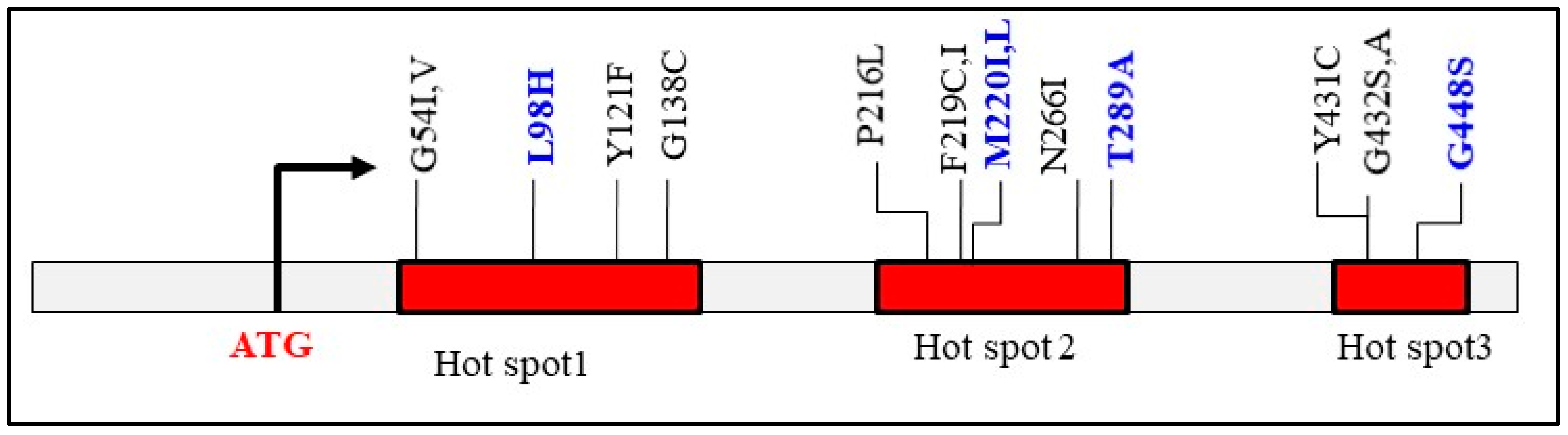
3. Intrinsic Azole Resistance of Fusarium

3.1. Resistance Evolution

Azole resistance in clinical Fusarium isolates may arise from prolonged antifungal therapy regimens for invasive and chronic infections (Figure 1) as well as from the hospital environment [8]. While both agricultural and medical triazoles share the same molecular target, it is plausible to assume that clinical resistance could be exacerbated by the extensive use of azole fungicides in agriculture. Azoles are widely applied in agriculture to protect food crops from fungal pathogens such as Fusarium. Their widespread use on every continent is driven by affordability, broad-spectrum antifungal activity, and long-lasting stability, making them a preferred choice for crop protection [46,47,48]. Since the first agricultural azole, imazalil, was introduced in 1973—two decades before fluconazole—over 25 azoles have been developed for agricultural applications, whereas fewer than 10 clinical triazoles have been introduced [49,50].
The cross-kingdom pathogenicity of Fusarium, affecting both humans and plants, results in significant exposure to azole antifungals from agricultural soil, antifungal-treated patients, and the hospital environment. In hospitals, Fusarium species have been detected in the air of patient rooms, water tanks, drains, showerheads, and aerosolized water following shower use [8]. In the agricultural field, the primary pathogenic species within the FSSC and the FOSC complexes are commonly found in soil, plants, and water, where they can also cause waterborne diseases in animals and crops [51,52,53,54]. Additionally, Fusarium species have been detected in wastewater treatment plant effluents, which can disseminate into rivers, further contributing to environmental exposure [55]. However, there is currently a lack of experimental evidence to support the connection between azole resistance in these two environmental niches.
The treatment of invasive fusariosis (IF) and localized fusariosis remains challenging, as Fusarium exhibits significant resistance to antifungal agents. In vitro studies have demonstrated that Fusarium isolates show poor sensitivity not only to first-generation triazoles such as fluconazole and itraconazole but also to amphotericin B (AmB) and the second generation of triazoles, including voriconazole (VOR), posaconazole (POS), and isavuconazole (ISA) [56,57]. Despite this resistance, voriconazole (VOR) and amphotericin B (AmB) remain the primary treatment options for invasive fusariosis (IF), with VOR frequently used prophylactically in clinical practice [58].
Our own studies, which include 29 clinical isolates provided by the CDC and The University of Texas at San Antonio, indicate that 85% of these strains are resistant to VOR. Currently, we are evaluating over 100 strains isolated from nail infections for resistance to azoles, amphotericin B, caspofungin, and other antifungals in vitro. Preliminary data suggest that cross-azole resistance and multidrug resistance (MDR) are prevalent among clinical isolates in the United States (manuscript in preparation).

3.2. Molecular Basis of Fusarium Azole Resistance

Three major azole resistance mechanisms have been extensively studied in C. albicans and the mold A. fumigatus: (i) mutations in the azole target genes (CYP51), which account for 50–80% of Aspergillus resistance cases; (ii) overexpression of the target gene; and (iii) upregulation of efflux pumps [59,60]. In Aspergillus, the first two mechanisms are dominant, with mutations in CYP51A playing a key role [57]. A well-characterized resistance-associated alteration in Aspergillus involves a leucine-to-histidine substitution at codon 98 (L98H), accompanied by a 34-base pair tandem repeat (TR34/L98H) insertion in the CYP51A promoter. This mutation, frequently identified in azole-naïve patients with aspergilloma and lung disease, is considered a hallmark of environmentally acquired resistance [61,62]. The tandem repeat in the promoter contributes to the overexpression of CYP51A.
In contrast, the molecular mechanisms of azole resistance in Fusarium, particularly those involving CYP51A, remain less well characterized in clinical isolates. While amino acid substitutions from point mutations alter target interactions with azoles in Aspergillus, this mechanism does not appear to be relevant to resistance in field-isolated F. graminearum and F. asiaticum [63,64], both of which belong to the Fusarium graminearum species complex (FGSC). However, laboratory-induced metconazole adaptation resulted in mutations associated with different expression patterns in F. graminearum CYP51 genes [65]. Fusarium possesses several genetic features that likely contribute to its intrinsic resistance to azoles and even amphotericin B (AmB) [57,66]. Notably, this intrinsic resistance has been reported in clinical isolates collected both before and after 1990 [57], when fluconazole was first introduced in clinical settings. This suggests that the evolutionary path of azole resistance in Fusarium differs from that of Aspergillus.
One possible explanation for Fusarium’s intrinsic resistance is its high genomic plasticity, which may be facilitated by horizontal gene transfer [67]. Furthermore, recent discoveries of aneuploidy-driven resistance in drug-resistant Cryptococcus neoformans [68,69] and RNA interference (RNAi)-mediated resistance in Mucor spp. [70,71] suggest that Fusarium may employ similar mechanisms, making its antifungal resistance more complex than previously anticipated. These findings highlight the need for further investigation into the genetic and epigenetic basis of Fusarium drug resistance.

3.2.1. Point Mutations in CYP51 Paralogs

Regarding the classic point mutation mechanism, Fusarium possesses two key genetic advantages that may contribute to its azole resistance. First, it has three paralogs of the azole target gene (CYP51A, CYP51B, and CYP51C), whereas C. albicans has only one (ERG11), and Aspergillus has two (CYP51A and CYP51B). Second, Fusarium has those known missense mutations within their three CYP51 paralogs (Table 1). In Aspergillus, three hotspot regions for point mutations in CYP51A have been identified with azole resistance (Figure 2) [72]. These mutations can occur independently (e.g., any or all of G54I, M220L, G138C, and Y121F) and are primarily observed in aspergilloma patients with lung cavities. Some of these hotspot point mutations occur in combination with tandem repeat promoter alterations, such as TR34/L98H and TR46/Y121F/T289A, which are common in environmentally resistant A. fumigatus strains. In silico modeling of A. fumigatus CYP51A has revealed that substitutions at residues L98, M220, or Y431 reduce azole binding affinity, while mutations at residues L98 or G432 decrease protein stability, potentially diminishing the antifungal effects of azoles [73].
BLAST analysis of randomly selected Fusarium genomes identified the M220L mutation in CYP51A of clinically relevant Fusarium species, along with six of twelve known Aspergillus CYP51A SNPs distributed across CYP51A, CYP51B, and CYP51C (Table 1). Four reported CYP51C missense mutations, including S240A—linked to voriconazole resistance in Aspergillus flavus [75]—had corresponding but distinct amino acid substitutions in Fusarium CYP51 paralogs. The impact of these SNPs on Fusarium azole resistance remains largely unexplored, particularly for CYP51C mutations located in non-conserved regions of Fusarium CYP51 paralogs. Nevertheless, the presence of preexisting CYP51A-associated SNPs across all three Fusarium CYP51 paralogs may contribute to its inherent molecular basis for azole resistance, as CYP51A plays a more significant role in drug response (see the next section). While mutations in hotspot 1 and hotspot 2 of CYP51A confer itraconazole resistance in Aspergillus [72], alterations such as L98H, M220I (L), and T289A could contribute to the intrinsic insensitivity of clinical Fusarium isolates to itraconazole, which was not specifically designed to target Fusarium CYP51 paralogs.
Independent research by the authors (now in press) also confirms that it is not even possible to determine an MIC for itraconazole, so pervasive is the resistance of clinical strains to this drug. In field-isolated F. graminearum from China, point mutations such as S28L, S256A, and V307A in CYP51C were observed in hexaconazole-resistant strains [76]. By contrast, laboratory-induced metconazole-resistant F. graminearum harbored mutations such as D243N, E103Q/V157L, and G443S in CYP51A [65], which differed from the CYP51A variants identified in hexaconazole-resistant strains [76]. Without phenotypic confirmation of drug susceptibility, it remains inconclusive whether these reported mutations in these plant-pathogenic Fusarium species are influenced by the specific azole used in the study or the induction conditions. Notably, while all CYP51C mutations fall outside conserved amino acid regions [76], CYP51A mutations occur within conserved regions or putative substrate-recognition sites (SRS) [77] in all three CYP51 paralogs across different Fusarium species. Therefore, verifying the association between these CYP51A SNPs or other distinct SNPs linked to clinical azole-resistant Fusarium species is of particular interest. The widespread occurrence of these SNPs in available genomic data, along with corresponding azole susceptibility profiles, warrants further large-scale investigation into their potential role in clinical azole resistance.

3.2.2. Overexpression of CYP51 Genes and Exclusion Drug by Efflux

Overexpression of the target gene enhances substrate binding, allowing competition with the azole drug. This enables the fungus to sustain ergosterol synthesis and survive to a certain extent [78,79]. Data on the overexpression of CYP51 genes in Fusarium are limited. Notably, all three Fusarium paralogs are transcriptionally active in both the mycelial and conidial phases [80] and can be up-regulated under voriconazole (VOR) treatment [9]. A study by James et al. examined the expression responses of these paralog genes in F. solani, showing that CYP51B is highly expressed under normal growth conditions without drug pressure [9]. In contrast, CYP51A was upregulated by several thousand-fold upon exposure to 1 µg/mL VOR. The same level of VOR led to a moderate increase of ~20-fold in CYP51B, and an increase of 3–6-fold in CYP51C expression. Apparently, CYP51A functions as an inducible responder to azole pressure, playing a crucial role in adaptive azole resistance. In agreement with the constitutive expression of CYP51B, deletion mutants of CYP51A (Δcyp51A) or CYP51C (Δcyp51C), but not CYP51B (Δcyp51B), in Fusarium graminearum were found to be more susceptible to agricultural azole inhibitors [77]. However, the ergosterol content in Δcyp51A, Δcyp51B, or Δcyp51C mutants did not differ from that of the wild-type strain, suggesting functional redundancy among these paralogs. This redundancy may also be reflected in laboratory-induced metconazole mutations in F. graminearum. Beyond the biological fitness penalty, the D243N mutation in CYP51A was associated with the overexpression of all three CYP51 genes, whereas E103Q/V157L led to the overexpression of CYP51A and CYP51B, and G443S was linked to CYP51A only. These findings suggest that the impact of single SNPs in any one paralog on azole resistance may be limited.
Although the efflux mechanism has been considered to play a minor role in azole resistance in clinical mold isolates, some studies on field-isolated F. graminearum suggest otherwise. The activation of ABC transporters, such as the plasma membrane-localized H+ antiporter FgQdr2, significantly reduces F. graminearum’s sensitivity to azoles [81,82]. However, the role of efflux pumps and their regulatory mechanisms in azole resistance remains poorly understood.

3.2.3. Promoter Mutations in CYP51A and Their Potential Impact on Signal Transduction Pathways

Interestingly, a recent study on 25 F. solani species complex (FSSC) isolates from Malaysia identified a 23 bp CYP51A promoter deletion (DEL) in nine isolates with voriconazole MICs exceeding 32 µg/mL [9]. These isolates originated from diverse clinical and environmental sources. Although the authors proposed this deletion as a potential marker for voriconazole resistance in FSSC isolates, its direct role remains uncertain, as it only led to a modest CYP51A upregulation (1.3- to 7.5-fold). This 23bpDEL mutation is located 120 bp upstream of a putative 16 bp sterol regulatory (SR) cis-element, which has been identified as a binding site for the zinc-cluster transcription factor FgSR in Fusarium graminearum [83]. Among 116 genes with FgSR binding sites, 20 encode ERG proteins involved in ergosterol biosynthesis, including CYP51A, CYP51B, and CYP51C. Notably, deletion of the FgSR gene resulted in an 85% reduction in ergosterol levels and increased susceptibility to azoles and DNA synthesis inhibitors. These findings highlight a more pronounced role of FgSR in regulating ergosterol biosynthesis and the DNA damage response under fungicidal stress, potentially overriding the resistance influence of CYP51 paralogs.
Notably, FgSR orthologs are found only in two other ascomycete fungal classes, Sordariomycetes and Leotiomycetes. The authors demonstrated that FgSR activation occurs through phosphorylation in the nucleus upon ergosterol depletion, mediated by the Hog1 MAPK pathway. This regulatory mechanism differs from the canonical pathways governing ergosterol biosynthesis, such as the nuclear and ER translocation of Upc2 in Candida and SREBP (sterol regulatory element-binding protein) in Aspergillus [84,85]. Further investigation is needed to determine how promoter mutations in Fusarium CYP51 paralogs influence FgSR binding activity, particularly in the inducible CYP51A.
Understanding the role of this Hog1-MAPK signaling pathway in azole resistance in Fusarium is particularly relevant given the differences in azole susceptibility between yeast pathogens and Fusarium mutants of this pathway. In Candida, the Hog1 MAPK pathway is upregulated in response to stress resistance, including oxidative, osmotic, and drug-induced stresses. This pathway plays a critical role in regulating cell morphology, aggregation, and virulence. As a result, the Hog1 MAPK pathway has been suggested as a potential target for antifungals, especially in combating emerging drug-resistant pathogens [86,87,88]. In Cryptococcus, deletion of HOG1 confers increased susceptibility to azoles [89]. Both yeast responses contrast with the loss-of-function mutations in HOG1 and related MAPK components in F. graminearum, which increase azole susceptibility [86]. This raises an intriguing question: how do host-derived reactive oxygen species (ROS) during infection regulate the Hog1-FgSR pathway? Further studies are needed to clarify the impact of this pathway on ergosterol levels and azole susceptibility in Fusarium infection.
In summary, although Fusarium and Aspergillus are both mold pathogens with trans-kingdom infection potential, the currently characterized azole resistance mechanisms in Aspergillus do not necessarily apply to Fusarium. Growing evidence highlights differences between these two genera in ergosterol regulation and the impact of known resistance-associated SNPs. These observations emphasize the need for further functional studies to determine the precise contributions of Fusarium CYP51 paralogs to azole resistance and to explore additional resistance mechanisms, including epigenetic regulation, genomic adaptation strategies, and the signal transduction pathways.

4. Fusarium Biofilms

Fungal biofilms are a critical virulence factor, providing protection against host (human/plant) defenses and environmental conditions while increasing drug resistance. In Fusarium, biofilm formation is particularly relevant in keratitis and onychomycosis [34,90] the most common clinical manifestations of fusariosis. Biofilms have been demonstrated in corneal infections, contact lenses, and nail infections, where they contribute to persistence and therapeutic challenges.

4.1. Biofilm Formation in Fusarium Infections in Humans

Fusarium keratitis is frequently observed in contact lens users, where fungal cells attach to the lens case, secrete proteins, and establish a biofilm [91]. Beyond colonizing living surfaces such as the cornea and nails, biofilm-like structures are also found on non-living medical devices, including contact lenses, prosthetic heart valves, coronary stents, and prosthetic joints. Biofilm development follows a five-stage process: (i) reversible attachment of floating fungal cells to the lens surface; (ii) irreversible attachment and colonization; (iii) expansion of fungal layers; (iv) formation of a protective extracellular matrix (ECM); and (v) detachment and dispersal of fungal cells into the surrounding environment. Scratches or micro-injuries on living surfaces, such as the corneal or nail epithelium, facilitate fungal penetration and biofilm establishment. Additionally, contact lenses reduce the antimicrobial efficacy of the tear film, further promoting keratitis infection.

4.2. In Vivo and Ex Vivo Models of Fusarium Biofilm Formation

Several in vivo models have been developed to study Fusarium biofilms in keratitis using immunocompromised rabbits and mice [92]. In immunosuppressed mice, co-infection of Fusarium falciforme with Staphylococcus aureus led to biofilm formation in the eye [93]. Histopathological evaluation of enucleated eye sections revealed an extracellular matrix (biofilm) on the surface of both organisms. In a cyclophosphamide-treated BALB/c mouse model, F. solani infection alone resulted in biofilm-like fungal masses in the cornea within 4–8 days, accompanied by fungal infiltration and neutrophil accumulation in the anterior chamber [94]. Similarly, in a rabbit model pretreated with corticosteroids and antibiotics, a biofilm-like mass formed one week after interlamellar injection of F. solani [95].
The role of biofilms in Fusarium onychomycosis has been investigated using an ex vivo infection model with F. oxysporum isolates from an onychomycosis patient [34]. Microscopic analysis revealed conidial transition to hyphae and biofilm formation by day six. ECM analysis detected lipids, proteins, polysaccharides, extracellular DNA (eDNA), and extracellular RNA (eRNA), consistent with ECM components observed in in vitro biofilms. These findings strongly suggest that biofilms in vivo may contribute to its antifungal resistance and recurrence in Fusarium nail infections [33].

4.3. In Vitro Studies on Fusarium Biofilms

In vitro biofilm formation follows the same progression as in vivo and ex vivo models, involving adherence, germination, hyphal development, maturation, and detachment [90]. Biofilm ECM in vitro also consists of carbohydrates, proteins, lipids, and eDNA. Optimal biofilm development occurred with an inoculum of 1 × 10⁶ F. falciforme conidia in a 96-well plate using RPMI medium at 28 °C for 96 h [96] in an in vitro biofilm testing system. Proteomic analysis identified 19 overexpressed proteins in biofilms, including transketolase, enolase, phosphoglycerate kinase, and ATP-citrate synthase, highlighting that glycolysis and gluconeogenesis play significant roles in biofilm formation [96].
The crucial role of these metabolic pathways aligns with our phenotypic observations [97]. In that study, clinical isolates from four major Fusarium species complexes (F. solani, F. fujikuroi, F. oxysporum, and F. incarnatum-equiseti) were analyzed for biofilm composition and antifungal response. Notably, these clinical Fusarium species, like plant pathogens, exhibit poor thermotolerance and anaerobic growth, which explains their preference for superficial infections in the eyes and nails, where oxygen and lower temperatures are available. Despite variability among species, glucose promotes biofilm formation more effectively than other less favorable sugars or glycerol. Among the four Fusarium species complexes, F. oxysporum demonstrates greater nutritional flexibility during biofilm formation. Our second key finding is that azole-resistant F. solani strains form more robust biofilms compared to azole-susceptible strains when exposed to antifungals, including voriconazole (VOR), amphotericin B (AmB), and 5-flucytosine (5-FC). This suggests that the metabolic flexibility inherent in Fusarium and the drug stress adaptation mechanisms, which likely develop pre-clinically, enhance the survival of these strains and facilitate biofilm formation within the host.

4.4. Biofilm Composition Analysis and Treatment Implications

Over time, ECM composition shifts from lipid-rich in early stages to polysaccharide- and protein-dominant in late stages in our recent study with clinical Fusarium isolates [97]. We found that VOR demonstrated greater efficacy than AmB in inhibiting Fusarium biofilm formation. Our findings also highlight the potential of antifungal combination therapies to disrupt Fusarium biofilms and improve treatment outcomes by targeting the distinct composition of the biofilm. For example, lipids in the ECM during the early stages of biofilm formation could serve as a potential intervention target. In an in vivo biofilm model, lipids in the nail biofilm extracellular matrix (ECM) were found to peak at 7 days post-infection [34]. These secreted lipids, detected using FTIR-ATR spectroscopy, exhibit characteristic functional groups, including C=O (associated with triglycerides and phospholipids), C-N (found in fatty acid amides, which function as signaling molecules), and CH2/CH3 structures. However, the specific lipid classes, such as phospholipids or sterol lipids, within the Fusarium biofilm remain to be fully characterized.
The amount of ergosterol in the ECM of C. albicans varies with biofilm development [98,99]. Consistent with the early appearance of lipids observed in clinical Fusarium isolates, ergosterol content in C. albicans is highest during the early stages of biofilm formation and is reduced by approximately 50% in mature biofilms [98]. Given that voriconazole targets sterol lipids and amphotericin B interferes with phospholipid structures, the superior anti-biofilm efficacy of VOR [97] suggests a crucial role for sterol lipids in biofilm development. Interestingly, biofilm compositions from Fusarium graminearum, a plant pathogen, exhibit distinct characteristics, including a lack of lipids [100]. When testing other biofilm characteristics of this plant pathogen, it was observed that under anaerobic conditions, short bulbous hyphae were surrounded by ECM in the absence of electron acceptors. The addition of electron acceptors restored filamentous growth. This suggests that elevated reactive oxygen species (ROS), typically produced by the host response during infection, may trigger hyphal development during biofilm formation.

5. Summary and Prospect

Fusarium spp. are opportunistic mold pathogens that cause a range of infections, including keratitis and onychomycosis. Biofilm formation plays a role in the persistence of these infections, providing protection against environmental stressors and antifungal treatment. While biofilms enhance resistance to some antifungal agents, emerging evidence suggests that Fusarium azole resistance is primarily driven by distinct molecular mechanisms rather than biofilm formation alone. In contrast to Aspergillus, Fusarium has unique ergosterol biosynthesis regulation, with multiple CYP51 paralogs playing a key role in its resistance. Current evidence suggests that the severe azole resistance observed in clinical Fusarium isolates may result from multiple mechanisms, which differ from the single nucleotide polymorphism (SNP)-driven mechanisms seen in Aspergillus. Regardless of the specific genomic adaptations, other resistance mechanisms—such as epigenetic modifications and signal transduction pathways—may also influence Fusarium’s azole resistance. Gaining a deeper understanding of these Fusarium-specific mechanisms is crucial for developing targeted antifungal strategies to improve treatment outcomes.

Author Contributions

Conceptualization was performed by R.C., T.Y.S. and D.L.; literature data were collected by D.L., K.A.-C. and E.A.; writing—original draft preparation by R.C. and D.L.; writing—review and editing by D.L., T.Y.S. and R.C.; All authors have read and agreed to the published version of the manuscript.

Funding

This research received no external funding.

Institutional Review Board Statement

Not applicable.

Informed Consent Statement

Not applicable.

Data Availability Statement

No new data were created or analyzed in this study. Data sharing is not applicable to this article.

Conflicts of Interest

The authors declare no conflicts of interest.

References

  1. van Diepeningen, A.D.; de Hoog, G.S. Challenges in Fusarium, a Trans-Kingdom Pathogen. Mycopathologia 2016, 181, 161–163. [Google Scholar] [CrossRef]
  2. Savary, S.; Willocquet, L.; Pethybridge, S.J.; Esker, P.; McRoberts, N.; Nelson, A. The global burden of pathogens and pests on major food crops. Nat. Ecol. Evol. 2019, 3, 430–439. [Google Scholar] [CrossRef]
  3. Casalini, G.; Giacomelli, A.; Antinori, S. The WHO fungal priority pathogens list: A crucial reappraisal to review the prioritisation. Lancet Microbe 2024, 7, 717–724. [Google Scholar] [CrossRef] [PubMed]
  4. World Health Organization. WHO Fungal Priority Pathogens List to Guide Research, Development and Public Health Action; World Health Organization: Geneva, Switzerland, 2022. [Google Scholar]
  5. O’Donnell, K.; Sutton, D.A.; Rinaldi, M.G.; Magnon, K.C.; Cox, P.A.; Revankar, S.G.; Sanche, S.; Geiser, D.M.; Juba, J.H.; van Burik, J.A.; et al. Genetic diversity of human pathogenic members of the Fusarium oxysporum complex inferred from multilocus DNA sequence data and amplified fragment length polymorphism analyses: Evidence for the recent dispersion of a geographically widespread clonal lineage and nosocomial origin. J. Clin. Microbiol. 2004, 42, 5109–5120. [Google Scholar] [CrossRef] [PubMed]
  6. Bahadur, A. Current status of Fusarium and their management strategies. In Fusarium—An Overview on Current Status of the Genus; Mirmajlessi, S.M., Ed.; IntechOpen: London, UK, 2021. [Google Scholar] [CrossRef]
  7. Chowdhary, C.; Kathuria, S.; Xu, J.; Meis, J.F. Emergence of azole resistant Aspergillus fumigatus strains due to agriculture azole use creates an increasing threat to human health. PLoS Pathog. 2013, 9, e1003633. [Google Scholar] [CrossRef]
  8. Nucci, M.; Anaissie, E. Invasive fusariosis. Clin. Microbiol. Rev. 2023, 36, e0015922. [Google Scholar] [CrossRef]
  9. James, J.E.; Lamping, E.; Santhanam, J.; Milne, T.J.; Abd Razak, M.F.; Zakaria, L.; Cannon, R.D. A 23 bp cyp51A promoter deletion associated with voriconazole resistance in clinical and environmental isolates of Neocosmospora keratoplastica. Front. Microbiol. 2020, 11, 272. [Google Scholar] [CrossRef]
  10. O’Donnell, K.; Ward, T.J.; Robert, V.A.R.G.; Crus, P.W.; Geisen, P.M.; Kang, S. DNA sequence-based identification of Fusarium: Current status and future directions. Phytoparasitica 2015, 43, 583–595. [Google Scholar] [CrossRef]
  11. Lockhart, S.R.; Guarner, J. Emerging and reemerging fungal infections. Semin. Diagn. Pathol. 2019, 36, 177–181. [Google Scholar] [CrossRef]
  12. Nucci, M.; Marr, K.A.; Vehreschild, M.J.; de Souza, C.A.; Velasco, E.; Cappellano, P.; Carlesse, F.; Queiroz-Telles, F.; Sheppard, D.C.; Kindo, A.; et al. Improvement in the outcome of invasive fusariosis in the last decade. Clin. Microbiol. Infect. 2014, 20, 580–585. [Google Scholar] [CrossRef]
  13. Nucci, M.; Anaissie, E. Cutaneous infection by Fusarium species in healthy and immunocompromised hosts: Implications for diagnosis and management. Clin. Infect. Dis. 2002, 35, 909–920. [Google Scholar] [CrossRef] [PubMed]
  14. Nucci, M.; Anaissie, E. Fusarium infections in immunocompromised patients. Clin. Microbiol. Rev. 2007, 20, 695–704. [Google Scholar] [CrossRef]
  15. Hoffman, J.J.; Burton, M.J.; Leck, A. Mycotic keratitis-A global threat from the filamentous Fungi. J. Fungi 2021, 7, 273. [Google Scholar] [CrossRef] [PubMed]
  16. Oechsler, R.A.; Fellmeter, M.R.; Miller, D. Fusarium keratitis genotyping, in vitro susceptibility, and clinical outcomes. Cornea 2013, 32, 667–673. [Google Scholar] [CrossRef]
  17. Ranawaka, R.R.; Nagahawatte, A.; Gunasekara, T.A. Fusarium onychomycosis: Prevalence, clinical presentations, response to itraconazole and terbinafine pulse therapy, and 1-year follow-up in nine cases. Int. J. Dermatol. 2015, 54, 1275–1282. [Google Scholar] [CrossRef] [PubMed]
  18. Cohen, I.L.; Knoeri, M.J.; Bourcier, T.; Merabet, L.; Borderie, V.M. Infection keratitis following corneal transplantation: A long-term cohort study. Clin. Exp. Ophthalmol. 2024, 52, 402–415. [Google Scholar] [CrossRef]
  19. Thomas, B.; Audonneau, N.C.; Machouart, M.; Debourgogne, A. Fusarium infections: Epidemiology aspects over 10 yrs in a university hospital in France. J. Infect. Public Health 2020, 13, 1089–1093. [Google Scholar] [CrossRef] [PubMed]
  20. Brown, L.; Kamwiziku, G.; Oladele, R.O.; Burton, M.J.; Prajna, N.V.; Leitman, T.M.; Denning, D.W. The case for fungal keratitis to be accepted as a neglected tropical disease. J. Fungi 2022, 8, 1047. [Google Scholar] [CrossRef]
  21. Green, M.; Apel, A.; Stapleton, F. Risk factors and causative organisms in microbial keratitis. Cornea 2008, 27, 22–27. [Google Scholar] [CrossRef]
  22. Brown, L.; Leck, A.K.; Gichangi, M.; Burton, M.J.; David, D.W. The global incidence and diagnosis of fungal keratitis. Lancet Infect. Dis. 2021, 21, e49–e57. [Google Scholar] [CrossRef]
  23. Liu, J.; Wei, Z.; Cao, K.; Zhang, Z.; Xu, X.; Liang, Q. Trends of ocular fungal infections in North China (2001–2020). J. Infect. Public Health 2023, 16, 71–77. [Google Scholar] [CrossRef]
  24. Chang, D.C.; Grant, G.B.; O’Donnell, K.; Wannemuehler, K.A.; Noble-Wang, J.; Rao, C.Y.; Jacobson, L.M.; Crowell, C.S.; Sneed, R.S.; Lewis, M.F.T.; et al. Multistate outbreak of Fusarium keratitis associated with use of a contact lens solution. JAMA 2006, 296, 953–963. [Google Scholar] [CrossRef]
  25. Szaliński, M.; Zgryźniak, A.; Rubisz, I.; Gajdzis, M.; Kaczmarek, R.; Przeździecka-Dołyk, J. Fusarium keratitis—review of current treatment possibilities. J. Clin. Med. 2021, 10, 5468. [Google Scholar] [CrossRef] [PubMed]
  26. Liang, Q.F.; Jin, X.Y.; Wang, X.L.; Sun, X.G. Effect of topical application of terbinafine on fungal keratitis. Chin. Med. J. 2009, 122, 1884–1888. [Google Scholar] [PubMed]
  27. Feng, Y.; Yang, Z.; Li, D.; Li, J.; Li, D.; de Hoog, S.; Shi, D. Nails and skin co-infection by Fusarium verticillioides and Proteus vulgaris secondary to arterial occlusion of lower extremity. Rev. Iberoam. Micol. 2024, 41, 37–42. [Google Scholar] [CrossRef] [PubMed]
  28. Gupta, A.K.; Taborda, V.B.A.; Taborda, P.R.O.; Shemer, A.; Summerbell, R.C.; Nakrieko, K.A. High prevalence of mixed infections in global onychomycosis. PLoS ONE 2020, 15, e0239648. [Google Scholar] [CrossRef]
  29. Gupta, A.K.; Wang, T.; Cooper, E.A.; Lincoln, S.A.; Foreman, H.C.; Scherer, W.P.; Bakotic, W.L. Clinical diagnosis and laboratory testing of abnormal appearing toenails: A retrospective assessment of confirmatory testing for onychomycosis in the United States, 2022–2023. J. Fungi 2024, 10, 149. [Google Scholar] [CrossRef]
  30. Mima, Y.; Yamamoto, M.; Makimura, K.; Iozumi, K. Onychomycosis with gray-green staining caused by Fusarium solani. Med. Mycol. Case Rep. 2024, 46, 100684. [Google Scholar] [CrossRef]
  31. Uemura, E.V.G.; Barbosa, M.D.S.; Simionatto, S.; Al-Harrasi, A.; Al-Hatmi, A.M.S.; Rossato, L. Onychomycosis caused by Fusarium species. J. Fungi 2022, 8, 360. [Google Scholar] [CrossRef]
  32. Tikvesli, M.; Gürcan, S.; Afyoncu, E.; Ürün, Y.G.; Solak, S.S. Mixed onychomycosis case with Fusarium solani: A case report. J. Immunol. Clin. Microbiol. 2022, 7, 45–50. [Google Scholar]
  33. Gupta, A.K.; Daigle, D.; Carviel, J.L. Evidence for biofilms in onychomycosis. J. Am. Acad. Dermatol. 2016, 74, 1241–1246. [Google Scholar] [CrossRef]
  34. Veiga, F.F.; de Castro-Hoshino, L.V.; Sato, F.; Baesso, M.L.; Silva, S.; Negri, M.; Svidzinski, T.I.E. Characterization of a biofilm formed by Fusarium oxysporum on the human nails. Int. J. Dermatol. 2022, 61, 191–198. [Google Scholar] [CrossRef] [PubMed]
  35. Schäfer, K.; Di Pietro, A.; Gow, N.A.; MacCallum, D. Murine model for Fusarium oxysporum invasive fusariosis reveals organ-specific structures for dissemination and long-term persistence. PLoS ONE 2014, 9, e89920. [Google Scholar] [CrossRef]
  36. Girmenia, C.; Pagano, L.; Corvatta, L.; Mele, L.; del Favero, A.; Martino, P. The epidemiology of fusariosis in patients with haematological diseases. Gimema Infection Programme. Br. J. Haematol. 2000, 111, 272–276. [Google Scholar] [CrossRef] [PubMed]
  37. Kontoyiannis, D.P.; Marr, K.A.; Park, B.J.; Alexander, B.D.; Anaissie, E.J.; Walsh, T.J.; Ito, J.; Andes, D.R.; Baddley, J.W.; Brown, J.M.; et al. Prospective surveillance for invasive fungal infections in hematopoietic stem cell transplant recipients, 2001–2006: Overview of the transplant-associated infection surveillance network (transnet) database. Clin. Infect. Dis. 2010, 50, 1091–1100. [Google Scholar] [CrossRef] [PubMed]
  38. Atalla, A.; Garnica, M.; Maiolino, A.; Nucci, M. Risk factors for invasive mold diseases in allogeneic hematopoietic cell transplant recipients. Transpl. Infect. Dis. 2015, 17, 7–13. [Google Scholar] [CrossRef]
  39. Nucci, M.; Varon, A.G.; Garnica, M.; Akiti, T.; Barreiros, G.; Trope, B.; Nouér, S.A. increased incidence of invasive fusariosis with cutaneous portal of entry. Braz. Emerg. Infect. Dis. 2013, 19, 1567–1572. [Google Scholar] [CrossRef]
  40. Rosa, P.D.D.; Ramirez-Castrillon, M.; Borges, R.; Aquino, V.; Fuentefria, M.A.; Goldani, Z.L. Epidemiological aspects and characterization of the resistance profile of Fusarium spp. in patients with invasive fusariosis. J. Med. Microbiol. 2019, 68, 1489–1496. [Google Scholar] [CrossRef]
  41. Pérez-Nadales, E.; Alastruey-Izquierdo, A.; Linares-Sicilia, M.J.; Soto-Debrán, J.C.; Abdala, E.; García-Rodríguez, J.; Montejo, M.; Muñoz, P.; Lletí, M.S.; Rezusta, A.; et al. Spanish fusariosis study group. invasive fusariosis in nonneutropenic patients, Spain, 2000–2015. Emerg. Infect. Dis. 2021, 27, 26–35. [Google Scholar] [CrossRef]
  42. Demonchy, J.; Biard, L.; Clere-Jehl, R.; Wallet, F.; Mokart, D.; Moreau, A.S.; Argaud, L.; Verlhac, C.; Pène, F.; Lautrette, A.; et al. Multicenter retrospective study of invasive fusariosis in intensive care units, France. Emerg. Infect. Dis. 2024, 30, 215–224. [Google Scholar] [CrossRef]
  43. Rychert, J. Benefits and limitations of maldi-tof mass spectrometry for the identification of microorganisms. J. Infect. 2019, 2, 1–5. [Google Scholar] [CrossRef]
  44. Hachem, R.Y.; Kontoyiannis, D.P.; Chemaly, R.F.; Jiang, Y.; Reitzel, R.; Raad, I. Utility of galactomannan enzyme immunoassay and (1,3) beta-D-glucan in diagnosis of invasive fungal infections: Low sensitivity for Aspergillus fumigatus infection in hematologic malignancy patients. J. Clin. Microbiol. 2009, 47, 129–133. [Google Scholar] [CrossRef] [PubMed]
  45. Dellière, S.; Guitard, J.; Marcela Sabou, M.; Angebault, C.; Moniot, M.; Cornu, M.; Hamane, S.; Bougnoux, M.E.; Imbert, S.; Pasquier, G.; et al. Detection of circulating DNA for the diagnosis of invasive fusariosis: Retrospective analysis of 15 proven cases. Med. Mycol. 2022, 60, myac049. [Google Scholar] [CrossRef] [PubMed]
  46. Pintye, A.; Bacsó, R.; Kovács, G.M. Trans-kingdom fungal pathogens infecting both plants and humans, and the problem of azole fungicide resistance. Front. Microbiol. 2024, 15, 1354757. [Google Scholar] [CrossRef]
  47. Bastos, R.W.; Rossato, L.; Goldman, G.H.; Santos, D.A. Fungicide effects on human fungal pathogens: Cross-resistance to medical drugs and beyond. PLoS Pathog. 2021, 17, e1010073. [Google Scholar] [CrossRef]
  48. Toda, M.; Beer, K.D.; Kuivila, K.M.; Chiller, T.M.; Jackson, B.R. Trends in agricultural triazole fungicide use in the united states, 1992-2016 and possible implications for antifungal-resistant fungi in human disease. Environ. Health Perspect. 2021, 129, 55001. [Google Scholar] [CrossRef]
  49. Kleinkauf, N.; Verweij, P.E.; Arendrup, M.C.; Donnelly, P.J.; Cuenca-Estrella, M.; Fraaije, B.; Melchers, W.J.G.; Adriaenssens, N.; Kema, G.H.J.; Ullmann, A.; et al. Risk Assessment on the Impact of Environmental Usage of Triazoles on the Development and Spread of Resistance to Medica Triazoles in Aspergillus Species; European Center for Disease and Control: Stockholm, Sweden, 2013. [Google Scholar] [CrossRef]
  50. Jørgensen, L.N.; Heick, T.M. Azole use in agriculture, horticulture, and wood preservation—Is it indispensable? Front. Cell Infect. Microbiol. 2021, 11, 730297. [Google Scholar] [CrossRef]
  51. Bilska, K.; Jurczak, S.; Kulik, T.; Ropelewska, E.; Olszewski, J.; Żelechowski, M.; Zapotoczny, P. Species composition and trichothecene genotype profiling of Fusarium field isolates recovered from wheat in Poland. Toxins 2018, 10, 325. [Google Scholar] [CrossRef]
  52. Ma, L.J.; Geiser, D.M.; Proctor, R.H.; Rooney, A.P.; O’Donnell, K.; Trail, F.; Gardiner, D.M.; Manners, J.M.; Kazan, K. Fusarium pathogenomics. Annu. Rev. Microbiol. 2013, 67, 399–416. [Google Scholar] [CrossRef]
  53. Srinivas, C.; Nirmala, D.D.; Narasimha, M.K.; Mohan, C.D.; Lakshmeesha, T.R.; Singh, B.; Kalagatur, N.K.; Niranjana, S.R.; Hashem, A.; Alqarawi, A.A.; et al. Fusarium oxysporum f. sp. lycopersici causal agent of vascular wilt disease of tomato: Biology to diversity—A review. Saudi J. Biol. Sci. 2019, 26, 1315–1324. [Google Scholar] [CrossRef]
  54. Pegg, K.G.; Coates, L.M.; O’Neill, W.T.; Turner, D.W. The epidemiology of Fusarium wilt of banana. Front. Plant Sci. 2019, 10, 1395. [Google Scholar] [CrossRef]
  55. Assress, H.A.; Selvarajan, R.; Nyoni, H.; Ogola, H.J.O.; Mamba, B.B.; Msagati, T.A.M. Azole antifungal resistance in fungal isolates from wastewater treatment plant effluents. Environ. Sci. Pollut. Res. Int. 2021, 28, 3217–3229. [Google Scholar] [CrossRef] [PubMed]
  56. Vermeulen, P.; Gruez, A.; Babin, A.L.; Frippiat, J.P.; Machouart, M.; Debourgogne, A. CYP51 Mutations in the Fusarium solani species complex: First clue to understand the low susceptibility to azoles of the genus Fusarium. J. Fungi 2022, 8, 533. [Google Scholar] [CrossRef] [PubMed]
  57. Al-Hatmi, A.M.; Meis, J.F.; Sybren de Hoog, G. Fusarium: Molecular diversity and intrinsic drug resistance. PLoS Pathog. 2016, 12, e1005464. [Google Scholar] [CrossRef]
  58. Al-Hatmi, A.M.S.; Bonifaz, A.; Ranque, S.; Sybren de Hoog, G.; Verweij, P.E.; Meis, J.F. Current antifungal treatment of fusariosis. Int. J. Antimicrob. Agents 2018, 51, 326–332. [Google Scholar] [CrossRef] [PubMed]
  59. Dudakova, A.; Spiess, B.; Tangwattanachuleeporn, M.; Sasse, C.; Buchheidt, D.; Weig, M.; Groß, U.; Bader, O. Molecular tools for the detection and deduction of azole antifungal drug resistance phenotypes in Aspergillus species. Clin. Microbiol. Rev. 2017, 30, 1065–1091. [Google Scholar] [CrossRef]
  60. Zheng, B.; Yan, L.; Liang, W.; Yang, Q. Paralogous Cyp51s mediate the differential sensitivity of Fusarium oxysporum to sterol demethylation inhibitors. Pest. Manag. Sci. 2019, 75, 396–404. [Google Scholar] [CrossRef]
  61. Bader, O.; Weig, M.; Reichard, U.; Lugert, R.; Kuhns, M.; Christner, M.; Held, J.; Peter, S.; Schumacher, U.; Buchheidt, D.; et al. Cyp51A-based mechanisms of Aspergillus fumigatus azole drug resistance present in clinical samples from Germany. Antimicrob. Agents Chemother. 2013, 57, 3513–3517. [Google Scholar] [CrossRef]
  62. Wiederhold, N.P.; Gil, V.G.; Gutierrez, F.; Lindner, J.R.; Albataineh, M.T.; McCarthy, D.I.; Sanders, C.; Fan, H.; Fothergill, A.W.; Sutton, D.A. First detection of TR34 L98H and TR46 Y121F T289A Cyp51 mutations in Aspergillus fumigatus isolates in the united states. J. Clin. Microbiol. 2016, 54, 168–171. [Google Scholar] [CrossRef]
  63. Yin, Y.; Liu, X.; Li, B.; Ma, Z. Characterization of sterol demethylation inhibitor-resistant isolates of Fusarium asiaticum and F. graminearum collected from wheat in China. Phytopathology 2009, 99, 487–497. [Google Scholar] [CrossRef]
  64. Liu, J.; Jiang, J.; Guo, X.; Qian, L.; Xu, J.; Che, Z.; Chen, G.; Liu, S. Sensitivity and resistance risk assessment of Fusarium graminearum from wheat to prothioconazole. Plant Dis. 2022, 106, 2097–2104. [Google Scholar] [CrossRef] [PubMed]
  65. Duan, Y.; Li, M.; Zhao, H.; Lu, F.; Wang, J.; Zhou, M. Molecular and biological characteristics of laboratory metconazole-resistant mutants in Fusarium graminearum. Pestic. Biochem. Physiol. 2018, 152, 55–61. [Google Scholar] [CrossRef] [PubMed]
  66. Arikan, S.; Lozano-Chiu, M.; Paetznick, V.; Nangia, S.; Rex, J.H. Microdilution susceptibility testing of Amphotericin B, Itraconazole, and Voriconazole against clinical isolates of Aspergillus and Fusarium species. J. Clin. Microbiol. 1999, 37, 3946–3951. [Google Scholar] [CrossRef] [PubMed]
  67. Ma, L.J.; van der Does, H.C.; Borkovich, K.A.; Coleman, J.J.; Daboussi, M.J.; Di Pietro, A.; Dufresne, M.; Freitag, M.; Grabherr, M.; Henrissat, B.; et al. Comparative genomics reveals mobile pathogenicity chromosomes in Fusarium. Nature 2010, 464, 367–373. [Google Scholar] [CrossRef]
  68. Kwon-Chung, K.J.; Chang, Y.C. Aneuploidy and drug resistance in pathogenic fungi. PLoS Pathog. 2012, 8, e1003022. [Google Scholar] [CrossRef]
  69. Zhang, Z.H.; Sun, L.L.; Fu, B.Q.; Deng, J.; Jia, C.L.; Miao, M.X.; Yang, F.; Cao, Y.B.; Yan, T.H. Aneuploidy underlies brefeldin A-induced antifungal drug resistance in Cryptococcus neoformans. Front. Cell Infect. Microbiol. 2024, 14, 1397724. [Google Scholar] [CrossRef]
  70. Chang, Z.; Billmyre, R.B.; Lee, S.C.; Heitman, J. Broad antifungal resistance mediated by RNAi-dependent epimutation in the basal human fungal pathogen Mucor circinelloides. PLoS Genet. 2019, 15, e1007957. [Google Scholar] [CrossRef]
  71. Calo, S.; Nicolás, F.E.; Lee, S.C.; Vila, A.; Cervantes, M.; Torres-Martinez, S.; Ruiz-Vazquez, R.M.; Cardenas, M.E.; Heitman, J. A non-canonical RNA degradation pathway suppresses RNAi-dependent epimutations in the human fungal pathogen Mucor circinelloides. PLoS Genet. 2017, 13, e1006686. [Google Scholar] [CrossRef]
  72. Macedo, D.; Leonardelli, F.; Gamarra, S.; Garcia-Effron, G. Emergence of triazole resistance in Aspergillus spp. in Latin America. Curr. Fungal Infect. Rep. 2021, 15, 93–103. [Google Scholar] [CrossRef]
  73. Liu, M.; Zheng, N.; Li, D.; Zheng, H.; Zhang, L.; Ge, H.; Liu, W. Cyp51A-based mechanism of azole resistance in Aspergillus fumigatus: Illustration by a new 3D structural model of Aspergillus fumigatus CYP51A protein. Med. Mycol. 2016, 54, 400–408. [Google Scholar] [CrossRef]
  74. Gonzalez-Jimenez, I.; Lucio, J.; Amich, J.; Cuesta, I.; Sanchez Arroyo, R.; Alcazar-Fuoli, L.; Mellado, E. A Cyp51B mutation contributes to azole resistance in Aspergillus fumigatus. J. Fungi 2020, 6, 315. [Google Scholar] [CrossRef]
  75. Liu, W.; Sun, Y.; Chen, W.; Liu, W.; Wan, Z.; Bu, D.; Li, R. The T788G mutation in the cyp51C gene confers voriconazole resistance in Aspergillus flavus causing aspergillosis. Antimicrob. Agents Chemother. 2012, 56, 2598–2603. [Google Scholar] [CrossRef] [PubMed]
  76. Zhou, F.; Zhou, X.; Jiao, Y.; Han, A.; Su, H.; Wang, L.; Zhou, H.; Li, W.; Liu, R. Potential mechanisms of hexaconazole resistance in Fusarium graminearum. Plant Dis. 2024, 108, 3133–3145. [Google Scholar] [CrossRef] [PubMed]
  77. Liu, X.; Yu, F.; Schnabel, G.; Wu, J.; Wang, Z.; Ma, Z. Paralogous cyp51 genes in Fusarium graminearum mediate differential sensitivity to sterol demethylation inhibitors. Fungal Genet. Biol. 2011, 48, 113–123. [Google Scholar] [CrossRef]
  78. Mair, W.J.; Deng, W.; Mullins, J.G.; West, S.; Wang, P.; Besharat, N.; Ellwood, S.R.; Oliver, R.P.; Lopez-Ruiz, F.J. Demethylase inhibitor fungicide resistance in Pyrenophora teres f. sp. teres associated with target site modification and inducible overexpression of Cyp51. Front. Microbiol. 2016, 7, 1279. [Google Scholar] [CrossRef]
  79. Jayawardana, M.A.; Fernando, W.G.D. the mechanisms of developing fungicide resistance in Fusarium graminearum causing Fusarium head blight and fungicide resistance management. Pathogens 2024, 13, 1012. [Google Scholar] [CrossRef] [PubMed]
  80. Fernández-Ortuño, D.; Loza-Reyes, E.; Atkins, S.L.; Fraaije, B.A. The CYP51C gene, a reliable marker to resolve interspecific phylogenetic relationships within the Fusarium species complex and a novel target for species-specific PCR. Int. J. Food Microbiol. 2010, 144, 301–309. [Google Scholar] [CrossRef]
  81. Abou Ammar, G.; Tryono, R.; Döll, K.; Karlovsky, P.; Deising, H.B.; Wirsel, S.G. Identification of ABC transporter genes of Fusarium graminearum with roles in azole tolerance and/or virulence. PLoS ONE 2013, 8, e79042. [Google Scholar] [CrossRef]
  82. Ma, T.; Li, Y.; Lou, Y.; Shi, J.; Sun, K.; Ma, Z.; Yan, L.; Yin, Y. The drug H+ antiporter FgQdr2 is essential for multiple drug resistance, ion homeostasis, and pathogenicity in Fusarium graminearum. J. Fungi 2022, 8, 1009. [Google Scholar] [CrossRef]
  83. Liu, Z.; Jian, Y.; Chen, Y.; Kistler, H.C.; He, P.; Ma, Z.; Yin, Y. A phosphorylated transcription factor regulates sterol biosynthesis in Fusarium graminearum. Nat. Commun. 2019, 10, 1228. [Google Scholar] [CrossRef]
  84. Maguire, S.L.; Wang, C.; Holland, L.M.; Brunel, F.; Neuvéglise, C.; Nicaud, J.M.; Zavrel, M.; White, T.C.; Wolfe, K.H.; Butler, G. Zinc finger transcription factors displaced SREBP proteins as the major sterol regulators during Saccharomycotina evolution. PLoS Genet. 2014, 10, e1004076. [Google Scholar] [CrossRef] [PubMed]
  85. Bien, C.M.; Espenshade, P.J. Sterol regulatory element binding proteins in fungi: Hypoxic transcription factors linked to pathogenesis. Eukaryot. Cell 2010, 9, 352–359. [Google Scholar] [CrossRef]
  86. Chauhan, N.; Inglis, D.; Roman, E.; Pla, J.; Li, D.; Calera, J.A.; Calderone, R. Candida albicans response regulator gene SSK1 regulates a subset of genes whose functions are associated with cell wall biosynthesis and adaptation to oxidative stress. Eukaryot. Cell 2003, 2, 1018–1024. [Google Scholar] [CrossRef] [PubMed]
  87. Chauhan, N.; Kruppa, M.; Calderone, R. The Ssk1p response regulator and Chk1p histidine kinase mutants of Candida albicans are hypersensitive to fluconazole and voriconazole. Antimicrob. Agents Chemother. 2007, 51, 3747–3751. [Google Scholar] [CrossRef]
  88. Day, A.M.; McNiff, M.M.; da Silva Dantas, A.; Gow, N.A.R.; Quinn, J. Hog1 regulates stress tolerance and virulence in the emerging fungal pathogen Candida auris. mSphere 2018, 3, e00506-18. [Google Scholar] [CrossRef] [PubMed]
  89. Ko, Y.J.; Yu, Y.M.; Kim, G.B.; Lee, G.W.; Maeng, P.J.; Kim, S.; Floyd, A.; Heitman, J.; Bahn, Y.S. Remodeling of global transcription patterns of Cryptococcus neoformans genes mediated by the stress-activated HOG signaling pathways. Eukaryot. Cell 2009, 8, 1197–1217. [Google Scholar] [CrossRef]
  90. Córdova-Alcántara, I.M.; Venegas-Cortés, D.L.; Martínez-Rivera, M.Á.; Pérez, N.O.; Rodriguez-Tovar, A.V. Biofilm characterization of Fusarium solani keratitis isolate: Increased resistance to antifungals and UV light. J. Microbiol. 2019, 57, 485–497. [Google Scholar] [CrossRef]
  91. Bispo, P.J.; Haas, W.; Gilmore, M.S. Biofilms in infections of the eye. Pathogens 2015, 4, 111–136. [Google Scholar] [CrossRef]
  92. Montgomery, M.L.; Fuller, K.K. Experimental models for fungal keratitis: An overview of principles and protocols. Cells 2020, 9, 1713. [Google Scholar] [CrossRef]
  93. Ponce-Angulo, D.G.; Bautista-Hernández, L.A.; Calvillo-Medina, R.P.; Castro-Tecorral, F.I.; Aparicio-Ozores, G.; López-Villegas, E.O.; Ribas-Aparicio, R.M.; Bautista-de Lucio, V.M. Microscopic characterization of biofilm in mixed keratitis in a novel murine model. Microb. Pathog. 2020, 140, 103953. [Google Scholar] [CrossRef]
  94. Wu, T.G.; Keasler, V.V.; Mitchell, B.M.; Wilhelmus, K.R. Immunosuppression affects the severity of experimental Fusarium solani keratitis. J. Infect. Dis. 2004, 190, 192–198. [Google Scholar] [CrossRef]
  95. Forster, R.K.; Rebell, G. Animal model of Fusarium solani keratitis. Am. J. Ophthalmol. 1975, 79, 510–515. [Google Scholar] [CrossRef] [PubMed]
  96. Calvillo-Medina, R.P.; Reyes-Grajeda, J.P.; Barba-Escoto, L.; Bautista-Hernandez, L.A.; Campos-Guillén, J.; Jones, G.H.; Bautista-de Lucio, V.M. Proteome analysis of biofilm produced by a Fusarium falciforme keratitis infectious agent. Microb. Pathog. 2019, 130, 232–241. [Google Scholar] [CrossRef]
  97. Zhang, R.; Wiederhold, N.; Calderone, R.; Li, D. Biofilm formation in clinical isolates of Fusarium. J. Fungi 2024, 10, 766. [Google Scholar] [CrossRef] [PubMed]
  98. Mukherjee, P.K.; Chandra, J.; Kuhn, D.M.; Ghannoum, M.A. Mechanism of fluconazole resistance in Candida albicans biofilms: Phase-specific role of efflux pumps and membrane sterols. Infect. Immun. 2003, 71, 4333–4340. [Google Scholar] [CrossRef] [PubMed]
  99. Ramage, G.; Rajendran, R.; Sherry, L.; Williams, C. Fungal biofilm resistance. Int. J. Microbiol. 2012, 2012, 528521. [Google Scholar] [CrossRef]
  100. Shay, R.; Wiegand, A.A.; Trail, F. Biofilm formation and structure in the filamentous fungus Fusarium graminearum, a plant pathogen. Microbiol. Spectr. 2022, 10, e0017122. [Google Scholar] [CrossRef]
Figure 1. Triazole resistance in Fusarium may have evolved in two environmental niches: (i) soil, crops, and water exposed to agricultural azole use and (ii) hospital environments where Fusarium is shed from patients. Fusarium species have been detected in hospital environments, including air in patient rooms, water tanks, showerheads, and drains. Agricultural triazoles have a broader range and a longer history of use compared to clinical triazoles. However, there is currently no genetic evidence to distinguish the origin of triazole resistance in Fusarium (dashed arrow), as seen with the L98H/TR34 mutations in A. fumigatus, which indicate an environmental origin. Resistance may also emerge during prolonged antifungal treatment, particularly in superficial fusariosis—such as keratitis and onychomycosis—which often require extended therapy, potentially driving resistance development in clinical settings. Resistance mechanisms may involve CYP51 paralog-associated mechanisms (magenta boxes) and non-CYP51 mechanisms (green box).
Jof 11 00294 g001
Figure 2. Known missense mutations in A. fumigatus CYP51A that confer azole resistance are concentrated in three hotspot regions (Ref. [72]). Mutations L98H and G138A in hotspot 1, M220I (L), N266I, and T289A in hotspot 2, as well as G448S in hotspot 3, are also present in orthologous CYP51 proteins of wild-type Fusarium spp.
Jof 11 00294 g002
Table 1. Corresponding amino acid alterations in three Fusarium species associated with known azole resistance missense mutations in A. fumigatus and A. flavus.
Residues * of Missense Mutations in A. fumigatus CYP 51 ProteinsF. solani CYP51F. oxysporum CYP51F. fujikuroi CYP51
ABCABCABC
A. fumigatus CYP51A
 G54I, Y121F---------
 L98H-H--H--H-
 G138C--A--A-A-
 P216L, F219C----- ---
 M220ILI-LV-L-V
 N266I-TH-TH-HT
 T289A-AA-AA-AA
 Y431C, G432S ---------
 G448S--A--S--S
A. fumigatus CYP51B (Ref. [74])
 G457S---------
A. flavus CYP51C (Ref. [75])
 M54TL-IL-IL-I
 S240A **DKGEKEEKE
 P419TV-D--E--E
 N423DAGAAGKVAV
*: Amino acid and position in A. fumigatus CYP51A and CYP51B; **: S240A missense mutation in CYP51C confers voriconazole resistance in A. flavus; -: no change observed; mutations in hotspot 1 hotspot 2, and hotspot 3 are marked in yellow, green, and grey backgrounds, respectively; F. solani CYP51A (QGR26263.1), CYP51B (QGR26271.1), and CYP51C (QGR26274.1); F. oxysporum CYP51A (EXK90156.1), CYP51B (EXK89719.1), and CYP51C (RKK08703.1); F. fujikuroi CYP51 (CCT73654.1), CYP51B (CCT62283.1), and CYP51C (KAF4444228.1).
Disclaimer/Publisher’s Note: The statements, opinions and data contained in all publications are solely those of the individual author(s) and contributor(s) and not of MDPI and/or the editor(s). MDPI and/or the editor(s) disclaim responsibility for any injury to people or property resulting from any ideas, methods, instructions or products referred to in the content.

