Case Report

Invasive Fungal Infection by Scedosporium apiospermum with Cerebral Involvement in a Pediatric Patient Affected by Chronic Granulomatous Disease After Hematopoietic Cell Transplant

1
Pediatric Hematology Oncology, Department of Mother and Child, Azienda Ospedaliera Universitaria Integrata Verona, 37126 Verona, Italy
2
Microbiology Unit, Azienda Ospedaliera Universitaria Integrata Verona, 37126 Verona, Italy
3
Department of Diagnostic and Public Health, Microbiology Section, University of Verona, 37134 Verona, Italy
*
Author to whom correspondence should be addressed.
J. Fungi 2025, 11(4), 270; https://doi.org/10.3390/jof11040270
Submission received: 21 February 2025 / Revised: 25 March 2025 / Accepted: 28 March 2025 / Published: 1 April 2025

Abstract

:
A 5-year-old boy affected by chronic granulomatous disease (CGD) underwent two allogeneic hematopoietic cell transplants (HCT) from the same unrelated donor. The first HCT was complicated by prolonged fever and primary graft failure. While fully aplastic, the patient developed a disseminated infection by Scedosporium apiospermum involving the knee and parasternal skin (day +34 and +40 post-HCT). The patient was treated with voriconazole and granulocyte transfusions followed by a second HCT 80 days after the first HCT. At day +105, the patient developed fever, headache, and altered level of consciousness associated with multiple bilateral cerebral abscesses at magnetic resonance imaging. The serum B-D-glucan test was positive. Micafungin was added to voriconazole. Despite an initial clinical improvement, the patient developed hydrocephalus. Scedosporium apiospermum was cultured from cerebrospinal fluid. Liposomal amphotericin B, instead of micafungin, was combined with voriconazole as salvage therapy. Unfortunately, the patient developed uncal herniation and died at day +193 from HCT. This case shows that the prognosis of scedosporiosis remains poor despite adequate antifungal treatment. Noteworthy, the B-D-Glucan test is confirmed useful as a non-invasive marker for early diagnosis and may help the differential diagnosis of mycoses.

1. Introduction

Invasive fungal infections have been emerging globally in the last few decades, leading to elevated rates of morbidity and mortality [1,2]. Although most invasive infections by mold pathogens can be attributed to Aspergillus spp., other molds, such as Mucorales spp., Scedosporium spp., Lomentospora prolificans, and Fusarium spp., are increasingly reported, accounting for 10–25% of all invasive mold disease in patients with hematological malignancy or after hematopoietic cell transplant (HCT) [3]. Scedosporium spp. are saprophytes that can be ubiquitously found in the environment, especially in soil, sewage, and polluted waters [4]. Species from the Scedosporium (S.) species complex, such as S. apiospermum and S. boydii, and S. aurantiacum, are the most common [3]. The incidence of scedosporiosis increased through the years, with 0.82 vs. 1.33 cases per 100,000 patient-inpatient days from 1993 to 2005, respectively [5]. In immunocompromised patients, the infection is frequently disseminated with high mortality (46%) [6]. Since S. spp. are intrinsically resistant to many antifungals, an early diagnosis is fundamental to starting the proper therapy [7].
We report a clinical case of scedosporiosis with cerebral involvement in a 5-year-old child who underwent two HCTs for chronic granulomatosis disease (CGD).

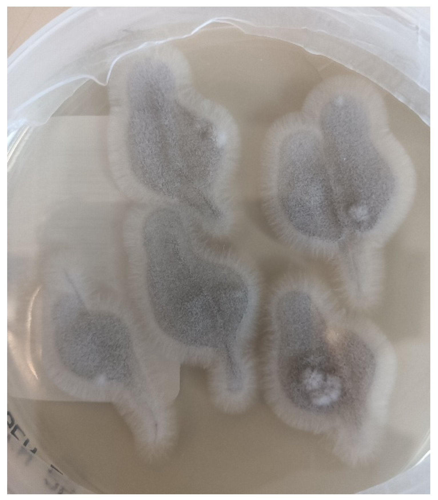
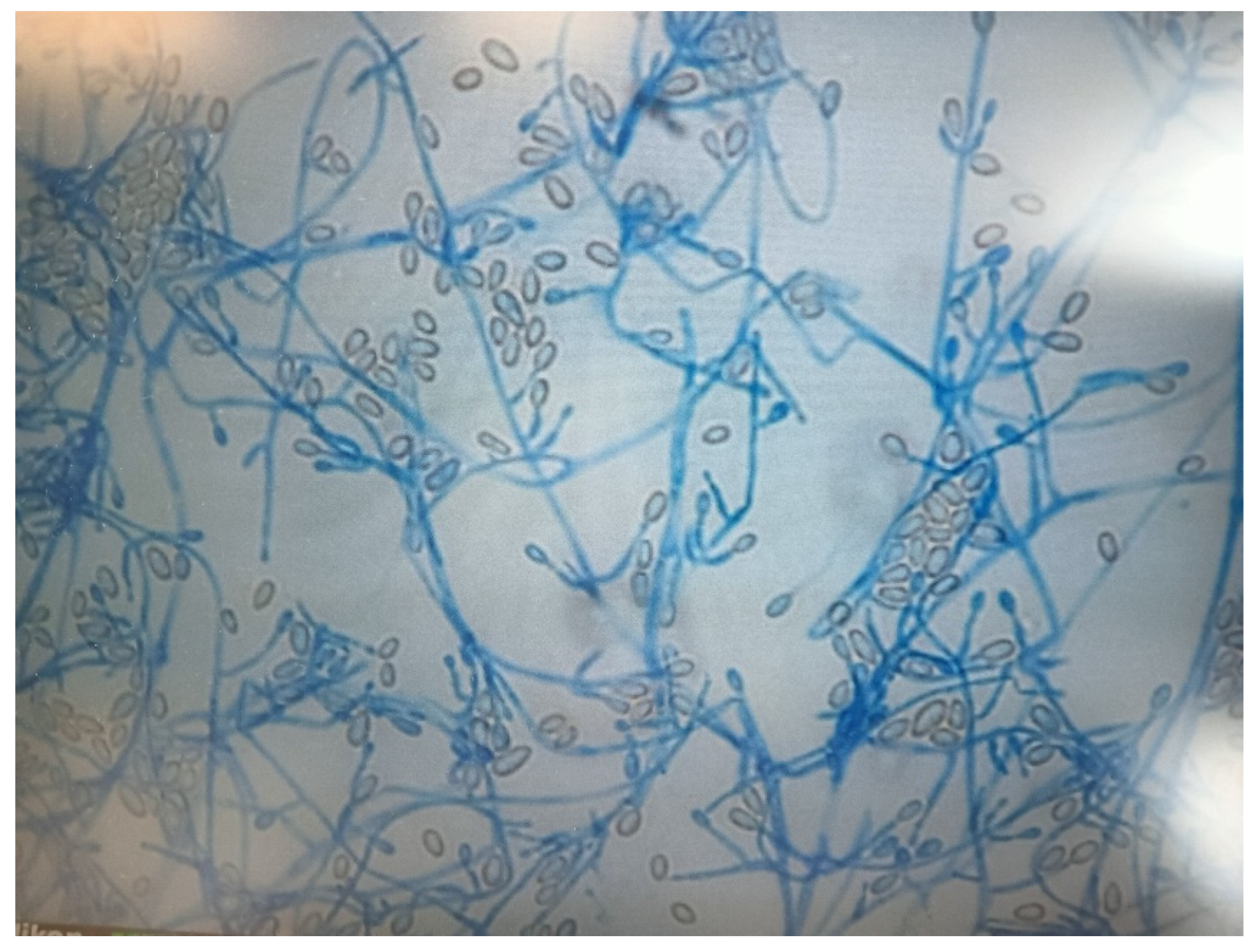
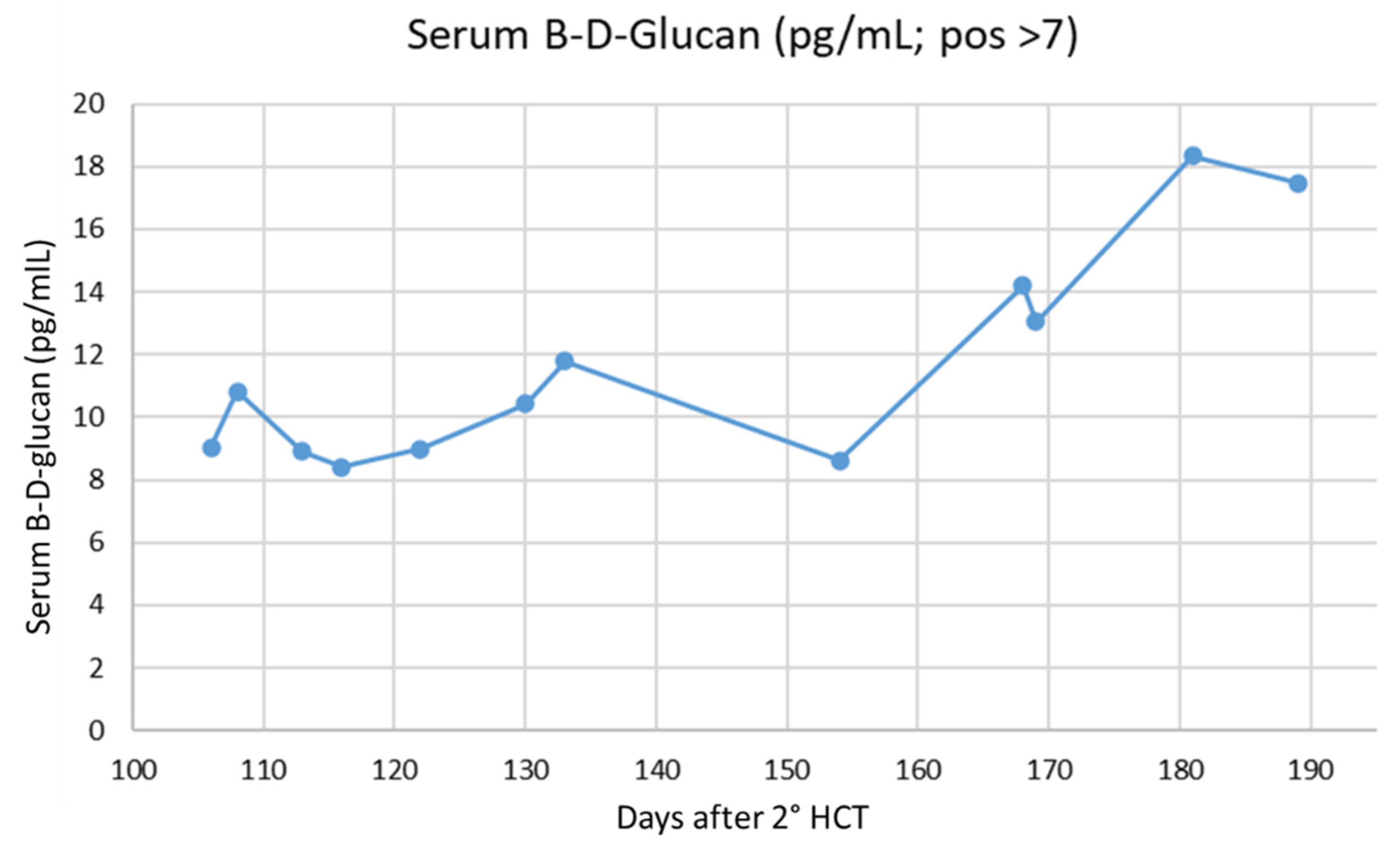
2. Case Presentation

A 5-year-old boy was diagnosed with X-linked CGD by genetic testing (a pathogenic variant of the CYBB gene) after a history characterized by multiple lymphadenopathies and recurrent mycobacterial and Gram-positive infections (S. aureus) with lung involvement. At the diagnosis of CGD, the patient was receiving anti-tubercular therapy because of a previous Mycobacterium tuberculosis complex positivity obtained from a lymph node drainage. After the diagnosis of CGD, the patient was started on cotrimoxazole prophylaxis for Pneumocytis jirovecii pneumonia and antifungal prophylaxis with liposomal amphotericin B (3 mg/kg/dose, twice a week; then 5 mg/kg/dose, once a week). This last was preferred to azole antifungals to prevent drug-to-drug interactions with the concomitant anti-tubercular therapy (isoniazid, rifampin, ethambutol, and pyrazinamide).
After 13 months from CGD diagnosis, the patient underwent allogeneic HCT from an unrelated donor (male donor, 45 years, recipient/donor HLA A, B, C, DR match: 7/8). The conditioning regimen and the graft-versus-host disease (GVHD) prophylaxis are reported in Table 1. The patient received a bone marrow graft containing 5 × 10⁶/kg CD34+ cells. The HCT was complicated by prolonged fever (from day +3 to +47 post-HCT), treated empirically with broad-spectrum antibiotics (meropenem, amikacin, and teicoplanin; then switched to ceftazidime/avibactam and Fosfomycin; this last one changed to clindamycin; with transient de-escalation to piperacillin/tazobactam during a phase of clinical improvement) and liposomal amphotericin B (3 mg/kg/dose every 24 h), primary graft failure (GF, day +41), and sepsis by Bacillus cereus (day +69, treated with meropenem and vancomycin). While fully aplastic, the patient developed abscesses in the knee joint and parasternal site (day +34 and +40, respectively). The drainage of the parasternal abscess (day +60) resulted in the isolation of Scedosporium apiospermum. Concomitant serum B-D-glucan tests (Toxinometer MT-6500, FUJIFILM Wako Pure Chemical Corporation) resulted in negative (<4 pg/mL). The patient was treated empirically with voriconazole (load: 9 mg/kg/dose every 12 h for two doses, then 8 mg/kg/dose every 12 h) and five granulocyte transfusions (GTX) using the father as a donor. Therapeutic drug monitoring (TDM) of voriconazole was performed (target trough level: 1–5 μg/mL). At the in vitro susceptibility test (broth dilution, Thermo Scientific (Waltham, MA, USA), Sensititre TM YeastOne ITAMYUCC, manufactured by TREK Diagnostic Systems, Units 17-19 Birches Industrial Estate, East Grinstead, West Sussex, Crawley, UK), a MIC of 0.25 resulted in voriconazole (Table 2).
A second HCT was performed at +80 days from the first HCT, using the same donor. The conditioning regimen and the GVHD prophylaxis are reported in Table 1. The patient received a bone marrow graft infusion containing 3.65 × 10⁶/kg CD34+ cells. This HCT was complicated by hepatic veno-occlusive disease, cytomegalovirus infection, respiratory syncytial virus, influenza infections, recurrent pericardial effusion (microbiologically negative, including fungal cultures), and grade III skin GVHD, treated with intravenous steroids and ruxolitinib. Nonetheless, with the gradual myeloid recovery, fungal abscesses in the knee joint and parasternal progressively improved. Neutrophil engraftment occurred at day + 29 post-second HCT. The patient was discharged from the hospital on day +88 from the second HCT, still on oral voriconazole therapy (9 mg/kg/dose every 12 h). Due to poor graft function, the patient needed occasional platelets, red blood cell transfusions, and granulocyte-colony stimulating factor administrations.
At day +105, the child was admitted to the hospital due to fever, headache, and altered level of consciousness. At that time, GVHD prophylaxis was with ruxolitinib twice a week and prednisone (on tapering). Antifungal therapy had been switched to Amphotericin B (3 mg/kg/dose, twice a week) about two weeks before, due to hepatic toxicity attributed to voriconazole. Blood counts were WBC 1.57 × 10⁹/L (PMN 1 × 10⁹/L) and platelets 16 × 109/L. Magnetic resonance imaging revealed multiple bilateral cerebral abscesses in the cortical-subcortical regions. The serum B-D-glucan test was positive (9.05 pg/mL, positive if >7), while serum galactomannan resulted in negative. In the hypothesis of a possible mycosis [8], the patient started voriconazole (load: 9 mg/kg/dose every 12 h for two doses, then 8 mg/kg/dose every 12 h), then micafungin was added (2 mg/kg/dose every 24 h), and immunosuppression was reduced. The analysis of cerebrospinal fluid obtained from a spinal tap showed low glucose levels, elevated proteins, and white blood cell counts. Microbiological investigations (including fungal culture) on cerebrospinal fluid resulted in negative, except for the B-D-glucan test (7.35 pg/mL, at day + 114). At the following cerebrospinal fluid examination (day +126), fungal culture resulted in negative, as well as the B-D-glucan. Therapeutic drug monitoring of voriconazole was periodically performed to optimize the drug dose, with a target trough level between 1 and 5 μg/mL. Noteworthy, from day +118 to +139, voriconazole was temporarily switched to posaconazole (6.5 mg/kg/day) due to gastrointestinal toxicity (vomiting). Meanwhile, chimerism analysis, performed both on peripheral blood and bone marrow, showed full donor chimerism, and neutrophil superoxide production at the dihydrorhodamine oxidation assay resulted in normal.
Despite an initial clinical neurological improvement, the patient developed pericardial effusion with hemodynamic instability that required a cardiosurgical intervention of pericardiotomy, pleuro-pericardial window, and positioning of drainage (day +140). The pericardial fluid was negative at microbiological culture (including fungal cultures). In the following days, the child developed a progressive neurological impairment associated with triventricular hydrocephalus that required the positioning of external drainage at day + 172. This time, S. apiospermum was identified by the culture of cerebrospinal fluid (Figure 1 and Figure 2) and confirmed at the Matrix-Assisted Laser Desorption Ionization-Time of Flight Mass Spectrometry (Vitek® MS, bioMérieux, Marcy-l’Étoile, France), along with high B-D-glucan levels in cerebrospinal fluid (173.3 pg/mL). Considering the worsening of S. apiospermum infection despite long-lasting voriconazole therapy, liposomal amphotericin B (5 mg/kg/dose every 24 h) was empirically added as salvage therapy instead of micafungin. Antifungal susceptibility testing is reported in Table 2. Figure 3a,b represent the MRI at day + 183. Unfortunately, in the following days, the patient developed progressive cerebral edema, uncal herniation, coma, and died on day + 193. Noteworthy, fungal susceptibility testing for antifungal combinations was performed at a national reference center. For the determination of the interaction, a microdilution checkerboard technique based on the Clinical and Laboratory Standards Institute (CLSI) reference method for antifungal susceptibility testing was used [9], showing that the more efficient combination in vitro seemed to be voriconazole plus micafungin. Still, unfortunately, this result arrived only post-mortem.

3. Discussion

We reported the case of a fatal fungal infection with CNS involvement by S. apiospermum. Few studies reported cases of scedosporiosis in HCT recipients. In 2005, Husain et al. analyzed 23 cases of S. spp. infections (including 9 Lomentospora prolificans infections, previously named S. prolificans) in HCT recipients. Overall, 69% of infections were disseminated with a mortality rate of 68%. Antifungal therapy with voriconazole was associated with a trend of better survival, compared with amphotericin B [10]. A fatal CNS infection by S. apiospermum after HCT was described in 2002 and treated with amphotericin B (voriconazole not available) [11]. A rhinoencephalitis due to S. apiospermum, 8 months after allogeneic-HCT, was reported in a 17-year-old boy with concomitant Rhizomucor pneumonia. The patient was treated with posaconazole and liposomal amphotericin B, but he had no response and, subsequently, experienced a fatal outcome [12]. Moreover, a 65-year-old HCT recipient with acute myeloid leukemia (AML) developed a disseminated scedosporiosis involving the lungs and CNS, probably from a foot source (toe wound). A combination therapy with voriconazole and liposomal amphotericin B successfully treated the infection, despite physical and neurological sequelae that took a long recovery [13]. Interestingly, a 12-year-old boy with CGD and pericardial infection by S. apiospermum, initially treated with voriconazole and GTX, underwent HCT to control the persistent and life-threatening infection. Radiologic investigations documented a post-transplant resolution of the S. spp. infection [14]. Finally, ocular complications have also been reported after HCT, including a case of chorioretinitis associated with disseminated scedosporiosis with osteomyelitis, endocarditis, and brain abscess, successfully treated with voriconazole, amphotericin B, and surgery, and a case of endophthalmitis after a second allogeneic HCT for AML [15,16]. Studies and case reports of scedosporiosis are summarized in Table 3.
In this case, we reported several cofactors predisposing to the mycosis. First, the long-lasting defective function of granulocytes was due to the underlying disease that resulted in the defective bactericidal and fungicidal activity [17]. Moreover, the prolonged aplasia that had followed HCT, due to the primary GF, and the deep post-transplant immunosuppressive therapy used as GVHD prophylaxis played a role in the development and dissemination of the infection. Finally, the poor graft function might have contributed to the scarce control of the disease.
HCT is a potentially definitive treatment for CGD. A recent multicenter study demonstrated that allogeneic HCT could reduce the burden of the disease, improving growth and nutrition, resolving infections and inflammatory disease, and reducing rates of antimicrobial prophylaxis or corticosteroid use. The study reported a 3-year overall survival of 82% and event-free survival of 69% [18]. However, in patients with reduced performance status, high inflammation before HCT, and HLA-mismatched donors, there were augmented rates of both GF and chronic GVHD [18,19].
We also found that abnormal results of the B-D-glucan test were associated with the occurrence and worsening of scedosporiosis (serum B-D-glucan levels are reported in Figure 4). Indeed, the B-D-glucan test was already positive at the time of the first central nervous system symptoms (9.05 pg/mL, positive if >7). This is consistent with a recent meta-analysis that reported a sensitivity of the B-D-glucan test for the diagnosis of scedosporiosis of 81.5%. The meta-analysis showed that positive results of B-D-glucan preceded the diagnosis by conventional methods (i.e., culture, histopathology) in 94% of cases. Finally, the combined use of the B-D-glucan test and galactomannan may help differentiate between invasive mold infections [20]. The B-D-glucan test in cerebrospinal fluid was also helpful, being the only positivity detected during the first examinations (Table 4).
Despite the parasternal and knee joint abscesses by Scedosporium improving with voriconazole therapy, the patient died from the cerebral localization of the mycosis. Unfortunately, we do not know if cerebral involvement was present since the initial diagnosis of scedosporiosis, though neurologically asymptomatic, because neither a head MRI nor computed tomography was performed.
The recommended treatment for scedosporiosis is voriconazole, which showed better outcomes compared with amphotericin B. Therefore, the use of amphotericin B monotherapy is discouraged whenever voriconazole is available [3,7,21]. Alternatives are posaconazole and voriconazole-based antifungal combinations, including voriconazole plus terbinafine or echinocandin or amphotericin B [3,21]. Instead, neither isavuconazole nor itraconazole is currently recommended [7]. Salvage therapy can be necessary in case of refractory infection or toxicity/intolerance, using a voriconazole-based antifungal combination or posaconazole, in case of intolerance or resistance to VCZ [7]. Due to the pharmacokinetic unpredictability and the inter-patient variability, the TDM is recommended during azole use to guide the dosing and avoid treatment failure and toxicities [3]. In our case, the antifungal susceptibility testing confirmed the sensitivity of voriconazole. However, due to suspected drug toxicities, we suspended voriconazole twice. In the first case for hepatic toxicity, since parasternal and knee joint abscesses were clinically and radiologically improved, voriconazole was temporarily switched to amphotericin B. In the second case, voriconazole was temporarily switched to posaconazole due to gastrointestinal toxicity. Moreover, we hypothesize that the non-response to antifungals might be related to profound and prolonged immunosuppression and insufficient concentration in the cerebral lesions.
New promising antifungal drugs are under investigation. For scedosporiosis, olorofilm and fosmanogepix have shown activity, both in vitro and in animal studies [7]. Olorofilm belongs to the new orotomide class that inhibits the fungal dihydroorotate dehydrogenase responsible for fungal pyrimidine and DNA synthesis. Fosmanogepix is a pro-drug of manogepix, a glycosylphosphatidylinositol anchor inhibitor [3]. Therefore, we expect to have new weapons against fungal infections in the near future.

4. Conclusions

Invasive fungal infections by Scedosporium spp. are an emerging global issue due to the high morbidity and mortality rates, especially in HCT recipients. In this case, we conclude that the long-lasting defective function of PMN due to CGD, the prolonged aplasia due to primary graft failure, and deep post-transplant immunosuppression have been significant factors in the occurrence of this rare mycosis. What we have learned is that the screening of CNS localization by MRI in severely immunocompromised patients with Scedosporium infections would be useful to define the extension of the disease, even though neurologically asymptomatic. Moreover, we found that the B-D-glucan test was useful for diagnosing and monitoring invasive scedosporiosis. Finally, the introduction of new antifungal drugs in the next years may improve the prognosis of this severe infection.

Author Contributions

Conceptualization, C.G. and S.C.; writing—original draft preparation, C.G. and S.C.; writing—review and editing, C.G., M.C., G.C., E.B., M.P.E., V.P., V.V., A.Z., A.S., D.G. and S.C. All authors have read and agreed to the published version of the manuscript.

Funding

This research received no external funding.

Institutional Review Board Statement

Ethical review and approval were waived for this manuscript because all procedures and treatments applied in the management of the patient were part of institutional clinical practice, and no experimental procedures or treatments were used.

Informed Consent Statement

Parent informed consent was obtained, and reported data were treated according to the Italian regulation for patient confidentiality and good clinical practice.

Data Availability Statement

The original contributions presented in this study are included in the article. Further inquiries can be directed to the corresponding author.

Acknowledgments

We would like to thank R. Torelli and M. Sanguinetti, Dipartimento di Scienze di Laboratorio e Infettivologiche, Fondazione Policlinico Universitario A. Gemelli IRCSS, Rome, Italy, for the performance and the information about the combined antifungal susceptibility testing on S. apiospermum.

Conflicts of Interest

The authors declare no conflicts of interest.

Abbreviations

The following abbreviations are used in this manuscript:
AMLacute myeloid leukemia
CGDchronic granulomatous disease
GFgraft failure
GTXgranulocyte transfusion
GVHDgraft-versus-host disease
HCThematopoietic cell transplant
S.scedosporium
TDMTherapeutic Drug Monitoring

References

  1. Fang, W.; Wu, J.; Cheng, M.; Zhu, X.; Du, M.; Chen, C.; Liao, W.; Zhi, K.; Pan, W. Diagnosis of invasive fungal infections: Challenges and recent developments. J. Biomed. Sci. 2023, 30, 42. [Google Scholar] [CrossRef] [PubMed] [PubMed Central]
  2. McCarthy, M.W.; Kontoyiannis, D.P.; Cornely, O.A.; Perfect, J.R.; Walsh, T.J. Novel Agents and Drug Targets to Meet the Challenges of Resistant Fungi. J. Infect. Dis. 2017, 216 (Suppl. S3), S474–S483. [Google Scholar] [CrossRef] [PubMed]
  3. Bupha-Intr, O.; Butters, C.; Reynolds, G.; Kennedy, K.; Meyer, W.; Patil, S.; Bryant, P.; Morrissey, C.O.; Australasian Antifungal Guidelines Steering Committee. Consensus guidelines for the diagnosis and management of invasive fungal disease due to moulds other than Aspergillus in the haematology/oncology setting, 2021. Intern. Med. J. 2021, 51 (Suppl. S7), 177–219. [Google Scholar] [CrossRef] [PubMed]
  4. Cortez, K.J.; Roilides, E.; Quiroz-Telles, F.; Meletiadis, J.; Antachopoulos, C.; Knudsen, T.; Buchanan, W.; Milanovich, J.; Sutton, D.A.; Fothergill, A.; et al. Infections caused by Scedosporium spp. Clin. Microbiol. Rev. 2008, 21, 157–197. [Google Scholar] [CrossRef] [PubMed] [PubMed Central]
  5. Lamaris, G.A.; Chamilos, G.; Lewis, R.E.; Safdar, A.; Raad, I.I.; Kontoyiannis, D.P. Scedosporium infection in a tertiary care cancer center: A review of 25 cases from 1989–2006. Clin. Infect. Dis. 2006, 43, 1580–1584. [Google Scholar] [CrossRef]
  6. Seidel, D.; Meißner, A.; Lackner, M.; Piepenbrock, E.; Salmanton-García, J.; Stecher, M.; Mellinghoff, S.; Hamprecht, A.; Durán Graeff, L.; Köhler, P.; et al. Prognostic factors in 264 adults with invasive Scedosporium spp. and Lomentospora prolificans infection reported in the literature and FungiScope®. Crit. Rev. Microbiol. 2019, 45, 1–21, Erratum in: Crit. Rev. Microbiol. 2019, 45, 238. [Google Scholar] [CrossRef] [PubMed]
  7. Boutin, C.A.; Luong, M.L. Update on therapeutic approaches for invasive fungal infections in adults. Ther. Adv. Infect. Dis. 2024, 11, 20499361231224980. [Google Scholar] [CrossRef] [PubMed] [PubMed Central]
  8. Donnelly, J.P.; Chen, S.C.; Kauffman, C.A.; Steinbach, W.J.; Baddley, J.W.; Verweij, P.E.; Clancy, C.J.; Wingard, J.R.; Lockhart, S.R.; Groll, A.H.; et al. Revision and Update of the Consensus Definitions of Invasive Fungal Disease From the European Organization for Research and Treatment of Cancer and the Mycoses Study Group Education and Research Consortium. Clin. Infect. Dis. 2020, 71, 1367–1376. [Google Scholar] [CrossRef] [PubMed]
  9. Bellio, P.; Fagnani, L.; Nazzicone, L.; Celenza, G. New and simplified method for drug combination studies by checkerboard assay. MethodsX 2021, 8, 101543. [Google Scholar] [CrossRef]
  10. Husain, S.; Muñoz, P.; Forrest, G.; Alexander, B.D.; Somani, J.; Brennan, K.; Wagener, M.M.; Singh, N. Infections due to Scedosporium apiospermum and Scedosporium prolificans in transplant recipients: Clinical characteristics and impact of antifungal agent therapy on outcome. Clin. Infect. Dis. 2005, 40, 89–99. [Google Scholar] [CrossRef] [PubMed]
  11. Safdar, A.; Papadopoulos, E.B.; Young, J.W. Breakthrough Scedosporium apiospermum (Pseudallescheria boydii) brain abscess during therapy for invasive pulmonary aspergillosis following high-risk allogeneic hematopoietic stem cell transplantation. Scedosporiasis and recent advances in anti-fungal therapy. Transpl. Infect. Dis. 2002, 4, 212–217. [Google Scholar] [CrossRef] [PubMed]
  12. Marques, D.S.; Pinho Vaz, C.; Branca, R.; Campilho, F.; Lamelas, C.; Afonso, L.P.; Jacome, M.; Breda, E.; Monteiro, E.; Campos Júnior, A. Rhizomucor and Scedosporium infection post hematopoietic stem-cell transplant. Case Rep. Med. 2011, 2011, 830769. [Google Scholar] [CrossRef] [PubMed] [PubMed Central]
  13. Marinovic, D.A.; Bhaimia, E.; Forrest, G.N.; LaRue, R.; Nathan, S.; Ustun, C.; Ward, A. Scedosporium infection disseminated “from toe to head” in allogeneic stem cell transplant recipient: A case report. BMC Infect. Dis. 2023, 23, 353. [Google Scholar] [CrossRef]
  14. Parta, M.; Hilligoss, D.; Kelly, C.; Kwatemaa, N.; Theobald, N.; Malech, H.; Kang, E.M. Haploidentical Hematopoietic Cell Transplantation with Post-Transplant Cyclophosphamide in a Patient with Chronic Granulomatous Disease and Active Infection: A First Report. J. Clin. Immunol. 2015, 35, 675–680. [Google Scholar] [CrossRef] [PubMed] [PubMed Central]
  15. Stoyanova, E.I.; Riemens, A.; Lokhorst, H.M.; te Boome, L.; Rothova, A. Absence of intraocular infections after hematopoietic stem cell transplantation at a single center: The experience with current preventive regimens. Ocul. Immunol. Inflamm. 2014, 22, 116–120. [Google Scholar] [CrossRef] [PubMed]
  16. Narita, K.; Ikeda, D.; Matsue, K. Scedosporium endophthalmitis in a patient with second allogeneic stem cell transplantation for acute myeloid leukemia. Transpl. Infect. Dis. 2024, 27, e14397. [Google Scholar] [CrossRef] [PubMed]
  17. Bassiri-Jahromi, S.; Doostkam, A. Fungal infection and increased mortality in patients with chronic granulomatous disease. J. Mycol. Med. 2012, 22, 52–57. [Google Scholar] [CrossRef] [PubMed]
  18. Leiding, J.W.; Arnold, D.E.; Parikh, S.H.; Logan, B.R.; Marsh, R.A.; Griffith, L.M.; Wu, R.; Kidd, S.; Mallhi, K.K.; Chellapandian, D.; et al. Genotype, oxidase status, and preceding infection or autoinflammation do not affect allogeneic HCT outcomes for CGD. Blood 2023, 142, 2105–2118. [Google Scholar] [CrossRef] [PubMed]
  19. Güngör, T. HCT alleviates disease burden in CGD. Blood 2023, 142, 2043–2045. [Google Scholar] [CrossRef] [PubMed]
  20. Lamoth, F.; Nucci, M.; Fernandez-Cruz, A.; Azoulay, E.; Lanternier, F.; Bremerich, J.; Einsele, H.; Johnson, E.; Lehrnbecher, T.; Mercier, T.; et al. Performance of the beta-glucan test for the diagnosis of invasive fusariosis and scedosporiosis: A meta-analysis. Med. Mycol. 2023, 61, myad061. [Google Scholar] [CrossRef] [PubMed] [PubMed Central]
  21. Hoenigl, M.; Salmanton-García, J.; Walsh, T.J.; Nucci, M.; Neoh, C.F.; Jenks, J.D.; Lackner, M.; Sprute, R.; Al-Hatmi, A.M.S.; Bassetti, M.; et al. Global guideline for the diagnosis and management of rare mould infections: An initiative of the European Confederation of Medical Mycology in cooperation with the International Society for Human and Animal Mycology and the American Society for Microbiology. Lancet Infect. Dis. 2021, 21, e246–e257, Erratum in: Lancet Infect. Dis. 2021, 21, e81. [Google Scholar] [CrossRef] [PubMed]
Figure 1. Scedosporium apiospermum on Sabouraud Dextrose agar culture (photography of culture plate, plate diameter: 8.5 cm).
Jof 11 00270 g001
Figure 2. Scedosporium apiospermum at microscopic examination (lactophenol cotton blue staining; microscope objective 25/0.63, eyepiece GF 12.5×/20).
Jof 11 00270 g002
Figure 3. Brain MRI ((a,b): different slices), day + 183 post-2nd HCT.
Jof 11 00270 g003
Figure 4. Serum B-D-glucan test results (Wako turbidimetric assay).
Jof 11 00270 g004
Table 1. Conditioning regimen and GVHD prophylaxis used in the HCT.
Drug, DoseTiming
First HCT
Conditioning regimenBusulfan, 1 mg/kg/dose for 16 doses over 5 daysFrom day −11 to day −7
Fludarabine, 40 mg/m2/day for 4 daysFrom day −2 to −5
ATG Genzyme®, 1.5 mg/kg/dose for 6 doses in totalFrom day −7 to day −3
GVHD prophylaxisCyclophosphamide, 50 mg/kgDays +4 and +5 post-HCT
Cyclosporine, 3 mg/kg/dayFrom day +5
Mycophenolate mofetil, 60 mg/kg/day in 3 doses From day +5
Second HCT
Conditioning regimenTotal body irradiation, 4 Gy, single fractionDay −7
Fludarabine, 35 mg/m2/dayFrom day −6 to day −3
Cyclophosphamide, 30 mg/kg/dayFrom day −6 to day −3
ATG Neovii®, 12.5 mg/kg/day Days −2 and −3
GVHD prophylaxisMycophenolate mofetil, 60 mg/kg/dayFrom day +1
Tacrolimus, 0.1 mg/kg intravenously, modulated according to blood levelsFrom day −1
ATG, anti-thymocyte globulin; GVHD, graft-versus-host disease; HCT, hematopoietic cell transplant.
Table 2. Antifungal susceptibility testing from parasternal abscess drainage (A) and cerebrospinal fluid (B) *.
MIC (A)MIC (B)Antifungal Drug
0.250.25Voriconazole
0.51Posaconazole
11Itraconazole
12Isavuconazole
88Amphotericin
88Micafungin
* CLSI interpretation not available.
Table 3. Summary of available evidence of scedosporiosis after HCT.
ReferenceOutcomeTreatmentOrgan InvolvementAge (Years)N. of Cases
[10]Mortality rate 68%AmB/ICZ/VCZCNS 36%
Pulmonary 40%
Skin 38%
Blood 25%
Disseminated 69%
(mean +/− SD)
32.9 +/− 3.3
23 (of which 9
L. prolificans)
[11]DiedAmBCNS341
[12]DiedPCZ + AmBRhinoencephalitis171
[13]SurvivedVCZ + AmBDisseminated
Pulmonary
CNS
651
[15]SurvivedVCZ + AmBOcular
Bone
Heart
CNS
591
[16]DiedIntravitreal VCZOcular
CNS
581
AmB, amphotericin B; CNS, central nervous system; ICZ, itraconazole; PCZ, posaconazole; VCZ, voriconazole; SD, standard deviation.
Table 4. B-D-glucan test in cerebrospinal fluid (Wako turbidimetric assay).
BDG (pg/mL, Pos > 7)Days Post-2nd HCT
7.35114
3126
173172
26.4189
Disclaimer/Publisher’s Note: The statements, opinions and data contained in all publications are solely those of the individual author(s) and contributor(s) and not of MDPI and/or the editor(s). MDPI and/or the editor(s) disclaim responsibility for any injury to people or property resulting from any ideas, methods, instructions or products referred to in the content.

