Case Report

Erythema Nodosum Associated with Kerion: A Case Series and Narrative Review of the Literature

by
Teerapong Rattananukrom
1,
Isaías Uh-Sánchez
2,
Carlos Atoche-Dieguez
3,
Nixma Eljure
3,
Carlos Garcia-Rementeria
4 and
Roberto Arenas
5,*
1
Division of Dermatology, Department of Medicine, Faculty of Medicine Ramathibodi Hospital, Mahidol University, Bangkok 10400, Thailand
2
Servicio de Dermatología, Hospital General Dr. Adolfo López Mateos, Mexico City 01030, Mexico
3
Centro Dermatológico de Yucatán, Mérida 97000, Mexico
4
Southwestern Dermatology, Oklahoma City, OK 73139, USA
5
Mycology Section, “Dr. Manuel Gea Gonzalez” General Hospital, Mexico City 14080, Mexico
*
Author to whom correspondence should be addressed.
J. Fungi 2025, 11(2), 103; https://doi.org/10.3390/jof11020103
Submission received: 17 September 2024 / Revised: 19 December 2024 / Accepted: 2 January 2025 / Published: 29 January 2025
(This article belongs to the Section Fungal Pathogenesis and Disease Control)

Abstract

:
Kerion is a form of inflammatory tinea capitis, a fungal infection caused by various zoophilic, geophilic, and anthropophilic pathogens. Erythema nodosum (EN), a form of septal panniculitis, can be considered a dermatophyte id reaction that occurs outside the primary site of dermatophyte infection. The association between EN and kerion is rarely reported, with most cases following Trichophyton mentagrophytes scalp infections. Here, we describe three cases of EN associated with kerion caused by T. mentagrophytes, successfully treated with itraconazole or griseofulvin plus prednisone. Additionally, we conducted a narrative review of the literature, identifying 23 reported cases of EN associated with kerion on PubMed. The most commonly reported fungus was T. mentagrophytes (78.25%). In 52.17% of cases, patients developed EN after initiating antifungal treatment, with a mean onset time of 11.58 days (SD 7.3). Griseofulvin remains a mainstay treatment. The mean time for EN resolution was 8.31 days (SD 4.15), and the median duration of treatment for kerion leading to a complete response was 6 weeks (IQR 6–8). Scarring alopecia is a common sequela following kerion, and the use of corticosteroids has been recommended as adjunct therapy to minimize the risk of scarring.

1. Introduction

Erythema nodosum (EN), a septal panniculitis, is associated with various diseases, infections, drugs and systemic conditions. Clinically, EN is typically characterized by episodes of painful subcutaneous nodules, predominantly located on the pretibial regions, although it may also appear on the upper extremities. EN is regarded as a dermatophyte id reaction (dermatophytid), manifesting at a site distinct from the primary dermatophyte infection [1,2,3,4].
Kerion is a severe inflammatory variant of fungal infection, most frequently affecting the scalp. This condition predominantly occurs in children from rural areas and arises from zoophilic, geophilic and anthropophilic pathogens, including Trichophyton and Microsporum spp. M. canis is the most common causative agent in Europe, China, and South America, while T. tonsurans is more commonly found in North America and the UK [5,6]. Kerions are typically marked by a raised, spongy, and swollen area of skin, often accompanied by pustules, crusting, and hair loss. The condition can be painful and may lead to scarring if left untreated.
Kerion and EN are frequently linked to an intense immune response to the fungal infection. The co-occurrence of EN and kerion is infrequently reported, with only 23 cases documented in the literature available on PubMed. Most of these cases develop following scalp infections caused by T. mentagrophytes, often emerging after antifungal therapy, however, EN may also present before treatment begins [7]. In this report, we describe three cases kerion-associated EN due to T. mentagrophytes, emphasizing the variability in clinical presentation and treatment outcomes.

2. Materials and Methods

2.1. Case Series

We report three cases of EN-associated kerion caused by T. mentagrophytes. Species identification was performed using conventional microbiological techniques, including growth on Sabouraud chloramphenicol gentamicin agar, followed by phenotypic identification. However, a limitation of our case series is the lack of molecular identification to confirm the fungal species. Written informed consent was obtained from all patients prior to their participation in this research.

2.2. Review of the Literature

We conducted a narrative review of the literature using PubMed with the search terms: ‘Erythema nodosum’ AND ‘Kerion’ OR ‘(Inflammatory) Tinea Capitis’. Relevant articles published between 1948 and 2024 were screened, and only confirmed cases of EN-associated kerion were included. The review incorporated studies published in English, Spanish, Italian, and French. Data extracted included demographic characteristics, clinical presentation, time to onset of EN, fungal species involved, treatment regimens, and clinical outcomes following antifungal therapy. Diagnostic criteria for an EN-type dermatophytid reaction included the following: (I) confirmed dermatophytosis, (II) eruption occurring at a site distant from the primary fungal infection, and (III) resolution of the eruption after antifungal treatment [8]. Based on these criteria, the patient was diagnosed with EN as a dermatophytid reaction. A ‘complete response’ was defined as patients achieving full recovery after antifungal therapy. The time to develop EN was categorized as occurring either before or after the initiation of antifungal treatment.
Demographic data, baseline characteristics, clinical findings, and outcomes were analyzed using descriptive statistics. Categorical variables were presented as numbers and percentages, while continuous variables were reported as means and standard deviations (SD) or as medians and interquartile ranges (IQRs).

3. Results

3.1. Case 1

A healthy 8-year-old boy presented with a one-week history of painful lesions on the scalp, neck, and legs. On physical examination, a 3 cm tender, erythematous, and edematous plaque with well-defined borders and crusting was observed on the occipital region of the scalp (Figure 1a). Additionally, the patient exhibited tender cervical lymphadenopathy and multiple erythematous, painful subcutaneous nodules, measuring 1–2 cm in diameter, on both shins (Figure 1b). The patient reported no history of animal contact. A clinical diagnosis of kerion and EN was made. Potassium hydroxide (KOH) examination of the scalp lesion revealed numerous ecto-endothrix parasitizations of hairs, with a positive hair perforation test. Fungal culture showed white to cream-colored, cottony, mounded colonies characteristic of T. mentagrophytes (Figure 1c,d). The patient was treated with griseofulvin at a dose of 25 mg/kg/day for 30 days. His mother declined to administer prednisone due to concerns about side effects. EN resolved within one week. At the one-month follow-up, the scalp lesion had resolved, leaving only a small area of alopecia.

3.2. Case 2

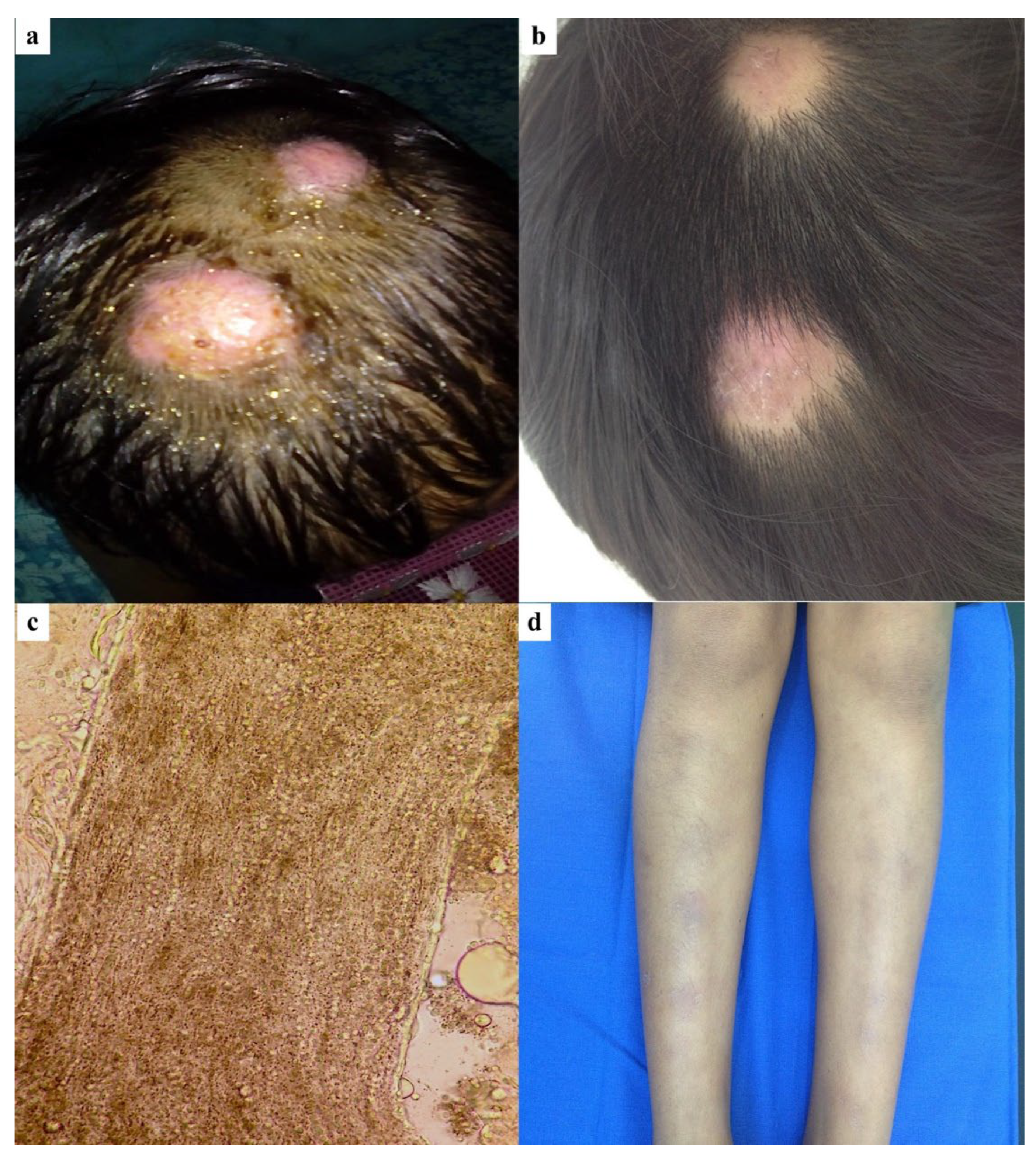
A healthy 8-year-old girl presented for evaluation with a 15-day history of tender, erythematous, and edematous plaques located on the left occipital and parietal regions of the scalp (Figure 2a). She denied any history of animal contact. The plaques, measuring 2 cm in diameter, displayed irregular borders, broken hairs, and crusting. KOH examination revealed hairs with ecto-endothrix parasitization (Figure 2c), with a positive hair perforation test. Fungal culture showed white to cream-colored, cottony colonies and confirmed the presence of T. mentagrophytes. Treatment was initiated with itraconazole at 5 mg/kg/day and prednisone at a dose of 1 mg/kg/day. During the 1-week follow-up, the patient developed multiple erythematous, painful subcutaneous nodules on both shins, suggestive of EN (Figure 2d). The treatment regimen, which included a tapering schedule for prednisone, was continued alongside itraconazole. After an additional 2 weeks, there was a 90% improvement in both the scalp and EN lesions (Figure 2b). At the 6-week follow-up, all scalp lesions had resolved, though focal areas of alopecia remained.

3.3. Case 3

A healthy 5-year-old girl presented with a one-week history of a painful lesion on the occipital region of the scalp. The patient reported no history of animal contact. On physical examination, a tender, erythematous–edematous plaque with irregular borders and crusting was observed (Figure 3a). Additionally, the patient had multiple erythematous, painful subcutaneous nodules on both shins, suggestive of EN (Figure 3c), along with scattered erythematous, scaly plaques on the trunk and face. KOH examination of the scalp lesion revealed hairs consistent with ecto-endothrix parasitization, with a positive hair perforation test. Fungal culture showed white to cream-colored, cottony colonies characteristic of T. mentagrophytes. KOH examination of the facial lesions was negative for fungal elements. Treatment was initiated with oral itraconazole at 5 mg/kg/day and prednisone at 1 mg/kg/day, following a tapering schedule. At the two-week follow-up, the lesions on the trunk, face, and shins had resolved, and the scalp lesion showed complete healing within six weeks, leaving a small plaque of alopecia (Figure 3b).

3.4. Literature Review

A narrative review of the literature identified a total of 18 published studies, encompassing 23 cases from various countries, including Italy, Tunisia, Morocco, the UK, Spain, and the USA. The majority of cases reported before 2000 originated from Spain and the USA, whereas those published after 2000 predominantly came from Italy and Tunisia. Of the 23 cases, 16 patients were male, and 7 were female, with a median age of 8 years (IQR 6.5–9). All patients presented with either painful or painless erythematous patches or plaques, along with boggy lesions on the scalp, typically accompanied by varying degrees of pus, hair loss, and crusting, commonly referred to as “kerion” (Table 1).
The median time to onset of kerion was 14.5 days (IQR 13.75–28.5). The most common locations of kerion were the scalp (34.78%), vertex (26%), and occipital area (17.39%). Additionally, 26% of patients exhibited lymphadenitis, with cervical lymphadenitis being the most frequent type. A history of pet contact was reported in 30.43% of cases, while 17.39% of patients denied any history of pet exposure. In 52.18% of the cases, data regarding pet contact history were unavailable (Table 2).
Regarding the etiological agent, T. mentagrophytes was identified as the most common causative organism in 18 out of 23 cases (78.25%). Other identified agents included M. canis (n = 1), Epidermophyton floccosum (n = 1), and T. verrucosum (n = 1). In one case, the species could not be identified, with only mega-sporic parasitism observed. Additionally, hair direct examination was performed in only four cases, all of which revealed ecto-endothrix parasitism.
Regarding the development of EN-type dermatophytid reactions, 43.49% of patients developed EN before initiating antifungal therapy, while 52.17% developed EN after receiving antifungal treatment, with a mean onset time of 11.58 days (SD 7.3). The most common location of EN was the shins or lower legs (43.48%), followed by the legs (34.78%). Skin biopsy confirmed EN in only six cases, while the remaining cases were diagnosed based on clinical presentation and previously mentioned diagnostic criteria. Two patients exhibited not only typical EN but also erythematous papular lesions on the ears, abdomen, and back, as well as erythematous patches on the trunk, suggesting a double dermatophytid reaction. The mean healing time of EN following antifungal therapy was 8.31 days (SD 4.15).
Griseofulvin was the most commonly used antifungal agent, administered in 95.65% of cases at a dose of 10–25 mg/kg/day. Only one case was treated with terbinafine. Additionally, two patients received systemic corticosteroids (prednisolone) at a dose of 1 mg/kg/day. Topical antimycotics were added as adjunctive therapy in 43.47% of cases. The median duration of treatment for kerion and complete response was 6 weeks (IQR 6–8). All patients experienced focal scarring alopecia as a sequela.

4. Discussion

Erythema nodosum (EN) is the most common form of acute panniculitis, with an incidence of approximately 1 in 100,000 individuals [7]. It is more prevalent in adults, particularly affecting women between the ages of 15 and 40 years. EN is less frequently observed in children, typically between the ages of 4 and 14 years. The etiology of EN is diverse, involving a range of microorganisms and non-microbial agents, with approximately 30–50% of cases considered idiopathic.
According to the literature, the clinical manifestation of dermatophytid reactions may vary based on the host’s immunological response, presenting either in a localized or generalized form [22]. Approximately 4–5% of patients with dermatophyte infections develop a dermatophytid reaction, which can occur either concurrently with or after the primary infection. In cases of kerion, the dermatophytid reaction is commonly characterized by papules and vesicles on the face, which may subsequently spread to the trunk. Other dermatophytid-associated skin lesions include urticarial papules, follicular papules, erythema multiforme, erythema annulare centrifugum, erysipelas-like dermatitis, generalized exanthematous pustular eruption, and EN [12]. Notably, ‘the ear sign’ is a clinical feature of the id reaction observed in some cases of inflammatory tinea capitis, particularly those caused by T. mentagrophytes. It is characterized by redness, swelling, and pain around the ear, which may accompany typical signs of tinea capitis, such as scalp scaling, hair loss, or pustules.
We present the first case series of kerion reported in Mexico. The patients, aged between 5 and 8 years, included two girls and one boy. The disease onset ranged from 7 to 15 days. All patients exhibited kerion in the occipital region and denied any history of pet contact. One patient also had cervical lymphadenopathy. The causative agent in all cases was T. mentagrophytes, with ecto-endothrix parasitization observed in the hair shafts. Our findings are consistent with previous reports, which primarily involved children, with a mean age of 8 years and a median onset time of 14.5 days.
T. mentagrophytes was the most common pathogen associated with kerion and EN, aligning with previous literature. Notably, all cases in our series exhibited kerion with T. mentagrophytes as the causative agent, characterized by ecto-endothrix parasitization, typically associated with zoonotic pathogens. However, despite the absence of pet contact in our patients, T. mentagrophytes may have the potential for human-to-human transmission. We recommend investigating contact history in all cases to assess possible transmission routes. Another important consideration is that T. indotineae was first identified and described in 2020 as a distinct dermatophyte species, previously classified as a genotype variant of T. mentagrophytes. T. indotineae can also cause tinea capitis. However, a limitation of earlier reports is that species-specific identification methods were not used to distinguish T. mentagrophytes from T. indotineae, as these cases were studied before 2020.
In our case series, all patients presented with EN localized on the shins, which is consistent with previous reports identifying the lower extremities as the most common location for EN. One patient also exhibited EN in combination with erythematous, scaly plaques on the trunk and face, suggestive of a double dermatophytid reaction. Based on the literature review, only two cases of this type of reaction have been previously reported. One of our cases demonstrated a similar double dermatophytid reaction. Additionally, no prior studies have compared the clinical response between EN and double dermatophytid reactions, highlighting an area that warrants further investigation.
The pathogenetic mechanism of EN associated with kerion is not yet fully understood. It is thought to be a hypersensitivity reaction to fungal antigens at a site distant from the primary dermatosis. This immunological response involves the release of fungal antigens from the infection site, followed by their opsonization by antibodies and the spread of T-helper (Th)1 cells and cytokines to other areas of the body [15].
EN development appears to be a delayed hypersensitivity reaction to fungal antigens or auto-antigens triggered by the fungal infection. The correlation between the peak of inflammation and EN suggests that the massive antigenic release from the primary inflammatory response may be a key factor, supporting the hypothesis of reactive T-cells activated by this antigenic load [3,4]. In vivo studies have demonstrated delayed hypersensitivity following intradermal injections of trichophytin, while in vitro studies using leukocyte migration inhibition and lymphocyte transformation tests show that Trichophyton species can elicit significant cellular immune responses and delayed-type hypersensitivity reactions [23].
Llorente et al. demonstrated elevated Th1 cytokine expression in the skin nodules and peripheral blood of most patients with EN [24]. Using semiquantitative reverse transcriptase-polymerase chain reaction (RT-PCR), they examined the messenger RNA (mRNA) expression of Th 1 (interleukin [IL] 2 and interferon-γ [IFN-γ]) and TH2 (IL-4 and IL-10) cytokines in skin biopsies and peripheral blood from 11 patients with EN and 9 healthy controls. Their findings revealed increased Th1 cytokine expression in the skin lesions and peripheral blood of most patients with EN, while no cytokine expression was observed in the skin or blood of the control group.
In a mouse model, Nakamura et al. demonstrated that skin lesions induced by T. mentagrophytes, a zoophilic dermatophyte, are driven by a Th1 response involved in the host defense against dermatophytosis [25]. Similarly, Koga et al. [23] found increased IFN-γ production by peripheral blood mononuclear cells following trichophytin stimulation, with RT-PCR revealing IFN-γ mRNA in tinea lesions. These findings support the hypothesis that skin lesions caused by dermatophytic infections of zoophilic origin, particularly those involving T. mentagrophytes, are the result of a Th1 immune response. This response may also account for the associated erythema nodosum and the spontaneous regression observed in many cases.
Collectively, these findings reinforce the hypothesis that skin lesions caused by T. mentagrophytes infections are mediated by a Th1 response, which plays a critical role in the host’s defense against dermatophytosis.
However, there remains debate about whether EN is due to the deposition of C3 and IgM in venules of the deep dermis and adipose tissue or whether it arises from a delayed hypersensitivity reaction [1]. One hypothesis suggests an antigen-antibody reaction, as immune complex deposits have been found around venules in the septa of the hypodermis. This theory is supported by the detection of circulating immune complexes and complement activation in patients with EN. Furthermore, direct immunofluorescence studies have shown immunoglobulin deposits on the vascular walls of the septa. However, such findings are inconsistent, indicating that other mechanisms, such as type IV hypersensitivity, may also play a significant role in the development of EN [21].
EN is a rare complication associated with kerion, typically caused by T. mentagrophytes. Molecular techniques such as Internal Transcribed Spacer (ITS) sequencing are valuable in identifying dermatophyte strains, though no specific ITS genotype has been consistently linked to EN-complicating kerion. ITS sequencing has revealed multiple genotypes within T. mentagrophytes, such as Type I, II, and III ITS sequences that may exhibit varied pathogenic behavior. Further molecular research is needed to explore potential associations between specific ITS genotypes and the occurrence of EN in kerion cases, as this could enhance understanding of strain virulence and guide clinical management.
EN lesions associated with kerion commonly appear following the initiation of antifungal therapy [1]. In our case series, two patients developed EN prior to receiving griseofulvin or a combination of itraconazole and prednisolone, while one patient developed EN seven days after beginning treatment with itraconazole and prednisolone. In the literature review, the onset of EN-type dermatophytid reactions following tinea capitis varies. EN develops in 43.49% of cases before treatment and more frequently (52.17%) near the infection’s peak, with a mean onset of 11.58 days after antifungal administration. Latency periods for EN after treatment range from one to 26 days, suggesting that the pathogenesis may involve the massive release of antigens triggered by therapy.
Several authors have associated the flare of erythema nodosum (EN) with the administration of antifungal agents such as griseofulvin and terbinafine, likely due to the release of fungal antigens during therapy. This release is thought to increase immunoreactivity, leading to a heightened response to intradermal trichophytin antigen [26]. However, in some cases, EN has appeared before the administration of griseofulvin, suggesting that other mechanisms may also be involved in the pathogenesis of this dermatophytid eruption.
EN-type dermatophytid reactions typically respond rapidly to oral antifungal therapy, with griseofulvin being the most commonly used agent. The mean healing time for EN after antifungal treatment was 8.31 days. This underscores the importance of recognizing the link between EN and kerion to ensure a combined diagnosis and an effective, unified therapeutic approach. However, due to the unavailability of griseofulvin in Mexico, two of our cases were treated with itraconazole, while one patient obtained griseofulvin from another region. In our cases, EN lesions resolved within seven days of antifungal treatment, and the duration of treatment for kerion ranged from four to six weeks, with residual scalp scarring as a sequela.
The use of corticosteroids has been recommended as an adjunct therapy to minimize the risk of scarring associated with kerion. However, a randomized comparative trial found no significant difference between treatment with griseofulvin combined with oral corticosteroids and griseofulvin alone in terms of clinical outcomes [27]. We recommend systemic corticosteroid therapy in addition to antifungal treatment when the dermatophytid reaction is particularly widespread or severe. In our cases, corticosteroid therapy was added to alleviate symptoms, provide an anti-inflammatory effect, and reduce the risk of scarring alopecia caused by kerion.
The association between kerion and EN is rare, with T. mentagrophytes identified as the most common causative agent. To date, only 23 cases have been reported in the literature on PubMed. In most cases, EN lesions appear after the initiation of antifungal treatment. However, in two of our cases, the lesions developed before therapy, though the significance of this finding remains unclear. Treatment with oral griseofulvin or itraconazole has been highly effective, resulting in rapid resolution of lesions in all cases.

Author Contributions

Conceptualization, T.R., I.U.-S., C.A.-D., N.E., C.G.-R. and R.A.; methodology, T.R., I.U.-S., C.A.-D., N.E., C.G.-R. and R.A.; writing—original draft preparation, T.R. and I.U.-S.; writing—review and editing, T.R. and R.A.; supervision, R.A. All authors have read and agreed to the published version of the manuscript.

Funding

This review did not receive any specific grant from funding agencies in the public, commercial, or not-for-profit sectors.

Institutional Review Board Statement

All patient material was obtained in accordance with the Declaration of Helsinki. Ethical review and approval were not applicable to this case report, and signed informed consent was obtained from the patient.

Informed Consent Statement

Written informed consent has been obtained from the patients to publish this paper.

Data Availability Statement

The original contributions presented in the study are included in the article, further inquiries can be directed to the corresponding author.

Conflicts of Interest

The authors declare no conflicts of interest.

References

  1. Zaraa, I.; Trojjet, S.; El Guellali, N.; El Euch, D.; Chelly, I.; Mokni, M.; Zitouna, M.; Osman, A.B. Childhood erythema nodosum associated with kerion celsi: A case report and review of literature. Pediatr. Dermatol. 2012, 29, 479–482. [Google Scholar] [CrossRef] [PubMed]
  2. Romano, C.; Gaviria, E.M.; Feci, L.; Fimiani, M. Erythema nodosum complicating kerion of the scalp caused by Trichophyton mentagrophytes. J. Eur. Acad. Dermatol. Venereol. 2016, 30, 357–359. [Google Scholar] [CrossRef] [PubMed]
  3. Ilkit, M.; Durdu, M.; Karakaş, M. Cutaneous id reactions: A comprehensive review of clinical manifestations, epidemiology, etiology, and management. Crit. Rev. Microbiol. 2012, 38, 191–202. [Google Scholar] [CrossRef] [PubMed]
  4. Trapani, S.; Rubino, C.; Lodi, L.; Resti, M.; Indolfi, G. Erythema Nodosum in Children: A Narrative Review and a Practical Approach. Children 2022, 9, 511. [Google Scholar] [CrossRef]
  5. Gupta, A.K.; Mays, R.R.; Versteeg, S.G.; Piraccini, B.M.; Shear, N.H.; Piguet, V.; Tosti, A.; Friedlander, S.F. Tinea capitis in children: A systematic review of management. J. Eur. Acad. Dermatol. Venereol. 2018, 32, 2264–2274. [Google Scholar] [CrossRef]
  6. Zhan, P.; Li, D.; Wang, C.; Sun, J.; Geng, C.; Xiong, Z.; Seyedmousavi, S.; Liu, W.; de Hoog, G.S. Epidemiological changes in tinea capitis over the sixty years of economic growth in China. Med. Mycol. 2015, 53, 691–698. [Google Scholar] [CrossRef]
  7. Mohamed, M.; Belkahla, M.; Hammedi, F.; Belhadjali, H.; Zili, J. Erythema nodosum induced by kerion Celsi in a Tunisian child: A case report. Our Dermatol. Online 2016, 7, 351–352. [Google Scholar] [CrossRef]
  8. Calista, D.; Schianchi, S.; Morri, M. Erythema nodosum induced by kerion celsi of the scalp. Pediatr. Dermatol. 2001, 18, 114–116. [Google Scholar] [CrossRef]
  9. Herzum, A.; Garibeh, E.; Gariazzo, L.; Occella, C.; Viglizzo, G. Erythema nodosum triggered by kerion celsi in pediatrics: Literature review and case report. An. Bras. Dermatol. 2024, 99, 312–315. [Google Scholar] [CrossRef]
  10. Ben Salah, N.; Korbi, M.; Soua, Y.; Youssef, M.; Belhadjali, H.; Zili, J. Erythema nodosum in patients with kerion of scalp. Clin. Exp. Dermatol. 2021, 46, 1577–1578. [Google Scholar] [CrossRef]
  11. Kelati, A.; Meziane, M.; Soughi, M.; Mernissi, F. Erythema nodosum due to tinea. Arch. Pediatr. 2016, 23, 727–730. [Google Scholar] [CrossRef] [PubMed]
  12. Castriota, M.; Ricci, F.; Paradisi, A.; Fossati, B.; De Simone, C.; Capizzi, R.; Guerriero, C. Erythema nodosum induced by kerion celsi of the scalp in a child: A case report and mini-review of literature. Mycoses 2013, 56, 200–203. [Google Scholar] [CrossRef] [PubMed]
  13. Bassi, N.; Kersey, P. Erythema nodosum complicating a case of kerion celsi of the scalp due to Trichophyton mentagrophytes. Clin. Exp. Dermatol. 2009, 34, 621–622. [Google Scholar] [CrossRef] [PubMed]
  14. Morrone, A.; Calcaterra, R.; Valenzano, M.; Fazio, R.; Franco, G. Erythema nodosum induced by kerion celsi of the scalp in a woman. Mycoses 2011, 54, e237–e239. [Google Scholar]
  15. Soria, X.; Sanmartín, V.; Martí, R.M.; Baradad, M.; Casanova, J.M. Erythema nodosum associated with inflammatory tinea capitis (kerion celsi). Actas Dermosifiliogr. 2008, 99, 319–321. [Google Scholar] [CrossRef]
  16. Provini, A.; Cacciaguerra, M.G.; Angelo, C.; Pedicelli, C.; Paradisi, M. Erythema nodosum induced by kerion celsi in a child with hypomelanosis of Ito. Minerva Pediatr. 2003, 55, 621–624. [Google Scholar]
  17. de las Heras, C.; Borbujo, J.; Pizarro, A.; Casado, M. Erythema nodosum caused by kerion of the scalp. Clin. Exp. Dermatol. 1990, 15, 317–318. [Google Scholar] [CrossRef]
  18. Martinez-Roig, A.; Llorens-Terol, J.; Torres, J.M. Erythema nodosum and kerion of the scalp. Am. J. Dis. Child. 1982, 136, 440–442. [Google Scholar] [CrossRef]
  19. Stocker, W.W.; Richtsmeier, A.J.; Rozycki, A.A.; Baughman, R.D. Kerion caused by Trichophyton verrucosum. Pediatrics 1977, 59, 912–915. [Google Scholar] [CrossRef]
  20. Velasco, J.A.; Martín-Pascual, A.; García-Pérez, A. Trichophyton erythema nodosum. Actas Dermosifiliogr. 1975, 66, 493–496. [Google Scholar]
  21. Smith, J.F. Erythema nodosum in association with pustular ringworm. Br. Med. J. 1963, 1, 1592. [Google Scholar] [CrossRef] [PubMed]
  22. Topaloğlu Demir, F.; Karadag, A.S. Are Dermatophytid Reactions in Patients with Kerion Celsi Much More Common Than Previously Thought? A Prospective Study. Pediatr. Dermatol. 2015, 32, 635–640. [Google Scholar] [CrossRef] [PubMed]
  23. Koga, T. Immune response in dermatophytosis. Nihon Ishinkin Gakkai Zasshi 2003, 44, 273–275. [Google Scholar] [CrossRef] [PubMed]
  24. Llorente, L.; Richaud-Patin, Y.; Alvarado, C.; Reyes, E.; Alcocer-Varela, J.; Orozco-Topete, R. Elevated Th1 cytokine mRNA in skin biopsies and peripheral circulation in patients with erythema nodosum. Eur. Cytokine Netw. 1997, 8, 67–71. [Google Scholar] [PubMed]
  25. Nakamura, T.; Nishibu, A.; Yasoshima, M.; Tanoue, C.; Yoshida, N.; Hatta, J.; Miyamoto, T.; Nishii, M.; Yanagibashi, T.; Nagai, Y.; et al. Analysis of Trichophyton antigen-induced contact hypersensitivity in mouse. J. Dermatol. Sci. 2012, 66, 144–153. [Google Scholar] [CrossRef]
  26. Elewski, B.E.; El Charif, M.; Cooper, K.D.; Ghannoum, M.; Birnbaum, J.E. Reactivity to trichophytin antigen in patients with onychomycosis: Effect of terbinafine. J. Am. Acad. Dermatol. 2002, 46, 371–375. [Google Scholar] [CrossRef]
  27. Hussain, I.; Muzaffar, F.; Rashid, T.; Ahmad, T.J.; Jahangir, M.; Haroon, T.S. A randomized, comparative trial of treatment of kerion celsi with griseofulvin plus oral prednisolone vs. griseofulvin alone. Med. Mycol. 1999, 37, 97–99. [Google Scholar] [CrossRef]
Figure 1. An 8-year-old boy: (a) Inflammatory tinea capitis on the occipital scalp; (b) Erythema nodosum on both shins developed prior to antifungal treatment; (c) Cultures of T. mentagrophytes showing white to creamy colonies with a cottony, mounded surface; (d) Microscopic features of T. mentagrophytes showing clustered round microconidia and cigar-shaped macroconidia. Magnification: 40×.
Jof 11 00103 g001
Figure 2. An 8-year-old girl: (a) Inflammatory tinea capitis on the left occipital and parietal scalp regions; (b) At 6-week follow-up after treatment with itraconazole and prednisone, all scalp lesions resolved, leaving focal scarring alopecia; (c) Ecto-endothrix parasitization in the hair shaft; (d) Erythema nodosum on both shins developed one week after antifungal treatment.
Jof 11 00103 g002
Figure 3. A 5-year-old girl: (a) Inflammatory tinea capitis on the occipital scalp; (b) Complete healing of the scalp lesion within six weeks, leaving a small plaque of alopecia after treatment with itraconazole and prednisone; (c) Erythema nodosum on both shins developed prior to antifungal treatment.
Jof 11 00103 g003
Table 1. Demographic data and clinical outcomes of the literature review of cases of erythema nodosum (EN) associated with kerion.
Number of Patients (n = 23)
Gender, n (%)
  • Male
16 (69.57%)
  • Female
7 (30.43%)
Age, years ± IQR8 (6.5–9)
Onset of kerion, days ± IQR14.5 (13.75–28.5)
Location of kerion, n (%)
  • Scalp
8 (34.78%)
  • Vertex
6 (26.08%)
  • Occipital area
4 (17.39%)
  • Occipitoparietal area
2 (8.7%)
  • Parietal area
2 (8.7%)
  • Temporal area
1 (4.35%)
Lymphadenopathy, n (%)6 (26.08%)
History of contact animals, n (%)7 (30.43%)
Culture of pathogen, n (%)
  • Trichophyton mentagrophytes
18 (78.25%)
  • Microsporum canis
1 (4.35%)
  • Epidermophyton floccosum
1 (4.35%)
  • Trichophyton verrucosum
1 (4.35%)
  • T. mentagrophytes complex
1 (4.35%)
  • Negative (seen mega sporic parasitism)
1 (4.35%)
EN location, n (%)
  • Shins or lower legs
10 (43.48%)
  • Legs
8 (34.78%)
  • Shins and thighs
3 (13.04%)
  • Legs and forearms
1 (4.35%)
  • Pretibial area
1 (4.35%)
  • Dermatophytid (id) reaction, n (%)
  • EN
21 (91.3%)
  • EN + other id reaction
2 (8.7%)
EN develop before receiving antifungal treatment10 (43.49%)
EN developed after initiating antifungal treatment12 (52.17%)
Time from treatment to onset EN, days ± SD11.58 (7.3%)
Treatment, n (%)
  • Griseofulvin
20 (86.95%)
  • Terbinafine
1 (4.35%)
  • Griseofulvin and systemic steroids
2 (8.7%)
Healing time of EN, days ± SD8.31 (4.15)
Treatment duration for kerion, weeks ± IQR6 (6–8)
Table 2. Literature review of cases of erythema nodosum (EN) associated with kerion.
Authors/YearCountryGender/Age (Years)Onset of Kerion (Days)Kerion LocationHistory of Contact AnimalsCultureEN Location/Other Id ReactionTime in Days from Treatment to Onset ENTreatmentHealing Time of EN (Days)Treatment Duration for Kerion (Weeks)
Our seriesMexicoM/87Occipital scalp + cervical lymphadenopathyNoTrichophyton mentagrophytes ^ShinsBefore treatmentGriseofulvin 25 MKD74
F/815Occipitoparietal scalpNoTrichophyton mentagrophytes ^Shins7Itraconazole 5 MKD + prednisolone 1 MKD76
F/57Occipital scalpNoTrichophyton mentagrophytes ^Shins/Erythmatous scaly plaques on trunk and faceBefore treatmentItraconazole 5 MKD + prednisolone 1 MKD76
Herzum et al. 2024 [9]ItalyM/730Scalp + occipital lymphadinopathyNoMicrosporum canisLegsBefore treatmentGriseofulvin 20 MKDNA8
Salah et al. 2021 [10]TunisiaM/14NAVertexNATrichophyton mentagrophytesLegs14Griseofulvin1NA
M/9NAVertexNATrichophyton mentagrophytesLegs7
M/4NAVertexNATrichophyton mentagrophytesLegs20
Romano et al. 2016 [2]ItalyF/413Scalp + cervical lymphadenopathyCatTrichophyton mentagrophytes ^Lower legs/Erythematous papular lesions on ears, abdomen and back2Griseofulvin 20 MKD + oral betamethasone98
Kelati et al. 2016 [11]MorrocoM/915VertexDogsTrichophyton mentagrophytesLegs, forearms *Before treatmentGriseofulvin 25 MKDNA6
Mohamed et al. 2016 [7]TunisiaM/714Temporal scalpRabbitTrichophyton mentagrophytesLegs *Before treatmentGriseofulvin 20 MKD + topical econazole + mefenamic acid 500 mg/day76
Castriota et al. 2013 [12]ItalyF/948Occipital scalp + Retronuchal lymphadenopathyRabbit, dogsTrichophyton mentagrophytes ^Legs *14Griseofulvin 25 MKD + topical mupirocin and tioconazole + prednisone 1 MKD106
Zaraa et al. 2012 [1]TunisiaM/760Occipitopariental scalp + cervical lymphadenopathyNoMega sporic parasitism ^Shins, thighs */Erythematous patches on trunk18Griseofulvin 25 MKD + ciclopirox olamine cream712
Bassi et al. 2009 [13]UKF/88Vertex + cervical lymphadenopathyPet ratsTrichophyton mentagrophytesShins, thighs1Griseofulvin 10 MKD106
Morrone et al. 2011 [14]Italy (Philippine nationality)F/3521Vertex ^NoTrichophyton mentagrophytes ^Pretibial area *Before treatmentTerbinafine 250 mg/day + naproxen 1 g/day158
Soria et al. 2008 [15]SpainM/11NAParietal scalpNATrichophyton mentagrophytesLower legs26Griseofulvin + Ibuprofen7NA
M/9NAScalpNATrichophyton mentagrophytesLower legs16Griseofulvin7NA
Provini et al. 2003 [16]ItalyM/356ScalpNAEpidermophyton floccosumLower legsBefore treatmentGriseolfuvin+ topical eosine and myconazoleNANA
Calista et al. 2001 [8]ItalyF/514Right parietal scalpNoTrichophyton mentagrophytesShins, thighsBefore treatmentGriseofulvin 18 MKD + topical crystal violet106
De las Heras et al. 1991 [17]SpainM/97ScalpRabbitsdogsTrichophyton mentagrophytesShins *Before treatmentGriseofulvin 10 MKD + topical tioconazole126
Martinez-Roig et al. 1982 [18]SpainM/8NAOccipital scalpNATrichophyton mentagrophytesShins7Griseofulvin + topical potassium permanganate solution12NA
M/6NAOccipital scalpNATrichophyton mentagrophytesShins712NA
M/3NAOccipital scalpNATrichophyton mentagrophytesShins712NA
Stocker et al. 1977 [19]USAF/12NAScalpNATrichophyton verrucosumShinsBefore treatmentGriseofulvinNANA
Velasco et al. 1975 [20]SpainM/7NAScalpNATrichophyton mentagrophytesLegsNAGriseofulvinNANA
Smith et al. 1963 [21]UKM/714Occipitopariental scalp + cervical adenitisWhite mouseTrichophyton mentagrophytesLower legsBefore treatmentGriseofulvin + topical tioconazoleNANA
M, male; F, female; ^ ectoendothrix hair parasitism; * erythema nodosum biopsy-proven; NA, no available data; MKD, mg/kg/day.
Disclaimer/Publisher’s Note: The statements, opinions and data contained in all publications are solely those of the individual author(s) and contributor(s) and not of MDPI and/or the editor(s). MDPI and/or the editor(s) disclaim responsibility for any injury to people or property resulting from any ideas, methods, instructions or products referred to in the content.