Article

A Class II Glutamine Amidotransferase FgDUG3 Is Involved in the Differentiation and Full Virulence of Fusarium graminearum

1
MARA Key Laboratory of Sustainable Crop Production in the Middle Reaches of the Yangtze River (Co-Construction by Ministry and Province)/Hubei Key Laboratory of Waterlogging Disaster and Agricultural Use of Wetland, College of Agriculture, Yangtze University, Jingzhou 434025, China
2
State Key Laboratory for Crop Stress Resistance and High-Efficiency Production, College of Plant Protection, Northwest A&F University, Yangling 712100, China
3
Jiangsu Product Quality Testing & Inspection Institute, 5 Guanghua Street, Nanjing 210007, China
*
Authors to whom correspondence should be addressed.
These authors contributed equally to this work.
J. Fungi 2025, 11(11), 763; https://doi.org/10.3390/jof11110763
Submission received: 20 September 2025 / Revised: 13 October 2025 / Accepted: 21 October 2025 / Published: 23 October 2025
(This article belongs to the Special Issue Growth and Virulence of Plant Pathogenic Fungi, 2nd Edition)

Abstract

Fusarium head blight caused by Fusarium graminearum is a serious fungal disease on wheat and maize worldwide, resulting in a significant economic loss. DUG3-mediated glutathione utilization has been revealed to play important roles in fungal differentiation, metabolism, stress adaptation, and plant infection. However, functional roles of the DUG3 homolog in F. graminearum remain uncharacterized. In the present study, FgDUG3 was knocked-out via homologous recombination to investigate functions of this gene. The deletion mutant (ΔFgDUG3) was normal in mycelial growth, but showed impairments in conidiation, conidial germination, and plant infection, compared to the wild-type strain. The defects of ΔFgDUG3 were recovered in the complemented strain (ΔFgDUG3-C). Transcriptomic analysis revealed that deletion of FgDUG3 caused significantly differential expression of genes, mainly related to metabolism, catabolism, cellular structure organization, and signal transduction. Taken together, these results suggest that FgDUG3 plays important roles in the differentiation and pathogenicity of F. graminearum.

1. Introduction

Fusarium graminearum is a globally distributed filamentous fungus and a major causal agent of Fusarium head blight on cereal crops, particularly maize and wheat barley [1,2]. This pathogen poses a significant threat to global food security by reducing crop yields and grain quality [3]. Beyond its agricultural impact, F. graminearum is notorious for producing harmful mycotoxins, especially deoxynivalenol (DON), which contaminate food and feed, posing serious health risks to humans and animals [4,5]. The fungus exhibits high genetic diversity and adaptability, with a complex life cycle that involves survival in crop residues, soil, and seeds, facilitating both local persistence and long-distance dispersal.
Glutathione (GSH), a ubiquitous tripeptide composed of γ-glutamyl, cysteinyl, and glycine residues, functions as a stress metabolite across a wide range of prokaryotes and eukaryotes [6,7]. GSH not only acts as the primary redox stabilizer to resist heavy metals, xenobiotics, and reactive oxygen species (ROS), but also serves as a nitrogen and sulfur reservoir [8,9,10]. In plant-pathogenic fungi, GSH homeostasis is tightly linked to virulence, enabling the pathogens to mitigate oxidative bursts from host immune responses [11]. Moreover, GSH plays an important role in supporting fungal growth and survival under nutrition-restricted conditions [12]. It is well established that GSH is biosynthesized by GSH synthetases, and biodegraded by γ-glutamyl transpeptidase [13].
However, some fungal organisms have evolved alternative pathways to utilize GSH, which is not dependent on γ-glutamyl transpeptidase. For example, the disruption mutant of Saccharomyces cerevisiae lacking γ-glutamyl transpeptidase grew well with glutathione as the sole sulfur source, proving the existence of an alternative pathway [14]. Further research confirmed that a protein complex, containing DUG1, DUG2, and DUG3, mediates the degradation of GSH in S. cerevisae [15]. DUG3 encodes a glutamine amidotransferase-like protein, acting as a central component of the glutathione degradation complex. Unlike the extracellular γ-glutamyl transpeptidase pathway, the DUG pathway allows fungi to recycle GSH directly in the cytoplasm, particularly under nutrient stress conditions. The loss of function of DUG3 leads to glutathione utilization defects, making cells unable to use GSH as a sole sulfur or nitrogen source.
Although the DUG3 orthologs are present in a wide range of fungi, their functional roles have been characterized in a few species. For example, the DUG3 homolog has been revealed to play important roles in stress adaptation, metabolism, conidiogenesis, and sexual differentiation in Aspergillus nidulans [7]. In the rice blast fungus Magnaporthe oryzae, the disruption of the DUG3 homolog caused defects in conidiation, appressorium development, and plant infection [16]. Although F. graminearum has been studied for the roles of glutathione-related genes in antioxidant stress response and pathogenicity [11,17], the functional roles of DUG3 remain unknown in this fungus.
In this study, we aimed to investigate the functions of FgDUG3 in F. graminearum using deletion mutants and transcriptomic analysis. The results showed that the deletion of FgDUG3 does not affect mycelial growth and sexual differentiation, but caused defects in conidiation, conidial germination, and plant infection. Transcriptomic analysis revealed that deletion of FgDUG3 resulted in significantly differential expression of genes associated with metabolism, catabolism, cellular structure organization, and signal transduction.

2. Materials and Methods

2.1. Fungal Strains and Culture Conditions

The F. graminearum strain PH-1 was used as the wild-type strain in this study. The strain PH-1 was kindly provided by Prof. Huaigu Chen and his Wheat Disease Control research team at the Institute of Plant Protection, Jiangsu Academy of Agricultural Sciences, China.
In this study, the wild-type strain, knockout mutants, and complemented strains were regularly cultured on potato dextrose agar medium (PDA) and complete agar medium (CM) for the evaluation of mycelial growth as previously described [18]. Mycelia were inoculated to carboxymethyl cellulose (CMC) liquid medium and cultured in a shaker at 150 rpm and 25 °C for producing conidia [19]. The CMC liquid medium was used for conidial germination, following a previous method [20].

2.2. Bioinformatic Analysis

The amino acid sequences used in this study were downloaded from GenBank (https://www.ncbi.nlm.nih.gov/genbank/, accessed on 1 March 2024). The domain of selected proteins was predicted using InterProScan (https://www.ebi.ac.uk/interpro/search/sequence/, accessed on 1 March 2024). Phylogenetic analysis of FgDUG3 and its homologs was performed using MEGA12 (https://www.megasoftware.net/, accessed on 1 March 2024).

2.3. Target Gene Deletion and Complementation

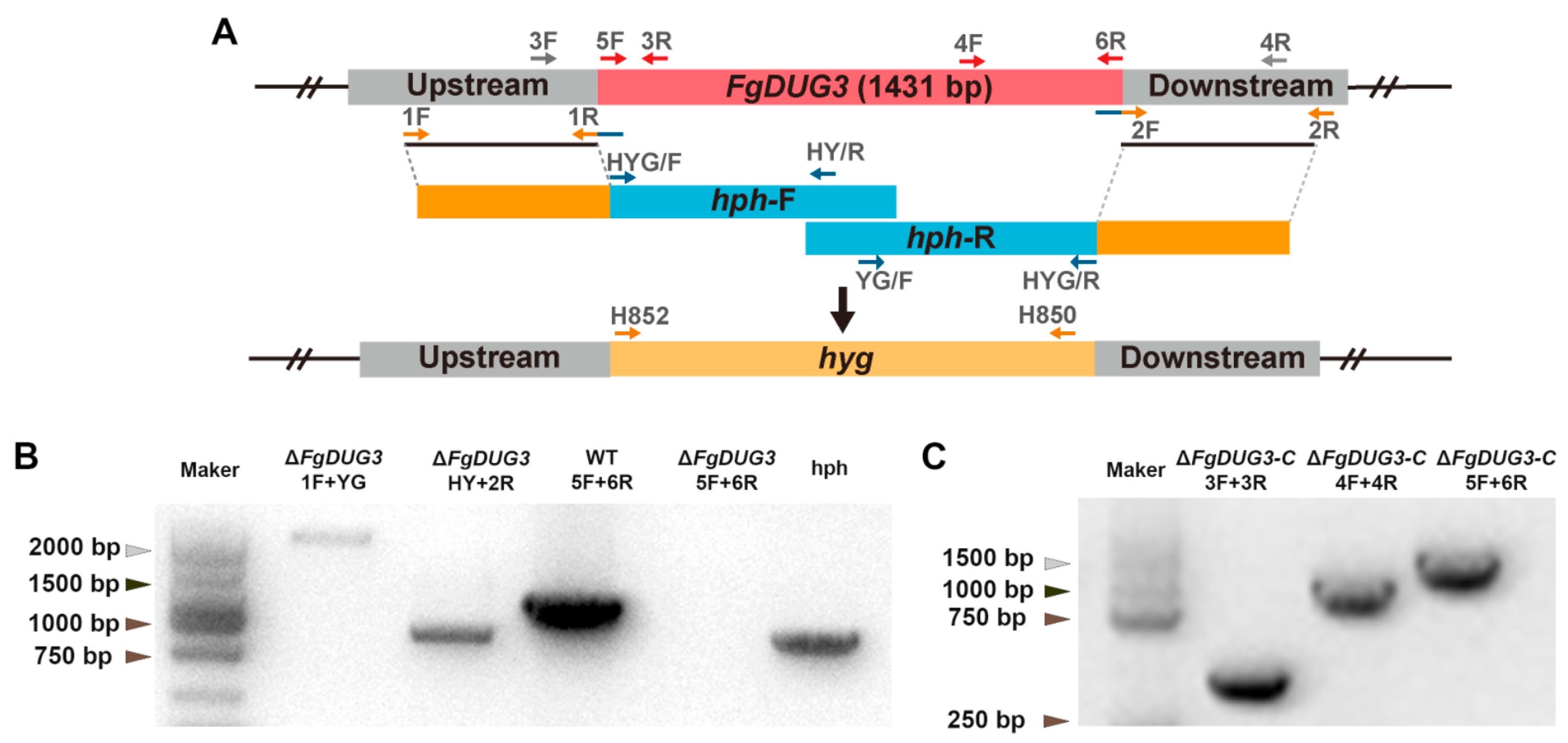
The split-marker method was used to generate a knockout mutant of the FgDUG3 gene [21]. Briefly, the upstream and downstream fragments of FgDUG3 gene were amplified from genomic DNA of a PH-1 strain with paired primers 1F/1R and 2F/2R, respectively (Table A1). The upstream fragment hph-F and downstream fragment hph-R of hph cassette were amplified with paired primers HYG/F/HY/R and YG/F/HYG/R, respectively, as described previously [19]. The upstream fragments of FgDUG3 gene and upstream fragments of hph cassette were fused and amplified with paired primers 1F/HY/R (Table A1). Similarly, both downstream fragments were fused and amplified with paired primers YG/F/2R (Table A1). The two purified fusion segments were introduced into protoplasts of PH-1 strain using a PEG-mediated transformation [19]. The transformants were grown on medium amended with hygromycin B, and then screened with paired primers 5F/6R and H856F/H855R (Table A1).
For complementation, the construct containing the FgDUG3 open reading frame and the upstream and downstream fragments were transformed into protoplasts of ΔFgDUG3. Transformants of deletion mutants and complemented strains were initially selected on TB3 agar media containing 200 µg/mL G418, respectively, and next screened by PCR with paired primers 5F/6R, 3F/3R, and 4F/4R (Table A1).

2.4. Characterization of Phenotypes

For the evaluation of mycelial growth, fungal strains were grown on PDA at 25 °C for two days [22]. Mycelial discs from PDA were inoculated to new PDA and cultured at 25 °C for five days to evaluate colony diameters. For the examination of conidiation and conidium morphology, mycelia obtained from PDA were inoculated to CMC liquid medium and rotated at 150 rpm and 25 °C for five days. The conidia were harvested by filtering through a single layer of sterile filter paper. Then, concentrations of conidial suspensions were evaluated using a hemocytometer. Conidial morphology was examined using a fluorescence microscope (Nikon DS-Ri2, Tokyo, Japan). These experiments were performed on three independent experiments with at least three replicates per experiment. The significant difference was estimated using Tukey’s HSD test (p < 0.05).

2.5. Sexual Reproduction

The designated strains were cultured on carrot agar at 25 °C for 7 days. Aerial mycelia were scraped off using a sterile medical spoon and cultured under black light at 25 °C. Once the mycelia began to grow, 1% Tween was applied to adhere the mycelia to the medium. After two weeks, photographs were taken using a Leica M205 FA stereoscope (Wetzlar, Germany), with ten replicates for each strain [23]. For ascospore observation, mycelial plugs (10 mm in diameter) containing ascus structures were bisected and placed vertically on the surface of a fungal culture block in a disposable Petri dish, followed by incubation at 25 °C for 24–48 h. The discharged asci were collected from the medium, transferred to a glass slide, and gently pressed under a coverslip. Ascospore morphology was examined using a Nikon ECLIPSE Ni–U microscope equipped with a Nikon DS–Ri2 digital camera (Tokyo, Japan) [24].

2.6. Pathogenicity Assay

To evaluate pathogenicity in flowering wheat heads, conidia were harvested from Fg strains after four days of cultivation in CMC liquid medium, resuspended in 0.01% (v/v) Tween 20, and adjusted to a final concentration of 105 conidia/mL. For pathogenicity in wheat spikelets, the prepared conidial suspensions (10 µL) were injected into grains in the middle wheat spikelet. For the control, wheat grains were inoculated with sterile distilled water (SDW) through the same method described above. Next, the bag was sprayed with SDW to maintain humidity for two days. At 14 days post-inoculation, the number of symptomatic spikelets exhibiting blight symptoms on each wheat head was assessed [20].
For pathogenicity on corn silks, sterile filter papers were placed in a glass dish, and moistened with SDW. Using a sterile blade, both ends of four corn silks were cleanly cut and laid flat on the moistened filter papers. Five replicates were applied for each strain. The inoculated corn husks were placed in an incubator at 25 °C [18]. After four days, the lesion sizes were measured and photographs were taken for record.

2.7. Transcriptomic Analysis

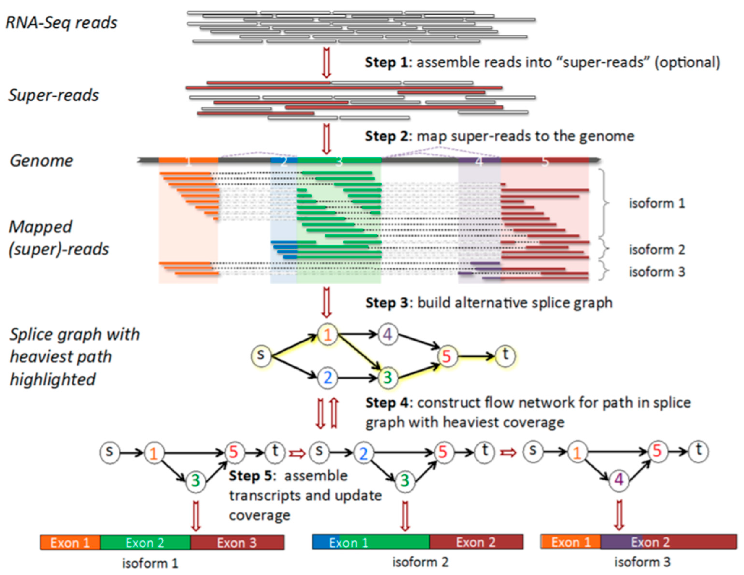
For transcriptome profiling, wheat heads were injected with conidial suspensions of the strains PH-1 and ΔFgDUG3, respectively (Genome Sequence Archive, CRA030539). Inoculated spikelets were harvested at 7 dpi, and immediately frozen in liquid nitrogen for total RNA extraction. Three biological replicates of extracted RNA samples were sequenced by Illumina Hiseq 2500 [25]. The RNA-seq reads were filtered, mapped, and assembled with a reference genome of F. graminearum PH-1, according to a pipeline (Figure A1). The distribution and mapping of total reads were shown in Table A2 and Table A3. The differentially expressed genes (DEGs) between PH-1 and ΔFgDUG3 were identified using DEGSeq2 with a threshold of |log2 fold change| > 1 and p < 0.05.

3. Results

3.1. Phylogenetic Analysis of DUG3 Proteins

Using the amino acid sequence of S. cerevisiae DUG3 (YNL_191W) as a query search in the genome of F. graminearum PH-1, we identified a sequence FGSG_06147 (named FgDUG3). The FgDUG3 gene is predicted to encode a protein (476 amino acids) comprising a glutamine amidotransferase type 2 domain (Figure 1A). To analyze phylogenetic relationships, a neighbor-joining tree was constructed based on FgDUG3 and its homologs from other selected fungi. FgDUG3 was found to be closely related to its homologs from Fusarium oxysporum, and distantly related to homologs from S. cerevisiae, Candida albicans, and Cryptococcus neoformans (Figure 1B). This result suggests that FgDUG3 is conserved among fungi.

3.2. Generation of FgDUG3 Deletion Mutants

To characterize the functions of FgDUG3, a split-marker method was used to generate the ΔFgDUG3 (Figure 2A). To restore FgDUG3 gene function, complementation strains ΔFgDUG3-C were constructed by introducing the native promoter-driven coding sequence of FgDUG3 under the control of its native promoter-driven coding sequence of FgDUG3. PCR was performed to verify the genotypes of ΔFgDUG3 and ΔFgDUG3-C (Figure 2B,C). Two independent ΔFgDUG3 and ΔFgDUG3-C strains were generated for subsequent functional analyses. Since the results were highly consistent between biological replicates, data from one representative strain of each genotype are shown herein.

3.3. FgDUG3 Is Dispensable for Mycelial Growth

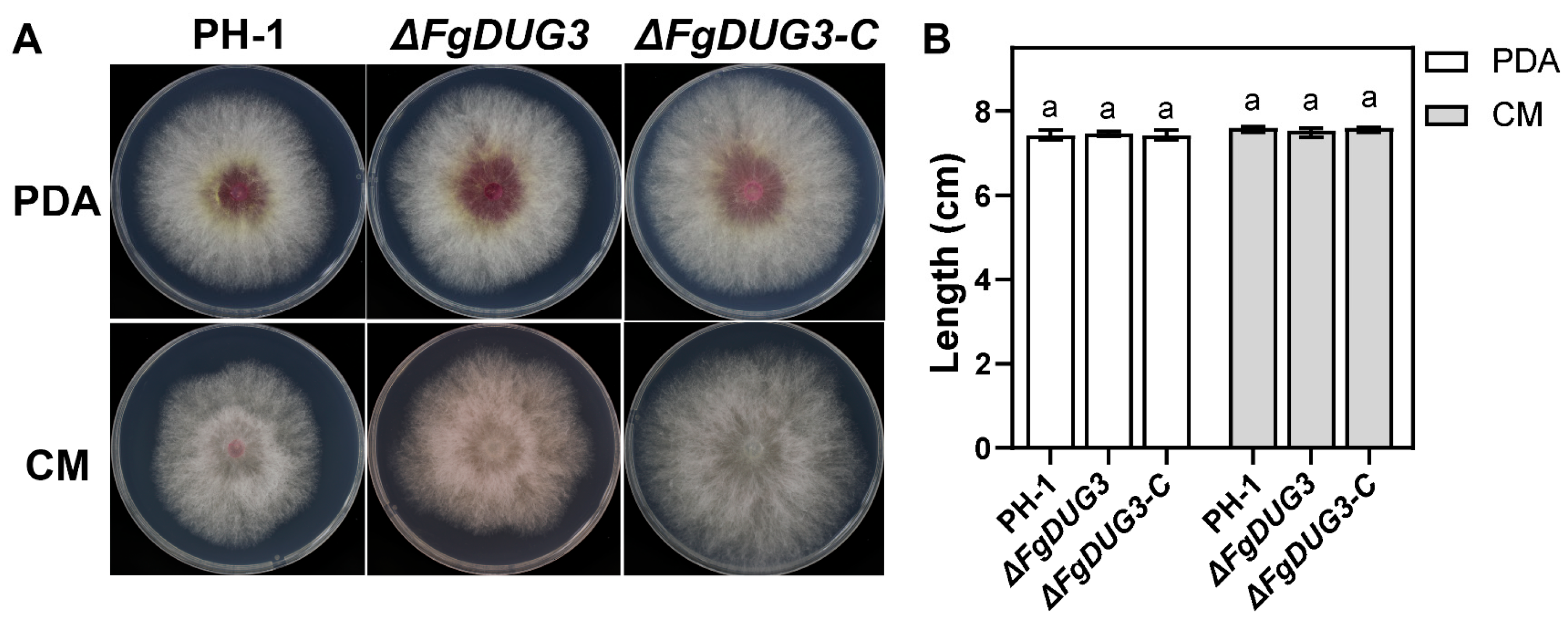
To investigate the role of FgDUG3 in fungal growth, mycelial discs were inoculated onto PDA and CM. After 5 days, colony diameter and morphology were evaluated. ΔFgDUG3 was found to be normal in mycelial growth rate and colony morphology, compared to the PH-1 and ΔFgDUG3-C (Figure 3A,B). This result suggests that FgDUG3 is dispensable for mycelial growth of F. graminearum.

3.4. FgDUG3 Is Involved in Asexual Reproduction

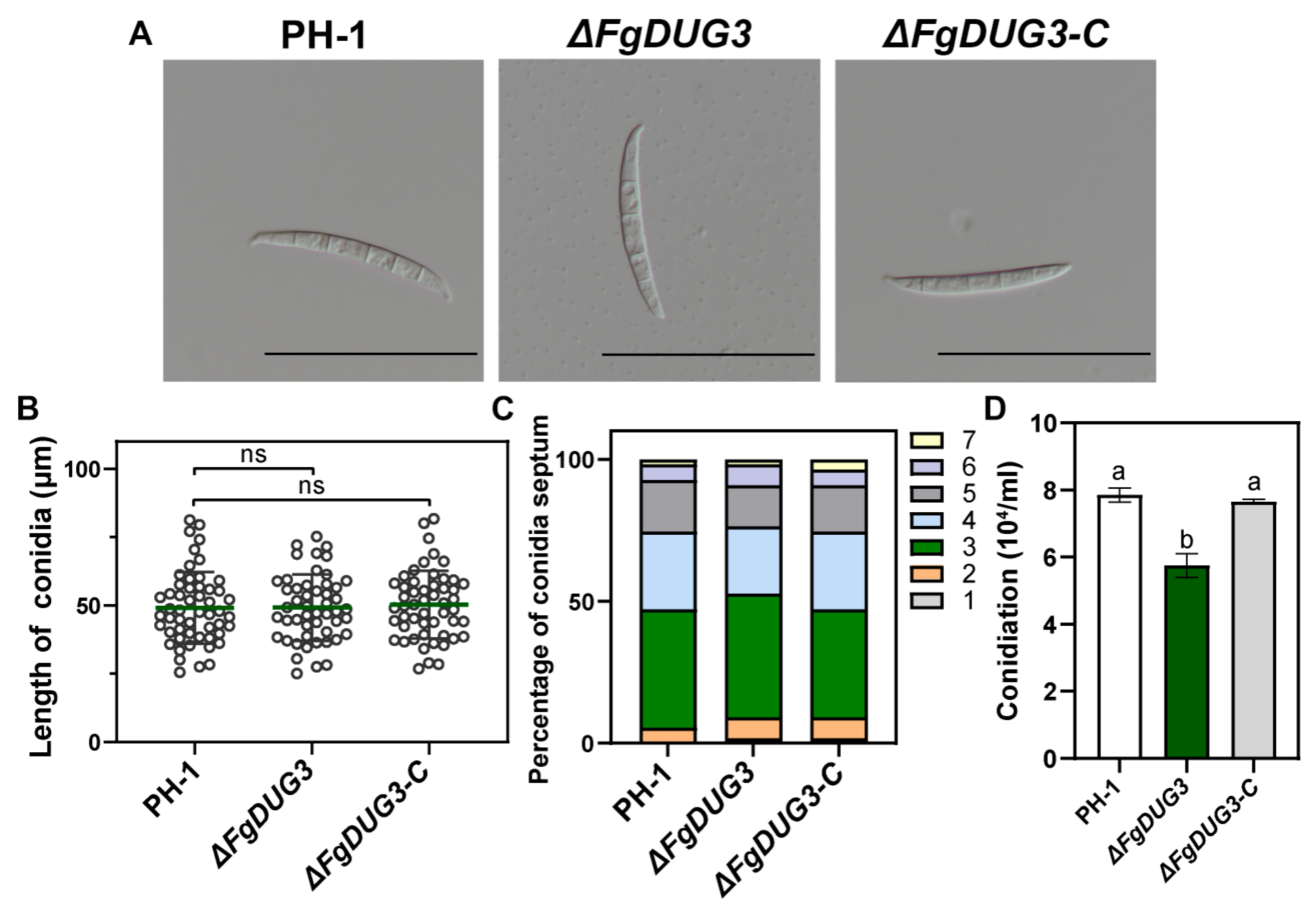
As asexual reproduction plays an important role in the dissemination of F. graminearum, we evaluated conidiation and conidium morphology in CMC broth. The ΔFgDUG3 mutant and its complemented strain (ΔFgDUG3-C) showed conidial morphology comparable to the wild-type PH-1, with no significant differences in the mean conidial length (∼50 μm) or in the distribution of septation numbers, where conidia with three septa were predominant (∼40%) (Figure 4A–C). Quantitative evaluation of conidiation showed that PH-1 and ΔFgDUG3-C produced (8.2 ± 0.7) × 104 conidia/mL and (8.0 ± 0.6) × 104 conidia/mL, respectively. However, ΔFgDUG3 produced (6.0 ± 0.5) × 104 conidia/mL, which is significantly less than that of PH-1 and ΔFgDUG3-C (Figure 4D).
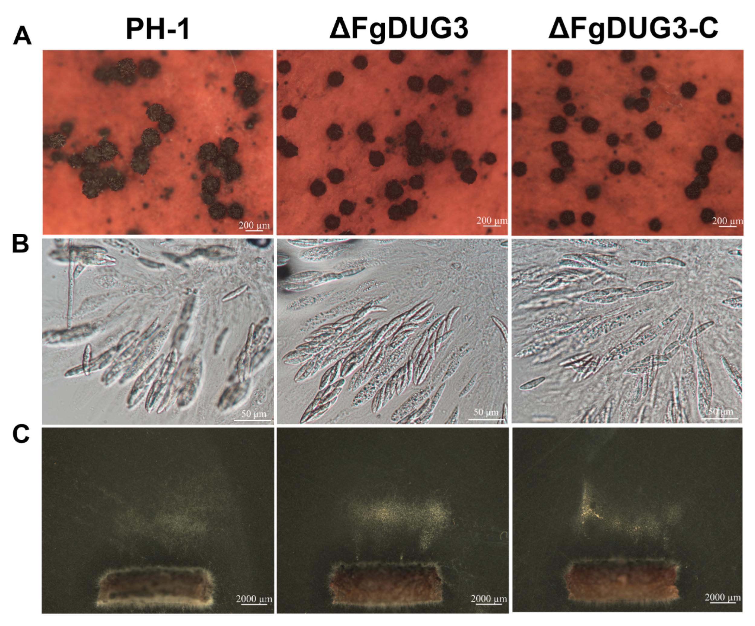
To investigate whether FgDUG3 is related to sexual reproduction, we inoculated strains on carrot agar medium. ΔFgDUG3 was found to be normal in the production of perithecia and ascospores, compared to PH-1 or ΔFgDUG3-C (Figure 5A–C), revealing that FgDUG3 is not involved in sexual reproduction. These results suggest that FgDUG3 is involved in conidiation but not conidium morphology of F. graminearum.

3.5. FgDUG3 Is Associated with Germ Tube Development

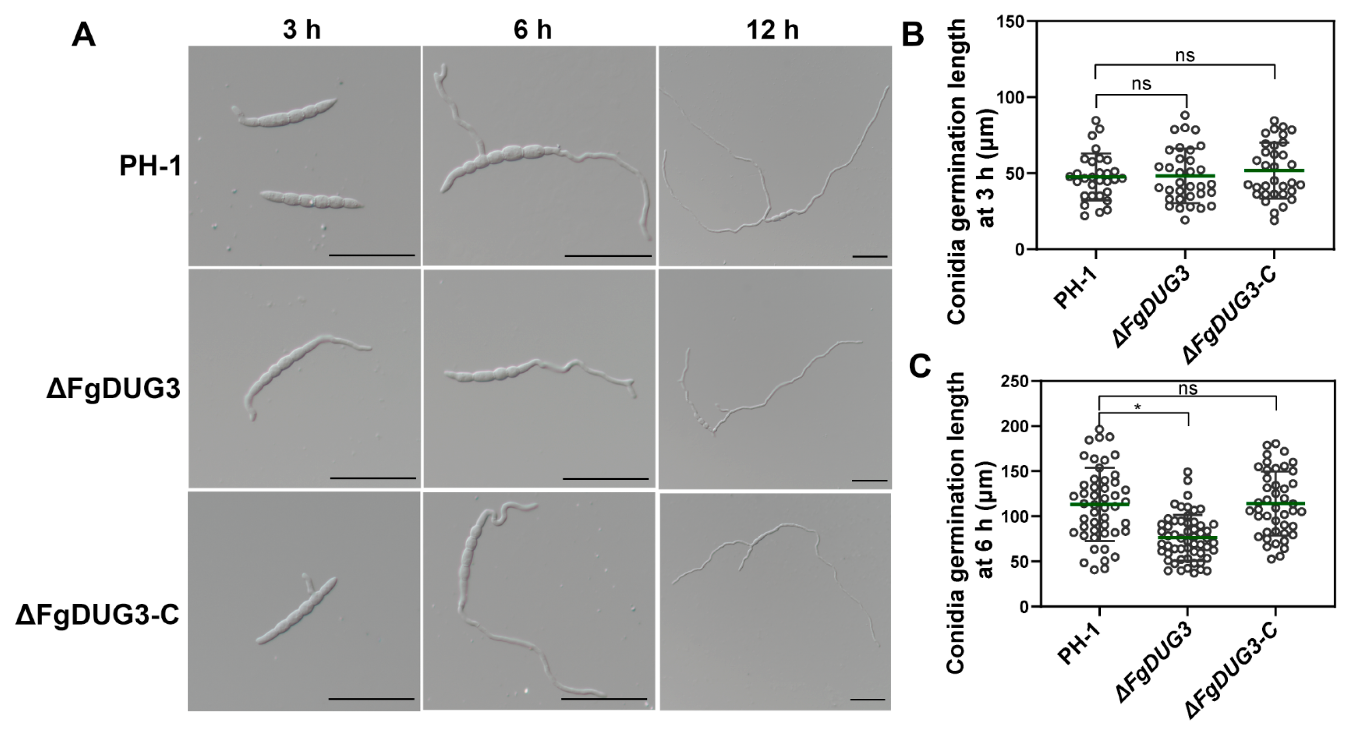
The conidial germination rate was found to be normal in ΔFgDUG3, compared to PH-1 or ΔFgDUG3-C (Figure 6A), suggesting that FgDUG3 is dispensable for the conidial germination rate of F. graminearum. Notably, the germ tube length of ΔFgDUG3 was significantly shorter than that of PH-1 or ΔFgDUG3-C at 6 h (Figure 6B). These results suggest that FgDUG3 is associated with the germ tube development of F. graminearum.

3.6. FgDUG3 Is Required for Full Virulence

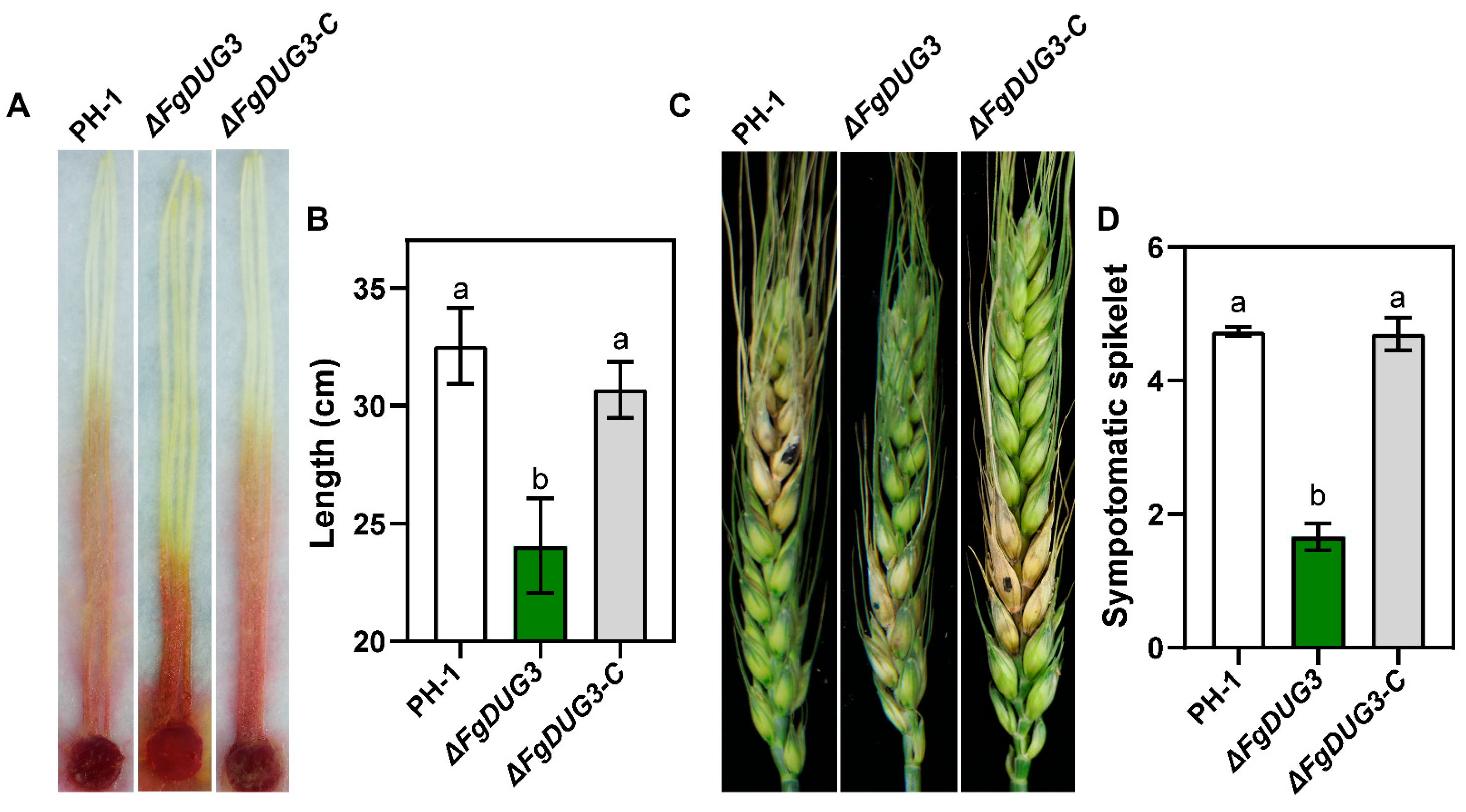
To test whether FgDUG3 is related to the virulence of F. graminearum, mycelial plugs were inoculated to corn silks. After 4 d, the lesion length caused by ΔFgDUG3 was significantly shorter than that of PH-1 or ΔFgDUG3-C (Figure 7A,B). We next investigated the role of FgDUG3 in infection on flowering wheat head by inoculating conidial suspensions. After 14 d, ΔFgDUG3 infected significantly fewer spikelets than WT or ΔFgDUG3-C (Figure 7C,D). These results suggest FgDUG3 is required for full the virulence of F. graminearum on corn and wheat.

3.7. Differentially Expressed Genes Regulated by FgDUG3

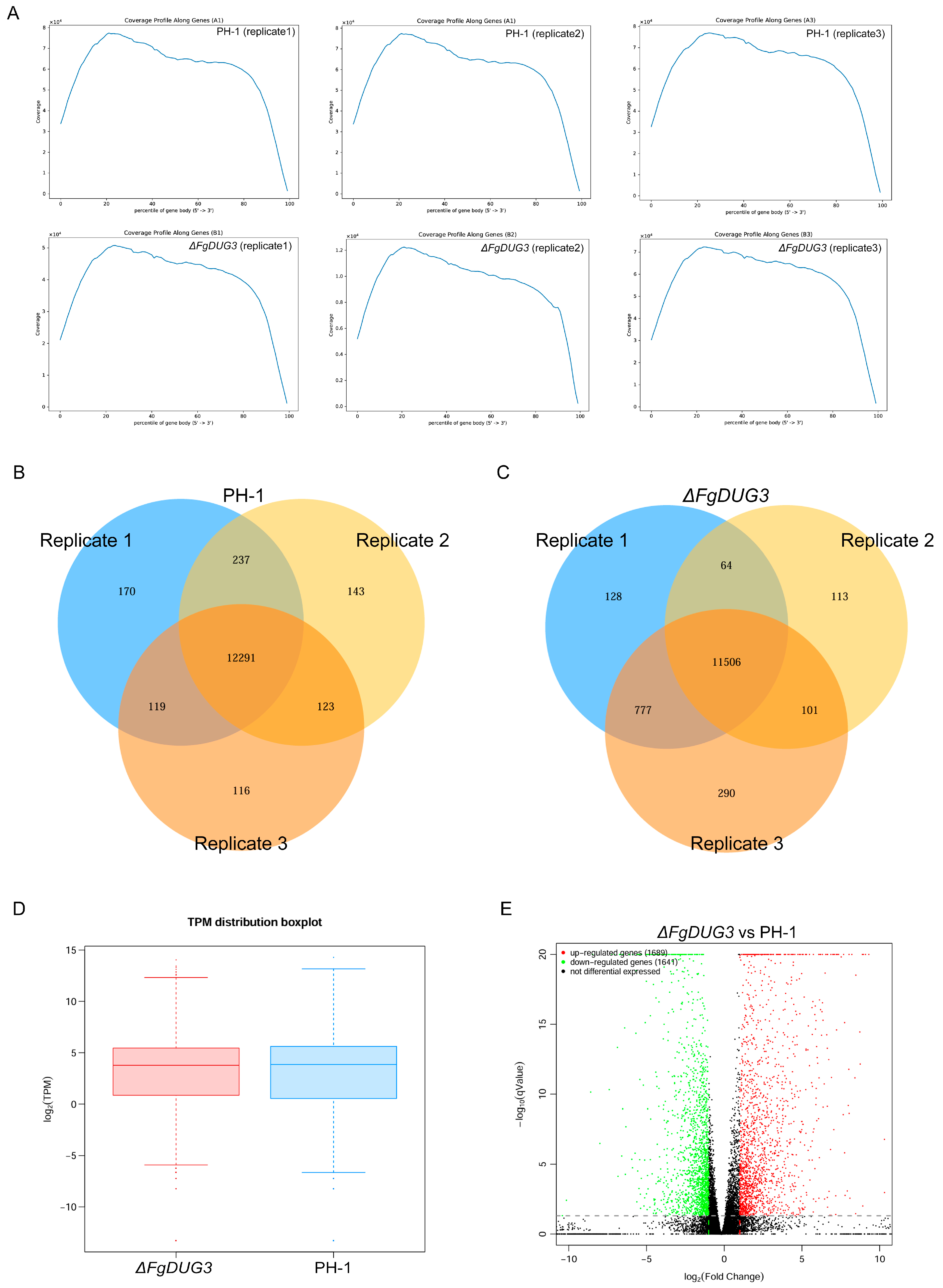
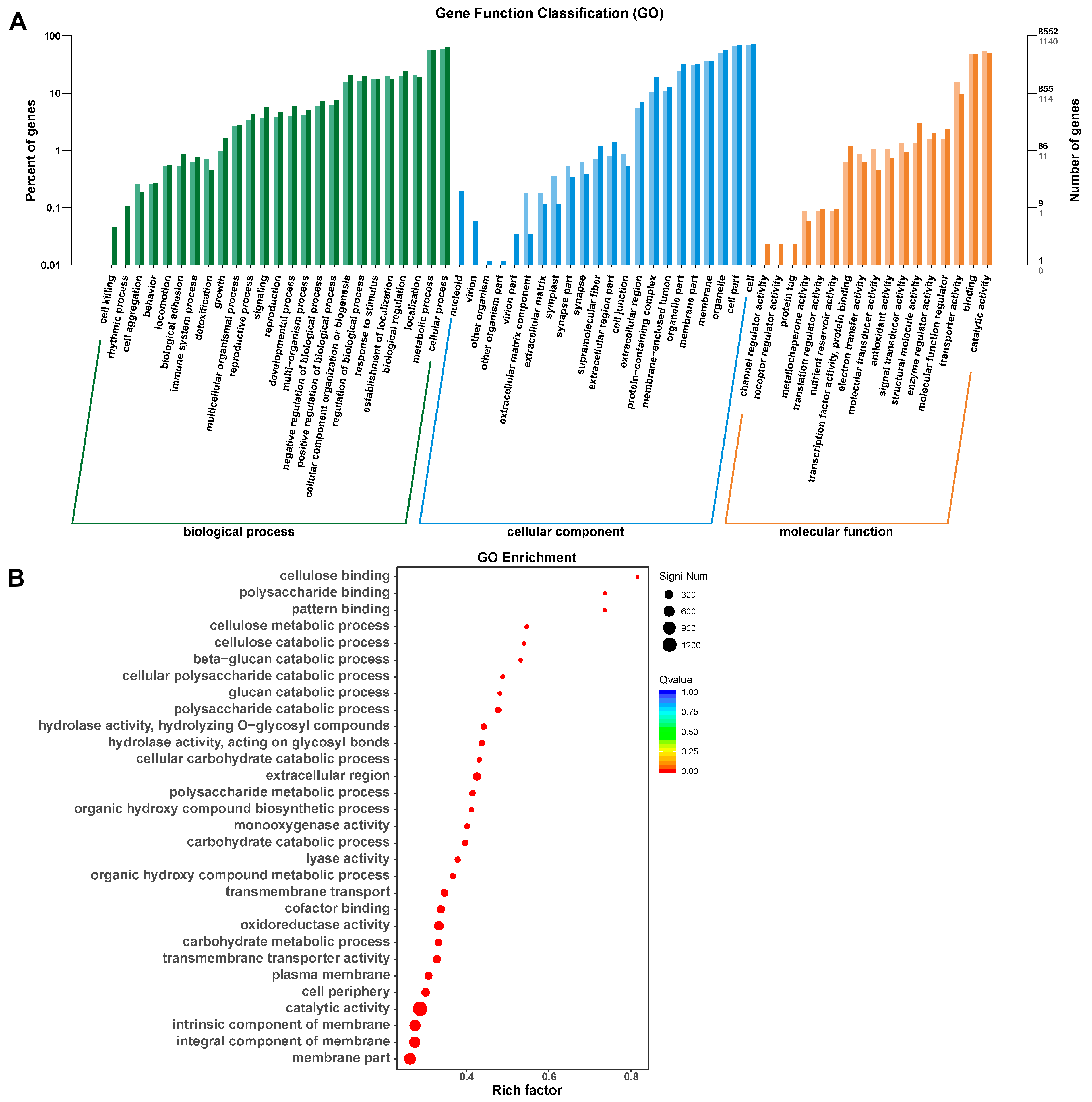
To further investigate the genes regulated by FgDUG3 during plant infection, the RNA-seq analysis was performed to profile the gene expression of ΔFgDUG3 and the PH-1 strain during wheat head infection at 7 dpi. A total of 3330 genes were identified as significantly differentially expressed (p < 0.05), with 1689 upregulated genes (log2 fold change > 1) and 1641 downregulated genes (log2 fold change < 1) (Figure 8 and Figure A2, Table A2). The Gene Ontology (GO) analysis showed that DEGs were classified into biological processes, cellular components, and molecular functions. The GO enrichment analysis revealed that DEGs were enriched in categories related to polysaccharide metabolism, catabolic processes, enzymatic activity, and structural components of the cell (Figure 8A,B).
The Kyoto Encyclopedia of Genes and Genomes (KEGG) enrichment analyses revealed that the DEGs were enriched in signal transduction and multiple metabolic pathways, including caffeine metabolism, starch and sucrose metabolism, and amino acid metabolic pathways (Figure 9A,B). Collectively, these results indicate that the deletion of FgDUG3 caused the major alterations in metabolism, catabolism, cellular structure organization, and signal transduction.

4. Discussion

The present study aims to investigate the functional roles of FgDUG3 in the wheat head blight pathogen F. graminearum. Our results demonstrated that FgDUG3 plays important roles in asexual reproduction, germ tube development, and virulence, while being dispensable for vegetative growth and sexual reproduction. Furthermore, transcriptomic analysis revealed significantly differential expression of genes involved in metabolism, signaling, and structural components following FgDUG3 deletion. The findings suggest that FgDUG3 is important for the fungal differentiation and pathogenicity of F. graminearum, which would provide novel insight into the molecular mechanisms governing Fusarium head blight development.
FgDUG3 was predicted to encode a protein containing a glutamine amidotransferase type 2 domain (Figure 1A), a conserved enzymatic motif involved in amino group transfer and nitrogen metabolism [26]. In terms of phylogenetic relationships, FgDUG3 is closely related to its homologs from Fusarium oxysporum, M. oryzae, and Botrytis cinerea, and more distantly related to those from S. cerevisiae, Candida albicans, and Cryptococcus neoformans (Figure 1B). This is consistent with the divergence between ascomycete fungal plant pathogens and yeast-like fungi. The conservation of domain architecture indicates that FgDUG3 may retain core enzymatic activities, yet functional divergence is also plausible. In yeast, the DUG pathway contributes to glutathione degradation and nitrogen utilization [13,14]. However, our findings indicate that FgDUG3 additionally regulates developmental processes, such as conidiation and germ tube elongation, as well as pathogenicity in F. graminearum. Consistently, functions of DUG3 were reported to be involved in conidiation, appressorium development, and plant infection in M. oryzae [16]. Thus, FgDUG3 might integrate metabolic activity with developmental regulation in a plant pathogenic context, reflecting the adaptation of conserved proteins to the specific lifestyle of fungal organism.
The deletion of FgDUG3 did not affect mycelial growth and morphology on PDA and CM (Figure 3), indicating that vegetative hyphal growth under nutrient-rich conditions is maintained without FgDUG3, possibly due to redundancy in glutamine amidotransferase. However, ΔFgDUG3 significantly reduced conidiation, suggesting that FgDUG3 is involved in regulating asexual reproduction in F. graminearum. Importantly, conidial morphology of ΔFgDUG3 remained unaltered, suggesting that FgDUG3 specifically influences conidiation rather than morphogenesis. This finding supports that fungal reproduction is tightly linked to nutrient metabolism [27,28,29]. As glutamine amidotransferases participate in amino acid metabolism and nitrogen assimilation [26,30], FgDUG3 may act as a metabolic regulator for the conidiation of F. graminearum. Interestingly, ΔFgDUG3 was normal in the production of perithecia and ascospores (Figure 5), indicating that sexual reproduction relies on distinct regulatory circuits that bypass FgDUG3. The molecular network regulating asexual and sexual reproduction has been previously documented in F. graminearum [31,32,33]. Our findings reinforce the idea that asexual and sexual cycles are governed by partially overlapping but distinct networks, with FgDUG3 being specific to asexual reproduction.
ΔFgDUG3 was normal in conidial germination rate, but defective in germ tube elongation (Figure 6), suggesting that FgDUG3 is dispensable for initial polarity establishment but required for sustained polarized growth. Germ tube elongation is a complex process involving cytoskeletal organization, vesicle trafficking, and cell wall remodeling [34,35]. Metabolism is also essential for germ tube elongation, as the synthesis of macro-molecules contributes to tip growth. For example, nitrogen starvation induces morphogenetic transitions critical for infection structure formation in M. oryzae [36]. Therefore, the defect in germ tube elongation in ΔFgDUG3 might arise from metabolic insufficiency or the misregulation of developmental signaling.
ΔFgDUG3 significantly attenuated virulence on both corn silks and wheat heads, indicating the important function of FgDUG3 in pathogenicity (Figure 7). During host colonization, fungal plant pathogens encounter nutrient limitations, oxidative stress, and plant defense responses [37,38]. Glutamine amidotransferase may be involved in synthesizing or recycling key metabolites to support infection. The defect of ΔFgDUG3 in pathogenicity suggests that the metabolic change conferred by FgDUG3 is essential for host colonization. This was supported by the transcriptomic profiling, as widespread alterations of gene expressions involved in polysaccharide metabolism, amino acid metabolism, and signal transduction (Figure 8). The enrichment of carbohydrate metabolism-related genes is particularly relevant. Successful colonization of host tissues requires the efficient degradation and utilization of plant polysaccharides. Downregulation of such genes in ΔFgDUG3 mutants may compromise nutrient acquisition, leading to impaired colonization in plants. Similarly, amino acid metabolism is critical for nitrogen assimilation and secondary metabolite biosynthesis. Given that the biosynthesis of trichothecene mycotoxins in F. graminearum is nitro-gen-regulated [39], the disruption of nitrogen metabolism through the loss of function of FgDUG3 could indirectly affect secondary metabolism and virulence. Furthermore, enrichment of DEGs in signal transduction pathways suggests that FgDUG3 influences cellular communication and environmental sensing. Metabolic enzymes are increasingly recognized to play a role in signaling [40,41]. Thus, FgDUG3 may serve as both a metabolic enzyme and a regulator of signaling cascades, orchestrating the transcriptional programs essential for adaptation to the host environment.

5. Conclusions

In conclusion, FgDUG3 is involved in regulating asexual reproduction and contributes to the pathogenicity of F. graminearum, likely through integrating nitrogen metabolism with developmental and signaling pathways. Its absence impairs germ tube elongation and host colonization, underscoring its adaptive role in fungal pathogenicity while highlighting the metabolic basis of infection-related morphogenesis.

Author Contributions

C.S. and P.C. conceived the study and designed the experiments. P.C., Y.D. and C.H. developed the methodology. C.S. and P.C. conducted the investigation and data collection. C.S. wrote the original draft. C.S., W.X., A.G. and Z.F. reviewed and edited the manuscript. C.H. and C.S. contributed to software development. D.M. and T.F. oversaw the project administration, provided supervision, and performed validation. All authors have read and agreed to the published version of the manuscript.

Funding

This work was supported by the Key Research and Development Program of Hubei Province (2024BBB004) and Jiangsu Province Science and Technology of Market Supervision and Administration Foundation (KJ2024003).

Institutional Review Board Statement

Not applicable.

Informed Consent Statement

Not applicable.

Data Availability Statement

The transcriptomic data presented in the study are openly available in CNCB at CRA030539.

Acknowledgments

We thank Huaigu Chen of the Institute of Plant Protection, Jiangsu Academy of Agri-cultural Sciences, China, for supplying the fungal strains for this research.

Conflicts of Interest

The authors declare no conflicts of interest.

Abbreviations

The following abbreviations are used in this manuscript:
DONDeoxynivalenol
GSHGlutathione
ROSReactive oxygen species
PDAPotato dextrose agar
DEGsDifferentially expressed genes
CMComplete agar
CMCCarboxymethyl cellulose

Appendix A

Table A1. Primers used in the present study.
PrimerSequence (5′-3′)
FgDUG3-1FAGTGACGCCAGCAGGATA
Hyg-FgDUG3-1RTTGACCTCCACTAGCTCCAGCCAAGCCTGATTGCGTGTTACGGAT
FgDUG3-2FTATGGATCGAGGCAAGGT
Hyg-FgDUG3-1RGAATAGAGTAGATGCCGACCGCGGGTT
FgDUG3-2RCCTGTGATGGAAAAGAGG
FgDUG3-5FATGTGCCGTTTTCTGGTG
FgDUG3-6RTCACCCCGCATGGGAGGC
HYG/FGGCTTGGCTGGAGCTAGTGGAGGTCAA
HYG/RAACCCGCGGTCGGCATCTACTCTATTC
HY/RGTATTGACCGATTCCTTGCGGTCCGAA
YG/FGATGTAGGAGGGCGTGGATATGTCCT
H852TTCCTCCCTTTATTTCAGATTCAA
H850ATGTTGGCGACCTCGTATTGG
Table A2. Mapping of total reads.
A1A2A3B1B2B3
Total reads62,670,288(100.00%)65,215,212(100.00%)64,694,700(100.00%)75,654,640(100.00%)32,819,352(100.00%)62,749,358(100.00%)
Total mapped59,858,562(95.51%)61,760,765(94.70%)63,100,844(97.54%)40,018,705(52.90%)10,521,595(32.06%)57,468,385(91.58%)
Mutiple mapped265,266(0.42%)233,445(0.36%)270,593(0.42%)126,563(0.17%)835,181(2.54%)206,012(0.33%)
Uniquely mapped59,593,296(95.09%)61,527,320(94.35%)62,830,251(97.12%)39,892,142(52.73%)9,686,414(29.51%)57,262,373(91.26%)
Read-1 mapped29,879,586(47.68%)30,870,888(47.34%)31,526,988(48.73%)20,012,291(26.45%)4,856,115(14.80%)28,732,486(45.79%)
Read-2 mapped29,713,710(47.41%)30,656,432(47.01%)31,303,263(48.39%)19,879,851(26.28%)4,830,299(14.72%)28,529,887(45.47%)
Reads map to ‘+’29,786,332(47.53%)30,753,179(47.16%)31,403,465(48.54%)19,941,079(26.36%)4,840,809(14.75%)28,626,222(45.62%)
Reads map to ‘-’29,806,964(47.56%)30,774,141(47.19%)31,426,786(48.58%)19,951,063(26.37%)4,845,605(14.76%)28,636,151(45.64%)
Non-splice reads45,069,390(71.92%)46,582,022(71.43%)48,344,264(74.73%)30,475,976(40.28%)7,369,629(22.46%)43,701,686(69.64%)
Splice reads14,523,906(23.18%)14,945,298(22.92%)14,485,987(22.39%)9,416,166(12.45%)2,316,785(7.06%)13,560,687(21.61%)
Reads mapped in proper pairs58,975,354(94.10%)60,862,436(93.33%)62,138,088(96.05%)39,439,416(52.13%)9,576,482(29.18%)56,594,788(90.19%)
Table A3. Distribution of total reads.
A1A2A3B1B2B3
NC_026474.120,227,94820,774,44721,217,35112,787,7283,032,29918,272,007
NC_026475.113,050,35613,511,95913,890,9679,574,9122,153,42313,694,690
NC_026476.113,039,23213,411,78013,738,6618,712,2632,042,99712,571,361
NC_026477.113,265,09013,819,34513,970,1998,810,2272,123,57512,715,866
NW_001837897.11630160316747731861081
NW_001837898.1001000
NW_001837899.15413000
NW_001837900.175482050
NW_001837902.140753674427716515102433
NW_001837903.180579280395169
NW_001837904.12191743341
NW_001837905.12912400210
NW_001837906.17610345112

Appendix B

Figure A1. An analysis pipeline for assembling, mapping, and annotation of RNA-seq reads. The numbers 1, 2, 3, 4, and 5 within the circles represent exons.
Jof 11 00763 g0a1
Figure A2. Global analysis of RNA-seq data. (A) Coverage profile along genes of six samples. (B,C) Venn diagram of detected genes among three replicates of PH-1 (B) and ΔFgDUG3 (C). (D) TPM distribution of detected genes in PH-1 and ΔFgDUG3. (E) Volcano diagram of differentially expressed genes (DEGs) in ΔFgDUG3 referenced to PH-1.
Jof 11 00763 g0a2

References

  1. Bekele, B.; Dawit, W. Review on the status and management strategies of Fusarium head blight (Fusarium graminearum) of wheat. Int. J. Agric. Sustain. 2018, 4, 2348–3997. [Google Scholar]
  2. Goswami, R.S.; Kistler, H.C. Heading for disaster: Fusarium graminearum on cereal crops. Mol. Plant Pathol. 2004, 5, 515–525. [Google Scholar] [CrossRef] [PubMed]
  3. Valverde-Bogantes, E.; Bianchini, A.; Herr, J.R.; Rose, D.J.; Wegulo, S.N. Hallen-Adams, H.E. Recent population changes of Fusarium head blight pathogens: Drivers and implications. Can. J. Plant Pathol. 2020, 42, 315–329. [Google Scholar] [CrossRef]
  4. Akinmoladun, O.F.; Fon, F.N.; Nji, Q.; Adeniji, O.O.; Tangni, E.K.; Njobeh, P.B. Multiple mycotoxin contamination in livestock feed: Implications for animal Health, productivity, and food safety. Toxins 2025, 17, 365. [Google Scholar] [CrossRef]
  5. Kamle, M.; Mahato, D.K.; Gupta, A.; Pandhi, S.; Sharma, B.; Dhawan, K.; Vasundhara; Mishra, S.; Kumar, M.; Tripathi, A.D. Deoxynivalenol: An overview on occurrence, chemistry, biosynthesis, health effects and its detection, management, and control strategies in food and feed. Microbiol. Res. 2022, 13, 292–314. [Google Scholar] [CrossRef]
  6. Penninckx, M.J.; Elskens, M.T. Metabolism and functions of glutathione in micro-organisms. Adv. Microb. Physiol. 1993, 34, 239–301. [Google Scholar] [PubMed]
  7. Gila, B.C.; Moon, H.; Antal, K.; Hajdu, M.; Kovács, R.; Jónás, A.P.; Pusztahelyi, T.; Yu, J.-H.; Pócsi, I.; Emri, T. The DUG pathway governs degradation of intracellular glutathione in Aspergillus nidulans. Appl. Environ. Microbiol. 2021, 87, e01321-20. [Google Scholar] [CrossRef]
  8. Georgiou-Siafis, S.K.; Tsiftsoglou, A.S. The key role of GSH in keeping the redox balance in mammalian cells: Mechanisms and significance of GSH in detoxification via formation of conjugates. Antioxidants 2023, 12, 1953. [Google Scholar] [CrossRef]
  9. Jozefczak, M.; Remans, T.; Vangronsveld, J.; Cuypers, A. Glutathione is a key player in metal-induced oxidative stress defenses. Int. J. Mol. Sci. 2012, 13, 3145–3175. [Google Scholar] [CrossRef]
  10. Di Giulio, R.T.; Meyer, J.N. Reactive oxygen species and oxidative stress. In The Toxicology of Fishes; CRC Press: Boca Raton, FL, USA, 2008; Volume 10. [Google Scholar] [CrossRef]
  11. Park, J.; Han, J.W.; Lee, N.; Kim, S.; Choi, S.; Lee, H.-H.; Kim, J.-E.; Seo, Y.-S.; Choi, G.J.; Lee, Y.-W. Sulfur metabolism-mediated fungal glutathione biosynthesis is essential for oxidative stress resistance and pathogenicity in the plant pathogenic fungus Fusarium graminearum. mBio 2024, 15, e02401-23. [Google Scholar] [CrossRef]
  12. Wangsanut, T.; Pongpom, M. The role of the glutathione system in stress adaptation, morphogenesis and virulence of pathogenic fungi. Int. J. Mol. Sci. 2022, 23, 10645. [Google Scholar] [CrossRef] [PubMed]
  13. Bachhawat, A.K.; Kaur, A. Glutathione degradation. Antioxid. Redox Signal. 2017, 27, 1200–1216. [Google Scholar] [CrossRef]
  14. Kumar, C.; Sharma, R.; Bachhawat, A.K. Utilization of glutathione as an exogenous sulfur source is independent of γ-glutamyl transpeptidase in the yeast Saccharomyces cerevisiae: Evidence for an alternative gluathione degradation pathway. FEMS Microbiol. Lett. 2003, 219, 187–194. [Google Scholar] [CrossRef]
  15. Ganguli, D.; Kumar, C.; Bachhawat, A.K. The alternative pathway of glutathione degradation is mediated by a novel protein complex involving three new genes in Saccharomyces cerevisiae. Genetics 2007, 175, 1137–1151. [Google Scholar] [CrossRef]
  16. Reza, M.H.; Sanyal, K. Defective in utilizing glutathione 3, DUG3, is required for conidiation and host infection in the rice blast fungus Magnaporthe oryzae. Micropubl. Biol. 2022, 2022. [Google Scholar] [CrossRef]
  17. Sevastos, A.; Labrou, N.; Flouri, F.; Malandrakis, A. Glutathione transferase-mediated benzimidazole-resistance in Fusarium graminearum. Pestic. Biochem. Physiol. 2017, 141, 23–28. [Google Scholar] [CrossRef]
  18. Fu, Z.; Chen, Y.; Cai, G.; Peng, H.; Wang, X.; Li, P.; Gu, A.; Li, Y.; Ma, D. An antisense long non-coding RNA, LncRsn, is involved in sexual reproduction and full virulence in Fusarium graminearum. J. Fungi 2024, 10, 692. [Google Scholar] [CrossRef]
  19. Li, P.; Fu, Z.; Wang, M.; Yang, T.; Li, Y.; Ma, D. Functional Characterization of FgAsp, a Gene Coding an Aspartic Acid Protease in Fusarium graminearum. J. Fungi 2024, 10, 879. [Google Scholar] [CrossRef]
  20. Cai, L.; Xu, X.; Dong, Y.; Jin, Y.; Rashad, Y.M.; Ma, D.; Gu, A. Roles of Three FgPel Genes in the Development and Pathogenicity Regulation of Fusarium graminearum. J. Fungi 2024, 10, 666. [Google Scholar] [CrossRef] [PubMed]
  21. Fu, J.; Zhang, X.; Chen, X.; Yin, Y.; Ma, Z. Serine O-acetyltransferase is important, but not essential for cysteine–methionine synthesis in Fusarium graminearum. World J. Microbiol. Biotechnol. 2014, 30, 1219–1228. [Google Scholar] [CrossRef] [PubMed]
  22. Cao, S.; Jiang, W.; Shu, Y.; Li, W.; Zhang, Y.; Zhang, A.; Chen, H. UDP-galactopyranose mutase mediates cell wall integrity, polarity growth, and virulence in Fusarium graminearum. Appl. Environ. Microbiol. 2023, 89, e01235-22. [Google Scholar] [CrossRef]
  23. Chen, A.; Ju, Z.; Wang, J.; Wang, J.; Wang, H.; Wu, J.; Yin, Y.; Zhao, Y.; Ma, Z.; Chen, Y. The RasGEF FgCdc25 regulates fungal development and virulence in Fusarium graminearum via cAMP and MAPK signalling pathways. Environ. Microbiol. 2020, 22, 5109–5124. [Google Scholar] [CrossRef] [PubMed]
  24. Zhang, C.; Wang, Y.; Wang, J.; Zhai, Z.; Zhang, L.; Zheng, W.; Zheng, W.; Yu, W.; Zhou, J.; Lu, G. Functional characterization of Rho family small GTPases in Fusarium graminearum. Fungal Genet. Biol. 2013, 61, 90–99. [Google Scholar] [CrossRef]
  25. Fu, T.; Han, J.-H.; Shin, J.-H.; Song, H.; Ko, J.; Lee, Y.-H.; Kim, K.-T.; Kim, K.S. Homeobox transcription factors are required for fungal development and the suppression of host defense mechanisms in the Colletotrichum scovillei-pepper pathosystem. mBio 2021, 12, e0162021. [Google Scholar] [CrossRef]
  26. Massière, F.; Badet-Denisot, M.-A. The mechanism of glutamine-dependent amidotransferases. Cell. Mol. Life Sci. CMLS 1998, 54, 205–222. [Google Scholar] [CrossRef]
  27. Walker, G.M.; White, N.A. Introduction to fungal physiology. In Fungi: Biology and Applications; Wiley: Hoboken, NJ, USA, 2017; pp. 1–35. [Google Scholar]
  28. Calvo, A.M.; Wilson, R.A.; Bok, J.W.; Keller, N.P. Relationship between secondary metabolism and fungal development. Microbiol. Mol. Biol. Rev. 2002, 66, 447–459. [Google Scholar] [CrossRef]
  29. Calvo, A.M.; Cary, J.W. Association of fungal secondary metabolism and sclerotial biology. Front. Microbiol. 2015, 6, 62. [Google Scholar] [CrossRef] [PubMed]
  30. Kishorekumar, R.; Bulle, M.; Wany, A.; Gupta, K.J. An overview of important enzymes involved in nitrogen assimilation of plants. In Nitrogen Metabolism in Plants: Methods and Protocols; Humana: New York, NY, USA, 2019; pp. 1–13. [Google Scholar]
  31. Kim, W.; Kim, D.-W.; Wang, Z.; Liu, M.; Townsend, J.P.; Trail, F. Transcription factor-dependent regulatory networks of sexual reproduction in Fusarium graminearum. mBio 2025, 16, e0303024. [Google Scholar] [CrossRef] [PubMed]
  32. Brauer, E.K.; Subramaniam, R.; Harris, L.J. Regulation and dynamics of gene expression during the life cycle of Fusarium graminearum. Phytopathology 2020, 110, 1368–1374. [Google Scholar] [CrossRef]
  33. Niu, G.; Yang, Q.; Liao, Y.; Sun, D.; Tang, Z.; Wang, G.; Xu, M.; Wang, C.; Kang, J. Advances in understanding Fusarium graminearum: Genes involved in the regulation of sexual development, pathogenesis, and deoxynivalenol biosynthesis. Genes 2024, 15, 475. [Google Scholar] [CrossRef]
  34. Steinberg, G.; Peñalva, M.A.; Riquelme, M.; Wösten, H.A.; Harris, S.D. Cell biology of hyphal growth. Microbiol. Spectr. 2017, 5. [Google Scholar] [CrossRef]
  35. Riquelme, M. Tip growth in filamentous fungi: A road trip to the apex. Annu. Rev. Microbiol. 2013, 67, 587–609. [Google Scholar] [CrossRef] [PubMed]
  36. Kershaw, M.J.; Talbot, N.J. Genome-wide functional analysis reveals that infection-associated fungal autophagy is necessary for rice blast disease. Proc. Natl. Acad. Sci. USA 2009, 106, 15967–15972. [Google Scholar] [CrossRef]
  37. Segal, L.M.; Wilson, R.A. Reactive oxygen species metabolism and plant-fungal interactions. Fungal Genet. Biol. 2018, 110, 1–9. [Google Scholar] [CrossRef]
  38. Divon, H.H.; Fluhr, R. Nutrition acquisition strategies during fungal infection of plants. FEMS Microbiol. Lett. 2007, 266, 65–74. [Google Scholar] [CrossRef]
  39. Chen, Y.; Kistler, H.C.; Ma, Z. Fusarium graminearum trichothecene mycotoxins: Biosynthesis, regulation, and management. Annu. Rev. Phytopathol. 2019, 57, 15–39. [Google Scholar] [CrossRef]
  40. Li, F.; Xu, W.; Zhao, S. Regulatory roles of metabolites in cell signaling networks. J. Genet. Genom. 2013, 40, 367–374. [Google Scholar] [CrossRef] [PubMed]
  41. Chandel, N.S. Signaling and metabolism. Cold Spring Harb. Perspect. Biol. 2021, 13, a040600. [Google Scholar] [CrossRef] [PubMed]
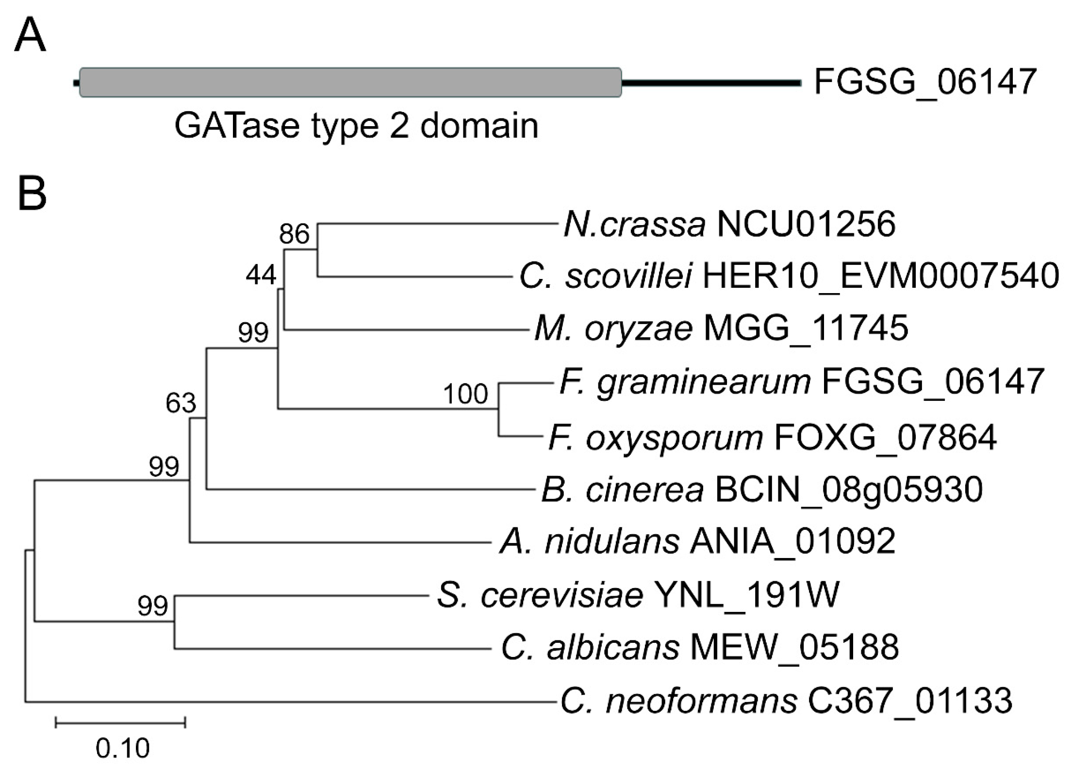
Figure 1. Domain representation and phylogenetic relationship. (A) Domain representation. Black line and gray box represent amino acid sequence and GATase type 2 domain (IPR017932), respectively. (B) Phylogenetic relationship. The phylogenetic tree was constructed based on FgDUG3 and its homologs using the neighbor-joining method with 1000 bootstraps. The full Latin names are as follows: N. crassa (Neurospora crassa), C. scovillei (Colletotrichum scovillei), M. oryzae (Magnaporthe oryzae), F. graminearum (Fusarium graminearum), F. oxysporum (Fusarium oxysporum), B. cinerea (Botrytis cinerea), A. nidulans (Aspergillus nidulans), S. cerevisiae (Saccharomyces cerevisiae), C. albicans (Canidia Albicans), C. neoformans (Cryptococcus neoformans).
Jof 11 00763 g001
Figure 2. Detection of FgDUG3 knockout and complementation transformants. (A) Schematic of gene knockout via homologous recombination. (B) Four pairs of primers PCR to detect transformant electrophoresis results. (C) Positive transformants of the FgDUG3 complementation strain were screened by PCR.
Jof 11 00763 g002
Figure 3. Examination of colony growth. Mycelial growth and colony morphology of the indicated strains were evaluated on PDA and CM. (A) Observation of colony diameter. (B) Length of colony diameter. The same letter in group indicate significant difference according to Tukey’s HSD test (p < 0.05).
Jof 11 00763 g003
Figure 4. Evaluation of conidiation. (A) Conidium morphology. Scale bar = 50 μm. (BD) Statistics of conidial size, septation number, and conidial production. Different letters in groups indicate significant difference according to Tukey’s HSD test (p < 0.05). ns, no significant difference.
Jof 11 00763 g004
Figure 5. Sexual reproduction. After being cultivated on CA plates for 3–5 days on carrot agar (CA) medium, the mycelia of each strain were gently scraped with a sterilized spoon, and added with 1 mL 0.2% Tween 20 to each dish. The plates were incubated at 18–24 °C under a 12 h light/12 h dark cycle for 7–14 days. (A) Photographs of perithecia production. (B) Microscopic observation of ascospores. (C) Ascospore discharge.
Jof 11 00763 g005
Figure 6. Conidial germination and germ tube morphology. (A) Microscopic observation of morphology of germ tube at 3, 6, and 12 h. Scale bar = 50 μm. (B) Quantitative measurement of germ tube length at 3 h and (C) 6 h. 30 to 60 conidia were examined in each replicate. Statistically significant analyses were based on Student’s t-test: ns, no significant difference; *, p < 0.05.
Jof 11 00763 g006
Figure 7. Pathogenicity assays. (A,B) Lesions developed on corn silks. Mycelial plugs were inoculated to corn silks and incubated for 4 days. Photographs of lesions on corn silks (A). Statistical analysis of lesion length on corn silk (B). (C,D) The flowering wheat heads were injected with conidial suspensions (105 conidia/mL) via syringe needles and incubated for 14 days. Photographs of lesions on flowering wheat heads (C). Statistical analysis of lesion length on flowering wheat heads (D). Three independent biological experiments were conducted. In each experiment, measurements were obtained from 5 to 10 individual corn silks or wheat spikes per biological replicate (n = 5–10). Data are presented as the mean ± SEM of three biological replicates and were analyzed by one-way ANOVA. Significant differences (p < 0.05) according to Tukey’s test are indicated by different lowercase letters.
Jof 11 00763 g007
Figure 8. Transcriptomic analysis. The flowering wheat heads were ejected with conidial suspensions (105 conidia/mL) via syringe needles. After 7 days, the gene expression was determined via RNA-seq. (A) Classification of differentially expressed genes (DEGs) based on gene ontology (GO). (B) GO enrichment of DEGs.
Jof 11 00763 g008
Figure 9. Functional enrichment analysis. (A) Classification of DEGs based on Kyoto Encyclopedia of Genes and Genomes (KEGG). (B) KEGG enrichment of DEGs.
Jof 11 00763 g009
Disclaimer/Publisher’s Note: The statements, opinions and data contained in all publications are solely those of the individual author(s) and contributor(s) and not of MDPI and/or the editor(s). MDPI and/or the editor(s) disclaim responsibility for any injury to people or property resulting from any ideas, methods, instructions or products referred to in the content.