Article

Phylogenetic and Morphological Perspectives on Crepidotus subg. Dochmiopus: Exploratively Unveiling Hidden Diversity in China

1
Institute of Mycological Science and Technology, School of Horticulture, Ludong University, Yantai 264025, China
2
Institute of Edible Fungi, Fujian Academy of Agricultural Sciences, Fuzhou 350014, China
3
National and Local Joint Engineering Research Center for Breeding & Cultivation of Features Edible Fungi, Fuzhou 350014, China
4
Nanjing Institute of Environmental Sciences, Ministry of Ecology and Environment, State Environmental Protection Scientific Observation and Research Station for Ecological Environment of Wuyi Mountains, Nanjing 210042, China
5
Institute of Ecology, Chinese Research Academy of Environmental Science, Beijing 100012, China
6
College of Forestry, Northeast Forestry University, Harbin 150040, China
7
Wufeng Tujia Autonomous County Environmental Monitoring Station, Yichang 443400, China
*
Author to whom correspondence should be addressed.
These authors contributed equally to this work.
J. Fungi 2024, 10(10), 710; https://doi.org/10.3390/jof10100710
Submission received: 28 August 2024 / Revised: 7 October 2024 / Accepted: 9 October 2024 / Published: 11 October 2024
(This article belongs to the Special Issue Taxonomy, Systematics and Evolution of Forestry Fungi, 2nd Edition)

Abstract

:
Crepidotus subg. Dochmiopus contributes to more than half of Crepidotus species and exhibits highly hidden diversity. However, C. subg. Dochmiopus is challenging to study because the basidiomata of C. subg. Dochmiopus species are usually small and white, inconspicuous interspecific distinctions, and possess a familiar complex. In this study, we utilized a variety of characteristics for species identification, including habitat, presence or absence of a stipe in mature specimens, pileipellis and cheilocystidia patterns, whether the lamellae edges are fimbriated, and other characteristics. Above all, cheilocystidia and pileipellis patterns will be important in C. subg. Dochmiopus research. Based on the present specimens, we constructed a multigene phylogenetic tree (ITS + LSU) and recognized four new species: C. lamellomaculatus sp. nov., C. capitatocystidiatus sp. nov., C. succineus sp. nov., C. clavocystidiatustustus sp. nov. Detailed morphological descriptions, photographs, line drawings and comparisons with closely related taxa for the new species are provided. The current phylogenetic analysis does not support the previously classifications, indicating that the classification of Crepidotus requires re-evaluation. But the existing molecular datasets and species’ descriptions are insufficient to fully resolve the classification. Further integration of new gene segments and a comprehensive review of morphological characteristics will reveal a natural classification for Crepidotus.

1. Introduction

Crepidotus (Fr.) Staude are notable saprophytic fungi, primarily utilizing decaying wood and branches as growth substrates, characterized by small basidiomata with dark-colored basidiospores, and these mostly exhibit an underdeveloped stipe [1,2,3,4]. Since Staude elevated the Agaricus L. tribe Crepidotus Fr. to generic rank in 1857, a total of 586 Crepidotus taxa have been recorded, encompassing approximately 400 species (https://www.indexfungorum.org/Names/Names.asp, accessed on 5 October 2024). Taxonomic research about this genus has traditionally been concentrated in Europe and America [1,2,3,5,6]. Since the early 21st century, studies in Asia have gradually increased, and 34 species have been published, with 13 of these species described by Chinese scholars [7,8,9,10,11,12,13,14,15]. In 1965, based on morphological studies, Hesler and Smith classified Crepidotus into three subgenera [5], and documented 123 taxa: 19 clampless taxa in C. subg. Crepidotus Hesler & A.H. Sm, 61 elongated-spored taxa in C. subg. Dochmiopus (Pat.) Pilát, 43 globose-spored taxa in C. subg. Sphaerula Hesler & A.H. Sm. In 2008, Consiglio and Setti [6], primarily focused on European specimens, and revised the classification by dividing the genus into two subgenera and combining globose-spored species into C. subg. Dochmiopus as sect. Sphaeruli Hesler & A.H. Sm. They documented including 25 taxa in the genus Crepidotus: 6 in C. subg. Crepidotus and 19 in C. subg. Dochmiopus 12 elongated-spored and 7 globose-spored taxa [6]. Both classifications indicate that C. subg. Dochmiopus exhibited abundant species diversity, representing about 50% of taxa of the genus [5,6]. Despite different definitions of C. subg. Dochmiopus, most taxa within this subgenus are characterized by ornamented basidiospores and hyphae with clamp connections [1,5,6].
Crepidotus subg. Dochmiopus exhibits simple characteristics, limited taxonomic characteristics, insufficiently detailed morphological descriptions, and absent available molecular sequences, presenting significant challenges for precise identification and systematic classification. Initially described by Pilát in 1948, C. subg. Dochmiopus is characterized by its small and white basidiomata, non-hygrophanous, and fibrillose-covered pileus [1,5,16]. The macroscopic characteristics within this subgenus are highly similar, and only basidiospores, cystidia and pileipellis demonstrate morphological value. The restriction of utilized morphological characteristics brought obstacles to the efficiency of identification and classification. The majority of species within C. subg. Dochmiopus present cheilocystidia while lacking pleurocystidia or pileocystidia, only with a few exceptions. For instance, four species within C. subg. Dochmiopus sect. Cystiodiosi Hesler & A.H. Sm (C. pseudoflammeus Hesler & A.H. Sm., C. albatus Hesler & A.H. Sm., C. luteicolor Hesler & A.H. Sm., C. rainierensis Hesler & A.H. Sm.) possess pleurocystidia [5]. In addition, C. autochthonus J. Lang, C. herrerae Bandala & Montoya, C. malachioides Consiglio, Prydiuk & Setti, and others possess pileocystidia [6,17]. Given the high species diversity within the genus Crepidotus, the scarcity of valuable morphological characteristics further complicates identification and classification. Additionally, some species exhibit variability in cheilocystidia shape, which obscures determinations between inter- and intraspecific variations [18]. For example, C. variabilis P. Kummer, the type species of C. subg. Dochmiopus, possesses six varieties and exhibits diverse morphologies in its cheilocystidia [18]. Jančovičová studied the holotype of C. variabilis and noted that the cheilocystidia were predominantly narrowly lageniform or utriform to lobate, with a few being clavate to lageniform with forked and antler-like shapes [18]. Factors such as the maturing status of basidiomata and environmental humidity may lead to the secondary development of cheilocystidia, resulting in substantial intraspecific morphological variability [19]. Such phenomena complicate the accurate determination of the mature morphology of cheilocystidia. Furthermore, accurate recognition is further encumbered by subtle distinctions among closely related species in basidiospore ornamentations, which may vary from smooth to punctate or reticulate patterns [1,6,7]. Presently, GenBank houses fewer than 600 Crepidotus sequences, with about 65% being ITS sequences and 35% LSU sequences. These cover 96 identified species and 70 uncertain taxa (https://www.ncbi.nlm.nih.gov/genbank/, accessed on 5 October 2024). Some sequences are less reliable, due to incorrect submissions, thereby reducing their utility. Only around 40 taxa within C. subg. Dochmiopus have associated sequences, providing limited data for taxonomic and phylogenetic study. Some species were published based on traditional morphological studies and lacked sequences, which may lead to nomenclatural synonyms and cause more difficulties for future research.
The species diversity in this genus has been consistently underestimated. In recent years, our research group has published 12 new species of Crepidotus from China [4,7,12,15,20]. However, our ongoing studies of collected specimens have revealed more unknown species. Among these, the number of C. subg. Dochmiopus is the highest, indicating significant hidden diversity. The simple morphological characteristics, the scarcity of useful taxonomic characteristics, and the limited molecular datasets pose challenges for species recognition and classification. Current phylogenetic analyses do not support the morphologically established classification, and the absence of clamp connections and smooth basidiospores is no longer an exclusive characteristic of C. subg. Crepidotus. In this study, we introduce four new species of C. subg. Dochmiopus collected from various regions in China, provide a preliminary discussion of their key taxonomic characteristics and build a multigene phylogenetic tree to discuss taxonomic problems. We consider C. subg. Dochmiopus contains a hidden species diversity, relying solely on morphological characteristics to distinguish subgenera is not suitable, and there may be new sections within C. subg. Dochmiopus. More molecular datasets and key taxonomic characteristics could potentially offer robust support to resolve C. subg. Dochmiopus’s taxonomic problems and establish a more natural classification.

2. Materials and Methods

2.1. Specimens and Morphological Examination

The studied specimens were collected from Heilongjiang Province, Jilin Province, Shandong Province, Hubei Province, and Fujian Province of China. Macro-morphological descriptions relied on field records of fresh specimens, while micro-morphological descriptions were based on observations of dry specimens. During fieldwork, high-resolution photographs of fresh basidiomata and their surrounding habitat were taken by an Olympus E-M1 Mark III camera equipped with an M. Zuiko Digital Ed 12–40 mm or 60 mm lens (Olympus, Tokyo, Japan) and a Sony ILCE-A7M3 (Sony, Tokyo, Japan) with a Sigma 70 mm f2.8 EX DG MACRO (Sigma, Kobe, Japan). Comprehensive details of the specimens were recorded, including the size, color, shape, and characteristics of pileus, lamellae, stipe, odor and taste of fresh specimens, and their location information. More characters of pileus and lamellae were recorded, including whether the pileus surface was hygrophanous, striate, smooth, villose, or scaly, and whether the lamellae edge was fimbriated. In our descriptions, the color codes and notations for the macroscopic characteristics of the basidiomata followed the work of Robert Ridgway [21]. Freshly collected specimens were dried with portable dryers at 40 °C (Stöckli, Netstal), and then sealed and stored with allochroic silica gel [22]. Microscopic examinations were carried out on dried materials rehydrated with 5% potassium hydroxide (KOH) aqueous solution and, if necessary, stained with 1% Congo red aqueous solution [23,24]. Each microstructure was observed using a Lab A1 microscope (Carl Zeiss AG, Jena, Germany), and then photographed and measured with ZEN 2.3 software [25].At least 20 matured basidiospores of each basidiomata were measured in lateral view (under oil, 1000×), and one or two basidiomata were recorded for each specimen. The notation [a/b/c] used at the beginning of each basidiospore description indicates that a number of basidiospores from b number of basidiomata of c number of specimens were measured; the dimensions (length × width) are presented as (d)e–f–g(h) × (i)j–k–l(m), in which d is the minimum length, e–g represents the range of at least 90% of values, f is the average length and h is the maximum length; the width (i–m) is expressed in the same way [23,26]. Furthermore, Q denotes the ratio of length and width, and Qm refers to the average Q value of all basidiospores ± the sample standard deviation [24,27,28]. The type of basidiospore ornamentation referred to Consiglio & Setti’s descriptions [6]. The length and width of the hyphae of pileipellis, basidia, cheilocystidia, and pileocystidia were measured through at least 20 measurements of each species; for basidia and cystidia, the widest point value was chosen as the width, and the length of the sterigma was excluded from the measurement and measured separately. Based on the observation of the macro- and microstructures of multiple specimens, illustrations of the basidiomata and microstructures of the new species were created. First, pencil sketches were drawn on A4 paper, and then transposed to tracing paper using a needle pen. Subsequently, the line drawings on the tracing paper were digitized into TIFF format using a Canon LiDE120 scanner (Canon, Tokyo, Japan), and post-processed using Photoshop 2020. The voucher specimens used were deposited in the Fungarium of the Fujian Academy of Agricultural Sciences (FFAAS), Fuzhou, China.

2.2. DNA Extraction, PCR Amplification and DNA Sequencing

Genomic DNA was extracted from each specimen using the NuClean Plant Genomic DNA Kit (Kangwei Century Biotechnology Co., Beijing, China). The nuclear internal transcribed spacer (ITS) region (ITS1-5.8s-ITS2) and the large subunit region (LSU) of rDNA were amplified by primer pair ITS1/ITS4 and LROR/LR7 [29,30]. Amplifications were performed in a total volume of 25 μL, containing 1 μL of each primer, 12.5 μL 2 × Utaq PCR MasterMix (ZomanBio, Beijing, China), 8.5 μL ddH2O, and 2 μL DNA template [31]. The PCR thermocycling protocol for amplification of the ITS region was 94 °C for 4 min, followed by 34 cycles of 94 °C for 45 s, 52 °C for 45 s, and 72 °C for 1 min and final extension for 10 min at 72 °C; for LSU, it was 95 °C for 5 min, followed by 30 cycles of 95 °C for 30 s, 55 °C for 30 s, 72 °C for 1 min and final extension for 10 min at 72 °C [23]. The PCR product was sequenced by Bgi Tech Solutions (Beijing Liuhe) Co. (Beijing, China). The BioEdit v7.0.4.1 was used to inspect chromatograms to ensure the new sequence quality [32,33]. The low-quality sequences were enhanced using plasmid vectors by using VELUTE Gel Mini Purification and pBLUE-T kits (Beijing Zoman Biotechnology Co., Beijing, China) to improve [4]. All obtained sequences in this study were submitted to GenBank. The BLAST and the GenBank (https://www.ncbi.nlm.nih.gov/genbank/, accessed on 5 October 2024) were used to download homologous ITS and LSU sequences of Crepidotus and outgroup taxa sequences. Representative species that share a morphological or phylogenetic affinity with the new species were selected for phylogenetic analysis. Detailed information on these specimens is given in Table 1.

2.3. Sequence Alignment and Molecular Phylogeny

This study is based on the use of combined ITS and LSU analyses to investigate the relationship of the new taxa with other species of Crepidotus. Maximum likelihood (ML) and Bayesian inferences (BI) were implemented to construct phylogenetic trees. Sequences used for phylogenetic analysis were aligned using MAFFT v7.487, strategy Auto (FFT-NS-1, FFT-NS-2, FFT-NS-i or L-INS-i), and then manually adjusted and trimmed using BioEdit v7.0.4.1. MEGA 10 was used to generate matrixes [32,33,54,55]. The raxmlGUI v2.0 inferred the best-fit substitution model for each gene partition and performed the ML analysis with 1000 bootstrap replicates [56]. For BI analysis with MrBayes v3.2.6, the best-fit substitution model for each gene partition was inferred by MrModeltest 2.3 [57,58]. MCMC generations were run 8 chains for 5,000,000 generations, sampled every 1000 generations, until the average standard deviation of split frequencies was under 0.01. In total, 25% of the trees were removed as a burn-in phase of each analysis [57]. Tracer v1.7.2 was used for the visualization and examination of MCMC trace files from Bayesian phylogenetic inference [59]. Phylogenetic trees were visualized and checked in Figtree v1.4.3 and Photoshop 2020 was used for the final editing of the trees.

3. Results

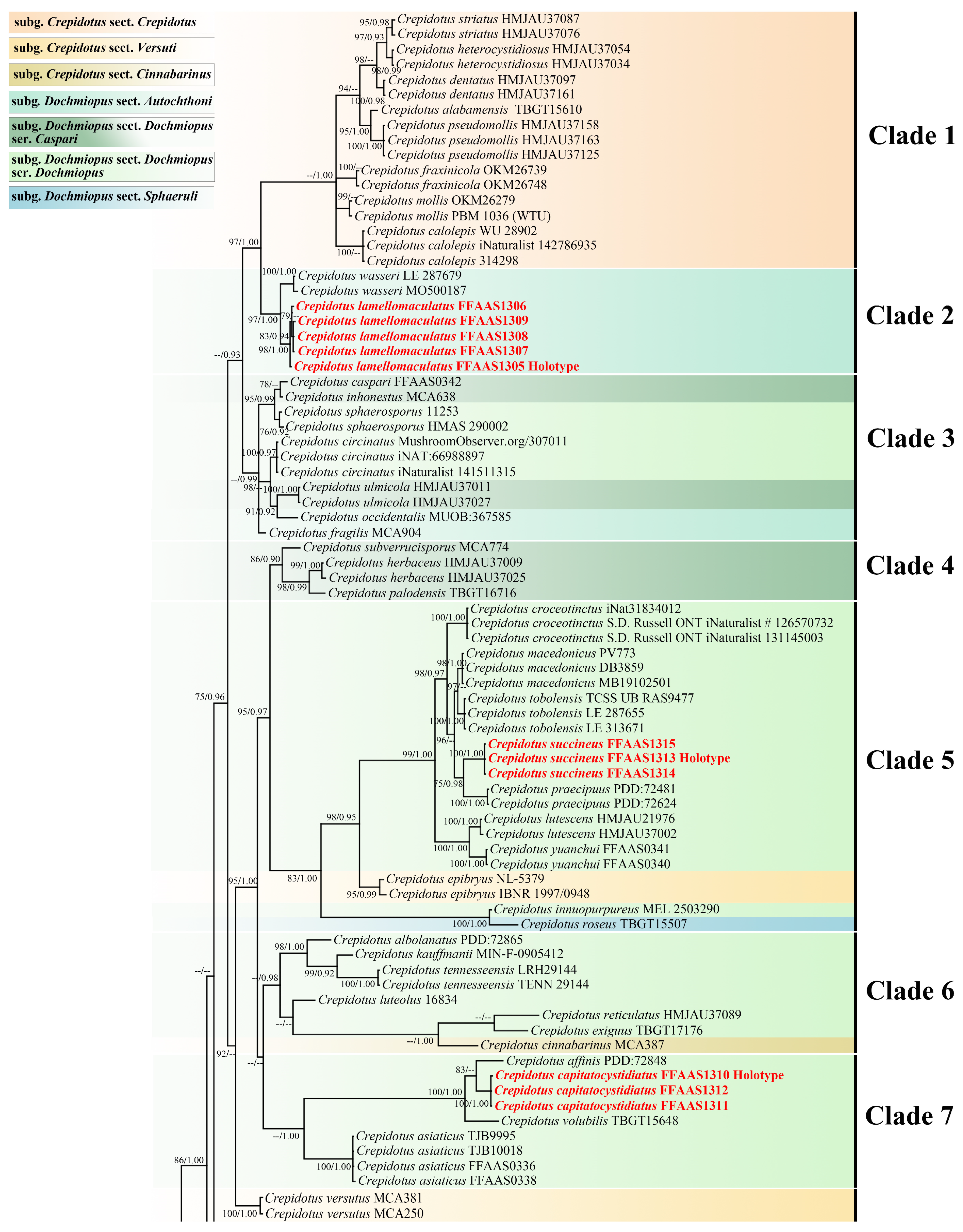
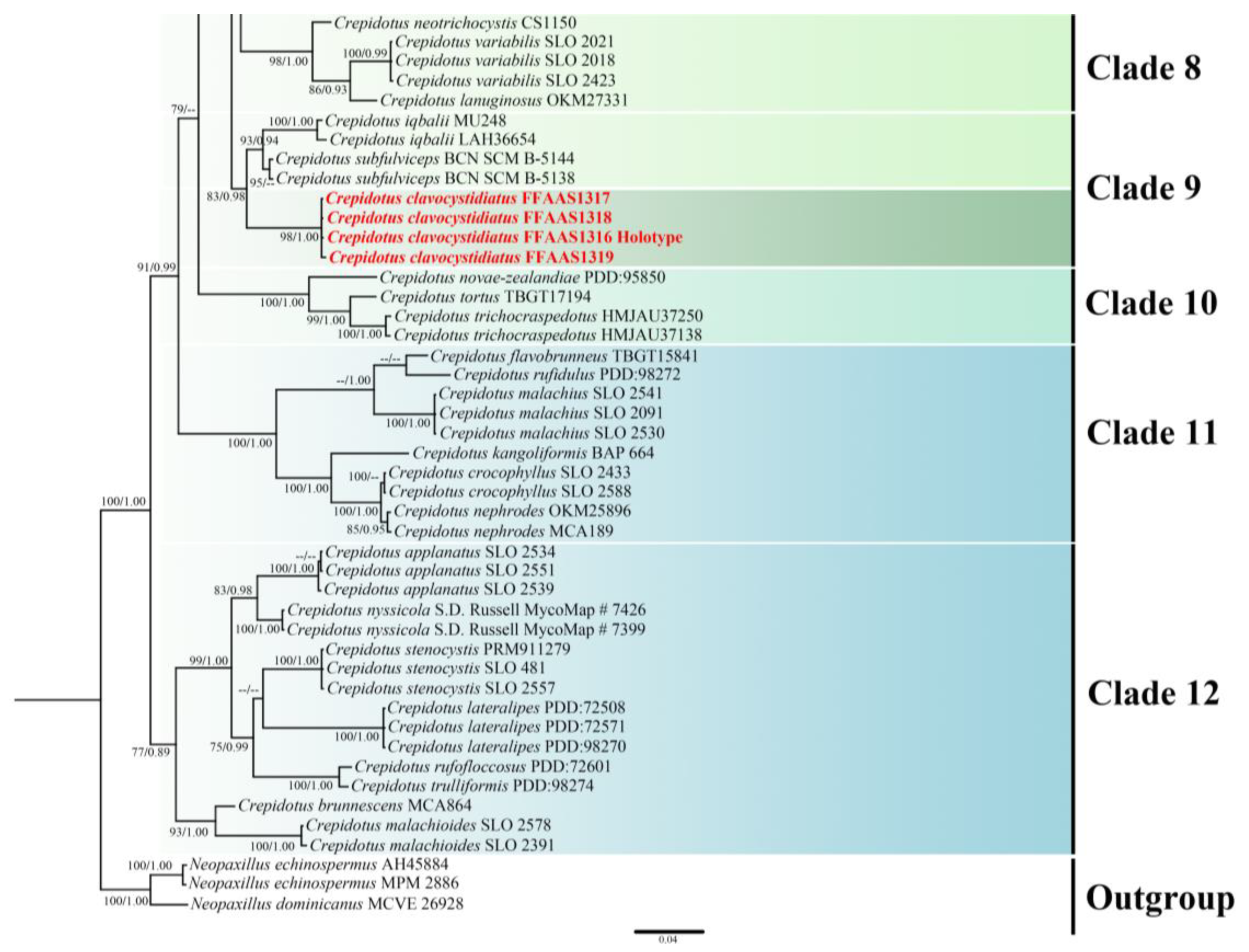
3.1. Phylogenetic Analysis
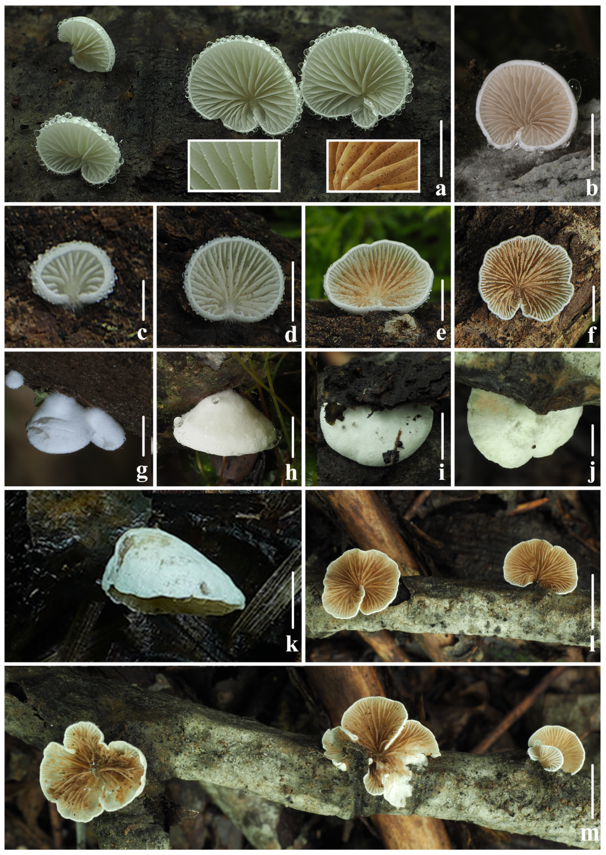
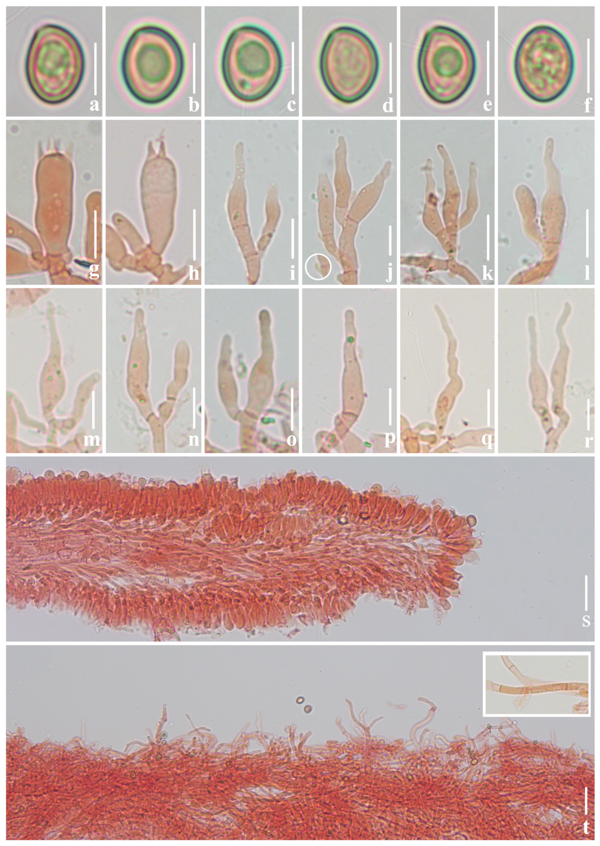
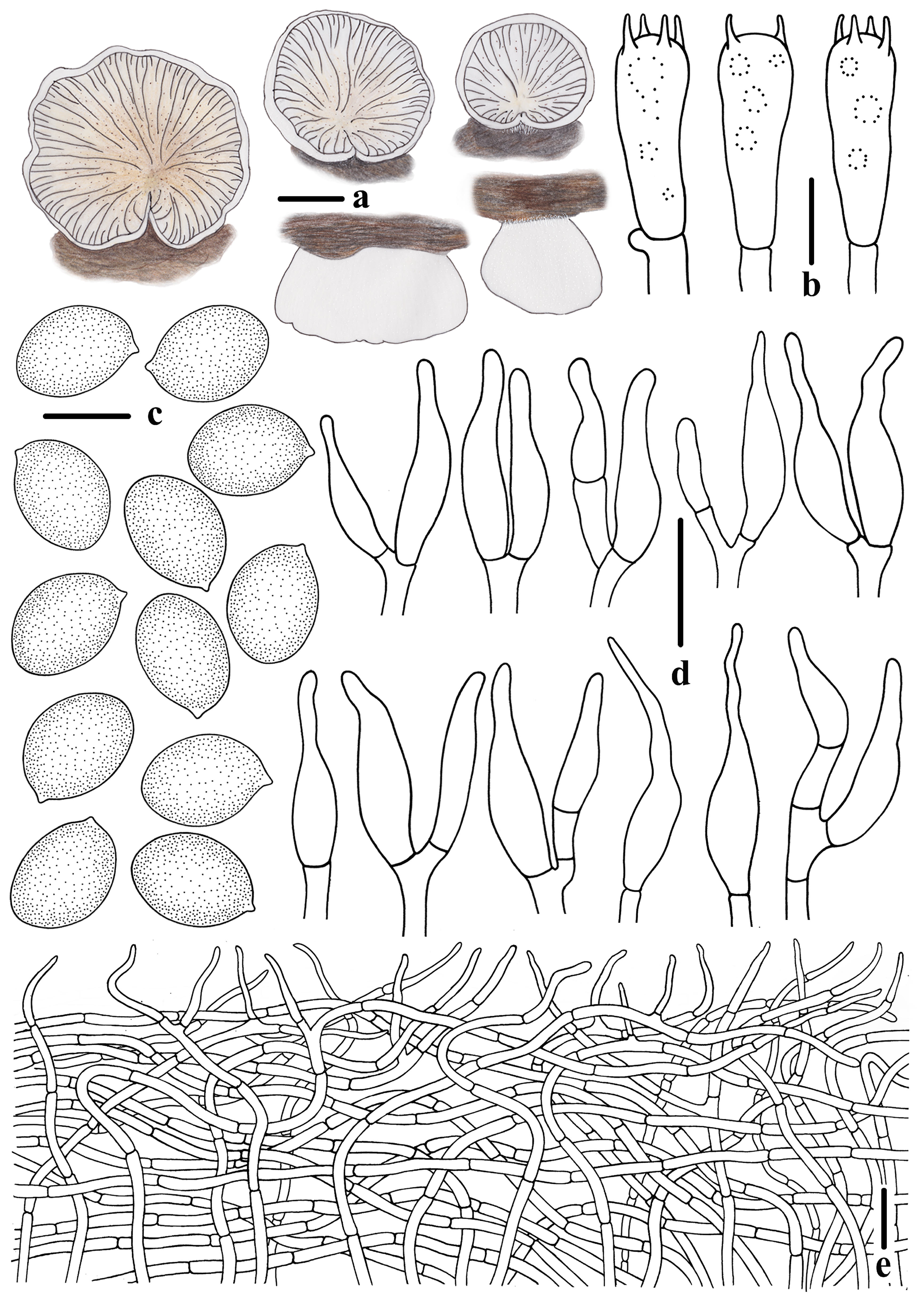
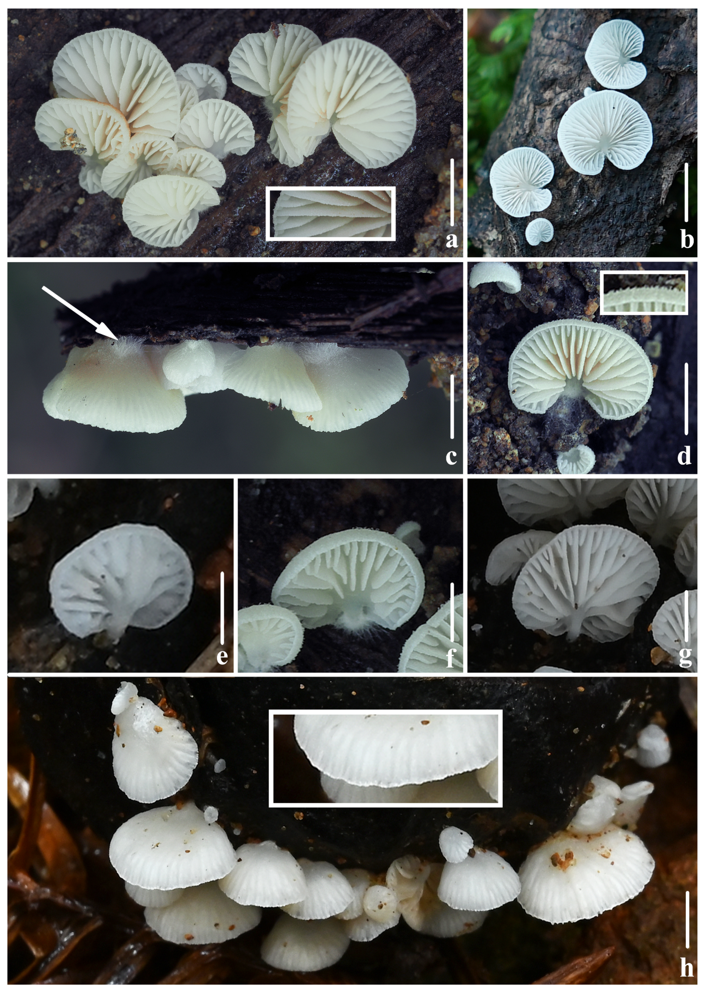
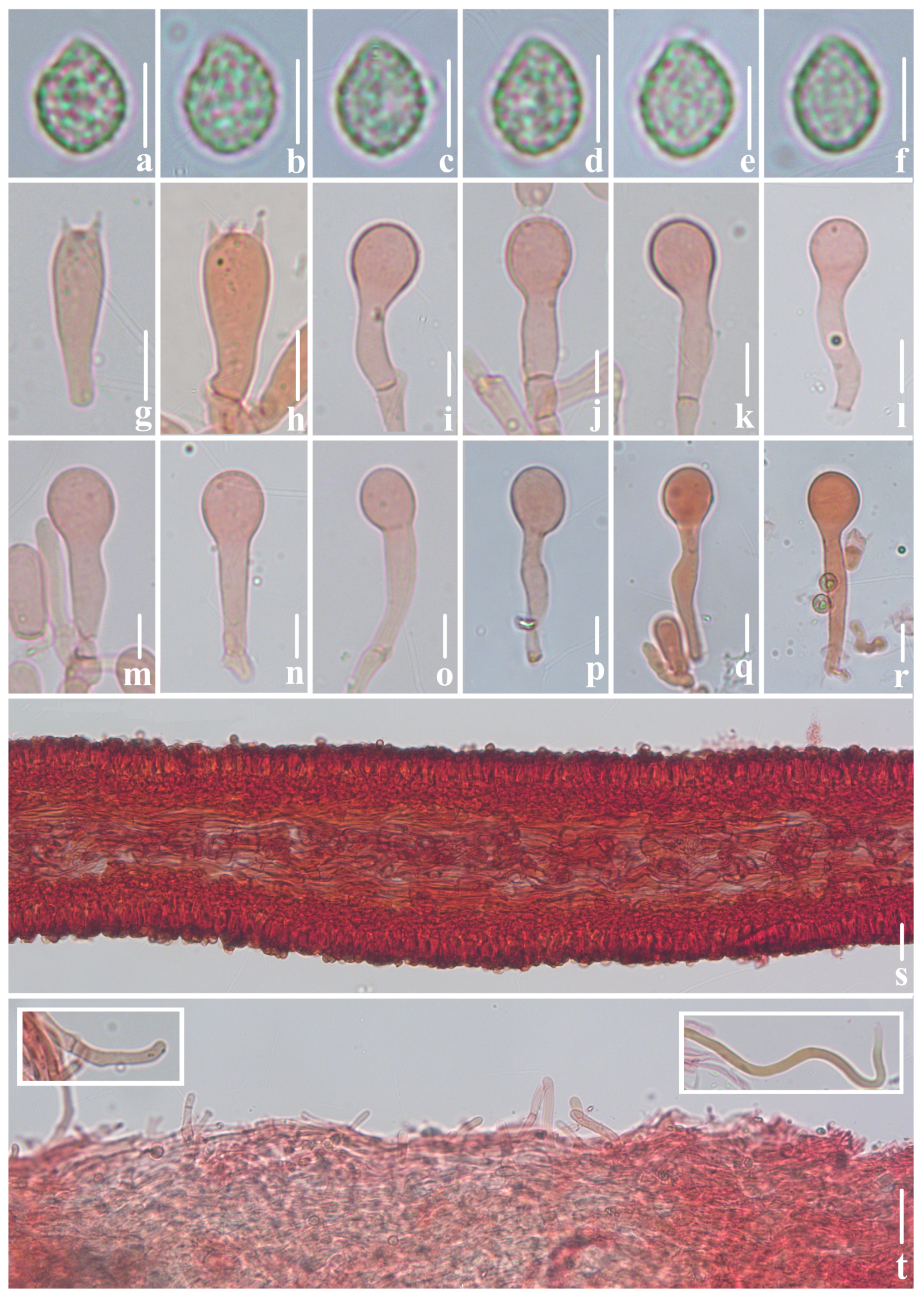
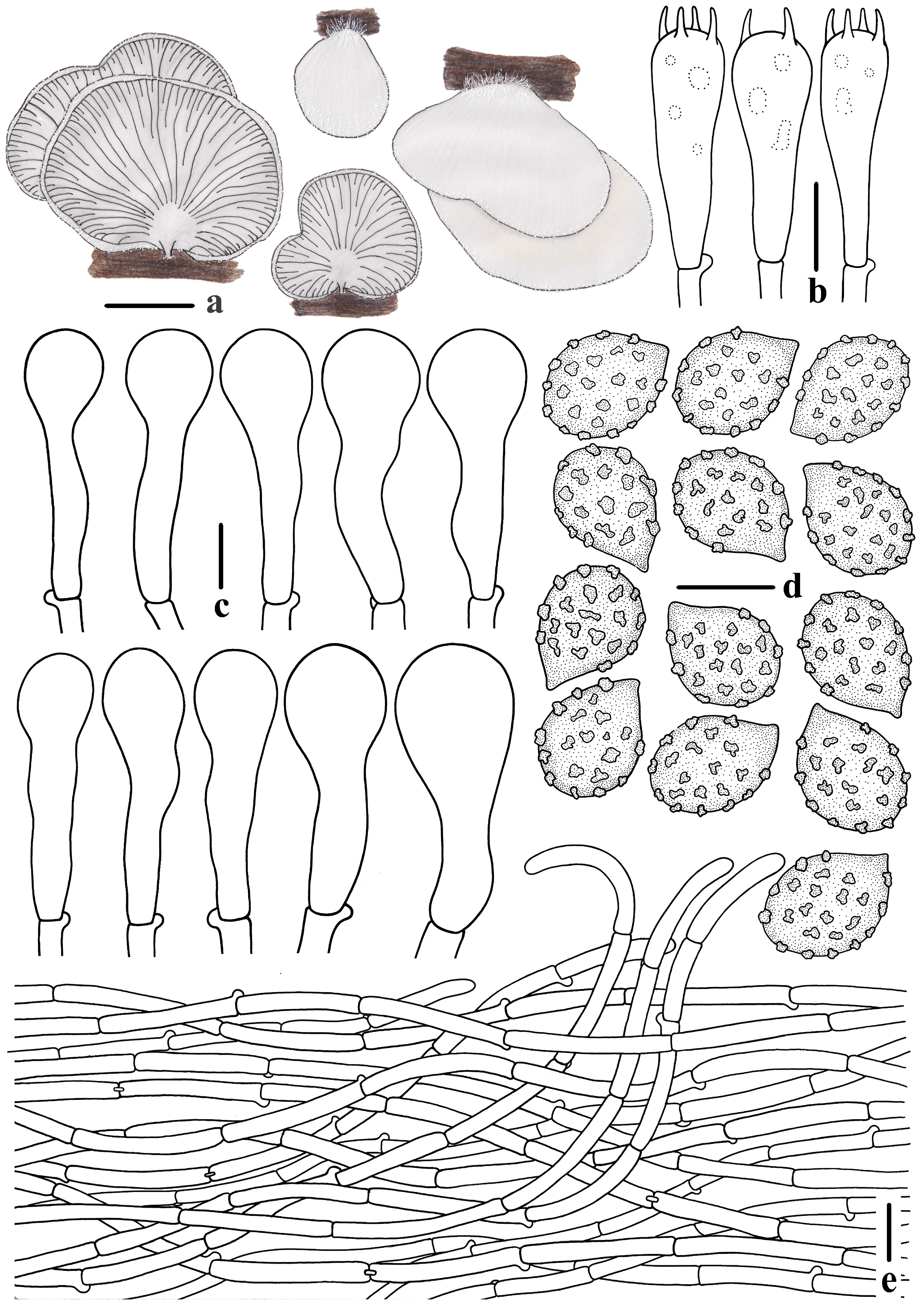
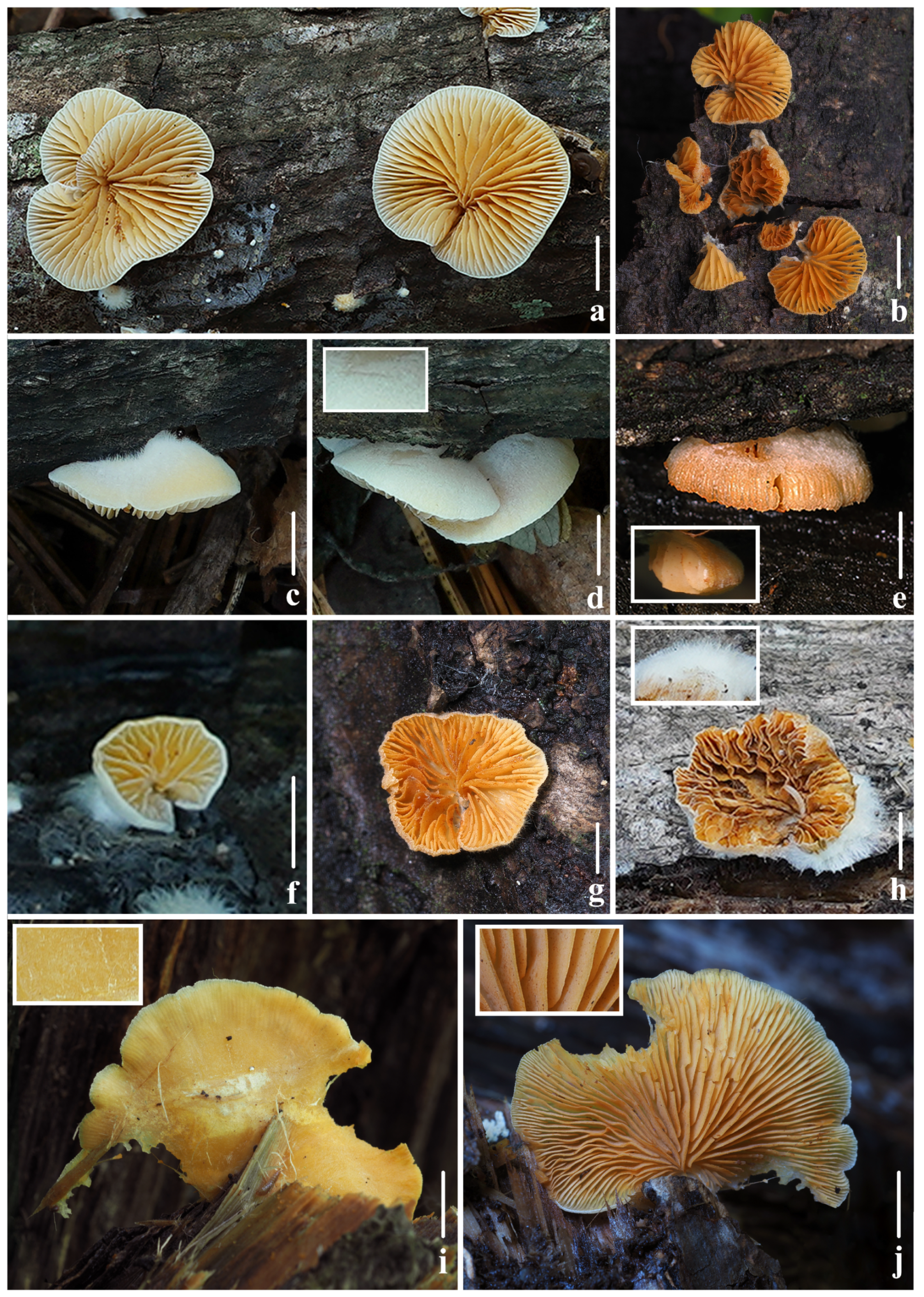
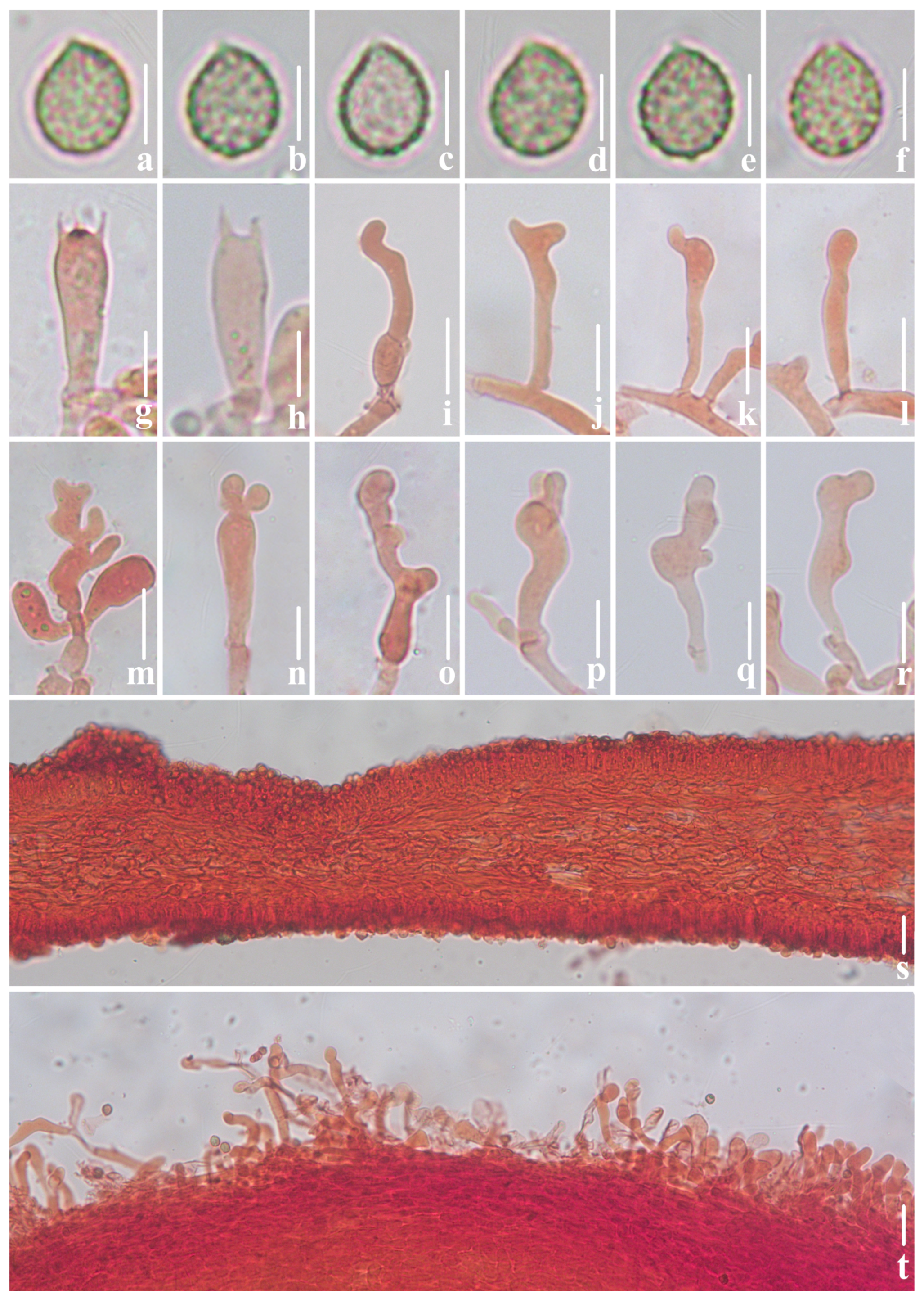
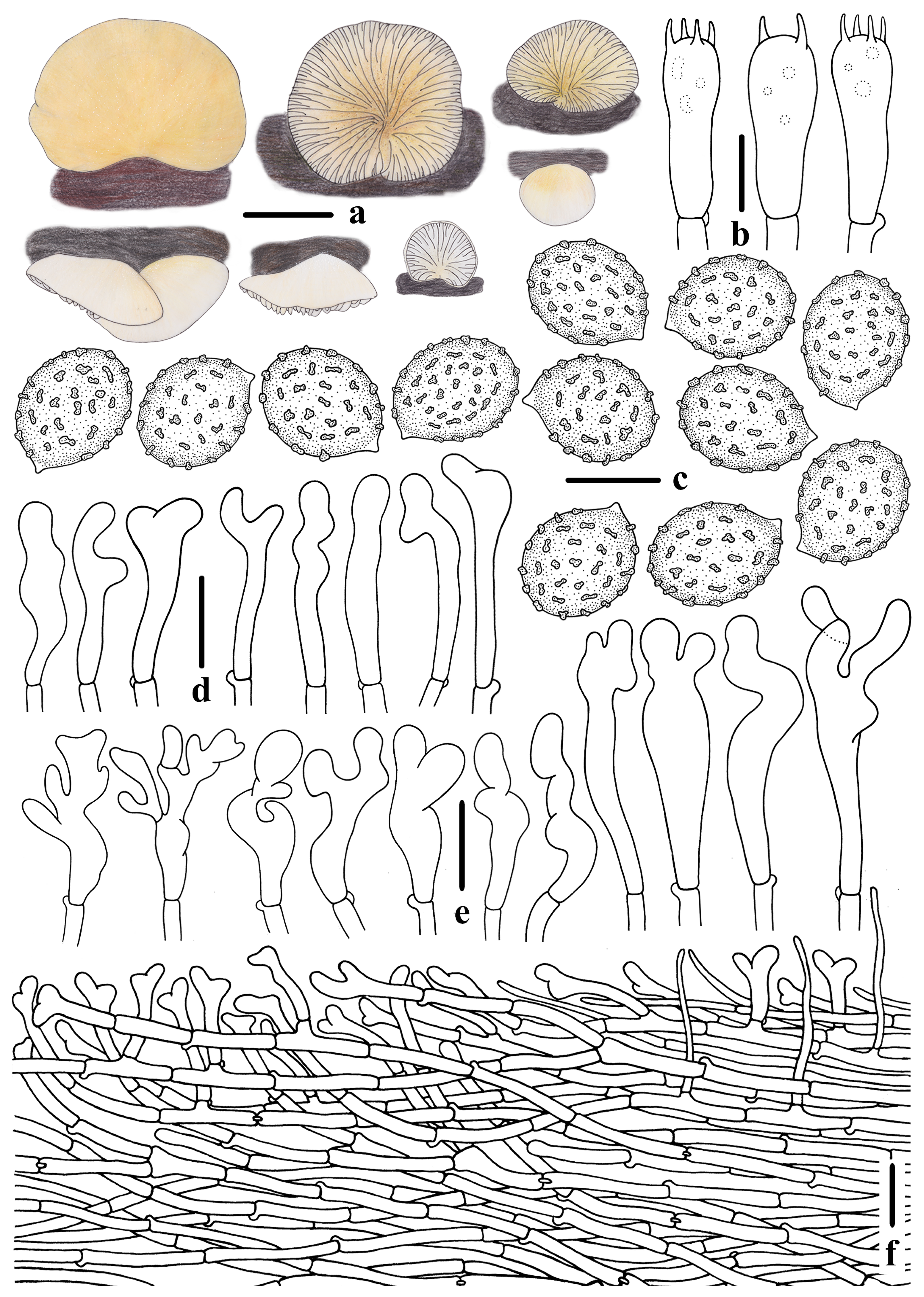
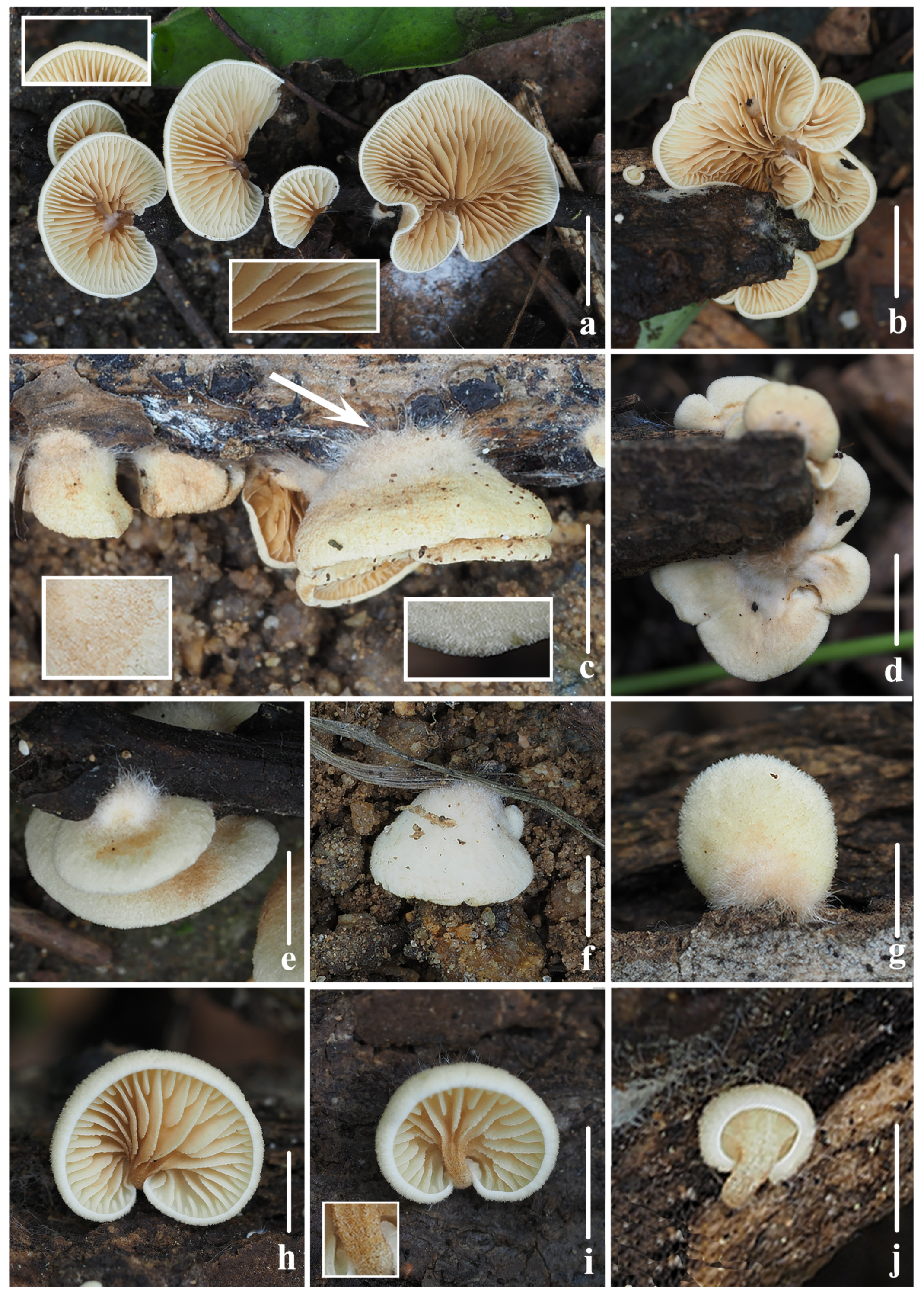
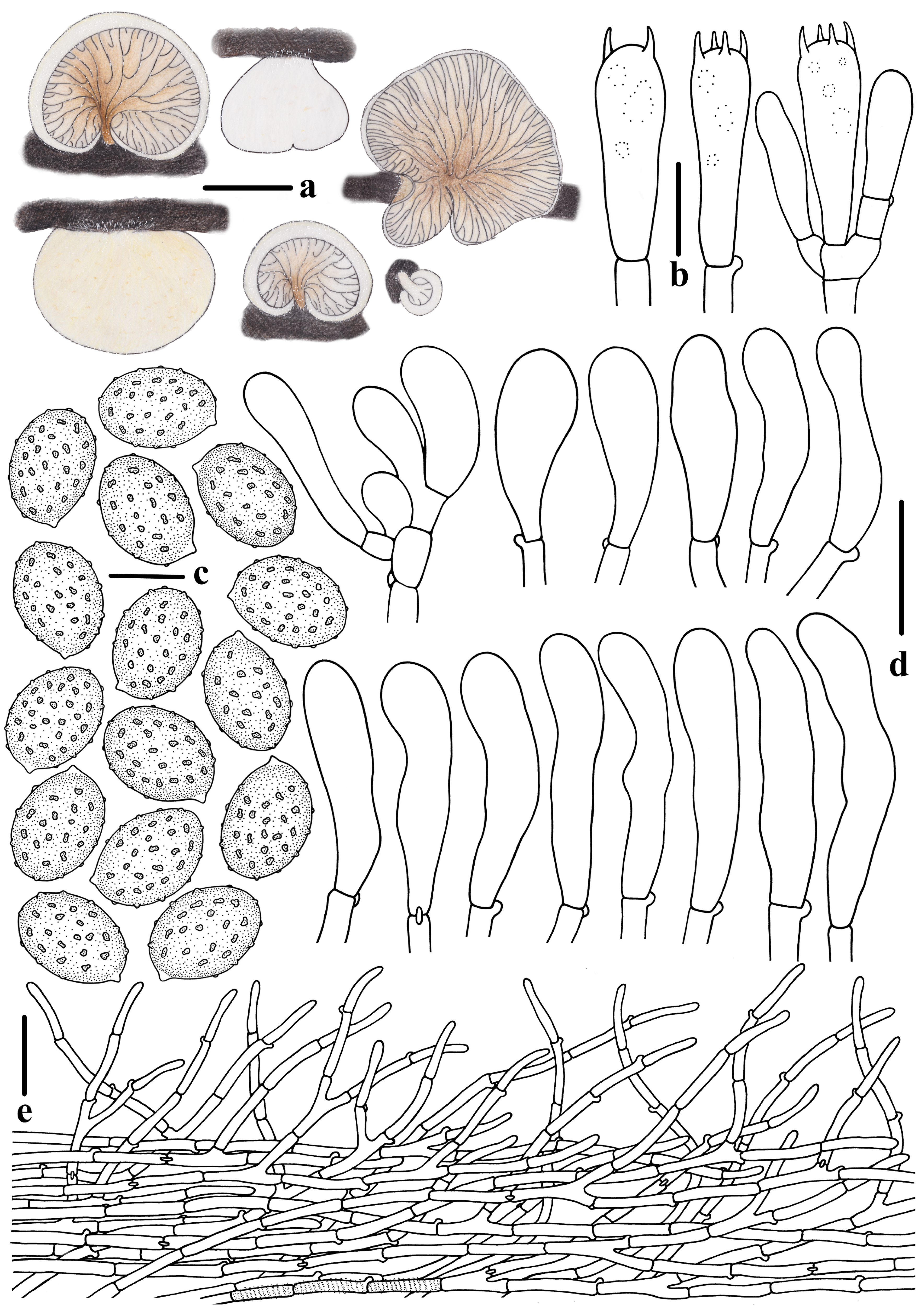
The dataset matrix was composed of 68 taxa, 126 samples, 174 sequences (104 ITS, and 70 LSU). Among these, 144 sequences were downloaded from GenBank (89 ITS, and 55 LSU), and 30 sequences (15 ITS, and 15 LSU) were amplified in this study. Neopaxillus dominicanus Angelini & Vizzini and N. echinospermus (Speg.) Singer were selected as the outgroups [53]. The aligned length, including gaps, encompassed 1782 nucleotide sites, with 861 for ITS and 921 for LSU. For BI analysis, the best-fit substitution model for both ITS and LSU was GTR + I + G. BI analysis resulted in an average standard deviation of split frequencies = 0.005577, the effective sample size (ESS) was 1333.8, and the average Potential Scale Reduction Factor (PSRF) parameter values = 1.000 after 5,000,000 MCMC generations. For ML analysis, the best-fit substitution model selected for ITS was TVM + I + G4 and that for LSU was GTR + I + G4, with a final log-likelihood value of −14,404.410766. The average bootstrap value was 87.17%, indicating generally high confidence in the tree’s branches. The BI and ML trees exhibited similar topologies, and the BI tree is shown in Figure 1. Those nodes supported by bootstrap of at least 75% and posterior probabilities exceeding 0.90 were incorporated.
According to the phylogenetic tree in Figure 1, twelve high-support clades were delineated. Clade 1 comprises eight species: C. striatus T. Bau & Y.P. Ge, C. heterocystidiosus T. Bau & Y.P. Ge, C. dentatus T. Bau & Y.P. Ge, C. alabamensis Murrill, C. pseudomollis T. Bau & Y.P. Ge, C. fraxinicola Murrill, C. mollis (Schaeff.) Staude, and C. calolepis (Fr.) P. Karst. This clade has strong support in BI analysis (BS/BPP = --/1.00); the species in this clade exhibit smooth elongated spores and their tissues do not have clamp connections. Based on Consiglio and Setti’s classification, all eight species in this clade belong to C. subg. Crepidotus sect. Crepidotus [6]. Clade 2 includes C. wasseri Kapitonov, Biketova, Zmitr. & Á. Kovács and C. lamellomaculatus, with strong support (BS/BPP = 97/1.00). The new species C. lamellomaculatus forms a well-supported clade (BS/BPP = 98/1.00).Both C. wasseri and C. lamellomaculatus possess smooth elongated spores and clamp connections [52]. Typically, species in Crepidotus with clamp connections exhibit ornamented basidiospores [4,6,12]. However, some species exhibit smooth basidiospores and clamp connections simultaneously, such as C. wasseri and C. lamellomaculatus in Clade 2, and C. novae-zealandiae Pilát, C. tortus A.M. Kumar & C.K. Pradeep, and C. trichocraspedotus T. Bau & Y.P. Ge in Clade 10 [11,12,34,52]. According to Consiglio & Setti’s classification, Clade 2 and Clade 10, characterized by clamp connections and smooth elongated spores, should be classified within C. subg. Dochmiopus sect. Autochthoni (Sennn-Irlet) Consiglio & Setii [6]. However, the current phylogenetic tree does not support this classification. Additionally, Clade 2 and Clade 10 exhibit significant differences in the morphology of their cheilocystidia. In Clade 2, the cheilocystidia of C. wasseri and C. lamellomaculatus are both lageniform and ventricose, whereas in Clade 10, the cheilocystidia are sinuously cylindrical to vine-like [52]. Clade 3 comprises C. caspari Velen., C. inhonestus P. Karst., C. sphaerosporus (Pat.) J.E. Lange, C. circinatus Hesler & A.H. Sm., C. ulmicola T. Bau & Y.P. Ge, C. occidentalis Hesler & A.H. Sm., and C. fragilis Joss. Among these, C. occidentalis and C. fragilis are classified in C. subg. Dochmiopus sect. Autochthoni, due to their smooth basidiospores and clamp connections [5,6]. The remaining taxa belong to C. subg. Dochmiopus sect. Dochmiopus. Specifically, C. sphaerosporus and C. circinatus are part of C. subg. Dochmiopus sect. Dochmiopus ser. Dochmiopus Consiglio & Setii, whereas C. caspari, C. inhonestus and C. ulmicola are classified in C. subg. Dochmiopus sect. Dochmiopus ser. Caspari Consiglio & Setii [4,6,20]. Although C. ulmicola was initially placed in C. subg. Dochmiopus sect. Dochmiopus ser. Dochmiopus when published, its smooth to nearly smooth basidiospores suggest a reclassification to C. subg. Dochmiopus sect. Dochmiopus ser. Caspari [20]. Additionally, C. sphaerosporus has been accepted as C. cesatii (Rabenh.) Sacc., and C. fragilis was combined into C. autochthonus [1,6]. Clade 4 includes three species, C. subverrucisporus Pilát, C. herbaceus T. Bau & Y.P. Ge and C. palodensis C.K. Pradeep & A.M. Kumar, all of which are regarded as C. subg. Dochmiopus sect. Dochmiopus ser. Caspari. These species are characterized by clavate cheilocystidia and a gregarious habit [1,10,20]. Clade 5 incorporates the new species C. succineus (BS/BPP = 100/1.00) along with nine other taxa. Except for C. roseus Singer and C. epibryus (Fr.) Quél., other taxa in this clade belong to C. subg. Dochmiopus sect. Dochmiopus ser. Dochmiopus. Crepidotus roseus is classified in C. subg. Dochmiopus sect. Sphaeruli due to its nearly globose basidiospores (Qm < 1.10) [6,11]. As C. epibryus has smooth elongated spores and absent clamp connections, it is placed in C. subg. Crepidotus sect. Versuti Hesler & A.H. Sm.; C. versutus (Peck) Sacc. also belongs to this section for similar reasons [5,6]. Clade 5 contains several brightly colored species, including the yellow-to-orange C. tobolensis Kapitonov, Biketova & Zmitr., C. succineus, C. praecipuus E. Horak, C. lutescens T. Bau & Y.P. Ge, C. yuanchui Q. Na, Z.W. Liu & Y.P. Ge, and pink-to-red-colored C. innuopurpureus McMull.-Fish., T. Lebel & Senn-Irlet, and C. roseus. In Clade 6, except C. cinnabarinus Peck, the other taxa belong to C. subg. Dochmiopus sect. Dochmiopus ser. Dochmiopus, and C. kauffmanii Hesler & A.H. Sm., C. reticulatus T. Bau & Y.P. Ge and C. cinnabarinus are notably colored species. Crepidotus cinnabarinus Peck is classified in C. subg. Crepidotus sect. Cinnabarinus Hesler & A.H. Sm., due to its ornamented basidiospores and absence of clamp connections [6,27]. Additionally, we concur with the assessment by Jančovičová et al. regarding the GenBank accession numbers for C. reticulatus [60]. Upon reviewing the original publication, we confirmed that the holotype of C. reticulatus is correctly associated with the GenBank sequence MF461346. The sequence was previously misassigned, and we have submitted a correction request to GenBank to rectify this error. Among Clade 5 and Clade 6, C. epibryus, C. versutus and C. cinnabarinus, which present absent clamp connections, cannot be classified in C. subg. Crepidotus based on the current phylogenetic analysis. This suggests that the absence of clamp connections is not an exclusive characteristic of C. subg. Crepidotus, and other subgenera also include taxa without clamp connections. Therefore, the defining characteristic of C. subg. Crepidotus requires further study and refinement. All species in Clade 7 belong to C. subg. Dochmiopus sect. Dochmiopus ser. Dochmiopus. Within this clade, the new species C. capitatocystidiatus is well supported (BS/BPP = 100/1.00). Crepidotus capitatocystidiatus clusters with C. affinis E. Horak and C. volubilis C.K. Pradeep & A.M. Kumar, sharing similar guttiform basidiospores covered by irregularly punctate-verrucae [10,34]. Notably, C. asiaticus Guzm.-Dáv., C.K. Pradeep & T.J. Baroni in Clade 7, C. iqbalii A. Izhar, Usman & Khalid and C. subfulviceps (Murrill) Aime, Vila & P.-A. Moreau in Clade 9 differ from the typical Crepidotus species due to their agaricoid basidiomata, representing the stipitate group within Crepidotus. Clade 8 comprises three species within C. subg. Dochmiopus sect. Dochmiopus ser. Dochmiopus, characterized by their white basidiomata with a finely villous pileus surface. Clade 9 includes three species, C. iqbalii, C. subfulviceps and C. clavocystidiatus. The new species C. clavocystidiatus is strongly supported (BS/BPP = 98/1.00). Crepidotus clavocystidiatus belongs to C. subg. Dochmiopus sect. Dochmiopus ser. Caspari, and the other two species are classified in C. subg. Dochmiopus sect. Dochmiopus ser. Dochmiopus. The taxa of Clade 11 and Clade 12 all possess clamp connections and globose ornamented basidiospores and are classified as C. subg. Dochmiopus. sect. Sphaeruli. But in the phylogenetic tree, Clade 11 and Clade 12 do not form a monophyletic group, indicating that C. subg. Dochmiopus sect. Sphaeruli is polyphyletic. This aligns with the viewpoint proposed by Ge, which is further corroborated by our current phylogenetic analysis [61].

3.2. Taxonomy

Crepidotus lamellomaculatus M.H. Han, Q. Na, Y.P. Hu & Y.P. Ge, sp. nov., Figure 2, Figure 3 and Figure 4.
MycoBank no: 854914
Etymology: The epithet lamellomaculatae combines the Latin word ‘lamellae’ and the Latin adjective ‘maculatae’, meaning spotted. This name accurately describes the lamellae surface, which shows a few dark spots when matured.
Holotype: China. Hubei Province, Yichang City, Wufeng County, 19 June 2024, leg. Menghui Han, Jingwen Guo, Guanyu Qiu, Lijun Wang, Qin Na, and Yupeng Ge, 553 m asl, FFAAS1305 (collection number NJ5410).
Diagnosis: Pileus white, covered by short white felted tomentum, margin incurved, lamellae edge nearly smooth when matured, stipe observed in early stage; basidiospores (5.6)6.0–6.5–7.1(7.6) × (4.2)4.4–4.8–5.2(5.7) μm, broadly ellipsoid to ellipsoid, smooth, cheilocystidia narrowly lageniform with a curved elongated neck, ventricose. Differs from C. wasseri by smaller cheilocystidia and basidiospores, and absent of pileocystidia.
Description: Pileus 4–18 mm broad, in young stage conchoidal to flabelliform, White (LIII), when matured semicircular to reniform, sometimes petaloid, nearly applanate, White (LIII), when old or dry Pale Olive Buff (XL21‴f), margin incurved in all stages; dry, covered by short White (LIII) felted tomentum, margin glabrescent, non-striated, not hygrophanous, attached laterally or dorsally to the substratum, White (LIII) villosity observed near the point of attachment. Lamellae less than 1mm broad, L = 12–17, l = 3–7, subdecurrent, ventricose, when young White (LIII), edge slightly fimbriated, when matured Isabella Color (XXX19″i) to Light Brownish Olive (XXX19″k), surface with a few dark spots, and edge nearly smooth. Stipe observed in young stage, clavate to cylindrical, lateral, White (LIII), with age persisting as a knob in lateral, indistinct. Context thin (<5 mm thick), White (LIII). Odor and taste not distinctive.
Basidiospores (417/8/5)(5.6)6.0–6.5–7.1(7.6) × (4.2)4.4–4.8–5.2(5.7) μm, Q = (1.24)1.26–1.46(1.50), Qm = 1.35 ± 0.061, [HOLOTYPE (68/2/1)(6.2)6.3–6.8–7.2(7.4) × (4.3)4.6–4.9–5.2(5.3) μm, Q = (1.30)1.32–1.47(1.49), Qm = 1.39 ± 0.039], broadly ellipsoid to ovoid in frontal view, broadly ellipsoid to ellipsoid in lateral view, greyish orange to yellowish brown in 5% KOH aqueous solution, smooth (under oil), sometimes granular contents or large oil drop observed. Basidia 16–25 × 5–7 μm, short clavate, apex obtuse, base slightly constricted, four-spored, rarely two-spored, sterigmata 1.9–3.8 μm long, thin-walled (<0.5 μm thick), hyaline. Pleurocystidia absent. Cheilocystidia 17–41 × 3–6 μm, slightly clavate to utriform, narrowly lageniform with a curved neck, apex rounded, the middle part ventricose and slightly attenuated at the base, clumped, hyaline, thin-walled (<0.5 μm thick). Pileipellis a tomentum, composed of interwoven long cylindrical hyphae, 3–6 μm in diameter, sometimes branched, terminal cells flexuous, sometimes vertical to the pileus, thin-walled (<0.5 μm thick), hyaline, non-gelatinized. Lamellae trama subregular, composed of cylindrical hyphae, 3–6 μm in diameter, interwoven, at times inflated, non-gelatinized. Clamp connections present only in basidia and hymenium, occasionally observed in cheilocystidia.
Habit and habitat. Scattered on rotten branches and woods in the mixed broadleaf-conifer forests of temperate and subtropical zones, mainly found under Betula sp., Castanopsis sp., Cypressi sp., Cunninghamia sp., and Fraxinus sp.
Other specimens examined: China. Jilin Province, Yanbian Korean Autonomous Prefecture, Antu County, Changbaishan National Nature Reserve, 16 August 2021, leg. Menghui Han, Zewei Liu, Qin Na, and Yupeng Ge, 831m asl, FFAAS1306 (collection number GN1378), same location, 17 August 2021, leg. Menghui Han, Zewei Liu, Qin Na, and Yupeng Ge 843 m asl, FFAAS1307 (collection number GN1398), 841 m asl, FFAAS1308 (collection number GN1399); Heilongjiang Province, Hegang City, Luobei County, Taipinggou National Natural Reserve, 6 July 2023, leg. Menghui Han, Renxiu Wei, Tingting Sun, Zengcai Liu, Weiguo Chen, Xinyu Tong, Yawei Li, Nannan Geng, Li Zou, Qin Na, and Yupeng Ge, 476 m asl, FFAAS1309 (collection number GN1878).
Notes: Within the genus Crepidotus, the species with clamp connections typically exhibit ornamented basidiospores, while those lacking clamp connections generally present smooth basidiospores [1,5,6]. However, C. lamellomaculatus is an exceptional species within the genus, possessing both clamp connections and smooth basidiospores, and it is considered to belong to C. subg. Dochmiopus sect. Autochthoni. Before that, the only species in this section was C. autochthonus; it was distinguished from C. lamellomaculatus by bigger basidiospores (7.0–9.0 × 5.0–6.0 μm or 7.1–8.5 × 4.9–5.7 μm) and cylindrical to narrowly utriform cheilocystidia [1,6]. Phylogenetic analysis showed the C. wasseri is most related to C. lamellomaculatus, and there are many similarities in macroscopical, but C. wasseri can be distinguishable by its narrowly lageniform pileocystidia, bigger cheilocystidia [(33.1)37.1–59.6(74.0) × (3.9)4.6–7.3(8.4) μm] and basidiospores [(6.5)7.1–8.6(9.0) × (4.5)4.8–5.5(5.9) μm, av. = 7.8 × 5.2 μm] [50]. Crepidotus trichocraspedotus is a species discovered in China, possessing smooth basidiospores and clamp connections similar to C. lamellomaculatus, but it can be easily distinguished from bigger basidiospores [(8.8)9.1–10.5(10.7) × 6.0–6.8(7.1) μm], vine-shape with bifurcation cheilocystidia and a trichoderm pileipellis [12]. Crepidotus tortus and C. novae-zelandiae Pilát are characterized by white woolly-to-appressed squamulose pileus, smooth basidiospores, and clamp connections that resemble C. lamellomaculatus. However, both C. tortus and C. novae-zelandiae are distinguished from C. lamellomaculatus by possessing bigger basidiospores [C. novae-zealandiae (9.5)10–12 × (7)7.5–8 μm, C. tortus 8–10.5 × 5.5–6.5(6.8) μm] and flexuous cylindrical cheilocystidia [11,34].
Crepidotus capitatocystidiatus M.H. Han, H. Zeng, Q. Na & Y.P. Ge, sp. nov., Figure 5, Figure 6 and Figure 7.
MycoBank no: 854915
Etymology: The epithet capitatocystidiatus derives from the Latin words ‘capitata’, meaning capitate, and ‘cystidia’. The name is chosen to describe the cheilocystidia apex globose to capitate.
Holotype: China. Fujian Province, Nanping City, Wuyishan National Park, 13 August 2021, leg. Menghui Han, Zewei Liu, Junqing Yan, Hui Zeng, Qin Na, and Yupeng Ge, 728 m asl, FFAAS1310 (collection number NJ3779).
Diagnosis: Pileus white, margin tomentose, translucent striate, not hygrophanous; lamellae edge smooth, stipe present in young stage; basidiospores 4–6.2–7.3(7.7) × (4.1)4.3–5.0–5.7(6.2) μm, guttiform, covered by irregularly punctate-verrucae, cheilocystidia clavate with a globose apex, clamp connections present. Differs from C. palmarum Singer by pileus hygrophanous and distinguishable habitat.
Description: Pileus 6–25 mm broad, in young stage spatuliform, ungulate to conchoid, margin incurved, when matured flabelliform to rounded flabelliform, occasionally lobed, plano-convex, margin gradually straight; White (LIII), at times Chamois (XXX19″b) to Isabella color (XXX19″i) by basidiospores depositing on surface; White (LIII) pubescence when young, with age gradually sparse, when matured almost smooth, in all stages margin weakly tomentose; dry, translucent striate, not hygrophanous; pileus attached almost laterally, but some dorsally to the substratum, clear villosity near the point of attachment. Lamellae 0.5–1 mm broad, L = 11–23, l = 3–7, subdecurrent to decurrent, subventricose, edge weakly fimbriated; White (LIII) when young, becoming *Buff-Yellow (IV19d) to Baryta Yellow (IV21f), when matured Orange-Citrine (IV19k) to Buckthom Brown (XV17′i). Stipe present when young, cylindrical, when matured as a lateral knob or covered by lamellae, subtransparent. Context thin (<5 mm thick), White (LIII), hyaline. Odor and taste not distinctive.
Basidiospores (141/4/3)(5.3)5.4–6.2–7.3(7.7) × (4.1)4.3–5.0–5.7(6.2) μm, Q = (1.13)1.20–1.35(1.40), Qm = 1.26 ± 0.051, [HOLOTYPE (80/2/1)(5.3)5.4–5.9–6.5(7.4) × (4.2)4.3–4.7–5.4(5.8) μm, Q = (1.13)1.19–1.35(1.40), Qm = 1.25 ± 0.051], broadly fusiform to ovoid in frontal view, broadly ellipsoid to guttiform bottom slightly pointed in lateral view, greyish yellow to olive brown in 5% KOH aqueous solution, irregularly punctate-verrucose to verrucose (ornamentation verrucose type I), suprahilar depression weakly developed (under oil). Basidia 19–26 × 6–8 μm, clavate to broadly clavate, slightly constricted towards the base, four-spored, rarely two-spored, sterigmata 1.8–4.5 μm long, thin-walled (<0.5 μm thick), hyaline. Pleurocystidia absent. Cheilocystidia 29–60 × 8–18 μm, clavate to narrowly clavate with a globose to ovoid apex, middle portion more or less ventricose, constrict to base, clumped, hyaline, thin- to thick-walled (≤0.6 μm thick). Pileipellis a rectocutis, composed of cylindrical parallel hyphae, 4–8 μm diameter, sometimes interwoven, occasionally branched, thin-walled (<0.5 μm thick); some terminal cells semierect and bundled, contributed to the formation of short pubescence on pileus surface, hyaline, not-gelatinized, oleiferous hyphae observed occasionally. Lamellae trama subregular, composed of subcylindrical hyphae, 6–11 μm diameter, nearly parallel arrangement, more or less inflated, non-gelatinized. Clamp connections present in all tissues.
Habit and habitat: gregarious on deciduous twigs, rotten woods, and nutshell in mixed broadleaf-conifer forests of temperate and subtropical zones, mainly found under Betula sp., Tilia sp., Larix sp., and Pinus sp.
Other specimens examined: China. Fujian Province, Quanzhou City, Dehua County, Lingjiao Village, 30 April 2021, leg. Menghui Han, Zewei Liu, Hui Zeng, Qin Na, and Yupeng Ge, 457 m asl, FFAAS1311 (collection number GN0812); Jilin Province, Tonghua City, Baijifeng National Forest Park, 26 June 2023, leg. Menghui Han, Xiaoliang Liu, Renxiu Wei, Qin Na, and Yupeng Ge, 588 m asl, FFAAS1312 (collection number GN1547).
Notes: Crepidotus capitatocystidiatus is classified within C. subg. Dochmiopus sect. Dochmiopus ser. Dochmiopus due to its verrucose basidiospores and clamped hyphae [6]. Within this series, C. cesatii bears a resemblance to C. capitatocystidiatus, but it can be distinguished by its pileus margin striated, fimbriated lamellae edge, and irregular cheilocystidia, which generally knobby or branched [1,6]. Crepidotus palmarum also shares many similarities with C. capitatocystidiatus due to guttiform basidiospores and capitate cheilocystidia. However, C. palmarum is macroscopically distinct by its hygrophanous pileus, smooth margin, and sessile. Furthermore, C. palmarum and C. capitatocystidiatus exhibit significant differences in habitat and distribution. Crepidotus palmarum was recorded in tropical and subtropical America, gregarious on leaves and leaf petioles of Trachycarpus sp. or rotten trunk of Quercus sp., whereas C. capitatocystidiatus is a species that occurred in temperate and subtropical China, gregarious on rotten branches and woods of Betula sp., Tilia sp., Larix sp., and Pinus sp. [27,62]. Additionally, C. affinis and C. volubilis are close to the new species in phylogeny analysis, while C. affinis can be distinguished by its globose basidiospores and slender cheilocystidia [(35)45–65 × 5–10 μm] [34]. Crepidotus volubilis is different from the new species in ventricose cheilocystidia without capitate and pileus margin straight to slightly upturned [10].
Crepidotus succineus M.H. Han, L. Zou, Q. Na & Y.P. Ge, sp. nov., Figure 8, Figure 9 and Figure 10.
MycoBank no: 854916
Etymology: The epithet is from the Latin word ‘succineus’, meaning amber. The name reflects the color of the basidiomata.
Holotype: China. Heilongjiang Province, Hegang City, Luobei County, Taipinggou National Natural Reserve, 5 July 2023, leg. Menghui Han, Renxiu Wei, Tingting Sun, Zengcai Liu, Weiguo Chen, Xinyu Tong, Yawei Li, Nannan Geng, Li Zou, Qin Na, and Yupeng Ge, 454 m asl, FFAAS1313 (collection number GN1824).
Diagnosis: Pileus buff yellow to apricot yellow, covered by short villosity, margin straight, translucent striate, not hygrophanous; basidiospores (5.2)5.7–6.3–7.0(7.4) × (4.5)4.7–5.3–5.8(6.0) μm, guttiform and verrucose, cheilocystidia usually irregularly knobbed in upper portion, pileipellis a clavicutis, flexuous clavate pileocystidia observed. Differs from C. praecipuus by variable cheilocystidia and a clavicutis pileipellis with pileocystidia.
Description: Pileus 6–26 mm broad, in young stage spathuliform to rounded reniform to petaloid, margin weakly incurved, Naphthalene Yellow (XVI23′f) to *Cream Color (XVI19′f), with age flabelliform to semicircular, when matured applanate and margin progressively straight, *Buff Yellow (IV19d) to Apricot Yellow (IV19b); dry, covered by White (LIII) short tomentum when immature, transitioning to slight pubescence when matured, translucent striate, not hygrophanous; mostly dorsally attached to the substratum, attached point covered by White (LIII) tomentum. Lamellae less than 2 mm broad, L = 9–20, l = 3–9, adnate to subdecurrent, ventricose, when young *Cream Color (XVI19′f) to *Naples Yellow (XVI19′d), edge slightly fimbriated, when matured *Buff Yellow (IV19d) to Mustard Yellow (XVI19′b), surface with a few dark spots, edge smooth. Stipe obvious when young, nearly cylindrical, White (LIII), subtransparent, when matured becoming an eccentric knob in lateral. Context thin (<5 mm thick), subtranslucent. Odor and taste not distinctive.
Basidiospores (238/5/3)(5.2)5.7–6.3–7.0(7.4) × (4.5)4.7–5.3–5.8(6.0) μm, Q =(1.09)1.13–1.27(1.38), Qm = 1.20 ± 0.045, [HOLOTYPE (146/3/1)(5.2)5.7–6.3–6.9(7.4) × (4.5)4.8–5.3–5.8(6.0) μm, Q = (1.09)1.12–1.26(1.38), Qm = 1.19 ± 0.043], subglobose to broadly ellipsoid in frontal view, broadly ellipsoid to guttiform in lateral view, greyish yellow to olive brown in 5% KOH aqueous solution, irregularly punctate-verrucose to verrucose (ornamentation verrucose type I), suprahilar depression weakly developed (under oil). Basidia 18–26 × 5–8 μm, clavate, gradually constricted to base, four-spored, rarely two-spored, sterigmata 2.2–4.2 μm long. Pleurocystidia absent. Cheilocystidia 27–70 × 3–7 μm, predominantly contorted narrowly utriform to slender clavate, bifurcated, digitiform to irregularly knobbed branching at middle part and apex, occasionally coralline in densely branched specimens, and taper towards a constricted base, hyaline, thin-walled (<0.5 μm thick). Pileipellis a clavicutis, composed of cylindrical hyphae, 4–7 μm diameter, sometimes interwoven, occasionally branched, nearly hyaline, thin-walled (<0.5 μm thick), not-gelatinized; a few terminal cells slender and semierect, contributing to short villosity on pileus surface, majority terminal cells form pileocystidia, 24–63 × 3–9 μm, clavate, flexuous, sometimes the upper portion branched. Lamellae trama subregular, composed of cylindrical hyphae, 5–11 μm diameter, tightly arranged, slightly inflated, non-gelatinized. Clamp connections present in all tissues.
Habit and habitat: scattered on rotten branches in temperate mixed broadleaf-conifer forests, mainly found under Betula sp., Fraxinus sp., Pinus sp., and Tilia sp.
Other specimens examined: China. Jilin Province, Yanbian Korean Autonomous Prefecture, Antu County, Changbaishan National Nature Reserve, 28 June 2023, leg. Menghui Han, Renxiu Wei, Bai Wang, Qin Na, and Yupeng Ge, 1309 m asl, FFAAS1314 (collection number GN1064). Heilongjiang Province, Hegang City, Luobei County, Taipinggou National Natural Reserve, 6 July 2023, leg. Menghui Han, Renxiu Wei, Tingting Sun, Zengcai Liu, Weiguo Chen, Xinyu Tong, Yawei Li, Nannan Geng, Li Zou, Qin Na, and Yupeng Ge, 462 m asl, FFAAS1315 (collection number GN1917).
Notes: Due to clamp connections and irregularly punctate-verrucose to verrucose basidiospores, C. succineus is considered a member of C. subg. Dochmiopus sect. Dochmiopus ser. Dochmiopus [6]. In this series, C. luteolus Sacc. shares similar basidiomata, but it can be distinguished by oblong-to-cylindrical basidiospores (Q = 1.63–1.99 or Q = 1.55–2.10), cylindrical to narrowly lageniform cheilocystidia, and absent pileocystidia [6,62]. Phylogenetically, C. praecipuus is closed to the new species, resembling white tomentum at the point of attachment and guttiform basidiospores [34]. However, cheilocystidia of C. praecipuus are broadly clavate or vesiculose, and pileipellis is a cutis without pileocystidia [34]. Crepidotus tobolensis possesses similar basidiomata color and basidiospores to C. succineus, but is distinguished by hygrophanous pileus, denser lamellae, and a cutis pileipellis [50]. There are two species with similar basidiomata color to C. succineus from China, C. yuanchui and C. lutescens, but they can be easily distinguished by pileipellis type and morphology of cheilocystidia [4,7].
Crepidotus clavocystidiatus M.H. Han, Q. Na & Y.P. Ge, sp. nov., Figure 11, Figure 12 and Figure 13.
MycoBank no: 854917
Etymology: The epithet clavocystidiatus combines the Latin adjective ‘clavatus’ and the Latin word ‘cystidia’. This name describes the shape of the cheilocystidia, which is clavate to narrowly clavate.
Holotype: China. Shandong Province, Tai’an City, Culaishan National Forest Park, 23 July 2021, leg. Menghui Han, Zewei Liu, Yulan Sun, Qin Na, and Yupeng Ge, 883 m asl, FFAAS1316 (collection number GN1297).
Diagnosis: Pileus ivory yellow, covered by dense white flat villosity in all stages, white dense fibrillose hyphae observed on attachment of the substratum, non-striated; stipe present, pruinose; basidiospores (5.9)6.1–6.7–7.3(7.7) × (4.1)4.3–4.7–5.1(5.3) μm, smooth to faintly verrucose, pileipellis a rectocutis to plagiotrichoderm, clamp connections present. Differs from C. caspari var. caspari Velen. by colored stipe and smaller basidiospores.
Description: Pileus 5–14 mm broad, in young stage umbonate to conchoid or ungulate, White (LIII) to *Suphur Yellow (V25f), becoming flabelliform to semicircular, at times somewhat petaloid, when matured nearly applanate, Ivory Yellow (XXX21″f) to Naphthalene Yellow (XVI23′f), margin incurved, at times Isabella Color (XXX19″i) by basidiospores depositing on surface; dry, covered by White (LIII) flat villosity, margin weakly tomentose, non-striated, not hygrophanous, pileus attached laterally to the substratum, clearly fibrillose hyphae observed near the point of attachment. Lamellae less than 1 mm broad, L = 12–22, l = 3–9, free or subdecurrent, ventricose, edge fimbriated, when young Marguerite Yellow (XXX23″f) to Old Gold (XV19′i), becoming Pale Oliver-Buff (XL21″f) to Deep Olive-Buff (XL21″b), when matured Chamois (XXX19″b). Stipe observed in all stages, slightly clavate to cylindrical, lateral, *Olive-Buff (XL21″d) to Isabella Color (XXX19″i), pruinose, with age persisting as a bent cylinder or occasionally lateral knob. Context thin (<5 mm thick), White (LIII), subtranslucent. Odor and taste not distinctive.
Basidiospores (174/5/4)(5.9)6.1–6.7–7.3(7.7) × (4.1)4.3–4.7–5.1(5.3) μm, Q = (1.25)1.30–1.55(1.61), Qm = 1.41 ± 0.074, [HOLOTYPE (77/2/1)(5.9)6.2–6.7–7.2(7.7) × (4.2)4.4–4.8–5.2(5.3) μm, Q = (1.25)1.31–1.52(1.59), Qm = 1.40 ± 0.065], broadly ellipsoid to ellipsoid in frontal view, ovoid to ellipsoid in lateral view, greyish yellow to olive brown in 5% KOH aqueous solution, smooth to faintly verrucose (under oil, ornamentation rugulose-verruculose type I), sometimes granular contents or large oil drop are observed. Basidia 21–31 × 6–8 μm, clavate, four-spored, rarely two-spored, sterigmata 1.8–4.8 μm long, thin-walled (<0.5 μm thick), hyaline. Pleurocystidia absent. Cheilocystidia 28–45 × 6–12 μm, clavate to narrowly clavate, sometimes apex round or subcapitate, not forked, base contracted, flexuous, clumped, hyaline, thin-walled (<0.5 μm thick). Pileipellis a rectocutis to plagiotrichoderm, composed of densely arranged cylindrical hyphae, interwoven and branched, 5–9 μm wide, thin- to thick-walled (0.3–0.9 μm), weakly yellow-brown hyphae, some terminal cells erected, forming short-villosity on pileus surface, nearly hyaline, a few hyphae encrusted, non-gelatinized. Lamellae trama intermixed, composed of subparallel cylindrical hyphae 3–6 μm, more or less inflated, physalohyphae observed near trama base, 8–18 μm, measured up to 33 μm diameter, subregular, non-gelatinized. Clamp connections present in all tissues.
Habit and habitat: scattered on rotten twigs in temperate mixed broadleaf-conifer forests, mainly were found under Fagaceae sp., Pinaceae sp., Populus sp., and Quercus sp.
Other specimens examined: China. Shandong Province, Tai’an City, Culaishan National Forest Park, 23 July 2021, leg. Menghui Han, Zewei Liu, Yulan Sun, Qin Na, and Yupeng Ge, 909 m asl, FFAAS1317(collection number GN1294), 899 m asl, FFAAS1318 (collection number GN1296); Shandong Province, Yantai City, Luoshan National Forest Park, 19 July 2021, Menghui Han, Zewei Liu, Yulan Sun, Qin Na, and Yupeng Ge, 223 m asl, FFAAS1319 (collection number GN1157).
Notes: Crepidotus clavocystidiatus is classified into C. subg. Dochmiopus sect. Dochmiopus ser. Caspari by clamp connections and smooth to rugulose-verruculose basidiospores [6]. Within this series, C. caspari var. caspari and C. subverrucisporus share similar morphological characteristics to C. clavocystidiatus, but can be easily distinguished from C. clavocystidiatus by white to cream pileus and larger basidiospores (C. caspari var. caspari 7.1–8.9 × 4.6–5.6 μm, C. subverrucisporus 7.7–9.4 × 5.1–6.3 μm or (7.0)7.5–10.0(11.0) × (4.5)5.0–6.0(7.0) μm or 7.2–9.1 × 4.3–5.3 μm) [6,62,63]. In C. subg. Dochmiopus sect. Dochmiopus ser. Caspari, there is a species, C. furcaticystidiosus Q. Na, M.H. Han, R.X. Wei, H. Zeng & Y.P. Ge, which was discovered in Changbaishan Nature Reserve (China, Yanbian), and has faintly verrucose ellipsoid basidiospores similar to C. clavocystidiatus, but it can be readily distinguished by its furcate cheilocystidia and smooth to nearly smooth pileus surface [15]. Crepidotus subfulviceps (Murrill) Aime, Vila & P.-A. Moreau and C. iqbalii are close to the new species phylogenetically, but they are both stipitate Crepidotus, which can be readily distinguished [13,48].

4. Discussion

Crepidotus subg. Dochmiopus likely harbors a rich diversity of species in China. Despite numerous records of C. subg. Dochmiopus species in regional diversity studies in China, the documentation of new taxa remains limited [64,65,66,67,68]. For researchers new to Crepidotus, accurately identifying species is challenging due to the scarcity of molecular data and the limited number of available morphological characteristics, often leading to overlooked new taxa. Through thorough morphological analyses, the collation of key taxonomic features and the enrichment of molecular datasets can improve the accuracy of species identification, thereby increasing the likelihood of discovering new species and providing basic information to reveal the phylogenetic and taxonomic problems.
Microscopic characters are essential for identifying species within C. subg. Dochmiopus, and requiring detailed morphological examinations to reveal more referable taxonomic characters for classification. This study utilized Clémençon’s description of pileipellis types, recognized patterns such as rectocutis, plagiotrichoderm and clavicutis based on hyphae arrangement and the orientation of terminal cells, thereby enhancing the referential value of pileipellis for interspecific identification, and considered that pileipellis and cheilocystidia patterns play a role in species recognition [69]. For species that are challenging to differentiate, we attempted to employ more characteristics for identification, including, habitat, stipe present or absence in mature specimens, and whether the lamellae edge is fimbriated. For example, C. capitatocystidiatus and C. cesatii were recognized by whether the lamellae edge is fimbriated and the pileus margin is striated [6]. To identify species more conveniently and precisely, we provide an identification key for elongated-spored species of C. subg. Dochmiopus known in China, 30 elongated-spored species of C. subg. Dochmiopus, including four new species described in this study. The use of DNA sequences, however, has simplified species recognition and improved accuracy. This study contributed by sequencing 15 specimens from four new species in both ITS and LSU regions, offering valuable molecular insights for future identification efforts. Thorough morphological research combined with molecular phylogenetics is crucial for the precise identification of C. subg. Dochmiopus species and the discovery of new taxa.
The classification of Crepidotus needs to be re-evaluated, as some sections may be merged or new sections built in Crepidotus. Based on the phylogenetic tree constructed by combining ITS and LSU sequences, the phylogenetic framework of Crepidotus cannot support the classification of Hesler & Smith or Consiglio & Setti, which were proposed by morphological characteristics [5,6]. Both Hesler & Smith and Consiglio & Setti considered the distinction between C. subg. Crepidotus and C. subg. Dochmiopus to be the presence of clamp connections [5,6]. Crepidotus epibryus, C. versutus, and C. cinnabarinus were classified in subg. Crepidotus by absent clamp connections, but they are located in C. subg. Dochmiopus group in the phylogenetic tree. Additionally, the species of C. subg. Dochmiopus sect. Autochthoni possess smooth basidiospores. That hints smooth basidiospores and the absence of clamp connections may no longer be exclusive characteristics of C. subg. Crepidotus. Consiglio & Setti divided C. subg. Crepidotus into two sections and C. subg. Dochmiopus into three sections, including five series [6]. Previously, due to an incomplete comprehension of key taxonomic characteristics and the lack of phylogenetic studies, many sections were overlooked. Within this phylogenetic tree, the same sections were present in different clades. For example, Clade 2 and Clade 10 are both C. subg. Dochmiopus sect. Autochthoni. But morphologically, the cheilocystidia of Clade 2 species are lageniform, while the cheilocystidia of Clade 10 species are sinuously cylindrical to vine-like. There may be a new section among Clade 2 and Clade 10. However, relying on the current phylogenetic tree and morphological characteristics to build a new section is insufficient. More specimens, sequences and new gene segments are urgently needed to analyze and reveal the phylogenetic relationships within C. subg. Dochmiopus and Crepidotus to build a distinct classification based on morphology and phylogeny.
Key to the elongated-spored species of C. subg. Dochmiopus known in China
1 Basidiospores smooth to nearly smooth2
1 Basidiospores ornamented10
2 Pileocystidia presentC. autochthonus
2 Pileocystidia absent3
3 Pileipellis gelatinousC. betulae
3 Pileipellis non-gelatinous4
4 Cheilocystidia vine-shapedC. trichocraspedotus
4 Cheilocystidia not vine-shaped5
5 Pileipellis hyphae clamplessC. albidus
5 Pileipellis hyphae clamped6
6 Lamellae edge not fimbriate7
6 Lamellae edge fimbriate8
7 Pileipellis a cutisC. caspari
7 Pileipellis a tomentoseC. lamellomaculatus
8 Basidiospores width < 4.5 μmC. albissimus
8 Basidiospores width ≥ 4.5 μm9
9 Pileipellis a cutis, pileus greyish white to silver-greyC. occidentalis
9 Pileipellis a trichoderm, pileus white to light orange-yellowC. ulmicola
10 Inhabit on living plantsC. herbaceus
10 Inhabit on rotten branches and woods11
11 Pileus reddishC. reticulatus
11 Pileus not reddish12
12 Pleurocystidia presentC. luteicolor
12 Pleurocystidia absent13
13 Pileocystidia present14
13 Pileocystidia absent15
14 Pileus buff yellow to apricot yellowC. succineus
14 Pileus whiteC. vulgaris
15 Basidiospores subglobose to ellipsoid, Qm ≤ 1.6016
15 Basidiospores amygdaliform, oblong to cylindrical, Qm > 1.60 26
16 Pileus brown, at point of attachment smoothC. payettensis
16 Pileus not brown, at point of attachment villous, fibrillose or tomentose17
17 Pileus surface smooth18
17 Pileus surface not smooth20
18 Pileus translucent striateC. capitatocystidiatus
18 Pileus not translucent striate19
19 Lamellae edge not fimbriate, cheilocystidia apex bifurcateC. furcaticystidiosus
19 Lamellae edge fimbriate, cheilocystidia apex branched or stag antlersC. macedonicus
20 Pileus hygrophanous21
20 Pileus non-hygrophanous22
21 Cheilocystidia lageniformC. lutescens
21 Cheilocystidia narrowly clavate to narrowly utriformC. croceotinctus
22 Pileipellis with incrusted hyphae23
22 Pileipellis without incrusted hyphae24
23 Cheilocystidia clavate or ventricose, sessileC. kauffmanii
23 Cheilocystidia clavate to narrowly clavate, pruinose stipeC. clavocystidiatus
24 Pileus orange-yellow to brownish-yellowC. yuanchui
24 Pileus white to cream25
25 Basidiospores subglobose to broadly ellipsoid, Q ≤ 1.30C. cesatii
25 Basidiospores ellipsoid to ovoid, Q > 1.30C. subverrucisporus
26 Pileus light smoky or rusty brownC. herbarum
26 Pileus white to light yellow or orange27
27 Basidiospores length ≥ 8.0 μmC. luteolus
27 Basidiospores length < 8.0 μm28
28 Cheilocystidia with a long cylindrical protuberance at apexC. tomentellus
28 Cheilocystidia without a long cylindrical protuberance at apex29
29 Cheilocystidia filiform, narrowly lageniform, at times diverticular or knobby in the upper portionC. neotrichocystis
29 Cheilocystidia clavate, utriform, strangulated, sometimes stag antlersC. variabilis

Author Contributions

Conceptualization and methodology: Y.G.; validation, investigation, data curation, and writing—original draft preparation: M.H., Q.N. and R.W.; validation, investigation and data curation: H.Z., Y.H., L.Z. (Libo Zhang), J.D., L.Z. (Li Zou), W.T. and X.C.; writing—review and editing: M.H. and Q.N. All authors have read and agreed to the published version of the manuscript.

Funding

This study was supported by the National Natural Science Foundation of China (grant No. 32200008), the Natural Science Foundation of Shandong Province (grant No. ZR2020QC001), the Project of Biological Diversity Protection in Yichang City (grant No. 202309076), the 5511 Collaborative Innovation Project of Fujian Province (grant No. XTCXGC2021007), the Central Public Interest Scientific Institution Basal Research Fund (grant No. GYZX200203), the east-west cooperation project, FAAS (grant No. DKBF-2022-12), the Natural Science Foundation of Fujian Province (grant No. 2023J01379), the Project of Biological Resources Survey in Wuyishan National Park (grant No. HXQT2020120701), the Shandong Agricultural Industry Technology System (2021 grant No. 26, SDAIT-07-03), and the Project of Investigate and Assess of Biodiversity Conservation Priority Areas in Yellow River Delta, Taishan-Culai Mountain in Shandong Province (Phase II) (grant No. 37000000040200120230029).

Institutional Review Board Statement

Not applicable.

Informed Consent Statement

Not applicable.

Data Availability Statement

The original contributions presented in the study are included in the article, further inquiries can be directed to the corresponding author.

Acknowledgments

We express our gratitude to Zewei Liu (Kunming Institute of Botany, Chinese Academy of Sciences), Junqing Yan (Jiangxi Agriculture University), Binrong Ke (Institute of Edible Fungi, Fujian Academy of Agricultural Sciences), Zengcai Liu, Nannan Geng, Xinyu Tong, and Yawei Li (the College of Forestry, Northeast Forestry University), Tingting Sun (Harbin University), Xiaoliang Liu (Tonghua Normal University) and Yulan Sun (the College of Agriculture, Ludong University) for assisting in field investigations in this study. We are very grateful to the reviewers for their comments and suggestions for improving this manuscript.

Conflicts of Interest

The authors declare no conflicts of interest.

References

  1. Senn-Irlet, B. The Genus Crepidotus (Fr.) Staude in Europe. Persoonia 1995, 16, 1–80. [Google Scholar]
  2. Bandala, V.; Montoya, L. A taxonomic revision of some American Crepidotus. Mycologia 2000, 92, 341–353. [Google Scholar] [CrossRef]
  3. Aime, M.C.; Miller, O.K., Jr. Delayed germination of basidiospores in temperate species of Crepidotus (Fr.) Staude. Can. J. Bot. 2002, 80, 280–287. [Google Scholar] [CrossRef]
  4. Na, Q.; Liu, Z.; Zeng, H.; Cheng, X.; Ge, Y. Crepidotus yuanchui sp. nov. and C. caspari found in subalpine areas of China. Mycoscience 2022, 63, 1–11. [Google Scholar] [CrossRef]
  5. Hesler, L.R.; Smith, A.H. North American Species of Crepidotus; Hafner Publishing Company: New York, NY, USA, 1965. [Google Scholar]
  6. Consiglio, G.; Setti, L. Il Genere Crepidotus in Europa; A. M. B. Fondazione Centro Studi Micologici: Vicenza, Italy, 2008. [Google Scholar]
  7. Ge, Y.; Yang, S.; Bau, T. Crepidotus lutescens sp. nov. (Inocybaceae, Agaricales), an ochraceous salmon colored species from northeast of China. Phytotaxa 2017, 297, 189. [Google Scholar] [CrossRef]
  8. Guzmán-Dávalos, L.; Pradeep, C.K.; Vrinda, K.B.; Manoj Kumar, A.; Ramírez-Cruz, V.; Herrera, M.; Villalobos-Arámbula, A.R.; Soytong, K.; Baroni, T.J.; Aime, M.C. A new stipitate species of Crepidotus from India and Thailand, with notes on other tropical species. Mycologia 2017, 109, 804–814. [Google Scholar] [CrossRef]
  9. Manoj Kumar, A.; Vrinda, K.B.; Pradeep, C.K. New and Noteworthy Crepidotoid Agarics from India. Cryptogam. Mycol. 2018, 39, 287–298. [Google Scholar] [CrossRef]
  10. Manoj Kumar, A.; Vrinda, K.B.; Pradeep, C.K. Two new species of Crepidotus (Basidiomycota, Agaricales) from peninsular India. Phytotaxa 2018, 372, 67. [Google Scholar] [CrossRef]
  11. Manoj Kumar, A.; Pradeep, C.K.; Aime, M.C. New species and new records of Crepidotus (Crepidotaceae) from India. Mycol. Prog. 2022, 21, 311–326. [Google Scholar] [CrossRef]
  12. Ge, Y.; Bau, T. Descriptions of six new species of Crepidotus from China. Mycosystema 2020, 39, 238–255. [Google Scholar] [CrossRef]
  13. Izhar, A.; Usman, M.; Khalid, A.N. Crepidotus iqbalii (Crepidotaceae, Agaricales) a new stipitate species, from Pakistan. Phytotaxa 2021, 500, 95–107. [Google Scholar] [CrossRef]
  14. Takahashi, H. New species of Clitocybe and Crepidotus (Agaricales) from eastern Honshu, Japan. Mycoscience 2003, 44, 103–107. [Google Scholar] [CrossRef]
  15. Liu, S.-L.; Zhao, P.; Cai, L.; Shen, S.; Wei, H.-W.; Na, Q.; Han, M.; Wei, R.; Ge, Y.; Ma, H.; et al. Catalogue of fungi in China 1. New Taxa of Plant-Inhabiting Fungi. Mycology 2024, 3, 1–58. [Google Scholar] [CrossRef]
  16. Pilát, A. Revision of the types of some extra-European species of the genus Crepidotus Fr. Trans. Br. Mycol. Soc. 1950, 33, 215–249. [Google Scholar] [CrossRef]
  17. Montoya, L.; Mata, M. New species and records of Crepidotus from Costa Rica and Mexico. Fungal Divers. 2008, 32, 9–29. [Google Scholar]
  18. Jančovičová, S.; Adamčíková, K.; Caboň, M.; Adamčík, S. How variable is Crepidotus variabilis? Phytotaxa 2020, 449, 3. [Google Scholar] [CrossRef]
  19. Aime, M.C. Biosystematic studies in Crepidotus and the Crepidotaceae (Basidiomycetes, Agaricales). Ph.D. Thesis, Virginia Polytechnic Institute and State University, Blacksburg, VA, USA, 2001. [Google Scholar]
  20. Zhang, P.; Ge, Y.; Bau, T. Two new species of Crepidotus (Crepidotaceae) from China. Phytotaxa 2022, 552, 22–34. [Google Scholar] [CrossRef]
  21. Ridgway, R. Color Standards and Color Nomenclature; August Hoen Company: Washington, DC, USA, 1912; pp. 1–172. [Google Scholar]
  22. Na, Q.; Hu, Y.; Zeng, H.; Song, Z.; Ding, H.; Cheng, X.; Ge, Y. Updated taxonomy on Gerronema (Porotheleaceae, Agaricales) with three new taxa and one new record from China. MycoKeys 2022, 89, 87–120. [Google Scholar] [CrossRef]
  23. Ge, Y.; Liu, Z.; Zeng, H.; Cheng, X.; Na, Q. Updated description of Atheniella (Mycenaceae, Agaricales), including three new species with brightly coloured pilei from Yunnan Province, southwest China. MycoKeys 2021, 81, 139–164. [Google Scholar] [CrossRef]
  24. Na, Q.; Hu, Y.; Liu, Z.; Zeng, H.; Qi, L.; Ding, H.; Cheng, X.; Ge, Y. The first reported occurrence of Leucoinocybe (Porotheleaceae, Agaricales) in China: Leucoinocybe lishuiensis sp. nov. from Zhejiang Province. Nova Hedwig. 2021, 113, 453–469. [Google Scholar] [CrossRef]
  25. Carl Zeiss Microscopy GmbH. ZEN 2.3 (Blue Edition); Carl Zeiss Microscopy GmbH: Jena, Germany, 2016. [Google Scholar]
  26. Liu, Z.; Na, Q.; Cheng, X.; Wu, X.; Ge, Y. Mycena yuezhuoi sp. nov. (Mycenaceae, Agaricales), a purple species from the peninsula areas of China. Phytotaxa 2021, 511, 2. [Google Scholar] [CrossRef]
  27. Bandala, V.M.; Montoya, L. A revision of some Crepidotus species related to Mexican Taxa. Mycol. Res. 2000, 104, 495–506. [Google Scholar] [CrossRef]
  28. Dramani, R.; Hegbe, A.D.M.T.; Tabe, A.; Badou, A.S.; Furneaux, B.; Ryberg, M.; Yorou, N.S. How are basidiospore size measurements affected by drying? Curr. Res. Environ. Appl. Mycol. 2020, 10, 63–70. [Google Scholar] [CrossRef]
  29. White, T.; Bruns, T.; Lee, S.; Taylor, J.; Innis, M.; Gelfand, D.; Sninsky, J. Amplification and Direct Sequencing of Fungal Ribosomal RNA Genes for Phylogenetics. In PCR Protocols; Elsevier: Amsterdam, The Netherlands, 1990; Volume 31, pp. 315–322. [Google Scholar] [CrossRef]
  30. Hopple, J.S., Jr.; Vilgalys, R. Phylogenetic Relationships in the Mushroom Genus Coprinus and Dark-Spored Allies Based on Sequence Data from the Nuclear Gene Coding for the Large Ribosomal Subunit RNA: Divergent Domains, Outgroups, and Monophyly. Mol. Phylogenetics Evol. 1999, 13, 1–19. [Google Scholar] [CrossRef]
  31. Liu, Z.; Ge, Y.; Zeng, H.; Cheng, X.; Na, Q. Four new species of Mycena sect. Calodontes (Agaricales, Mycenaceae) from northeast China. MycoKeys 2022, 93, 23–56. [Google Scholar] [CrossRef]
  32. Hall, T.A. BioEdit: A User-Friendly Biological Sequence Alignment Editor and Analysis Program for Windows 95/98/NT. Nucleic Acids Symp. Ser. 1999, 41, 95–98. [Google Scholar] [CrossRef]
  33. Alzohairy, A.M. BioEdit: An important software for molecular biology. GERF Bull. Biosci. 2011, 2, 60–61. [Google Scholar]
  34. Horak, E. Fungi of New Zealand Volume 6 Agaricales (Basidiomycota) of New Zealand 2 Brown Spored Genera; Westerdijk Fungal Biodiversity Institute: Utrecht, The Netherlands, 2018; ISBN 978-94-91751-13-4. [Google Scholar]
  35. Jančovičová, S.; Adamčíková, K.; Caboň, M.; Adamčík, S. Phylogeny of Crepidotus applanatus Look-Alikes Reveals a Convergent Morphology Evolution and a New Species C. pini. J. Fungi 2022, 8, 489. [Google Scholar] [CrossRef]
  36. Jančovičová, S. New records and epitypification of Crepidotus malachioides (Crepidotaceae, Agaricales). Sydowia Int. J. Mycol. 2014, 66, 79–97. [Google Scholar] [CrossRef]
  37. Aime, M.C. Generic Concepts in the Crepidotaceae as Inferred from Nuclear Large Subunit Ribosomal DNA Sequences, Morphology, and Basidiospore Dormancy Patterns. Master’s Thesis, Virginia Polytechnic Institute and State University, Blacksburg, VA, USA, 1998. [Google Scholar]
  38. Aime, M.; Vilgals, R.; Miller, O. The Crepidotaceae (Basidiomycota, Agaricales): Phylogeny and taxonomy of the genera and revision of the family based on molecular evidence. Am. J. Bot. 2005, 92, 74–82. [Google Scholar] [CrossRef]
  39. Varga, T.; Krizsán, K.; Földi, C.; Dima, B.; Sánchez-García, M.; Sánchez-Ramírez, S.; Szöllősi, G.J.; Szarkándi, J.G.; Papp, V.; Albert, L.; et al. Megaphylogeny resolves global patterns of mushroom evolution. Nat. Ecol. Evol. 2019, 3, 668–678. [Google Scholar] [CrossRef] [PubMed]
  40. Moncalvo, J.-M.; Vilgalys, R.; Redhead, S.A.; Johnson, J.E.; James, T.Y.; Catherine Aime, M.; Hofstetter, V.; Verduin, S.J.W.; Larsson, E.; Baroni, T.J.; et al. One hundred and seventeen clades of euagarics. Mol. Phylogenetics Evol. 2002, 23, 357–400. [Google Scholar] [CrossRef] [PubMed]
  41. Crous, P.W.; Osieck, E.R.; Jurjevi, Ž.; Boers, J.; Van Iperen, A.L.; Starink-Willemse, M.; Dima, B.; Balashov, S.; Bulgakov, T.S.; Johnston, P.R.; et al. Fungal Planet Description Sheets: 1284–1382. Persoonia 2021, 47, 178–374. [Google Scholar] [CrossRef]
  42. Desjardin, D. Dark-spored species of Agaricineae from Republic of São Tomé and Príncipe, West Africa. Mycosphere 2016, 7, 359–391. [Google Scholar] [CrossRef]
  43. Osmundson, T.W.; Robert, V.A.; Schoch, C.L.; Baker, L.J.; Smith, A.; Robich, G.; Mizzan, L.; Garbelotto, M.M. Filling Gaps in Biodiversity Knowledge for Macrofungi: Contributions and Assessment of an Herbarium Collection DNA Barcode Sequencing Project. PLoS ONE 2013, 8, e62419. [Google Scholar] [CrossRef]
  44. Liu, L.-N.; Razaq, A.; Atri, N.S.; Bau, T.; Belbahri, L.; Bouket, A.C.; Chen, L.-P.; Deng, C.; Ilyas, S.; Khalid, A.N.; et al. Fungal Systematics and Evolution: FUSE 4. Sydowia 2018, 70, 286. [Google Scholar] [CrossRef]
  45. Matheny, P.B.; Vellinga, E.C.; Bougher, N.L.; Ceska, O.; Moreau, P.-A.; Neves, M.A.; Ammirati, J.F. Taxonomy of displaced species of Tubaria. Mycologia 2007, 99, 569–585. [Google Scholar] [CrossRef]
  46. Sammut, C. Further additions to the Mycobiota of Malta. Ecol. Mediterr. 2021, 47, 85–135. [Google Scholar] [CrossRef]
  47. Jančovičová, S.; Adamčík, S.; Looney, B.P.; Caboň, M.; Čaplovičová, M.; Kopáni, M.; Pennycook, S.R.; Adamčíková, K. Delimitation of European Crepidotus stenocystis as different from the North American species C. brunnescens (Inocybaceae, Agaricales). Phytotaxa 2017, 328, 17. [Google Scholar] [CrossRef]
  48. Aime, M.C.; Vila, J.; Moreau, P.-A. Crepidotus subfulviceps comb. nov., a stipitate Crepidotus from temperate North America and Europe. Mycotaxon 2009, 110, 283–287. [Google Scholar] [CrossRef]
  49. Horak, E.; Matheny, P.B.; Desjardin, D.E.; Soytong, K. The genus Inocybe (Inocybaceae, Agaricales, Basidiomycota) in Thailand and Malaysia. Phytotaxa 2015, 230, 201–238. [Google Scholar] [CrossRef]
  50. Kapitonov, V.I. Crepidotus tobolensis Kapitonov, Biketova & Zmitr., sp. nov./Fungal Planet 918. Persoonia Mol. Phylogeny Evol. Fungi 2019, 2, 404–405. [Google Scholar]
  51. Malysheva, E.F.; Kiyashko, A.A.; Malysheva, V.F.; Shikalova, E.A. A survey of rare species of agaricoid fungi (Basidiomycota) from South Siberia, Russia. Turczaninowia 2022, 25, 52–72. [Google Scholar] [CrossRef]
  52. Kapitonov, V.I. Crepidotus wasseri Kapitonov, Biketova, Zmitrovich & Á. Kovács, sp. nov./Fungal Planet 1236. Persoonia Mol. Phylogeny Evol. Fungi 2021, 46, 434–435. [Google Scholar]
  53. Vizzini, A.; Angelini, C.; Ercole, E. A new Neopaxillus species (Agaricomycetes) from the Dominican Republic and the status of Neopaxillus within the Agaricales. Mycologia 2012, 104, 138–147. [Google Scholar] [CrossRef]
  54. Katoh, K.; Misawa, K.; Kuma, K.; Miyata, T. MAFFT: A Novel Method for Rapid Multiple Sequence Alignment Based on Fast Fourier Transform. Nucleic Acids Res. 2002, 30, 3059–3066. [Google Scholar] [CrossRef]
  55. Katoh, K.; Standley, D.M. A simple method to control over-alignment in the MAFFT multiple sequence alignment program. Bioinformatics 2016, 32, 1933–1942. [Google Scholar] [CrossRef]
  56. Edler, D.; Klein, J.; Antonelli, A.; Silvestro, D. raxmlGUI 2.0: A graphical interface and toolkit for phylogenetic analyses using RAxML. Methods Ecol. Evol. 2021, 12, 373–377. [Google Scholar] [CrossRef]
  57. Ronquist, F.; Huelsenbeck, J.P. MrBayes 3: Bayesian phylogenetic inference under mixed models. Bioinformatics 2003, 19, 1572–1574. [Google Scholar] [CrossRef]
  58. Nylander, J.A.A.; Wilgenbusch, J.C.; Warren, D.L.; Swofford, D.L. AWTY (are we there yet?): A system for graphical exploration of MCMC convergence in Bayesian phylogenetics. Bioinformatics 2008, 24, 581–583. [Google Scholar] [CrossRef]
  59. Rambaut, A.; Drummond, A.J.; Xie, D.; Baele, G.; Suchard, M.A. Posterior Summarization in Bayesian Phylogenetics Using Tracer 1.7. Syst. Biol. 2018, 67, 901–904. [Google Scholar] [CrossRef] [PubMed]
  60. Jančovičová, S.; Adamčíková, K.; Caboň, M.; Graddy, M.G.; Matheny, P.B.; Noffsinger, C.R.; Wheeler, T.B.; Adamčík, S. Taxonomic reintroduction of the holarctic saprotrophic fungus Crepidotus cinnamomeus. Mycol Prog. 2024, 23, 49. [Google Scholar] [CrossRef]
  61. Ge, Y. Taxonomy and Molecular Phylogeny of Crepidotaceae in China. Ph.D. Thesis, Jilin Agricultural University, Changchun, China, 2017. [Google Scholar]
  62. Gonou-Zagou, Z.; Delivorias, P. Studies on Basidiomycetes in Greece 1: The Genus Crepidotus. Mycotaxon 2005, 94, 15–42. [Google Scholar]
  63. Krisai-Greilhuber, I.; Senn-Irlet, B.; Voglmayr, H. Notes on Crepidotus from Mexico and the South-eastern USA. Persoonia Mol. Phylogeny Evol. Fungi 2002, 17, 515–539. [Google Scholar]
  64. Fan, Y.G. Studies on Conservation Biology of Endangered Macrofungi in Changbai Mountain Nature Reserve. Master’s Thesis, Jilin Agricultural University, Changchun, China, 2007. [Google Scholar]
  65. Wang, W. Biodiversity of Macrofungi in Changbai Mountain Region. Ph.D. Thesis, Jilin Agricultural University, Changchun, China, 2014. [Google Scholar]
  66. Zhang, P. Diversity of Macrofungi in the Greater and Lesser Khinggan Moutains. Ph.D. Thesis, Jilin Agricultural University, Changchun, China, 2019. [Google Scholar]
  67. Wang, F.J. Diversity of Macrofungi in Western Hubei. Ph.D. Thesis, Jilin Agricultural University, Changchun, China, 2019. [Google Scholar]
  68. Wang, J.R. Biodiversity of Macrofungi in Shandong Province. Ph.D. Thesis, Jilin Agricultural University, Changchun, China, 2013. [Google Scholar]
  69. Clémençon, H. Cytology and Plectology of the Hymenomycetes, 2nd ed.; Gebr. Borntraeger Verlagsbuchhandlung: Stuttgart, Germany, 2012. [Google Scholar]
Figure 1. Phylogenetic tree inferred from the Bayesian Inference (BI) analysis based on a concatenated ITS and LSU dataset; bootstrap (BS) values over 75% and Bayesian posterior probabilities (BPP) over 0.90 are indicated. The new species are marked in red. In the top left corner, the figure caption indicates that different sections and series are marked with different colors.
Jof 10 00710 g001aJof 10 00710 g001b
Figure 2. Fresh basidiomata of Crepidotus lamellomaculatus sp. nov. (a,i) FFAAS1307; (b,g) FFAAS1305 (holotype); (cf) FFAAS1306; (h,k) FFAAS1309; (j,l,m) FFAAS1308; (a) lamellae edge when young and matured. Bars: (a,b,df,hk) = 5 mm; (c,g) = 2 mm; (l,m) = 10 mm. Photos by Yupeng Ge and Menghui Han.
Jof 10 00710 g002
Figure 3. Microscopic features of Crepidotus lamellomaculatus (FFAAS1305, holotype). (ae) Lateral view of basidiospores; (f) frontal view of basidiospores; (g,h) basidia; (ir) cheilocystidia; (j) clamp connection; (s) lamellae trama; (t) pileipellis, encrusted hyphae. Bars: (af) = 5 μm; (gr) = 10 μm; (s,t) = 30 μm. Structures (af) were rehydrated in 5% KOH aqueous solution and (gt) were stained in 1% Congo red aqueous solution.
Jof 10 00710 g003
Figure 4. Morphological features of Crepidotus lamellomaculatus (FFAAS1305, holotype). (a) Basidiomata; (b) basidia; (c) basidiospores; (d) cheilocystidia; (e) pileipellis. Bars: (a) = 3 mm; (b) = 10 μm; (c) = 5 μm; (d,e) = 20 μm. Drawing by Menghui Han.
Jof 10 00710 g004
Figure 5. Fresh basidiomata of Crepidotus capitatocystidiatus sp. nov. (a,c,d,f) FFAAS1311; (b) FFAAS1312; (e,g,h) FFAAS1310 (holotype); (a) lamellae edge fimbriated when matured; (c) clear villosity near the point of attachment; (d,h) pileus margin tomentose. Bars: (ad,h) = 5 mm; (eg) = 1 mm. Photos by Yupeng Ge, Junqing Yan and Menghui Han.
Jof 10 00710 g005
Figure 6. Microscopic features of Crepidotus capitatocystidiatus (FFAAS1310, holotype). (ae) Lateral view of basidiospores; (f) frontal view of basidiospores; (g,h) basidia; (ir) cheilocystidia; (s) lamellae trama; (t) pileipellis, clamp connection of pileipellis cell and oleiferous hyphae. Bars: (af) = 5 μm; (gr) = 10 μm; (s,t) = 20 μm. Structures (af) were rehydrated in 5% KOH aqueous solution and (gt) were stained in 1% Congo red aqueous solution.
Jof 10 00710 g006
Figure 7. Morphological features of Crepidotus capitatocystidiatus (FFAAS1310, holotype). (a) Basidiomata; (b) basidia; (c) cheilocystidia; (d) basidiospores; (e) pileipellis. Bars: (a) = 5 mm; (bc) = 10 μm; (d) = 5 μm; (e) = 20 μm. Drawing by Menghui Han.
Jof 10 00710 g007
Figure 8. Fresh basidiomata of Crepidotus succineus sp. nov. (a,c,d,f,h) FFAAS1313 (holotype); (b,e,g) FFAAS1315; (i,j) FFAAS1314; (d) short tomentums when immature in pileus; (e) lamellae in side view; (h) tomentums near the point of attachment; (i) pubescence when matured on pileus; (j) lamellae edge smooth when matured. Bars: (a,cf,hj) = 5 mm; (b,g) = 10 mm. Photos by Yupeng Ge and Menghui Han.
Jof 10 00710 g008
Figure 9. Microscopic features of Crepidotus succineus (FFAAS1313, holotype). (ae) Lateral view of basidiospores; (f) frontal view of basidiospores; (g,h) basidia; (il) pileocystidia; (mr) cheilocystidia; (s) lamellae trama; (t) pileipellis. Bars: (af) = 5 μm; (g,h) = 10 μm; (it) = 20 μm. Structures (af) were rehydrated in 5% KOH aqueous solution and (gt) were stained in 1% Congo red aqueous solution.
Jof 10 00710 g009
Figure 10. Morphological features of Crepidotus succineus (FFAAS1313, holotype). (a) Basidiomata; (b) basidia; (c) basidiospores; (d) pileocystidia; (e) cheilocystidia; (f) pileipellis. Bars: (a) = 10 mm; (b) = 10 μm; (c) = 5 μm; (df) = 20 μm. Drawing by Menghui Han.
Jof 10 00710 g010
Figure 11. Fresh basidiomata of Crepidotus clavocystidiatus sp. nov. (a,b,d,e,gj) FFAAS1319; (c,f) FFAAS1316 (holotype); (a) lamellae edge fimbriated when matured; (c) villosity and tomentums on pileus surface; (i) stipe pruinose. Bars: (ad) = 5 mm; (ei) = 3 mm; (j) = 1 mm. Photos by Yupeng Ge and Menghui Han.
Jof 10 00710 g011
Figure 12. Microscopic features of Crepidotus clavocystidiatus (FFAAS1316, holotype). (ae) Lateral view of basidiospores; (f) frontal view of basidiospores; (g,h) basidia; (ir) cheilocystidia; (s) lamellae trama; (t) pileipellis, pigment and encrusted hyphae. Bars: (af) = 5 μm; (gr) = 10 μm; (s,t) = 30 μm. Structures (af) were rehydrated in 5% KOH aqueous solution and (gt) were stained in 1% Congo red aqueous solution.
Jof 10 00710 g012
Figure 13. Morphological features of Crepidotus clavocystidiatus (FFAAS1316, holotype). (a) Basidiomata; (b) basidia; (c) basidiospores; (d) cheilocystidia; (e) pileipellis. Bars: (a) = 5 mm; (b) = 10 μm; (c) = 5 μm; (d,e) = 20 μm. Drawing by Menghui Han.
Jof 10 00710 g013
Table 1. DNA sequences used in the phylogenetic analysis in this study.
TaxaVoucher/Strain No.LocationGenBank Sequence IDReference
ITS nLSU
C. affinisPDD:72848New ZealandKY827291[34]
C. alabamensisTBGT15610IndiaMK459545MK459543[34]
C. albolanatusPDD:72865New ZealandKY827292[34]
C. applanatusSLO 2534SlovakiaOM832521OM832556[35]
C. applanatusSLO 2539SlovakiaOM832523OM832558[35]
C. applanatusSLO 2551SlovakiaOM832526OM832560[35]
C. asiaticusTJB9995ThailandMF077337MF077336[8]
C. asiaticusTJB10018ThailandMF077339MF077338[8]
C. asiaticusFFAAS0336ChinaMW580871Direct Sub.
C. asiaticusFFAAS0338ChinaMW580872Direct Sub.
C. brunnescensMCA864Not indicatedAF367936Unpublished
C. calolepisWU 28902HungaryKF879617[36]
C. calolepisiNaturalist 142786935USAOR824560Unpublished
C. calolepis314298USAMH188448Direct Sub.
C. capitatocystidiatusFFAAS1310 HolotypeChinaPQ061270PQ061255This study
C. capitatocystidiatusFFAAS1311ChinaPQ061271PQ061256This study
C. capitatocystidiatusFFAAS1312ChinaPQ061272PQ061257This study
C. caspariFFAAS0342ChinaMZ401361MW581521[4]
C. cinnabarinusMCA387USAAF205686[37]
C. circinatusMushroomObserver.
org/307011
USAMH087459Direct Sub.
C. circinatusiNAT:66988897USAOQ147188Direct Sub.
C. circinatusiNaturalist 141511315USAOR824682Direct Sub.
C. clavocystidiatusFFAAS1316 HolotypeChinaPQ061276PQ061261This study
C. clavocystidiatusFFAAS1317ChinaPQ061277PQ061262This study
C. clavocystidiatusFFAAS1318ChinaPQ061278PQ061263This study
C. clavocystidiatusFFAAS1319ChinaPQ061279PQ061264This study
C. croceotinctusiNat31834012USAMN498116Direct Sub.
C. croceotinctusS.D. Russell ONT iNaturalist # 126570732USAOP470431Direct Sub.
C. croceotinctusS.D. Russell ONT iNaturalist 131145003USAOP549098Direct Sub.
C. crocophyllusSLO 2433SlovakiaOM832529OM832562[35]
C. crocophyllusSLO 2588SlovakiaOM832530OM832563[35]
C. dentatusHMJAU37097ChinaMH320736[12]
C. dentatusHMJAU37161ChinaMH320737[12]
C. epibryusIBNR 1997/0948RussiaAF367934[38]
C. epibryusNL-5379HungaryMK277884[39]
C. exiguusTBGT17176IndiaMK567974[11]
C. flavobrunneusTBGT15841IndiaMK567981[11]
C. fragilisMCA 904USAAF367931[38]
C. fraxinicolaOKM26739USAAF205699[37]
C. fraxinicolaOKM26748USAAF205697[37]
C. herbaceusHMJAU37009ChinaMW080327[20]
C. herbaceusHMJAU37025ChinaMW080326[20]
C. heterocystidiosusHMJAU37054ChinaMF461342[12]
C. heterocystidiosusHMJAU37034ChinaMF461344[12]
C. inhonestusMCA638Not indicatedAF205704[40]
C. innuopurpureusMEL 2503290AustraliaNR_182391MZ870346[41]
C. iqbaliiMU248PakistanOQ672617Direct Sub.
C. iqbaliiLAH36654PakistanMT973498MW888515[13]
C. kangoliformisBAP 664São Tomé and PríncipeKX017199[42]
C. kauffmaniiMIN-F-0905412USAMK277887[39]
C. lamellomaculatusFFAAS1306ChinaPQ061266PQ061251This study
C. lamellomaculatusFFAAS1309ChinaPQ061269PQ061254This study
C. lamellomaculatusFFAAS1308ChinaPQ061268PQ061253This study
C. lamellomaculatusFFAAS1307ChinaPQ061267PQ061252This study
C. lamellomaculatusFFAAS1305 HolotypeChinaPQ061265PQ061250This study
C. lanuginosusOKM27331USAAF367940Unpublished
C. lateralipesPDD:72508New ZealandKY827293[34]
C. lateralipesPDD:72571New ZealandKY827294[34]
C. lateralipesPDD:98270New ZealandKY827295[34]
C. luteolus16834ItalyJF907963[43]
C. lutescensHMJAU 21976ChinaKU762016[7]
C. lutescensHMJAU 37002ChinaKU762017[7]
C. macedonicusPV773HungaryMH780921[44]
C. macedonicusDB3859HungaryMH780922[44]
C. macedonicusMB19102501ItalyPP131267PP125747Direct Sub.
C. malachioidesSLO 2578SlovakiaOM832538OM832568[35]
C. malachioidesSLO 2391SlovakiaOM832536OM832567[35]
C. malachiusSLO 2541SlovakiaOM832546OM832575[35]
C. malachiusSLO 2091SlovakiaOM832541OM832571[35]
C. malachiusSLO 2530SlovakiaOM832543OM832573[35]
C. mollisOKM26279USAAF205677[37]
C. mollisPBM 1036 (WTU)USADQ986293[45]
C. neotrichocystisCS1150MaltaOL672745OL672702[46]
C. nephrodesOKM25896Not indicatedAF205693[37]
C. nephrodesMCA189Not indicatedAF205670[37]
C. novae-zealandiaePDD:95850New ZealandHQ533046Direct Sub.
C. nyssicolaS.D. Russell
MycoMap # 7426
USAMN906237Direct Sub.
C. nyssicolaS.D. Russell
MycoMap # 7399
USAMN906236Direct Sub.
C. occidentalisMUOB:367585USAOK376745Direct Sub.
C. palodensisTBGT16716IndiaMH844890MH310743[10]
C. praecipuusPDD:72624New ZealandKY827312[34]
C. praecipuusPDD:72481New ZealandKY827311[34]
C. pseudomollisHMJAU37158ChinaMH320739[12]
C. pseudomollisHMJAU37163ChinaMH320740[12]
C. pseudomollisHMJAU37125ChinaMH320738[12]
C. reticulatusHMJAU37089ChinaMF461346Direct Sub.
C. roseusTBGT15507IndiaMK567976MK567977[11]
C. rufidulusPDD 98272New ZealandNR_159823[34]
C. rufofloccosusPDD 72601New ZealandNR_159822[34]
C. sphaerosporus11253ItalyJF907960Direct Sub.
C. sphaerosporusHMAS 290002ChinaMK966514Direct Sub.
C. stenocystisPRM911279Czech RepublicMF621030MF621024[47]
C. stenocystisSLO 481SlovakiaOM832552[35]
C. stenocystisSLO 2557SlovakiaOM832553OM832581[35]
C. striatusHMJAU37087ChinaMH320742[12]
C. striatusHMJAU37076ChinaMH320741[12]
C. subfulvicepsBCN SCM B-5144SpainFJ947116[48]
C. subfulvicepsBCN SCM B-5138SpainFJ947117[48]
C. subverrucisporusMCA774USAAF367948[19]
C. succineusFFAAS1313 HolotypeChinaPQ061273PQ061258This study
C. succineusFFAAS1314ChinaPQ061274PQ061259This study
C. succineusFFAAS1315ChinaPQ061275PQ061260This study
C. tennesseensisTENN 29144 USAFJ601806Unpublished
C. tennesseensisLRH29144USANR_119720GQ892981[49]
C. tobolensisTCSS UB RAS9477RussiaMK522392[50]
C. tobolensisLE 287655RussiaMK522393MK560762[50]
C. tobolensisLE313671RussiaOL739885[51]
C. tortusTBGT17194IndiaMK462161MK462162[11]
C. trichocraspedotusHMJAU37250ChinaMH320744[12]
C. trichocraspedotusHMJAU37138ChinaMH320743[12]
C. trulliformisPDD:98274New ZealandKY827298[34]
C. ulmicolaHMJAU37011ChinaKX456184[20]
C. ulmicolaHMJAU37027ChinaMW080328[20]
C. variabilisSLO 2021SlovakiaMT055889OM832585[18]
C. variabilisSLO 2018SlovakiaMT055890OM832583[18]
C. variabilisSLO 2423SlovakiaMT055887OM832584[18]
C. versutusMCA381USAAF205683[37]
C. versutusMCA250USAAF205695[37]
C. volubilisTBGT15648IndiaMH845231MH310742[11]
C. wasseriLE 287679RussiaMW722981MW723022[52]
C. wasseriMO500187USAOR203555 Direct Sub.
C. yuanchuiFFAAS0340ChinaMZ401362[4]
C. yuanchuiFFAAS0341ChinaMZ401363MW581519[4]
N. dominicanusMCVE 26928Dominican
Republic
JN033216JN033217[53]
N. echinospermusAH45884BrazilKY468512KY468511Direct Sub.
N. echinospermusMPM 2886BrazilJN033222[53]
Remarks: New generated sequences are emphasized in bold; “–” show missing sequence.
Disclaimer/Publisher’s Note: The statements, opinions and data contained in all publications are solely those of the individual author(s) and contributor(s) and not of MDPI and/or the editor(s). MDPI and/or the editor(s) disclaim responsibility for any injury to people or property resulting from any ideas, methods, instructions or products referred to in the content.

