Article

A Taxonomic and Phylogenetic Study of Anamorphic Strains of Daldinia (Hypoxylaceae, Xylariales) in Southern China

1
College of Life Sciences, Shandong Normal University, Jinan 250300, China
2
Shandong Provincial Key Laboratory for Biology of Vegetable Diseases and Insect Pests, College of Plant Protection, Shandong Agricultural University, Taian 271000, China
*
Author to whom correspondence should be addressed.
J. Fungi 2024, 10(10), 700; https://doi.org/10.3390/jof10100700
Submission received: 25 July 2024 / Revised: 3 October 2024 / Accepted: 5 October 2024 / Published: 7 October 2024

Abstract

:
In an extensive fungal investigation conducted in southern China, a large number of fungal strains were isolated by collecting and treating diseased and decayed leaves. Using internal transcribed spacer regions (ITSs) sequence data for a BLAST search to screen for suspected strains of Daldinia, followed by phylogenetic analysis using internal transcribed spacer regions, partial sequences of the large subunit of the rDNA (LSU), RNA polymerase II (rpb2), and beta tubulin (tub2) sequence data, combined with morphological characteristics of anamorphic species, ninety-four strains of Daldinia were identified. Furthermore, their geographical distribution and host specificity of the genus were thoroughly analyzed and summarized. Additionally, seven new anamorphic species of the genus Daldinia were also detected, Daldinia ehretiae sp. nov., D. jianfengensis sp. nov., D. ledongensis sp. nov., D. menghaiensis sp. nov., D. rhododendri sp. nov., D. spatholobi sp. nov., and D. thunbergiae sp. nov.

1. Introduction

The genus Daldinia (Hypoxylaceae, Xylariales) was introduced by Cesati and De Notaris, as a tribute to the Swiss monk, Agostino Daldini [1], and it is now recognized as one of the largest genera within Hypoxylaceae (Xylariales, Ascomycota). The identification of Daldinia species traditionally relies on the presence of internal concentric zones beneath the perithecial layer and the detection of KOH-extractable pigments below and on the stromatal surface [2]. The current common view is that Daldinia species usually inhabited dicots. However, there have been reports of Daldinia bambusicola isolated from bamboo (monocot) in Thailand, and records of isolated Daldinia from monocot plant hosts can even be traced back to the 19th century [2,3]. Daldinia graminis and D. sacchari were reported to occur on sugarcane in India [4]. The latest world monograph of Daldinia species has employed a combination of morphological, molecular phylogenetic characteristics, and chemical classification [3]. It has also compiled morphological, structural, and chemotaxonomic data for over a thousand cultures and specimens, including preliminary phylogenetic data based on ITS sequences. Notably, several Daldinia species, including D. caldariorum, D. gelatinoides, D. loculata, D. loculatoides, and D. vernicosa, have been documented to exhibit stromata production on wood after fire events [3]. In order to determine the phylogenetic relationships of Daldinia and its allied genera, the study conducted by Wendt et al. incorporated various species from different genera into a multi-locus phylogenetic analysis, revealing that Daldinia and its relatives form a distinct branch within the Hypoxylaceae, separate from Hypoxylon and Pyrenopolyporus, and Daranagama et al. presented a comprehensive analysis and introduction of the phylogeny for key taxa within Xylariaceae, accompanied by updated illustrations and descriptions of all taxa [5,6]. Wongkanoun et al. unveiled a new record and a new species of the genus Daldinia in northern Thailand by employing traditional morphological and a multi-locus phylogenetic analysis of ITS, LSU, tub2, and rpb2 [7]. It is worth mentioning that Pažoutová et al. identified a new insect-associated, endophytic species, Daldinia hawksworthii, based on molecular data of anamorphic species and structural characteristics of their conidiophores, conidiogenous cells, and conidia, which provided a strong reference for our research [8].
China boasts abundant microbial resources across its vast territory, particularly in the southern regions characterized by intricate topography, diverse vegetation, and favorable climatic conditions. In this investigation, an extensive sampling effort was conducted across Yunnan, Hainan, Sichuan, Fujian, and Guizhou provinces of China to study fungal resources comprehensively. At present, there are few relevant articles on the systematic study of the genus Daldinia in China [3]. Therefore, the primary objective of this study was to comprehensively analyze the geographical distribution and host species exhibited by the genus Daldinia in southern China, as well as to provide detailed descriptions and explanations regarding the morphological and phylogenetic analysis of the anamorphic species of Daldinia encountered in this investigation.

2. Materials and Methods

2.1. Sample Treatment and Morphological Characterizations

Between 2022 and 2023, more than 6000 samples were collected from diseased and decayed leaves in densely vegetated areas such as forests and mountains in the Yunnan, Hainan, Sichuan, Guizhou, and Fujian provinces of China, based on the principle of collecting samples from different locations and plants as far as possible. Cut 3 × 3 mm small square leaves from the fungal infection site of the diseased and decayed leaves (the location selected for cutting is marked with a black circle and arrow in subsequent figure), disinfect the surface in 75% alcohol for 30 s, rinse once with sterile water, immerse in a 5% sodium hypochlorite solution for disinfection for 1 min, and finally rinse three times with sterile water. After disinfection, the small square leaves were dried on sterile filter paper and then transferred to the surface of Potato Dextrose Agar (PDA) (200 g potato, 20 g agar powder, 20 g dextrose, 1000 mL distilled water, and pH adjusted to 7.0) and Oatmeal Agar (OA) (25 g oats, 20 g agar powder, 1000 mL distilled water, and pH adjusted to 7.0). Three to five small square leaves can be placed on each medium; the strain number and date were marked on the medium and cultured in a constant temperature incubator at 25 °C, and the fungal growth was regularly observed. When there were more mycelia around the small square leaves, 5 × 5 mm agar blocks with mycelia were cut down with the inoculation needle and inoculated on the new PDA and OA medium and then placed in the incubator for culture. A digital camera (Canon Powershot G7X; Canon (China) Co., Ltd., Beijing, China) was utilized to capture mycelia growing on PDA medium and OA medium on day 7, day 14, and day 31, respectively. In addition, the color formed by the colonies during the cultivation process was recorded using the Pantone Colour Chart (https://www.pantone-colours.com/) (accessed on 20 June 2024). The asexual morphology of Daldinia was observed and recorded using a stereomicroscope (Olympus SZX10) and microscope (Olympus BX53). Additionally, a high-definition color digital camera (Olympus DP80) was employed to photograph the conidiogenous cells, conidia, and other structures. The microstructure of each strain (20–30 measurements per strain) was measured using the Digimizer v.5.4.7 (https://www.digimizer.com/) (accessed on 20 June 2024). All strains were stored in sterile 10% glycerol at 4 °C. Ex-type cultures were stored in the Shandong Agricultural University Culture Collection (SAUCC). Furthermore, voucher specimens were dried in the oven, sealed, and stored in the Herbarium Mycologicum Academiae Sinicae, Institute of Microbiology, Chinese Academy of Sciences, Beijing, China (HMAS), as well as in the Herbarium of the Department of Plant Pathology, Shandong Agricultural University, Taian, China (HSAUP). Taxonomic information on all new species of the genus Daldinia has been uploaded to MycoBank (http://www.mycobank.org/) (accessed on 20 June 2024).

2.2. DNA Extraction and Sequencing

The mycelium on PDA medium was subjected to the CTAB method and BeaverBeads Plant DNA Kit (Cat. No.: 70409-20; BEAVER Biomedical Engineering Co., Ltd., Suzhou, China) for genomic DNA extraction of each of the strains [9,10,11]. PCR amplification of the genomic DNA was performed using ITSs as primers. The reaction mixture (25 μL) consisted of 2 × Hieff Canace ® Plus PCR Master Mix (Shanghai, China) (with dye) (Yeasen Bio-technology, Shanghai, China, Cat No. 10154ES03) (12 μL), 10 μmol/μL forward primer (1 μL), 10 μmol/μL reverse primer (1 μL), 10–20 μmol/μL genomic DNA (1 μL), and distilled deionized water (10 μL). The amplified products were detected on 2% agarose electrophoresis gel and the amplification effect was examined under UV light [12]. After the gel strips with the correct band were cut off, they were recycled through the Gel Extraction Kit (Cat: AE0101-C) (Shandong Sparkjade Biotechnology Co., Ltd., Jinan, China), and the obtained DNA solutions were sent to the biological company for sequencing. Primer synthesis and DNA sequencing were conducted by Tsingke Biotechnology Co., Ltd. (Qingdao, China). The obtained sequencing results were analyzed and assembled using MEGA v.7.0 [13]. The assembled ITS sequences were BLAST searched (https://blast.ncbi.nlm.nih.gov/) (accessed on 20 June 2024) to screen out suspected Daldinia strains, and then LSU, rpb2, and tub2 primers were used to repeat the operation of the above ITS primers, and finally the sequences of ITS, LSU, rpb2, and tub2 of all suspected strains were obtained. The primer sequences and reaction conditions used in this study are presented in the Supplementary Materials [14,15,16,17]. Finally, the sequences of all strains of the Daldinia genus have been uploaded to Genbank; GenBank accession numbers are shown in Table 1.

2.3. Phylogenetic Analyses

Based on the recently published literature on the genus Daldinia, the sequences for phylogenetic analysis of the Daldinia genus were downloaded from the National Center for Biotechnology Information (https://www.ncbi.nlm.nih.gov/) (accessed on 20 June 2024) [19]. The GenBank accession numbers of all sequences used in this experiment are shown in Table 1. All sequences for phylogenetic analysis were compared and manually corrected using MEGA v.7.0 [13]. The maximum likelihood (ML), Bayesian algorithm (MB), and maximum parsimony (MP) were used to analyze all the sequence of Daldinia. The RaxML 8.2.4 of CIPRES Science Gateway V.3.3 (https://www.phylo.org/) (accessed on 20 June 2024) was used for ML trees and bootstrap analyses (MLBS), and the GTR rate parameters were optimized with the BFGS method [22,23]. The AIC of Mr Modeltest 2.2 selected the best-fit model (GTR + I + G), MrBayes 3.0B4 computed branches’ Bayesian posterior probabilities (BPP), and hierarchical likelihood ratios (hLRTs) tested results [24,25]. Three million generations in four Markov chains were ran, sampled every one hundred generations, and a burn-in value to three thousand sampled trees was set. The PAUP*4.0b10 was used for the MP analysis. All characters used for analysis were weighted equally, and gaps were missing data [26]. When MLBS ≥ 70 or BPP ≥ 0.9, it can be considered as having a good support rate [7,19]. The phylogenetic trees were viewed and adjusted with FigTree v.1.4.4 (http://tree.bio.ed.ac.uk/software/figtree) (accessed on 20 June 2024) and beautified with Adobe Illustrator CC 2019.

3. Results

3.1. Phylogenetic Analyses

The phylogenetic analysis of ITS, LSU, rpb2, and tub2 sequence data was performed to verify the interspecific relationships of Daldinia. A total of 84 sequences were used in this phylogenetic analysis, including 52 sequences of Daldinia, 12 sequences of Hypoxylon, 6 sequences of Pyrenopolyporus, 5 sequences of Annulohypoxylon, 4 sequences of Hypomontagnella, 3 sequences of Jackrogersella, and 1 sequence each of Graphostroma and Xylaria. A total of 6622 characters including gaps, 1714 of ITS, 2273 of LSU, 925 of rpb2, and 1710 of tub2 were used in this phylogenetic analysis, including 4263 constant characters, 767 variable characters that are parsimony uninformative, and 1592 parsimony informative characters. The final ML Optimization Likelihood was −56,104.543344. The trees obtained through the employment of maximum likelihood (ML) and Bayesian algorithm (MB) methods exhibit a high degree of similarity. Figure 1 shows the ML tree with the most superior score, and the corresponding maximum likelihood bootstrap support values and Bayesian posterior probabilities (MLBS/BPP) were displayed above each of the branches. This study introduced seventeen strains of the genus Daldinia for phylogenetic analysis, which were divided into 10 clades on the phylogenetic tree, representing seven new species and three known species. It is worth noting that the sequences of a large number of strains discovered during this survey were the same, belonging to three different species. Therefore, to maintain the simplicity of the phylogenetic tree, Daldinia bambusicola (SAUCC197001), D. childiae (SAUCC133401), and D. eschscholtzii (SAUCC265301) were used as representative for phylogenetic analysis. Finally, 84 strains were divided into 56 taxa on the phylogenetic tree.

3.2. Sample Information Statistics

In this investigation, a total of over 6000 samples were collected resulting in the isolation of Daldinia strains from 94 samples; we identified a total of three known species and seven new anamorphic species of the genus Daldinia; the collected information and host of each specimen are shown in Table 2, and the geographical distribution and host species are shown in Figure 2.

3.3. Taxonomy

  • Daldinia bambusicola Y.M. Ju, J.D. Rogers and F. San Martín, Figure 3 and Figure 4.
Description—conidiophores exhibit a virgariella-like to nodulisporium-like branching pattern [27]. Conidiophores dichotomously or trichotomously branched, finely roughened, hyaline, and aseptate, with 2–3 conidiogenous cells at each terminus, 110–160 × 2.1–2.7 µm (x = 132 × 2.5 µm, n = 22). Conidiogenous cells are cylindrical, finely roughened, and hyaline, with a flattened base, bearing conidia on their apical region, 10.1–15.3 × 2.5–3.1 μm ( x ¯ = 12.2 × 2.7 µm, n = 24). Conidia are subglobose or ellipsoid, smooth or finely roughened, hyaline, aseptate, solitary, and produced holoblastically in sympodial sequence, 3.4–4.5 × 2.5–3.1 μm ( x ¯ = 3.9 × 2.8 µm, n = 30). The teleomorph was not discovered.
Culture characteristics—after 7 days of cultivation on PDA medium at 25 °C, the colony exhibited a diameter of 90 mm and demonstrated a growth rate of approximately 12.9 mm/day. Colonies radiated and ringed from the middle to the periphery; flat, medium dense, and aerial mycelium was more on the periphery than in the center and was white (7443); the reverse center was black (419), and the periphery was white (7443). After 7 days of cultivation on OA medium at 25 °C, the colony exhibited a diameter of 75 mm and demonstrated a growth rate of approximately 10.7 mm/day. Colonies were rough, medium dense, and aerial mycelium distributed evenly in the middle and formed irregular continuous protrusions on the periphery and was white (7541); the reverse center was dark brown (1545), and the periphery was white (7443).
Notes—based on phylogenetic analysis and spore characteristics, the strains represented by SAUCC197001 were identified as Daldinia bambusicola, as shown in Figure 1 and Figure 3 [2,3]. These strains were distributed in the Yunnan, Hainan, Sichuan, and Guizhou provinces of China, especially in warm and humid forests and mountainous areas, which belong to a tropical monsoon climate and subtropical monsoon climate, and the altitude range was 151.57 m to 1876.71 m. These strains differ from those previously reported in that they were isolated from 12 new plant hosts (Viburnum rhytidophyllum, Spatholobus suberectus, Piper nigrum, Cinnamomum verum, Koelreuteria paniculata, Ficus hirta, Schima superba, Citrus maxima, Phyllostachys heteroclada, Ageratina adenophora, Lophatherum gracile, and Ulmus pumila), as shown in Figure 4.
Figure 3. Daldinia bambusicola (type: SAUCC197001). (a) leaf of host Viburnum rhytidophyllum; (b) colony front and back after 7 days of culture on PDA; (c) colony front and back after 7 days of culture on OA; (d,e) conidiogenous cells and conidia. Scale bars: (d,e) 10 μm.
Jof 10 00700 g003
Figure 4. The host leaves of Daldinia bambusicola; relevant information is shown in Table 2. Scale bars: (ak) 2 cm.
Jof 10 00700 g004
Description—conidiophores exhibit a virgariella-like to nodulisporium-like branching pattern [27]. Conidiophores dichotomously or trichotomously branched and are smooth or finely roughened, hyaline, and aseptate, with 2–3 conidiogenous cells at each terminus, 150–220 × 2.5–3 µm ( x ¯ = 181 × 2.7 µm, n = 20). Conidiogenous cells are clavate, terminal enlargement, roughened, and hyaline, bearing conidia on their apical region, 14.9–32.7 × 2.8–4.4 μm ( x ¯ = 23.2 × 3.5 µm, n = 26). Conidia are subglobose or ellipsoid, smooth, pale yellow, hyaline, aseptate, solitary, and produced holoblastically in sympodial sequence, 5.8–9.1 × 4.1–5.8 μm ( x ¯ = 7.6 × 4.9 µm, n = 30). The teleomorph was not discovered.
Culture characteristics—after 7 days of cultivation on PDA medium at 25 °C, the colony exhibited a diameter of 90 mm and demonstrated a growth rate of approximately 12.9 mm/day. Colonies were rough, dense, and aerial mycelium distributed evenly and was white (7443); the reverse center was black (419), light brown (1395) outwards, and the colony radiated from the middle to the periphery. After 7 days of cultivation on OA medium at 25 °C, the colony exhibited a diameter of 80 mm and demonstrated a growth rate of approximately 11.4 mm/day. Colonies were rough, dense, and aerial mycelium distributed evenly and formed many punctiform protrusions and was white (7541); the reverse center was brown (1615), and the periphery was white (7541).
Notes—J.D. Rogers et al. had reported Daldinia childiae, but it was not included in the phylogenetic analysis of Daldinia in recent publications [7,19,21,28]. In this paper, the complete sequence information of ITS, LSU, rpb2, and tub2 of D. childiae was provided and included in the phylogenetic analysis. Based on sequence alignment and spore characteristics, the strains represented by SAUCC133401 were identified as D. childiae, as shown in Figure 1 and Figure 5 [3,28]. These strains were distributed in the Fujian, Yunnan, Hainan, and Sichuan provinces of China; their geographical distribution characteristics were similar to those of the D. bambusicola strains identified in this study. It is worth noting that Stadler et al. previously reported a Chinese specimen that resembles D. childiae but has an aberrant teleomorphic morphology [3]. The difference from previous reports is that the D. childiae found in this investigation was isolated from 10 new plant hosts (Machilus nanmu, Eurya japonica, Piper nigrum, Microstegium vimineum, Pseudosasa japonica, Litsea cubeba, Quercus glauca, Schima superba, Castanopsis calathiformis, and Symplocos sumuntia), as shown in Figure 6.
Figure 5. Daldinia childiae (type: SAUCC133401). (a) leaf of host Machilus nanmu; (b) colony front and back after 7 days of culture on PDA; (c) colony front and back after 7 days of culture on OA; (d,e) conidiogenous cells and conidia. Scale bars: (d,e) 10 μm.
Jof 10 00700 g005
Figure 6. The host leaves of Daldinia childiae; relevant information is shown in Table 2. Scale bars: (ai) 2 cm.
Jof 10 00700 g006
Description—conidiophores exhibit a virgariella-like to nodulisporium-like branching pattern [27]. Conidiophores mononematous, dichotomously or trichotomously branched, finely roughened, hyaline, and aseptate, with 2–3 conidiogenous cells at each terminus, 120–214 × 2.3–4.1 µm ( x ¯ = 176 × 3.4 µm, n = 21). Conidiogenous cells are cylindrical, finely roughened, and hyaline, with a flattened base, bearing conidia on their apical region, 14.9–22.7 × 2.1–3.6 μm ( x ¯ = 18.3 × 2.7 µm, n = 26). Conidia are ellipsoid or dacryoid, smooth to finely roughened, hyaline, aseptate, and solitary, mostly with a flattened base and produced holoblastically in sympodial sequence, 4.9–6.8 × 2.3–3.5 μm ( x ¯ = 5.7 × 2.8 µm, n = 30). The teleomorph was not discovered.
Culture characteristics—after 7 days of cultivation on PDA medium at 25 °C, the colony exhibited a diameter of 88 mm and demonstrated a growth rate of approximately 12.6 mm/day. Colonies radiated from the middle to the periphery, rough, medium dense, aerial mycelium was more on the periphery than in the center, and formed some protrusions and was white (7443); the reverse center formed a black (419) area with irregular edges surrounded by white (7443). After 7 days of cultivation on OA medium at 25 °C, the colony exhibited a diameter of 86 mm and demonstrated a growth rate of approximately 12.2 mm/day. Colonies were rough, medium dense, and aerial mycelium distributed evenly and was white (7443); the reverse center was light brown (1395), the periphery was white (7443), and multiple rings appeared from the middle to the periphery.
Notes—based on phylogenetic analysis and spore characteristics, the strains represented by SAUCC265301 were identified as Daldinia eschscholtzii, as shown in Figure 1 and Figure 7 [3]. According to the survey, it was extensively found in the Fujian, Yunnan, Hainan, Sichuan, and Guizhou provinces of China. Particularly prevalent in warm, moist and vegetated forests, mountains, plains, and wetlands at elevations ranging from 154.85 m to 1876.7 m. This survey has discovered 58 new plant hosts of Daldinia; they belong to 38 families, mainly Lauraceae, Moraceae, and Poaceae, as shown in Figure 8.
Figure 7. Daldinia eschscholtzii (type: SAUCC265301). (a) leaf of host Lysimachia clethroides; (b) colony front and back after 7 days of culture on PDA; (c) colony front and back after 7 days of culture on OA; (d,e) conidiogenous cells and conidia. Scale bars: (d,e) 10 μm.
Jof 10 00700 g007
Figure 8. The host leaves of Daldinia eschscholtzii; relevant information is shown in Table 2. Scale bars: (a1o) 2 cm.
Jof 10 00700 g008
  • Daldinia ehretiae C.Z. Yin, Z.X. Zhang and X.G. Zhang, sp. nov. Figure 9.
MycoBank—MB853131
Etymology—the epithet “ehretiae” refers to the name of the host plant, Ehretia acuminata.
Material studied—China, Yunnan Province, Jinghong City, Sancha River, 22°10′10″ N, 100°51′49″ E, on diseased leaves of Ehretia acuminata, 19 March 2023, C.Z. Yin, Z.X. Zhang and X.G. Zhang, holotype HMAS352914, ex-type culture SAUCC228302.
Description—conidiophores exhibit a virgariella-like to nodulisporium-like branching pattern [27]. Conidiophores mononematous or dichotomously branched and are finely roughened, hyaline, and aseptate, with 1–2 conidiogenous cells at each terminus, 100–210 × 3.1–4.3 µm ( x ¯ = 165 × 3.6 µm, n = 23). Conidiogenous cells are cylindrical, finely roughened, and hyaline, bearing conidia on their apical region, 16.8–24.5 × 2.7–4.1 µm ( x ¯ = 19.3 × 3.5 µm, n = 25). Conidia are ellipsoid or cylindrical, smooth to finely roughened, hyaline, aseptate, and solitary, mostly with a flattened base, and produced holoblastically in sympodial sequence, 4.2–6.6 × 1.7–2.8 µm ( x ¯ = 5.4 × 2.2 µm, n = 30). The teleomorph was not discovered.
Culture characteristics—after 7 days of cultivation on PDA medium at 25 °C, colonies radiated and ringed from the middle to the periphery, showing black (419), pale green (365), grey (422), and white (7541) in sequence, flat, medium dense, and aerial mycelium distributed evenly; the reverse was similar to the front. After 14 days, colonies formed multiple white (7443) rings, dense, and aerial mycelium grey (423); the reverse center and periphery was black (426), and the intermediate part was light green (373). After 31 days, colonies changed from gray to turquoise brown (403); the reverse was mostly black (426). The average growth rate on PDA medium was 11.7 mm/day. After 7 days of cultivation on OA medium at 25 °C, colonies formed a dark green (378) ring, and the rest was mostly white (7443), rough, medium dense, and aerial mycelium was more on the periphery than in the center; the reverse was similar to the front. After 14 days, colonies alternated gray (425) and white (7541) rings from the middle to the periphery, dense, and aerial mycelium distributed evenly; the reverse alternated brown (1615) and white (7443) rings from the middle to the periphery. After 31 days, colonies changed from gray to light gray (427); the reverse was mostly brown (1615). The average growth rate on OA medium was 11.6 mm/day.
Additional material studied—China, Yunnan Province, Jinghong City, Sancha River, 22°10′10″ N, 100°51′49″ E, on diseased leaves of Ehretia acuminata, 19 March 2023, C.Z. Yin, Z.X. Zhang and X.G. Zhang, HSAUP 228303, living culture SAUCC228303.
Notes—the phylogenetic analysis of ITS, LSU, rpb2, and tub2 sequences revealed a close relationship between Daldinia ehretiae and another newly discovered species, D. thunbergiae. Daldinia ehretiae is different from D. thunbergiae by 69/559 bp in ITS, 17/1259 bp in LSU, and 201/1393 bp in tub2. Morphologically, D. ehretiae and D. thunbergiae differ in that there are a fewer number of conidiogenous cells (1–2 × 1–3) at the terminus of conidiophores and larger conidiogenous cells (16.8–24.5 × 2.7–4.1 vs. 6.7–17.3 × 1.9–2.5 µm). In addition, there is little difference in conidia size between D. ehretiae and D. thunbergiae, but the conidial shape of D. ehretiae is mostly ellipsoid or cylindrical, and the conidia of D. thunbergiae are mostly teardrop shaped except ellipsoid. Therefore, D. ehretiae can be identified as a new anamorphic species of Daldinia via phylogenetic and morphological comparison.
Figure 9. Daldinia ehretiae (holotype: HMAS352914). (a) leaf of host plant Ehretia acuminata; (bd) colony front and back after 7 days, 14 days, and 31 days culture on PDA; (eg) colony front and back after 7 days, 14 days, and 31 days culture on OA; (hj) conidiogenous cells and conidia. Scale bars: (hj) 10 μm.
Jof 10 00700 g009
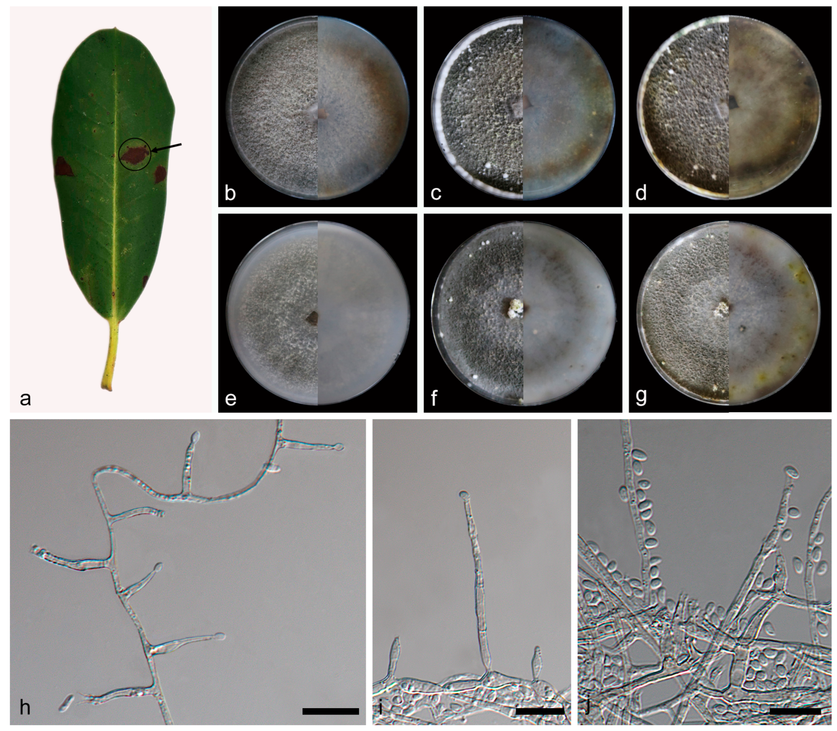
  • Daldinia rhododendri C.Z. Yin, Z.X. Zhang and X.G. Zhang, sp. nov. Figure 10.
MycoBank—MB853135
Etymology—the epithet “rhododendri” refers to the name of the host plant, Rhododendron decorum.
Material studied—China, Yunnan Province, Diqing Prefecture, Shangri-la City, 27°58′43″ N, 99°34′24″ E, on diseased leaves of Rhododendron decorum, 28 June 2023, C.Z. Yin, Z.X. Zhang and X.G. Zhang, holotype HMAS352918, ex-type culture SAUCC460001.
Description—conidiophores exhibit a virgariella-like to nodulisporium-like branching pattern [27]. Conidiophores mononematous or dichotomously branched and are rare, smooth or finely roughened, hyaline, and septate, with conidiogenous cells at each terminus, 40–90 × 1.4–2.0 µm ( x ¯ = 74 × 1.7 µm, n = 21). Conidiogenous cells are cylindrical or ampuliform, rare, smooth to finely roughened, and hyaline, bearing conidia on their apical region, 5.9–11.6 × 1.1–2.9 µm ( x ¯ = 8.6 × 2.3 µm, n = 26). Conidia are ellipsoid, cylindrical or banana shaped, smooth, hyaline, aseptate, solitary, and produced from percurrently proliferating conidiogenous cells, 3.2–5.1 × 1.1–2.3 µm ( x ¯ = 4.1 × 1.7 µm, n = 30). The teleomorph was not discovered.
Culture characteristics—after 7 days of cultivation on PDA medium at 25 °C, colonies were rough, dense, and aerial mycelium distributed evenly and was white (7443) and grey (424) from the middle out; the reverse center was white (7443), with irregular areas of reddish brown (469) around. After 14 days, colonies formed a few white (7541) fluffy balls and a circle of white (7541) fluffy mycelium around; aerial mycelium was dark grey (425); the reverse center was white (7443), with irregular areas of reddish brown (469) and pale green (384) around. After 31 days, colonies had not changed significantly; the reverse was predominantly brown (1405) and black (426). The average growth rate on PDA medium was 12 mm/day. After 7 days of cultivation on OA medium at 25 °C, colonies alternated dark gray (425) and white (7443) with rings from the middle to the periphery and was rough, medium dense, with aerial mycelium distributed evenly; the reverse was white (7443). After 14 days, colonies formed a few white (7541) fluffy balls, dense; the reverse exhibited an irregular dark gray (425) ring. After 31 days, colonies changed from dark gray to gray (423); the reverse exhibited some irregular yellow green (582) areas. The average growth rate on OA medium was 11.1 mm/day.
Additional material studied—China, Yunnan Province, Diqing Prefecture, Shangri-la City, 27°58′43″ N, 99°34′24″ E, on diseased leaves of Rhododendron decorum, 28 June 2023, C.Z. Yin, Z.X. Zhang and X.G. Zhang, HSAUP 460002, living culture SAUCC460002.
Notes—the phylogenetic analysis of ITS, LSU, rpb2, and tub2 sequences revealed a close relationship between Daldinia rhododendri, D. pyrenaica, and D. childiae, and they are both on a branch with better support (MLBS/BPP = 100/1). Daldinia rhododendri is different from D. pyrenaica by 21/552 bp in ITS, 4/982 bp in LSU, and 29/1342 bp in tub2 and different from D. childiae by 20/552 bp in ITS, 6/982 bp in LSU, and 18/1368 bp in tub2. Morphologically, D. rhododendri and D. pyrenaica differ obviously in conidiogenous cells (5.9–11.6 × 1.1–2.9 vs. 10.0–25.0 × 2.5–3.0 µm) and conidia (3.2–5.1 × 1.1–2.3 vs. 6.5–7.0 × 4.0–5.0 μm) [3,29]. Daldinia rhododendri and D. childiae also differ obviously in conidiogenous cells (5.9–11.6 × 1.1–2.9 vs. 10.0–25.0 × 3.0–4.0 µm) and conidia (3.2–5.1 × 1.1–2.3 vs. 7.0–9.0 × 4.5–5.5 μm) [3,28]. Therefore, D. rhododendri can be identified as a new anamorphic species of Daldinia by phylogenetic and morphological comparison.
Figure 10. Daldinia rhododendri (holotype: HMAS352918). (a) leaf of host plant Rhododen dron decorum; (bd) colony front and back after 7 days, 14 days, and 31 days culture on PDA; (eg) colony front and back after 7 days, 14 days, and 31 days culture on OA; (hj) conidiogenous cells and conidia. Scale bars: (hj) 10 μm.
Jof 10 00700 g010
  • Daldinia spatholobi C.Z. Yin, Z.X. Zhang and X.G. Zhang, sp. nov. Figure 11.
MycoBank—MB853136
Etymology—the epithet “spatholobi” refers to the name of the host plant, Spatholobus suberectus.
Material studied—China, Yunnan Province, Jinghong City, Xishuangbanna primitive Forest Park, 22°1′52″ N, 100°52′36″ E, on diseased leaves of Spatholobus suberectus, 17 March 2023, C.Z. Yin, Z.X. Zhang and X.G. Zhang, holotype HMAS352919, ex-type culture SAUCC203501.
Description—Conidiophores exhibit a virgariella-like to nodulisporium-like branching pattern [27]. Conidiophores mononematous, dichotomously, or trichotomously branched and are roughened, hyaline, and septate, with 1–3 conidiogenous cells at each terminus, 90–150 × 2.9–4.1 µm ( x ¯ = 135 × 3.4 µm, n = 22). Conidiogenous cells are cylindrical or clavate, roughened, and hyaline, with a flattened base, bearing conidia on their apical region, 9.5–14.9 × 2.4–3.0 µm ( x ¯ = 12.3 × 2.6 µm, n = 25). Conidia are ellipsoid or teardrop shaped, smooth to finely roughened, hyaline, aseptate, solitary, and produced holoblastically in sympodial sequence, 4.3–6.1 × 2.6–3.7 µm ( x ¯ = 5.6 × 3.4 µm, n = 30). The teleomorph was not discovered.
Culture characteristics—after 7 days of cultivation on PDA medium at 25 °C, colonies alternated pale brown (1245), white (7443), pale green (390), and white (7443) with rings from the middle to the periphery and were flat, medium dense, and with aerial mycelium more in the center than on the periphery; the reverse was similar to the front, but the center was black (419). After 14 days, colonies formed irregular green (377) areas and was dense with aerial mycelium distributed evenly; the reverse center was black (419), brown (1255) with irregular area outside, and white (7443) outwards. After 31 days, colonies were white (7527) in the center, and the rest was mostly black (419); the reverse was also mostly brown (1405). The average growth rate on PDA medium was 10.6 mm/day. After 7 days of cultivation on OA medium at 25 °C, colonies formed a black (426) circular area in the center and was white (7443) outwards and flat, medium dense, and with aerial mycelium distributed evenly; the reverse center was brown (1405) and mostly white (7443) on the outside. After 14 days, colonies ringed from the middle to the periphery, showing black (426), pale brown (1395), grey (423), dark green (371), and white (7541) colors and were dense; the reverse center was black (419) and formed a regular brown (1405) ring. After 31 days, colonies darkened in color; the reverse has not changed significantly. The average growth rate on OA medium was 9.7 mm/day.
Additional material studied—China, Yunnan Province, Jinghong City, Xishuangbanna primitive Forest Park, 22°1′52″ N, 100°52′36″ E, on diseased leaves of Spatholobus suberectus, 17 March 2023, C.Z. Yin, Z.X. Zhang and X.G. Zhang, HSAUP 203502, living culture SAUCC203502.
Notes—the phylogenetic analysis of ITS, LSU, rpb2, and tub2 sequences revealed a close relationship between Daldinia spatholobi and D. kretzschmarioides, and they are both on a branch with better support (MLBS/BPP = 99/1). Daldinia spatholobi was different from D. kretzschmarioides by 6/961 bp in LSU and 25/1066 bp in tub2. Morphologically, compared to D. kretzschmarioides, D. spatholobi has shorter conidiogenous cells (2.4–3.0 vs. 3.0–4.0 µm) and similar conidia sizes. But the conidia of D. spatholobi are teardrop shaped in addition to ellipsoid, with a finely roughened surface. Under the same conditions, D. spatholobi exhibits regular circular pigment precipitation on OA medium with fewer mycelium, while D. kretzschmarioides exhibits irregular pigment precipitation with more mycelium [7]. Therefore, D. spatholobi can be identified as a new anamorphic species of Daldinia by phylogenetic and morphological comparison.
Figure 11. Daldinia spatholobi (holotype: HMAS352919). (a) leaf of host plant Spatholobus suberectus; (bd) colony front and back after 7 days, 14 days, and 31 days culture on PDA; (eg) colony front and back after 7 days, 14 days, and 31 days culture on OA; (hj) conidiogenous cells and conidia. Scale bars: (hj) 10 μm.
Jof 10 00700 g011
  • Daldinia thunbergiae C.Z. Yin, Z.X. Zhang and X.G. Zhang, sp. nov. Figure 12.
MycoBank—MB853137
Etymology—the epithet “thunbergiae” refers to the name of the host plant, Thunbergia grandiflora.
Material studied—China, Yunnan Province, Jinghong City, Sancha River, 22°10′10″ N, 100°51′49″ E, on diseased leaves of Thunbergia grandiflora, 19 March 2023, C.Z. Yin, Z.X. Zhang and X.G. Zhang, holotype HMAS352920, ex-type culture SAUCC228601.
Description—Conidiophores exhibit a virgariella-like to nodulisporium-like branching pattern [27]. Conidiophores mononematous, dichotomously, or trichotomously branched and are roughened, hyaline, and septate, with 1–4 conidiogenous cells at each terminus, 70–220 × 1.8–4.1 µm ( x ¯ = 150 × 3.3 µm, n = 21). Conidiogenous cells are cylindrical or clavate, finely roughened, and hyaline, bearing conidia on their apical region, 6.7–17.3 × 1.9–2.5 µm ( x ¯ = 11.5 × 2.2 µm, n = 26). Conidia are ellipsoid or teardrop shaped, finely roughened, hyaline, aseptate, solitary, mostly with a flattened base, and produced holoblastically in sympodial sequence, 3.2–5.0 × 2.2–3.1 µm ( x ¯ = 4.2 × 2.7 µm, n = 30). The teleomorph was not discovered.
Culture characteristics—after 7 days of cultivation on PDA medium at 25 °C, colonies radiated from the middle to the periphery, showing white (7443), dark green (385), and white (7443) colors and were rough, medium dense, and with aerial mycelium more in the center than on the periphery; the reverse center was dark green (385), with an incomplete dark green (385) ring outwards, and the rest was white (7443). After 14 days, colonies were mostly dark gray (425), plat, dense, and with aerial mycelium distributed evenly; the reverse center was mostly black (426), surrounded by many black (426) spots and irregular pale green (391) areas, and the rest was white (7443). After 31 days, colonies changed from dark gray to dark brown (1545); white (7541) mycelium appeared in the center; the reverse periphery was mostly brown (1615). The average growth rate on PDA medium was 11.1 mm/day. After 7 days of cultivation on OA medium at 25 °C, colonies formed multiple white (7541) rings and the center was black (419), plat, and medium dense, with aerial mycelium distributed evenly; the reverse was similar to the front, but the center was brown (1615). After 14 days, colonies formed a white (7443) ring in the outermost ring and the rest was mostly gray (424) and dense; the reverse was similar to the front, but the center and some irregular parts were dark green (378). After 31 days, colonies changed from gray to light brown (1255); the reverse was also mostly brown (1265). The average growth rate on OA medium was 11.3 mm/day.
Additional material studied—China, Yunnan Province, Jinghong City, Sancha River, 22°10′10″ N, 100°51′49″ E, on diseased leaves of Thunbergia grandiflora, 19 March 2023, C.Z. Yin, Z.X. Zhang and X.G. Zhang, HSAUP 228602, living culture SAUCC228602.
Notes—Daldinia thunbergiae is closely related to another newly discovered species in this study, D. ehretiae, on the phylogenetic tree of Daldinia. Based on the differences in sequence, conidiophores, conidiogenous cells, and conidia between the two species mentioned above, D. thunbergiae can be identified as a new anamorphic species of Daldinia.
Figure 12. Daldinia thunbergiae (holotype: HMAS352920). (a) leaf of host plant Thunbergia grandiflora; (bd) colony front and back after 7 days, 14 days, and 31 days culture on PDA; (eg) colony front and back after 7 days, 14 days, and 31 days culture on OA; (hj) conidiogenous cells and conidia. Scale bars: (hj) 10 μm.
Jof 10 00700 g012
  • Daldinia jianfengensis C.Z. Yin, Z.X. Zhang and X.G. Zhang, sp. nov. Figure 13.
MycoBank—MB853132
Etymology—the epithet “jianfengensis” refers to the name of the strain location, Jianfeng Town.
Material studied—China, Hainan Province, Ledong County, Jianfeng Town, 18°41′19″ N, 108°51′31″ E, on decayed leaves, 12 April 2023, C.Z. Yin, Z.X. Zhang and X.G. Zhang, holotype HMAS352915, ex-type culture SAUCC373804.
Description—conidiophores exhibit a virgariella-like to nodulisporium-like branching pattern [27]. Conidiophores mononematous, dichotomously, or trichotomously branched and are finely roughened, hyaline, and septate, with 1–4 conidiogenous cells at each terminus, 70–120 × 2.9–4.4 µm ( x ¯ = 97 × 3.6 µm, n = 22). Conidiogenous cells are cylindrical, finely roughened, and hyaline, bearing conidia on their apical region, 12.1–16.9 × 2.6–3.6 µm ( x ¯ = 15.1 × 3.2 µm, n = 25). Conidia are subglobose or ellipsoid, smooth to finely roughened, hyaline, aseptate, solitary, mostly with a flattened base, and produced holoblastically in sympodial sequence, 3.2–5.5 × 2.6–3.7 µm ( x ¯ = 4.3 × 3.5 µm, n = 30). The teleomorph was not discovered.
Culture characteristics—after 7 days of cultivation on PDA medium at 25 °C, colonies exhibited alternated white (7443) and gray (422) rings from the middle to the periphery, with periphery irregularity, flat, and medium dense; the reverse center was black (426), dark green (385) irregular area outside, and white (7443) outwards. After 14 days, colonies formed a round, white (7443) fluff-like area in the center, dense, and aerial mycelium distributed evenly, grey (424); the reverse exhibited alternating black (419) and white (7443) irregular rings from the middle to the periphery. After 31 days, colonies and reverse had not changed significantly. The average growth rate on PDA medium was 11.6 mm/day. After 7 days of cultivation on OA medium at 25 °C, colonies were black (419), gray (425), and white (7443) from the middle out, flat, and medium dense; the reverse center was gray (424), with a circle of black (419) on the outside and mostly white (7541) on the outside. After 14 days, colonies alternated gray (424) and white (7443) from the middle to the periphery, dense, and aerial mycelium distributed evenly; the reverse alternated gray (423) and white (7443) with regular rings from the middle to the periphery. After 31 days, colonies changed from gray to pale brown (1265); the reverse was slightly darker. The average growth rate on OA medium was 11.4 mm/day.
Additional material studied—China, Hainan Province, Ledong County, Jianfeng Town, 18°41′19″ N, 108°51′31″ E, on decayed leaves, 12 April 2023, C.Z. Yin, Z.X. Zhang and X.G. Zhang, HSAUP 373805, living culture SAUCC373805.
Notes—The phylogenetic analysis of ITS, LSU, rpb2, and tub2 sequences revealed a close relationship between Daldinia jianfengensis and another newly discovered species, D. ledongensis, and they are both on a branch with better support (MLBS/BPP = 100/1). D. jianfengensis was different from D. ledongensis by 56/549 bp in ITS, 12/1277 bp in LSU, and 5/1038 bp in tub2. Morphologically, the difference between D. jianfengensis and D. ledongensis is that there are more conidiogenous cells (1–3 × 1–2) at the terminus of conidiophores and conidiogenous cells are generally long (12.1–16.9 vs. 8.6–15.1 µm) and are mostly distributed at the terminus of conidiophores. In addition, the conidia of D. jianfengensis are wider (2.6–3.7 vs. 1.4–2.0 μm) and produce more conidia under the same conditions. The conidial shape of D. jianfengensis is subglobose or ellipsoid, which is different from that of the conidia of D. ledongensis with an ellipsoid or fusiform shape. It is worth noting that under the same culture conditions, whether in OA medium or PDA medium, the medium of D. jianfengensis will show circular dark pigmentation, and the mycelium will be darker, while the medium of D. ledongensis only has a small part of pigmentation and the mycelium was always white. Therefore, D. jianfengens can be identified as a new anamorphic species of Daldinia by phylogenetic and morphological comparison.
Figure 13. Daldinia jianfengensis (holotype: HMAS352915). (ac) colony front and back after 7 days, 14 days, and 31 days culture on PDA; (df) colony front and back after 7 days, 14 days, and 31 days culture on OA; (gi) conidiogenous cells and conidia. Scale bars: (gi) 10 μm.
Jof 10 00700 g013
  • Daldinia ledongensis C.Z. Yin, Z.X. Zhang and X.G. Zhang, sp. nov. Figure 14.
MycoBank—MB853133
Etymology—the epithet “ledongensis” refers to the name of the strain location, Ledong County.
Material studied—China, Hainan Province, Ledong County, 18°41′18″ N, 108°51′31″ E, on decayed leaves, 12 April 2023, C.Z. Yin, Z.X. Zhang and X.G. Zhang, holotype HMAS352916, ex-type culture SAUCC393602.
Description—Conidiophores exhibit a virgariella-like to nodulisporium-like branching pattern [27]. Conidiophores mononematous or dichotomously branched and are rare, smooth or finely roughened, hyaline, and aseptate, with a conidiogenous cells at each terminus, 120–200 × 1.7–2.1 µm ( x ¯ = 157 × 1.8 µm, n = 21). Conidiogenous cells are clavate, rare, smooth to finely roughened, and hyaline, with a flattened base, bearing conidia on their apical region, 8.6–15.1 × 1.2–3.4 µm ( x ¯ = 11.3 × 2.4 µm, n = 24). Conidia are ellipsoid or fusiform, smooth to finely roughened, hyaline, aseptate, solitary, and produced from percurrently proliferating conidiogenous cells, 3.2–4.0 × 1.4–2.0 µm ( x ¯ = 3.5 × 1.6 µm, n = 30). The teleomorph was not discovered.
Culture characteristics—after 7 days of cultivation on PDA medium at 25 °C, colonies radiated from the middle to the periphery, rough, medium dense, with aerial mycelium more on the periphery than in the center and was white (7443); the reverse was similar to the front, but the center was hazel (471). After 14 days, colonies were dense, and aerial mycelium distributed evenly; the reverse was similar to the front, but the center was brown (1405). After 31 days, colonies became denser; the reverse had not changed significantly. The average growth rate on PDA medium was 10.7 mm/day. After 7 days of cultivation on OA medium at 25 °C, colonies were rough, medium dense, and aerial mycelium was more on the periphery than in the center and was white (7443); the reverse center was brown (1615), mostly white (7443) on the outside. After 14 days, colonies were dense; the reverse exhibited some dark brown (1545) areas. After 31 days, colonies became denser; the reverse exhibited many irregular brown (1395) areas. The average growth rate on OA medium was 11.4 mm/day.
Additional material studied—China, Hainan Province, Ledong County, 18°41′18″ N, 108°51′31″ E, on decayed leaves, 12 April 2023, C.Z. Yin, Z.X. Zhang and X.G. Zhang, HSAUP 393603, living culture SAUCC393603.
Notes—Daldinia ledongensis was closely related to another newly discovered species, D. jianfengensis, on the phylogenetic tree of Daldinia. Based on the differences in sequence, conidiophores, conidiogenous cells, conidia, and medium between the two species mentioned above, D. ledongensis can be identified as a new anamorphic species of Daldinia.
Figure 14. Daldinia ledongensis (holotype: HMAS352916). (ac) colony front and back after 7 days, 14 days, and 31 days culture on PDA; (df) colony front and back after 7 days, 14 days, and 31 days culture on OA; (g,h) conidiogenous cells and conidia. Scale bars: (g,h) 10 μm.
Jof 10 00700 g014
  • Daldinia menghaiensis C.Z. Yin, Z.X. Zhang and X.G. Zhang, sp. nov. Figure 15.
MycoBank—MB853134
Etymology—the epithet “menghaiensis” refers to the name of the strain location, Menghai County.
Material studied—China, Yunnan Province, Menghai County, 21°55′25″ N, 100°35′41″ E, on decayed leaves, 18 March 2023, C.Z. Yin, Z.X. Zhang and X.G. Zhang, holotype HMAS352917, ex-type culture SAUCC242404.
Description—Conidiophores exhibit a virgariella-like to nodulisporium-like branching pattern [27]. Conidiophores dichotomously or trichotomously branched, occasionally branched from the conidiogenous region, and are finely roughened, hyaline, and septate, with 1–2 conidiogenous cells at each terminus, 80–150 × 1.9–3.4 µm ( x ¯ = 115 × 2.6 µm, n = 22). Conidiogenous cells are cylindrical or clavate, finely roughened, and hyaline, bearing conidia on their apical region, 16.9–23.5 × 2.0–3.5 µm ( x ¯ = 19.3 × 2.8 µm, n = 27). Conidia are ellipsoid, subglobose or dacryoid, roughened, hyaline, aseptate, solitary, mostly with flattened base, and produced holoblastically in sympodial sequence, 4.7–8.2 × 3.1–4.0 µm ( x ¯ = 5.7 × 3.5 µm, n = 30). The teleomorph was not discovered.
Culture characteristics—after 7 days of cultivation on PDA medium at 25 °C, colonies radiated and ringed from the middle to the periphery, flat, medium dense, and with aerial mycelium more on the periphery than in the center and was white (7443); the reverse was similar to the front, but the center was pale brown (1375). After 14 days, colonies were dense; the reverse center was brown (1395), there appeared some light green (382) areas. After 31 days, colonies became rough; the reverse center was black (419), and the shape of the colonies became irregular. The average growth rate on PDA medium was 12.8 mm/day. After 7 days of cultivation on OA medium at 25 °C, colonies were flat, medium dense, and aerial mycelium was rare in the central circular region and was white (7443); the reverse was similar to the front. After 14 days, colonies formed many dark green (378) spots, dense, with aerial mycelium distributed evenly; the reverse was similar to the front. After 31 days, colonies changed from white to gray (424); the reverse exhibited many small irregular brown (1615) areas. The average growth rate on OA medium was 10.3 mm/day.
Additional material studied—China, Yunnan Province, Menghai County, 21°55′25″ N, 100°35′41″ E, on decayed leaves, 18 March 2023, C.Z. Yin, Z.X. Zhang and X.G. Zhang, HSAUP 242405, living culture SAUCC242405.
Notes—the phylogenetic analysis of ITS, LSU, rpb2, and tub2 sequences revealed a close relationship between Daldinia menghaiensis, D. subvernicosa, and D. vernicosa, and they are both on a branch with better support (MLBS/BPP = 99/1). Daldinia menghaiensis was different from D. subvernicosa by 25/464 bp in ITS, 16/1107 bp in LSU, 39/684 bp in rpb2, and 57/949 bp in tub2 and different from D. vernicosa by 35/552 bp in ITS, 19/1226 bp in LSU, 54/867 bp in rpb2, and 62/1080 bp in tub2. Morphologically, compared with Daldinia vernicosa, the number of conidiogenous cells at the terminus of conidiophores in D. menghaiensis is less (1–2 vs. 1–4) and is generally long (16.9–23.5 vs. 8.0–23.0 µm), but the conidia are narrower (3.1–4.0 vs. 4.5–6.0 µm) [3]. Under the same conditions, the medium of D. menghaiensis always produces pigmentation, and mycelia become darker, but D. subvernicosa does not [7]. Therefore, D. menghaiensis can be identified as a new anamorphic species of Daldinia by phylogenetic and morphological comparison.
Figure 15. Daldinia menghaiensis (holotype: HMAS352917). (ac) colony front and back after 7 days, 14 days, and 31 days culture on PDA; (df) colony front and back after 7 days, 14 days, and 31 days culture on OA; (g,h) conidiogenous cells and conidia. Scale bars: (g,h) 10 μm.
Jof 10 00700 g015

4. Discussion

This study investigated the fungal resources in five provinces in southern China, and the strains of Daldinia isolated in this survey were mainly collected from forests and mountainous areas, with altitudes ranging from 151.57 to 1876.7 m.a.s.l. These strains were found in all five provinces of southern China, which proved that they were also widely distributed in southern China. The number and types of strains were the highest in Yunnan Province, followed by Hainan Province, and the least were in Guizhou Province. The overall distribution of all strains in each province is shown in Figure 2a. We noticed that with similar sample sizes collected in each province, more strains of Daldinia were collected in the Yunnan and Hainan provinces in the tropical monsoon climate zone than in the other three provinces in the subtropical monsoon climate zone. Our preliminary hypothesis posits that these primary forests and mountains, boasting warm and humid climate conditions and abundant vegetation, may harbor more Daldinia as well as other fungal. The plant specimens collected in this survey were divided into diseased and decayed leaves, and 84 new host plants were determined by identifying the strains of Daldinia. They belong to 43 plant families, among which Lauraceae (10.84%), Poaceae (9.64%), Moraceae (6.02%), Theaceae (6.02%), and Fagaceae (4.82%) have a relatively high proportion, and there is little difference in the proportion of other families. The number of each plant family is shown in Figure 2b. In addition, dicots accounted for 84.34% of all plant hosts, while monocots were only Poaceae, Zingiberaceae, Iridaceae, and Asparagaceae. Therefore, it can be preliminarily inferred that the genus Daldinia does not exhibit strong specificity of host, and dicots account for the majority of hosts.
Since the establishment of the genus Daldinia, continuous advancements have been made in the methods utilized for its identification. Now, a widely accepted approach for identifying Daldinia involves phylogenetic analysis based on ITS, LSU, rpb2, and tub2 combined with morphological comparison. A total of 104 records encompassing various species of Daldinia were retrieved from the Index Fungorum (https://www.indexfungorum.org/, accessed on 7 September 2024), among which most of the species with complete ITS, LSU, rpb2, and tub2 sequences are included in Table 1. We also used this method to identify three known species (Daldinia bambusicola, D. childiae, and D. eschscholtzii) and seven new anamorphic species (D. ehretiae, D. jianfengensis, D. ledongensis, D. menghaiensis, D. rhododendri, D. spatholobi, and D. thunbergiae). In this study, we not only provided sequence information for new species but also perfected sequence data for known species, such as the rpb2 and tub2 sequence data of D. childiae, after species were confirmed through anamorph characteristics and multiple sequence alignments. The Daldinia strains were cultured using PDA and OA mediums, and the colony morphology at 7 days, 14 days, and 31 days of culture was observed and recorded, which could also assist in the identification of the strain while showing the colony changes. In the process of cultivating the strains of Daldinia, we also noticed an interesting phenomenon: taking the new species discovered this time as an example, whether on PDA medium or OA medium, Daldinia can be divided into two categories. One type produces a large number of spores and forms a regular large-area pigment precipitation area on the medium, and most of them form circular pigment precipitation around 2 weeks, such as D. ehretiae, D. jianfengensis, D. menghaiensis, D. rhododendri, D. spatholobi, and D. thunbergiae. And another type features the production of very small amounts of spores and the formation of small areas of pigment precipitation on the culture medium, such as D. ledongensis. Furthermore, it is worth noting that there have been numerous reports that Daldinia are endophytes, some even related to insects, and spores of Daldinia typically exhibit both teleomorph and anamorph modes, but the specimens obtained during this survey were found on diseased leaves and did not directly produce spores of teleomorph in their natural habitat. So, inducing Daldinia to produce spores of teleomorph in the laboratory presents an intriguing yet challenging task. At the same time, we will continue to conduct field collection work on teleomorphic species of Daldinia in the next step and improve the teleomorphic data of Daldinia found in this survey as soon as possible. Interestingly, due to the abundance of secondary metabolites within the genus Daldinia, there has been a growing trend towards utilizing HPLC profiling for metabolite analysis and incorporating chemical classification into the identification process. In their investigation of Xylariales in Thailand, Wongkanoun et al. employed morphological characters, phylogenetic analysis based on multi-locus sequences, and comprehensive analysis of secondary metabolites based on high-performance liquid chromatography-diode array detection and mass spectrometry, leading to the identification of three novel species, D. flavogranulata, D. phadaengensis, and D. chiangdaoensis [19]. Moving forward, we will continue conducting comprehensive research on resource exploration and metabolic characteristic analysis of the genus Daldinia.

Supplementary Materials

The following supporting information can be downloaded at: https://www.mdpi.com/article/10.3390/jof10100700/s1, Table S1: PCR primer sequence and reaction conditions used in this experiment.

Author Contributions

Conceptualization, C.Y.; methodology, Z.Z.; software, C.Y.; validation, S.W. and Z.Z.; formal analysis, C.Y.; investigation, W.L.; resources, C.Y. and W.L.; data curation, C.Y. and W.L.; writing—original draft preparation, C.Y.; writing—review and editing, C.Y.; visualization, Z.Z.; supervision, X.Z.; project administration, X.Z.; funding acquisition, X.Z. All authors have read and agreed to the published version of the manuscript.

Funding

This research was funded by the National Natural Science Foundation of China (nos. 32170012, 32300011, 32470004, 31400019), Ji’nan City’s ‘New University 20 Policies’ Initiative for Innovative Research Teams Project (202228028), Innovative Agricultural Application Technology Project of Jinan City (CX202210), and Key Technological Innovation Program of Shandong Province, China (2022CXGC020710).

Institutional Review Board Statement

Not applicable.

Informed Consent Statement

Not applicable.

Data Availability Statement

All datasets in this study are included in this article/Supplementary Materials.

Conflicts of Interest

The authors declare no conflicts of interest.

References

  1. Cesati, V.; De Notaris, G. Schema di classificazione degle sferiacei italici aschigeri piu’ o meno appartenenti al genere Sphaeria nell’antico significato attribuitoglide Persono. Comment. Della Soc. Crittogamologica Ital. 1 1863, 4, 177–420. [Google Scholar]
  2. Ju, Y.M.; Rogers, J.D.; San Martín, F. A revision of the genus Daldinia. Mycotaxon 1997, 61, 243–293. [Google Scholar]
  3. Stadler, M.; Læssøe, T.; Fournier, J.; Decock, C.; Schmieschek, B.; Tichy, H.V.; Peršoh, D. A polyphasic taxonomy of Daldinia (Xylariaceae). Stud. Mycol. 2014, 77, 1–143. [Google Scholar] [CrossRef]
  4. Dargan, J.S.; Thind, K.S. Xylariaceae of India. VIII Genus Daldinia Ces & de Not-a further segregation into two new subgenera. Kavaka 1985, 12, 113–118. [Google Scholar]
  5. Wendt, L.; Sir, E.B.; Kuhnert, E.; Heitkämper, S.; Lambert, C.; Hladki, A.I.; Romero, A.I.; Luangsa-ard, J.J.; Srikitikulchai, P.; Peršoh, D.; et al. Resurrection and emendation of the Hypoxylaceae, recognised from a multi-gene genealogy of the Xylariales. Mycol. Prog. 2018, 17, 115–154. [Google Scholar] [CrossRef]
  6. Daranagama, D.A.; Hyde, K.D.; Sir, E.B.; Thambugala, K.M.; Tian, Q.; Samarakoon, M.C.; McKenzie, E.H.C.; Jayasiri, S.C.; Tibpromma, S.; Bhat, J.D.; et al. Towards a natural classification and backbone tree for Graphostromataceae, Hypoxylaceae, Lopadostomataceae and Xylariaceae. Fungal Divers. 2018, 88, 1–165. [Google Scholar] [CrossRef]
  7. Wongkanoun, S.; Wendt, L.; Stadler, M.; Luangsa-ard, J.J.; Srikitikulchai, P. A novel species and a new combination of Daldinia from Ban Hua Thung community forest in the northern part of Thailand. Mycol. Prog. 2019, 18, 553–564. [Google Scholar] [CrossRef]
  8. Pažoutová, S.; Follert, S.; Bitzer, J.; Keck, M.; Surup, F.; Šrůtka, P.; Holuša, J.; Stadler, M. A new endophytic insect-associated Daldinia species, recognised from a comparison of secondary metabolite profiles and molecular phylogeny. Fungal Divers. 2013, 60, 107–123. [Google Scholar] [CrossRef]
  9. Doyle, J.J. Isolation of plant DNA from fresh tissue. Focus 1990, 12, 13–15. [Google Scholar]
  10. Guo, L.D.; Hyde, K.D.; Liew, E.C.Y. Identification of endophytic fungi from Livistona chinensis based on morphology and rDNA sequences. New Phytol. 2000, 147, 617–630. [Google Scholar] [CrossRef]
  11. Wang, S.; Liu, X.; Xiong, C.; Gao, S.; Xu, W.; Zhao, L.; Song, C.; Liu, X.; James, T.Y.; Li, Z.; et al. ASF1 regulates asexual and sexual reproduction in Stemphylium eturmiunum by DJ-1 stimulation of the PI3K/AKT signaling pathway. Fungal Divers. 2023, 123, 159–176. [Google Scholar] [CrossRef]
  12. Zhang, Z.X.; Liu, R.Y.; Liu, S.B.; Mu, T.C.; Zhang, X.G.; Xia, J.W. Morphological and phylogenetic analyses reveal two new species of Sporocadaceae from Hainan, China. MycoKeys 2023, 88, 171–192. [Google Scholar] [CrossRef] [PubMed]
  13. Kumar, S.; Stecher, G.; Tamura, K. MEGA7: Molecular Evolutionary Genetics Analysis Version 7.0 for Bigger Datasets. Mol. Biol. Evolut. 2016, 33, 1870–1874. [Google Scholar] [CrossRef] [PubMed]
  14. White, T.J.; Bruns, L.; Lee, S.; Taylor, J. Amplification and direct sequencing of fungal ribosomal RNA genes for phylogenetics. PCR Protoc. A Guide Methods Appl. 1990, 38, 315–322. [Google Scholar] [CrossRef]
  15. Bunyard, B.A.; Nicholson, M.S.; Royse, D.J. A systematic assessment of Morchella using RFLP analysis of the 28S ribosomal RNA gene. Mycologia 1994, 86, 762–772. [Google Scholar] [CrossRef]
  16. Liu, Y.L.; Whelen, S.; Hall, B.D. Phylogenetic relationships among ascomycetes: Evidence from and RNA polymerase II subunit. Mol. Biol. Evolut. 1999, 16, 1799–1808. [Google Scholar] [CrossRef]
  17. O’Donnell, K.; Cigelnik, E. Two divergent intragenomic rDNA ITS2 types within a monophyletic lineage of the fungus Fusarium are nonorthologous. Mol. Phylogenet. Evolut. 1997, 7, 103–116. [Google Scholar] [CrossRef]
  18. Daranagama, D.A.; Camporesi, E.; Tian, Q.; Liu, X.; Chamyuang, S.; Stadler, M.; Hyde, K.D. Anthostomella is polyphyletic comprising several genera in Xylariaceae. Fungal Divers. 2015, 73, 203–238. [Google Scholar] [CrossRef]
  19. Wongkanoun, S.; Becker, K.; Boonmee, K.; Srikitikulchai, P.; Boonyuen, N.; Chainuwong, B.; Luangsa-ard, J.; Stadler, M. Three novel species and a new record of Daldinia (Hypoxylaceae) from Thailand. Mycol. Prog. 2020, 19, 1113–1132. [Google Scholar] [CrossRef]
  20. U’Ren, J.M.; Miadlikowska, J.; Zimmerman, N.B.; Lutzoni, F.; Stajich, J.E.; Arnold, A.E. Contributions of North American endophytes to the phylogeny, ecology, and taxonomy of Xylariaceae (Sordariomycetes, Ascomycota). Mol. Phylogenet. Evolut. 2016, 98, 210–232. [Google Scholar] [CrossRef]
  21. Sir, E.B.; Lambert, C.; Wendt, L.; Hladki, A.I.; Romero, A.I.; Stadler, M. A new species of Daldinia (Xylariaceae) from the Argentine subtropical montane forest. Mycosphere 2016, 7, 596–614. [Google Scholar] [CrossRef]
  22. Miller, M.A.; Pfeiffer, W.; Schwartz, T. Creating the CIPRES science gateway for inference of large phylogenetic trees. In Proceedings of the 2010 Gateway Computing Environments Workshop (GCE), New Orleans, LA, USA, 14 November 2010. [Google Scholar] [CrossRef]
  23. Stamatakis, A. RAxML version 8: A tool for phylogenetic analysis and post–analysis of large phylogenies. Bioinformatics 2014, 30, 1312–1313. [Google Scholar] [CrossRef] [PubMed]
  24. Huelsenbeck, J.P.; Ronquist, F. MrBayes: Bayesian inference of phylogenetic trees. Bioinformatics 2001, 17, 115–755. [Google Scholar] [CrossRef] [PubMed]
  25. Nylander, J.A.A. MrModeltest v. 2.0. Evolutionary Biology Centre; Uppsala University: Uppsala, Sweden, 2004. [Google Scholar]
  26. Swofford, D.L. PAUP*4.0b10: Phylogenetic Analysis Using Parsimony (*and Other Methods); Sinauer: Sunderland, UK, 2002. [Google Scholar] [CrossRef]
  27. Ju, Y.M.; Rogers, J.D. A Revision of the Genus Hypoxylon; Mycologia memoir no.° 20; APS Press: St. Paul, MN, USA, 1996; p. 365. [Google Scholar]
  28. Rogers, J.D.; Ju, Y.M.; Watling, R.; Whalley, A.J.S. A reinterpretation of Daldinia concentrica based upon a recently discovered specimen. Mycotaxon 1999, 72, 507–519. [Google Scholar]
  29. Stadler, M.; Baumgartner, M.; Wollweber, H. Three new Daldinia species with yellowish stromatal pigments. Mycotaxon 2001, 80, 179–196. [Google Scholar]
Figure 1. A maximum likelihood tree of Daldinia based on ITS, LSU, rpb2, and tub2 gene sequences, and CBS 270.87 of Graphostroma platystomum and CBS 12260 of Xylaria hypoxylon as the tree root of Daldinia. The nodes of the branches are labeled MLBS/BPP (MLBS ≥ 70, BPP ≥ 0.9). The known species in this study are shown in blue font, while new species are shown in red font. Ex-type or ex-epitype strains are shown in bold with an “*”. Yellow and green parts are used to distinguish different strains. The black line at the bottom right is the scale bar, indicating 0.1 nucleotide changes at each site.
Jof 10 00700 g001
Figure 2. The distribution and host of Daldinia. (a) the number of Daldinia strains in five provinces of China; (b) the number of families to which the plant hosts of Daldinia.
Jof 10 00700 g002
Table 1. Information on strains used in phylogenetic analysis of the genus Daldinia.
SpeciesStrainsCountryGenBank Accession NumbersReference
ITSLSUrpb2tub2
Annulohypoxylon annulatumCBS 140775USAKY610418KY610418KY624263KX376353[5]
Annulohypoxylon moriformeCBS 123579FranceKX376321KY610425KY624289KX271261[5]
Annulohypoxylon nitensMFLUCC 12.0823ThailandKJ934991KJ934992KJ934994KJ934993[18]
Annulohypoxylon stygiumMUCL 54601FrenchKY610409KY610475KY624292KX271263[5]
Annulohypoxylon truncatumCBS 140778USAKY610419KY610419KY624277KX376352[5]
Daldinia andinaCBS 114736 *EcuadorAM749918KY610430KY624239KC977259[5]
Daldinia bambusicolaCBS 122872 *ThailandKY610385KY610431KY624241AY951688[5]
Daldinia bambusicolaTBRC 8878ThailandMH922869MH922870MK165431MK165422[7]
Daldinia bambusicolaTBRC 8879ThailandMH922872MH938543MK165432MK165423[7]
Daldinia bambusicolaBCC27937ThailandMN153861MN153876MN172217N/A[19]
Daldinia bambusicolaBCC33678ThailandMN153860MN153877MN172218N/A[19]
Daldinia bambusicolaSAUCC197001ChinaPP145311PP198902PP263619PP277065This study
Daldinia brachyspermaBCC33676ThailandMN153854MN153871N/AMN172205[19]
Daldinia caldariorumMUCL 49211FranceAM749934KY610433KY624242KC977282[5]
Daldinia caldariorumCBS122874USAKU683756KU683756KU684289KU684128[20]
Daldinia chiangdaoensisBCC88220 *ThailandMN153850MN153867MN172208MN172197[19]
Daldinia chiangdaoensisBCC88221ThailandMN153851MN153868MN172209MN172198[19]
Daldinia childiaeSAUCC133401ChinaPP145313PP198904PP263621PP277067This study
Daldinia concentricaCBS 113277GermanyAY616683KY610434KY624243KC977274[5]
Daldinia dennisiiCBS 114741 *AustraliaJX658477KY610435KY624244KC977262[5]
Daldinia eschscholtziiMUCL 45435BeninJX658484KY610437KY624246KC977266[5]
Daldinia eschscholtziiTBRC 8876ThailandMH938532MH938541MK165429MK165420[7]
Daldinia eschscholtziiBCC27887ThailandMN153861MN153878MN172214N/A[19]
Daldinia eschscholtziiBCC28091ThailandMN153862MN153879MN172215N/A[19]
Daldinia eschscholtziiBCC62428ThailandMN153863MN153880MN172216N/A[19]
Daldinia eschscholtziiSAUCC265301ChinaPP145315PP198906PP263623PP277069This study
Daldinia ehretiaeSAUCC228302 *ChinaPP145319PP198888PP263613PP277051This study
Daldinia ehretiaeSAUCC228303ChinaPP145320PP198889PP263614PP277052This study
Daldinia flavogranulataBCC89363 *ThailandMN153856MN153873MN172211MN172200[19]
Daldinia flavogranulataBCC89365ThailandMN153857MN153874MN172212MN172201[19]
Daldinia flavogranulataBCC89376ThailandMN153858MN153875MN172213MN172202[19]
Daldinia jianfengensisSAUCC373804 *ChinaPP145325PP198890PP263615PP277053This study
Daldinia jianfengensisSAUCC373805ChinaPP145326PP198891PP263616PP277054This study
Daldinia korfiiEBS 067ArgentinaKY204018N/AN/AKY204014[21]
Daldinia korfiiEBS 473ArgentinaKY204020N/AN/AKY204016[21]
Daldinia kretzschmarioidesTBRC 8875ThailandMH938531MH938540MK165425MK165416[7]
Daldinia ledongensisSAUCC393602 *ChinaPP145327PP198892N/APP277055This study
Daldinia ledongensisSAUCC393603ChinaPP145328PP198893N/APP277056This study
Daldinia loculatoidesCBS 113279UKAF176982KY610438KY624247KX271246[5]
Daldinia macaronesicaCBS 113040SpainKY610398KY610477KY624294KX271266[5]
Daldinia menghaiensisSAUCC242404 *ChinaPP145323PP198894PP263617PP277057This study
Daldinia menghaiensisSAUCC242405ChinaPP145324PP198895PP263618PP277058This study
Daldinia phadaengensisBCC89349 *ThailandMN153852MN153869MN172206MN172195[19]
Daldinia phadaengensisBCC89350ThailandMN153853MN153870MN172207MN172196[19]
Daldinia petriniaeMUCL 49214AustriaAM749937KY610439KY624248KC977261[5]
Daldinia placentiformisMUCL 47603MexicoAM749921KY610440KY624249KC977278[5]
Daldinia pyrenaicaMUCL 53969FranceKY610413KY610413KY624274KY624312[5]
Daldinia rhododendriSAUCC460001 *ChinaPP145330PP198896N/APP277059This study
Daldinia rhododendriSAUCC460002ChinaPP145329PP198897N/APP277060This study
Daldinia spatholobiSAUCC203501 *ChinaPP145318PP198898N/APP277061This study
Daldinia spatholobiSAUCC203502ChinaPP145317PP198899N/APP277062This study
Daldinia steglichiiMUCL 43512Papua New GuineaKY610399KY610479KY624250KX271269[5]
Daldinia subvernicosaTBRC 8877 *ThailandMH938533MH938542MK165430MK165421[7]
Daldinia theisseniiCBS 113044ArgentinaKY610388KY610441KY624251KX271247[5]
Daldinia thunbergiaeSAUCC228601 *ChinaPP145322PP198900N/APP277063This study
Daldinia thunbergiaeSAUCC228602ChinaPP145321PP198901N/APP277064This study
Daldinia vernicosaCBS 119316GermanyKY610395KY610442KY624252KC977260[5]
Graphostroma platystomumCBS 270.87 *FranceJX658535DQ836906KY624296HG934108[5]
Hypomontagnella monticulosaMUCL 54604FrenchKY610404KY610487KY624305KX271273[5]
Hypomontagnella monticulosaBCC58592ThailandMN153864MN153881MN172219MN172204[19]
Hypomontagnella monticulosaBCC69203ThailandMN153865MN153882MN172220MN172203[19]
Hypomontagnella submonticulosaCBS 115280FranceKC968923KY610457KY624226KC977267[5]
Hypoxylon crocopeplumCBS 119004FranceKC968907KY610445KY624255KC977268[5]
Hypoxylon fragiformeMUCL 51264GermanyKC477229KM186295KM186296KX271282[5]
Hypoxylon fuscumCBS 113049FranceKY610401KY610482KY624299KX271271[5]
Hypoxylon haematostromaMUCL 53301FranceKC968911KY610484KY624301KC977291[5]
Hypoxylon haematostromaBCC50533ThailandMN153866MN153883MN172221N/A[19]
Hypoxylon investiensCBS 118183MalaysiaKC968925KY610450KY624259KC977270[5]
Hypoxylon lateripigmentumMUCL 53304 *FranceKC968933KY610486KY624304KC977290[5]
Hypoxylon lenormandiiCBS 119003EcuadorKC968943KY610452KY624261KC977273[5]
Hypoxylon petriniaeCBS 114746 *FranceKY610405KY610491KY624279KX271274[5]
Hypoxylon rickiiMUCL 53309FranceKC968932KY610416KY624281KC977288[5]
Hypoxylon rubiginosumMUCL 52887GermanyKC477232KY610469KY624266KY624311[5]
Hypoxylon samuelsiiMUCL 51843FranceKC968916KY610466KY624269KC977286[5]
Jackrogersella cohaerensCBS 119126GermanyKY610396KY610497KY624270KY624314[5]
Jackrogersella minutellaCBS 119015PortugalKY610381KY610424KY624235KX271240[5]
Jackrogersella multiformisCBS 119016GermanyKC477234KY610473KY624290KX271262[5]
Pyrenopolyporus hunteriMUCL 52673Ivory CoastKY610421KY610472KY624309KU159530[5]
Pyrenopolyporus laminosusMUCL 53305 *FranceKC968934KY610485KY624303KC977292[5]
Pyrenopolyporus laminosusTBRC 8871ThailandMH938527MH938536MK165424MK165415[7]
Pyrenopolyporus laminosusBCC89383ThailandMN153855MN153872MN172210MN172199[19]
Pyrenopolyporus nicaraguensisCBS 117739 *Burkina FasoAM749922KY610489KY624307KC977272[5]
Pyrenopolyporus symphyonTBRC 8873ThailandMH938529MH938538MK165428MK165419[7]
Xylaria hypoxylonCBS12260SwedenKY610407KY610495KY624231KX271279[5]
Notes: The new strains for phylogenetic analysis introduced in this experiment are shown in bold. Ex-type or ex-epitype strains are marked with an “*”. N/A: Not available.
Table 2. Collection information of Daldinia specimens.
SpeciesCollection LocationCollection TimeStrainsHostIllustration
Daldinia bambusicolaYunnan Province, Jinghong City, Xishuangbanna primitive forest Park, 22°1′52″ N, 100°52′36″ E17 March 2023SAUCC197001Viburnum rhytidophyllumFigure 3a
SAUCC203501Spatholobus suberectusFigure 4a
SAUCC204601Piper NigrumFigure 4b
SAUCC206901Cinnamomum verumFigure 4g
Yunnan Province, Menghai County, Nanuo Mountain, 21°55′25″ N, 100°35′41″ E18 March 2023SAUCC240501Koelreuteria paniculataFigure 4c
Hainan Province, Lingshui County, Diaoluo Mountain, 18°41′45″ N, 109°56′26″ E8 April 2023SAUCC283401Ficus hirtaFigure 4d
Hainan Province, Ledong County, Jianfengling National Forest Park, 18°41′18″ N, 108°51′31″ E12 April 2023SAUCC392801Schima superbaFigure 4e
Sichuan Province, Chengdu City, Dujiangyan, 30°59′58″ N, 108°51′31″ E24 June 2023SAUCC423801Citrus maximaFigure 4f
Sichuan Province, Leshan City, 29°34′56″ N, 103°17′20″ E25 June 2023SAUCC433001Phyllostachys heterocladaFigure 4h
Sichuan Province, Liangshan Prefecture, Xichang City, 27°45′29″ N, 102°18′31″ E2 July 2023SAUCC518401Ageratina adenophoraFigure 4i
Guizhou Province, Qiandongnan Prefecture, Majiang County, Gudong Town, 26°25′5″ N, 107°21′18″ E22 August 2023SAUCC551401Lophatherum gracileFigure 4j
Guizhou Province, Qiannan Prefecture, Sandu County, 25°55′52″ N, 107°57′34″ E24 August 2023SAUCC570801Ulmus pumilaFigure 4k
Daldinia childiaeFujian Province, Wuyishan City, Xingcun Town, 27°45′5″ N, 117°41′3″ E15 October 2022SAUCC133401Machilus nanmuFigure 5a
SAUCC148701Eurya japonicaFigure 6a
Yunnan province, Jinghong City, Xishuangbanna primitive Forest Park, 22°1′52″ N, 100°52′36″ E17 March 2023SAUCC209301Piper nigrumFigure 6e
Yunnan Province, Jinghong City, Binjiang Avenue, 22°0′41″ N, 100°48′15″ E20 March 2023SAUCC220001Microstegium vimineumFigure 6b
Hainan Province, Lingshui County, Benhao Town, 18°41′54″ N, 109°52′51″ E9 April 2023SAUCC314401Pseudosasa japonicaFigure 6f
Hainan Province, Ledong County, Jianfengling National Forest Park, 18°42′35″ N, 108°52′35″ E12 April 2023SAUCC387901Litsea cubebaFigure 6c
SAUCC388601Quercus glaucaFigure 6d
SAUCC392901Schima superbaFigure 6i
SAUCC397101Castanopsis calathiformisFigure 6g
Sichuan Province, Leshan City, Erhong Road, 29°35′31″ N, 103°22′39″ E25 June 2023SAUCC439301Symplocos sumuntiaFigure 6h
Daldinia eschscholtziiHainan Province, Sanya City, Jiyang District, Dongtian Ridge, 18°23′35″ N, 109°38′14″ E8 April 2023SAUCC265301Lysimachia clethroidesFigure 7a
Fujian Province, Wuyishan City, Xingcun Town, 27°44′47″ N, 117°40′36″ E15 October 2022SAUCC117601Machilus thunbergiiFigure 8(a1)
SAUCC123301Lindera aggregataFigure 8(a2)
SAUCC132901Maesa japonicaFigure 8(a3)
Yunnan Province, Xishuangbanna Tropical Botanical Garden, 21°55′52″ N, 101°14′50″ E15 March 2023SAUCC179601Gonocaryum lobbianumFigure 8(b1)
SAUCC179701Engelhardia spicataFigure 8(b2)
SAUCC182201Artocarpus heterophyllusFigure 8(b3)
SAUCC184501Tabernaemontana divaricataFigure 8(b4)
SAUCC187501Piper nigrumFigure 8(b5)
SAUCC191301Ficus tinctoriaFigure 8(b6)
SAUCC191401Lycianthes bifloraFigure 8(b7)
SAUCC192301Bischofia javanicaFigure 8(b8)
Yunnan province, Jinghong City, Xishuangbanna primitive Forest Park, 22°1′52″ N, 100°52′36″ E17 March 2023SAUCC197301Artocarpus hypargyreusFigure 8(c1)
SAUCC201001Murraya exoticaFigure 8(c2)
SAUCC205701Fargesia spathaceaFigure 8(c3)
SAUCC206001Zingiber zerumbetFigure 8(c4)
SAUCC206301Microstegium vimineumFigure 8(c5)
SAUCC215701Kadsura longipedunculataFigure 8(c6)
Yunnan Province, Xishuangbanna Prefecture, Jinghong City, 22°0′41″ N, 100°48′15″ E20 March 2023SAUCC217201Iris tectorumFigure 8(d3)
SAUCC217901Cordyline fruticosaFigure 8(d2)
SAUCC218101Graptophyllum pictumFigure 8(d1)
SAUCC220401Calliandra haematocephalaFigure 8(d4)
SAUCC220501Ficus subulataFigure 8(d5)
Yunnan Province, Menghai County, Nanuo Mountain, 21°55′25″ N, 100°35′41″ E18 March 2023SAUCC231401Castanopsis calathiformisFigure 8(e1)
SAUCC237801Betula utilisFigure 8(e2)
SAUCC239601Ageratina adenophoraFigure 8(e3)
SAUCC243701Dendrocalamus latiflorusFigure 8(e4)
SAUCC244401Tithonia diversifoliaFigure 8(e5)
Hainan Province, Lingshui County, Diaoluo Mountain, 18°43′35″ N, 109°52′1″ E9 April 2023SAUCC296301Mangifera indicaFigure 8(f1)
SAUCC297601Lindera nacusuaFigure 8(f2)
Hainan Province Lingshui County, Benhao town, 18°41′54″ N, 109°52′51″ E9 April 2023SUACC300301Syzygium levineiFigure 8(g1)
SAUCC309901Pseudosasa japonicaFigure 8(g3)
SAUCC311501Eurya groffiiFigure 8(g2)
SAUCC314101Exbucklandia populneaFigure 8(g4)
SAUCC314201Bridelia balansaeFigure 8(g5)
Hainan Province, Baoting County, Baocheng town, 109°41′35″ N, 18°42′6″ E10 April 2023SAUCC323201Pronephrium gymnopteridifronsFigure 8h
Hainan Province, Changjiang County, Qicha Town, 19°7′2″ N, 109°9′1″ E,11 April 2023SAUCC331301Parashorea chinensisFigure 8i
Hainan Province, Changjiang County, Bawangling National Forest Park, 19°7′17″ N, 109°7′6″ E11 April 2023SAUCC342001Clematis uncinataFigure 8(j1)
SAUCC343301Diplospora dubiaFigure 8(j2)
SAUCC364301Tilia cordataFigure 8(j3)
SAUCC367501Triadica cochinchinensisFigure 8(j4)
Hainan Province, Ledong County, Jianfeng town, 18°42′35″ N, 108°52′35″ E12 April 2023SAUCC367701Rhaphiolepis indicaFigure 8(k1)
SAUCC369301Dryopteris podophyllaFigure 8(k2)
SAUCC371901Chengiodendron matsumuranumFigure 8(k3)
SAUCC373001Schima superbaFigure 8(k4)
SAUCC373901Camellia oleiferaFigure 8(k5)
SAUCC374001Polyspora chrysandraFigure 8(k6)
SAUCC374501Lithocarpus henryiFigure 8(k7)
SAUCC374601Eurya nitidaFigure 8(k8)
SAUCC375901Elaeocarpus decipiensFigure 8(k9)
SAUCC376101Meliosma rigidaFigure 8(k10)
SAUCC376901Rhododendron latoucheaeFigure 8(k11)
SAUCC377201Litsea cubebaFigure 8(k12)
Hainan Province, Ledong County, Jianfengling National Forest Park, 18°41′18″ N, 108°51′31″ E12 April 2023SAUCC394901Psidium cattleyanumFigure 8l
Sichuan Province, Chengdu City, Dujiangyan, 30°59′58″ N, 108°51′31″ E24 June 2023SAUCC424501Phoebe zhennanFigure 8(m1)
SAUCC426101Rhus chinensisFigure 8(m2)
Sichuan Province, Yaan City, Tianquan County, 30°0′21″ N, 102°30′25″ E26 June 2023SAUCC440101Machilus nanmuFigure 8n
Guizhou Province, Qiannan Prefecture, Pingtang County, 25°47′42″ N, 107°23′10″ E23 August 2023SAUCC561601Liquidambar formosanaFigure 8o
Daldinia ehretiaeYunnan Province, Jinghong City, Sancha River, 22°10′10″ N, 100°51′49″ E19 March 2023SAUCC228302Ehretia acuminataFigure 9a
Daldinia rhododendriYunnan Province, Diqing Prefecture, Shangri-la City, 27°58′43″ N, 99°34′24″ E28 June 2023SAUCC460001Rhododendron decorumFigure 10a
Daldinia spatholobiYunnan Province, Jinghong City, Xishuangbanna primitive Forest Park, 22°1′52″ N, 100°52′36″ E17 March 2023SAUCC203501Spatholobus suberectusFigure 11a
Daldinia thunbergiaeYunnan Province, Jinghong City, San-cha River, 22°10′10″ N, 100°51′49″ E19 March 2023SAUCC228601Thunbergia grandifloraFigure 12a
Daldinia jianfengensisHainan Province, Ledong County, Jianfeng Town, 18°41′19″ N, 108°51′31″ E12 April 2023SAUCC373804decayed leaves
Daldinia ledongensisHainan Province, Ledong County, 18°41′18″ N, 108°51′31″ E12 April 2023SAUCC393602decayed leaves
Daldinia menghaiensisYunnan Province, Menghai County, 21°55′25″ N, 100°35′41″ E18 March 2023SAUCC242404decayed leaves
Disclaimer/Publisher’s Note: The statements, opinions and data contained in all publications are solely those of the individual author(s) and contributor(s) and not of MDPI and/or the editor(s). MDPI and/or the editor(s) disclaim responsibility for any injury to people or property resulting from any ideas, methods, instructions or products referred to in the content.

