Article

Biological Control Activities of Rhizosphere Fungus Trichoderma virens T1-02 in Suppressing Flower Blight of Flamingo Flower (Anthurium andraeanum Lind.)

by
Dusit Athinuwat
1,
On-Uma Ruangwong
2,
Dulanjalee L. Harishchandra
2,3,
Kitsada Pitija
4 and
Anurag Sunpapao
5,*
1
Department of Agricultural Technology, Faculty of Science and Technology, Thammasat University, Khlong Luang District, Pathum Thani 12120, Thailand
2
Department of Entomology and Plant Pathology, Faculty of Agriculture, Chiang Mai University, Mueang, Chiang Mai 50200, Thailand
3
Office of Research Administration, Chiang Mai University, Chiang Mai 50200, Thailand
4
PerkinElmer Scientific (Thailand) Co., Ltd., 290 Soi Soonvijai 4, Bang Kapi, Huai Kwang, Bangkok 10310, Thailand
5
Agricultural Innovation and Management Division (Pest Management), Faculty of Natural Resources, Prince of Songkla University, Hatyai 90110, Thailand
*
Author to whom correspondence should be addressed.
J. Fungi 2024, 10(1), 66; https://doi.org/10.3390/jof10010066
Submission received: 19 December 2023 / Revised: 11 January 2024 / Accepted: 12 January 2024 / Published: 15 January 2024

Abstract

:
Flower blight caused by Neopestalotiopsis clavispora is an emerging disease of flamingo flower (Anthurium andraeanum Lind.) that negatively impacts flower production. The use of rhizosphere fungi as biocontrol agents is an alternative way to control this disease instead of using synthetic fungicides. This research aimed to screen the potential of rhizosphere fungi, Trichoderma spp., with diverse antifungal abilities to control N. clavispora and to reduce flower blight in flamingo flowers. A total of ten isolates were tested against N. clavispora by dual culture assay, and T1-02 was found to be the most effective isolate against N. clavispora, with inhibition of 78.21%. Morphology and molecular phylogeny of multiple DNA sequences of the genes, the internal transcribed spacer (ITS), translation elongation factor 1-α (tef1-α), and RNA polymerase 2 (rpb2) identified isolate T1-02 as Trichoderma virens. Sealed plate method revealed T. virens T1-02 produced volatile antifungal compounds (VOCs) against N. clavispora, with inhibition of 51.28%. Solid-phase microextraction (SPME) was applied to trap volatiles, and GC/MS profiling showed VOCs emitted from T. virens T1-02 contained a sesquiterpene antifungal compound—germacrene D. The pre-colonized plate method showed that T. virens T1-02 aggressively colonized in tested plates with inhibition of 100% against N. clavispora, and microscopy revealed direct parasitism onto fungal hyphae. Furthermore, the application of T. virens T1-02 spore suspension reduced the disease severity index (DSI) of flower blight in flamingo flowers. Based on the results from this study, T. virens T1-02 displays multiple antagonistic mechanisms and has the potential ability to control flower blight of flamingo flowers caused by N. clavispora.

1. Introduction

The rhizosphere is the zone or region of soil surrounding the plant roots, and it is directly influenced by root activities and the uptake of nutrients and water [1]. The interaction between rhizosphere and plant roots plays an important role in soil health in natural and agricultural ecosystems [2,3]. The activity of soil microorganisms is generally enhanced by root exudates in the plant–rhizosphere system [4]. As soil fungal communities play a crucial role in plant growth and plant disease control [4], rhizosphere fungi are closely linked to plant growth and health by acting as antagonists against plant pathogens and plant growth promoters [5].
Filamentous fungi of the genus Trichoderma are known as common rhizosphere inhabitants [6]. Trichoderma species can produce a mass of conidia for asexual reproductive propagules [7]. Trichoderma species commonly inhabit surface soil, rhizosphere soil, and endophytes in plant tissues [8]. They have been widely used to control plant diseases due to their capacity to compete for nutrients and space [9], produce antimicrobial metabolites [10], possess valuable volatile compounds [11,12,13] and mycoparasites against plant pathogens [14,15], induce plant resistance against disease [16], and promote plant growth [17]. Due to the multifaceted mechanisms of Trichoderma spp., they have been used as biological control agents (BCAs) against several tropical plant diseases [17,18], such as stem canker of dragon fruit [15], gummy stem blight of muskmelon [16] and Cercospora leaf spot of sugar beet [18]. Hence, these rhizosphere fungi are a great source of natural fungicides, due to their potential to produce beneficial compounds or metabolites from those rhizosphere fungi that can be used to protect their plant host from plant pathogens.
Flamingo flower (Anthurium andraeanum Lind.) is a flowering plant belonging to the family Araceae, a monocotyledonous perennial that prefers warm humid climates in the tropical and subtropical regions. This plant is normally grown under humid and moderate temperature conditions, which are commonly found in Thailand, especially in the southern region. However, due to the tropical climate, flamingo flowers face several plant diseases caused by fungal phytopathogens and fungal-like organisms, for instance, black nose caused by Colletotrichum theobromicola [19], leaf spot caused by Myrothecium roridum [20], and stem rot caused by Phytopythium vixans [21].
Recently, an emerging disease of flamingo flowers has been noted in southern Thailand, including spadix rot caused by Lasiodiplodia theobromae [22] and flower blight caused by Neopestalotiopsis clavispora [23]. Flower blight is one of the emerging diseases that causes great loss in both the quality and quantity of flower production in southern Thailand [23]. The disease spread is about 20% in plantation areas and negatively impacts market value. As accurate identification of causal agents is considered the first step for disease management, finding appropriate methods to control the disease is an important next step.
Although the use of synthetic fungicides effectively reduces loss from diseases [24], extensive use of chemical fungicides leads to resistant fungal pathogens [25], can cause residue accumulation in the ecosystems [26], and causes harmful side effects on human health [27]. Developing biological control and management methods can be an effective strategy to control flower blight and may benefit disease management in flamingo flowers. The study of interactions between plant hosts and rhizosphere fungi provides basic knowledge about managing plant pests. However, biocontrol mechanisms of new strains of rhizosphere fungi of the genus Trichoderma are still of high interest in agricultural research, because they carry some valuable metabolites that can be used to explore disease control strategies in the plant pathology field. Therefore, the aim of this research was to screen and test the fungicidal activities of rhizosphere Trichoderma species against N. clavispora, the pathogen of flower blight of flamingo flowers.

2. Materials and Methods

2.1. Fungal Strains

A total of ten Trichoderma strains—K1-01, K2-02, K3-01, T1-02, T3-01, Z1-01, Z1-02, Z2-02, SP1-01, and SP2-02—had been isolated from rhizosphere soil of Para rubber tree (Hevea brasiliensis) in a plant-protected area in Songkhla province, southern Thailand using a soil dilution pour plate in a previous study [6] and deposited in the Culture Collection of the Pest Management Department, Faculty of Natural Resources, Prince of Songkla University, Thailand. The flower blight pathogen N. clavispora strain PSU-AAN2 was isolated from the flamingo flower in Thailand [23]. Both Trichoderma strains and N. clavispora strain PSU-AAN2 were obtained from the Culture Collection of the Pest Management Department, Faculty of Natural Resources, Prince of Songkla University, Thailand. Trichoderma spp. and N. clavispora were cultured on potato dextrose agar (PDA) and incubated at 28 ± 2 °C for 3 days before being used in this study.

2.2. Dual Culture Assay

An agar plug of a 3-day-old N. clavispora colony was placed on the side of a 9 cm petri dish, with an agar plug of each strain of Trichoderma spp. placed on the opposite side 5 cm away from the pathogen. PDA plates with pathogen alone served as the control. The experiment was designed as a complete randomized design (CRD) with five replicates and was repeated twice. The tested plates were incubated at ambient temperature (28 ± 2 °C) for 5 days. Colony radii of N. clavispora were measured and the percentage inhibition was calculated with the formula:
Percentage   of   inhibition   = R 1 R 2 R 1 × 100
where R1 is the colony radii of N. clavispora in the control plate and R2 is the colony radii of N. clavispora in tested plates [28].

2.3. Identification of Trichoderma Species

Selected Trichoderma isolates with the highest percentage inhibition against N. clavispora were cultured on PDA and incubated at ambient temperature for 5 days for morphological study. Macroscopic and microscopic features of Trichoderma strains were observed using a stereomicroscope (Leica S8AP0, Leica Microsystems, Wetzlar, Germany) and compound microscope (Leica DM750, Leica Microsystems). Colony growth on PDA and morphological characteristics and dimensions of phialides and conidia were observed and measured (n = 20).
For molecular study, selected Trichoderma strains were cultured at ambient temperature for 2 days, and young mycelia were harvested and subjected to DNA extraction by mini-preparation [29]. Internal transcribed spacer (ITS), translation elongation factor 1-α (tef1-α), and RNA polymerase II (rpb2) sequences were amplified by PCR using a BIORAD T100TM Thermal Cycler (Bio-rad, Hercules, CA, USA). Sequences of ITS, tef1-α and rpb2 were amplified by using primer pairs ITS1/ITS4 [30], EF1-728F/EF2 [31,32] and fRPB2-5F/fRPB2-7cr [33], respectively. The PCR reaction mixture contained 2× PCR DreamTaq Green PCR master mix (Thermo Scientific, Waltham, MA, USA), 10 pmol of each primer, DNA template, and nuclease distilled water (DW) in a 50 μL tube. The amplification temperature for initial denaturation was 94 °C for 3 min, followed by 35 cycles of denaturation at 94 °C for 1 min, annealing at 60 °C for 1 min, and extension at 72 °C for 3 min, with a final extension at 72 °C for 10 min. PCR products were visualized through 1% agarose gel electrophoresis. PCR products were sequenced at Macrogen Sequencing Service (Macrogen, Republic of Korea) with the same primers used in PCR amplification.
The chromatograms of each sequence were checked to verify their quality using BioEdit software v7.0.5.2 [34], and the ambiguous bases at the 5′ and 3′ ends were edited or deleted accordingly. Based on the results of BLASTn searches of GenBank with each of the newly sequenced isolates, sequences of species of Trichoderma were selected to be included in the phylogenetic analyses [35]. Individual datasets for each gene consisting of reference sequences and sequences obtained in the current study were aligned using the default settings of MAFFT 7 [36] and improved manually where necessary with BioEdit. The gene regions were combined in ITS, rpb2 and tef1.
Phylogenetic analyses of the combined data set of Trichoderma spp. were performed by maximum likelihood (ML) and Bayesian inference analysis (BI). ML analysis was performed in RAxML [37] implemented in the CIPRES Science Gateway web server RAxML-HPC2on XSEDE [38] with 1000 rapid bootstrap replicates, using the GTRGAMMA model of nucleotide evolution. Maximum-likelihood bootstrap values (MLBS) equal to or greater than 50% are given above each node in the phylogenetic tree.
The best-fit nucleotide substitution model for BI for each gene was separately determined using MrModeltest v. 2.3 [39] under the Akaike information criterion (AIC) implemented in PAUP v. 4.0b10. BI analysis was performed using the Markov Chain Monte Carlo (MCMC) method with MrBayes 3.2.2 [40]. Five million generations were run, with sampling frequency at every 1000th generation. Twenty-five percent of the trees were discarded, representing the burn-in phase.

2.4. Volatile Antifungal Bioassay

Several Trichoderma strains are able to produce and release volatile organic compounds responsible for suppressing fungal growth. In order to test the effect of volatiles emitted from Trichoderma in restricting the growth of N. clavispora in PDA plates, the sealed plate method was used according to the methodology provided in Dennis and Webster [41]. The selected Trichoderma isolate was cultured on PDA in 9 cm petri dishes for 5 days. A mycelial plug was cut from cultured plates and placed centrally, and the lid of each petri dish was removed. The bottom plate was replaced with a plate inoculated with N. clavispora, and the two bottom plates were sealed together with parafilm. The bottom plate with PDA alone served as the control. The experiment was designed as CRD with five replicates and was repeated twice. Tested plates were incubated at ambient temperature (28 ± 2 °C) for 5 days. Colony diameters of N. clavispora were measured and converted to percentage inhibition using the following formula:
Percentage   of   inhibition   = Dc Dt Dc × 100
where Dc is the mycelial growth of N. clavispora on the control plate and Dt is the mycelial growth of N. clavispora on the tested plate.

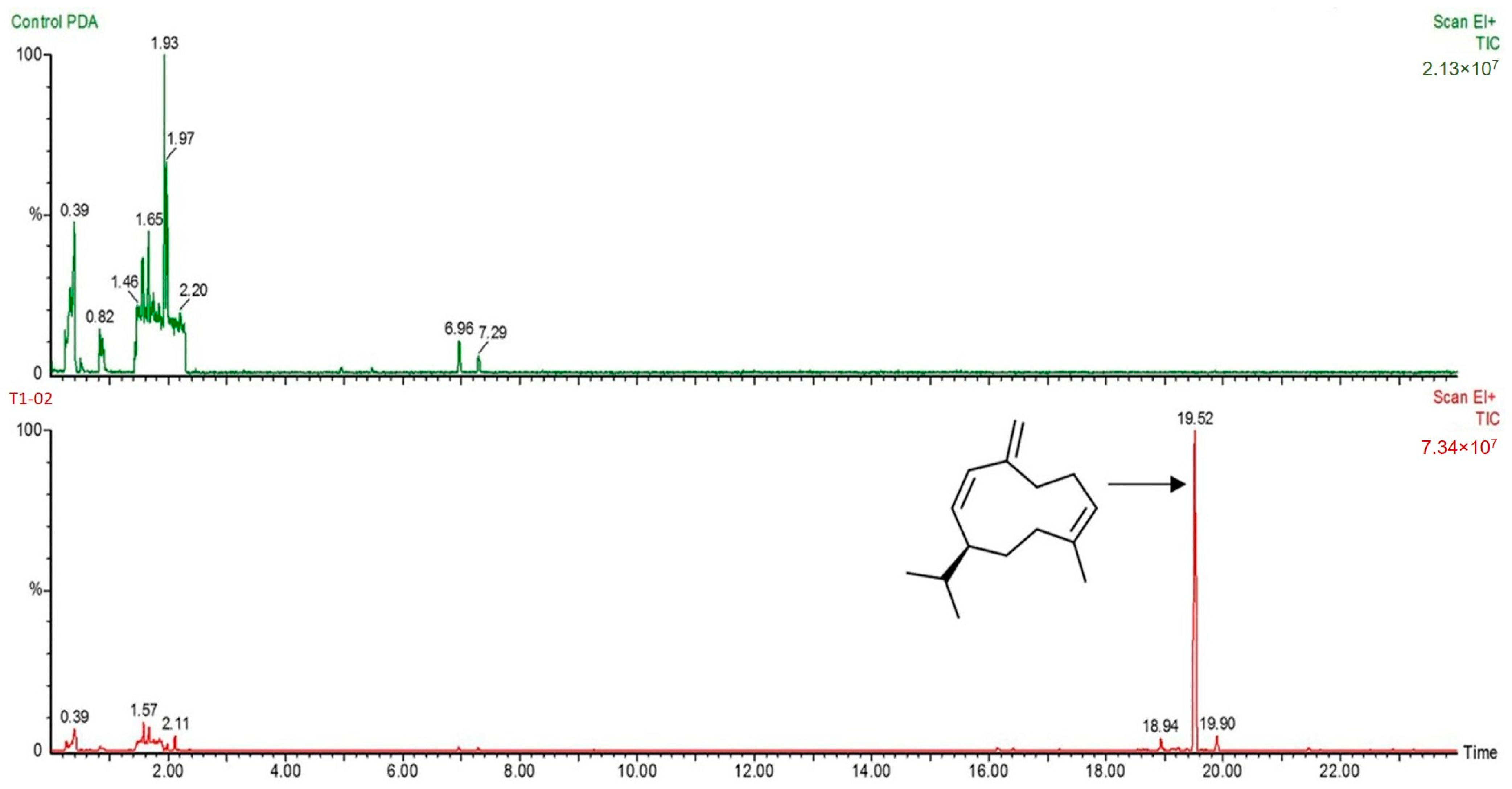
2.5. Solid-Phase Microextraction (SPME) Gas Chromatography–Mass Spectrometry (GC/MS) Analysis

To analyze volatile organic compounds (VOCs) emitted by Trichoderma spp., solid-phase microextraction (SPME) gas chromatography–mass spectrometry (SPME-GC/MS) was conducted [42]. The most effective strain of Trichoderma was cultured in a 20 mL chromatography vial (PerkinElmer, Waltham, MA, USA) and incubated at ambient temperature (28 ± 2 °C) for 7 days. Volatiles were extracted by SPME with headspace (HS) derivatization, and the extraction temperature was set to 125 °C for 25 min, needle temperature 130 °C for 1.5 min, and the transfer line 135 °C for 0.03 min. Separation of volatile compounds was conducted using a Clarus model 690 gas chromatograph (PerkinElmer, Waltham, MA, USA) coupled with an SQ8 mass-selective detector. The capillary column contained 5% phenylmethylpolysiloxane, 30 m × 250 μm ID × 0.25 μm film thickness (PerkinElmer, Waltham, MA, USA). The oven temperature was held at 60 ℃, then increased at a rate of 7 °C/min to a final temperature of 200 °C. The injector temperature was set at 200 °C and purified helium gas was used as the carrier gas at a flow rate of 1 mL/min. Electron impact (EI) mass spectra were collected at 70 eV ionization voltages over the range of m/z 45–550 and the electron multiplier voltage was set at 1400 V. The ion source and quadrupole temperatures were set at 200 °C. Volatile compounds were identified through a computer search of the National Institute of Standard and Technology (NIST) Mass Spectral Library Search Chromatogram.

2.6. Pre-Colonized Plate Method

To investigate the mycoparasitism of Trichoderma species, the pre-colonized plate method was used [10,15]. A mycelial plug of a 5-day-old colony of N. clavispora was cut and placed on one side of PDA and incubated at ambient temperature for 7 days. A strip of the most effective strain of Trichoderma (4 × 0.5 cm) was cut from a 7-day-old colony cultured on PDA, placed on the other side of PDA, and wholly pre-colonized by the isolate from N. clavispora plate. The tested plates were incubated at ambient temperature in dark conditions for 4 weeks. Ten agar plugs from the tested plates were then placed on 20% PDA to observe the growth of Trichoderma and N. clavispora. The percentage of colonization of each fungus was calculated. Growth of the Trichoderma colony in 20% PDA plates in comparison with the N. clavispora colony was counted and converted to percentage colonization. Mycoparasitism of Trichoderma was checked after 5 days of incubation with a compound microscope (Leica DM750, Leica Microsystems). After 10 days of incubation, parasitism of Trichoderma was checked through a thin polymer membrane nano-suit method [43], and was observed directly with a JSM-6100 SEM (JEOL, Peabody, MA, USA).

2.7. In Vivo Test

To test the efficacy of Trichoderma in reducing the flower blight of flamingo flower, an in vivo test was conducted with the flamingo flower Cheer cultivar. The selected Trichoderma isolates and N. clavispora were cultured on PDA for 7 days and incubated at ambient temperature (28 ± 2 °C) for sporulation. Conidia of both Trichoderma and N. clavispora were harvested. The conidial suspension of each fungus was adjusted in concentration as 106 conidia/mL by sterile DW. The experiment was set up as flower inoculation by spraying: (1) 50 mL DW (control group), (2) 50 mL N. clavispora conidial suspension, (3) 50 mL Trichoderma strain conidial suspension, then incubated at ambient temperature for 24 h and challenged similarly with N. clavispora conidial suspension, and (4) 50 mL Trichoderma conidial suspension. Five flowers were inoculated for each treatment and the experiment was repeated twice. The tested flowers were incubated in the moist chamber (approximately 85% relative humidity) at ambient temperature, and disease development was observed after 10 days postinoculation. Disease scores were determined by the method previously described by Sunpapao et al. [9], with some modifications, based on assessing the external symptoms of the flowers (0 = no symptom, 1 = small brown symptom < 1 cm, 2 = brown symptom > 1 cm, 3 = brown symptom that covers > 25% of flower, 4 = brown symptom that covers more than 50% of flower). Disease scores were converted to a disease severity index (DSI) using the following formula:
DSI   ( % ) = ( Scale × Amount   of   plants ) Maximum   level × Total   number   of   plants × 100

2.8. Statistical Analysis

Significant differences among fungal growth, mycoparasite test, and disease severity were subjected to one-way analysis of variance (ANOVA). Tukey’s test and Student’s t test were used to determine statistically significant differences.

3. Results

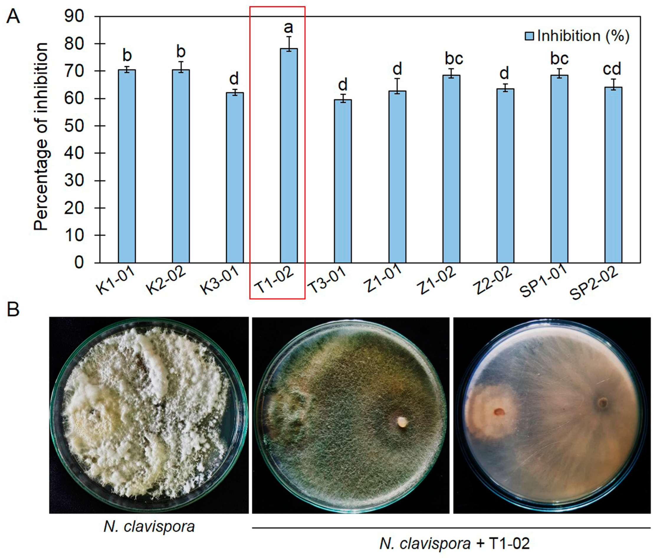
3.1. Primary Screening of Trichoderma spp., against Neopestalotiopsis clavispora

Dual culture assays showed that the ten isolates of Trichoderma spp. grew faster in tested PDA plates compared to N. clavispora and covered most of the petri dish after 5 days of incubation. Trichoderma spp. inhibited mycelial growth of N. clavispora, with inhibition ranging from 59.61% to 78.20%. Trichoderma sp. T1-02 presented the highest inhibition against N. clavispora, 78.20%, significantly higher than those of other isolates (Figure 1A). At 7 days after incubation at ambient temperature, Trichoderma sp. T1-02 effectively covered the whole colony of N. clavispora (Figure 1B).

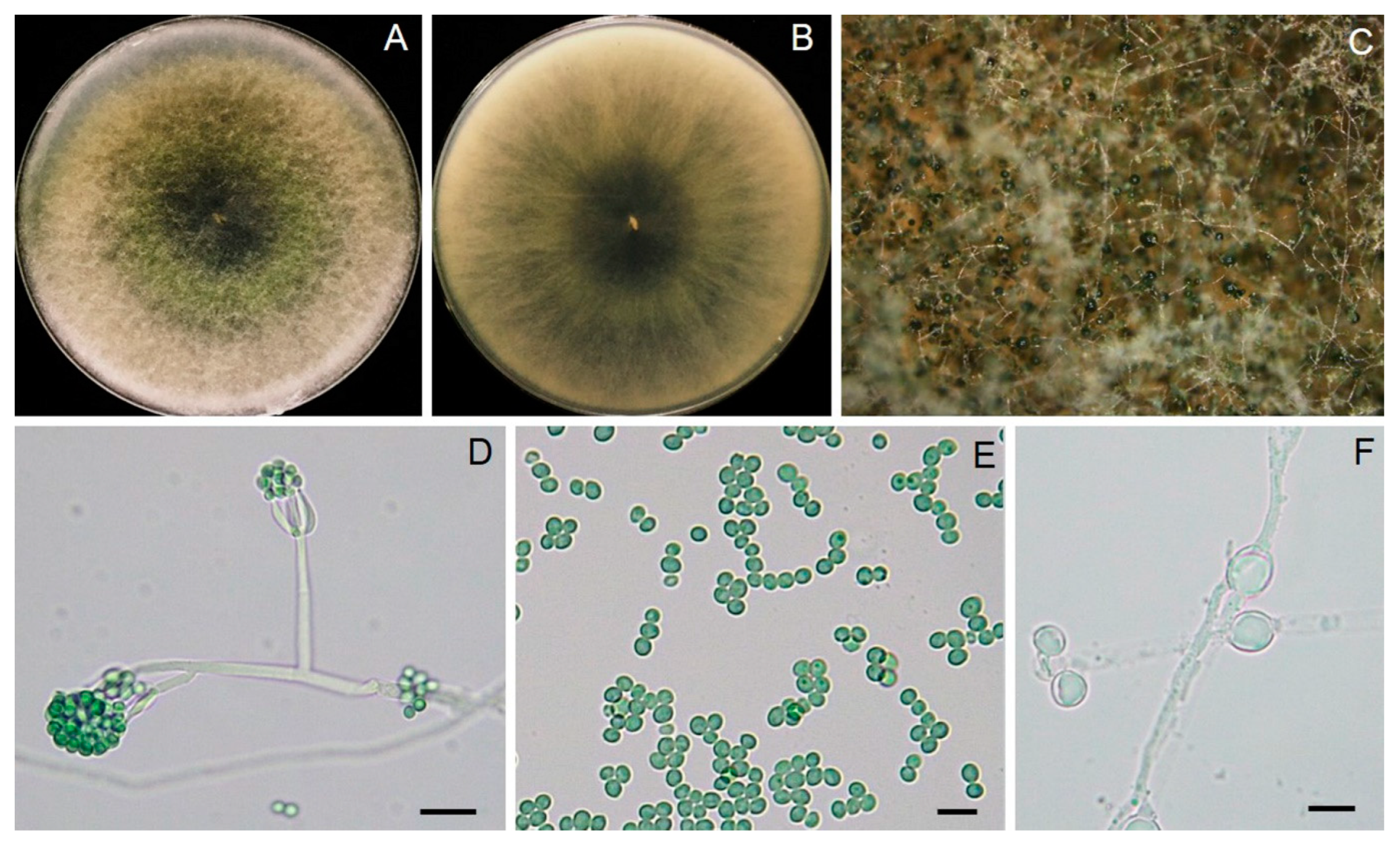
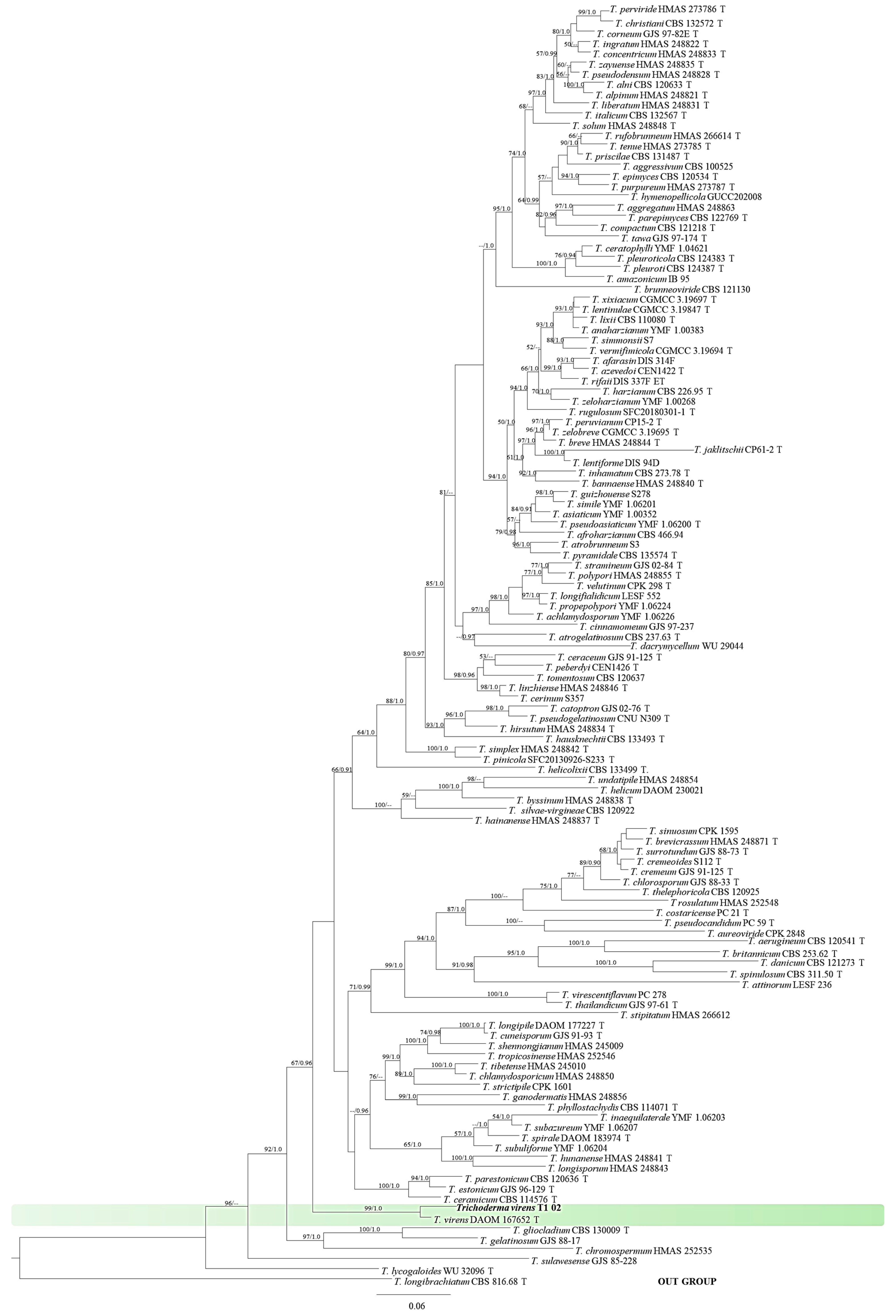
3.2. Identification of Trichoderma Strain T1-02

Trichoderma sp. T1-02 conidia germinated on PDA within 24 h and reached 9 cm in three days at ambient temperature (28 ± 2 °C), revealing a growth rate of 3 cm per day. Fungal colonies of Trichoderma sp. T1-02 displayed white, pale green, and turned to dark green after 5 days (Figure 2A,B). Conidiophores branched, terminating a whorl of two to three divergent phialides. Phialides were 6.22–16.38 μm long, 2.33–9.96 μm wide ( x ¯ = 10.79 × 4.02 μm, n = 30) and apulliform. Conidia were 3.39–5.16 μm long, 3.08–4.40 μm wide ( x ¯ = 4.31 × 3.78 μm, n = 30), and green to dark-green globose to ovule (Figure 2E).
For molecular identification, portions of ITS, rpb2 and tef1-α regions gave DNA fragments about 500, 1200, and 1200 bases long, respectively. The PCR products were then deposited in the GenBank database and assigned accession numbers OR578379, OR587842, and OR587841 for ITS, rpb2 and tef1-α regions, respectively, and they were compared with known sequences of Trichoderma spp. available at GenBank. A BLASTn search in GenBank indicated that Trichoderma sp. T1-02 was grouped within Trichoderma virens species with 100% identity of the all sequences (ITS, rpb2 and tef1-α regions). The combined ITS, rpb2 and tef1-α sequences dataset consisted of 44 taxa. The phylogram assigned the fungal isolate T1-02 used in this study to the same clade of Trichoderma virens containing species of ex-type (DAOM167652), with high BS (99%) and PP (1.0) supports. Therefore, the isolate T1-02 was identified as T. virens based on molecular properties (Figure 3).

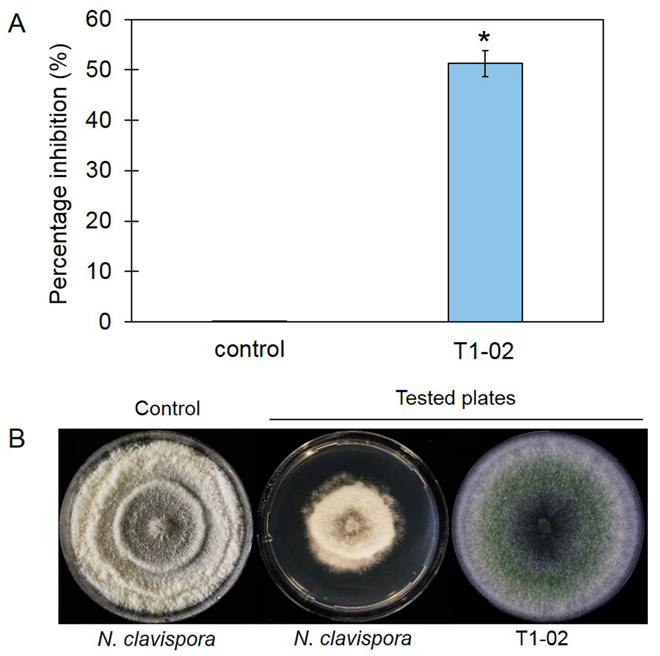
3.3. Production of Volatile Antifungal Compounds

The sealed plate method showed that T. virens T1-02 inhibited the growth of N. clavispora, with inhibition of 51.28% in PDA plates (Figure 4A), and suppressed growth of N. clavispora in the tested PDA plate (Figure 4B). This result revealed that VOCs emitted by T. virens T1-02 were able to restrict fungal growth. SPME-GC/MS showed a major volatile emitted by T. virens T1-02 at 15.22 min (Figure 5). This compound was tentatively identified as a sesquiterpene: germacrene D (IUPAC name: (1E,6E,8S)-1-methyl-5-methylidene-8-propan-2-ylcyclodeca-1,6-diene). The germacrene D contained C 15 atoms (C15) with a molecular weight of 204 and molecular formula of C15H24.

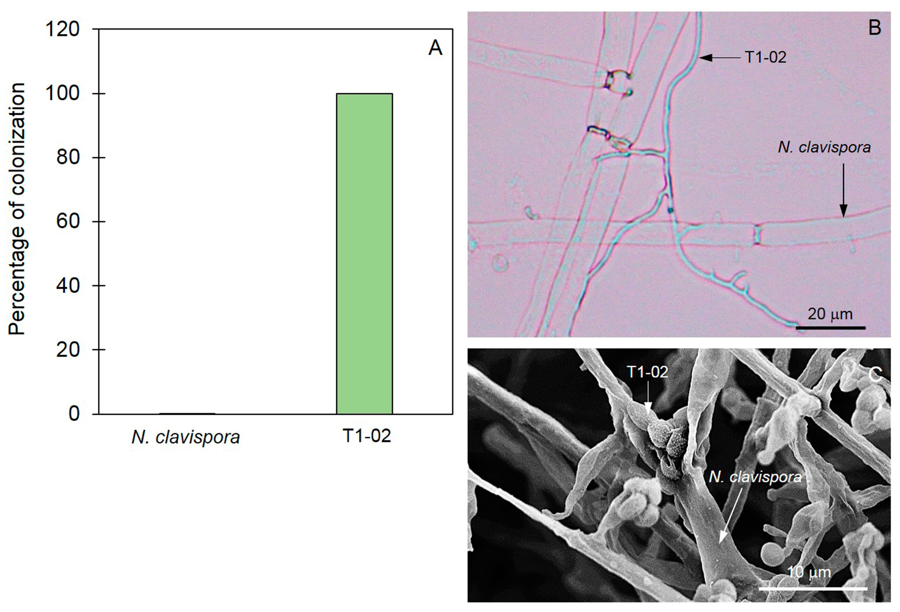
3.4. Mycoparasitic Properties of Trichoderma T1-02

Mycoparasitic ability of T. virens T1-02 was assessed through the pre-colonized plate method. After incubation for 5 days, hyphae of T1-02 attaching to N. clavispora hyphae were observed by compound microscopy (Figure 6B). SEM showed T. virens T1-02 conidia attached on N. clavispora hyphae and caused hyphae wilt after 10 days of incubation (Figure 6C). After incubation for 3 weeks, T. virens T1-02 was aggressively mycoparasitic to N. clavispora, with colonization of 100% (Figure 6A). The pathogen could not be recovered from this point of colonization on PDA.

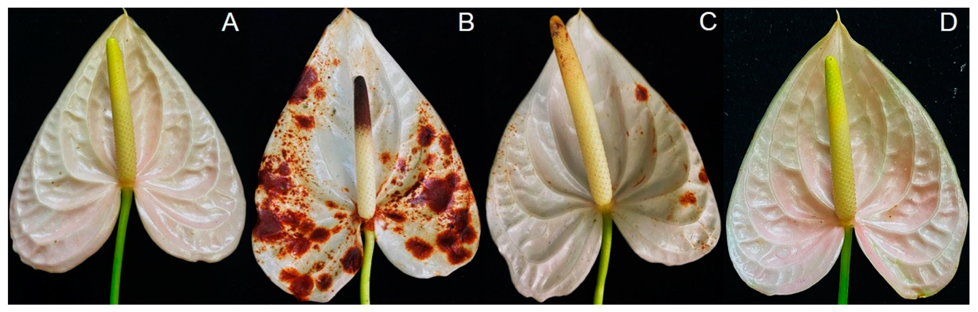
3.5. Trichoderma virens T1-02 Suppressed Symptom Development

An in vivo test was conducted to test the effect of Trichoderma on the reduction of flower blight on flamingo flowers. Application of the T. virens T1-02 conidia suspension reduced symptom development and disease severity index of flower blight on flamingo flowers (Figure 7C). After 10 days of incubation, brown lesions in control (inoculation with N. clavispora alone) showed the most severe symptoms compared to other treatments (Figure 7B). Disease severity index (DSI) of flamingo flowers inoculated with DW, N. clavispora alone, N. clavispora challenged with T. virens T1-02 and T. virens T1-02 alone was 0%, 85.52%, 12.50%, and 0%, respectively.

4. Discussion

In the current study, the newly isolated rhizosphere fungus displayed antagonistic ability against N. clavispora in dual culture assays. Based on morphological study and molecular properties of multiple DNA sequences of ITS, tef1-α and rpb2 this rhizosphere fungus was identified as T. virens strain T1-02. Generally, the plant pathogens were suppressed by the rhizosphere fungi through multiple mechanisms, including antibiosis, competition, mycoparasitism, and induction of defense responses in plants [9,10,15]. Meanwhile, the possible biocontrol mechanisms of T. virens T1-02 against N. clavispora were assessed both in vitro and in vivo. Interestingly, our consistent results from the two assessments indicate that T. virens T1-02 has the potential to be used as a BCA in inhibiting N. clavispora. The possible mechanisms of T. virens T1-02 include competition for nutrients and space, production of VOCs, and direct parasitism on pathogen hyphae, leading to reduction in disease severity in flamingo flowers.
Based on the above results, T1-02 effectively suppressed fungal growth of N. clavispora on dual culture assay. This may be due to competition and/or antifungal metabolites released from T. virens T1-02. The results from our study showed that T. virens T1-02 effectively inhibited the fungal growth of N. clavispora in dual culture assay plates due to a faster growth rate and covering the pathogen colony. The ability to compete for nutrients and space is one of the most effective mechanisms involved in fungicidal ability against pathogens. Due to the fast-growing ability of Trichoderma spp. [44], they were able to compete for nutrients and space in vitro, as observed by dual culture assay [28]. This method is widely used to primarily screen antagonistic activities of several pathogens [9,14]. This suggests that T. virens T1-02 exhibited competition mechanism that involved antagonistic ability by restricting the fungal growth of N. clavispora, the flower blight pathogen of the flamingo flower.
In the current study, the sealed plate method showed that VOCs emitted by T. virens T1-02 effectively inhibited the fungal growth of N. clavispora. Similar results were observed in several Trichoderma species that could produce and release VOCs responsible for restricting the growth of fungal pathogens [45,46]. For instance, volatiles emitted by T. asperelloides PSU-P1 mediated antifungal ability, induced defense response, and promoted plant growth in Arabidopsis thaliana [11]. Furthermore, volatiles emitted by T. asperellum T76-14 contained 2-phynyl ethanol (2-PE), associated with reduction of postharvest fruit rot of muskmelon [13]. The GC/MS profile also suggested that VOCs of T. virens T1-02 contained germacrene D. This compound is classified into sesquiterpene, which is commonly found in plant essential oil with antimicrobial activities [47,48]. However, this compound was detected in a different strain of T. virens by Nieto-Jacobo et al. [49]. Our result is in agreement with Nieto-Jacobo et al. [49] that different strains of T. virens can produce and emit germacrene D, and may contribute to antagonistic activity against N. clavispora.
The pre-colonized plate method was used in this study to reveal this mycoparasitism, and T. virens T1-02 aggressively colonized 100% of N. clavispora in tested plates. This method showed that hyphae and conidia of T. virens T1-02 had strong inhibitory and parasitic effects on hyphae of N. clavispora. This ability has been found in different species of Trichoderma by Bailey et al. [10]. They are aggressively mycoparasitic against Moniliophthora roreri, the fungal pathogen of pod rot of Theobroma cacao. A recent publication by Runagwong et al. [14] also revealed that T. asperelloides PSU-P1 acts as a mycoparasite against Stagonosporopsis cucurbitacearum, the fungal pathogen of gummy stem blight in muskmelon. The finding from the present study suggested that the T. virens T1-02 not only competed for nutrients and spaces but also parasitized and colonized fungal hyphae of N. clavispora.
This study aimed to reduce the excess use of synthetic fungicides in agricultural processes and to find environmentally friendly ways to combat N. clavispora in flamingo flowers. The results from in vivo testing revealed that the application of rhizosphere fungus T. virens T1-02 conidia suspension reduced the disease severity of flower blight in flamingo flowers caused by N. clavispora when compared to the control without rhizosphere fungus inoculation. As observed by Wonglom et al. [50], application of Trichoderma sp. T76-12/2 conidia suspension reduced disease severity of Sclerotium fruit rot of snake fruit and stem rot of lettuce. Furthermore, the development of an emulsion formulation of T. asperelloides PSU-P1 has been noted as an effective formulation in suppressing stem canker of dragon fruit [51]. Furthermore, the application of T. virens 6PS-2 spore suspensions changed the community of microorganisms through colonization in rhizosphere soil and reduced a pathogen of apples [52]. Our current study provides evidence on the possibilities of applying T. virens T1-02 as BCA for managing Neopestalotiopsis diseases including flower blight of flamingo flower, which would be an eco-friendly management strategy of N. clavispora and contribute to the healthy maintenance of plants.

5. Conclusions

The rhizosphere soil fungus was primarily screened for its fungicidal activity against N. clavispora and identified based on both morphology and molecular data as T. virens T1-02. The results revealed that this strain of T. virens has the potential to be used as a biological control against N. clavispora with multiple mechanisms, including antibiosis via the production of volatile antifungal compound germacrene D, competition and direct parasitism. An in vivo test suggested the potential of T. virens T1-02 to reduce the disease severity of flower blight on flamingo flowers. However, the bioformulation of this strain has yet to be investigated. To apply T. virens T1-02 as BCA on a large scale for commercial purposes, further experiments need to be verified in the near future.

Author Contributions

Conceptualization, D.A. and A.S.; methodology, A.S.; software, O.-U.R. and K.P.; validation, D.A., O.-U.R. and A.S.; formal analysis, K.P. and D.L.H.; investigation, A.S.; resources, A.S.; data curation, A.S.; writing—original draft preparation, A.S.; writing—review and editing, D.A., D.L.H. and O.-U.R.; visualization, A.S.; supervision, A.S.; project administration, A.S.; funding acquisition, D.A. and A.S. All authors have read and agreed to the published version of the manuscript.

Funding

This research was funded by Prince of Songkla University.

Institutional Review Board Statement

Not applicable.

Informed Consent Statement

Not applicable.

Data Availability Statement

The DNA sequence data obtained from this study were deposited in GenBank under accession numbers ITS (OR578379), rpb2 (OR587842) and tef1-α (OR587841).

Acknowledgments

This research was partially supported by the Center of Excellence in Agricultural and Natural Resources Biotechnology (CoE-ANRB), Phase 3, Prince of Songkla University, Chiang Mai University and Thammasat University. Special thanks go to PerkinElmer Co. Ltd. Thailand for GC/MS analysis.

Conflicts of Interest

The authors have received technical support from PerkinElmer Co. Ltd. Thailand.

References

  1. Hartmann, A.; Rothballer, M.; Schmid, M. Lorenz Hiltner, A pioneer in rhizosphere microbial ecology and soil bacteriology research. Plant Soil 2008, 312, 7–14. [Google Scholar] [CrossRef]
  2. Hinsinger, P.; Bengough, G.A.; Vetterlein, D.; Young, I.M. Rhizosphere: Biophysics, biogeochemistry and ecological relevance. Plant Soil 2009, 321, 117–152. [Google Scholar] [CrossRef]
  3. Lambers, H.; Mougel, C.; Jaillard, B.; Hinsinger, P. Plant-microbe-soil interactions in the rhizosphere: An evolutionary perspective. Plant Soil 2009, 321, 83–115. [Google Scholar] [CrossRef]
  4. Raaijmakers, J.M.; Paulitz, T.C.; Steinberg, C.; Alabouvette, C.; Moenne-Loccoz, Y. The rhizosphere: A playground and battlefield for soilborne pathogens and beneficial microorganisms. Plant Soil 2009, 321, 341–361. [Google Scholar] [CrossRef]
  5. Ehrmann, J.; Ritz, K. Plant: Soil interactions in temperate multi-cropping production systems. Plant Soil 2014, 376, 1–29. [Google Scholar] [CrossRef]
  6. Phoka, N.; Pornsuriya, C.; Sunpapao, A. High-throughput sequencing provides insight into soil fungal community structure and diversity in plant protected areas of Songkhla zoo in southern Thailand. Chiang Mai J. Sci. 2022, 49, 524–537. [Google Scholar] [CrossRef]
  7. Verma, M.; Brar, S.K.; Tyagi, R.D.; Surampalli, R.Y.; Valero, J.R. Dissolved oxygen as principal parameter for conidia production of biocontrol fungi Trichoderma viride in non-Newtonian wastewater. J. Ind. Microbiol. Biotechnol. 2006, 33, 941–952. [Google Scholar] [CrossRef]
  8. Tseng, Y.-H.; Rouina, H.; Groten, K.; Rajani, P.; Furch, A.C.U.; Reichelt, M.; Baldwin, I.T.; Nataraja, K.N.; Uma Shaanker, R.; Oelmüller, R. An Endophytic Trichoderma strain promotes growth of its hosts and defends against pathogen attack. Front. Plant Sci. 2020, 11, 573670. [Google Scholar] [CrossRef]
  9. Sunpapao, A.; Chairin, T.; Ito, S. The biocontrol by Streptomyces and Trichoderma of leaf spot disease caused by Curvularia oryzae in oil palm seedlings. Biol. Control. 2018, 123, 36–42. [Google Scholar] [CrossRef]
  10. Bailey, B.A.; Bae, H.; Strem, M.D.; Crozier, J.; Thomas, S.E.; Samuels, G.J.; Vinyard, B.T.; Holmes, K.A. Antibiosis, mycoparasitism, and colonization success for endophytic Trichoderma isolates with biological control potential in Theobroma cacao. Biol. Control 2008, 46, 24–35. [Google Scholar] [CrossRef]
  11. Phoka, N.; Suwannarach, N.; Lumyong, S.; Ito, S.; Matsui, K.; Arikit, S.; Sunpapao, A. Role of volatiles from the endophytic fungus Trichoderma asperelloides PSU-P1 in biocontrol potential and in promoting the plant growth of Arabidopsis thaliana. J. Fungi 2020, 6, 341. [Google Scholar] [CrossRef]
  12. Suebrasri, T.; Somteds, A.; Harada, H.; Kanokmedhakul, S.; Jogloy, S.; Ekprasert, J.; Lumyong, S.; Boonlue, S. Novel endophytic fungi with fungicidal metabolites suppress sclerotium disease. Rhizosphere 2020, 16, 100250. [Google Scholar] [CrossRef]
  13. Intana, W.; Kheawleng, S.; Sunpapao, A. Trichoderma asperellum T76-14 released volatile organic compounds against postharvest fruit rot in muskmelons (Cucumis melo) caused by Fusarium incarnatum. J. Fungi 2021, 7, 46. [Google Scholar] [CrossRef] [PubMed]
  14. Ruangwong, O.-U.; Wonglom, P.; Phoka, N.; Suwannarach, N.; Lumyong, S.; Ito, S.-I.; Sunpapao, A. Biological control activity of Trichoderma asperelloides PSU-P1 against gummy stem blight in muskmelon (Cucumis melo). Physiol. Mol. Plant Pathol. 2021, 115, 101663. [Google Scholar] [CrossRef]
  15. Intana, W.; Kumla, J.; Suwannarach, N.; Sunpapao, A. Biological control potential of a soil fungus Trichoderma asperellum K1-02 against Neoscytalidium dimidiatum causing stem canker of dragon fruit. Physiol. Mol. Plant Pathol. 2023, 128, 102151. [Google Scholar] [CrossRef]
  16. Intana, W.; Wonglom, P.; Suwannarach, N.; Sunpapao, A. Trichoderma asperelloides PSU-P1 induced expression of pathogenesis-related protein genes against gummy stem blight of muskmelon (Cucumis melo) in field evaluation. J. Fungi 2022, 8, 156. [Google Scholar] [CrossRef] [PubMed]
  17. Zhang, S.; Gan, Y.; Xu, B. Application of plant-growth-promoting fungi Trichoderma longibrachiatum T6 enhances tolerance of wheat to salt stress through improvement of antioxidative defense system and gene expression. Front. Plant Sci. 2016, 7, 1045. [Google Scholar] [CrossRef]
  18. Galletti, S.; Burzi, P.L.; Cerato, C.; Marinello, S.; Sala, E. Trichoderma as a potential biocontrol agent for Cercospora leaf spot of sugar beet. BioControl 2008, 53, 917–930. [Google Scholar] [CrossRef]
  19. Chaves, T.P.; Miranda, A.R.G.S.; da Paz, L.C.; Netto, M.S.B.; Lima, G.S.A.; Assunção, I.P. First report of Colletotrichum theobromicola causing anthracnose on Anthurium sp. Australas. Plant Dis. Notes 2020, 15, 27. [Google Scholar] [CrossRef]
  20. Ben, H.-Y.; Zhao, Y.-J.; Chai, A.-L.; Shi, Y.-X.; Xie, X.-W.; Li, B.-J. First Report of Myrothecium roridum Causing Leaf Spot on Anthurium andraeanum in China. J. Phytopathol. 2015, 163, 144–147. [Google Scholar] [CrossRef]
  21. Park, M.-J.; Back, C.-G.; Park, J.-H. Occurrence of Phytopythium vixans causing stem rot on Anthurium andraeanum in Korea. Korean J. Mycol. 2019, 47, 443–446. [Google Scholar]
  22. Daengsuwan, W.; Wonglom, P.; Sunpapao, A. First report of Lasiodiplodia theobromae causing spadix rot in Anthurium andraeanum. J. Phytopathol. 2020, 168, 129–133. [Google Scholar] [CrossRef]
  23. Daengsuwan, W.; Wonglom, P.; Arikit, S.; Sunpapao, A. Morphological and molecular identification of Neopestalotiopsis clavispora causing flower blight on Anthurium andraeanum in Thailand. Hortic. Plant J. 2021, 7, 573–578. [Google Scholar] [CrossRef]
  24. Poole, N.F.; Arnaudin, M.E. The role of fungicides for effective disease management in cereal crops. Can. J. Plant Pathol. 2014, 36, 1–11. [Google Scholar] [CrossRef]
  25. Hahn, M. The rising threat of fungicide resistance in plant pathogenic fungi: Botrytis as a case study. J. Chem. Biol. 2014, 7, 133–141. [Google Scholar] [CrossRef] [PubMed]
  26. Polat, B.; Tiryaki, O. Determination of fungicide residues in soil using QuEChERS coupled with LC-MS/MS, and environmental risk assessment. Environ. Monit. Assess. 2023, 195, 986. [Google Scholar] [CrossRef]
  27. Tao, H.; Bao, Z.; Jin, C.; Miao, W.; Fu, Z.; Jin, Y. Toxic effects and mechanisms of three commonly used fungicides on the human colon adenocarcinoma cell line Caco-2. Environ. Pollut. 2020, 263, 114660. [Google Scholar] [CrossRef]
  28. Rahman, M.A.; Begum, M.F.; Alam, M.F. Screening of Trichoderma isolates as a biological control agent against Ceratocystis paradoxa causing pineapple disease of sugarcane. Microbiology 2009, 37, 277–285. [Google Scholar]
  29. Saitoh, K.; Togashi, K.; Arie, T.; Teraoka, T. A simple method for a mini-preparation of fungal DNA. J. Gen. Plant Pathol. 2006, 72, 348–350. [Google Scholar] [CrossRef]
  30. White, T.J.; Bruns, T.; Lee, S.; Tayler, J. Amplification and direct sequencing of fungal ribosomal RNA genes for phylogenies. In PCR Protocols: A Guide to Methods and Applications; Innis, A.M., Gelfelfard, D.H., Snindky, J.J., White, T.J., Eds.; Academic Press: San Diego, CA, USA, 1990; pp. 315–322. [Google Scholar]
  31. Carbone, I.; Kohn, L.M. A method for designing primer sets for speciation studies in filamentous ascomycetes. Mycologia 1999, 91, 553–556. [Google Scholar] [CrossRef]
  32. O’Donnell, K.; Kistler, H.C.; Cigelnik, E.; Ploetz, R.C. Multiple evolutionary origins of the fungus causing Panama disease of banana: Concordant evidence from nuclear and mitochondrial gene genealogies. Proc. Nat. Acad. Sci. USA 1998, 95, 2044–2049. [Google Scholar] [CrossRef]
  33. Liu, Y.J.; Whelen, S.; Hall, B.D. Phylogenetic relationships among ascomycetes: Evidence from and RNA polymerase II subunit. Mol. Biol. Evol. 1999, 16, 1799–1808. [Google Scholar] [CrossRef] [PubMed]
  34. Hall, T.A. BioEdit: A User-Friendly Biological Sequence Alignment Editor and Analysis Program for Windows 95/98/NT. Nucleic Acids Symp. Ser. 1999, 41, 95–98. [Google Scholar]
  35. Zeng, X.-Y.; Yuan, X.-X.; Peng, K.-Q.; Pan, Y.-T.; Tan, T.-J.; Wu, N.; Tian, F.-H. Taxonomy and control of Trichoderma hymenopellicola sp. nov. responsible for the first green mold disease on Hymenopellis raphanipes. Front. Microbiol. 2022, 13, 991987. [Google Scholar] [CrossRef] [PubMed]
  36. Katoh, K.; Rozewicki, J.; Yamada, K.D. MAFFT online service: Multiple sequence alignment, interactive sequence choice and visualization. Brief Bioinform. 2019, 20, 1160–1166. [Google Scholar] [CrossRef]
  37. Stamatakis, A. RAxML version 8: A tool for phylogenetic analysis and post-analysis of large phylogenies. Bioinformatics 2014, 30, 1312–1313. [Google Scholar] [CrossRef]
  38. Miller, M.A.; Pfeiffer, W.; Schwartz, T. Creating the CIPRES Science Gateway for Inference of Large Phylogenetic Trees; Gateway Computing Environments Workshop (GCE): New Orleans, LA, USA, 2010; pp. 1–8. [Google Scholar]
  39. Nylander, J.A.A. MrModeltest Version 2. Program Distributed by the Author; Evolutionary Biology Centre, Uppsala University: Uppsala, Sweden, 2004. [Google Scholar]
  40. Ronquist, F.; Teslenko, M.; van der Mark, P.; Ayres, D.L.; Darling, A.; Höhna, S.; Larget, B.; Liu, L.; Suchard, M.A.; Huelsenbeck, J.P. MrBayes 3.2: Efficient Bayesian phylogenetic inference and model choice across a large model space. Syst Biol. 2012, 61, 539–542. [Google Scholar] [CrossRef]
  41. Dennis, C.; Webster, J. Antagonistic properties of species-groups of Trichoderma, II. Production of volatile antibiotic. Trans. Br. Mycol. Soc. 1971, 57, 41–48. [Google Scholar] [CrossRef]
  42. Ruangwong, O.-U.; Wonglom, P.; Suwannarach, N.; Kumla, J.; Thaochan, N.; Chomnunti, P.; Pitija, K.; Sunpapao, A. Volatile organic compound from Trichoderma asperelloides TSU1: Impact on plant pathogenic fungi. J. Fungi 2021, 7, 187. [Google Scholar] [CrossRef]
  43. Takaku, Y.; Suzuki, S.; Ohta, I.; Ishii, D.; Muranaka, Y.; Shimomura, M.; Hariyama, T. A thin polymer membrane, nano-suit, enhancing survival across the continuum between air and high vacuum. Proc. Natl. Acad. Sci. USA 2014, 110, 7631–7635. [Google Scholar] [CrossRef]
  44. Rossi-Rodrigues, B.C.; Brochetto-Braga, M.R.; Tauk-Tornisielo, S.M.; Carmona, E.C.; Arruda, V.M.; Chaud Netto, J. Comparative growth of Trichoderma strains in different nutritional sources, using bioscreen c automated system. Braz. J. Microbiol. 2009, 40, 404–410. [Google Scholar] [CrossRef] [PubMed]
  45. Guo, Y.; Ghirardo, A.; Weber, B.; Schnitzler, J.-P.; Benz, J.P.; Rosenkranz, M. Trichoderma species differ in their volatile profiles and in antagonism toward ectomycorrhiza Laccaria bicolor. Front. Microbiol. 2019, 10, 891. [Google Scholar] [CrossRef] [PubMed]
  46. Elsherbiny, A.E.; Amin, B.H.; Aleem, B.; Kingsley, K.L.; Bennett, J.W. Trichoderma volatile organic compounds as a biofumigation tool against late blight pathogen Phytophthora infestans in postharvest potato tubers. J. Agric. Food Chem. 2020, 68, 8163–8171. [Google Scholar] [CrossRef] [PubMed]
  47. Montanari, R.M.; Barbosa, L.C.A.; Demuner, A.J.; Silva, C.J.; Carvalho, L.S.; Andrade, N.J. Chemical composition and antibacterial activity of essential oils from verbenaceae species: Alternative sources of (E)-caryophyllene and germacrene-D. Quím. Nova 2011, 34, 1550–1555. [Google Scholar] [CrossRef]
  48. Santos, T.G.; Laemmle, J.; Rebelo, R.A.; Dalmarco, E.M.; Cruz, A.B.; Schmit, A.P. Chemical composition and antimicrobial activity of Aloysia gratíssima (Verbenaceae) leaf essential oil. J. Essent. Oil Res. 2015, 27, 125–130. [Google Scholar] [CrossRef]
  49. Nieto-Jacobo, M.F.; Steyaert, J.M.; Salazar-Badillo, F.B.; Nguyen, D.V.; Rostás, M.; Braithwaite, M.; De Souza, J.T.; Jimenez-Bremont, J.F.; Ohkura, M.; Stewart, A.; et al. Environmental growth conditions of Trichoderma spp. affects indole acetic acid derivatives, volatile organic compounds, and plant growth promotion. Front. Plant Sci. 2017, 8, 102. [Google Scholar] [CrossRef]
  50. Wonglom, P.; Daengsuwan, W.; Ito, S.; Sunpapao, A. Biological control of Sclerotium fruit rot of snake fruit and stem rot of lettuce by Trichoderma sp. T76-12/2 and the mechanism involved. Physiol. Mol. Plant Pathol. 2019, 107, 1–7. [Google Scholar] [CrossRef]
  51. Intana, W.; Wonglom, P.; Dy, K.S.; Sunpapao, A. Development of a novel emulsion formulation of Trichoderma asperelloides PSU-P1 conidia against stem canker on dragon fruit caused by Neoscytalidium dimidiatum. Microbiol. Res. 2023, 14, 1139–1149. [Google Scholar] [CrossRef]
  52. Wang, H.; Tang, W.; Mao, Y.; Ma, S.; Chen, X.; Shen, X.; Yin, C.; Mao, Z. Isolation of Trichoderma virens 6PS-2 and its effects on Fusarium proliferatum f. sp. malus domestica MR5 related to apple replant disease (ARD) in China. Hortic. Plant J. 2022; in press. [Google Scholar] [CrossRef]
Figure 1. Dual culture assays of Trichoderma spp. Percentage inhibition of Trichoderma spp. against Neopestalotiopsis clavispora (A) and dual culture assay plates containing control (left), tested plates with N. clavispora and Trichoderma sp. T1-02 in top (middle) and bottom (right) view (B). Error bars indicate standard deviation (SD) of percentage of inhibition (%), whereas letters indicate significant difference among treatments according to Tukey’s test (p < 0.05).
Jof 10 00066 g001
Figure 2. Morphological characteristics of T1-02, colony on PDA in top (A) and bottom view (B); pustule conidiation and mass of conidia developed on tip of conidiophores observed by stereomicroscope (C); details of conidiophore, phialides and conidia (D), conidia (E) and chlamydospores (F).
Jof 10 00066 g002
Figure 3. Phylogram derived from maximum-likelihood (ML) method of combined ITS, tef1-α and rpb2 sequences of 122 taxa with T. longibrachiatum used as the outgroup. Maximum-likelihood bootstrap support (BS) above 50% and Bayesian posterior probability (PP) above 0.9 are shown at nodes. Scale bar represents 6 substitutions per nucleotide position. The new isolate obtained in this study is in bold. “T” represents ex-type strains.
Jof 10 00066 g003
Figure 4. Volatile antifungal bioassay, percentage inhibition of Trichoderma virens T1-02 against Neopestalotiopsis clavispora (A), and sealed plate method on tested plate containing control and tested plates (B). Error bars indicate standard deviation (SD) of percentage inhibition (%), whereas asterisk (*) indicates significant difference between treatments according to Student’s t test (p < 0.05).
Jof 10 00066 g004
Figure 5. GC/MS profiling of PDA (control; up) and volatiles emitted by Trichoderma virens T1-02 (down), major peak at 19.52 min tentatively identified as germacrene D and its structure.
Jof 10 00066 g005
Figure 6. Percentage colonization of Trichoderma virens T1-02 against Neopestalotiopsis clavispora by pre-colonized plate method (A), hyphae of T1-02 attached on N. clavispora hyphae observed by compound microscope (B), conidia of T1-02 attached on N. clavispora hyphae observed by SEM (C).
Jof 10 00066 g006
Figure 7. Flower blight disease development of flamingo flower inoculated with DW (A), Neopestalotiopsis clavispora alone (B), N. clavispora challenged with T1-02 (C) and T1-02 alone (D).
Jof 10 00066 g007
Disclaimer/Publisher’s Note: The statements, opinions and data contained in all publications are solely those of the individual author(s) and contributor(s) and not of MDPI and/or the editor(s). MDPI and/or the editor(s) disclaim responsibility for any injury to people or property resulting from any ideas, methods, instructions or products referred to in the content.

