Article

Identification and Biological Characteristics of Mortierella alpina Associated with Chinese Flowering Cherry (Cerasus serrulata) Leaf Blight in China

1
China National Engineering Research Center of JUNCAO Technology, Fujian Agriculture and Forestry University, Fuzhou 350002, China
2
College of JunCao Science and Ecology (College of Carbon Neutrality), Fujian Agriculture and Forestry University, Fuzhou 350002, China
3
Fujian University Key Laboratory for Plant-Microbe Interaction, College of Life Sciences, Fujian Agriculture and Forestry University, Fuzhou 350002, China
4
College of Plant Protection, Fujian Agriculture and Forestry University, Fuzhou 350002, China
5
School of Environmental Engineering and Chemistry, Luoyang Institute of Science and Technology, Luoyang 471002, China
6
Xi’an Greening Management Center, Xi’an 710007, China
7
Department of Biochemistry, Faculty of Life Sciences, Ahmadu Bello University, Zaria 810281, Nigeria
8
Technology Innovation Center for Monitoring and Restoration Engineering of Ecological Fragile Zone in Southeast China, Ministry of Natural Resources, Fuzhou 350002, China
*
Authors to whom correspondence should be addressed.
J. Fungi 2024, 10(1), 50; https://doi.org/10.3390/jof10010050
Submission received: 5 December 2023 / Revised: 29 December 2023 / Accepted: 2 January 2024 / Published: 5 January 2024
(This article belongs to the Special Issue Fungal Pathogens and Host Plants)

Abstract

:
The Chinese flowering cherry (Cerasus serrulata), an ornamental tree with established medicinal values, is observed to suffer from leaf blight within Xi’an’s greenbelts. This disease threatens both the plant’s growth and its ornamental appeal. In this study, 26 isolates were obtained from plants with typical leaf blight, and only 3 isolates (XA-10, XA-15, and XA-18) were found to be pathogenic, causing similar symptoms on the leaves of the host plant. Based on sequence alignment, the ITS and LSU sequences of the three selected isolates were consistent, respectively. Following morphological and molecular analyses, the three selected isolates were further identified as Mortierella alpina. The three selected isolates exhibited similar morphological characteristics, including wavy colonies with dense, milky-white aerial mycelia on PDA medium. Therefore, isolate XA-10 was used as a representative strain for subsequent experiments. The representative strain XA-10 was found to exhibit optimal growth at a temperature of 30 °C and a pH of 7.0. Host range infection tests further revealed that the representative strain XA-10 could also inflict comparable disease symptoms on both the leaves and fruits of three different Rosaceae species (Prunus persica, Pyrus bretschneideri, and Prunus salicina). This study reveals, for the first time, the causative agent of leaf blight disease affecting the Chinese flowering cherry. This provides a deeper understanding of the biology and etiology of M. alpina. This study lays a solid foundation for the sustainable control and management of leaf blight disease in the Chinese flowering cherry.

1. Introduction

The Chinese flowering cherry, also known as sakura or cherry blossom, represents an ornamental tree species, blooming beautifully in spring and enjoying global appreciation [1]. The term “Sakura” is typically used to describe deciduous trees belonging to the family Rosaceae [2]. The Chinese flowering cherry holds significant economic value due to its considerable appeal to tourists during its spring bloom. In addition to its aesthetic appeal, it also offers a range of medicinal benefits, including anti-inflammatory properties for the skin, prevention of skin roughness, and suppression of melanin production. It is also used as an ingredient in tea [2,3,4,5]. The polyphenols contained in cherries also aid in improving abnormal serum lipid levels, regulating blood glucose, and reducing obesity [6]. Cerasus serrulata (Lindley) Loudon, a member of the Rosaceae family and Cerasus Miller [7,8], serves as an essential ornamental cherry germplasm resource that pervades widely across China, the Korean peninsula, and Japan [9,10,11,12]. Its beautiful flowers, wide adaptability, and high ornamental value contribute to its widespread use in urban and landscape designs [6,13].
With the increased cultivation of Cerasus spp., associated diseases have become increasingly prominent [14,15,16,17,18]. However, similar to other arboreal plants, Cerasus spp. is not immune to various pathogen infections. Previous research has documented leaf blight disease, caused by Phytophthora ramorum, impacting the growth of Prunus laurocerasus in Washington state [19]. Furthermore, Phytophthora cambivora was reported to have attacked the leaves of P. campanulata, causing leaf blight disease in Taiwan [20], and Fusarium proliferatum has been discovered causing leaf blight disease in sweet cherry trees in Liaoning province [21]. However, there have been few scientific literature reports concerning leaf blight disease or the associated pathogens in Chinese flowering cherry. Therefore, identification of the pathogens in Chinese flowering cherry is essential for establishing disease management programs.
Currently, an unknown disease causing wilting of the leaves in Chinese flowering cherry was found in Xi’an, China, and was thus named Chinese flowering cherry leaf blight based on its symptoms. This disease initially appears on the margin or tip of the leaves and enlarges consistently. The color of the infected leaves becomes chlorotic in the initial stage, and later gradually turns into a brown shade. In the later stages of the disease, a significant number of leaves become infected, inhibiting a tree’s growth. The horticultural value of C. serrulata in China and the presence of leaf blight stimulated our interest in understanding which pathogen causes Chinese flowering cherry leaf blight. Therefore, the aim of this study was to detect and identify the pathogen associated with leaf blight of Chinese flowering cherry and to investigate the biological characteristics and pathogenicity of this pathogen on other Rosaceae plants. This study provides essential information to better control the disease and further the sustainable development of the Chinese flowering cherry industry.

2. Materials and Methods

2.1. Disease Investigation and Isolate Collection

From 2019 to 2022, a field survey of Chinese flowering cherry leaf blight was conducted in the urban street greenbelt of Xi’an (elevation: 400 m; geographic position: 107°40′–109°49′ E, 33°42′–34°45′ N), Shaanxi province, China. A total of 1140 trees were surveyed, and the incidence of leaf blight was approximately 15% from the Xi’an Greening Management Center. To isolate the pathogens, 150 leaves with leaf blight symptoms were collected from 285 trees in three greenbelts. Among them, 30 leaves showing typical leaf blight symptoms and without other disease lesions were randomly selected for pathogen isolation. The junction between the diseased and healthy parts of the typical leaf blight samples was used as experimental material.
The tissue isolation method was utilized to isolate the pathogenic fungi [22]. Briefly, the leaves with lesions were cleaned with sterile water, the moisture was absorbed, and 2 cm2 of the disease area was excised. The samples were then surface sterilized in 75% ethanol (Shandong Anweishi Medical Technology Co., Ltd., Dezhou, China) for 30 s, soaked in 2.0% NaClO (Sinopharm Chemical Reagent Co., Ltd., Shanghai, China) for 2 min, rinsed 3 times with sterile water, and dried with sterile filter paper. Subsequently, the diseased leaves were placed onto potato dextrose agar (PDA) medium (prepared by adding 46 g PDA powder to 1 L of sterile double-distilled water) provided by Guangdong Huankai Microbial and Tech. Co., Ltd., Guangzhou, China, and were incubated at 28 °C in the dark until mycelia appeared. The mycelia surrounding the disinfected leaves were selected for further purification; pure cultures were obtained after more than 3 successive subcultures and then stored at 4 °C for further study.

2.2. Pathogenicity Tests on Host Leaves

All isolates were tested for pathogenicity on wounded leaves of the host plant (Chinese flowering cherry). The leaves used in the experiment were sourced from Fujian Agriculture and Forestry University. The healthy samples were cleaned with sterile water, sterilized with 75% ethanol for 30 s, soaked in 2.0% NaClO for 2 min, rinsed with sterile water more than 3 times, and then artificially wounded using sterile toothpicks. Mycelial discs (5 mm in diameter), from the edges of 7-day-old isolated fungal colonies, were placed on the surface of the detached leaf samples, ensuring the mycelia directly contacted the plant tissues. Leaves inoculated with sterile water and PDA without fungal mycelium were used as controls, respectively. To maintain high relative humidity, all inoculated leaves were placed in the culture dish with wet filter paper and then incubated at 28 °C for 5 days.
Meanwhile, mycelial discs (5 mm diameter) were also used to infect the leaves of live host plants at a temperature range of 28–30 °C. All the inoculated samples underwent daily observation, and symptoms were recorded and photographed. All experimental treatments were performed in triplicate. In addition, the pathogens were re-isolated from the leaves of the infected host, their morphological characteristics were analyzed, and the ITS and LSU sequences were amplified for sequence alignment to fulfill Koch’s postulates.

2.3. Molecular Identification of Fungal Isolates

Molecular identification of the 3 selected isolates was performed using the internal transcribed spacer (ITS) and large ribosomal subunit (LSU) gene sequences analysis methods with PCR (polymerase chain reaction) [23]. The isolates were cultured on PDA medium at 28 °C for 7 days. Mycelia were harvested from the cultures, and genomic DNA (gDNA) was extracted using the CTAB method [24]. The gDNA samples were then utilized as templates to amplify the ITS and LSU gene sequences, respectively. All amplified loci, primers, and PCR conditions are listed in Table 1. The TIANGEN Golden Easy PCR kit (TIANGEN Biotech, Beijing, China) was used to carry out the PCR. The PCR amplification reaction was adapted from Liu et al. [25] and Yan et al. [26]. The PCR products with evident bands, detected with 1% agarose gel electrophoresis, were sent to Tsingke Biotech, Fuzhou, China, for sequencing. The sequences were confirmed with a BLAST (Basic Local Alignment Search Tool) search of the NCBI (National Center for Biotechnology Information) database (https://www.ncbi.nlm.nih.gov/, accessed on 16 November 2023). The strains utilized in this study and their corresponding GenBank accession numbers are listed in Table 2.

2.4. Phylogenetic Analysis

A phylogram was constructed using PhyloSuite v.1.2.1 [33] software (https://github.com/dongzhang0725/PhyloSuite, accessed on 16 November 2023). The construction of the phylogram for the 3 selected isolates, which was based on the combined sequences of two genes (ITS and LSU), was carried out using maximum likelihood (ML) and Bayesian inference (BI) [34]. IQ-TREE v.1.5.5 [35] was used for ML analysis, with the selection of GTR+G (ITS) and TRN+I+G (LSU) models, and the estimation of branch stability was performed with 1000 bootstrap replicates. BI analysis was performed in PhyloSuite v.1.2.1 using Mr Bayes v3.2.6 [36] under a partition model (a total of 2 × 10⁷ generations were run, with trees sampled once every 100 generations). FigTree v.1.4.4 was used to display the phylogram.

2.5. Morphological Study

For the colony morphology, the 3 selected isolates were inoculated on PDA medium and incubated at 28 °C in the dark. After 7 days, the colony morphologies from both front and back were analyzed. To investigate the formation of sporangiospores, the representative strain was inoculated on oatmeal agar (OA) medium (50 g oatmeal, and 15 g agar in 1 L of sterile double-distilled water; Guangdong Huankai Microbial and Tech. Co., Ltd., Guangzhou, China) and incubated at 28 °C in the dark. After 7 days, sporangiospores were harvested, observed, and photographed with an Olympus-BX53F (Olympus Corporation Co., Ltd., Tokyo, Japan) light microscope. The representative strain was identified physically by colony characteristics, hyphal structures, and sporangiospores [37].

2.6. Growth Characteristics at Different Temperatures

The optimum temperature for the representative strain was evaluated. The representative strain on the PDA medium was incubated under varying temperatures of 15 °C, 20 °C, 25 °C, 30 °C, and 35 °C in the dark. For sporulation, the representative strain was inoculated on OA medium under the same conditions. After 5 days, the colony morphologies were observed and photographed, and the colony diameters of the resulting were measured using vernier calipers (Dongguan Sanliang Measuring Tools Co., Ltd., Dongguan, China). After 7 days of incubation, the sporangiospores were harvested and the sporulation ability was determined.

2.7. Growth Characteristics at Different pH Values

The optimum pH of the representative strain was assessed. The growth medium pH was adjusted using 1.0 mol/L HCl or NaOH (Sinopharm Chemical Reagent Co., Ltd., Shanghai, China). The representative strain was inoculated on PDA and OA medium with pH values of 5.0, 6.0, 7.0, 8.0, 9.0, and 10.0 for the growth assay and sporulation, respectively, and then incubated in the dark at 28 °C. After 5 days, the colony diameters were gauged using vernier calipers, and the colony morphologies were observed and photographed. After 7 days of incubation, the sporangiospores were collected for an assessment of their sporulation ability.

2.8. Pathogenicity Tests on Leaves and Fruits of the Other Three Species of Rosaceae

To test the host range of the representative strain, inoculation experiments were carried out on the leaves and fruits of other Rosaceae plants, including Prunus persica, Pyrus bretschneideri, and Prunus salicina. The leaves and fruits of the plants used in the experiment were sourced from Fujian Agriculture and Forestry University. Pathogenicity tests using the leaves and fruits of the three Rosaceae species were carried out as previously described in Section 2.2. The inoculated leaves and fruits were then placed in dishes with wet filter papers to maintain a moist microenvironment and incubated at 28 °C for 5 days. All inoculated samples were observed daily, and symptoms were recorded and photographed. All treatments were performed in triplicates.

2.9. Statistical Analysis

Statistical analysis and graphing were conducted using Graph Pad Prism version 8.0 (Graph Pad Software, San Diego, CA, USA). One-way analysis of variance (ANOVA) and the least significant difference (LSD) were utilized to evaluate the levels of significance among the samples. All experiments were conducted in triplicate.

3. Results

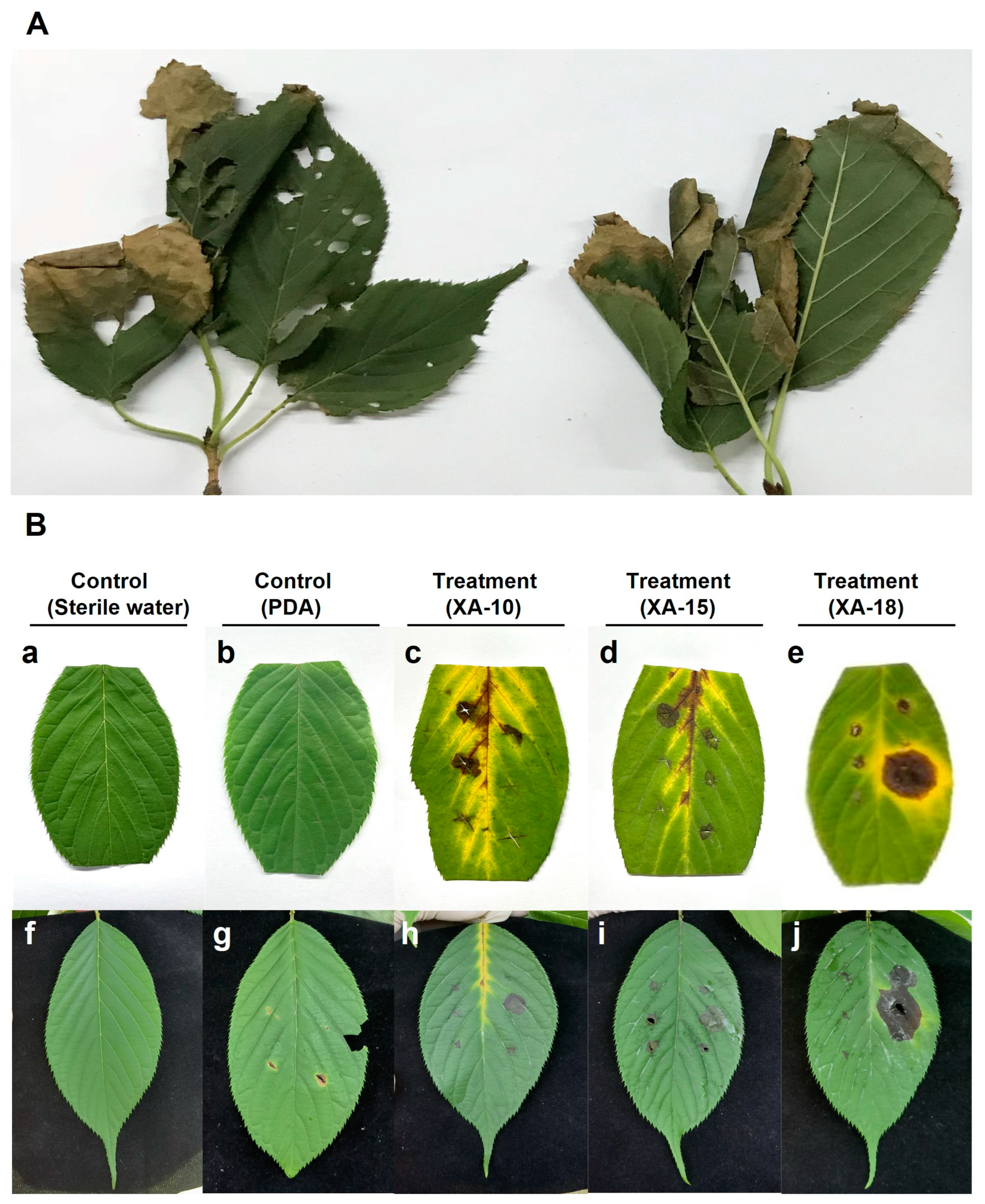
3.1. Natural Symptoms and Pathogen Isolation

The initial symptoms on C. serrulata leaves were small chlorotic lesions present on the leaf margin or tip. These lesions eventually enlarged to form larger brown lesions, resulting in leaf wilting and severely impeding the plant’s growth (Figure 1A). The results of the isolates showed that a total of 26 single hyphae fungal isolates, coded as XA-1 to XA-26, were obtained from the leaves with typical symptoms of C. serrulata.

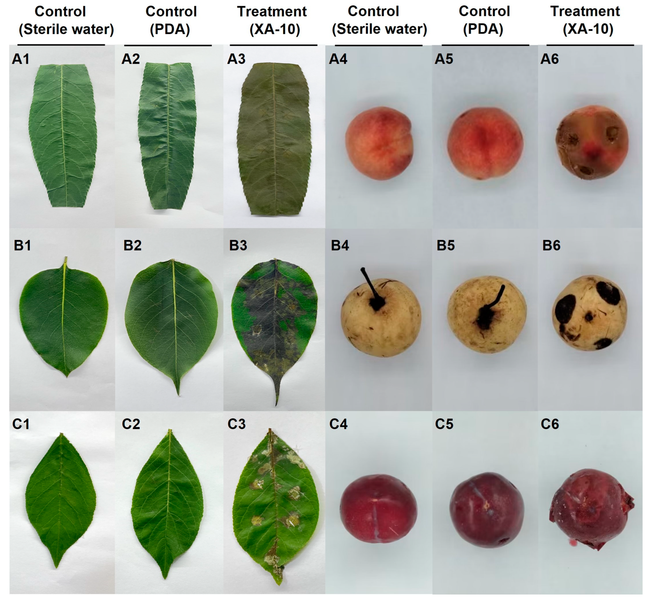
3.2. Pathogenicity Tests on Host Leaves

The pathogenicity of these isolates was tested, revealing discernible differences among the 26 isolates studied (Table S1). Among them, three isolates (XA-10, XA-15, and XA-18) were found to be pathogenic, exhibiting similar disease symptoms on the host leaves (Figure 1B).
Symptoms manifested on the leaves after 5 days post-inoculation. The detached leaves inoculated with the three selected isolates (XA-10, XA-15, and XA-18) initially showed small brown circular spots (Figure 1B). With increasing inoculation time, the spots gradually expanded, causing the surrounding tissues to turn yellow, with some leaves showing signs of wilting (Figure 1B(c–e)). Similarly, the respective inoculated sites on the leaves of the infected live host plants showed circular spots, and the lesion areas were dark brown. The lesions spread, and the central and inoculated sections of the leaves gradually wilted (Figure 1B(h–j)). Pronounced disease symptoms were identifiable on both the detached leaves as well as the leaves of live host plants. Leaves subjected to inoculation with sterile water (Figure 1B(a,f)) and those inoculated with PDA medium devoid of mycelia (Figure 1B(b,g)) exhibited no symptoms.
To confirm Koch’s postulates, the pathogens were successfully re-isolated from the infected leaves, and their morphological characteristics and gene sequence matched those of the original isolates.

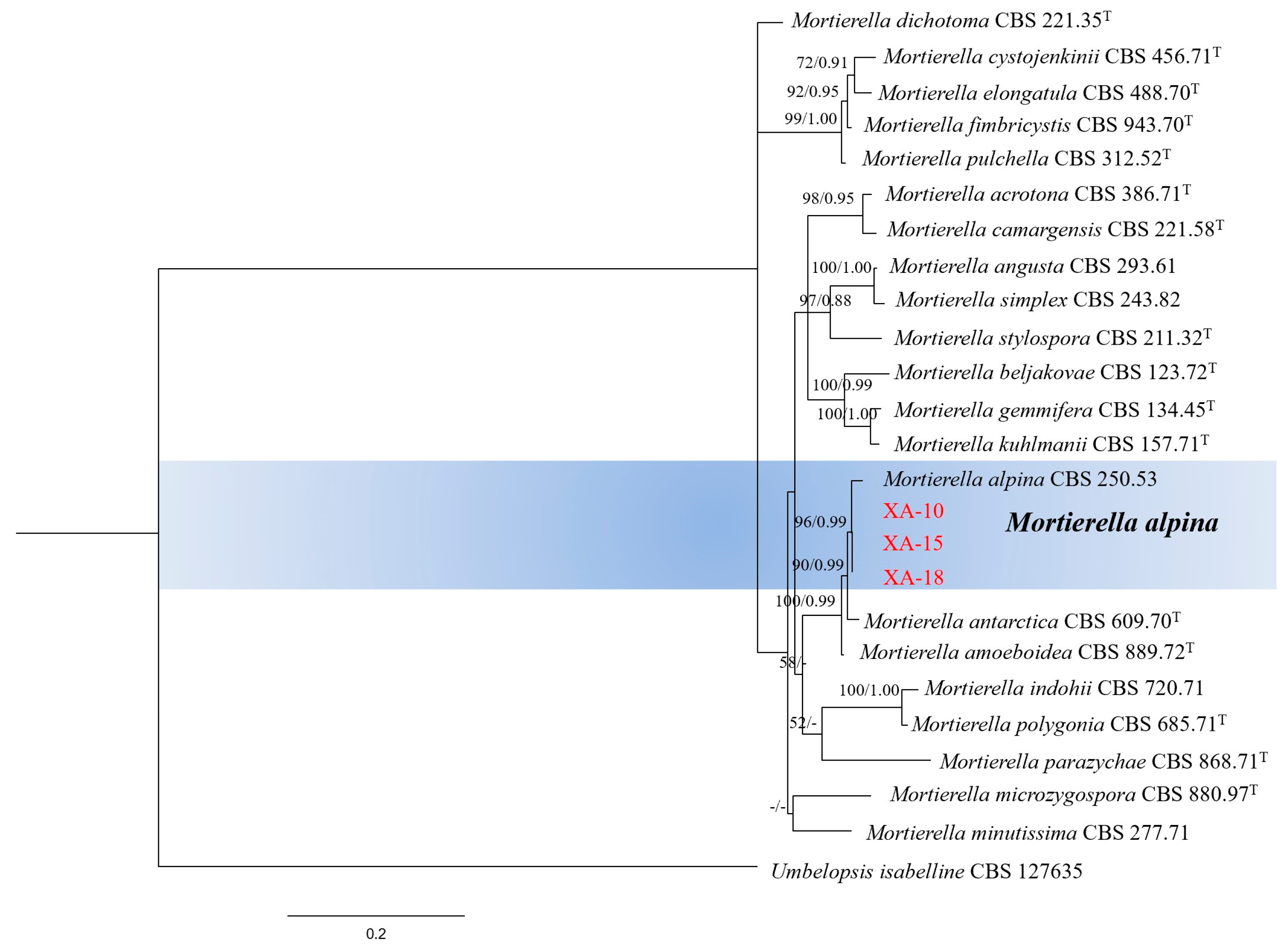
3.3. Molecular Identification of Fungal Isolates

The internal transcribed spacer (ITS) and large ribosomal subunit (LSU) regions of isolates XA-10, XA-15, and XA-18, exhibiting similar pathogenicity, were amplified and sequenced. The ITS and LSU sequences were submitted to the GenBank database under the accession numbers ITS: OR206530, OR817763, and OR817762; and LSU: OR206531, OR817765, and OR817764, respectively. The phylogram of the ITS and LSU gene sequence combinations was constructed using maximum likelihood (ML) and Bayesian inference (BI). The topological structures in the two phylograms were similar, and finally, the maximum likelihood phylogram was selected for presentation (Figure 2). Following sequence alignment, the ITS and LSU sequences of the three selected isolates (XA-10, XA-15, and XA-18), respectively, exhibited consistency. As shown in Figure 2, isolates XA-10, XA-15, and XA-18 (accession no. ITS: OR206530, OR817763, and OR817762; accession no. LSU: OR206531, OR817765, and OR817764) and Mortierella alpina (GenBank accession number: CBS 250.53) were present in the same cluster with a bootstrap support value of 96% and a posterior probability of 0.99. Based on ITS and LSU sequence analysis, the isolates XA-10, XA-15, and XA-18 were identified as M. alpina (Figure 2).

3.4. Morphology of Pathogenic Fungi

The three selected isolates (XA-10, XA-15, and XA-18) display similar milky-white aerial mycelia with wavy colony edges when observed from both the front and the back sides of their PDA medium (Figure 3A–C). The mycelium of the representative strain XA-10 (based on the similar morphological characteristics, pathogenicity to host plants, and molecular biological identification of the isolates XA-10, XA-15, and XA-18, the strain XA-10 was selected as the representative sample for the further analysis) exhibits multiple septa and branches (Figure 3D). The fungal sporangia appear to be spherical with smooth surfaces (Figure 3E). The sporangia have an average length and width of 10.09 ± 2.02 μm and 8.94 ± 1.92 μm, respectively. The sporangiospores appear spherical or nearly spherical (Figure 3F). The average length of the sporangiospores is 3.90 ± 0.8 μm, and the average width is 3.76 ± 0.77 μm.

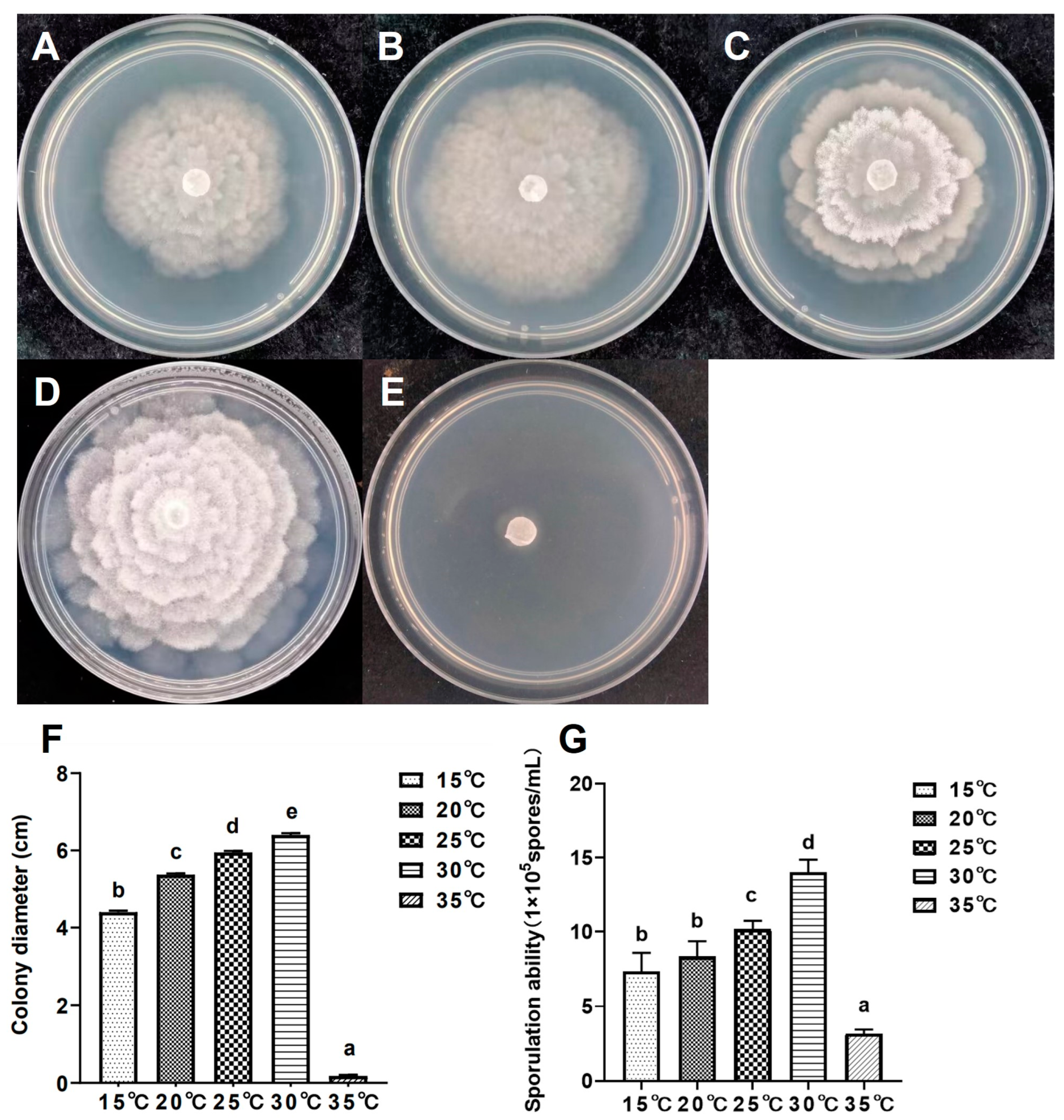
3.5. Growth Characteristics at Different Temperatures

The growth and sporulation capabilities of isolate XA-10 were assessed at various temperature conditions (Figure 4). After 5 days of culture on PDA medium at temperatures ranging from 15 to 35 °C, the mycelial growth was significantly (p < 0.05) different among different temperatures (Figure 4A–E). The colony diameters of the isolate XA-10 ranged from 0.18 ± 0.03 cm to 6.40 ± 0.04 cm. The maximum and minimum colony diameters were recorded at 30 °C and 35 °C, respectively (Figure 4F). Optimum sporulation was observed at a temperature of 30 °C (1.40 × 10⁶ spores/mL), while the lowest (3.17 × 10⁵ spores/mL) was recorded at 35 °C (Figure 4G). The results indicate that the optimal temperature for the growth and development of isolate XA-10 was 30 °C (Figure 4), with growth nearly ceasing at temperatures exceeding 35 °C (Figure 4D,E). Thus, temperature could significantly impact the sporulation ability of isolate XA-10.

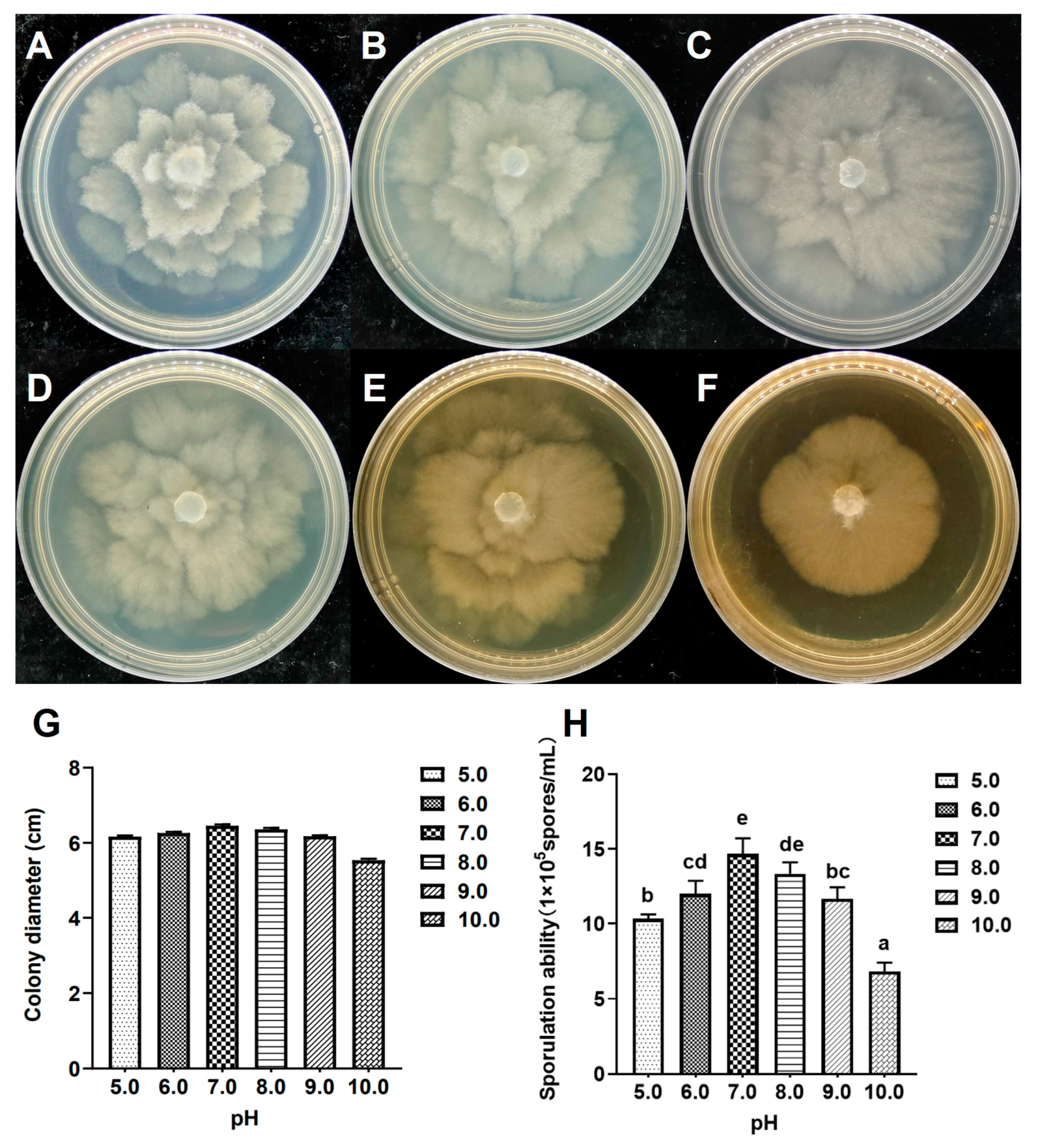
3.6. Growth Characteristics at Different pH Values

The effects of varying pH values on the mycelial growth and sporulation capacity of the isolate XA-10 are shown in Figure 5. The isolate showed similar growth characteristics within the pH value range of 5.0–10.0 (Figure 5A–F), and the colony diameters ranged from 5.53 ± 0.04 cm to 6.46 ± 0.03 cm (Figure 5G). The mycelia grew well at pH values between 5.0 and 10.0, and a pH range of 5.0 to 9.0 was more suitable for mycelial growth (Figure 5A–E). Sporulation was highest (1.47 × 10⁶ spores/mL) at neutral pH (7.0) (Figure 5H). The lowest sporulation was observed at pH 10.0 (6.83 × 10⁵ spores/mL), as shown in Figure 5H. Although no significant differences were found in mycelial growth under various pH conditions (p < 0.05), the sporulation of isolate XA-10 was significantly affected by pH.

3.7. Pathogenicity Tests on Leaves and Fruits of the Other Three Species of Rosaceae

The leaves and fruits of P. persica, P. bretschneideri, and P. salicina inoculated with isolate XA-10 showed severe symptoms, with an incidence of 100%, excluding the asymptomatic control (Figure 6).
After 2 days of inoculation, brown lesions emerged on the leaves and fruits of P. persica. These infected regions noticeably expanded after 4 days, causing extensive wilting of the leaves (Figure 6(A3)) and severe rotting of the pulp, producing a foul odor (Figure 6(A6)). On P. bretschneideri and P. salicina, however, black and brown lesions were observed 3 days post-inoculation. The symptoms on the leaves of P. bretschneideri continued to expand, displaying irregular shapes after 5 days (Figure 6(B3)). At this time, the infected fruits had significantly rotted internally, with the initially small, round, black lesions at the inoculation site gradually spreading (Figure 6(B6)). Similarly, brown lesion symptoms were observed on the leaves of P. salicina, and the distribution of these spots showed an irregular pattern (Figure 6(C3)). The fruits of P. salicina showed significant rot accompanied by a foul odor and disease spots (Figure 6(C6)). Thus, isolate XA-10 is pathogenic to P. persica, P. bretschneideri, and P. salicina, causing similar symptoms on the leaves and fruits following wound inoculation. The control group displayed no symptoms (Figure 6).

4. Discussion

The Chinese flowering cherry, renowned for its vibrant colors and 15-20-day flowering period, is a popular choice for urban green spaces and parks due to its significant ornamental value [38]. Leaf blight detrimentally impacts not only the aesthetics of plants but also their growth and development. In severe cases, it leads to the death of the plant, threatening China’s economic development and ecological environment [19,20,21]. In our study, the fungal pathogen causing leaf blight was isolated from a diseased plant, identified by morphological and cultural characteristics [39], and confirmed to be M. alpina using ITS [23] and LSU [27,28] sequence analyses. This report is the first to implicate M. alpina as the causative agent of leaf blight in the Chinese flowering cherry. Consequently, further exploration is required to understand the role of this fungus as a leaf blight pathogen that affects economically significant plant species.
M. alpina is a member of the fungus Zygomycotina [40]. The morphological traits of the pathogen described in this study, particularly the colony morphology and milky-white aerial mycelia, were consistent with the species description of M. alpina previously established by Wani et al. [41] and Wang et al. [42]. Other features, including the sporangia’s shape and the sporangiospores, correspond to those characterized by Yadav et al. [43] for M. alpina.
Environmental factors, such as temperature and pH, were the key factors influencing fungal mycelia’s growth. The peak temperature for mycelial growth in this study was similar to that of Streekstra et al. [44] and Zhang [45]. However, the optimal temperature for the growth of fungal mycelia differed slightly from that reported by Melo et al. [46]. These variations in biological characteristics may stem from the different hosts and regions evaluated in these studies. Most phytopathogenic fungi grow optimally within a pH range of 5.0 to 6.5 [47]. However, we found that M. alpina exhibited optimal mycelial growth at pH 7.0, which was consistent with the results of Zhang [45].
Results from some previous studies suggest that some members of the genus Mortierella are weak or conditioned pathogens, as well as rhizosphere microorganisms or endophytes of plants [41,46,48,49]. A common member of this genus, M. alpina, can induce several plant diseases, such as seedling necrosis of Pinus nigra [50] and seedling necrosis of the phloem of Araucaria araucana [51]. M. elongata has been found to cause diseases in avocado plants [52]. Furthermore, our study observed that M. alpina can severely infect the leaves and fruits of three other Rosaceae plants, including P. persica, P. bretschneideri, and P. salicina. Yet, the mechanism by which Mortierella spp. instigate plant diseases remains unidentified. In future studies, we aim to unravel the pathogenic mechanisms of these fungal isolates causing leaf blight in the Chinese flowering cherry.
Mortierella spp., acting as rhizospheric or endophytic fungi, plays essential roles in the plant–soil ecosystem, plant growth, and health [41,48,53,54,55]. Generally, Mortierella spp., being filamentous fungi, are commonly found in soil, the rhizosphere, and plant tissues [53,54,56,57,58]. Previous research has indicated that Mortierella species are present in the rhizosphere soil of Panax notoginseng and a poplar plantation, suggesting that they may play an important role in maintaining plant–soil balance or assisting host plants in the uptake of phosphorus and nitrogen [53,57]. Mortierella spp. may be an important component of the phosphorus cycle [54]. Certain Mortierella species have the capability to convert insoluble phosphorus into a soluble form, thereby promoting plant growth and development [56]. Mortierella species have been demonstrated to positively impact wheat yield [59], with M. elongata found abundantly in the rhizosphere of pea farms preceded by wheat in crop rotation [60]. M. capitata could increase maize biomass and promote plant growth [61]. Many Mortierella species can also improve soil nutrition, reconstruct microbial communities, and improve litter decomposition in the rhizosphere [56,61,62,63].
Further investigation is warranted into the prevalence of M. alpina infection among diverse horticultural plants. This study provided a theoretical foundation and practical guide for managing leaf blight disease in Chinese flowering cherry, among other Rosaceae plants. Nonetheless, additional studies are needed to elucidate the host range and the mechanisms underlying the pathogenicity of this fungus.

5. Conclusions

In summary, this study identified the pathogen causing Chinese flowering cherry leaf blight disease as M. alpina based on morphological observations, molecular identification, and pathogenicity testing. To the best of our knowledge, this is the first report of Chinese flowering cherry leaf blight disease caused by M. alpina in China. Its biological characteristics are as follows: the strain XA-10 is suitable for mycelial growth at 30 °C, and the optimal pH value is 7.0 for mycelial growth. This fungus causes varying degrees of symptoms on leaves and fruits of other Rosaceae plants including P. persica, P. bretschneideri, and P. salicina. Our results enrich the knowledge about the specific features of M. alpina and serve as important scientific foundations for efficient control and management of the studied plant disease.

Supplementary Materials

The following supporting information can be downloaded at: https://www.mdpi.com/article/10.3390/jof10010050/s1, Table S1: A brief description of the 26 isolates obtained from leaves with typical symptoms of C. serrulata.

Author Contributions

Conceptualization, W.Y. and G.L.; methodology, W.Y., D.S. and Y.X.; software, D.S. and J.S.; formal analysis, D.S., J.S. and Y.X.; investigation, X.J. and Y.X.; writing—original draft preparation, D.S., C.Z., A.F. and Y.S.A.; visualization, L.S., Z.L. and X.J.; project administration, W.Y. and R.Y.; funding acquisition, W.Y. and R.Y. All authors have read and agreed to the published version of the manuscript.

Funding

This research was funded by the Science and Technology Planning Major Project of Fujian province of China (2022N0010), the Natural Resources Science and Technology Innovation Project of Fujian province of China (KY-090000-04-2022-016), and the Science and Technology Program of Henan province of China (232102320111).

Institutional Review Board Statement

Not applicable.

Informed Consent Statement

Not applicable.

Data Availability Statement

Data are contained within the article and supplementary materials.

Conflicts of Interest

The authors declare no conflict of interest.

References

  1. Shirasawa, K.; Esumi, T.; Itai, A.; Isobe, S. Cherry blossom forecast based on transcriptome of floral organs approaching blooming in the flowering cherry (Cerasus × yedoensis) cultivar ‘Somei-Yoshino’. Front. Plant Sci. 2022, 13, 802203. [Google Scholar] [CrossRef] [PubMed]
  2. Shibato, J.; Takenoya, F.; Hirabayashi, T.; Kimura, A.; Iwasaki, Y.; Toyoda, Y.; Hori, M.; Tamogami, S.; Rakwal, R.; Shioda, S. Towards identification of bioactive compounds in cold vacuum extracted double cherry blossom (Gosen-Sakura) leaves. Plant Signal Behav. 2019, 14, e1644594. [Google Scholar] [CrossRef] [PubMed]
  3. Zhang, Y.Q.; Guan, L.; Zhong, Z.Y.; Chang, M.; Zhang, D.K.; Li, H.; Lai, W. The anti-inflammatory effect of cherry blossom extract (Prunus yedoensis) used in soothing skincare product. Int. J. Coxmetic Sci. 2014, 36, 527–530. [Google Scholar] [CrossRef] [PubMed]
  4. Shimoda, H.; Nakamura, S.; Morioka, M.; Tanaka, J.; Matsuda, H.; Yoshikawa, M. Effect of cinnamoyl and flavonol glucosides derived from cherry blossom flowers on the production of advanced glycation end products (AGEs) and AGE-induced fibroblast apoptosis. Phytother. Res. 2011, 25, 1328–1335. [Google Scholar] [CrossRef] [PubMed]
  5. Li, L.; Hwang, E.; Ngo Hien, T.T.; Lin, P.; Gao, W.; Liu, Y.; Yi, T.H. Antiphotoaging effect of Prunus yeonesis blossom extract via inhibition of MAPK/AP-1 and regulation of the TGF-βI/Smad and Nrf2/ARE signaling pathways. Photochem. Photobiol. 2018, 94, 725–732. [Google Scholar] [CrossRef] [PubMed]
  6. Yi, X.G.; Chen, J.; Zhu, H.; Li, X.X.; Li, M.; Duan, Y.F.; Chen, L.; Wang, X.R. Phylogeography and the population genetic structure of flowering cherry Cerasus serrulata (Rosaceae) in subtropical and temperate China. Ecol. Evol. 2020, 10, 11262–11276. [Google Scholar] [CrossRef] [PubMed]
  7. Liu, X.L. Study on the Ornamental Evaluation Of Fourteen Cerasus serrulata Cultivars and the Application in Landscape. Master’s Thesis, Zhejiang A&F University, Hangzhou, China, 2012. [Google Scholar]
  8. Zhang, J. Studies on the Cultivar Resources of Cerasus and the Application in Landscape. Master’s Thesis, Nanjing Forestry University, Nanjing, China, 2010. [Google Scholar]
  9. Yi, X.G.; Yu, X.Q.; Chen, J.; Zhang, M.; Liu, S.W.; Zhu, H.; Li, M.; Duan, Y.F.; Chen, L.; Wu, L.; et al. The genome of Chinese flowering cherry (Cerasus serrulata) provides new insights into Cerasus species. Hortic. Res. 2020, 7, 165. [Google Scholar] [CrossRef] [PubMed]
  10. Yi, X.G.; Chen, J.; You, L.X.; Cong, R.; Wang, H.C.; Duan, Y.F.; Wang, X.R. Genetic diversity of Cerasus serrulata populations assessed by SSR markers. J. Nanjing Forestry Univ. 2018, 42, 25–31. [Google Scholar]
  11. Yi, X.G. The Variation and Phylogeography of Cerasus serrulata Mill. Populations. Ph.D. Thesis, Nanjing Forestry University, Nanjing, China, 2018. [Google Scholar]
  12. Li, M.; Yin, X.G.; Wang, H.C.; Shang, T.; Gu, Y.; Wang, X.R. Studies on the relationship between Cerasus serrulata distribution region and the environmental factors. J. Nanjing Forestry Univ. 2014, 38, 74–80. [Google Scholar]
  13. Wang, X.R. An Illustrated Monograph of Cherry Cultivars in China; Science Press: Beijing, China, 2014; pp. 24–28. [Google Scholar]
  14. Liao, Z.J.; Zeng, H.L.; Gao, C.; Zhou, S.Y.; Liu, G.S.; Li, R.G. Identification of the pathogen causing brown spot in Cerasus in Jiangxi and its sensitivity to fungicides. J. Nanjing For. Univ. 2022, 48, 449–453. [Google Scholar]
  15. Chen, J.X.; Wu, Z.Q.; Wu, F.J.L.; Ma, H.C.; Han, Q.L.; Wu, J.R. First report of ‘Candidatus Phytoplasma asteris’ associated with witches’-broom and plexus bud disease of Cerasus serrula in China. Plant Dis. 2023, 107, 3274. [Google Scholar] [CrossRef] [PubMed]
  16. Zhang, S.M.; Liu, C.Y.; Xu, S.F. Occurrence and Control of Landscape Plant Diseases; China Agricultural University Press: Beijing, China, 2014; pp. 156–157. [Google Scholar]
  17. Wang, X.W.; Ma, X.Y.; Zhou, Q.; Long, J.B.; Tian, H.Q.; Liu, D.M.; Yu, C. First report of root rot in Cerasus subhirtella caused by Fusarium solani and Fusarium oxysporum in China. Plant Dis. 2023. [Google Scholar] [CrossRef] [PubMed]
  18. Wang, Y.; Chen, G.F.; Turatsinze, A.N.; Xie, X.F.; Sha, Y.X.; Wang, R.Y. First report of Fusarium tricinctum causing root rot on Chinese dwarf cherry (Cerasus humilis) in China. Plant Dis. 2023. [Google Scholar] [CrossRef] [PubMed]
  19. Elliott, M.; Rollins, L.; Bourret, T.; Chastagner, G. First report of leaf blight caused by Phytophthora ramorum on cherry laurel (Prunus laurocerasus) in Washington State, U.S.A. Plant Dis. 2021, 105, 712. [Google Scholar] [CrossRef] [PubMed]
  20. Huang, J.H.; Ann, P.J. First report of Phytophthora cambivora causing leaf and stem blight and root rot on Taiwan cherry (Prunus campanulata) in Taiwan. Plant Dis. 2012, 96, 1065–1066. [Google Scholar] [CrossRef] [PubMed]
  21. Dai, Q.D.; Zhou, C.H.; Ai, J.Y.; Zhang, Q.J. Pathogen identification of sweet cherry leaf blight in Liaoning Province. China Fruits 2022, 9, 12–19. [Google Scholar]
  22. Fang, Z.D. Research Methods for Plant Pathology, 3rd ed.; China Agriculture Press: Beijing, China, 1998; pp. 156–168. [Google Scholar]
  23. White, T.J.; Bruns, T.; Lee, S.; Taylor, F.J.R.M.; Lee, S.H.; Taylor, L.; Shawe-Taylor, J. Amplification and direct sequencing of fungal ribosomal RNA genes for phylogenetics. In PCR Protocols: A Guide to Methods and Applications; Innis, M.A., Gelfand, D.H., Sninsky, J.J., Eds.; Academic Press Inc.: New York, NY, USA, 1990; pp. 315–322. [Google Scholar]
  24. Lee, S.B.; Taylor, J.W. Isolation of DNA from fungal mycelia and single spores. In PCR Protocols: A Guide to Methods and Applications; Innis, M.A., Gelfand, D.H., Sninsky, J.J., Eds.; Academic Press Inc.: New York, NY, USA, 1990; pp. 282–287. [Google Scholar]
  25. Liu, S.B.; Liu, X.Y.; Zhang, Z.X.; Xia, J.W.; Zhang, X.G.; Meng, Z. Three New Species of Microdochium (Sordariomycetes, Amphisphaeriales) on Miscanthus sinensis and Phragmites australis from Hainan, China. J. Fungi 2022, 8, 577. [Google Scholar] [CrossRef] [PubMed]
  26. Yan, N.N.; Addrah, M.E.; Zhang, Y.Y.; Jia, R.F.; Kang, L.R.; Zhao, J.; Zhang, J. Identification and comparison of biological characteristics and pathogenicity of different mating types of V. dahliae isolated from potato and sunflower. Sci. Rep. 2022, 12, 12840. [Google Scholar]
  27. Vilgalys, R.; Hester, M. Rapid genetic identification and mapping of enzymatically amplified ribosomal DNA from several Cryptococcus species. J. Bacteriol. 1990, 172, 4238–4246. [Google Scholar] [CrossRef]
  28. Rehner, S.A.; Samuels, G.J. Taxonomy and phylogeny of Gliocladium analysed from nuclear large subunit ribosomal DNA sequences. Mycol. Res. 1994, 98, 625–634. [Google Scholar] [CrossRef]
  29. Wagner, L.; Stielow, B.; Hoffmann, K.; Petkovits, T.; Papp, T.; Vágvölgyi, C.; de Hoog, G.S.; Verkley, G.; Voigt, K. A comprehensive molecular phylogeny of the Mortierellales (Mortierellomycotina) based on nuclear ribosomal DNA. Persoonia 2013, 30, 77–93. [Google Scholar] [CrossRef] [PubMed]
  30. Petkovits, T.; Nagy, L.G.; Hoffmann, K.; Wagner, L.; Nyilasi, I.; Griebel, T.; Schnabelrauch, D.; Vogel, H.; Voigt, K.; Vagvoelgyi, C.; et al. Data partitions, bayesian analysis and phylogeny of the Zygomycetous fungal family Mortierellaceae, inferred from nuclear ribosomal DNA sequences. PLoS ONE 2011, 6, e27507. [Google Scholar] [CrossRef] [PubMed]
  31. Nguyen, T.T.T.; Park, S.W.; Pangging, M.; Lee, H.B. Molecular and morphological confirmation of three undescribed species of Mortierella from Korea. Mycobology 2019, 47, 31–39. [Google Scholar] [CrossRef] [PubMed]
  32. Vu, D.; Groenewald, M.; de Vries, M.; Gehrmann, T.; Stielow, B.; Eberhardt, U.; Al-Hatmi, A.; Groenewald, J.Z.; Cardinali, G.; Houbraken, J.; et al. Large-scale generation and analysis of filamentous fungal DNA barcodes boosts coverage for kingdom fungi and reveals thresholds for fungal species and higher taxon delimitation. Stud. Mycol. 2019, 92, 135–154. [Google Scholar] [CrossRef] [PubMed]
  33. Zhang, D.; Gao, F.; Jakovlić, I.; Zou, H.; Zhang, J.; Li, W.X.; Wang, G.T. PhyloSuite: An integrated and scalable desktop platform for streamlined molecular sequence data management and evolutionary phylogenetics studies. Mol. Ecol. Resour. 2020, 20, 348–355. [Google Scholar] [CrossRef] [PubMed]
  34. Fan, S.B.; Su, J.Y.; Fang, T.; Pan, A.F.; He, X.Y.; Fan, G.L.; Wang, Z.H.; Hu, H.L. Pathogen of liquidambar formosana leaf spot. Mycosystema 2022, 41, 420–434. [Google Scholar]
  35. Nguyen, L.T.; Schmidt, H.A.; von Haeseler, A.; Minh, B.Q. IQ-TREE: A fast and effective stochastic algorithm for estimating maximum-likelihood phylogenies. Mol. Biol. Evol. 2015, 32, 268–274. [Google Scholar] [CrossRef] [PubMed]
  36. Ronquist, F.; Teslenko, M.; van der Mark, P.; Ayres, D.L.; Darling, A.; Hohna, S.; Larget, B.; Liu, L.; Suchard, M.A.; Huelsenbeck, J.P. MrBayes 3.2: Efficient bayesian phylogenetic inference and model choice across a large model space. Syst. Biol. 2012, 61, 539–542. [Google Scholar] [CrossRef]
  37. Wei, J.C. Identification Manua; Shanghai Scientific & Technical Publishers: Shanghai, China, 1979; pp. 1–780. [Google Scholar]
  38. Li, C.L.; Bartholomew, B. Flora of China; Science Press: Beijing, China, 2003; pp. 404–420. [Google Scholar]
  39. Wang, Y. Mechanism Analysis of Inoculation with Mortierella alplina on Defense against Root of Cultivated Ginseng. Master’s Thesis, Northeast Forestry University, Harbin, China, 2022. [Google Scholar]
  40. Liu, Z. Studies on the Taxonomy and Molecular Phylogeny of Mortierella and Allied Genera in China. Master’s Thesis, Beijing Forestry University, Beijing, China, 2020. [Google Scholar]
  41. Wani, Z.A.; Kumar, A.; Sultan, P.; Bindu, K.; Riyaz-Ul-Hassan, S.; Ashraf, N. Mortierella alpina CS10E4, an oleaginous fungal endophyte of Crocus sativus L. enhances apocarotenoid biosynthesis and stress tolerance in the host plant. Sci. Rep. 2017, 7, 8598. [Google Scholar] [CrossRef]
  42. Wang, Y.; Wang, L.W.; Suo, M.; Qiu, Z.J.; Wu, H.; Zhao, M.; Yang, H.Y. Regulating root fungal community using Mortierella alpina for Fusarium oxysporum resistance in Panax ginseng. Front. Microbiol. 2022, 13, 850917. [Google Scholar] [CrossRef]
  43. Yadav, D.R.; Kim, S.W.; Babu, A.G.; Adhikari, M.; Kim, C.; Lee, H.B.; Lee, Y.S. First report of Mortierella alpina (Mortierellaceae, Zygomycota) isolated from crop field soil in Korea. Mycobiology 2014, 42, 401–404. [Google Scholar] [CrossRef] [PubMed]
  44. Streekstra, H. On the safety of Mortierella alpina for the production of food ingredients, such as arachidonic acid. J. Biotechnol. 1997, 56, 153. [Google Scholar] [CrossRef] [PubMed]
  45. Zhang, J.Z. Studies on the Diversity of Soil Fungi in the Alpine Grassland of Eastern Qilian Mountains. Ph.D. Thesis, Gansu Agricultural University, Lanzhou, China, 2010. [Google Scholar]
  46. Melo, I.; Santos, S.; Rosa, L.; Parma, M.; Silva, L.; Queiroz, S.; Pellizari, V. Isolation and biological activities of an endophytic Mortierella alpina strain from the Antarctic moss Schistidium antarctici. Extremophiles 2014, 18, 15–23. [Google Scholar] [CrossRef] [PubMed]
  47. Cochrane, V.W. Physiology of Fungi; John Wiley & Sons: New York, NY, USA, 1958; pp. 19–23. [Google Scholar]
  48. de León, Y.M.P.; Muñoz-Castellanos, L.N.; Ruiz-Cisneros, M.F.; Pérez-Corral, D.A.; Ornelas-Paz, J.J.; AcostaMuñiz, C.H.; Berlanga Reyes, D.I.; Rios Velasco, C. Morphological and molecular identification of Mortierella species associated to rhizosphere of apple trees with symptoms of root diseases. Rev. Mex. Fitopatol. 2017, 36, 184–195. [Google Scholar]
  49. Johnson, J.M.; Ludwig, A.; Furch, A.C.U.; Mithofer, A.; Scholz, S.; Reichelt, M.; Oelmtiller, R. The beneficial root-colonizing fungus Mortierella hyaline promotes the aerial growth of Arabidopsis and activates calcium-dependent responses that restrict Alternaria brassicae-induced disease development in roots. Mol. Plant Microbe Interact. 2019, 32, 351–363. [Google Scholar] [CrossRef]
  50. Li Destri Nicosia, M.G.; Mosca, S.; Mercurio, R.; Schena, L. Dieback of Pinus nigra seedlings caused by a strain of Trichoderma viride. Plant Dis. 2015, 99, 44–49. [Google Scholar] [CrossRef]
  51. Vélez, M.L.; Marfetán, J.A.; Salgado Salomón, M.E.; Taccari, L.E. Mortierella species from declining Araucaria araucana trees in Patagonia, Argentina. Forest Pathol. 2020, 50, e12591. [Google Scholar] [CrossRef]
  52. Hernández Pérez, A.; Cerna Chávez, E.; Delgado Ortiz, J.C.; Beltrán Beache, M.; Hernández Bautista, O.; Tapia Vargas, L.M.; Ochoa Fuentes, Y.M. First report of Mortierella elongata as a pathogen of avocado crop in Michoacán, Mexico. Sci. Fungorum. 2018, 48, 95–98. [Google Scholar] [CrossRef]
  53. Miao, C.P.; Mi, Q.L.; Qiao, X.G.; Zheng, Y.K.; Chen, Y.W.; Xu, L.H.; Guan, H.L.; Zhao, L.X. Rhizospheric fungi of Panax notoginseng: Diversity and antagonism to host phytopathogens. J. Ginseng Res. 2016, 40, 127–134. [Google Scholar] [CrossRef]
  54. Curlevski, N.J.A.; Xu, Z.H.; Anderson, I.C.; Cairney, J.W.G. Diversity of soil and rhizosphere fungi under Araucaria bidwillii (Bunya pine) at an Australian tropical montane rainforest site. Fungal Divers. 2010, 40, 12–22. [Google Scholar] [CrossRef]
  55. Huang, F.F. Effect of Pesticides on Mortierella in Rhizospheric soil of panax notoginseng and Their Colonization Ability in the Field. Master’s Thesis, Yunnan University, Kunming, China, 2017. [Google Scholar]
  56. Ozimek, E.; Hanaka, A. Mortierella Species as the Plant Growth-Promoting Fungi Present in the Agricultural Soils. Agriculture 2021, 11, 7. [Google Scholar] [CrossRef]
  57. Sang, Y.; Jin, L.; Zhu, R.; Yu, X.Y.; Hu, S.; Wang, B.T.; Ruan, H.H.; Jin, F.J.; Lee, H.G. Phosphorus-solubilizing capacity of Mortierella species isolated from rhizosphere soil of a poplar plantation. Microorganisms 2022, 10, 2361. [Google Scholar] [CrossRef] [PubMed]
  58. Vandepol, N.; Liber, J.; Desirò, A.; Na, H.; Kennedy, M.; Barry, K.; Grigoriev, I.V.; Miller, A.N.; O’Donnell, K.; Stajich, J.E. Resolving the Mortierellaceae phylogeny through synthesis of multi-gene phylogenetics and phylogenomics. Fungal Divers. 2020, 104, 267–289. [Google Scholar] [CrossRef] [PubMed]
  59. Borrell, A.N.; Shi, Y.; Gan, Y.; Bainard, L.D.; Germida, J.J.; Hamel, C. Fungal diversity associated with pulses and its influence on the subsequent wheat crop in the Canadian Prairies. Plant Soil 2017, 414, 13–31. [Google Scholar] [CrossRef]
  60. Niu, Y.N.; Bainard, L.D.; May, W.E.; Hossain, Z.; Hamel, C.; Gan, Y.T. Intensified pulse rotations buildup pea rhizosphere pathogens in cereal and pulse based cropping systems. Front. Microbiol. 2018, 9, 1909. [Google Scholar] [CrossRef]
  61. Li, F.; Zhang, S.Q.; Wang, Y.; Li, Y.; Li, P.P.; Chen, L.; Jie, X.L.; Hu, D.S.; Feng, B.; Yue, K.; et al. Rare fungus, Mortierella capitata, promotes crop growth by stimulating primary metabolisms related genes and reshaping rhizosphere bacterial community. Soil Biol. Biochem. 2020, 151, 108017. [Google Scholar] [CrossRef]
  62. Li, F.; Chen, L.; Gordon, M.R.; Zhang, J.B.; Zhang, C.Z.; Ning, Q.; Li, W. Mortierella elongata’s roles in organic agriculture and crop growth promotion in a mineral soil. Land Degrad. Dev. 2018, 29, 1642–1651. [Google Scholar] [CrossRef]
  63. Ning, Q.; Chen, L.; Li, F.; Zhang, C.Z.; Ma, D.H.; Cai, Z.J.; Zhang, J.B. Effects of Mortierella on nutrient availability and straw decomposition in soil. Acta Pedol. Sin. 2022, 59, 206–217. [Google Scholar]
Figure 1. Disease symptoms in greenbelts and pathogenicity tests of the 3 selected isolates (XA-10, XA-15, and XA-18) on host leaves. (A) Symptoms on naturally infected leaves of Cerasus serrulata. (B) Pathogenicity test for leaf blight symptoms caused by mycelial inoculation of the 3 selected isolates on the host plant (flowering cherry) leaves after 5 days. (B: ae) Pathogenicity on detached leaves of host plants after 5 days of inoculation. (B: fj) Pathogenicity on leaves of live host plants after 5 days of inoculation.
Jof 10 00050 g001
Figure 2. Maximum likelihood (ML) phylogram of the 3 selected isolates (XA-10, XA-15, and XA-18) based on the combined 2-locus (ITS and LSU) sequences. The tree is rooted in Umbelopsis isabellina (CBS 127635). Maximum likelihood bootstrap values ≥ 50% and Bayesian posterior probabilities ≥ 0.85 (PP/MLBS) are given at the nodes. Maximum likelihood bootstrap values < 50%, Bayesian posterior probabilities < 0.85 (PP/MLBS) are indicated with “-”. The type species is represented by “T”. The 3 selected isolates in this study are shown in red. The scale bar represents the expected number of changes per site.
Jof 10 00050 g002
Figure 3. Morphological features of Mortierella alpina. (AC) Colony morphologies (front and back) of the 3 selected isolates (XA-10, XA-15, and XA-18) on PDA medium incubated at 28 °C for 7 days. (DF) Septate hyphae (white arrows), sporangium, and spherical and nearly spherical sporangiospores (red arrows) of the representative strain XA-10 after 7 days of incubation on OA medium at 28 °C. Scale bar = 10 µm.
Jof 10 00050 g003
Figure 4. Colony morphology, diameter, and sporulation ability of the representative strain XA-10 at different temperatures. (AE) Colony morphology of isolate XA-10 at 15–35 °C on PDA medium. (F,G) Colony diameter and sporulation quantity of isolate XA-10 at different temperatures. Values of ±standard error are represented with error bars, and different letters indicate significant differences at p < 0.05.
Jof 10 00050 g004
Figure 5. Colony morphology, diameter, and sporulation ability of the representative strain XA-10 at different pH values. (AF) Colony morphologies of isolate XA-10 at pH 5.0–10.0 on PDA medium. (G,H) Colony diameters and sporulation ability of isolate XA-10 at different pH values. Values of ± standard error are represented with error bars, and different letters indicate significant differences at p < 0.05.
Jof 10 00050 g005
Figure 6. Pathogenicity of the representative strain XA-10 on leaves and fruits of three other Rosaceae plants. (A1A6) Detached leaves and fruits of Prunus persica; (B1B6) detached leaves and fruits of Pyrus bretschneideri; and (C1C6) detached leaves and fruits of Prunus salicina. The photographs of P. persica were taken 4 days post-inoculation and of P. bretschneideri and P. salicina were taken 5 days post-inoculated.
Jof 10 00050 g006
Table 1. Amplification sites, primer sequences, and PCR conditions used in this study.
Locus aPrimerPrimer Sequence (5′–3′)PCR ConditionsReference
ITSITS5GGAAGTAAACGTAACAAGG94 °C for 5 min (94 °C for 40 s, 58 °C for 40 s, and
72 °C for 60 s) × 35 cycles, 72 °C for 7 min
[23]
ITS4TCCTCCGCTTATTGATATGC
LSULR0RACCCGCTGAACTTAAGC94 °C for 5 min (94 °C for 45 s, 50 °C for 45 s, and
72 °C for 1 min) × 35 cycles, 72 °C for 7 min
[27,28]
LR5TCCTGAGGGAAACTTCG
a Genes: ITS, internal transcribed spacer; LSU, large ribosomal subunit.
Table 2. Strains used in this study.
Original
Name
Culture Accession Number(s)Type
Status
Accession
No. ITS
Accession
No. LSU
Reference
Mortierella acrotonaCBS 386.71, FSU 9788Type of Mortierella acrotonaJX975921HQ667405.1[29,30]
Mortierella alpinaCBS 250.53 JX975955KC018184[29]
Mortierella amoeboideaCBS 889.72Type of Mortierella amoeboideaJX976073HQ667422.1[29,30]
Mortierella angustaCBS 293.61Neotype of Mortierella polycephala var.angustaJX976061HQ667358.1[29,30]
Mortierella antarcticaCBS 609.70, FSU 9792Type of Mortierella antarcticaJX975907HQ667503.1[29,30]
Mortierella beljakovaeCBS 123.72, FSU 9794Type of Mortierella beljakovaeJX976126HQ667428.1[29,30]
Mortierella camargensisCBS 221.58, FSU 9796Type of Mortierella camargensisJX975949HQ667408.1[29,30,31]
Mortierella cystojenkiniiCBS 456.71, FSU 9803Type of Mortierella cystojenkiniiJX976030HQ667504.1[29,30]
Mortierella dichotomaCBS 221.35, FSU 9804Syntype of Mortierella dichotomaJX975842HQ667393.1[29,30]
Mortierella elongatulaCBS 488.70, FSU 9808Type of Mortierella elongatulaJX975967HQ667425.1[29,30]
Mortierella fimbricystisCBS 943.70Type of Mortierella fimbricystisGU559986.1JX976172[29]
Mortierella gemmiferaCBS 134.45, FSU 9815Type of Mortierella gemmiferaJX975931HQ667371.1[29,30]
Mortierella indohiiCBS 720.71, FSU 9826Isotype of Mortierella indohiiJX975856HQ667377.1[29,30]
Mortierella kuhlmaniiCBS 157.71, FSU 9827Type of Mortierella kuhlmaniiJX975846HQ667372.1[29,30]
Mortierella microzygosporaCBS 880.97, FSU 9831Type of Mortierella microzygosporaJX976027HQ667394.1[29,30]
Mortierella minutissimaCBS 277.71, FSU 832 JX975938KC018293[29]
Mortierella parazychaeCBS 868.71, FSU 9836Type of Mortierella parazychaeJX975985HQ667362.1[29,30]
Mortierella polygoniaCBS 685.71, FSU 9839Type of Mortierella polygoniaJX975900HQ667378.1[29,30]
Mortierella pulchellaCBS 312.52, FSU 9840Authentic strain of Mortierella pulchellaJX976054HQ667427.1[29,30]
Mortierella simplexCBS 243.82 JX975870JX976156[29,30]
Mortierella stylosporaCBS 211.32, FSU 9850Type of Mortierella stylosporaJX976086HQ667359.1[29,30]
Umbelopsis isabellinaCBS 127635 MH864646MH876082[32]
Disclaimer/Publisher’s Note: The statements, opinions and data contained in all publications are solely those of the individual author(s) and contributor(s) and not of MDPI and/or the editor(s). MDPI and/or the editor(s) disclaim responsibility for any injury to people or property resulting from any ideas, methods, instructions or products referred to in the content.

