Cardiometabolic Heart Failure with Preserved Ejection Fraction (HFpEF): Epidemiology, Mechanisms, and the Role of Lifestyle Modification
 ,
,
Abstract
1. Introduction
2. What Has Driven CM-HFpEF to the Forefront of Public Health Concerns?
3. How Do Sex-Specific Biological and Hormonal Factors Influence the Development and Progression of CM-HFpEF?
4. How Do SDoH Contribute to Disparities in Incidence and Prognosis of CM-HFpEF?
5. What Are the Mechanistic Underpinnings of CM-HFpEF?
6. How Is CM-HFpEF Different from Non-CM-HFpEF?
7. Can Lifestyle Interventions Modify the Course of CM-HFpEF?
8. Can Dietary Modification Improve Outcomes in CM-HFpEF?
9. What Is the Role of Weight Loss (WL) and Calorie Restriction (CR) in CM-HFpEF?
10. What Is the Role of Bariatric Surgery (BSx) in Preventing or Improving HFpEF?
11. How Does Physical Activity Influence Functional Capacity in CM-HFpEF?
12. What Is the Best Exercise Prescription?
13. What Is the Clinical Value of Cardiopulmonary Exercise Testing (CPET) in Guiding Therapeutic Interventions and Monitoring Treatment Response in CM-HFpEF?
14. How Do Emerging Pharmacological Therapies Improve CV Outcomes in Patients with CM- HFpEF?
15. How Do Current Heart Failure Guidelines Address CM-HFpEF?
16. What Are the Future Directions in CM-HFpEF?
Take Home Message
- CM risk factors are fueling the rapid global expansion of CM- HFpEF phenotype.
- CM-HFpEF is a multisystem inflammatory-metabolic syndrome.
- CM-HFpEF shows greater symptom burden and biventricular adverse remodeling.
- Dietary modification and caloric restriction improve functional capacity.
- SGLT2i and GLP-1RAs are effective due to their metabolic protective effects.
- ET is a cornerstone intervention for HFpEF symptoms.
17. Conclusions
Author Contributions
Funding
Institutional Review Board Statement
Informed Consent Statement
Data Availability Statement
Conflicts of Interest
References
- Pandey, A.; Shah, S.J.; Butler, J.; Kellogg, D.L., Jr.; Lewis, G.D.; Forman, D.E.; Mentz, R.J.; Borlaug, B.A.; Simon, M.A.; Chirinos, J.A.; et al. Exercise Intolerance in Older Adults with Heart Failure with Preserved Ejection Fraction: JACC State-of-the-Art Review. J. Am. Coll. Cardiol. 2021, 78, 1166–1187. [Google Scholar] [PubMed]
- Redfield, M.M.; Borlaug, B.A. Heart Failure with Preserved Ejection Fraction: A Review. JAMA 2023, 329, 827–838. [Google Scholar] [PubMed]
- Borlaug, B.A.; Sharma, K.; Shah, S.J.; Ho, J.E. Heart Failure with Preserved Ejection Fraction: JACC Scientific Statement. J. Am. Coll. Cardiol. 2023, 81, 1810–1834. [Google Scholar] [PubMed]
- Paulus, W.J.; Tschöpe, C. A novel paradigm for heart failure with preserved ejection fraction: Comorbidities drive myocardial dysfunction and remodeling through coronary microvascular endothelial inflammation. J. Am. Coll. Cardiol. 2013, 62, 263–271. [Google Scholar] [CrossRef] [PubMed]
- Daou, D.; Tong, D.; Schiattarella, G.G.; Gillette, T.G.; Hill, J.A. What Is Cardiometabolic HFpEF and How Can We Study it Preclinically? JACC Basic Transl. Sci. 2025, 10, 101295. [Google Scholar] [CrossRef] [PubMed]
- Verma, R.; Dhingra, N.K.; Connelly, K.A. Obesity/cardiometabolic phenotype of heart failure with preserved ejection fraction: Mechanisms to recent trials. Curr. Opin. Cardiol. 2024, 39, 92–97. [Google Scholar] [CrossRef] [PubMed]
- Obokata, M.; Reddy, Y.N.V.; Pislaru, S.V.; Melenovsky, V.; Borlaug, B.A. Evidence Supporting the Existence of a Distinct Obese Phenotype of Heart Failure with Preserved Ejection Fraction. Circulation 2017, 136, 6–19. [Google Scholar] [CrossRef] [PubMed]
- Paulus, W.J.; Zile, M.R. From Systemic Inflammation to Myocardial Fibrosis: The Heart Failure with Preserved Ejection Fraction Paradigm Revisited. Circ. Res. 2021, 128, 1451–1467. [Google Scholar] [PubMed]
- Reddy, Y.N.V.; Lewis, G.D.; Shah, S.J.; Obokata, M.; Abou-Ezzedine, O.F.; Fudim, M.; Sun, J.L.; Chakraborty, H.; McNulty, S.; LeWinter, M.M.; et al. Characterization of the Obese Phenotype of Heart Failure with Preserved Ejection Fraction: A RELAX Trial Ancillary Study. Mayo Clin. Proc. 2019, 94, 1199–1209. [Google Scholar] [CrossRef] [PubMed]
- Campbell, P.; Rutten, F.H.; Lee, M.M.; Hawkins, N.M.; Petrie, M.C. Heart failure with preserved ejection fraction: Everything the clinician needs to know. Lancet 2024, 403, 1083–1092. [Google Scholar] [CrossRef] [PubMed]
- Van Wagoner, D.R. Paracrine Signals Modulate Atrial Epicardial Progenitor Cells and Development of Subepicardial Adiposity and Fibrosis Implications for Atrial Fibrillation. Circ. Res. 2020, 126, 1343–1345. [Google Scholar] [CrossRef] [PubMed]
- Packer, M. The Adipokine Hypothesis of Heart Failure with a Preserved Ejection Fraction: A Novel Framework to Explain Pathogenesis and Guide Treatment. J. Am. Coll. Cardiol. 2025, 86, 1269–1373. [Google Scholar] [CrossRef] [PubMed]
- Schiattarella, G.G.; Rodolico, D.; Hill, J.A. Metabolic inflammation in heart failure with preserved ejection fraction. Cardiovasc. Res. 2021, 117, 423–434. [Google Scholar] [PubMed]
- Capone, F.; Vacca, A.; Bidault, G.; Sarver, D.; Kaminska, D.; Strocchi, S.; Vidal-Puig, A.; Greco, C.M.; Lusis, A.J.; Schiattarella, G.G. Decoding the Liver-Heart Axis in Cardiometabolic Diseases. Circ. Res. 2025, 136, 1335–1362. [Google Scholar] [CrossRef] [PubMed]
- Upadhya, B.; Pisani, B.; Kitzman, D.W. Evolution of a Geriatric Syndrome: Pathophysiology and Treatment of Heart Failure with Preserved Ejection Fraction. J. Am. Geriatr. Soc. 2017, 65, 2431–2440. [Google Scholar] [CrossRef] [PubMed]
- Bozkurt, B.; Ahmad, T.; Alexander, K.M.; Baker, W.L.; Bosak, K.; Breathett, K.; Fonarow, G.C.; Heidenreich, P.; Ho, J.E.; Hsich, E.; et al. Heart Failure Epidemiology and Outcomes Statistics: A Report of the Heart Failure Society of America. J. Card. Fail. 2023, 29, 1412–1451. [Google Scholar] [CrossRef] [PubMed]
- Walters, G.W.M.; Yeo, J.L.; Bilak, J.M.; Pepper, C.; Gulsin, G.S.; Freeman, S.C.; Gray, L.J.; Mc, C.G.; Brady, E.M. The Effectiveness of Lifestyle Interventions in Heart Failure with Preserved Ejection Fraction: A Systematic Review and Network Meta-Analysis. J. Card. Fail. 2024, 30, 994–1009. [Google Scholar] [CrossRef] [PubMed]
- Ostrominski, J.W.; Højbjerg Lassen, M.C.; Butt, J.H.; Claggett, B.L.; Anand, I.S.; Desai, A.S.; Jhund, P.S.; Lam, C.S.P.; Pfeffer, M.A.; Pitt, B.; et al. Adiposity-Related Anthropometrics and Clinical Outcomes in Heart Failure with Mildly Reduced or Preserved Ejection Fraction: A Participant-Level Pooled Analysis of Randomized Clinical Trials. J. Am. Coll. Cardiol. 2025, 86, 1760–1777. [Google Scholar] [PubMed]
- Bozkurt, B. Contemporary pharmacological treatment and management of heart failure. Nat. Rev. Cardiol. 2024, 21, 545–555. [Google Scholar] [CrossRef] [PubMed]
- Mahajan, R.; Stokes, M.; Elliott, A.; Munawar, D.A.; Khokhar, K.B.; Thiyagarajah, A.; Hendriks, J.; Linz, D.; Gallagher, C.; Kaye, D.; et al. Complex interaction of obesity, intentional weight loss and heart failure: A systematic review and meta-analysis. Heart 2020, 106, 58–68. [Google Scholar] [PubMed]
- Pandey, A.; Patel, K.V.; Vaduganathan, M.; Sarma, S.; Haykowsky, M.J.; Berry, J.D.; Lavie, C.J. Physical Activity, Fitness, and Obesity in Heart Failure with Preserved Ejection Fraction. JACC Heart Fail. 2018, 6, 975–982. [Google Scholar] [CrossRef] [PubMed]
- Savji, N.; Meijers, W.C.; Bartz, T.M.; Bhambhani, V.; Cushman, M.; Nayor, M.; Kizer, J.R.; Sarma, A.; Blaha, M.J.; Gansevoort, R.T.; et al. The Association of Obesity and Cardiometabolic Traits with Incident HFpEF and HFrEF. JACC Heart Fail. 2018, 6, 701–709. [Google Scholar] [CrossRef] [PubMed]
- Rao, V.N.; Zhao, D.; Allison, M.A.; Guallar, E.; Sharma, K.; Criqui, M.H.; Cushman, M.; Blumenthal, R.S.; Michos, E.D. Adiposity and Incident Heart Failure and its Subtypes: MESA (Multi-Ethnic Study of Atherosclerosis). JACC Heart Fail. 2018, 6, 999–1007. [Google Scholar] [PubMed]
- Borlaug, B.A.; Jensen, M.D.; Kitzman, D.W.; Lam, C.S.P.; Obokata, M.; Rider, O.J. Obesity and heart failure with preserved ejection fraction: New insights and pathophysiological targets. Cardiovasc. Res. 2023, 118, 3434–3450. [Google Scholar] [PubMed]
- Dunlay, S.M.; Roger, V.L.; Redfield, M.M. Epidemiology of heart failure with preserved ejection fraction. Nat. Rev. Cardiol. 2017, 14, 591–602. [Google Scholar] [CrossRef] [PubMed]
- Capone, F.; Vettor, R.; Schiattarella, G.G. Cardiometabolic HFpEF: NASH of the Heart. Circulation 2023, 147, 451–453. [Google Scholar] [CrossRef] [PubMed]
- Pandey, A.; Omar, W.; Ayers, C.; LaMonte, M.; Klein, L.; Allen, N.B.; Kuller, L.H.; Greenland, P.; Eaton, C.B.; Gottdiener, J.S.; et al. Sex and Race Differences in Lifetime Risk of Heart Failure With Preserved Ejection Fraction and Heart Failure With Reduced Ejection Fraction. Circulation 2018, 137, 1814–1823. [Google Scholar] [CrossRef] [PubMed]
- Radakrishnan, A.; Agrawal, S.; Singh, N.; Barbieri, A.; Shaw, L.J.; Gulati, M.; Lala, A. Underpinnings of heart failure with preserved ejection fraction in women—From prevention to improving function. A co-publication with the American Journal of Preventive Cardiology and the Journal of Cardiac Failure. Am. J. Prev. Cardiol. 2025, 23, 100928. [Google Scholar] [PubMed]
- Beale, A.L.; Meyer, P.; Marwick, T.H.; Lam, C.S.P.; Kaye, D.M. Sex Differences in Cardiovascular Pathophysiology: Why Women Are Overrepresented in Heart Failure With Preserved Ejection Fraction. Circulation 2018, 138, 198–205. [Google Scholar] [PubMed]
- Parikh, N.I.; Gonzalez, J.M.; Anderson, C.A.M.; Judd, S.E.; Rexrode, K.M.; Hlatky, M.A.; Gunderson, E.P.; Stuart, J.J.; Vaidya, D. Adverse Pregnancy Outcomes and Cardiovascular Disease Risk: Unique Opportunities for Cardiovascular Disease Prevention in Women: A Scientific Statement From the American Heart Association. Circulation 2021, 143, e902–e916. [Google Scholar] [CrossRef] [PubMed]
- Wu, P.; Haththotuwa, R.; Kwok, C.S.; Babu, A.; Kotronias, R.A.; Rushton, C.; Zaman, A.; Fryer, A.A.; Kadam, U.; Chew-Graham, C.A.; et al. Preeclampsia and Future Cardiovascular Health: A Systematic Review and Meta-Analysis. Circ. Cardiovasc. Qual. Outcomes 2017, 10, e003497. [Google Scholar] [PubMed]
- Upadhya, B.; Hegde, S.; Tannu, M.; Stacey, R.B.; Kalogeropoulos, A.; Schocken, D.D. Preventing new-onset heart failure: Intervening at stage A. Am. J. Prev. Cardiol. 2023, 16, 100609. [Google Scholar] [CrossRef] [PubMed]
- Havranek, E.P.; Mujahid, M.S.; Barr, D.A.; Blair, I.V.; Cohen, M.S.; Cruz-Flores, S.; Davey-Smith, G.; Dennison-Himmelfarb, C.R.; Lauer, M.S.; Lockwood, D.W.; et al. Social Determinants of Risk and Outcomes for Cardiovascular Disease: A Scientific Statement From the American Heart Association. Circulation 2015, 132, 873–898. [Google Scholar] [PubMed]
- Figueroa, J.F.; Frakt, A.B.; Jha, A.K. Addressing Social Determinants of Health: Time for a Polysocial Risk Score. JAMA 2020, 323, 1553–1554. [Google Scholar] [PubMed]
- Hurera, A.; Skowronski, J.N.; Zhu, J.; Thoma, F.W.; Magnani, J.W.; Hickey, G.W. Social Determinants of Health and Outcomes in Hospitalized Patients with Heart Failure With Preserved Ejection Fraction. J. Card. Fail. 2026; in press. [CrossRef] [PubMed]
- Heidenreich, P.A.; Bozkurt, B.; Aguilar, D.; Allen, L.A.; Byun, J.J.; Colvin, M.M.; Deswal, A.; Drazner, M.H.; Dunlay, S.M.; Evers, L.R.; et al. 2022 AHA/ACC/HFSA Guideline for the Management of Heart Failure: Executive Summary: A Report of the American College of Cardiology/American Heart Association Joint Committee on Clinical Practice Guidelines. J. Am. Coll. Cardiol. 2022, 79, 1757–1780. [Google Scholar] [CrossRef] [PubMed]
- Kitzman, D.W.; Shah, S.J. The HFpEF Obesity Phenotype: The Elephant in the Room. J. Am. Coll. Cardiol. 2016, 68, 200–203. [Google Scholar] [PubMed]
- Upadhya, B.; Kitzman, D.W. Heart failure with preserved ejection fraction: New approaches to diagnosis and management. Clin. Cardiol. 2020, 43, 145–155. [Google Scholar] [PubMed]
- Bertero, E.; Maack, C. Metabolic remodelling in heart failure. Nat. Rev. Cardiol. 2018, 15, 457–470. [Google Scholar] [CrossRef] [PubMed]
- Trimarchi, G.; Pizzino, F.; Paradossi, U. Inflammation in chronic heart failure: An unsolved puzzle. Int. J. Cardiol. 2025, 432, 133281. [Google Scholar] [CrossRef] [PubMed]
- Koepp, K.E.; Obokata, M.; Reddy, Y.N.V.; Olson, T.P.; Borlaug, B.A. Hemodynamic and Functional Impact of Epicardial Adipose Tissue in Heart Failure with Preserved Ejection Fraction. JACC Heart Fail. 2020, 8, 657–666. [Google Scholar] [CrossRef] [PubMed]
- Iacobellis, G. Epicardial adipose tissue in contemporary cardiology. Nat. Rev. Cardiol. 2022, 19, 593–606. [Google Scholar] [CrossRef] [PubMed]
- van Woerden, G.; van Veldhuisen, D.J.; Manintveld, O.C.; van Empel, V.P.M.; Willems, T.P.; de Boer, R.A.; Rienstra, M.; Westenbrink, B.D.; Gorter, T.M. Epicardial Adipose Tissue and Outcome in Heart Failure With Mid-Range and Preserved Ejection Fraction. Circ. Heart Fail. 2022, 15, e009238. [Google Scholar] [CrossRef] [PubMed]
- Pugliese, N.R.; Paneni, F.; Mazzola, M.; De Biase, N.; Del Punta, L.; Gargani, L.; Mengozzi, A.; Virdis, A.; Nesti, L.; Taddei, S.; et al. Impact of epicardial adipose tissue on cardiovascular haemodynamics, metabolic profile, and prognosis in heart failure. Eur. J. Heart Fail. 2021, 23, 1858–1871. [Google Scholar] [CrossRef] [PubMed]
- Wang, W.; Gao, Y.; Wang, J.; Ji, C.; Gu, H.; Yuan, X.; Yang, S.; Wang, X. Prognostic Value of Epicardial Adipose Tissue in Heart Failure With Mid-Range and Preserved Ejection Fraction: A Multicenter Study. J. Am. Heart Assoc. 2024, 13, e036789. [Google Scholar] [PubMed]
- Dalos, D.; Mascherbauer, J.; Zotter-Tufaro, C.; Duca, F.; Kammerlander, A.A.; Aschauer, S.; Bonderman, D. Functional Status, Pulmonary Artery Pressure, and Clinical Outcomes in Heart Failure with Preserved Ejection Fraction. J. Am. Coll. Cardiol. 2016, 68, 189–199. [Google Scholar] [CrossRef] [PubMed]
- He, J.; Yang, W.; Wu, W.; Sun, X.; Li, S.; Yin, G.; Zhuang, B.; Xu, J.; Zhou, D.; Zhang, Y.; et al. Clinical features, myocardial strain and tissue characteristics of heart failure with preserved ejection fraction in patients with obesity: A prospective cohort study. eClinicalMedicine 2023, 55, 101723. [Google Scholar] [PubMed]
- Kitzman, D.W.; Brubaker, P.; Morgan, T.; Haykowsky, M.; Hundley, G.; Kraus, W.E.; Eggebeen, J.; Nicklas, B.J. Effect of Caloric Restriction or Aerobic Exercise Training on Peak Oxygen Consumption and Quality of Life in Obese Older Patients with Heart Failure with Preserved Ejection Fraction: A Randomized Clinical Trial. JAMA 2016, 315, 36–46. [Google Scholar] [PubMed]
- Brubaker, P.H.; Nicklas, B.J.; Houston, D.K.; Hundley, W.G.; Chen, H.; Molina, A.J.A.; Lyles, W.M.; Nelson, B.; Upadhya, B.; Newland, R.; et al. A Randomized, Controlled Trial of Resistance Training Added to Caloric Restriction Plus Aerobic Exercise Training in Obese Heart Failure with Preserved Ejection Fraction. Circ. Heart Fail. 2023, 16, e010161. [Google Scholar] [CrossRef] [PubMed]
- Gupta, D.; Georgiopoulou, V.V.; Kalogeropoulos, A.P.; Dunbar, S.B.; Reilly, C.M.; Sands, J.M.; Fonarow, G.C.; Jessup, M.; Gheorghiade, M.; Yancy, C.; et al. Dietary sodium intake in heart failure. Circulation 2012, 126, 479–485. [Google Scholar] [CrossRef] [PubMed]
- Machado d’Almeida, K.S.; Rabelo-Silva, E.R.; Souza, G.C.; Trojahn, M.M.; Santin Barilli, S.L.; Aliti, G.; Rohde, L.E.; Biolo, A.; Beck-da-Silva, L. Aggressive fluid and sodium restriction in decompensated heart failure with preserved ejection fraction: Results from a randomized clinical trial. Nutrition 2018, 54, 111–117. [Google Scholar] [CrossRef] [PubMed]
- Hummel, S.L.; Seymour, E.M.; Brook, R.D.; Kolias, T.J.; Sheth, S.S.; Rosenblum, H.R.; Wells, J.M.; Weder, A.B. Low-sodium dietary approaches to stop hypertension diet reduces blood pressure, arterial stiffness, and oxidative stress in hypertensive heart failure with preserved ejection fraction. Hypertension 2012, 60, 1200–1206. [Google Scholar] [CrossRef] [PubMed]
- Hummel, S.L.; Seymour, E.M.; Brook, R.D.; Sheth, S.S.; Ghosh, E.; Zhu, S.; Weder, A.B.; Kovács, S.J.; Kolias, T.J. Low-sodium DASH diet improves diastolic function and ventricular-arterial coupling in hypertensive heart failure with preserved ejection fraction. Circ. Heart Fail. 2013, 6, 1165–1171. [Google Scholar] [PubMed]
- Hummel, S.L.; Karmally, W.; Gillespie, B.W.; Helmke, S.; Teruya, S.; Wells, J.; Trumble, E.; Jimenez, O.; Marolt, C.; Wessler, J.D.; et al. Home-Delivered Meals Postdischarge from Heart Failure Hospitalization. Circ. Heart Fail. 2018, 11, e004886. [Google Scholar] [CrossRef] [PubMed]
- Miró, Ò.; Estruch, R.; Martín-Sánchez, F.J.; Gil, V.; Jacob, J.; Herrero-Puente, P.; Herrera Mateo, S.; Aguirre, A.; Andueza, J.A.; Llorens, P. Adherence to Mediterranean Diet and All-Cause Mortality After an Episode of Acute Heart Failure: Results of the MEDIT-AHF Study. JACC Heart Fail. 2018, 6, 52–62. [Google Scholar] [PubMed]
- Billingsley, H.E.; Carbone, S.; Driggin, E.; Kitzman, D.W.; Hummel, S.L. Dietary Interventions in Heart Failure with Preserved Ejection Fraction: A Scoping Review. JACC Adv. 2025, 4, 101465. [Google Scholar] [PubMed]
- Vacca, A.; Wang, R.; Nambiar, N.; Capone, F.; Farrelly, C.; Mostafa, A.; Sechi, L.A.; Schiattarella, G.G. Lifestyle interventions in cardiometabolic HFpEF: Dietary and exercise modalities. Heart Fail. Rev. 2025, 30, 637–655. [Google Scholar] [PubMed]
- Gabel, K.; Hoddy, K.K.; Haggerty, N.; Song, J.; Kroeger, C.M.; Trepanowski, J.F.; Panda, S.; Varady, K.A. Effects of 8-hour time restricted feeding on body weight and metabolic disease risk factors in obese adults: A pilot study. Nutr. Healthy Aging 2018, 4, 345–353. [Google Scholar] [CrossRef] [PubMed]
- Azhar, G.; Raza, S.; Pangle, A.; Coleman, K.; Dawson, A.; Schrader, A.; Wolfe, R.R.; Wei, J.Y. Potential Beneficial Effects of Dietary Protein Supplementation and Exercise on Functional Capacity in a Pilot Study of Individuals with Heart Failure with Preserved Ejection Fraction. Gerontol. Geriatr. Med. 2020, 6, 2333721420982808. [Google Scholar] [CrossRef] [PubMed]
- Carbone, S.; Billingsley, H.E.; Canada, J.M.; Kadariya, D.; Medina de Chazal, H.; Rotelli, B.; Potere, N.; Paudel, B.; Markley, R.; Dixon, D.L.; et al. Unsaturated Fatty Acids to Improve Cardiorespiratory Fitness in Patients With Obesity and HFpEF: The UFA-Preserved Pilot Study. JACC Basic Transl. Sci. 2019, 4, 563–565. [Google Scholar] [PubMed]
- Lechner, K.; Scherr, J.; Lorenz, E.; Lechner, B.; Haller, B.; Krannich, A.; Halle, M.; Wachter, R.; Duvinage, A.; Edelmann, F. Omega-3 fatty acid blood levels are inversely associated with cardiometabolic risk factors in HFpEF patients: The Aldo-DHF randomized controlled trial. Clin. Res. Cardiol. 2022, 111, 308–321. [Google Scholar] [PubMed]
- Hearon, C.M., Jr.; Dias, K.A.; MacNamara, J.P.; Hieda, M.; Mantha, Y.; Harada, R.; Samels, M.; Morris, M.; Szczepaniak, L.S.; Levine, B.D.; et al. 1 Year HIIT and Omega-3 Fatty Acids to Improve Cardiometabolic Risk in Stage-A Heart Failure. JACC Heart Fail. 2022, 10, 238–249. [Google Scholar] [CrossRef] [PubMed]
- Lombardi, M.; Carbone, S.; Del Buono, M.G.; Chiabrando, J.G.; Vescovo, G.M.; Camilli, M.; Montone, R.A.; Vergallo, R.; Abbate, A.; Biondi-Zoccai, G.; et al. Omega-3 fatty acids supplementation and risk of atrial fibrillation: An updated meta-analysis of randomized controlled trials. Eur. Heart J. Cardiovasc. Pharmacother. 2021, 7, e69–e70. [Google Scholar] [CrossRef] [PubMed]
- Upadhya, B.; Kitzman, D.W. Inorganic Nitrates for HFpEF: Is the Juice Worth the Squeeze? Mayo Clin. Proc. 2024, 99, 185–190. [Google Scholar] [CrossRef] [PubMed]
- Sobirin, M.A.; Herry, Y.; Sofia, S.N.; Uddin, I.; Rifqi, S.; Tsutsui, H. Effects of coenzyme Q10 supplementation on diastolic function in patients with heart failure with preserved ejection fraction. Drug Discov. Ther. 2019, 13, 38–46. [Google Scholar] [CrossRef] [PubMed]
- Samuel, T.Y.; Hasin, T.; Gotsman, I.; Weitzman, T.; Ben Ivgi, F.; Dadon, Z.; Asher, E.; Amir, O.; Glikson, M.; Alcalai, R.; et al. Coenzyme Q10 in the Treatment of Heart Failure with Preserved Ejection Fraction: A Prospective, Randomized, Double-Blind, Placebo-Controlled Trial. Drugs R D 2022, 22, 25–33. [Google Scholar] [PubMed]
- Pierce, J.D.; Shen, Q.; Mahoney, D.E.; Rahman, F.; Krueger, K.J.; Diaz, F.J.; Clark, L.; Smith, C.; Vacek, J.; Hiebert, J.B. Effects of Ubiquinol and/or D-ribose in Patients with Heart Failure with Preserved Ejection Fraction. Am. J. Cardiol. 2022, 176, 79–88. [Google Scholar] [CrossRef] [PubMed]
- Kinugasa, Y.; Sota, T.; Ishiga, N.; Nakamura, K.; Kamitani, H.; Hirai, M.; Yanagihara, K.; Kato, M.; Yamamoto, K. l-Carnitine supplementation in heart failure patients with preserved ejection fraction; a pilot study. Geriatr. Gerontol. Int. 2020, 20, 1244–1245. [Google Scholar] [PubMed]
- Boxer, R.S.; Kenny, A.M.; Schmotzer, B.J.; Vest, M.; Fiutem, J.J.; Piña, I.L. A randomized controlled trial of high dose vitamin D3 in patients with heart failure. JACC Heart Fail. 2013, 1, 84–90. [Google Scholar] [PubMed]
- El Hajj, E.C.; El Hajj, M.C.; Sykes, B.; Lamicq, M.; Zile, M.R.; Malcolm, R.; O’Neil, P.M.; Litwin, S.E. Pragmatic Weight Management Program for Patients with Obesity and Heart Failure with Preserved Ejection Fraction. J. Am. Heart Assoc. 2021, 10, e022930. [Google Scholar] [CrossRef] [PubMed]
- Upadhya, B.; Brubaker, P.H.; Nicklas, B.J.; Houston, D.K.; Haykowsky, M.J.; Kitzman, D.W. Long-term Changes in Body Composition and Exercise Capacity Following Calorie Restriction and Exercise Training in Older Patients with Obesity and Heart Failure with Preserved Ejection Fraction. J. Card. Fail. 2025, 31, 497–507. [Google Scholar] [PubMed]
- Kostanjsek, L.; Ardissino, M.; Moussa, O.; Rayes, B.; Amin, R.; Collins, P.; Purkayastha, S. Bariatric Surgery and Incident Heart Failure: A Propensity Score Matched Nationwide Cohort Study. Int. J. Cardiol. 2023, 378, 42–47. [Google Scholar] [CrossRef] [PubMed]
- van Veldhuisen, S.L.; Gorter, T.M.; van Woerden, G.; de Boer, R.A.; Rienstra, M.; Hazebroek, E.J.; van Veldhuisen, D.J. Bariatric surgery and cardiovascular disease: A systematic review and meta-analysis. Eur. Heart J. 2022, 43, 1955–1969. [Google Scholar] [CrossRef] [PubMed]
- Sorimachi, H.; Obokata, M.; Omote, K.; Reddy, Y.N.V.; Takahashi, N.; Koepp, K.E.; Ng, A.C.T.; Rider, O.J.; Borlaug, B.A. Long-Term Changes in Cardiac Structure and Function Following Bariatric Surgery. J. Am. Coll. Cardiol. 2022, 80, 1501–1512. [Google Scholar] [CrossRef] [PubMed]
- Mikhalkova, D.; Holman, S.R.; Jiang, H.; Saghir, M.; Novak, E.; Coggan, A.R.; O’Connor, R.; Bashir, A.; Jamal, A.; Ory, D.S.; et al. Bariatric Surgery-Induced Cardiac and Lipidomic Changes in Obesity-Related Heart Failure with Preserved Ejection Fraction. Obesity 2018, 26, 284–290. [Google Scholar] [PubMed]
- Mentias, A.; Desai, M.Y.; Aminian, A.; Patel, K.V.; Keshvani, N.; Verma, S.; Cho, L.; Jacob, M.; Alvarez, P.; Lincoff, A.M.; et al. Trends and Outcomes Associated with Bariatric Surgery and Pharmacotherapies with Weight Loss Effects Among Patients with Heart Failure and Obesity. Circ. Heart Fail. 2024, 17, e010453. [Google Scholar] [CrossRef] [PubMed]
- Amjad, A.; Brubaker, P.H.; Upadhya, B. Exercise training for prevention and treatment of older adults with heart failure with preserved ejection fraction. Exp. Gerontol. 2021, 155, 111559. [Google Scholar] [CrossRef] [PubMed]
- Sachdev, V.; Sharma, K.; Keteyian, S.J.; Alcain, C.F.; Desvigne-Nickens, P.; Fleg, J.L.; Florea, V.G.; Franklin, B.A.; Guglin, M.; Halle, M.; et al. Supervised Exercise Training for Chronic Heart Failure with Preserved Ejection Fraction: A Scientific Statement From the American Heart Association and American College of Cardiology. Circulation 2023, 147, e699–e715. [Google Scholar] [CrossRef] [PubMed]
- Mueller, S.; Winzer, E.B.; Duvinage, A.; Gevaert, A.B.; Edelmann, F.; Haller, B.; Pieske-Kraigher, E.; Beckers, P.; Bobenko, A.; Hommel, J.; et al. Effect of High-Intensity Interval Training, Moderate Continuous Training, or Guideline-Based Physical Activity Advice on Peak Oxygen Consumption in Patients with Heart Failure with Preserved Ejection Fraction: A Randomized Clinical Trial. JAMA 2021, 325, 542–551. [Google Scholar] [PubMed]
- Mirzai, S.; Sandesara, U.; Haykowsky, M.J.; Brubaker, P.H.; Kitzman, D.W.; Peters, A.E. Aerobic, resistance, and specialized exercise training in heart failure with preserved ejection fraction: A state-of-the-art review. Heart Fail. Rev. 2025, 30, 1015–1034. [Google Scholar] [PubMed]
- Gary, R.A.; Sueta, C.A.; Dougherty, M.; Rosenberg, B.; Cheek, D.; Preisser, J.; Neelon, V.; McMurray, R. Home-based exercise improves functional performance and quality of life in women with diastolic heart failure. Heart Lung 2004, 33, 210–218. [Google Scholar] [CrossRef] [PubMed]
- Kitzman, D.W.; Brubaker, P.H.; Morgan, T.M.; Stewart, K.P.; Little, W.C. Exercise training in older patients with heart failure and preserved ejection fraction: A randomized, controlled, single-blind trial. Circ. Heart Fail. 2010, 3, 659–667. [Google Scholar] [PubMed]
- Haykowsky, M.J.; Brubaker, P.H.; John, J.M.; Stewart, K.P.; Morgan, T.M.; Kitzman, D.W. Determinants of exercise intolerance in elderly heart failure patients with preserved ejection fraction. J. Am. Coll. Cardiol. 2011, 58, 265–274. [Google Scholar] [CrossRef] [PubMed]
- Kitzman, D.W.; Brubaker, P.H.; Herrington, D.M.; Morgan, T.M.; Stewart, K.P.; Hundley, W.G.; Abdelhamed, A.; Haykowsky, M.J. Effect of endurance exercise training on endothelial function and arterial stiffness in older patients with heart failure and preserved ejection fraction: A randomized, controlled, single-blind trial. J. Am. Coll. Cardiol. 2013, 62, 584–592. [Google Scholar] [PubMed]
- Edelmann, F.; Gelbrich, G.; Düngen, H.D.; Fröhling, S.; Wachter, R.; Stahrenberg, R.; Binder, L.; Töpper, A.; Lashki, D.J.; Schwarz, S.; et al. Exercise training improves exercise capacity and diastolic function in patients with heart failure with preserved ejection fraction: Results of the Ex-DHF (Exercise training in Diastolic Heart Failure) pilot study. J. Am. Coll. Cardiol. 2011, 58, 1780–1791. [Google Scholar] [PubMed]
- Smart, N.A.; Haluska, B.; Jeffriess, L.; Leung, D. Exercise training in heart failure with preserved systolic function: A randomized controlled trial of the effects on cardiac function and functional capacity. Congest. Heart Fail. 2012, 18, 295–301. [Google Scholar] [CrossRef] [PubMed]
- Fu, T.C.; Yang, N.I.; Wang, C.H.; Cherng, W.J.; Chou, S.L.; Pan, T.L.; Wang, J.S. Aerobic Interval Training Elicits Different Hemodynamic Adaptations Between Heart Failure Patients with Preserved and Reduced Ejection Fraction. Am. J. Phys. Med. Rehabil. 2016, 95, 15–27. [Google Scholar] [CrossRef] [PubMed]
- Angadi, S.S.; Mookadam, F.; Lee, C.D.; Tucker, W.J.; Haykowsky, M.J.; Gaesser, G.A. High-intensity interval training vs. moderate-intensity continuous exercise training in heart failure with preserved ejection fraction: A pilot study. J. Appl. Physiol. 2015, 119, 753–758. [Google Scholar] [PubMed]
- Alves, A.J.; Ribeiro, F.; Goldhammer, E.; Rivlin, Y.; Rosenschein, U.; Viana, J.L.; Duarte, J.A.; Sagiv, M.; Oliveira, J. Exercise training improves diastolic function in heart failure patients. Med. Sci. Sports Exerc. 2012, 44, 776–785. [Google Scholar] [CrossRef] [PubMed]
- Shaltout, H.A.; Eggebeen, J.; Marsh, A.P.; Brubaker, P.H.; Laurienti, P.J.; Burdette, J.H.; Basu, S.; Morgan, A.; Dos Santos, P.C.; Norris, J.L.; et al. Effects of supervised exercise and dietary nitrate in older adults with controlled hypertension and/or heart failure with preserved ejection fraction. Nitric Oxide 2017, 69, 78–90. [Google Scholar] [CrossRef] [PubMed]
- Lang, C.C.; Smith, K.; Wingham, J.; Eyre, V.; Greaves, C.J.; Warren, F.C.; Green, C.; Jolly, K.; Davis, R.C.; Doherty, P.J.; et al. A randomised controlled trial of a facilitated home-based rehabilitation intervention in patients with heart failure with preserved ejection fraction and their caregivers: The REACH-HFpEF Pilot Study. BMJ Open 2018, 8, e019649. [Google Scholar] [PubMed]
- Palau, P.; Domínguez, E.; López, L.; Ramón, J.M.; Heredia, R.; González, J.; Santas, E.; Bodí, V.; Miñana, G.; Valero, E.; et al. Inspiratory Muscle Training and Functional Electrical Stimulation for Treatment of Heart Failure with Preserved Ejection Fraction: The TRAINING-HF Trial. Rev. Esp. Cardiol. (Engl. Ed.) 2019, 72, 288–297. [Google Scholar] [CrossRef] [PubMed]
- Donelli da Silveira, A.; Beust de Lima, J.; da Silva Piardi, D.; Dos Santos Macedo, D.; Zanini, M.; Nery, R.; Laukkanen, J.A.; Stein, R. High-intensity interval training is effective and superior to moderate continuous training in patients with heart failure with preserved ejection fraction: A randomized clinical trial. Eur. J. Prev. Cardiol. 2020, 27, 1733–1743. [Google Scholar] [CrossRef] [PubMed]
- Kinugasa, Y.; Sota, T.; Ishiga, N.; Nakamura, K.; Kamitani, H.; Hirai, M.; Yanagihara, K.; Kato, M.; Yamamoto, K. Home-based Inspiratory Muscle Training in Patients with Heart Failure and Preserved Ejection Fraction: A Preliminary Study. J. Card. Fail. 2020, 26, 1022–1023. [Google Scholar] [CrossRef] [PubMed]
- Kitzman, D.W.; Whellan, D.J.; Duncan, P.; Pastva, A.M.; Mentz, R.J.; Reeves, G.R.; Nelson, M.B.; Chen, H.; Upadhya, B.; Reed, S.D.; et al. Physical Rehabilitation for Older Patients Hospitalized for Heart Failure. N. Engl. J. Med. 2021, 385, 203–216. [Google Scholar] [CrossRef] [PubMed]
- Alonso, W.W.; Kupzyk, K.A.; Norman, J.F.; Lundgren, S.W.; Fisher, A.; Lindsey, M.L.; Keteyian, S.J.; Pozehl, B.J. The HEART Camp Exercise Intervention Improves Exercise Adherence, Physical Function, and Patient-Reported Outcomes in Adults with Preserved Ejection Fraction Heart Failure. J. Card. Fail. 2022, 28, 431–442. [Google Scholar] [PubMed]
- Liu, L.; Chen, J.; Zhao, N.; Zhang, M.; Zhou, L.; Ren, X.; Zhang, T.; Zhao, P.; Hu, D.; Pang, X.; et al. Early Exercise-Based Rehabilitation for Patients with Acute Decompensated Heart Failure: A Systemic Review and Meta-Analysis. Rev. Cardiovasc. Med. 2022, 23, 356. [Google Scholar] [CrossRef] [PubMed]
- Borlaug, B.A.; Koepp, K.E.; Reddy, Y.N.V.; Obokata, M.; Sorimachi, H.; Freund, M.; Haberman, D.; Sweere, K.; Weber, K.L.; Overholt, E.A.; et al. Inorganic Nitrite to Amplify the Benefits and Tolerability of Exercise Training in Heart Failure with Preserved Ejection Fraction: The INABLE-Training Trial. Mayo Clin. Proc. 2024, 99, 206–217. [Google Scholar] [PubMed]
- Obaya, H.E.; El-Moneim Abd El-Hakim, A.A.; Fares, H.M.; Eldin Saad, M.K.; Abo Elyazed, T.I. Effect of different types of aerobic training on peak VO2 and ejection fraction for diastolic heart failure patients; a comparative randomized control trial. Physiother. Res. Int. 2024, 29, e2044. [Google Scholar] [PubMed]
- Sharif, S.M.; Smith, J.R.; Bruhn, E.J.; Shultz, A.M.; Olson, T.P. Impact of Structured Resistance Training on Locomotor Muscle Composition in Patients with HFpEF. J. Card. Fail. 2024, 30, 410–414. [Google Scholar] [PubMed]
- Edelmann, F.; Wachter, R.; Duvinage, A.; Mueller, S.; Fegers-Wustrow, I.; Schwarz, S.; Christle, J.W.; Pieske-Kraigher, E.; Seyfarth, M.; Knapp, M.; et al. Combined endurance and resistance exercise training in heart failure with preserved ejection fraction: A randomized controlled trial. Nat. Med. 2025, 31, 306–314. [Google Scholar] [CrossRef] [PubMed]
- Palau, P.; de Amo, I.; Núñez, G.; Flor, C.; de la Espriella, R.; García-Conejo, C.; Berlanga-Tovar, S.E.; Casaña-Granell, J.; Calatayud, J.; Domínguez, E.; et al. Effect of exercise training in patients with chronotropic incompetence and heart failure with preserved ejection fraction: The TRAINING-HR randomized clinical trial. Eur. J. Prev. Cardiol. 2026, 33, 265–275. [Google Scholar] [PubMed]
- Upadhya, B.; Kitzman, D.W. Mechanisms of Exercise Intolerance in Chronic Heart Failure with Preserved Ejection Fraction: Challenging the Traditional Hypothesis. Chest 2023, 164, 574–577. [Google Scholar] [PubMed]
- Upadhya, B.; Kitzman, D.W. Mechanisms of Skeletal Muscle Dysfunction in Cardiometabolic HFpEF and Its Reversal with Exercise Training. JACC Basic Transl. Sci. 2024, 9, 1426–1428. [Google Scholar] [CrossRef] [PubMed]
- Trippel, T.D.; Holzendorf, V.; Halle, M.; Gelbrich, G.; Nolte, K.; Duvinage, A.; Schwarz, S.; Rutscher, T.; Wiora, J.; Wachter, R.; et al. Ghrelin and hormonal markers under exercise training in patients with heart failure with preserved ejection fraction: Results from the Ex-DHF pilot study. ESC Heart Fail. 2017, 4, 56–65. [Google Scholar] [PubMed]
- Medicare, C.F.; Services, M. Decision Memo for Cardiac Rehabilitation (CR) Programs—Chronic Heart Failure (CAG-00437N); Medicare Coverage Database; CMS: Baltimore, MD, USA, 2014. [Google Scholar]
- Mentz, R.J.; Whellan, D.J.; Reeves, G.R.; Pastva, A.M.; Duncan, P.; Upadhya, B.; Nelson, M.B.; Chen, H.; Reed, S.D.; Rosenberg, P.B.; et al. Rehabilitation Intervention in Older Patients with Acute Heart Failure with Preserved Versus Reduced Ejection Fraction. JACC Heart Fail. 2021, 9, 747–757. [Google Scholar] [CrossRef] [PubMed]
- Di Spigno, F.; Dall’Ospedale, V.; Gerra, L.; Breviario, F.; Tedeschi, A.; Trimarchi, G.; Aschieri, D. Cardiopulmonary Exercise Testing and HFpEF: Diagnostic and Therapeutic Perspectives. Healthcare 2025, 13, 3098. [Google Scholar] [CrossRef] [PubMed]
- Solomon, S.D.; McMurray, J.J.V.; Claggett, B.; de Boer, R.A.; DeMets, D.; Hernandez, A.F.; Inzucchi, S.E.; Kosiborod, M.N.; Lam, C.S.P.; Martinez, F.; et al. Dapagliflozin in Heart Failure with Mildly Reduced or Preserved Ejection Fraction. N. Engl. J. Med. 2022, 387, 1089–1098. [Google Scholar] [CrossRef] [PubMed]
- Anker, S.D.; Butler, J.; Filippatos, G.; Ferreira, J.P.; Bocchi, E.; Böhm, M.; Brunner-La Rocca, H.P.; Choi, D.J.; Chopra, V.; Chuquiure-Valenzuela, E.; et al. Empagliflozin in Heart Failure with a Preserved Ejection Fraction. N. Engl. J. Med. 2021, 385, 1451–1461. [Google Scholar] [CrossRef] [PubMed]
- Nassif, M.E.; Windsor, S.L.; Borlaug, B.A.; Kitzman, D.W.; Shah, S.J.; Tang, F.; Khariton, Y.; Malik, A.O.; Khumri, T.; Umpierrez, G.; et al. The SGLT2 inhibitor dapagliflozin in heart failure with preserved ejection fraction: A multicenter randomized trial. Nat. Med. 2021, 27, 1954–1960. [Google Scholar] [CrossRef] [PubMed]
- Kosiborod, M.N.; Bhatt, A.S.; Claggett, B.L.; Vaduganathan, M.; Kulac, I.J.; Lam, C.S.P.; Hernandez, A.F.; Martinez, F.A.; Inzucchi, S.E.; Shah, S.J.; et al. Effect of Dapagliflozin on Health Status in Patients With Preserved or Mildly Reduced Ejection Fraction. J. Am. Coll. Cardiol. 2023, 81, 460–473. [Google Scholar] [PubMed]
- Vaduganathan, M.; Docherty, K.F.; Claggett, B.L.; Jhund, P.S.; de Boer, R.A.; Hernandez, A.F.; Inzucchi, S.E.; Kosiborod, M.N.; Lam, C.S.P.; Martinez, F.; et al. SGLT-2 inhibitors in patients with heart failure: A comprehensive meta-analysis of five randomised controlled trials. Lancet 2022, 400, 757–767. [Google Scholar] [PubMed]
- Cinti, F.; Laborante, R.; Cappannoli, L.; Morciano, C.; Gugliandolo, S.; Pontecorvi, A.; Burzotta, F.; Donniacuo, M.; Cappetta, D.; Patti, G.; et al. The effects of SGLT2i on cardiac metabolism in patients with HFpEF: Fact or fiction? Cardiovasc. Diabetol. 2025, 24, 208. [Google Scholar] [CrossRef] [PubMed]
- Kosiborod, M.N.; Abildstrøm, S.Z.; Borlaug, B.A.; Butler, J.; Rasmussen, S.; Davies, M.; Hovingh, G.K.; Kitzman, D.W.; Lindegaard, M.L.; Møller, D.V.; et al. Semaglutide in Patients with Heart Failure with Preserved Ejection Fraction and Obesity. N. Engl. J. Med. 2023, 389, 1069–1084. [Google Scholar] [CrossRef] [PubMed]
- Butler, J.; Shah, S.J.; Petrie, M.C.; Borlaug, B.A.; Abildstrøm, S.Z.; Davies, M.J.; Hovingh, G.K.; Kitzman, D.W.; Møller, D.V.; Verma, S.; et al. Semaglutide versus placebo in people with obesity-related heart failure with preserved ejection fraction: A pooled analysis of the STEP-HFpEF and STEP-HFpEF DM randomised trials. Lancet 2024, 403, 1635–1648. [Google Scholar] [PubMed]
- Packer, M.; Zile, M.R.; Kramer, C.M.; Baum, S.J.; Litwin, S.E.; Menon, V.; Ge, J.; Weerakkody, G.J.; Ou, Y.; Bunck, M.C.; et al. Tirzepatide for Heart Failure with Preserved Ejection Fraction and Obesity. N. Engl. J. Med. 2024, 392, 427–437. [Google Scholar] [PubMed]
- Solomon, S.D.; McMurray, J.J.V.; Vaduganathan, M.; Claggett, B.; Jhund, P.S.; Desai, A.S.; Henderson, A.D.; Lam, C.S.P.; Pitt, B.; Senni, M.; et al. Finerenone in Heart Failure with Mildly Reduced or Preserved Ejection Fraction. N. Engl. J. Med. 2024, 391, 1475–1485. [Google Scholar] [CrossRef] [PubMed]
- Van Tassell, B.W.; Trankle, C.R.; Canada, J.M.; Carbone, S.; Buckley, L.; Kadariya, D.; Del Buono, M.G.; Billingsley, H.; Wohlford, G.; Viscusi, M.; et al. IL-1 Blockade in Patients With Heart Failure With Preserved Ejection Fraction. Circ. Heart Fail. 2018, 11, e005036. [Google Scholar] [CrossRef] [PubMed]
- Petrie, M.; Borlaug, B.; Buchholtz, K.; Ducharme, A.; Hvelplund, A.; Ping, C.L.S.; Mendieta, G.; Hardt-Lindberg, S.Ø.; Voors, A.A.; Ridker, P.M. HERMES: Effects of Ziltivekimab Versus Placebo on Morbidity and Mortality in Patients with Heart Failure with Mildly Reduced or Preserved Ejection Fraction and Systemic Inflammation. J. Card. Fail. 2024, 30, 126. [Google Scholar] [CrossRef]
- McDonagh, T.A.; Metra, M.; Adamo, M.; Gardner, R.S.; Baumbach, A.; Böhm, M.; Burri, H.; Butler, J.; Čelutkienė, J.; Chioncel, O.; et al. 2023 Focused Update of the 2021 ESC Guidelines for the diagnosis and treatment of acute and chronic heart failure: Developed by the task force for the diagnosis and treatment of acute and chronic heart failure of the European Society of Cardiology (ESC) With the special contribution of the Heart Failure Association (HFA) of the ESC. Eur. J. Heart Fail. 2024, 26, 5–17. [Google Scholar] [PubMed]



| Proteins | Protein supplementation (1.2 g/kg/day) alone failed to improve physical performance. However, when combined with light exercise (2 days of hydrotherapy and 1 day of gym sessions/week), there was significant improvement in some (6 min walk, 10 m walking speed, quadriceps strength) but not all physical function measurements [59]. |
| Unsaturated fatty acids (UFA) (Pilot study) | Studies are limited. Only one small trial, with 84 days of consumption in 9 patients, showed improved exercise time and O2 pulse (limited by the small sample size and single-arm intervention) [60]. This pilot study counseled participants to consume at least 54 g of extra-virgin olive oil or canola oil, and/or 28 g of unsalted or lightly salted mixed nuts daily, and demonstrated the feasibility of dietary UFA supplementation. |
| Omega-3 Fatty Acids (RCT) | A greater n-3 index (the sum of eicosapentaenoic acid and docosahexaenoic acid content of red blood cell membrane) was associated with a favorable cardiac and metabolic profile and greater 6MWD and VO2peak in patients with HFpEF. However, oral supplementation in patients with DD was not associated with improved cardiac function or body composition [61,62]. Linked to AF [63] Oral supplementation needs further safety evaluation. |
| Inorganic nitrate/Nitrite (RCT) | Inorganic nitrate supplementation, such as beetroot juice, can acutely improve VO2peak. However, chronic supplementation or nitrite therapies do not enhance submaximal endurance or long-term exercise capacity, indicating limited sustained clinical benefit [64]. |
| Coenzyme Q10 (RCT) Pilot study | Short-term (1–4 months) dosing with 100 mg 3 times/day did not demonstrate improved DD, nor did 300 mg twice daily for 12 weeks; however, this higher dose did improve the Kansas City Cardiomyopathy Questionnaire clinical summary score, LVEF, and brain natriuretic peptide relative to placebo [65,66,67]. Overall, clinical evidence remains inconsistent, and a definitive benefit remains unproven [56,57]. |
| L-carnitine (Pilot study) | It may aid weight loss and metabolic health, but evidence for improving diastolic function is lacking [68]. |
| Vitamin D3 (RCT) | In 64 participants with HF, weekly 50,000 IU vitamin D3 for 6 months did not increase the primary outcome of VO2peak compared with placebo, and adjustment for ejection fraction did not change the results [69]. |
| NCT# | Intervention | Primary Outcome/Study Completion Date |
|---|---|---|
| NCT05236413 (n = 36) | HIIT training versus DASH diet versus HIIT training and DASH diet, exercise supervised, all meals provided to participants, for 4 weeks | Change in VO2peak Completed |
| NCT04235699 (n = 24) | Energy-restricted ketogenic diet versus energy-restricted low-fat diet, all meals provided to participants, for 4 weeks | Change in VO2peak Completed |
| NCT06081543 (n = 90) | Ketogenic diet versus low-fat diet for 6 months—groceries provided for first 6 weeks, dietary counseling for duration of study | Change in VO2peak August 2026 |
| NCT06078683 (n = 30) | Ketone ester beverage or placebo beverage twice daily for 6 weeks, followed by a 4-week washout and 6 weeks of the alternate beverage | Change in VO2peak August 2027 |
| NCT05878912 (n = 120) | Energy-restricted diet—beginning with total meal replacement for 8 weeks with gradual food reintroduction vs. SOC control, lasting 3 to 6 months in total. | Change in left atrial volume index December 2026 |
| NCT06044194 (n = 56) | L-arginine and liposomal vitamin C supplement vs. placebo for 3 months | Mitochondrial function in peripheral blood mononuclear cells Completed |
| NCT05887271 (n = 102) | Energy-restricted total meal replacement vs. attention control for 12 weeks | Change in 6MWD Completed |
| First Author/Trial (Ref. Number) | Intervention | HFpEF Patient Type | LVEF | Primary Endpoint | Trial Result |
|---|---|---|---|---|---|
| Gary et al. [81] | Exercise training (n = 32) | Aged 67 ± 11, all women, NYHA class II/III diastolic HF, ECHO–DD or diastolic HF, LVEF ≥ 45% | 54 ± 7% (Mean ± SD) | 6MWD | * Improved 6MWD * Quality-of-life and depression scores |
| PARIS/Kitzman et al. [82] | Exercise training (n = 53) | Aged 70 ± 6 years, 87% female, ambulatory HF patients with NYHA class II-III symptoms, LVEF ≥ 50% | 61 ± 5% (Mean ± SD) | VO2peak | * Improved peak and submaximal exercise capacity This benefit was not associated with any measurable change in resting LV structure or function. |
| PARIS II/Haykowsky [83] | Exercise training (n = 40) | Aged 69 ± 6 years,82% female, ambulatory HF patients with NYHA class II-III symptoms, LVEF ≥ 50% | 61 ± 5% (Mean ± SD) | VO2peak | * Improved VO2peak. Arterial-venous oxygen difference was the primary contributor to improved VO2peak |
| Kitzman et al. [84] | Exercise training (n = 63) | Aged 70 ± 7 years, 76% female, ambulatory HF patients with NYHA class II-III symptoms, LVEF ≥ 50% | 58 ± 6% (Mean ± SD) | VO2peak, 6MWD | * Improved VO2peak without altering endothelial function |
| SECRET-1/Kitzman et al. [48] | Caloric restriction and exercise training (n = 100) | Aged 67 ± 6 years, 80% female, ambulatory HF patients with NYHA class II-III symptoms LVEF ≥ 50% | 60 ± 6% (Mean ± SD) | VO2peak, Quality of Life | * Increased VO2peak and the effects may be additive * Quality of life by KCCQ was improved, and the benefit was greatest for caloric restriction |
| Ex-DHF trial [85] | Exercise training (n = 64) | Aged 65 ± 7 years,56% female, symptomatic, ambulatory NYHA II/III symptoms, echo-DD, LVEF ≥ 50% | 68 ± 7% (Mean ± SD) | VO2peak | * Improved exercise capacity and quality of life scores by KCCQ This benefit was associated with atrial reverse remodeling and improved LV diastolic function |
| Smart et al. [86] | Exercise training (n = 25) | Aged 64 ± 8 years, 48% female, well compensated HF, LVEF > 45% | 57 ± 10% (Mean ± SD) | VO2peak | Improved VO2peak No significant changes in diastolic, systolic function, quality of life by KCCQ, and depression scores |
| Fu et al. [87] | Exercise training (n = 30) | Aged 61 ± 3 years, 33% female, NYHA class II/III HF, LVEF ≥ 50% with episodes of acute pulmonary edema. | 58 ± 2% (Mean ± SD) | VO2peak | Improved VO2peak Improved diastolic function with reduction of the E/e’ ratio Improved quality of life scores |
| Angadi SS et al. [88] | Exercise training (n = 9) | Aged 69 ± 6 years, 11% female, NYHA class II/III HF, ECHO-DD, LVEF ≥ 50% | 65 ± 5% (Mean ± SD) | VO2peak | Improved VO2peak Improved diastolic function |
| Alves et al. [89] | Exercise training (n = 31) | Aged 63 ± 11 years, 29% female, admission with clinical signs of HF. (LVEF > 55%) | 56 ± 3% (Mean ± SD) | Exercise tolerance (METS), LVEF, and E/e′ | * Improved exercise tolerance, cardiac systolic and diastolic function |
| Shaltout et al. [90] | Supervised aerobic exercise with vs. without dietary nitrate (n = 19) | 69 ± 7 years, LVEF ≥ 50%, HFpEF (NYHA classes II-III) | Not reported | Submaximal aerobic endurance, measured as cycling time to exhaustion at 75% of measured maximal power output | * Both groups improved in submaximal aerobic endurance. There was no significant difference in submaximal aerobic endurance (the primary outcome) between the dietary nitrate and placebo groups. |
| REACH-HF/Lang et al. [91] | Comprehensive self-management rehabilitation program, including progressive exercise training program (n = 50) | 72 ± 10 years, 64% female, HFpEF with LVEF ≥ 45% | Not reported | MLHFQ total score | * REACH-HF intervention improves exercise capacity and health-related quality of life. The program is feasible |
| Training-HF/Palau et al. [92] | IMT, FES, IMT + FES (n = 59) | Aged 74 ± 9 years, 58% female, clinically stable HFpEF, LVEF ≥ 50%, LVH/LAH or diastolic dysfunction, end-diastolic diameter < 60 mm | 67 ± 10% (Mean ± SD) | VO2peak | * All interventional groups showed improved peak VO2 and quality of life. Effects were sustained at 24 weeks. |
| Azhar et al. [59] | ET + Protein, Protein only (n = 16) | Aged 70 ± 2 years, 50% female, HFpEF with LVEF ≥ 50% | 58 ± 1% (Mean ± SD) | 6MWD | * ET + Protein group showed improved 6MWD and blood pressure |
| Donelli da Silveira et al. [93] | HIIT, moderate continuous training (n = 19) | Aged 60 ± 9 years, 63% female, LVEF ≥ 50%, NYHA II–III | 65 ± 5% (Mean ± SD) | VO2peak | * Improved VO2peak in both groups, but more pronounced in HIIT group. * Improved diastolic function |
| Kinugasa et al. [94] | IMT (n = 20) | 76 ± 10 years, 15% female, HFpEF with LVEF ≥ 45% | Not reported | Maximum inspiratory muscle pressure | * Improved maximum inspiratory muscle pressure and aerobic capacity |
| REHAB-HF/Kitzman et al. [95] | Tailored rehabilitation training (n = 185) | Aged 73 ± 9 years, 49% female, during or early after hospitalization for HF (HFpEF cohort had LVEF ≥ 45%) | Not reported | SPPB | * Improved physical function in multiple domains as measured by SPPB |
| Mueller et al. (OptimEx-CLIN) [79] | HIIT, MCT (n = 180) | HIIT: aged 70 ± 7 years, 71% female MCT: aged 70 ± 8 years, 60% female Control: aged 69 ± 10 years, 68% female LVEF ≥ 50% | HIIT: 62 ± 6% MCT: 62 ± 6% Control: 62 ± 5% (Mean ± SD) | VO2peak | Improvements in VO2peak for both HIIT and MCT, without a significant difference between HIIT and MICT. |
| Alonso et al. (HEART camp) [96] | Multicomponent behavioral program including aerobic + resistance training with adherence support (n = 59) | Aged 64.6 ± 9.3 years, 44% female, HFpEF with LVEF ≥ 50% | 55 ± 6% (Mean ± SD) | Adherence to exercise | * Improved adherence and functional capacity |
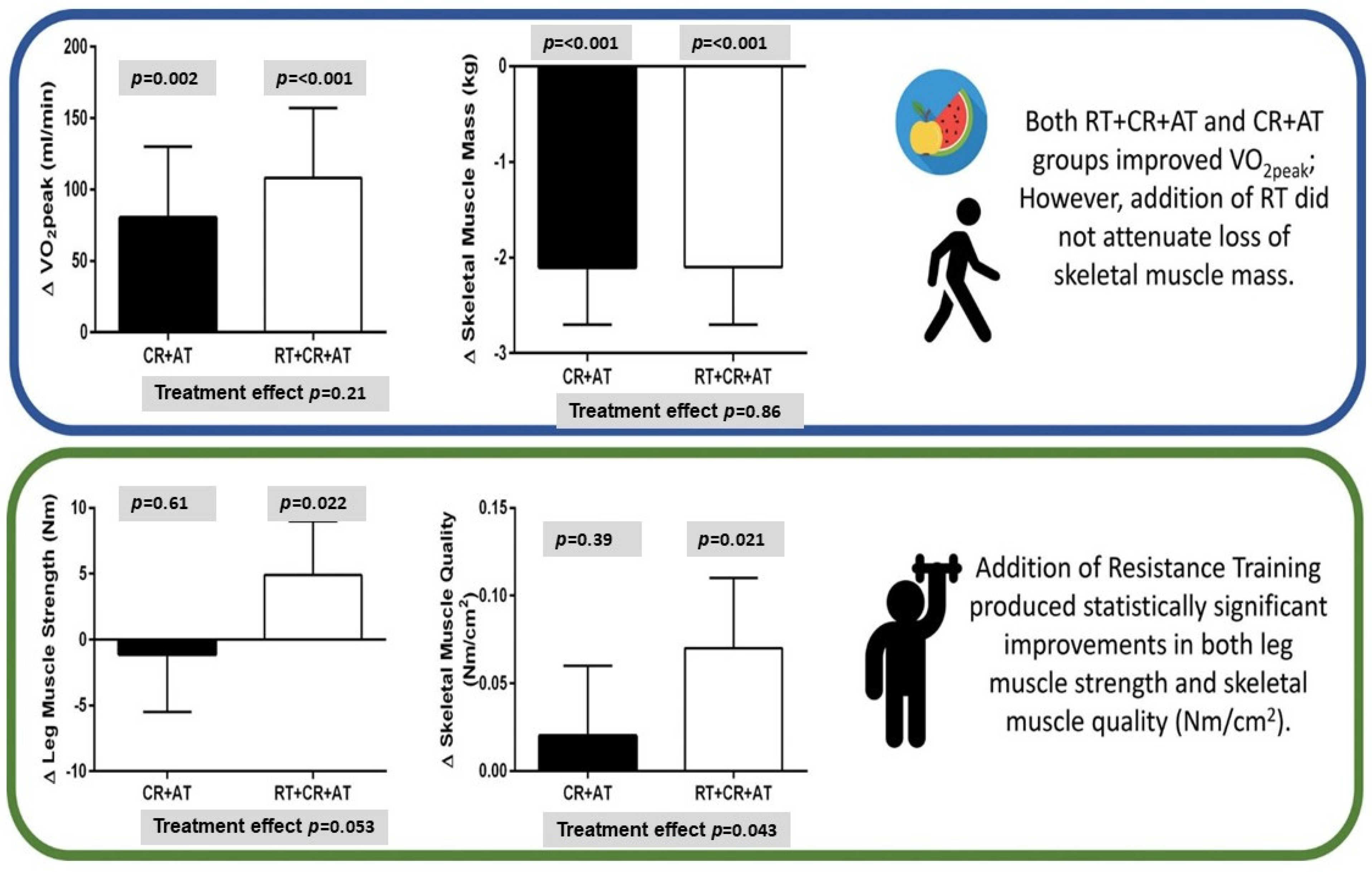
| SECRET-2/Brubaker et al. [49] | CR + AT, CR + AT + RT (n = 88) | Aged 70 ± 9 years, 85% female, chronic HFpEF, and BMI ≥ 28 kg/m2, LV EF ≥ 50% | 61 ± 6% (Mean ± SD) | VO2peak | * CR + AT produces large improvements in VO2peak and quality-of-life. Adding RT to CR + AT increased leg strength and muscle quality without attenuating skeletal muscle loss or further increasing VO2peak or quality of life. |
| Liu et al. [97] | ET + pill, ET only (n = 60) | Aged 67 ± 7, 38% female, HFpEF with LVEF ≥ 50% and NYHA class II-III symptoms | Not reported | VO2peak | * ET improved VO2peak, anaerobic threshold, 6MWD, sleep, and quality of life |
| INABLE-training/Borlaug et al. [98] | ET + nitrites, ET + placebo (n = 92) | Aged 73 (66–76), 34% female, HFpEF with LVEF ≥ 50% | Mean: 60, Range: 55–64 | VO2peak | * ET improved VO2peak and quality of life No enhanced effect from nitrites |
| Obaya et al. [99] | Upper limb aerobic exercise, lower limb aerobic exercise (n = 48) | Aged 55 ± 7, 100% male, HFpEF with left ventricular end diastolic dimension > 5.5 cm and LVEF ≥ 50% | 59 ± 4% (Mean ± SD) | VO2peak, LVEF | * Improved VO2peak No change in LVEF |
| Sharif et al. [100] | RT (n = 24) | Aged 70 ± 7, 5% female, ambulatory HFpEF with LVEF ≥ 45%, NYHA class I-III | RT: 56 ± 5% Control: 54 ± 6 (Mean ± SD) | VO2peak | * Improved locomotor muscle composition, peak VO2peak and muscle quality |
| Edelmann et al. [101] | Endurance ET (n = 322) | Aged 70 ± 7, 60% female, NYHA class II-III, clinically stable HFpEF with LVEF ≥ 50% | ET: 60 ± 5% Control: 62 ± 6% | Modified Packer score | * Improved VO2peak and NYHA class Did not significantly improve the modified Packer score |
| Training-HR/Palau et al. [102] | AT, AT/LRT, AT/MCT to HIIT, ER (n = 80) | Aged 75 ± 7 years, 59.6% female, symptomatic (NYHA classes II-III/IV) patients with the ChI HFpEF phenotype, LVEF > 55% | 65 ± 7% (Mean ± SD) | VO2peak | * All supervised training programs led to improvements in VO2peak compared to ER |
Disclaimer/Publisher’s Note: The statements, opinions and data contained in all publications are solely those of the individual author(s) and contributor(s) and not of MDPI and/or the editor(s). MDPI and/or the editor(s) disclaim responsibility for any injury to people or property resulting from any ideas, methods, instructions or products referred to in the content. |
© 2026 by the authors. Licensee MDPI, Basel, Switzerland. This article is an open access article distributed under the terms and conditions of the Creative Commons Attribution (CC BY) license.
Share and Cite
Yang, D.G.; Thakur, S.; Akunor, H.; Stacey, R.B.; Upadhya, B. Cardiometabolic Heart Failure with Preserved Ejection Fraction (HFpEF): Epidemiology, Mechanisms, and the Role of Lifestyle Modification. J. Cardiovasc. Dev. Dis. 2026, 13, 291. https://doi.org/10.3390/jcdd13070291
Yang DG, Thakur S, Akunor H, Stacey RB, Upadhya B. Cardiometabolic Heart Failure with Preserved Ejection Fraction (HFpEF): Epidemiology, Mechanisms, and the Role of Lifestyle Modification. Journal of Cardiovascular Development and Disease. 2026; 13(7):291. https://doi.org/10.3390/jcdd13070291
Chicago/Turabian StyleYang, Daniel G., Shaleen Thakur, Harriet Akunor, Richard B. Stacey, and Bharathi Upadhya. 2026. "Cardiometabolic Heart Failure with Preserved Ejection Fraction (HFpEF): Epidemiology, Mechanisms, and the Role of Lifestyle Modification" Journal of Cardiovascular Development and Disease 13, no. 7: 291. https://doi.org/10.3390/jcdd13070291
APA StyleYang, D. G., Thakur, S., Akunor, H., Stacey, R. B., & Upadhya, B. (2026). Cardiometabolic Heart Failure with Preserved Ejection Fraction (HFpEF): Epidemiology, Mechanisms, and the Role of Lifestyle Modification. Journal of Cardiovascular Development and Disease, 13(7), 291. https://doi.org/10.3390/jcdd13070291