Article

Characterization of Biofilm Producing Coagulase-Negative Staphylococci Isolated from Bulk Tank Milk

College of Veterinary Medicine & Zoonoses Research Institute, Kyungpook National University, Daegu 41566, Korea
*
Author to whom correspondence should be addressed.
Vet. Sci. 2022, 9(8), 430; https://doi.org/10.3390/vetsci9080430
Submission received: 19 June 2022 / Revised: 30 July 2022 / Accepted: 8 August 2022 / Published: 13 August 2022
(This article belongs to the Special Issue Resistant Staphylococci in Animals)

Abstract

:

Simple Summary

Coagulase-negative staphylococci are considered less virulent than other variants. However, they have been increasingly recognized as an important cause of bovine mastitis. Moreover, the biofilm-forming ability appears to be important in CoNS pathogenicity, which leads more resistance to antimicrobials. This study investigated the pathogenic potential by assessing the biofilm-forming ability of CoNS isolated from bulk tank milk and analyzed the biogilm-associated resistance to antimicrobial agents. The results indicate that various CoNS isolated from bulk tank milk, not from bovine with mastitis, exhibited a high prevalence of biofilm-forming ability with a high prevalence of MDR, and also biofilm-associated genes with a high prevalence. Therefore, developing a strong monitoring and sanitation program for dairy factories is important to ensure hygienic milk production.

Abstract

Coagulase-negative staphylococci (CoNS) are considered less virulent as they do not produce a large number of toxic enzymes and toxins; however, they have been increasingly recognized as an important cause of bovine mastitis. In particular, the ability to form biofilms appears to be an important factor in CoNS pathogenicity, and it contributes more resistance to antimicrobials. The aim of this study was to investigate the pathogenic potential by assessing the biofilm-forming ability of CoNS isolated from normal bulk tank milk using the biofilm formation assay and to analyze the biofilm-associated resistance to antimicrobial agents using the disc diffusion method. One hundred and twenty-seven (78.4%) among 162 CoNS showed the ability of biofilm formation, and all species showed a significantly high ability of biofilm formation (p < 0.05). Although the prevalence of weak biofilm formers (39.1% to 80.0%) was significantly higher than that of other biofilm formers in all species (p < 0.05), the prevalence of strong biofilm formers was significantly higher in Staphylococcus haemolyticus (36.4%), Staphylococcus chromogenes (24.6%), and Staphylococcus saprophyticus (21.7%) (p < 0.05). Also, 4 (11.4%) among 35 non-biofilm formers did not harbor any biofilm-associated genes, whereas all 54 strong or moderate biofilm formers harbored 1 or more of these genes. The prevalence of MDR was significantly higher in biofilm formers (73.2%) than in non-formers (20.0%) (p < 0.05). Moreover, the distribution of MDR in strong or moderate biofilm formers was 81.5%, which was significantly higher than in weak (67.1%) and non-formers (20.0%) (p < 0.05). Our results indicated that various CoNS isolated from bulk tank milk, not from bovine with mastitis, have already showed a high ability to form biofilms, while also displaying a high prevalence of MDR.

1. Introduction

Staphylococci, a title which includes at least 40 species, are divided into two groups according to their ability to produce the enzyme coagulase: coagulase-negative staphylococci (CoNS) and coagulase-positive staphylococci [1,2,3]. Coagulase-positive staphylococci, including Staphylococcus aureus, are a well-known cause of staphylococcal food poisoning, whereas CoNS are considered less virulent as they do not produce a large number of toxic enzymes and toxins compared to coagulase-positive staphylococci [1,2]. However, CoNS have been increasingly recognized as an important cause of bovine mastitis worldwide, with a significant increase in the incidence of intramammary infections in cows based on recent studies [3,4,5]. In particular, the ability to form biofilms appears to be an important factor in CoNS pathogenicity [6]. Biofilm formation occurs when bacteria switch from a planktonic state to a surface-attached state. It ensures bacterial survival by making them less accessible to the host’s defense system [2,7]. Moreover, biofilm exhibit resistance to antimicrobials because these impair their action [8], and act as a chronic source of microbial contamination that may lead to food spoilage in food processing [9]. In Korea, the prevalence and characteristics of CoNS from milk and dairy products have been reported [10,11,12], but there are no reports about their biofilm-forming ability. Thus, the aim of this study was to investigate their pathogenic potential by assessing the biofilm-forming ability of CoNS isolated from normal bulk tank milk, not from bovine with mastitis, and to analyze the biofilm-associated resistance to antimicrobial agents.

2. Materials and Methods

2.1. CoNS Isolates

A total of 1588 batches of bulk tank milk were collected from 396 dairy farms managed by four companies in Korea. Milk samples were aseptically collected twice each in the summer and winter seasons. Each 50 mL of bulk tank milk was tested for the isolation and identification of Staphylococcus spp. according to the standard microbiological protocols published by the Ministry of Food and Drug Safety (2018) [13]. Briefly, 1 mL of each milk sample was cultured in 9 mL of tryptic soy broth with 6% NaCl (BD Biosciences, Sparks, MD, USA). After incubation at 37 °C for 24 h, each medium was streaked onto 5% sheep blood agar (KOMED, Seoul, Korea). Confirmation of Staphylococcus spp. was performed using PCR with a species-specific primer as described previously [14]. The classification of CoNS was performed by MALDI-TOF mass spectrometry (Biomerieux, Marcy-l’Étoile, France) based on protein expression profiles using a VITEK MS system (Biomerieux). If two isolates from the same sample origin showed the same antimicrobial susceptibility patterns, only one isolate was randomly chosen. In this study, 162 CoNS were included: 65 Staphylococcuschromogenes, 46 Staphylococcus saprophyticus, 17 Staphylococcus xylosus, 11 Staphylococcus haemolyticus, 4 Staphylococcus simulans, 5 Staphylococcus sciuri, and 14 others.

2.2. Biofilm Formation Assay

Biofilm formation was estimated using the standard microtiter plate test, as described with some modifications [15]. In brief, all CoNS isolates were cultured on a brain heart infusion agar (BD Biosciences, Sparks, MD, USA) overnight at 37 °C. Five hundred ul of bacterial suspension adjusted to the 0.5 McFarland standard were inoculated into 3 mL of fresh brain heart infusion broth (BD Biosciences) supplemented with glucose (0.25% wt/vol), and 200 uL of mixture was transferred into 3 wells of a 96 well microtiter plate. After incubation for 18–24 h at 37 °C, planktonic cells were removed by washing with sterile saline. Attached bacteria were fixed with 200 uL of methanol for 15 min, and the bacterial biomass was quantified by measuring the absorbance at 490 nm (A490) after staining with safranin solution (0.1% wt/vol) for 10 min and destaining with 50% ethanol−50% glacial acetic acid solution. The ability to form biofilms was classified as negative (A490 < 0.110), weak (0.110 ≤ A490 < 0.500), moderate (0.500 ≤ A490 ≤ 1.500), and strong (A490 > 1.500). To certify the analysis, Staphylococcus epidermidis ATCC 35984 and Staphylococcus epidermidis ATCC 12228 were used as reference strains of strong and weak biofilm producers, respectively, and a sterile medium was used as a contamination control, as described previously [16,17,18].

2.3. Antimicrobial Susceptibility Testing

Based on the Clinical and Laboratory Standards Institute guidelines (CLSI, 2018) [19], the antimicrobial resistance of all CoNS isolates was determined using the disc diffusion method with the following discs (BD Biosciences): amikacin (A, 30 µg), ampicillin (AM, 10 µg), amoxicillin-clavulanate (AMC, 20 µg), ceftazidime (CAZ, 30 µg), clindamycin (CC, 2 µg), cefadroxil (CDX, 30 µg), cephalothin (CF, 30 µg), ciprofloxacin (CIP, 5 µg), colistin (CL, 10 µg), cefotaxime (CTX, 30 µg), cefuroxime (CXM, 30 µg), cefazoline (CZ, 30 µg), chloramphenicol (C, 30 µg), doxycycline (DOX, 30 µg), erythromycin (E, 15 µg), cefepime (FEP, 30 µg), cefoxitin (FOX, 30 µg), gentamicin (G, 10 µg), imipenem (IPM, 10 µg), kanamycin (K, 30 µg), oxacillin (OX, 1 µg), penicillin (P, 10 units), tetracycline (TE, 30 µg), teicoplanin (TEC, 30 µg), and vancomycin (VA, 30 µg). Staphylococcus aureus ATCC 29213 was used as a quality control. Multidrug resistance (MDR) was defined as an acquired resistance to at least one agent in three or more antimicrobial classes [20].

2.4. Detection of Biofilm-Associated Genes

DNA extraction was prepared by the boiling method, as reported [21]. The presence of biofilm-associated genes, such as aap (accumulation-associated protein), atlE (adhesion and autolysin), bap (biofilm-associated protein), embP (fibronectin adhesion), eno (laminin-binding protein), fbe (fibrinogen adhesion), and icaA (intercelluar adhesion protein A) was determined by PCR using previously published primer sequences for aap, atlE, bap, embP, eno, fbe, and icaA [22,23,24,25].

2.5. Statistical Analysis

Statistical analysis using Pearson’s chi-square tests and Fisher’s exact tests with Bonferroni correction was performed in Statistical Package for the Social Science version 25 (SPSS; IBM, Korea). Significant differences were considered at p < 0.05.

3. Results

3.1. Biofilm Formation Potential

The distribution of the ability to form biofilms based on the microtiter plate assay of 162 CoNS isolates is shown in Table 1. One hundred and twenty-seven (78.4%) CoNS showed the ability of biofilm formation, and all species showed a significantly high ability of biofilm formation (p < 0.05). Moreover, 73 (45.1%), 23 (14.2%), and 31 (19.1%) among the 127 biofilm-forming isolates were weak, moderate, and strong biofilm formers, respectively. However, the strength to form biofilms showed significant differences in CoNS species. Although the prevalence of weak biofilm formers (39.1% to 80.0%) was significantly higher than that of other biofilm formers in all species (p < 0.05), the prevalence of strong biofilm formers was significantly higher in Staphylococcus haemolyticus (36.4%), Staphylococcus chromogenes (24.6%), and Staphylococcus saprophyticus (21.7%), whereas that of moderate biofilm formers was significantly higher in Staphylococcus chromogenes (27.7%) and Staphylococcus simulans (25.0%) (p < 0.05). The distribution of strength to form biofilms by CoNS species is shown in Figure 1. Staphylococcus chromogenes had the highest median value followed by Staphylococcus haemolyticus. Although Staphylococcus saprophyticus showed a significantly high ability of strong biofilm formation (Table 1), Staphylococcus sciuri had the lowest median value followed by Staphylococcus saprophyticus. Moreover, Staphylococcus haemolyticus showed the widest deviation range of biofilm formation ability followed by Staphylococcus chromogenes, whereas Staphylococcus xylosus showed the narrowest deviation range.

3.2. Distribution of Biofilm-Associated Genes

The distribution of biofilm-associated genes in 162 CoNS isolates is also shown in Table 1. One hundred and fifty-two (93.8%) isolates harbored at least one of the seven biofilm-associated genes. Among seven biofilm-associated genes, the eno gene showed the highest prevalence (50.0% to 82.6%) in all species, except in Staphylococcus chromogenes (p < 0.05). However, Staphylococcus chromogenes carried the icaA gene which had the highest prevalence (61.5%) (p < 0.05). Moreover, 76 (46.9%) and 64 (39.5%) among 162 isolates carried the fbe and icaA genes, respectively. In particular, the fbe gene appeared in a significantly higher frequency in Staphylococcus chromogenes, Staphylococcus saprophyticus, and Staphylococcus sciuri, whereas the icaA gene appeared in a significantly higher frequency in Staphylococcus chromogenes and Staphylococcus saprophyticus (p < 0.05). Forty-nine (30.2%) and thirty (18.5%) isolates carried the aap and atlE genes, respectively, but their prevalence showed no significant differences among the CoNS species.

3.3. Relationship between Biofilm-Associated Genes and Biofilm-Forming Ability

The distribution of the biofilm-associated genes according to the ability of biofilm formation in 162 CoNS isolates is shown in Table 2. A total of 4 (11.4%) among 35 non-biofilm formers did not harbor any of the biofilm-associated genes, whereas all 54 strong or moderate biofilm formers harbored one or more of these genes. In particular, the prevalence of four genes (aap, atlE, bap, and icaA) was significantly higher in strong or moderate biofilm formers than in weak and non-formers (p < 0.05).

3.4. Relationship between MDR and Biofilm-Forming Ability

The distribution of MDR according to the ability of biofilm formation in 162 CoNS isolates is shown in Table 3. The prevalence of MDR was significantly higher in biofilm formers (73.2%) than in non-formers (20.0%) (p < 0.05). Moreover, the distribution of MDR in strong or moderate biofilm formers was 81.5%, which was significantly higher than that in weak (67.1%) and non-formers (20.0%) (p < 0.05).

4. Discussion

Bovine mastitis is the most important disease that leads to economic loss in dairy cattle worldwide [26]. Recently, CoNS are also described as the most common bovine mastitis isolates in many countries, and these emerged as pathogens associated with clinical and subclinical intramammary infection [3,27,28]. Park et al. (2011) [29] reported that S. chromogenes (72.2%) was the most distributed CoNS isolate from bovine mastitis in the United States, followed by Staphylococcus xylosus (9.1%) and Staphylococcus haemolyticus (6.1%). Walid et al. (2021) [30] also reported that most CoNS isolates from bovine mastitis in Egypt were Staphylococcus epidermidis (48.4%), Staphylococcus saprophyticus (32.3%), and Staphylococcus haemolyticus (19.4%). In particular, several CoNS, such as Staphylococcus epidemidis, Staphylococcus chromogenes, and Staphylococcus xylosus, showed a higher pathogenicity by forming biofilms for bacterial aggregation for a better growth and resistance to adverse conditions [31]. In this study, 162 CoNS isolates, including Staphylococcus chromogenes (65 isolates), Staphylococcus saprophyticus (46 isolates), Staphylococcus xylosus (17 isolates), Staphylococcus haemolyticus (11 isolates), Staphylococcus sciuri (5 isolates), Staphylococcus simulans (4 isolates), and others (14 isolates) were isolated from bulk tank milk, not from bovine with mastitis; however, 127 (78.4%) CoNS isolates showed various abilities to form biofilms. Moreover, 54 (33.3%) CoNS isolates were classified as strong or moderate biofilm formers. Tremblay et al. (2013) and Srednik et al. (2017) [32,33] reported that 48.6% and 44.0% of biofilm-forming CoNS isolates from bovine mastitis in Canada and Argentina, respectively, were classified as strong or moderate biofilm formers. If milk samples were derived from bovine with clinical mastitis rather than from normal bulk tank, a higher prevalence of biofilm formers in CoNS might be confirmed. The prevalence of strong or moderate biofilm formers in this study was significantly higher in Staphylococcus haemolyticus and Staphylococcus chromogenes, which was similar to previous reports [6,32,33]. The highest median value and widest deviation range were also observed in these two CoNS species. The presence of biofilm-associated genes confers a greater ability to form biofilms [34,35]. In this study, 152 (93.8%) among 162 CoNS isolates harbored at least one or more of the seven biofilm-associated genes. Although 62.3% of 162 CoNS isolates carried the eno gene, which showed significantly the highest prevalence, it appears to be distributed regardless of species and biofilm-forming ability, as previous described [2,36]. The icaA and bap genes are commonly involved in biofilm formation, and their prevalence in this study was 39.5% and 26.5%, respectively. In particular, Staphylococcus chromogenes and Staphylococcus saprophyticus significantly had a higher prevalence in icaA, while Staphylococcus saprophyticus significantly had a higher prevalence in bap. Interestingly, Staphylococcus chromogenes and Staphylococcus saprophyticus had a significantly higher prevalence in strong biofilm formers. Staphylococcus haemolyticus also had the highest prevalence in strong biofilm formers in this study. However, the prevalence of Staphylococcus haemolyticus carrying the bap gene was significantly lower than that of Staphylococcus saprophyticus, and no Staphylococcus haemolyticus isolates harbored the icaA gene. Moreover, the prevalence of the fbe and embP genes, which are involved in surface-adhesins for biofilm formation, was 46.9% and 18.5%, respectively. The highest prevalence of the fbe and embP genes was observed in Staphylococcus sciui and Staphylococcus simulans, respectively, for which none of the isolates had a strong biofilm former. The prevalence of four biofilm-associated genes (aap, atlE, bap, and icaA) was significantly higher in strong or moderate biofilm formers than weak or non-formers in this study, and other studies have reported a high prevalence of biofilm-associated genes in biofilm-producing staphylococci [37,38,39]. However, the link between the presence of biofilm-associated genes and the ability to form biofilms is not clear and needs to be better understood.
Moreover, the ability to form biofilms is associated with the capacity of bacteria to adhere to a surface and form a layer, so the density of the layer was directly related to the strength of the biofilm produced [40]. Therefore, the strength of biofilm formation was higher in antimicrobial-resistant strains than in antimicrobial-sensitive strains, and a remarkable correlation was found between antimicrobial resistance and biofilm formation strength [41,42,43]. In this study, the prevalence of MDR was significantly higher in biofilm formers than in non-formers. Interestingly, strong and moderate biofilm formers also had a significantly higher prevalence of MDR than weak biofilm formers. Recently, Phophi et al. (2019) [44] reported that biofilm-forming CoNS from mastitis in South Africa showed the significantly higher prevalence in MDR. Moreover, Oliveira et al. (2016) [45] reported that bacteria living in biofilms are up to 1000 times more resistant compared to planktonic bacteria. Therefore, our results support that the biofilm-forming ability limits the treatment strategies for mastitis and might increase morbidity and mortality if biofilm-forming CoNS isolated from bulk tank milk develop as a cause of mastitis. In this study, various CoNS isolated from bulk tank milk, not bovine with mastitis, have already showed their high ability to form biofilms, with a high prevalence of MDR. Therefore, an improved hygiene program should be proposed to control the intramammary infection of environmental bacteria like CoNS.

Author Contributions

Conceptualization, Y.J.L. (Yu Jin Lee) and Y.J.L. (Young Ju Lee); Data curation, Y.J.L. (Yu Jin Lee); Formal analysis, Y.J.L. (Yu Jin Lee) and Y.J.L. (Young Ju Lee); Funding acquisition, Y.J.L. (Young Ju Lee); Investigation, Y.J.L. (Yu Jin Lee); Methodology, Y.J.L. (Yu Jin Lee) and Y.J.L. (Young Ju Lee); Project administration, Y.J.L. (Young Ju Lee); Resources, Y.J.L. (Yu Jin Lee) and Y.J.L. (Young Ju Lee); Software, Y.J.L. (Yu Jin Lee); Supervision, Y.J.L. (Young Ju Lee); Validation, Y.J.L. (Yu Jin Lee) and Y.J.L. (Young Ju Lee); Visualization, Y.J.L. (Yu Jin Lee); Writing—original draft, Y.J.L. (Yu Jin Lee); Writing—review & editing, Y.J.L. (Yu Jin Lee) and Y.J.L. (Young Ju Lee). All authors have read and agreed to the published version of the manuscript.

Funding

This research received no external funding.

Institutional Review Board Statement

Not applicable.

Informed Consent Statement

Not applicable.

Data Availability Statement

Not applicable.

Conflicts of Interest

The authors declare no conflict of interest.

References

  1. França, A.; Gaio, V.; Lopes, N.; Melo, D.R. Virulence Factors in Coagulase-Negative Staphylococci. Pathogens 2021, 10, 170. [Google Scholar] [CrossRef] [PubMed]
  2. Marek, A.; Pyzik, E.; Stępień-Pyśniak, D.; Dec, M.; Jarosz, Ł.S.; Nowaczek, A.; Sulikowska, M. Biofilm-formation ability and the presence of adhesion genes in coagulase-negative staphylococci isolates from chicken broilers. Animals 2021, 11, 728. [Google Scholar] [CrossRef] [PubMed]
  3. Melo, D.A.; Motta, C.C.; Rojas, A.C.C.M.; Soares, B.S.; Coelho, I.S.; Coelho, S.M.O.; Souza, M.M.S. Characterization of Coagulase-Negative Staphylococci and pheno-genotypic beta lactam resistance evaluation in samples from bovine Intramammary infection. Arq. Bras. De Med. Veteriná Ria E Zootec. 2018, 70, 368–374. [Google Scholar] [CrossRef]
  4. Klibi, A.; Maaroufi, A.; Torres, C.; Jouini, A. Detection and characterization of methicillin-resistant and susceptible coagulase-negative staphylococci in milk from cows with clinical mastitis in Tunisia. Int. J. Antimicrob. Agents 2018, 52, 930–935. [Google Scholar] [CrossRef] [PubMed]
  5. Kirwa, E.; Gabriel, A.O.; Maitho, T.E.; Mbindyo, C.M.; Abuom, T.O.; Mainga, A.O. Antibiotic profile of Staphylococcus aureus and Coagulase negative Staphylococci species isolated from raw camel milk from Garissa County, Kenya. East African J. Sci. Technol. Innov. 2021, 2, 1–15. [Google Scholar] [CrossRef]
  6. Goetz, C.; Tremblay, Y.D.N.; Lamarche, D.; Blondeau, A.; Gaudreau, A.M.; Labrie, J.; Malouin, F.; Jacques, M. Coagulase-negative staphylococci species affect biofilm formation of other coagulase-negative and coagulase-positive staphylococci. J. Dairy Sci. 2017, 100, 6454–6464. [Google Scholar] [CrossRef] [PubMed]
  7. Coffey, B.M.; Anderson, G.G. Biofilm formation in the 96-well microtiter plate. Methods Mol. Biol. 2014, 1149, 631–641. [Google Scholar] [CrossRef] [PubMed]
  8. Gajewska, J.; Chajęcka-Wierzchowska, W. Biofilm formation ability and presence of adhesion genes among coagulase-negative and coagulase-positive staphylococci isolates from raw cow’s milk. Pathogens 2020, 9, 654. [Google Scholar] [CrossRef]
  9. Jayaweera, T.S.P.; Ruwandeepika, H.A.D.; Deekshit, V.K.; Kodithuwakku, S.P.; Cyril, H.W.; Karunasagar, I.; Vidanarachchi, J.K. Biofilm Forming Ability of Broiler Chicken Meat Associated Salmonella spp. on Food Contact Surfaces. Trop. Agric. Res. 2021, 32, 17–26. [Google Scholar] [CrossRef]
  10. Nam, H.M.; Lim, S.K.; Moon, J.S.; Kang, H.M.; Kim, J.M.; Jang, K.C.; Kim, J.M.; Kang, M.I.; Joo, Y.S.; Jung, S.C. Antimicrobial resistance of enterococci isolated from mastitic bovine milk samples in Korea. Zoonoses Public Health 2010, 57, 698–701. [Google Scholar] [CrossRef] [PubMed]
  11. Kim, S.J.; Moon, D.C.; Park, S.C.; Kang, H.Y.; Na, S.H.; Lim, S.K. Antimicrobial resistance and genetic characterization of coagulase-negative staphylococci from bovine mastitis milk samples in Korea. J. Dairy Sci. 2019, 102, 11439–11448. [Google Scholar] [CrossRef] [PubMed]
  12. Yun, M.J.; Yoon, S.; Lee, Y.J. Monitoring and characteristics of major mastitis pathogens from Bulk tank milk in Korea. Animals 2020, 10, 1562. [Google Scholar] [CrossRef] [PubMed]
  13. Ministry of Food and Drug Safety (MFDS). Processing Standards and Ingredient Specifications for Livestock Products; NIFDS: Cheong ju, Korea, 2018. [Google Scholar]
  14. Martineau, F.; Picard, F.J.; Ke, D.; Paradis, S.; Roy, P.H.; Ouellette, M.; Bergeron, M.G. Development of a PCR assay for identification of staphylococci at genus and species levels. J. Clin. Microbiol. 2001, 39, 2541–2547. [Google Scholar] [CrossRef] [PubMed]
  15. Mitchell, G.; Séguin, D.L.; Asselin, A.E.; Déziel, E.; Cantin, A.M.; Frost, E.H.; Michaud, S.; Malouin, F. Staphylococcus aureus sigma B-dependent emergence of small-colony variants and biofilm production following exposure to Pseudomonas aeruginosa 4-hydroxy-2-heptylquinoline-N-oxide. BMC Microbiol. 2010, 10, 33. [Google Scholar] [CrossRef] [PubMed]
  16. Zhang, Y.Q.; Ren, S.X.; Li, H.L.; Wang, Y.X.; Fu, G.; Yang, J.; Qin, Z.Q.; Miao, Y.G.; Wang, W.Y.; Chen, R.S.; et al. Genome-based analysis of virulence genes in a non-biofilm-forming Staphylococcus epidermidis strain (ATCC 12228). Mol. Microbiol. 2003, 49, 1577–1593. [Google Scholar] [CrossRef] [PubMed]
  17. Gill, S.R.; Fouts, D.E.; Archer, G.L.; Mongodin, E.F.; DeBoy, R.T.; Ravel, J.; Paulsen, I.T.; Kolonay, J.F.; Brinkac, L.; Beanan, M.; et al. Insights on evolution of virulence and resistance from the complete genome analysis of an early methicillin-resistant Staphylococcus aureus strain and a biofilm-producing methicillin-resistant Staphylococcus epidermidis strain. J. Bacteriol. 2005, 187, 2426–2438. [Google Scholar] [CrossRef] [PubMed]
  18. Toledo-Silva, B.; de Souza, F.N.; Mertens, K.; Piepers, S.; Haesebrouck, F.; De Vliegher, S. Bovine-associated non-aureus staphylococci suppress Staphylococcus aureus biofilm dispersal in vitro yet not through agr regulation. Vet. Res. 2021, 52, 114. [Google Scholar] [CrossRef] [PubMed]
  19. Clinical and Laboratory Standards Institute (CLSI). Performance Standards for Antimicrobial Susceptibility Testing, 28th ed.; CLSI: Wayne, PA, USA, 2018; ISBN 156238838X. [Google Scholar]
  20. Magiorakos, A.P.; Srinivasan, A.; Carey, R.B.; Carmeli, Y.; Falagas, M.E.; Giske, C.G.; Harbarth, S.; Hindler, J.F.; Kahlmeter, G.; Olsson-Liljequist, B.; et al. Multidrug-resistant, extensively drug-resistant and pandrug-resistant bacteria: An international expert proposal for interim standard definitions for acquired resistance. Clin. Microbiol. Infect. 2012, 18, 268–281. [Google Scholar] [CrossRef] [PubMed]
  21. Bakshi, C.S.; Shah, D.H.; Verma, R.; Singh, R.K.; Malik, M. Rapid differentiation of Mycobacterium bovis and Mycobacterium tuberculosis based on a 12.7-kb fragment by a single tube multiplex-PCR. Vet. Microbiol. 2005, 109, 211–216. [Google Scholar] [CrossRef] [PubMed]
  22. Cucarella, C.; Tormo, M.Á.; Úbeda, C.; Trotonda, M.P.; Monzón, M.; Peris, C.; Amorena, B.; Lasa, Í.; Penadés, J.R. Role of Biofilm-Associated Protein Bap in the Pathogenesis of Bovine Staphylococcus aureus. Infect. Immun. 2004, 72, 2177–2185. [Google Scholar] [CrossRef]
  23. Rohde, H.; Burdelski, C.; Bartscht, K.; Hussain, M.; Buck, F.; Horstkotte, M.A.; Knobloch, J.K.M.; Heilmann, C.; Herrmann, M.; Mack, D. Induction of Staphylococcus epidermidis biofilm formation via proteolytic processing of the accumulation-associated protein by staphylococcal and host proteases. Mol. Microbiol. 2005, 55, 1883–1895. [Google Scholar] [CrossRef] [PubMed]
  24. Rohde, H.; Burandt, E.C.; Siemssen, N.; Frommelt, L.; Burdelski, C.; Wurster, S.; Scherpe, S.; Davies, A.P.; Harris, L.G.; Horstkotte, M.A.; et al. Polysaccharide intercellular adhesin or protein factors in biofilm accumulation of Staphylococcus epidermidis and Staphylococcus aureus isolated from prosthetic hip and knee joint infections. Biomaterials 2007, 28, 1711–1720. [Google Scholar] [CrossRef] [PubMed]
  25. Simojoki, H.; Hyvönen, P.; Plumed Ferrer, C.; Taponen, S.; Pyörälä, S. Is the biofilm formation and slime producing ability of coagulase-negative staphylococci associated with the persistence and severity of intramammary infection? Vet. Microbiol. 2012, 158, 344–352. [Google Scholar] [CrossRef] [PubMed]
  26. Kovačević, Z.; Radinović, M.; Čabarkapa, I.; Kladar, N.; Božin, B. Natural agents against bovine mastitis pathogens. Antibiotics 2021, 10, 205. [Google Scholar] [CrossRef] [PubMed]
  27. Pyatov, V.; Vrtková, I.; Knoll, A. Detection of selected antibiotic resistance genes using multiplex PCR assay in mastitis pathogens in the Czech Republic. Acta Vet. Brno 2017, 86, 167–174. [Google Scholar] [CrossRef]
  28. Mbindyo, C.M.; Gitao, G.C.; Mulei, C.M. Prevalence, Etiology, and Risk Factors of Mastitis in Dairy Cattle in Embu and Kajiado Counties, Kenya. Vet. Med. Int. 2020, 2020, 1–12. [Google Scholar] [CrossRef]
  29. Park, J.Y.; Fox, L.K.; Seo, K.S.; McGuire, M.A.; Park, Y.H.; Rurangirwa, F.R.; Sischo, W.M.; Bohach, G.A. Comparison of phenotypic and genotypic methods for the species identification of coagulase-negative staphylococcal isolates from bovine intramammary infections. Vet. Microbiol. 2011, 147, 142–148. [Google Scholar] [CrossRef] [PubMed]
  30. Walid, M.S. Antibiogram and antibiotic resistance genes among coagulase-negative staphylococci recovered from bovine mastitis. Arch. Anesthesiol. Crit. Care 2021, 9, 1267–1274. [Google Scholar] [CrossRef]
  31. Maity, S.; Ambatipudi, K. Mammary microbial dysbiosis leads to the zoonosis of bovine mastitis: A One-Health perspective. FEMS Microbiol. Ecol. 2021, 97, 1–17. [Google Scholar] [CrossRef] [PubMed]
  32. Tremblay, Y.D.N.; Lamarche, D.; Chever, P.; Haine, D.; Messier, S.; Jacques, M. Characterization of the ability of coagulase-negative staphylococci isolated from the milk of Canadian farms to form biofilms. J. Dairy Sci. 2013, 96, 234–246. [Google Scholar] [CrossRef] [PubMed]
  33. Srednik, M.E.; Tremblay, Y.D.N.; Labrie, J.; Archambault, M.; Jacques, M.; Cirelli, A.F.; Gentilini, E.R. Biofilm formation and antimicrobial resistance genes of coagulase-negative staphylococci isolated from cows with mastitis in Argentina. FEMS Microbiol. Lett. 2017, 364, 1–8. [Google Scholar] [CrossRef] [PubMed]
  34. Becker, K.; Heilmann, C.; Peters, G. Coagulase-negative staphylococci. Clin. Microbiol. Rev. 2014, 27, 870–926. [Google Scholar] [CrossRef]
  35. Van Meervenne, E.; De Weirdt, R.; Van Coillie, E.; Devlieghere, F.; Herman, L.; Boon, N. Biofilm models for the food industry: Hot spots for plasmid transfer? Pathog. Dis. 2014, 70, 332–338. [Google Scholar] [CrossRef] [PubMed]
  36. Lin, J.; Secondary, C.A.; Author, C.; Lin, J.; Jin, Y.; Pang, Q.; Lin, J. Application of ica D, eno, sar A and agr gene testing in early diagnosis of periprosthetic joint infection. Int. Surg. 2021, 106, 82–94. [Google Scholar] [CrossRef]
  37. Ibtissem, K.T.; Hafida, H.; Salwa, O.; Samia, B.; Imen, M.; Meriem, L.; Mohammed, T. Detection of icaA and icaD genes and biofilmformation in Staphylococcus spp. isolated from urinary catheters at the University Hospital of Tlemcen (Algeria). African J. Microbiol. Res. 2013, 7, 5350–5357. [Google Scholar] [CrossRef]
  38. Osman, K.M.; Abd El-Razik, K.A.; Marie, H.S.H.; Arafa, A. Relevance of biofilm formation and virulence of different species of coagulase-negative staphylococci to public health. Eur. J. Clin. Microbiol. Infect. Dis. 2015, 34, 2009–2016. [Google Scholar] [CrossRef] [PubMed]
  39. Saidi, R.; Cantekin, Z.; Mimoune, N.; Ergun, Y.; Solmaz, H.; Khelef, D.; Kaidi, R. Investigation of the presence of slime production, VanA gene and antiseptic resistance genes in Staphylococci isolated from bovine mastitis in Algeria. Vet. Stanica 2021, 52, 57–63. [Google Scholar] [CrossRef]
  40. Machado, T.S.; Pinheiro, F.R.; Soares, L.; Andre, P.; Freire, R.; Pereira, A.; Correa, R.F.; De Mello, G.C.; Aparecida, T.; Ribeiro, N.; et al. Virulence Factors Found in Nasal Colonization and Infection of Methicillin-Resistant Staphylococcus aureus (MRSA) Isolates and Their Ability to Form a Biofilm. Toxins 2020, 13, 14. [Google Scholar] [CrossRef]
  41. Poppele, E.H.; Hozalski, R.M. Micro-cantilever method for measuring the tensile strength of biofilms and microbial flocs. J. Microbiol. Methods 2003, 55, 607–615. [Google Scholar] [CrossRef]
  42. Lu, D.; Bai, H.; Kong, F.; Liss, S.N.; Liao, B. Recent advances in membrane aerated biofilm reactors. Crit. Rev. Environ. Sci. Technol. 2021, 51, 649–703. [Google Scholar] [CrossRef]
  43. Karimi, K.; Zarei, O.; Sedighi, P.; Taheri, M.; Doosti-Irani, A.; Shokoohizadeh, L. Investigation of Antibiotic Resistance and Biofilm Formation in Clinical Isolates of Klebsiella pneumoniae. Int. J. Microbiol. 2021, 2021, 5573388. [Google Scholar] [CrossRef] [PubMed]
  44. Phophi, L.; Petzer, I.M.; Qekwana, D.N. Antimicrobial resistance patterns and biofilm formation of coagulase-negative Staphylococcus species isolated from subclinical mastitis cow milk samples submitted to the Onderstepoort Milk Laboratory. BMC Vet. Res. 2019, 15, 1–9. [Google Scholar] [CrossRef] [PubMed]
  45. de Oliveira, A.; Pereira, V.C.; Pinheiro, L.; Riboli, D.F.M.; Martins, K.B.; Ribeiro de Souza da Cunha, M.D.L. Antimicrobial resistance profile of planktonic and biofilm cells of staphylococcus aureus and coagulase-negative staphylococci. Int. J. Mol. Sci. 2016, 17, 1423. [Google Scholar] [CrossRef] [PubMed]
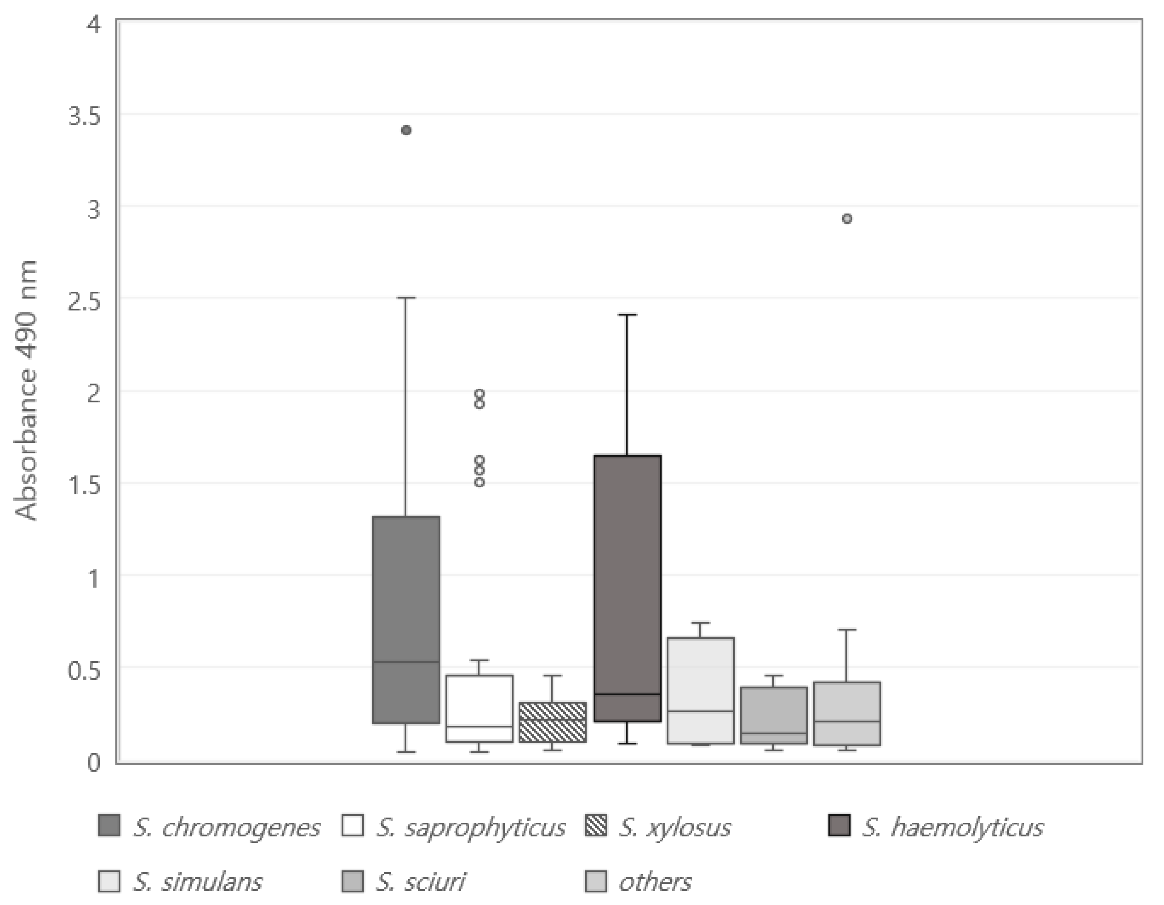
Figure 1. Distribution of strength to form biofilm in 162 coagulase negative staphylococci isolated from bulk tank milk. Points outside the box and whiskers are considered as outliers.
Vetsci 09 00430 g001
Table 1. Distribution of biofilm formation potential and biofilm-associated genes in 162 coagulase negative staphylococci from milk.
Staphylococcus chromogenes
(n = 65)
Staphylococcus saprophyticus
(n = 46)
Staphylococcus xylosus
(n = 17)
Staphylococcus haemolyticus
(n = 11)
Staphylococcus simulans
(n = 4)
Staphylococcus sciuri
(n = 5)
Others 2
(n = 14)
Total (%)
Biofilm formation(A490) 1
Negative5 (7.7) c17 (37.0) a5 (29.4) a,b1 (9.1) b,c1 (25.0) a,b1 (20.0) a,b5 (35.7) a35 (21.6)
Positive60 (92.3) a*29 (63.0) c*12 (70.6) c*10 (90.9) a,b*3 (75.0) b,c*4 (80.0) a,b,c*9 (64.3) c*127 (78.4) *
Weak26 (40.0) cA18 (39.1) cA12 (70.6) a,bA5 (45.5) cA2 (50.0) b,cA4 (80.0) aA6 (42.9) c A73 (45.1) A
Moderate18 (27.7) aB1 (2.2) b,cC0 (0.0) cB1 (9.1) b,cC1 (25.0) aB0 (0.0) cB2 (14.3) a,bB23 (14.2) B
Strong16 (24.6) a,bB10 (21.7) a,bB0 (0.0) cB4 (36.4) aB0 (0.0) cC0 (0.0) cB1 (7.1) b,cC31 (19.1) B
Biofilm-associated gene
None4 (6.2) b,cD2 (4.3) b,cD2 (11.8) a,bC1 (9.1) a,bC1 (25.0) aB0 (0.0) cC0 (0.0) cD10 (6.2) D
aap20 (30.8) B,C15 (32.6) B,C3 (17.6) B,C4 (36.4) B,C1 (25.0) B1 (20.0) B5 (35.7) B,C49 (30.2) B,C
atlE12 (18.5) C,D 6 (13.0) C,D4 (23.5) B,C2 (18.2) C1 (25.0) B1 (20.0) B4 (28.6) C30 (18.5) C
bap15 (23.1) b,cC,D17 (37.0) aB,C2 (11.8) cC3 (27.3) b,cB,C1 (25.0) b,cB0 (0.0) cC5 (35.7) a,bB,C43 (26.5) C
embP14 (21.5) b,cC,D5 (10.9) b,cC,D3 (17.6) b,cB,C1 (9.1) cC1 (25.0) a,bB0 (0.0) cC6 (42.9) aB,C30 (18.5) C
eno27 (41.5) cB,C38 (82.6) aA11 (64.7) a,bA9 (81.8) aA2 (50.0) b,cA4 (80.0) aA10 (71.4) a,bA101 (62.3) A
fbe32 (49.2) a,bB,C25 (54.3) a,bB7 (41.2) b,cB3 (27.3) cB,C1 (25.0) cB3 (60.0) aB5 (35.7) b,cB,C76 (46.9) B
icaA40 (61.5) aA15 (32.6) a,bB,C4 (23.5) b,cB,C0 (0.0) cD1 (25.0) b,cB1 (20.0) b,cB3 (21.4) b,cC64 (39.5) B,C
The superscript letter represents significant difference of the column, while the subscript letter represents significant difference of the row (p < 0.05). * Statistically significant difference between biofilm-positive isolates and biofilm-negative isolates (p < 0.05). 1 A490 = Absorbance at 490 nm. 2 Others: Staphylococcus arlettae (n = 1), Staphylococcus capitis (n = 3), Staphylococcus cohnii (n = 2), Staphylococcus epidermidis (n = 1), Staphylococcus equorum (n = 2), Staphylococcus gallinarum (n = 2), Staphylococcus lentus (n = 1), Staphylococcus succinus (n = 2).
Table 2. Relationship between biofilm-associated genes and biofilm-forming ability in 162 coagulase negative staphylococci from milk.
Antimicrobial ResistanceBiofilm Producer
Strong or Moderate
Biofilm Former
(n = 54)
Weak Biofilm Former (n = 73)Non-Former
(n = 35)
Non-MDR10 (18.5) cB24 (32.9) bB28 (80.0) aA
MDR44 (81.5) aA49 (67.1) bA7 (20.0) cB
The superscript letter represents significant difference of the column, while the subscript letter represents significant difference of the row (p < 0.05).
Table 3. Relationship between multidrug resistance (MDR) and biofilm-forming ability in 162 coagulase negative staphylococci from milk.
Biofilm-Associated GeneBiofilm Producer
Strong or Moderate
Biofilm Former
(n = 54)
Weak Biofilm Former
(n = 73)
Non-Former
(n = 35)
None0 (0.0) bE6 (8.2) a,bD4 (11.4) aC,D
aap28 (51.9) aA,B,C16 (21.9) bB,C5 (14.3) cC,D
atlE18 (33.3) aC,D9 (12.3) bC,D3 (8.6) cD
bap22 (40.7) aB,C,D16 (21.9) bB,C5 (14.3) cC,D
embP12 (22.2) a,bD9 (12.3) bC,D9 (25.7) aB,C
eno35 (64.8) a,bA38 (52.1) bA28 (80.0) aA
fbe32 (59.3) aA,B28 (38.4) bA,B16 (45.7) a,bB
icaA35 (64.8) aA24 (32.9) bA,B5 (14.3) cC,D
The superscript letter represents significant difference of the column, while the subscript letter represents significant difference of the row (p < 0.05).
Publisher’s Note: MDPI stays neutral with regard to jurisdictional claims in published maps and institutional affiliations.