Article

Molecular Identification of Parasitic Protozoa Sarcocystis in Water Samples

by
Živilė Strazdaitė-Žielienė
,
Agnė Baranauskaitė
,
Dalius Butkauskas
,
Elena Servienė
and
Petras Prakas
*
Nature Research Centre, Akademijos Str. 2, LT-08412 Vilnius, Lithuania
*
Author to whom correspondence should be addressed.
Vet. Sci. 2022, 9(8), 412; https://doi.org/10.3390/vetsci9080412
Submission received: 2 May 2022 / Revised: 13 June 2022 / Accepted: 3 August 2022 / Published: 5 August 2022
(This article belongs to the Special Issue Diagnostic PCR on Animal Diseases: From Extraction to Amplification)

Abstract

:

Simple Summary

Members of the genus Sarcocystis are protozoan parasites having two-host prey–predator cycle. These parasites are widespread in farm animals. Sarcocystis species are characterized morphologically in intermediate hosts, and these parasites are identified in definitive hosts by molecular methods. Thus far, only few studies have been conducted on Sarcocystis parasites in environmental samples. The aim of the present work was to evaluate several sample preparation and polymerase chain reaction methods for the identification of several Sarcocystis species in water samples. Overall, 114 samples collected from various water sources, ponds, canals, lakes, lagoons, and rivers in Lithuania were tested for the presence of Sarcocystis spp. Based on molecular methods, eight Sarcocystis species, S. bovifelis, S. cruzi, S. hirsuta, S. arieticanis, S. tenella, S. capracanis, S. bertrami, and S. miescheriana, were identified. The main intermediate hosts of detected Sarcocystis parasites are cattle, sheep, goats, horses, and pigs. Further, more sensitive molecular techniques are needed for the development of the diagnosis of Sarcocystis species in water bodies.

Abstract

Sarcocystis parasites are among the most common parasitic protozoa in farm animals. So far, the diversity of these parasites has been mainly studied in animal carcasses by morphological or molecular methods. Research on parasitic protozoa in environmental samples is scarce due to the lack of an appropriate methodology and low concentrations of parasites. For these reasons, there is a paucity of validated methods for Sarcocystis identification from environmental samples. Therefore, the present study aims to investigate various molecular methods for Sarcocystis parasite identification in water samples. In the present study, the sample volume, sporocysts isolation, and various conventional PCR were evaluated, and species-specific primers for the identification of different Sarcocystis species have been developed. Of the methods studied, based on data the most appropriate method for the identification of analyzed Sarcocystis spp. in water bodies is nested PCR, using species-specific primers targeting the cox1 gene. Sarcocystis DNA was detected in 111 out of 114 (97.4%) samples. This paper represents the first identification of S. bovifelis, S. cruzi, S. hirsuta, S. arieticanis, S. tenella, S. capracanis, S. bertrami, and S. miescheriana by PCR and sequencing in environmental water samples. Our pilot study is useful in developing techniques for the identification of Sarcocystis species from water samples.

1. Introduction

Sarcocystis parasites are members of the Apicomplexa phylum that infect mammals, birds, and reptiles. They have an obligatory life cycle of two hosts, intermediate and definitive, that implies a predator–prey interaction. Sarcocysts of these parasites are mainly formed in muscle tissues of intermediate hosts (asexual stage), while sporocysts develop in the intestine of the definitive host and are spread into the environment in feces (sexual stage) [1]. Meat of farm animals showing an eosinophilic myositis related to the presence of Sarcocystis spp. is withdrawn from the market [2]. Furthermore, intense Sarcocystis infection might result in reduced meat, wool, and milk production, causing huge losses in the livestock industry annually [2,3].
To date, the muscle tissues of intermediate hosts have been studied mainly for the purpose of identifying different Sarcocystis species. Morphological characterization of these parasites includes principally the analysis of sarcocyst size and shape, morphometric parameters of bradyzoites located inside the sarcocyst, and the structure of the sarcocyst wall [1]. Sarcocystis species are distinguished by a combination of morphological analysis and sequence data of 18S ribosomal DNA (rDNA) [1,4]. Significantly less frequently, the identification of these parasites is performed in definitive hosts by examining intestinal samples and feces of predators or scavengers [5]. Sarcocystis species cannot be distinguished by the morphology of sporocysts. Therefore, species of these parasites in the definitive host and environmental samples are identified by using nested PCR for the detection and sequencing of cytochrome c oxidase subunit I (cox1), 18S rRNA, 28S rRNA, and ITS1 [6].
Until now, only two studies on Sarcocystis spp. diversity in water samples have been conducted due to the lack of validated methods [7,8]. In these studies, samples were collected from water sources located near villages on Tioman Island, Malaysia, where cases of muscular sarcocystosis were recorded. Sporocysts were isolated from water samples using the sucrose gradient method [9]. The nested PCR and Sanger sequencing of 18S rRNA fragments allowed the identification of S. nesbitti, S. singaporensis, Sarcosystis sp. YLL-2013, and Sarcocystis sp. [7]. On the other hand, S. nesbitti, S. singaporensis, S. zamani, and several unnamed Sarcocystis species were defined by an improved Sarcocystis spp. identification method, relying on the next-generation sequencing (NGS) of 18S rRNA and 28S rRNA coding fragments [8]. Thus, water samples collected near pastures and human bathing areas could be used for epidemiology research aimed at detecting pathogenic species and those involving economically important animals as their hosts.
In the present study, we chose to examine the presence of eight Sarcocystis species (S. bovifelis, S. cruzi, S. hirsuta, S. arieticanis, S. tenella, S. capracanis, S. bertrami, and S. miescheriana) in water samples. Three species, S. bovifelis, S. cruzi, and S. hirsuta, form sarcocysts in muscles of cattle, S. arieticanis and S. tenella parasitize sheep, while S. capracanis, S. bertrami, and S. miescheriana are specific to goats, horses, and pig/wild boar, respectively. There is considerable confusion over classification of Sarcocystis species found in horses. Sarcocysts of S. bertrami and S. fayeri detected in muscles of horses show significant morphological differences [1]. However, S. bertrami and S. fayeri are the same species based on molecular markers, and S. fayeri should be considered a junior synonym of S. bertrami [10]. In the present study, we used S. bertrami to describe any Sarcocystis species from horses. So far Sarcocystis species composition in Lithuania was investigated only in cattle. It was shown that S. bovifelis, S. cruzi and S. hirsuta are the most common species in diaphragm muscles of cattle [11]. According to current knowledge, the other species selected in this work are most detected in sheep, goats, horses, and pigs in Europe [1].
The objective of the present study was to evaluate various sample preparation and conventional PCR methods that may aid in species-specific molecular identification of Sarcocystis residing in watersheds.

2. Materials and Methods

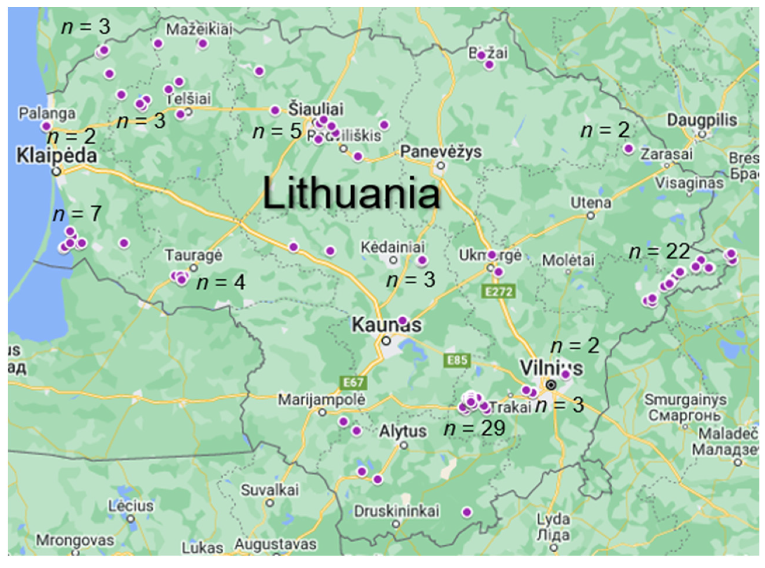
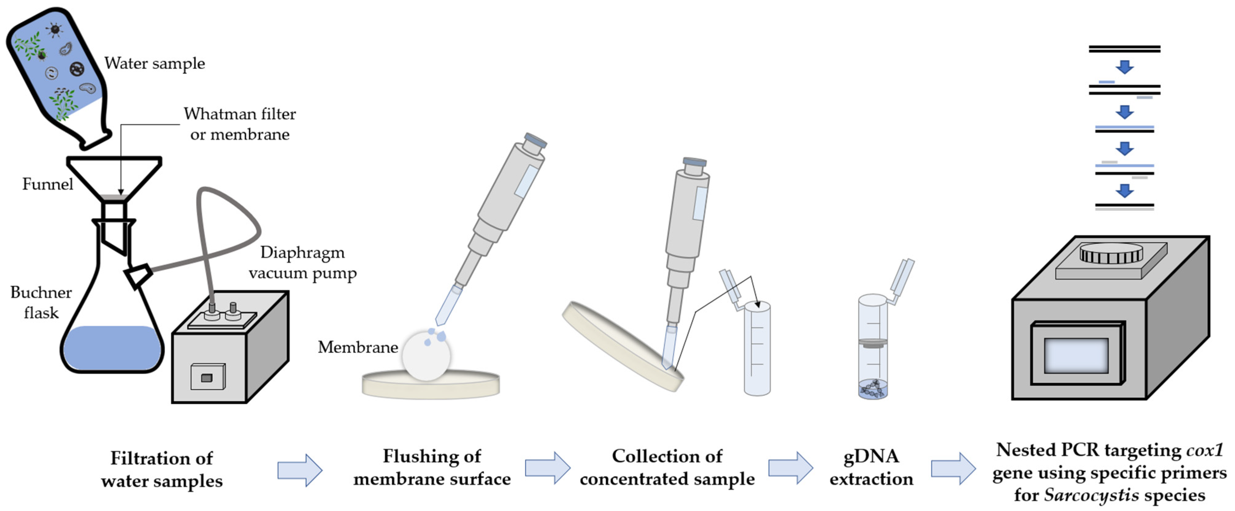
2.1. Sample Collection

Water samples (n = 114) were collected from different regions of Lithuania in 2020 and were analyzed for the presence of tested Sarcocystis spp. (Figure 1). Samples were collected during the summer, when the highest animal activity and migration rate is observed in Lithuania. Samples were gathered not only near pastures, but also near human bathing and remote wooded areas from various water sources such as ponds, canals, rivers, lakes, and lagoons. Water samples were collected up to one meter from the shore, gently stirring if taken from a standing water body. More than 20 L of water is usually collected for studies on parasitic protozoa in water [12,13]. Due to the relatively high Sarcocystis infection prevalence and intensity in farm animals from Lithuania [14], it was hypothesized that a smaller sample volume will be sufficient for the identification of these parasites in water samples. In this way, the conditions for transposition and laboratory examinations were facilitated.
To evaluate sample volume for testing, 3 different volumes of water samples (200 mL, 1 L, and 3 L) were collected in sterile containers and stored in portable coolers with ice batteries. Since water samples were collected in small-capacity vessels, sample collection and transportation were a simple and effortless process; we were able to collect up to 10–20 water samples per day. In the pilot study, 20 water samples of each volume (200 mL, 1 L, and 3 L) were collected and tested by nested PCR. For the first round of nested PCR, 20 samples were analyzed using three primer pairs SF1/SR8D, SF1/SR9, and SF1/SR12H, which were known to be suitable for amplification of the cox1 gene of six of the tested Sarcocystis species (Table 1) [11,15,16,17,18,19]. Meanwhile, species-specific primers were applied to the second round of PCR. Only two samples were positive when a volume of 200 mL was used, while five samples were positive in both larger volume cases. It was decided that a 1 L volume of sample is appropriate for further large-scale analysis. The decision was made taking into consideration technical aspects of material processing at our laboratory. When 3 L of water was treated, it was more difficult to remove impurities such as algae, sand, and plant parts.

2.2. Isolation of Sporocysts and Genomic DNA Extraction

To isolate Sarcocystis spp. sporocysts, three different techniques, including sucrose gradient (specific gravity = 1.15) [9], sedimentation, and filtration, were used. During the pilot study, 35 samples were analyzed by each of these methods to select the one for the further examination of all collected samples. All three methods were performed at room temperature. For the sucrose density gradient concentration, the sample was centrifuged in 250 mL bottles at 800× g for 10 min, then the supernatant was discarded, and the precipitate was suspended in 50 mL of 2 M sucrose solution. The prepared sample was centrifuged for 10 min at 800× g, and after centrifugation, the upper part of the supernatant (5–10 mL) was transferred to a new 50 mL tube. The sample was washed with 45 mL of sterile water and centrifuged for 10 min at 800× g. The supernatant was discarded, and the pellet was suspended in a mixture of 3 mL of HBSS antibiotics. The sample was stored at 4 °C. For sedimentation concentration, the sample was centrifuged in 250 mL bottles at 5000× g for 25 min, then the supernatant was discarded, and the pellet was suspended in sterile water and transferred to 1.5 mL tubes. The prepared sample was centrifuged for 10 min at 14,000× g. After centrifugation, the supernatant was discarded, and the pellet was stored at −20 °C. To concentrate sporocysts by filtration, water samples were filtrated using a 1 mm pore metal sieve, then Whatman™ Qualitative Filter Paper Grade 4, and finally MF-Millipore® 5 μm pore membranes. Membranes were washed in a sterile dish with 2 mL of distilled water. Concentrated samples were placed in sterile 2 mL tubes and stored at +4 °C.
The isolation of genomic DNA (gDNA) of Sarcocystis parasites from dry pellets (after sedimentation) was performed using the Genomic DNA Purification Kit (Thermo Fisher Scientific Baltics, Vilnius, Lithuania) and following the manufacturer’s protocol. The extraction of gDNA from water samples (after sucrose gradient and filtration) was accomplished using the GeneJET Genomic DNA Purification Kit (Thermo Fisher Scientific Baltics, Vilnius, Lithuania), according to the manufacturer’s recommendations. The maximum sample volume of 200 μL was selected according to the administration protocol. The resulting DNA samples were kept frozen at −20 °C until PCR was performed.
At the beginning of the study, the sucrose gradient method [9] and sediment collection from water were used for isolating sporocysts. PCR procedures were the same as described previously. After 35 water samples had been examined, it was observed that PCR fragments were more frequently obtained in samples using centrifugation at sucrose gradient (8/35, 22.9%) than sedimentation (3/35, 8.6%). Based on the results obtained, it was concluded that none of these methods is optimal for the isolation of sporocysts. It has been hypothesized that effective isolation of sporocysts may be hindered by a variety of impurities in water samples. As a result, it was decided to test the filtration of water samples. After analyzing 35 water samples by this method, the highest Sarcocystis spp. detection rate (14/35, 40.0%) was achieved. Consequently, this method was further applied to isolate sporocysts.

2.3. Identification of Sarcocystis Species Using PCR Procedures

Water samples were tested for the presence of eight Sarcocystis species (S. bovifelis, S. cruzi, S. hirsuta, S. arieticanis, S. tenella, S. capracanis, S. bertrami, and S. miescheriana) known to use agricultural species (cattle, sheep, goats, horses, and pigs) as intermediate hosts.
During the optimization of DNA fragment amplification, different conditions, and different types of conventional PCR, such as direct PCR, multiplex-nested PCR, semi-nested PCR, and nested PCR, were tested. Primers targeting the cox1 gene were designed using Primer3Plus [20]. Some primers were chosen to be specific to several Sarcocystis spp., while others were specific to one Sarcocystis species (Table 1). The specificity of the primers was tested with negative, no template, and positive controls, i.e., DNA extracted from sarcocysts of the selected Sarcocystis species. Positive DNA controls of Sarcocystis species analyzed were accumulated during previous studies, confirmed by sequencing, and stored at the Laboratory of Molecular Ecology, Vilnius, Lithuania. Both positive and negative DNA controls were used in all PCR reactions.
Each PCR was performed in a final volume of 25 μL containing 0.5 μM of each primer (Invitrogen by Thermo Fisher Scientific, Waltham, MA, USA), 2.5 μL of dNTP mix (0.2 mM of each), 2.5 μL of 10× DreamTaq buffer with 20 mM MgCl2, 1.25 U of DreamTaq polymerase (Thermo Fisher Scientific Baltics, Vilnius, Lithuania), and 5 μL of genomic DNA and nuclease-free water. For nested PCR, 5 μL of genomic DNA was used for the first round, and 5 μL of products of the first round were then subjected to the second round of amplification. Conditions for nested PCR were as follows: initial denaturation at 95 °C for 5 min, followed by 30 (1st PCR) or 40 (2nd PCR) cycles of 94 °C for 45 s, 56−65 °C (depending on primers—see Table 1 for annealing temps for each primer set) for 45 s and 72 °C for 30−60 s (depending on the length of fragments (see Table 1 for extension time for each primer set)), and a final extension with one cycle at 72 °C for 10 min. PCR products were visualized by 1% agarose gel electrophoresis.
The selected PCR products were purified using the GeneJET PCR Purification Kit (Thermo Fisher Scientific Baltics, Vilnius, Lithuania) according to the manufacturer’s recommendations. Purified PCR fragments were sequenced in both directions using forward and reverse primers. The sequencing was performed using the Big-Dye® Terminator v3.1 Cycle Sequencing Kit (Thermo Fisher Scientific, Vilnius, Lithuania) and the 3500 Genetic Analyzer (Applied Biosystems, Foster City, CA, USA). The obtained sequences were edited manually for ambiguously placed nucleotides and compared by BLAST analysis (http://blast.ncbi.nlm.nih.gov/, accessed on 22 March 2022).

2.4. Statistical Analysis

Statistical tests were performed using Quantitative Parasitology 3.0 software [21]. Sterne’s exact method was applied to compute the 95% confidence interval (CI) for the prevalence of identified Sarcocystis spp. [22]. Differences in the occurrence rates of the detected Sarcocystis species were evaluated using a Chi-squared test.

3. Results

3.1. Selection of PCR Conditions

We aimed to investigate conventional PCR methods for the identification of Sarcocystis parasites. As a result, several PCR approaches were analyzed in the present study, and the obtained results are summarized in Table 2. Briefly, to achieve the research objectives, direct, multiplex-nested, semi-nested, and nested PCRs were used. A series of primers (Table 1), theoretically amplifying single, several, or all eight species, were tested.
Finally, after sequencing obtained PCR fragments, we concluded that the application of nested PCR, using species-specific primers for both PCR rounds, is the best option. For each examined Sarcocystis spp., nested PCR with four different species-specific primers was used (Table 1). Having examined 35 samples, at least one species of Sarcocystis was detected in 34 samples (34/35, 97.1%). In addition, DNA fragments corresponding to the theoretical size were obtained using all species-specific primers. Therefore, this PCR type was used to analyze a total set of 114 samples.

3.2. Identification of Sarcocystis spp. from in Water Bodies

Overall, we obtained 274 PCR fragments. Of these, 73 theoretically representing 8 Sarcocystis spp. were sequenced. In total, we obtained four to twenty sequences for each species. The BLAST analysis showed that all of them represented tested species. To have an equal number of sequences, four sequences of each species were deposited in NCBI GenBank with Accession Numbers ON211315–ON211346 (Table 3). The obtained sequences were truncated by discarding the nucleotide sites, where DNA binds to the primers. Each set of four sequences generated by the same primer pair showed intraspecific genetic variability. Comparing the obtained sequences and those of the same Sarcocystis species available in GenBank, relatively large sequence similarity intervals were observed for S. arieticanis (92.3–99.4%) and S. miescheriana (93.9–99.5%). Low similarity values were established comparing the isolates identified in the current work with African isolates of S. arieticanis (92.3–93.5%; MH413047-48) and Asian isolates of S. miescheriana (93.9–95.3%; LC349977-80, MK867462-64). In general, interspecific, and intraspecific genetic variability values of tested Sarcocystis spp. did not overlap. Thus, the species-specific primers for nested PCR presented in Table 1 are suitable for the identification of eight Sarcocystis spp. (S. bovifelis, S. cruzi, S. hirsuta, S. arieticanis, S. tenella, S. capracanis, S. bertrami, and S. miescheriana).

3.3. Summary of Molecular-Based Sarcocystis Identification from Water Samples

During this work, suitable conditions for the identification of Sarcocystis spp. using farm animals as their intermediate hosts from water samples were selected (Figure 2). We used 1L of water sample for the detection of DNA of Sarcocystis spp. Impurities in water samples were removed by filtration using a 1 mm pore metal sieve, followed by Whatman™ Qualitative Filter Paper Grade 4. Sporocysts were collected on MF-Millipore® 5 μm pore membranes and washed with 2 mL of sterile distilled water on a glass plate. Concentrated samples were transferred to sterile 2 mL Eppendorf tubes. The prepared and concentrated water sample was used for gDNA extraction employing the GeneJET Genomic DNA Purification Kit. Different types of PCR were tested during optimization: direct, multiplex-nested, semi-nested, and nested. Based on the results obtained, it was concluded that the most appropriate method for the identification of analyzed Sarcocystis spp. in water bodies is nested PCR, using species-specific primers in both rounds of PCR. The comparison of DNA sequences obtained confirmed that the developed primer sets presented in Table 1 were appropriate for the identification of eight species of Sarcocystis from farm animals.

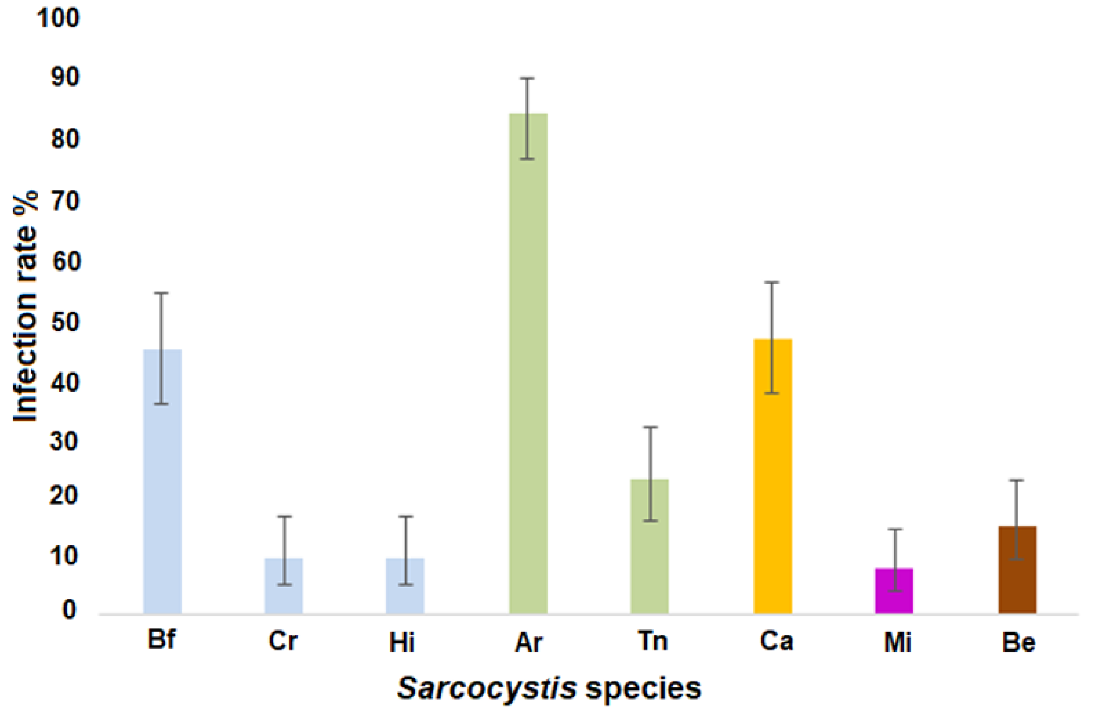
3.4. Sarcocystis spp. Occurrence Rates in the Analyzed Water Samples

Based on the applied molecular diagnostic technique, Sarcocystis spp. were detected in 111/114 analyzed water samples (97.4%; CI = 92.5–99.5). It should be noted that protozoans examined were confirmed in different water sources, ponds, canals, rivers, lakes, and lagoons. Comparing the analyzed species, the highest occurrence rate (96/114, 84.2%; CI = 76.4–90.0) was observed for S. arieticanis (Figure 3). The prevalence of S. arieticanis was significantly higher than that of S. capracanis2 = 35.81, p < 0.00001), which was the second most frequently detected species (53/114, 46.5%; CI = 37.2–55.7). The third most often detected species was S. bovifelis (51/114, 44.7%; CI = 35.6–54.0). The prevalence of the fourth most detected species, S. tenella (26/114, 22.8%; CI = 15.7–31.5), was significantly lower than that of S. capracanis2 = 14.12, p < 0.001) and that of S. bovifelis2 = 12.26, p < 0.001). The occurrence rate of the remaining species was lower than 20%, i.e., 14.9% (17/114; CI = 9.3–22.6) for S. bertrami, 9.6% (11/114; CI = 5.1–16.6) for both S. cruzi and S. hirsuta, and 7.9% (9/114; CI = 4.0–14.4) for S. miescheriana. The prevalence of S. tenella was higher than that of S. miescheriana2 = 9.75, p < 0.01), as well as S. cruzi and S. hirsuta2 = 12.26, p < 0.01). The overall prevalence of Sarcocystis spp. using sheep as an intermediate host (88.6%) was significantly higher (χ2 = 32.73, p < 0.00001) than that of Sarcocystis spp. using cattle as an intermediate host (54.4%). Whereas no significant difference (p > 0.05) between the occurrence of Sarcocystis spp. employing cattle as an intermediate host and those employing goats as an intermediate host has been noticed. The number of Sarcocystis species per sample varied from one to five. Most commonly three Sarcocystis species (37 samples, 32.5%) were identified, while two species were detected in 34 samples (29.8%), one species was found in 23 samples (20.2%), four species were confirmed in 14 samples (12.3%) and five species were established in three samples (2.6%).

4. Discussion

In the present study, we aimed to select suitable conditions for rapid identification of Sarcocystis spp. in water samples. For this purpose, a sufficient amount of water was selected to evaluate the presence of these protozoan parasites in water samples. One liter was shown to be the suitable volume, in terms of sensitivity and technical factors of our laboratory. The chosen amount of a sample is considerably smaller as compared to that in other studies on the identification of protozoan parasites in water bodies. For instance, 100 L of water or more is taken for the detection of Giardia spp. and Cryptosporidium spp. [12,13,23]. The choice of high volumes of water is based on a low concentration of protozoan oocysts in water [24]. However, large quantities of water samples not only complicate the work in laboratories, but also require special containers and transport. Low concentration of protozoan parasites, morphological similarities of (oo)cysts, and various impurities (e.g., plant parts, sand, sludge, and different microorganisms) complicate the identification of these parasites in water samples. Thus, the identification of protozoan parasites from water often consists of several steps. Typically, the samples are first concentrated by filtration or centrifugation [24,25]. Due to the different sizes of parasitic protozoan oocysts (for example, cysts of Cryptosporidium spp. are about 4 × 6 μm, while Sarcocystis spp. are about 10 × 15 μm), it is impossible to concentrate parasites belonging to different genera during filtration [1,26]. Filtration is usually followed by purification using immunomagnetic separation and immunofluorescence microscopy [26]. During immunomagnetic separation, (oo)cysts are isolated using antibody-coated particles, and impurities that impede isolation and further identification are removed. However, the use of immunological and fluorescent methods often makes it impossible to accurately identify many species belonging to protozoan parasites because of their low concentration in water and morphological similarities of (oo)cysts. In the present study, we suggest using filters with different porosities to remove impurities, collect sporocysts on MF-Millipore® 5 μm pore membranes, and wash with 2 mL of sterile distilled water.
Even though the Sarcocystis is one of the most abundant genera in the phylum Apicomplexa, consisting of more than 200 species, a reliable species identification system based on several nuclear and organelle markers has not been developed for these parasites yet [1]. It should be noted that the genus Sarcocystis is a heterogeneous group of organisms having complex evolutionary history depending on their host [27]. The largest DNA sequences database of Sarcocystis species has been accumulated for the 18S rDNA [28]. However, this gene appeared to have low resolution power in separating closely related Sarcocystis species whose intermediate hosts are ungulates, including cervids, cattle, water buffalo (Bubalus bubalis), and sheep [29,30,31]. Some Sarcocystis species using cattle, sheep, goat, horse, and pig/wild boar as their intermediate host were characterized within nuclear 18S rDNA, 28S rDNA, ITS1, and cox1 [15,30,32,33]. Cox1 was found to be most suitable for the identification of Sarcocystis species using farm animals as their intermediate hosts [29,32,33]. In the present study, the compared 248–554 bp long cox1 fragments appeared to be variable enough for the identification of the eight Sarcocystis species examined. Relatively high intraspecific variability values were observed when comparing the obtained sequences with those of African isolates of S. arieticanis [34] and Asian isolates of S. miescheriana [32]. In the current study, tested Sarcocystis species were mainly molecularly investigated in Europe and Asia [15,30,32,33]. Therefore, molecular characterization of Sarcocystis spp. from farm animals collected in different geographic regions and belonging to different animal breeds is necessary for accurate diagnosis of these species. In our research, the best results were obtained with the help of nested PCR and species-specific primers in both rounds of PCR. By contrast, in some cases, the use of primers specific for several Sarcocystis spp. resulted in the amplification of non-target Sarcocystis species (Table 2). Low specificity of primers can be explained by small interspecific differences in primer binding sites. In summary, the PCR results of the current study can be used to further develop Sarcocystis species identification techniques from environmental samples.
The present study is the first to identify S. bovifelis, S. cruzi, S. hirsuta, S. arieticanis, S. tenella, S. capracanis, S. bertrami, and S. miescheriana in environmental water samples. Furthermore, this is the first investigation devoted to diagnosing eight Sarcocystis species in water bodies. In the current work, using nested PCR and species-specific primers in both rounds, 274 DNA fragments were obtained; 73 of them were sequenced and showed the highest similarity with tested species. Since not all positive PCR fragments were sequenced, the obtained results should be considered with caution. Previously, up to three different Sarcocystis species were detected in water samples [7,8]. Using NGS, S. nesbitti, S. singaporensis, and S. zamani were identified, and based on the frequency of the presence, abundance, and identity of sequences, at least nine more individual but not yet formally described species were detected in the water samples analyzed in Malaysia [8]. The latter study showed that the diversity of Sarcocystis species has not been fully revealed yet.
Of the Lithuanian wild fauna, the European bison (Bison bonasus) can possibly act as an intermediate host of S. bovifelis, S. cruzi, and S. hirsuta [35,36], while S. arieticanis, S. capracanis, and S. tenella can form sarcocysts in muscles of European mouflon (Ovis aries musimon) [18]. However, populations of both wild animal species are limited to mainly central Lithuania and are composed of less than 500 individuals [37]. Thus, we assume that in Lithuania, the main intermediate hosts of the identified Sarcocystis species in the present work are farm animals.
According to the Department of Statistics of Lithuania, about 635,000 cattle and dairy cows, 152,000 sheep, 15,000goats, 13,000 horses, and 551,000 pigs were raised on Lithuanian farms in 2020 (https://osp.stat.gov.lt/lietuvos-aplinka-zeme-ukis-ir-energetika-2020/zemes-ukis/gyvulininkyste, accessed on 25 April 2022). The prevalence of Sarcocystis spp. and infection intensity in cattle, sheep, pigs, and horses were exhaustively examined in Lithuania [14]. The analysis of the esophagus, diaphragm, heart, neck, and jaw muscles showed high infection rates in sheep (100%) and cattle (88.0%), and moderate infection rates in horses (47.2%) and pigs (40.2%). The highest infection intensity was detected in sheep (Median, Md = 34.0), lower infection intensity was found in cattle (Md = 11.5) and pigs (Md = 11.50), and the lowest infection intensity in horses (Md = 2.0). Intense infection (>40 cysts per gram of muscle) was observed relatively often in sheep (44.9%), less frequently in cattle (19.1%), and occasionally in pigs (3.7%). The prevalence of different Sarcocystis species was performed only in the diaphragm muscles of cattle [11]. The analyzed diaphragm samples were most often infected with S. cruzi (96.1%), less frequently with S. bovifelis (71.6%) and S. hirsuta (30.4%), and the lowest with S. hominis (13.7%). The results obtained in this work are partially consistent with the abundance of tested farm animals and with the data of Sarcocystis spp. infection in muscles [11,14]. In the current study, we found a significantly higher infection prevalence of Sarcocystis spp. employing sheep as an intermediate host (88.6%) compared to cattle (54.4%). In addition, low infection rates were observed for S. bertrami (14.9%) and for S. miescheriana (7.9%), which are specific to horses and pigs, respectively. An attempt was made to identify Sarcocystis hominis in water samples by different conventional PCR methods using species-specific primers, but we were unable to detect them. The concentration of S. hominis in water bodies is considered to be too low to be identified by nested PCR. In previous morphological studies that we conducted [11], S. hominis was not detected by morphological methods in cattle carcasses. As a result, it was assumed that the concentration of S. hominis is very low, even in the cattle carcasses, so that few sporocysts enter the environment. However, the detection of S. cruzi in only 11 samples (9.6%) contradicts the data from the previous study. We assume that the primers developed for the identification of S. cruzi were not specific enough to some isolates of this species. In general, the results on infection rates of individual Sarcocystis species should be considered with caution. Hence, further development of molecular-based diagnosis of Sarcocystis spp. in water samples is required. In the future, to develop the identification of Sarcocystis spp. DNA from water samples, an attempt should be made to perform qPCR, which is a more sensitive and specific method. Furthermore, amplification of internal controls would improve the methodology for detecting Sarcocystis species in water samples.

5. Conclusions

This study presents the first identification of Sarcocystis species using farm animals as their intermediate hosts in water bodies. Sarcocystis spp. were detected in different water sources, such as ponds, canals, rivers, lakes, and lagoons. During the study, the volume of samples, the techniques for isolating sporocysts from water, and molecular detection procedures were investigated. Nested PCR using species-specific primers in both rounds of PCR gave the best results. The prevalence of Sarcocystis species analyzed significantly varied in the examined water samples. Based on the molecular method used, eight Sarcocystis species were identified, S. cruzi (9.6%), S. bovifelis (44.7%), S. hirsuta (9.6%), S. tenella (22.8%), S. arieticanis (84.2%), S. capracanis (46.5%), S. miescheriana (7.9%), and S. bertrami (14.9%). Thus, a convenient molecular method was developed for the identification of Sarcocystis parasites from farm animals in water samples. The suggested procedure can be used for epidemiological research and for tracking infection outbreaks.

Author Contributions

Conceptualization, Ž.S.-Ž. and P.P.; methodology, Ž.S.-Ž. and A.B.; software, P.P.; validation, P.P., D.B. and E.S.; formal analysis, P.P.; investigation, Ž.S.-Ž. and A.B.; resources D.B. and E.S.; data curation, P.P.; writing—original draft preparation, A.B. and P.P.; writing—review and editing, P.P., D.B., Ž.S.-Ž. and E.S.; visualization, A.B.; supervision, D.B. and E.S.; project administration, D.B.; funding acquisition, D.B. All authors have read and agreed to the published version of the manuscript.

Funding

This work was funded by the Research Council of Lithuania (grant number S-MIP-20-24).

Institutional Review Board Statement

Not applicable.

Informed Consent Statement

Not applicable.

Data Availability Statement

Data supporting the conclusions of this article are included in the article. The sequences generated in the present study were submitted to the GenBank database under Accession Numbers ON211315–ON211346.

Acknowledgments

This study was supported by the Open Access research infrastructure of the Nature Research Centre under the Lithuanian open access network initiative. The authors are grateful to V. Pabrinkis for his great support in water sampling.

Conflicts of Interest

The authors declare that they have no conflict of interest.

References

  1. Dubey, J.P.; Calero-Bernal, R.; Rosenthal, B.M.; Speer, C.A.; Fayer, R. Sarcocystosis of Animals and Humans, 2nd ed.; CRC Press: Boca Raton, FL, USA, 2016. [Google Scholar]
  2. Florin-Christensen, M.; Schnittger, L. Sarcocystis. In Parasitic Protozoa of Farm Animals and Pets; Florin-Christensen, M., Schnittger, L., Eds.; Springer Science and Business Media LLC: Berlin, Germany, 2018; pp. 103–124. [Google Scholar]
  3. Fayer, R. Sarcocystis spp. in Human Infections. Clin. Microbiol. Rev. 2004, 17, 894–902. [Google Scholar]
  4. Fayer, R.; Esposito, D.H.; Dubey, J.P. Human infections with Sarcocystis species. Clin. Microbiol. Rev. 2015, 28, 295–311. [Google Scholar] [CrossRef] [Green Version]
  5. Basso, W.; Alvarez Rojas, C.A.; Buob, D.; Ruetten, M.; Deplazes, P. Sarcocystis infection in red deer (Cervus elaphus) with eosinophilic myositis/fasciitis in Switzerland and involvement of red foxes (Vulpes vulpes) and hunting dogs in the transmission. Int. J. Parasitol. Parasites Wildl. 2020, 13, 130–141. [Google Scholar] [CrossRef]
  6. Juozaitytė-Ngugu, E.; Švažas, S.; Šneideris, D.; Rudaitytė-Lukošienė, E.; Butkauskas, D.; Prakas, P. The role of birds of the family corvidae in transmitting Sarcocystis protozoan parasites. Animals 2021, 11, 3258. [Google Scholar] [CrossRef]
  7. Shahari, S.; Tengku-Idris, T.I.N.; Fong, M.Y.; Lau, Y.L. Molecular evidence of Sarcocystis nesbitti in water samples of Tioman Island, Malaysia. Parasites Vectors 2016, 9, 598. [Google Scholar] [CrossRef] [Green Version]
  8. Lee, F.C.H. Finding Sarcocystis spp. on the Tioman island: 28S rRNA gene next-generation sequencing reveals nine new Sarcocystis species. J. Water Health 2019, 17, 416–427. [Google Scholar] [CrossRef] [Green Version]
  9. Verma, S.K.; Lindsay, D.S.; Grigg, M.E.; Dubey, J.P. Isolation, culture and cryopreservation of Sarcocystis species. Curr. Protoc. Microbiol. 2017, 45, 11–127. [Google Scholar] [CrossRef] [Green Version]
  10. Ma, C.L.; Ye, Y.L.; Wen, T.; Huang, Z.M.; Pan, J.; Hu, J.J.; Tao, J.P.; Song, J.L. Prevalence and morphological and molecular characteristics of Sarcocystis bertrami in horses in China. Parasite 2020, 27, 1. [Google Scholar] [CrossRef]
  11. Prakas, P.; Strazdaitė-Žielienė, Ž.; Januškevičius, V.; Chiesa, F.; Baranauskaitė, A.; Rudaitytė-Lukošienė, E.; Servienė, E.; Petkevičius, S.; Butkauskas, D. Molecular identification of four Sarcocystis species in cattle from Lithuania, including S. hominis, and development of a rapid molecular detection method. Parasites Vectors 2020, 13, 610. [Google Scholar] [CrossRef]
  12. Cheun, H.I.; Kim, C.H.; Cho, S.H.; Ma, D.W.; Goo, B.L.; Na, M.S.; Youn, S.K.; Lee, W.J. T The first outbreak of Giardiasis with drinking water in Korea. Osong Public Health Res. Perspect. 2013, 4, 89–92. [Google Scholar] [CrossRef] [Green Version]
  13. Hassan, E.M.; Örmeci, B.; DeRosa, M.C.; Dixon, B.R.; Sattar, S.A.; Iqbal, A. A review of Cryptosporidium spp. and their detection in water. Water Sci. Technol. 2021, 83, 1–25. [Google Scholar] [CrossRef]
  14. Januskevicius, V.; Januskeviciene, G.; Prakas, P.; Butkauskas, D.; Petkevicius, S. Prevalence and intensity of Sarcocystis spp. infection in animals slaughtered for food in Lithuania. Vet. Med. Czech 2019, 64, 149–157. [Google Scholar] [CrossRef]
  15. Gjerde, B. Phylogenetic relationships among Sarcocystis species in cervids, cattle and sheep inferred from the mitochondrial cytochrome c oxidase subunit I gene. Int. J. Parasitol. 2013, 43, 579–591. [Google Scholar] [CrossRef]
  16. Gjerde, B. Sarcocystis species in red deer revisited: With a re-description of two known species as Sarcocystis elongata n. sp. and Sarcocystis truncata n. sp. based on mitochondrial cox1 sequences. Parasitology 2014, 141, 441–452. [Google Scholar] [CrossRef]
  17. Prakas, P.; Rudaitytė, E.; Butkauskas, D.; Kutkienė, L. Sarcocystis entzerothi n. sp. from the European roe deer (Capreolus capreolus). Parasitol. Res. 2017, 116, 271–279. [Google Scholar] [CrossRef]
  18. Prakas, P.; Rehbein, S.; Rudaitytė-Lukošienė, E.; Butkauskas, D. Molecular identification of Sarcocystis species in diaphragm muscle tissue of European mouflon (Ovis Gmelini Musimon) from Austria. Parasitol. Res. 2021, 120, 2695–2702. [Google Scholar] [CrossRef]
  19. Prakas, P.; Balčiauskas, L.; Juozaitytė-Ngugu, E.; Butkauskas, D. The role of mustelids in the transmission of Sarcocystis spp. using cattle as intermediate hosts. Animals 2021, 11, 822. [Google Scholar] [CrossRef]
  20. Untergasser, A.; Nijveen, H.; Rao, X.; Bisseling, T.; Geurts, R.; Leunissen, J.A.M. Primer3Plus, an enhanced web interface to Primer3. Nucleic Acids Res. 2007, 35, W71–W74. [Google Scholar] [CrossRef] [Green Version]
  21. Rózsa, L.; Reiczigel, J.; Majoros, G. Quantifying parasites in samples of hosts. J. Parasitol. 2000, 86, 228–232. [Google Scholar] [CrossRef]
  22. Reiczigel, J. Confidence intervals for the binomial parameter: Some new considerations. Stat. Med. 2003, 22, 611–621. [Google Scholar] [CrossRef]
  23. Omarova, A.; Tussupova, K.; Berndtsson, R.; Kalishev, M.; Sharapatova, K. Protozoan parasites in drinking water: A system approach for improved water, sanitation and hygiene in developing countries. Int. J. Environ. Res. Public Health 2018, 15, 495. [Google Scholar] [CrossRef] [Green Version]
  24. Moreno, Y.; Moreno-Mesonero, L.; Amorós, I.; Pérez, R.; Morillo, J.A.; Alonso, J.L. Multiple identification of most important waterborne protozoa in surface water used for irrigation purposes by 18S rRNA amplicon-based metagenomics. Int. J. Hyg. Environ. Health 2018, 221, 102–111. [Google Scholar] [CrossRef]
  25. Prystajecky, N.; Huck, P.M.; Schreier, H.; Isaac-Renton, J.L. Assessment of Giardia and Cryptosporidium spp. as a microbial source tracking tool for surface water: Application in a mixed-use watershed. Appl. Environ. Microbiol. 2014, 80, 2328–2336. [Google Scholar] [CrossRef] [Green Version]
  26. Schets, F.M.; Engels, G.B.; During, M.; De Roda Husman, A.M. Detection of infectious Cryptosporidium oocysts by cell culture immunofluorescence assay: Applicability to environmental samples. Appl. Environ. Microbiol. 2005, 71, 6793–6798. [Google Scholar] [CrossRef] [Green Version]
  27. Doležel, D.; Koudela, B.; Jirků, M.; Hypša, V.; Oborník, M.; Votýpka, J.; Modrý, D.; Šlapeta, J.R.; Lukeš, J. Phylogenetic analysis of Sarcoystis spp. of mammals and reptiles supports the coevolution of Sarcocystis spp. with their final hosts. Int. J. Parasitol. 1999, 29, 795–798. [Google Scholar] [CrossRef]
  28. Lee, F.C.H.; Muthu, V. From 18S to 28S rRNA gene: An improved targeted Sarcocystidae PCR amplification, species identification with long DNA sequences. Am. J. Trop. Med. Hyg. 2021, 104, 1388–1393. [Google Scholar] [CrossRef]
  29. Gjerde, B. Molecular characterisation of Sarcocystis bovifelis, Sarcocystis bovini n. sp., Sarcocystis hirsuta and Sarcocystis cruzi from cattle (Bos taurus) and Sarcocystis sinensis from water buffaloes (Bubalus bubalis). Parasitol. Res. 2016, 115, 1473–1492. [Google Scholar] [CrossRef]
  30. Gjerde, B.; de la Fuente, C.; Alunda, J.M.; Luzón, M. Molecular characterisation of five Sarcocystis species in domestic sheep (Ovis aries) from Spain. Parasitol. Res. 2020, 119, 2145–2150. [Google Scholar] [CrossRef]
  31. Prakas, P.; Butkauskas, D.; Rudaitytė, E.; Kutkienė, L.; Sruoga, A.; Pūraitė, I. Morphological and molecular characterization of Sarcocystis taeniata and Sarcocystis pilosa n. sp. from the sika deer (Cervus nippon) in Lithuania. Parasitol. Res. 2016, 115, 3021–3032. [Google Scholar] [CrossRef]
  32. Huang, Z.; Ye, Y.; Zhang, H.; Deng, S.; Tao, J.; Hu, J.; Yang, Y. Morphological and molecular characterizations of Sarcocystis miescheriana and Sarcocystis suihominis in domestic pigs (Sus scrofa) in China. Parasitol. Res. 2019, 118, 3491–3496. [Google Scholar] [CrossRef]
  33. Metwally, D.M.; Al-Damigh, M.A.; Al-Turaiki, I.M.; El-Khadragy, M.F. Molecular characterization of Sarcocystis species isolated from sheep and goats in Riyadh, Saudi Arabia. Animals 2019, 9, 256. [Google Scholar] [CrossRef] [Green Version]
  34. El-Morsey, A.; Abdo, W.; Sultan, K.; Elhawary, N.M.; AbouZaid, A.A. Ultrastructural and molecular identification of the sarcocysts of Sarcocystis tenella and Sarcocystis arieticanis infecting domestic sheep (Ovis aries) from Egypt. Acta Parasitol. 2019, 64, 501–513. [Google Scholar] [CrossRef]
  35. Odening, K.; Wesemeier, H.H.; Walter, G.; Bockhardt, I. The wisent (Bison Bonasus, Bovidae) as an intermediate host of three Sarcocystis species (Apicomplexa: Sarcocystidae) of cattle. Folia Parasitol. 1994, 41, 115–121. [Google Scholar]
  36. Cabaj, W.; Grzelak, S.; Moskwa, B.; Bień-Kalinowska, J. Sarcocystis cruzi infection in free-living European bison (Bison bonasus bonasus L.) from the Białowieża forest, Poland—A molecular analysis based on the cox1 gene. Int. J. Parasitol. Parasites Wildl. 2021, 16, 59–63. [Google Scholar] [CrossRef]
  37. Balčiauskas, L.; Trakimas, G.; Juškaitis, R.; Ulevičius, A.; Balčiauskienė, L. Atlas of Lithuanian Mammals, Amphibians and Reptiles, 2nd ed.; Akstis: Vilnius, Lithuania, 1999. [Google Scholar]
Figure 1. Water sampling sites in Lithuania in 2020.
Vetsci 09 00412 g001
Figure 2. Scheme of molecular identification of Sarcocystis parasites from water samples.
Vetsci 09 00412 g002
Figure 3. Prevalence of examined Sarcocystis species in Lithuanian water bodies in 2020 based on species-specific nested PCR. Bf—S. bovifelis, Cr—S. cruzi, Hi—S. hirsuta, Ar—S. arieticanis, Tn—S. tenella, Ca—S. capracanis, Mi—S. miescheriana, Be—S. bertrami.
Vetsci 09 00412 g003
Table 1. Oligonucleotide primers used in this study for PCR procedures.
SpeciesPrimersTa,
°C
Extension Time, sProduct
Size (bp)
Reference
NameOrientationSequence (5′–3′)
Primers for Direct and Nested PCR
Sarcocystis spp.SF1ForwardATGGCGTACAACAATCATAAAGAA [15]
SR8DReverseCATTGCCCATDACTACGCC55701072[15]
SR9ReverseATATCCATACCRCCATTGCCCAT60701085[16]
SR12HReverseAAATACCTTGGTGCCCGTAG5660952[17]
S. bovifelisGaBfEFForwardATCAACTTCCTAGGTACAGCGGTATT5645523[11]
GaBfERReverseCCACATCATTGGTGCTTAGTCTAGTA[11]
S. cruziGaCrEFForwardGCTATGTATCTACTTACGGCAGGTATC5645608[11]
GaCrERReverseGAATATAATGGCCCAGGTAAATAATG[11]
S. hirsutaGaHiEFForwardGTTGTGCGGTATGAATTATCAACCT5645513[11]
GaHiERReverseGGTAAGAACTGGAATGGTTAATATCAG[11]
S. arieticanisGsSariFForwardTTCTTGGTATGGCTATTCTTGGACT6545586[18]
GsSariRReverseGATATGTCAATCCAGAGATCGGTAG[18]
S. tenellaGsStenFForwardTACTCGGAGCGGTGAACTTCTTA6335451[18]
GsStenRReverseATAGTCACGGCAGAGAAGTAGGAC[18]
S. capracanisGsScapFForwardAGCGGTAAACTTCCTGGGTACT6335467[18]
GsScapRReverseGCCTATCCAGTTGAATATCTTGGT[18]
Primers for multiplex-nested PCR
1st round
Sarcocystis spp.SF1ForwardATGGCGTACAACAATCATAAAGAA [15]
S. arieticanis,
S. cruzi
SsunR2ReverseGTGCCTCCCAGGCTGAAYAG56701055[19]
S. bertrami,
S. tenella,
S. capracanis
SsunR1ReverseGTACCGCCCAGGCTGAAYAG56701055[18]
S. bovifelis,
S. hirsuta
SkatRReverseCAGGCTGAACAGHABTACGA56701042[19]
S. miescherianaSmieFForwardACGCTGTATGCACCACTGAG5645658Present study
SmieRReverseCTGAACAGCGCTACAAATGC
2nd round
S. bertramiGsSberFForwardGTATGAACTGTCAACGGATGGAGTA6535482Present study
GsSberRReverseTCAACATTAGCGAGGTAAATACTATC
S. miescherianaGsSmieFForwardGTTCCTCGGTATTAGCAGCGTACT6535509Present study
GsSmieRReverseAGTTAAATATTTTAGTGCCCGTTGGA
Primers for semi-nested PCR
S. bovifelisVoboFForwardGATCGGTATTACTGTTGCACTCATT5845701Present study
VoboRReverseAGGCCACATCATTGGTGCTTA
GaBfEFForwardATCAACTTCCTAGGTACAGCGGTATT5735521[11]
S. tenellaVoteF1ForwardAGCGGTGAACTTCTTAGGAACC5935526Present study
VoteRReverseAATAATCCGCTGTTAACGTATGCPresent study
VoteF2ForwardCATTGTAATGCTCCTCGACGATA5930401Present study
S. capracanisVocaFForwardGTAAACTTCCTGGGTACTGTGCTGT6035526Present study
VocaR1ReverseCCAGTAATCCGCTGTCAAGATACPresent study
VocaR2ReverseAGTACCCATCACGGTGCCTATC6335500Present study
Species-specific primers for nested PCR
S. bovifelisV2bo1ForwardAACTTCCTAGGTACAGCGGTATTCG6040556Present study
V2bo2ReverseTGAACAGCAGTACGAAGGCAACPresent study
V2bo3ForwardATATTTACCGGTGCCGTACTTATGTT6030410Present study
V2bo4ReverseGCCACATCATTGGTGCTTAGTCTPresent study
S. cruziV2cr1ForwardTACAATGTGCTGTTTACGCTCCA5750776Present study
V2cr2ReverseGCAATCATGATAGTTACGGCAGAPresent study
V2cr3ForwardACCATCCTGTTCTGTGGTGCTATG6530298Present study
V2cr4ReverseAAACTACTTTACTGCCTACGGTACTCPresent study
S. hirsutaV2hi5ForwardTATGTTGGTTCTGCCGAAGTCAT6045686Present study
V2hi6ReverseGGTATGGCAATCATTATGGTTACAGPresent study
V2hi7ForwardGCACCGTAATATTTCAGGGATGT6030299Present study
V2hi8ReverseAACCTGCTTGCCGGAGTAAGTAPresent study
S. arieticanisV2arie1ForwardCTCTTTGCCGTAGATTCGCTAGTTA6355884Present study
V2arie2ReverseCAAAGATCGGTAGATATCCAATGCPresent study
V2arie3ForwardTAGTTCTTGGCCTGGCTATTCTT5925371Present study
V2arie4ReverseCTGACCTCCAAAAACTGGCTTACPresent study
S. tenellaV2te1ForwardGAGCGGTGAACTTCTTAGGAACC6040537Present study
V2te2ReverseCCCAATAATCCGCTGTTAACGTAPresent study
V2te3bForwardATTGTAATGCTCCTCGACGATATG5730314Present study
V2te4ReverseATAGTCACGGCAGAGAAGTAGGACPresent study
S. capracanisVocaFForwardGTAAACTTCCTGGGTACTGTGCTGT6040531Present study
VocaR1ReverseCCAGTAATCCGCTGTCAAGATACPresent study
V2cap3ForwardATACCGATCTTTACGGGAGCAGTA6330330Present study
V2cap4ReverseGGTCACCGCAGAGAAGTACGATPresent study
S. bertramiV2ber1ForwardGTATGAACTGTCAACGGATGGAGTA5860883Present study
V2ber2ReverseAGAAGCCATGTTCGTGACTACCPresent study
V2ber3ForwardGTACTACCTCCTTCCAGTCGGTTC5740600Present study
V2ber4ReverseCGGGTATCCACTTCAAGTCCAGPresent study
S. miescherianaV2mie1ForwardTGCTGCGGTATGAACTATCTACCT6160922Present study
V2mie2ReverseGCCCAGAGATCCAAATCCAGPresent study
V2mie3ForwardCTTGGTTCAACGTTACTCCTCCA6130474Present study
V2mie4ReverseCTTCGATCCAGCTGAACTAAAGCPresent study
SF1 is specific to genus Sarcocystis. SR8D, SR9, and SR12H are specific to some of the tested species or to some isolates of analyzed species. The remaining primer pairs were designed to be specific to certain Sarcocystis species. R = A or G, D = A or G or T2.4. PCR product purification, sequencing, and sequence analysis.
Table 2. Comparison of the used PCR approaches.
MethodSporocysts
Isolation
gDNA
Extraction
Number
of Water
Samples (n)
Positive
Samples *
CasesSpecies
Detected ****
Positive **False
Positive ***
Direct PCR
(species-specific
primers)
Sucrose
gradient
“GeneJET Genomic DNA
Purification Kit”
200 (0.0%)000
Nested PCR
(species-specific
primers only in
2nd round of PCR)
Sucrose
gradient
“GeneJET Genomic DNA
Purification Kit”
358 (22.9%)1222
Sedimentation“Genomic DNA
Purification Kit”
353 (8.6%)412
Filtration“GeneJET Genomic DNA
Purification Kit”
3514 (40.0%)1513
Multiplex-nested PCR
(species-specific
primers only in 2nd round of PCR)
Filtration“GeneJET Genomic DNA
Purification Kit”
357 (20.0%)1032
Semi-nested PCR
(species-specific
primers)
Filtration“GeneJET Genomic DNA
Purification Kit”
350 (0.0%)000
Nested PCR
(species-specific
primers in both rounds)
Filtration“GeneJET Genomic DNA
Purification Kit”
3534 (97.1%)8408
* The number of samples in which at least one of targeted Sarcocystis species was identified. ** The number of positive PCR fragments. *** Based on sequencing the number of incorrectly identified species. **** Based on sequencing the number of targeted species identified in samples.
Table 3. Genetic identification of Sarcocystis spp. detected in watershed samples using nested PCR.
SpeciesGenBank
Acc. No.
(Length, bp)
Sequence Similarity, %
Comparing Obtained Sequences with Each OtherComparing Obtained Sequences and Those of the Same Species Available in GenBankComparing Obtained
Sequences with Those of Most Closely Related Species
S. bovifelisON211315-18 (361)99.7–10099.5–100S. bovini 93.1–94.5
S. cruziON211319-22 (248)98.8–10098.0–99.6S. alceslatrans 85.1–86.8
S. hirsutaON211323-26 (254)99.6–10098.4–100S. buffalonis 92.8–93.2
S. arieticanisON211327-30 (325)99.7–10092.3–99.4S. hircicanis 86.2–87.4
S. tenellaON211331-34 (263)99.2–10096.2–100S. capracanis 91.3–93.2
S. capracanisON211335-38 (284)99.7–10096.8–99.7S. tenella 90.4–92.9
S. bertramiON211339-42 (554)99.1–99.897.5–99.8S. bovifelis 77.0–78.4
S. miescherianaON211343-46 (428)99.5–10093.9–99.5S. rangiferi 79.6–80.2
Publisher’s Note: MDPI stays neutral with regard to jurisdictional claims in published maps and institutional affiliations.

