Simple Summary
Glaesserella parasuis, one of the opportunistic pathogens causing Glässer’s disease in piglets, has become a significant concern for pig farmers. Vaccination has been shown to be effective in preventing Glaesserella parasuis infection by inducing the protective immune response. Notably, a humoral immune response plays an important role in protection of Glaesserella parasuis infection. The mechanism of protection by antibodies has been shown to be associated with antibody-opsonized phagocytosis, which facilitates uptake of Glaesserella parasuis by phagocytes such as macrophages. Outer membrane proteins of Glaesserella parasuis, as the promising candidates, are often chosen to develop subunit vaccines. HPS_06257 is one of the outer membrane proteins that has been shown to confer protection against Glaesserella parasuis infection. However, little is known about the role of HPS_06257 in the protective immune response. We demonstrate that antibody-dependent phagocytosis is involved in the protective effects of HPS_06257. Our findings extend our understanding of how antibody-dependent phagocytosis may contribute to the immune protection afforded by other outer membrane proteins. Thus, our study provides insight into the protective antigens of Glaesserella parasuis and useful information for the development of novel vaccines to prevent Glaesserella parasuis infection.
Abstract
HPS_06257 has been identified as an important protective antigen against Glaesserella parasuis infection. However, little is known about the role of HPS_06257 in the protective immune response. A whole-genome data analysis showed that among 18 isolates of Glaesserella parasuis, 11 were positive for the HPS_06257 gene, suggesting that not every strain contains this gene. We used PCR to investigate the presence of the HPS_06257 gene among 13 reference strains and demonstrated that 5 strains contained the gene. A polyclonal antibody against HPS_06257 was generated with a recombinant protein to study the expression of HPS_06257 in those 13 strains. Consistent with the PCR data, five strains expressed HPS_06257, whereas eight strains were HPS_06257 null. We also compared the protective effects of HPS_06257 against an HPS_06257-expressing strain (HPS5) and an HPS_06257-null strain (HPS11). Immunization with HPS_06257 only protected against HPS5 and not HPS11. Moreover, phagocytosis of antibody-opsonized bacteria demonstrates that the antibody against HPS_06257 increased the phagocytosis of the HPS5 strain by macrophages but not the phagocytosis of the HPS11 strain, suggesting that antibody-dependent phagocytosis is responsible for the protective role exerted by HPS_06257 in the immune response to HPS5. Our data also show that the antibody against HPS_06257 increased the phagocytosis of the other HPS_06257-expressing strains by macrophages but not that of HPS_06257-null strains. In summary, our findings demonstrate that antibody-dependent phagocytosis contributes to the protective immune response induced by immunization with HPS_06257 against HPS_06257-expressing strains.
1. Introduction
Glaesserella parasuis (G. parasuis), formerly known as Haemophilus parasuis (HPS), is one of the opportunistic pathogens causing Glässer’s disease in piglets, which is characterized by fibrinous polyserositis, polyarthritis, and meningitis []. With increasing reports of this disease, G. parasuis has become a significant concern for pig farmers. Vaccination has been shown to be effective in preventing G. parasuis infection by inducing the protective immune response. Notably, a humoral immune response plays an important role in protection of G. parasuis infection. For example, passive immunization of pigs with anti-serum against G. parasuis protected against lethal challenge []. The mechanism of protection by antibodies has been shown to be associated with antibody-opsonized phagocytosis, which facilitates uptake of G. parasuis by phagocytes such as macrophages [].
Regarding vaccination, as a promising approach to prevention and control of Glässer’s disease, different kinds of vaccines have been developed. While inactive G. parasuis vaccines have some limitations, including the different levels of cross-protection between G. parasuis serovars, a short time of protection, and so on, they are widely used throughout the world. In order to overcome the limitations of inactivated vaccines, several other methods have been used to develop novel vaccines. Among them, subunit vaccines have been extensively investigated for this purpose []. Several different kinds of proteins have been used to develop subunit vaccines [,,,,,]. Notably, outer membrane proteins are often chosen and confer effective protection against G. parasuis infection, mainly of homologous strains [,,].
HPS_06257 is one of the outer membrane proteins that has been shown to confer protection against G. parasuis infection []. It has a molecular weight of 28 kDa and contains a lipoprotein, GNA1870, domain at its C-terminus. Although data have shown that anti-HPS_06257 serum may protect against G. parasuis infection, the specific mechanism of this protective effect remains unclear. Antibody-dependent phagocytosis is an important mechanism underlying the protective roles of some immune antigens []. However, little is known about the role of antibody-dependent phagocytosis in HPS_06257-mediated protection against G. parasuis infection.
Previous G. parasuis genome analyses have identified similarities and divergence in the genomes of different G. parasuis strains [,,]. Whether HPS_06257 is present in all strains G. parasuis remains unknown. In this study, we identified the presence and absence of HPS_06257 in different G. parasuis stains and clarified the role of antibody-dependent phagocytosis in the protection it affords against HPS_06257-expressing strains.
2. Materials and Methods
2.1. Glaesserella parasuis
Glaesserella parasuis was recovered from a stock frozen in 20% glycerol and stored at −80 °C, and was grown on tryptic soy agar (TSA; Difco, Detroit, MI, USA) supplemented with 20 μg/mL nicotinamide adenine dinucleotide (NAD; Sigma, St. Louis, MO, USA) and 5% fetal calf serum (FCS; Thermo Fisher Scientific, Shanghai, China) overnight at 37 °C. A colony was picked from the TSA plate and grown in tryptic soy broth supplemented with 20 μg/mL NAD and 5% FCS for about 12 h at 37 °C with shaking.
2.2. DNA Isolation and PCR
The 13 G. parasuis reference strains for this study were obtained from the China Institute of Veterinary Drug Control []. They were representative of 13 different serovars: serovar 1 (as Glaesserella parasuis is formerly known as Haemophilus parasuis, in this study, Glaesserella parasuis serovar 1, simply named HPS1), serovar 2 (HPS2), serovar 3 (HPS3), serovar 4 (HPS4), serovar 5 (HPS5), serovar 6 (HPS6), serovar 8 (HPS8), serovar 9 (HPS9), serovar 10 (HPS10), serovar 11 (HPS11), serovar 13 (HPS13), serovar 14 (HPS14), and serovar 15 (HPS15). All of these strains were grown as described above and harvested for genomic DNA extraction with the TIANamp Bacteria DNA Kit (Tiangen Biotech Co., Ltd., Beijing, China). All 13 serovars were confirmed with a previously reported PCR method [].
2.3. Preparation of Polyclonal Antibody against HPS_06257
Primers (shown in Table S1) based on the complete genome sequence of G. parasuis isolate SH0104 (GenBank accession number NZ_CP024412) were designed to clone the HPS_06257 gene. The genomic DNA of G. parasuis isolate SH0104 was prepared as described above, and PCR was performed to amplify the gene. The amplification product (about 700 bp) was inserted into the BamHI and XhoI sites of the pET28a-vector (Novagen, Darmstadt, Germany) to generate the construct pET-HPS_06257. The recombinant HPS_06257 protein (histidine [His]–HPS_06257) was expressed in Escherichia coli BL21 cells and purified with the His·Bind Purification Kit (Novagen). A polyclonal antibody against HPS_06257 was then prepared as described previously []. Briefly, 8-week-old female BALB/c mice were obtained from JieSiJie Laboratory Animal Co., Ltd. (Shanghai, China) and immunized with His–HPS_06257 combined with complete or incomplete Freund’s adjuvant. After the seventh immunization, sera were collected from the immunized mice. All animal experiments were approved by the Institutional Animal Care and Use Committee of Shanghai Veterinary Research Institute, Chinese Academy of Agricultural Science, Shanghai, China (IACUC no. SHVRI-PO-2016060501) and were performed in compliance with the Guidelines on the Human Treatment of Laboratory Animals (Ministry of Science and Technology of the People’s Republic of China, policy no. 2006398).
2.4. Western Blotting
The specificity of the polyclonal antibody against HPS_06257 was examined by Western blotting, as described previously []. Briefly, recombinant His–HPS_06257 was transferred to membrane with the control protein, His–SRA (Xiang et al., 2020), and the membrane was blocked with 5% skim milk for 1 h at room temperature. The membrane was incubated overnight at 4 °C with the anti-HPS_06257 antibody (1:1000) or anti-His antibody (1:1000; Sigma), and then incubated for 1 h at room temperature with a horseradish peroxidase (HRP)–conjugated secondary antibody. The membrane was then treated with HRP substrate, according to the instructions for Enhanced Chemiluminescence (ECL) Reagent (Pierce) and exposed to x-ray film. Images were captured with the Gel Doc™ EZ System (Bio-Rad Laboratories, Hercules, CA, USA). A whole-cell lysate of each strain of G. parasuis was prepared, and the expression of HPS_06257 in the different strains was determined with the methods described above.
2.5. Immunization of Mice and Challenge with G. parasuis
Eight-week-old female BALB/c mice were immunized by subcutaneous injection with His–HPS_06257 combined with complete or incomplete Freund’s adjuvant (His–HPS_06257, 60 μg/mouse). Control mice were injected subcutaneously with phosphate-buffered saline (PBS) or adjuvant alone. Fourteen days after the second immunization, the mice were challenged with 2 × 10⁹ colony-forming units (CFU) of HPS5 or HPS11 by intraperitoneal injection. After the challenge, the mice were monitored twice daily and were euthanized after a bodyweight loss of >10%.
2.6. Antibody-Mediated Phagocytosis
G. parasuis was grown as described above and inactivated with heat. The heat-inactivated bacteria were surface labeled with fluorescein isothiocyanate (FITC), as described previously []. The FITC-labeled bacteria (108 cells) were opsonized with 10 μL of serum for 45 min at 37 °C, with occasional agitation. The opsonized bacteria were incubated at a multiplicity of infection of 300 bacteria per RAW264.7 cell for 2 h at 37 °C, as previously described []. Trypan blue was then added to quench the extracellular fluorescence. The fluorescence was measured with a microplate fluorometer (at 485 nm excitation/535 nm emission, M3; Molecular Devices, Sunnyvale, CA, USA). Among the PBS-, adjuvant-, and HPS_06257-treated groups of mice, the percentage phagocytosis was calculated by comparing the fluorescence of the adjuvant or HPS_06257 group with the fluorescence of the PBS group (control). Normal mouse serum was also used as a control for the calculation of anti-HPS_06257-antibody-mediated phagocytosis.
2.7. Statistical Analysis
All data were analyzed with the GraphPad Prism software version 5.01 (GraphPad Software, Inc., La Jolla, CA, USA). An unpaired Student’s t test was used to determine the significance of differences. Data were considered statistically significant at p < 0.05. Data are given as means ± standard deviations, as indicated; n refers to the sample size.
3. Results
3.1. Differential Presence of HPS_06257 Gene in Different G. parasuis Strains
The presence of the HPS_06257 gene was investigated in the whole-genome sequences of G. parasuis (Table 1), which have been deposited in GenBank. Among the 18 sequenced strains, 11 strains contained the HPS_06257 gene sequence, whereas 7 strains lacked the HPS_06257 gene sequence. In the meanwhile, PalA, Omp2, and D15, the reported antigens [] of G. parasuis, were also analyzed and their gene information is included in Table 1.

Table 1.
Genome analysis reveals the differential existence of HPS_06257 gene in different strains of Glaesserella parasuis.
To confirm the bioinformatics data, we examined the presence of the HPS_06257 gene in different G. parasuis strains (a total of 13 standard strains) with a PCR assay. Five of these strains (HPS5, HPS10, HPS13, HPS14, and HPS15) contained the HPS_06257 gene and eight strains (HPS1, HPS2, HPS3, HPS4, HPS6, HPS8, HPS9, and HPS11) did not (Figure 1A–C). The HPS_06257 gene was confirmed in strain SH0104 (included in Table 1) with gene cloning and plasmid construction. Collectively, our data show that the HPS_06257 gene occurs in only some strains of G. parasuis.

Figure 1.
Detection of HPS_06257 gene in different G. parasuis strains. Genomic DNA was extracted from different strains of G. parasuis for PCR analysis. (A) HPS_06257 gene in different strains of G. parasuis was detected with PCR. (B) PCR assay was used to identify different serotypes of G. parasuis. (C) The HPS_219690793 housekeeping gene was detected with PCR in all the strains described as above.
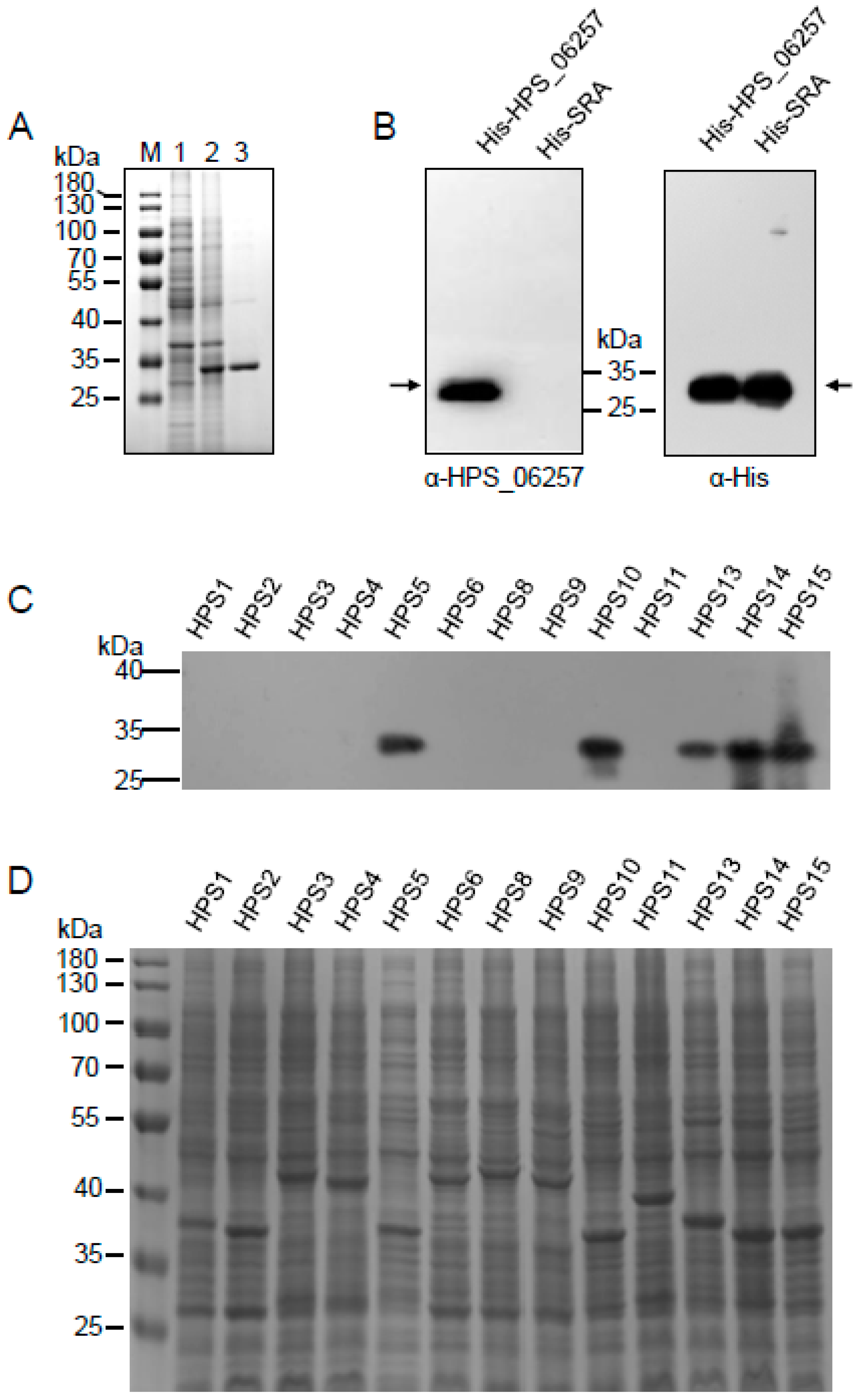
3.2. Differential Expression of HPS_06257 Protein in Different Strains of G. parasuis
Although we showed that the HPS_06257 gene occurs in only some strains of G. parasuis, it was unclear whether it was expressed in those strains. Therefore, we developed a polyclonal antibody against the recombinant HPS_06257 protein to detect the expression of native HPS_06257 in those strains. Purified recombinant HPS_06257 expressed in E. coli (Figure 2A) was used to immunize mice and generate a polyclonal antibody. To examine the specificity of this antibody, we first investigated its reaction with recombinant HPS_06257 and reported His-SRA [] with Western blotting. The polyclonal antibody specifically reacted with recombinant HPS_06257 but not with recombinant SRA (Figure 2B). In contrast, an anti-His antibody detected both of these recombinant (His-conjugated) proteins. Thus, we confirmed the successful generation of a polyclonal antibody against the HPS_06257 protein.

Figure 2.
Detection of HPS_06257 protein expression in different strains of G. parasuis. (A) SDS-PAGE gel shows the expression of HPS_06257 protein in E. coli (lane 2) and purified HPS_06257 (lane 3). Lane M, protein molecular weight marker; lane 1, E. coli transformed with empty control vector. (B) Polyclonal antibody against HPS_06257 was generated by immunizing BALB/C mice with purified HPS_0625. Western blotting was performed to determine the specificity of the anti-HPS_0625 antibody. The anti-HPS_0625 antibody specifically recognized recombinant His–HPS_0625 protein but not His–SRA; in contrast, an anti-His antibody specifically recognized both recombinant proteins. Black arrows indicate the target bands. (C) Expression of HPS_0625 in different strains of G. parasuis was determined with Western blotting using anti-HPS_0625 antibody. (D) SDS-PAGE gel shows the total cell lysates of different strains of G. parasuis, which were used for the Western blotting analysis.
The polyclonal antibody was then used to determine the expression of the HPS_06257 protein in different strains of G. parasuis. Our results showed that HPS_06257 was only expressed by strains containing the HPS_06257 gene (Figure 2C), consistent with the PCR results. The protein expressed by each strain was detected with SDS-PAGE (Figure 2D). These data confirm the existence of HPS_06257-expressing G. parasuis strains and HPS_06257-null G. parasuis strains in the field.
3.3. Differential Immunoprotective Effects of HPS_06257 against HPS_06257-Expressing Strains and HPS_06257-Null Strains
Because HPS_06257 is an important protective antigen against G. parasuis infection, we investigated the immunoprotective effects of HPS_06257 against HPS_06257-expressing strains and HPS_06257-null strains. We examined the immunoprotective role of HPS_06257 against HPS5 (an HPS_06257-expressing strain) and HPS11 (an HPS_06257-null strain) infections. Fourteen days after the booster immunization, each group was challenged with five median lethal doses of HPS5 or HPS11. The mice immunized with HPS_06257 showed 80% survival within 72 h of challenge with HPS5, whereas the mice treated with PBS or adjuvant all died within 60 h of challenge with HPS5 (Figure 3A). All the mice challenged with HPS11 died within 36 h of challenge (Figure 3B). These results indicate that HPS_06257 is protective against infection with HPS5 (an HPS_06257-expressing strain) and not against HPS11 (an HPS_06257-null strain).

Figure 3.
Comparative analysis of immunoprotective effects of HPS_06257 against G. parasuis strains HPS5 and HPS11. (A) Immunoprotective effect of HPS_06257 against HPS5 infection. Mice were immunized with PBS, adjuvant alone, or HPS_06257. The different groups of mice were challenged by intraperitoneal inoculation with 2 × 10⁹ CFU of HPS5. Notably, the HPS_06257 group showed 80% survival after challenge; in contrast, the mice in neither the PBS nor adjuvant-alone group survived after challenge. (B) Immunoprotective effects of HPS_06257 against HPS11 infection. Mice were immunized as described above. The different groups of mice were challenged by intraperitoneal inoculation with 2 × 10⁹ CFU of HPS11. Notably, none of the groups showed a protective effect against HPS11 infection.
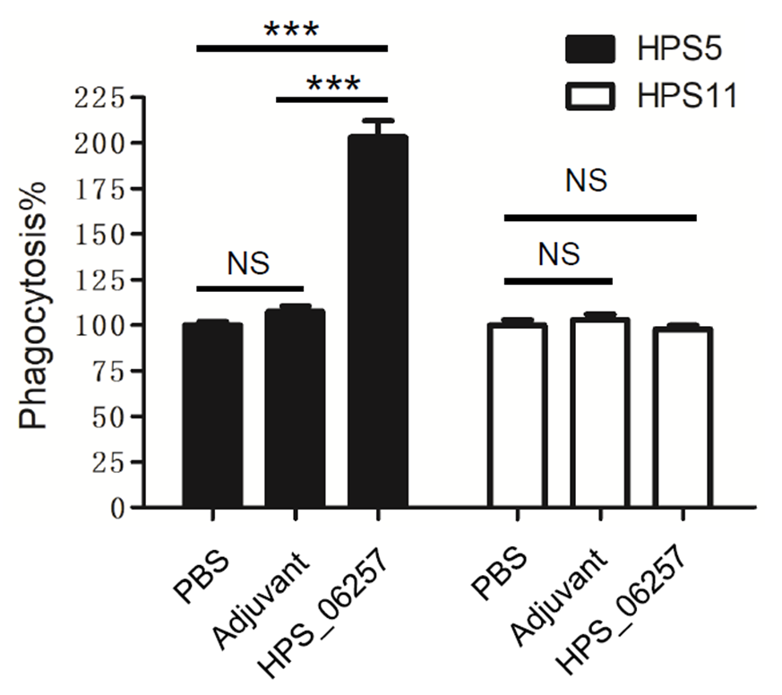
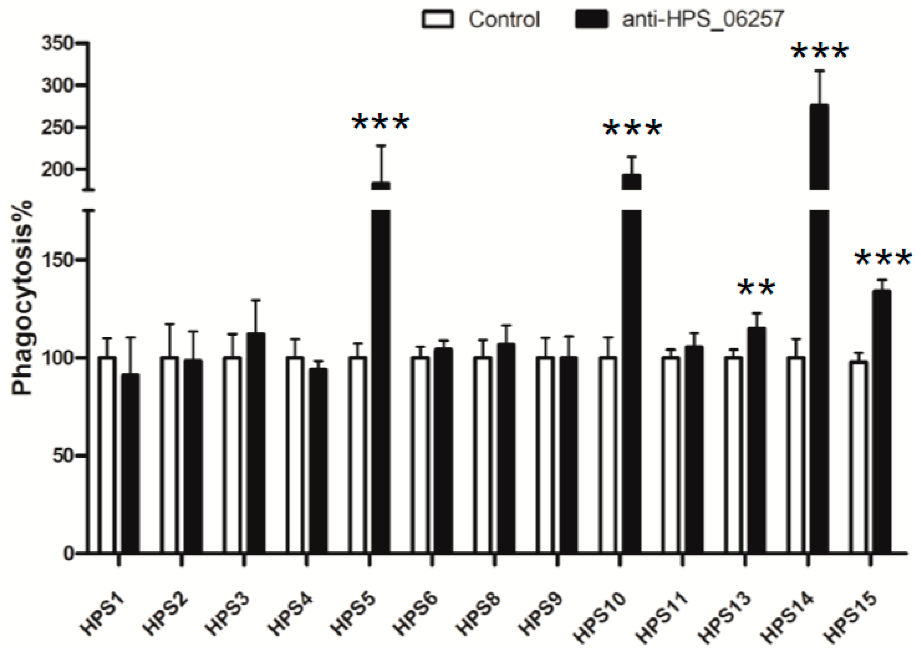
3.4. Antibody-Dependent Phagocytosis Is Associated with the Immunoprotective Effects of HPS_06257 against HPS_06257-Expressing Strains
Antibody-dependent phagocytosis has been shown to contribute to the protective effects of antigens against microbial infections. However, whether antibody-dependent phagocytosis is associated with the immunoprotective effects of HPS_06257 against HPS_06257-expressing strains was unclear. Therefore, we next examined the effect of the mouse serum isolated from each group in the challenge experiment on the phagocytosis of G. parasuis by macrophages. The sera from HPS_06257-immunized mice increased the phagocytosis of HPS5, whereas the sera from mice not immunized with HPS_06257 (PBS or adjuvant group) did not, consistent with the protective effect of HPS_06257 against HPS5 challenge (Figure 4). In contrast, the sera from HPS_06257-immunized mice did not affect the phagocytosis of HPS11 compared with the serum from unimmunized mice (PBS or adjuvant group) (Figure 4). These results indicate that the sera from HPS_06257-immunized mice increased the phagocytosis of HPS5 but not HPS11, consistent with the protective effect of HPS_06257 against HPS_06257-expressing strains but not against HPS_06257-null strains.

Figure 4.
Comparative analysis of effects of different mouse antisera on phagocytosis of G. parasuis strains HPS5 and HPS11. HPS5 was opsonized with mouse antisera from the PBS, adjuvant-alone, or HPS_06257-treated group, and phagocytosis was analyzed in RAW264.7 cells, as described in the Methods section. Notably, the antiserum from the HPS_06257 group significantly promoted the phagocytosis of HPS5 by macrophages, whereas the antiserum from neither the PBS group nor adjuvant group did so. HPS11 was opsonized with mouse antisera from the different groups, as described above, and phagocytosis was analyzed in RAW264.7 cells, as described in the Methods section. Notably, the mouse antiserum from the HPS_06257 group did not enhance the phagocytosis of HPS11 by macrophages compared with the effects of antiserum from either the PBS group or adjuvant group. Data are means ± SD of data pooled from one independent experiment; n ≥ 3 for each of the analyzed parameters. NS, not statistically significant. *** p < 0.001 in a comparison of the PBS and HPS_06257 groups or of the adjuvant-alone and HPS_06257 groups.
To confirm the effect of the anti-HPS_06257 antiserum on phagocytosis by macrophages, we examined effects of the polyclonal antibody against HPS_06257 on the 13 different strains shown in Figure 1C compared with the effects of normal mouse serum treated bacteria. The anti-HPS_06257 serum significantly increased the phagocytosis of all the HPS_06257-expressing strains compared with the treatment of some of these strains with normal mouse serum (Figure 5). However, the anti-HPS_06257 serum had no effect on the phagocytosis of any HPS_06257-null strain compared with the effect normal mouse serum had on some of these strains (Figure 5). Collectively, these findings demonstrate that antibody-dependent phagocytosis contributes to the immunoprotective effect of HPS_06257 against infections with HPS_06257-expressing G. parasuis strains.

Figure 5.
Effects of anti-HPS_06257 antibody on phagocytosis by macrophages of different strains of G. parasuis. Different strains of G. parasuis were opsonized with anti-HPS_06257 antibody, as shown in Figure 2, or control antibody (normal mouse serum). Notably, compared with control-antibody-opsonized phagocytosis, the anti-HPS_06257 antibody only affected the phagocytosis of HPS_06257-expressing strains. Data are means ± SD of data pooled from two independent experiment; n ≥ 6 for each of the analyzed parameters. ** p < 0.01; *** p < 0.001 in a comparison of the control and anti-HPS_06257 groups.
4. Discussion
This study provides novel information on the expression characteristics of HPS_06257 and its role in the development of a protective immune response against G. parasuis infection. Using a genomic analysis, PCR, and Western blotting, we demonstrated the presence of HPS_06257-expressing G. parasuis strains and HPS_06257-null G. parasuis strains. Furthermore, by comparing the immune protection afforded by HPS_06257 against HPS_06257-expressing and HPS_06257 null strain infections, we showed that antibody-dependent phagocytosis contributes to the protective immune response induced by HPS_06257 immunization against HPS_06257-expressing strains. Collectively, these data provide novel evidence for the role of HPS_06257, an important immune antigen, in the protective immune response to G. parasuis infection.
Since HPS_06257 was identified in an immunoproteomic analysis [], it has been studied as an immune antigen effective against the parental G. parasuis infection. Regarding the strong identity shared by the protein sequences of different strains (based on the BLASTP analysis, data not shown), HPS_06257 would be a good candidate for a protective antigen for subunit vaccine development. In this study, we demonstrated the existence of HPS_06257-expressing and HPS_06257-null G. parasuis strains. Thus, this finding gives some clue that HPS_06257 could not protect against all of strains in the field despite of the strong identity shared by the HPS_06257 protein sequences of different strains.
A previous study showed that antiserum against HPS_06257 had bactericidal activity in a whole-blood killing assay []; however, the specific mechanism of this protective effect remains unclear. In the present study, we demonstrated that antibody-dependent phagocytosis contributes to the protective effect induced by HPS_06257, providing further insight into the protective effect mediated by anti-HPS_06257 antiserum.
Because HPS_06257 was identified as an outer membrane protein in an immunoproteomic analysis, it is not unexpected that antibody-dependent phagocytosis is involved in its involvement in the protective immune response it induces against G. parasuis infection. The immunogenic actions of bacterial outer membrane proteins have been extensively studied [,,]. Notably, antibody-dependent phagocytosis has been investigated as the underlying mechanism of the protective immune response []. A study of the transferrin binding protein B (TbpB) expressed on the surface of G. parasuis showed that antibody-dependent phagocytosis contributes to its protective role in the development of immune response against G. parasuis infection []. Therefore, antibody-dependent phagocytosis could be an important mechanism underlying the role of HPS_06257 in the protective humoral immune response to G. parasuis infection.
In summary, this is the first study to demonstrate that antibody-dependent phagocytosis is involved in the protective effects of HPS_06257. These findings extend our understanding of how antibody-dependent phagocytosis may contribute to the immune protection afforded by other outer membrane proteins. Thus, future studies are needed to further verify this. In the meanwhile, these findings also provide insight into the protective antigens of G. parasuis and useful information for the development of a subunit vaccine to prevent G. parasuis infection.
Supplementary Materials
The following supporting information can be downloaded at: https://www.mdpi.com/article/10.3390/vetsci9070342/s1, Table S1: Primers used to amplify HPS_06257 gene.
Author Contributions
Conceptualization, X.C. (Xiaojun Chen), H.S., Z.M. and Y.Q.; methodology, X.C. (Xiaojun Chen), Z.L., D.S., K.L., J.W. (Jianchao Wei) and B.L.; formal analysis, X.C. (Xiaojun Chen), H.S. and Y.Q.; investigation, X.C. (Xingyu Chen) and H.S.; resources, X.W., J.W. (Jian Wang) and B.Z.; data curation, X.C. (Xiaojun Chen), H.S. and Y.Q.; writing—original draft preparation, X.C. (Xiaojun Chen), H.S. and Y.Q.; writing—review and editing, X.C. (Xiaojun Chen) and Y.Q.; supervision, Y.Q.; funding acquisition, Z.M. and Y.Q. All authors have read and agreed to the published version of the manuscript.
Funding
Funding was from the National Natural Science Foundation of China (31972693), the National Key R&D Program of China (2018YFD0500101), the Chinese Special Fund for Ago-scientific Research in the Public Interest (no. 2020JB04), and the Elite Program of CAAS (to Y.Q.).
Institutional Review Board Statement
All animal experiments were approved by the Institutional Animal Care and Use Committee of Shanghai Veterinary Research Institute, Chinese Academy of Agricultural Science, Shanghai, China (IACUC no. SHVRI-PO-2016060501) and were performed in compliance with the Guidelines on the Human Treatment of Laboratory Animals (Ministry of Science and Technology of the People’s Republic of China, policy no. 2006398).
Informed Consent Statement
Not applicable.
Data Availability Statement
Data will be made available upon request to the authors.
Acknowledgments
We acknowledge the technical assistance of Yanbing Zhang, Zhitao Wang, and Jiayun Wang.
Conflicts of Interest
The authors declare no conflict of interest.
References
- Oliveira, S.; Pijoan, C. Haemophilus parasuis: New trends on diagnosis, epidemiology and control. Vet. Microbiol. 2004, 99, 1–12. [Google Scholar] [CrossRef] [PubMed]
- Nedbalcova, K.; Kucerova, Z.; Krejci, J.; Tesarik, R.; Gopfert, E.; Kummer, V.; Leva, L.; Kudlackova, H.; Ondriasova, R.; Faldyna, M. Passive immunisation of post-weaned piglets using Hyperimmune serum against experimental Haemophilus parasuis infection. Res. Vet. Sci. 2011, 91, 225–229. [Google Scholar] [CrossRef]
- Olvera, A.; Ballester, M.; Nofrarías, M.; Sibila, M.; Aragon, V. Differences in phagocytosis susceptibility in Haemophilus parasuis strains. Vet. Res. 2009, 40, 24. [Google Scholar] [CrossRef] [PubMed]
- Liu, H.; Xue, Q.; Zeng, Q.; Zhao, Z. Haemophilus parasuis vaccines. Vet. Immunol. Immunopathol. 2016, 180, 53–58. [Google Scholar] [CrossRef] [PubMed]
- Zhou, M.; Guo, Y.; Zhao, J.; Hu, Q.; Hu, Y.; Zhang, A.; Chen, H.; Jin, M. Identification and characterization of novel immunogenic outer membrane proteins of Haemophilus parasuis serovar 5. Vaccine 2009, 27, 5271–5277. [Google Scholar] [CrossRef]
- Yuan, F.; Fu, S.; Hu, J.; Li, J.; Chang, H.; Hu, L.; Chen, H.; Tian, Y.; Bei, W. Evaluation of recombinant proteins of Haemophilus parasuis strain SH0165 as vaccine candidates in a mouse model. Res. Vet. Sci. 2012, 93, 51–56. [Google Scholar] [CrossRef]
- Li, M.; Song, S.; Yang, D.; Li, C.; Li, G. Identification of secreted proteins as novel antigenic vaccine candidates of Haemophilus parasuis serovar 5. Vaccine 2015, 33, 1695–1701. [Google Scholar] [CrossRef]
- Wen, Y.; Yan, X.; Wen, Y.; Cao, S.; He, L.; Ding, L.; Zhang, L.; Zhou, P.; Huang, X.; Wu, R.; et al. Immunogenicity of the recombinant HxuCBA proteins encoded by hxuCBA gene cluster of Haemophilus parasuis in mice. Gene 2016, 591, 478–483. [Google Scholar] [CrossRef]
- Li, G.; Xie, F.; Li, J.; Liu, J.; Li, D.; Zhang, Y.; Langford, P.R.; Li, Y.; Liu, S.; Wang, C. Identification of novel Haemophilus parasuis serovar 5 vaccine candidates using an immunoproteomic approach. J. Proteomics. 2017, 163, 111–117. [Google Scholar] [CrossRef]
- Barasuol, B.M.; Guizzo, J.A.; Fegan, J.E.; Martínez-Martínez, S.; Rodríguez-Ferri, E.F.; Gutiérrez-Martín, C.B.; Kreutz, L.C.; Schryvers, A.B.; Frandoloso, R. New insights about functional and cross-reactive properties of antibodies generated against recombinant TbpBs of Haemophilus parasuis. Sci. Rep. 2017, 7, 10377. [Google Scholar] [CrossRef]
- Fu, S.; Zhang, M.; Xu, J.; Ou, J.; Wang, Y.; Liu, H.; Liu, J.; Chen, H.; Bei, W. Immunogenicity and protective efficacy of recombinant Haemophilus parasuis SH0165 putative outer membrane proteins. Vaccine 2013, 31, 347–353. [Google Scholar] [CrossRef] [PubMed]
- McCaig, W.D.; Loving, C.L.; Hughes, H.R.; Brockmeier, S.L. Characterization and Vaccine Potential of Outer Membrane Vesicles Produced by Haemophilus parasuis. PLoS ONE 2016, 11, e0149132. [Google Scholar] [CrossRef] [PubMed]
- Xu, Z.; Yue, M.; Zhou, R.; Jin, Q.; Fan, Y.; Bei, W.; Chen, H. Genomic characterization of Haemophilus parasuis SH0165, a highly virulent strain of serovar 5 prevalent in China. PLoS ONE 2011, 6, e19631. [Google Scholar] [CrossRef] [PubMed]
- Li, Y.; Kwok, A.H.; Jiang, J.; Zou, Y.; Zheng, F.; Chen, P.; Hou, C.; Leung, F.C.; Jiang, P. Complete genome analysis of a Haemophilus parasuis serovar 12 strain from China. PLoS ONE 2013, 8, e68350. [Google Scholar] [CrossRef]
- Nicholson, T.L.; Brunelle, B.W.; Bayles, D.O.; Alt, D.P.; Shore, S.M. Comparative genomic and methylome analysis of non-virulent D74 and virulent Nagasaki Haemophilus parasuis isolates. PLoS ONE 2018, 13, e0205700. [Google Scholar] [CrossRef]
- Wang, X.; Xu, F.; Ning, K.; Shen, L.; Qi, X.; Wang, J. Construction and Application of MALDI-TOF Mass Spectrometry for the Detection of Haemophilus parasuis. Biomed. Res. Int. 2021, 2021, 5588855. [Google Scholar] [CrossRef]
- Howell, K.J.; Peters, S.E.; Wang, J.; Hernandez-Garcia, J.; Weinert, L.A.; Luan, S.L.; Chaudhuri, R.R.; Angen, Ø.; Aragon, V.; Williamson, S.M.; et al. Development of a Multiplex PCR Assay for Rapid Molecular Serotyping of Haemophilus parasuis. J. Clin. Microbiol. 2015, 53, 3812–3821. [Google Scholar] [CrossRef]
- Qiu, Y.; Shen, Y.; Li, X.; Liu, Q.; Ma, Z. Polyclonal antibody to porcine p53 protein: A new tool for studying the p53 pathway in a porcine model. Biochem. Biophys. Res. Commun. 2008, 377, 151–155. [Google Scholar] [CrossRef]
- Huang, H.; Sharma, M.; Zhang, Y.; Li, C.; Liu, K.; Wei, J.; Shao, D.; Li, B.; Ma, Z.; Cao, R.; et al. Expression Profile of Porcine TRIM26 and Its Inhibitory Effect on Interferon-β Production and Antiviral Response. Genes 2020, 11, 1226. [Google Scholar] [CrossRef]
- Aronoff, D.M.; Hao, Y.; Chung, J.; Coleman, N.; Lewis, C.; Peres, C.M.; Serezani, C.H.; Chen, G.H.; Flamand, N.; Brock, T.G.; et al. Misoprostol impairs female reproductive tract innate immunity against Clostridium sordellii. J. Immunol. 2008, 180, 222–230. [Google Scholar] [CrossRef]
- Xiang, X.; Zhang, Y.; Li, Q.; Wei, J.; Liu, K.; Shao, D.; Li, B.; Olszewski, M.A.; Ma, Z.; Qiu, Y. Expression profile of porcine scavenger receptor A and its role in bacterial phagocytosis by macrophages. Dev. Comp. Immunol. 2020, 104, 103534. [Google Scholar] [CrossRef] [PubMed]
- van der Pol, L.; Stork, M.; van der Ley, P. Outer membrane vesicles as platform vaccine technology. Biotechnol. J. 2015, 10, 1689–1706. [Google Scholar] [CrossRef] [PubMed]
- Sawa, T.; Kinoshita, M.; Inoue, K.; Ohara, J.; Moriyama, K. Immunoglobulin for Treating Bacterial Infections: One More Mechanism of Action. Antibodies 2019, 8, 52. [Google Scholar] [CrossRef] [PubMed]
Publisher’s Note: MDPI stays neutral with regard to jurisdictional claims in published maps and institutional affiliations. |
© 2022 by the authors. Licensee MDPI, Basel, Switzerland. This article is an open access article distributed under the terms and conditions of the Creative Commons Attribution (CC BY) license (https://creativecommons.org/licenses/by/4.0/).