Article

Analysis of the Expression of Neurotrophins and Their Receptors in Adult Zebrafish Kidney

1
Department of Pathology and Immunology, School of Medicine, University of Geneva, Rue Michel-Servet 1, 1206 Geneva, Switzerland
2
Department of Veterinary Medicine, University of Naples Federico II, 80138 Naples, Italy
*
Author to whom correspondence should be addressed.
Vet. Sci. 2022, 9(6), 296; https://doi.org/10.3390/vetsci9060296
Submission received: 7 April 2022 / Revised: 7 June 2022 / Accepted: 12 June 2022 / Published: 15 June 2022
(This article belongs to the Special Issue Neuropeptides: Role and Function in Species of Veterinary Interest)

Abstract

:
Neurotrophins and their receptors are involved in the development and maintenance of neuronal populations. Different reports have shown that all neurotrophin/receptor pathways can also play a role in several non-neuronal tissues in vertebrates, including the kidney. These signaling pathways are involved in different events to ensure the correct functioning of the kidney, such as growth, differentiation, and regulation of renal tubule transport. Previous studies in some fish species have identified the neurotrophins and receptors in the kidney. In this study, for the first time, we compare the expression profiles (mRNA and protein) of all neurotrophin/receptor pathways in the kidney of the adult zebrafish. We quantify the levels of mRNA by using qPCR and identify the expression pattern of each neurotrophin/receptor pathway by in situ hybridization. Next, we detect the proteins using Western blotting and immunohistochemistry. Our results show that among all neurotrophins analyzed, NT-3/TrkC is the most expressed in the glomerule and tubule and in the hematopoietic cells, similar to what has been reported in the mammalian kidney.

1. Introduction

Neurotrophins include brain-derived neurotrophic factor (BDNF), nerve growth factor (NGF), NT-3, NT-4/5, and, only in fish, NT 6/7 [1,2,3,4,5,6,7]. The binding to three distinct tyrosine kinase receptors, TrkA, TrkB, and TrkC, mediates their activities concerning neuronal proliferation [8], survival [9,10], differentiation, and plasticity [11] in many neuronal populations of the central and peripheral nervous systems in different vertebrates [12,13,14,15,16]. In mammals, as in fish, besides this essential role, neurotrophins and receptors are also important in non-neuronal cells, as suggested by their detection in different organs [17,18,19,20,21,22,23], including the kidney [24,25,26]. Neurotrophins and receptors are involved in different events to ensure the correct functioning of the kidney, such as growth, differentiation, and regulation of renal tubule transport. Indeed, recent functional in vivo studies, in mice models and in vitro in human patients, have revealed a close association between neurotrophin/receptor pathways and kidney diseases [27,28,29]. However, further studies using emerging animal models [30], such as zebrafish (Danio rerio), could be useful to elucidate the specific mechanisms of these pathologies. Zebrafish is a teleost fish of the family of Cyprinidae. This fish presents different characteristics, making it an excellent animal model. The most important advantages are the small size (reduction in maintenance costs and spaces to be used), high fecundity, and rapidity of embryonic development [31]. This fascinating model can be utilized in mutagenesis analyses and drug screening tests [32,33,34]. It has become a popular model organism used to conduct experiments related to different research fields, such as embryology, genetics, cancer, cardiovascular, and organ and tissue regeneration [35,36,37,38]. To study the anatomy, cellular biology, and molecular mechanisms of the vertebrate kidney, the zebrafish is an excellent model [39,40,41,42,43].
The zebrafish kidney is located in the retroperitoneal area. It consists of two different regions, the head and trunk [44,45,46]. Zebrafish and mammalian kidneys have several similarities at the microscopic level; both have nephrons with a glomerulus, proximal tubules, distal tubules, and collecting ducts. The renal interstitium of the zebrafish contains hematopoietic cells [47,48,49,50,51]; thus, it could be considered equivalent to the bone marrow in mammals [52,53,54]. During development, the hematopoietic stem progenitor cells (HSPCs) emerge from the dorsal aorta, colonize the caudal hematopoietic tissue (equivalent to the mammalian fetal liver), and migrate to the kidney marrow [55,56].
To our knowledge, there are no investigations concerning the distribution of mRNA and proteins of neurotrophic factors and their receptors in the adult zebrafish kidney. Therefore, the purpose of the present report is to gain insights into the anatomical localization of neurotrophins and their high-affinity Trk receptors in the kidney of the zebrafish to inspire further experimental investigations in a non-mammal model. This study was carried out using different approaches. We used qPCR and Western blotting, respectively, to quantify the expression of mRNA and proteins in the kidney. Next, we used in situ hybridization and immunohistochemistry, to localize the distribution of all neurotrophins and receptors in the adult zebrafish kidney.

2. Materials and Methods

2.1. Animals and Kidney Dissection

All procedures were approved by the Institutional Committee of the University of Naples Federico II (CNR, n°2/2020-PR), and according to the Italian Decree 26/2014. Adult females and males (1 year) of Danio rerio zebrafish were anesthetized by 0.1% ethyl 3-aminobenzoate and methanesulfonate (Sigma Chemicals Co., St. Louis, MO, USA), and kidneys were excised.

2.2. qPCR

From 15 kidneys, the total RNA was extracted using the RNeasy minikit (Qiagen, Frankfurt, Germany) and reverse-transcribed into cDNA using a Superscript kit (Invitrogen, Waltham, MA, USA). Quantitative real-time PCR (qPCR) was performed using the Universal qPCR Kit (KAPA BIOSYSTEMS, Wilmington, DC, USA) and run on a CFX connect real-time system (Bio-Rad, Tokyo, Japan), Fast Sybr Green (Universal Bioline). All data indicate the expression relative to ef1a, calculated by [2Ct(geneX)-Ct(ef1a)]. Each qPCR experiment was performed using biological triplicates. No difference in expression between females and males was found. For experiments using qPCR, each n represents the average of biological triplicates from a single experiment. Experiments were repeated at least three times. The primers used have also been described in previous studies [20,57,58,59,60]. Primers used:
bdnfF: CGAGGAATAGACAAGCGGCA;R: ATCCGTATAAACCGCCAGCC
ngfF: GAGAAGACTACAAGCGAAT;R: CGACAACAATAAGGAGGAT
nt3F: CCCATCAGTGCGCTCATC;R: TCCGAACTGTCCACCATG
nt4F: GCTCCTCCTAGAACAGAGGG;R: CGTCCTGGATGCATCTTCTCTG
trkaF: GCATTTACAATGGCAGCCAG;R: CTTCTTGAGTGGTCACTGTC
trkbF: TCACCTATGGCAAGCAACCC;R: CTTTGGGGCAAGTACGAGGT
trkcF: CGGAAGTGGATTGGACAGTT;R: CATGAAGCCGTTATCGTCC
ef1aF: TCAAGGACATCCGTCGTGGTA;R: ACAGCAAAGCGACCAAGAGG

2.3. Statistical Analysis

Statistical analysis was completed using a one-way ANOVA, adjusted for multiple comparisons; center values denote the mean, and error values denote s.e.m. The software GraphPad Prism (version 7.0a) has been used for all statistical analysis.

2.4. Chromogenic and Fluorescence In Situ Hybridization (ISH)

For the preparation of digoxigenin (DIG)-labeled antisense riboprobes, the protocol has also been described in previous studies [13,55]. In detail, full-bdnf, ngf, nt3, nt4, trka, full-trkb, and trkc expression was investigated using in situ hybridization (ISH). RNA probes were produced by linearization of TOPO-TA or ZeroBlunt vectors (Invitrogen) containing the amplified polymerase chain reaction (PCR). Antisense and sense riboprobes were generated with DIG RNA Labelling Mix (Roche Diagnostic, Indianapolis, IN, USA) by in vitro transcription, using T7 polymerase (Roche-Diagnostic) and SP6 polymerase (Roche-Diagnostic) on plasmid linearized by EcoRI and Not1. To check the specificity of the staining, the sense and antisense riboprobes were always hybridized on adjacent sections. The following primers were used to generate the riboprobes:
NgfGGAGCACAGGAGATCTACGC and CGTGGAAAAACCCAACTCAT
bdnf-full (fw)5′-ATAGTAACGAACAGGATGG-3′ and 5′-GCTCAGTCATGGGAGTCC-3′
nt3ATGGTTACCTTTATTACGATC and CCACCATTTTTCACGTCC
nt4CAGAGAAGATGCATCCAGGACG and CACTGCCGTTTCCTGACACGCG
trkaAGTTGTTGCTTGCAGGGTGG and TGGGTCAATCATGACCTCAG
trkb-fullCCAGAGATGTGTACAGCACC and CATTGTTTGAGAGCTGATACC
trkcGGACACTGAGAGCATCTCTATGA and CACGTTGTTCATCCCGACCACATT
Kidneys were quickly removed and fixed overnight at 4 °C in PBS-PAF 4%. Next, they were processed for cryostat. The sections (15 µm) were mounted on slides. They were washed in PBS-NaCl (0.85%) and post-fixed for 20 min in PBS-PFA 4%. The kidney sections were rinsed in PBS and treated for 10 min with proteinase K (2 mg/mL) diluted in PBS at room temperature. Slides were then rinsed as follows: 20 min in 4% PBS-PAF, 10 min in PBS, and 10 min in standard saline citrate (SSC 2x). The sections were incubated overnight at 64.5 °C in a moist chamber with the probes (1.5 µg/mL) diluted in hybridization buffer, as in previous studies [55]. Next, they were dipped in Tris-HCl/NaCl buffer (100 mM, Tris-HCl pH 7.5, 150 mM NaCl) and washed in the same buffer containing 0.1% Triton and 0.5% milk powder. For chromogenic ISH, all sections were incubated overnight at room temperature (RT) with anti-digoxigenin alkaline phosphatase Fab fragments (1:5000, Roche Diagnostic). On the next day, slides were rinsed in Tris-HCl/NaCl buffer and washed three times with Tris-HCl 100 mM (pH 8) containing NaCl (100 mM) and MgCl2 (10 mM). Staining was performed with NBT/BCIP buffer (pH 9.5). For fluorescence ISH, all paraffin sections were deparaffinized with ultraclear and rehydrated through a series of graded ethanol (100–30%). Next, the sections were washed and fixed, as mentioned before. In contrast to the chromogenic protocol, the sections were incubated with anti-DIG POD antibody (Roche) at a 1:200 dilution in the above blocking solution overnight at RT. The next day, the sections were washed 4 × 20 min in 1× maleic acid buffer, 4 × 10 min in PBS, and incubated for 1 h in Perkin Elmer amplification diluent buffer. For the reaction, we diluted Cy3 tyramide (TSA plus Cyanine 3, Perkin Elmer, Waltham, MA, USA) reagent 1:100 in amplification buffer and AlexaFluor488 reagent (TSA™ Reagent, Alexa Fluor 488 Tyramide Reagent, Invitrogen) after washing each section three times for 10 min and then observing them with a confocal Nikon Eclipse 90i microscope. The images were cataloged using software (NIS-Elements 4.2).

2.5. Immunohistochemistry (IHC)

A total of 5 adult zebrafish kidneys were fixed in Bouin’s solution for 12 h and processed for paraffin embedding. Transverse microtome sections were mounted on poly-lysine slides. Sections were deparaffinized in xylene, rehydrated through graded ethanol, treated with 3% H2O2 for 30 min, and rinsed in PBS (pH 7.4), followed by antigen retrieval in sodium citrate buffer (pH 6; 80 °C) for 30 min. After rinsing twice in 0.2% Triton PBS (PBT), non-specific binding was blocked by treating sections with 1/5 normal goat serum (Vector, Burlingame, CA, USA; cod S-1000-20) for 30 min at RT. Then, sections were incubated overnight at RT in a humidified chamber with rabbit antibody against NGF, BDNF, NT-3, NT-4, TrkA, TrkB, and TrkC. The antibodies used (see Table 1) have been described in previous studies [61]; we used a dilution of 1/100. The next day, the sections were incubated with the EnVision horseradish anti-peroxidase (HRP) system (Dako, Santa Barbara, CA, USA; cod. K4002).

2.6. Protein Analysis by Western Blot

To perform the evaluation of neurotrophins and their receptor proteins, 5 kidneys of adult zebrafish were homogenized using a Tissue Lyzer homogenization system (Qiagen) in 200 μL of ice-cold lysis buffer (50 mM Tris pH 7.5; 150 mM NaCl; 1 mM EDTA; 0.25% deoxicolic acid, 1% Triton X 100) added with 20 mM sodium pyrophosphate, 0.1 mg/mL aprotinin, 2 mM phenylmethylsulphony fluoride (PMSF), 10 mM sodium orthovanadate (Na2VO3), and 50 mM NaF. The number of total proteins was determined by the use of a protein assay kit (Bio-Rad Laboratories). Equal amounts of lysate samples (60 μg) were boiled and loaded on bis/acrylamide gel 15% (for neurotrophin) or 8% (for Trk receptors) and electrophoresed. All proteins obtained were blotted from the gel onto nitrocellulose membranes. Next, all membranes were blocked with 5% bovine serum albumin (BSA) in Tris-buffered saline (TBS, pH 7.5) containing 0.1% Tween 20 (TBS-T) at room temperature (RT), washed with TBS-T, and incubated with primary antibody (all antibodies are listed in Table 1) diluted 1:500 in TBS-T. After appropriate washing steps in TBS-T, anti-rabbit peroxidase-conjugated secondary antibodies (Amersham, Gel Health Care) were applied for 1 h at RT, at 1:2000 dilution. Finally, the reaction was developed using a chemiluminescent reagent (ECL, Amersham Pharmacia Biotech, Buckinghamshire, UK) and exposed to ChemiDoc gel scanner (Bio-Rad Laboratories). As control, a goat-anti-β-actin antibody (42 kDa) was used. For further details, see Figures S2 and S3.

2.7. Microscopy

The stained sections were photographed using a Nikon microscope. The raw digital images were optimized for image resolution, contrast, evenness of illumination, and background by using Adobe Photoshop CS5 (Adobe Systems, San Jose, CA, USA).

3. Results

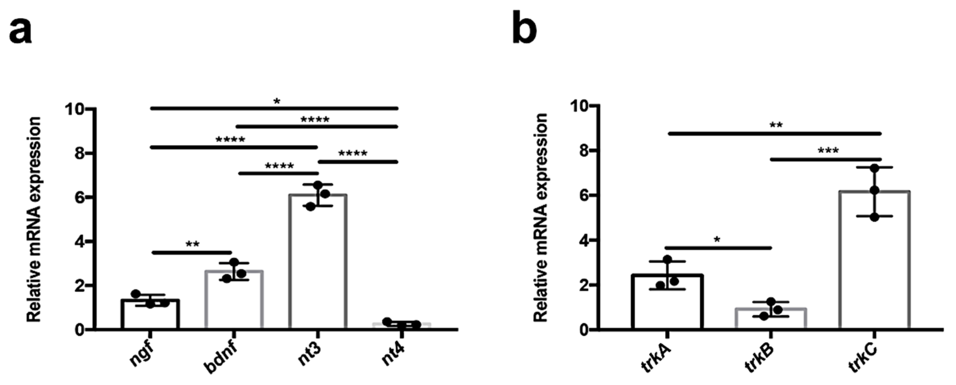
3.1. Quantitative Analysis of Neurotrophin and Receptor Expression in Adult Zebrafish Kidney

In order to evaluate neurotrophin and receptor mRNA levels, we performed quantitative PCR experiments in adult zebrafish kidneys. We found that in this organ, nt3 is highly expressed (compared with the other neurotrophins, ngf, bdnf). Surprisingly, nt4 is not significatively expressed (Figure 1a). All receptors of tyrosine kinase are expressed in this organ. In detail, we found abundant expression of receptor trkc compared to trka and trkb (Figure 1b).

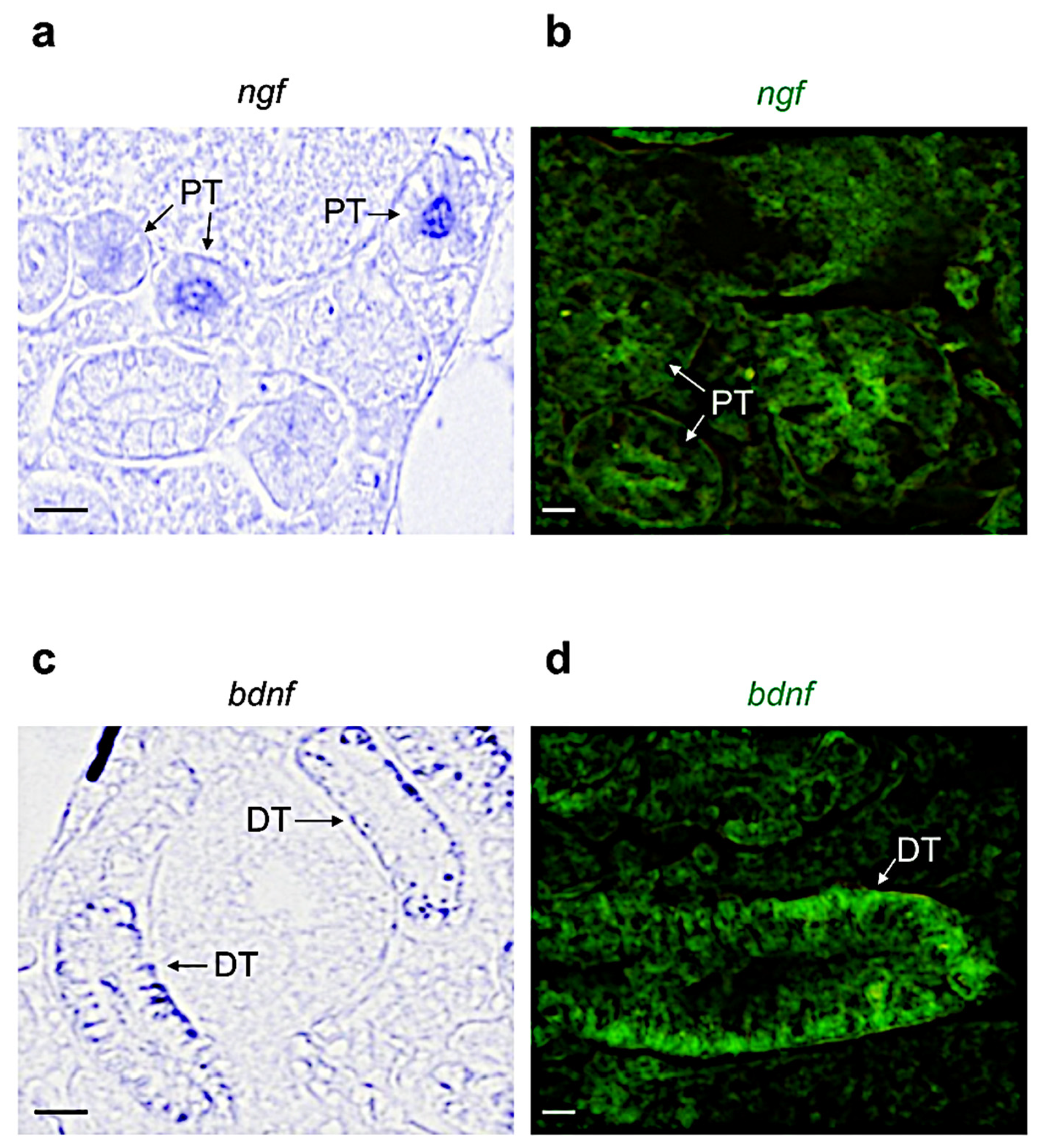
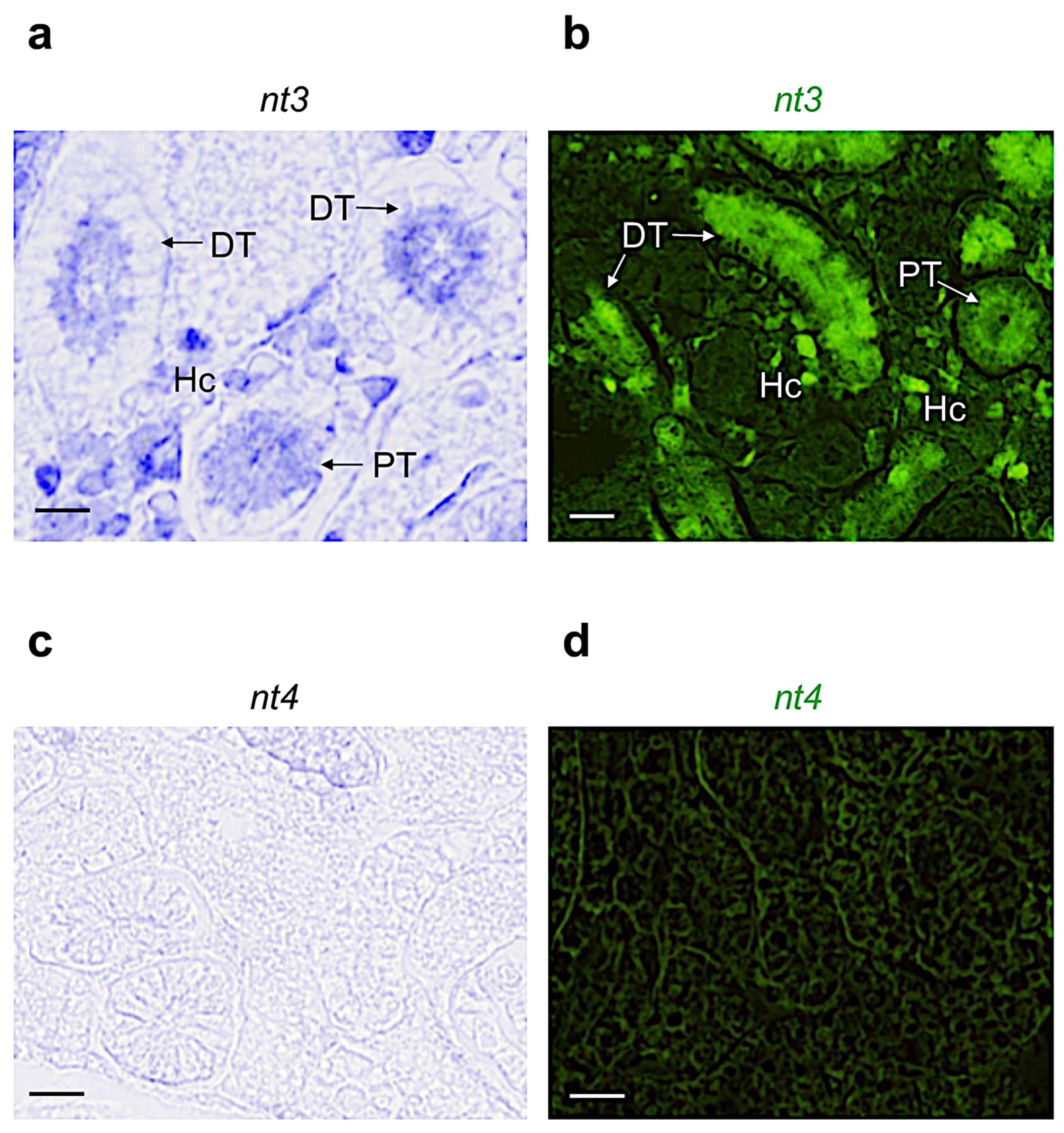
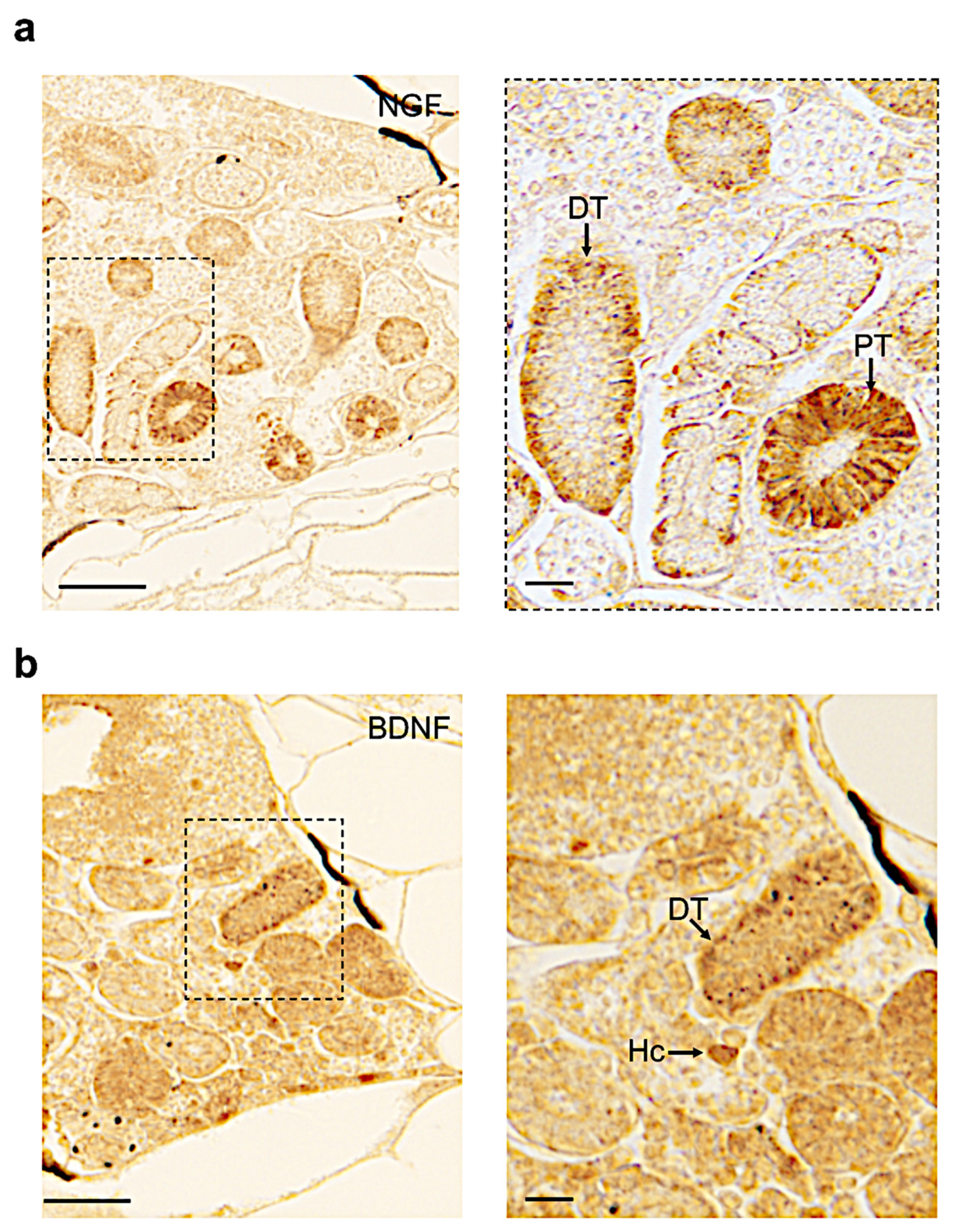
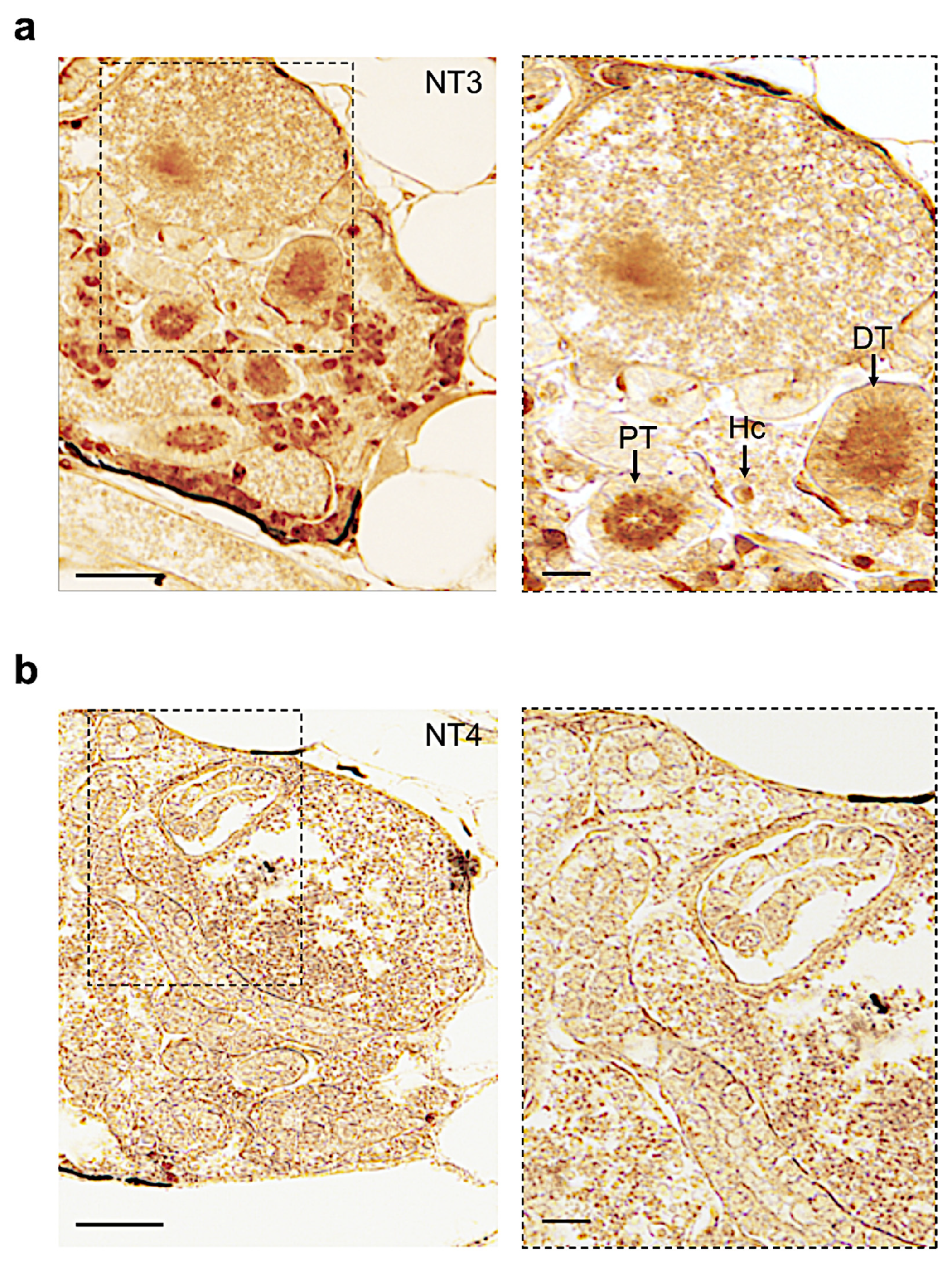
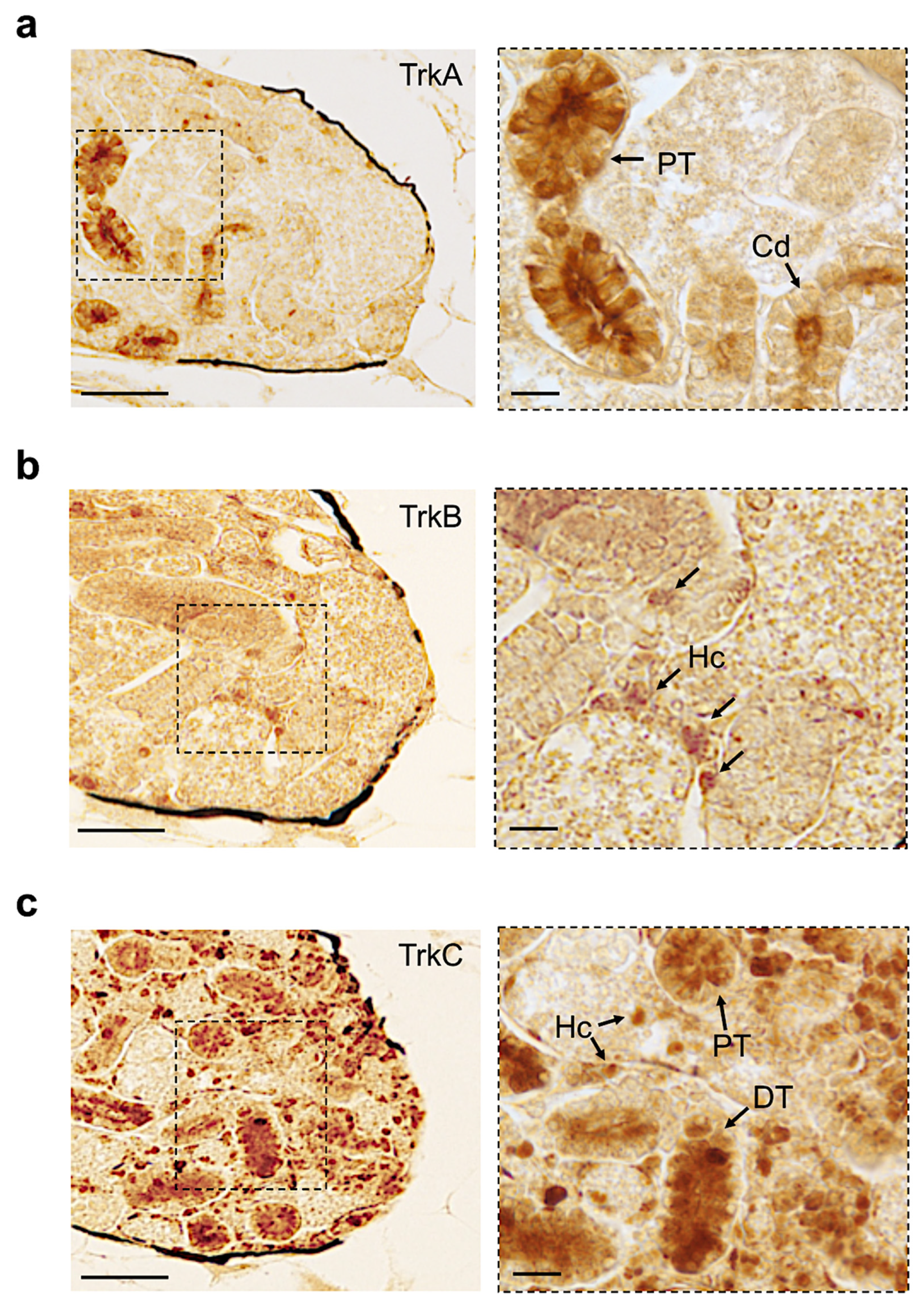
3.2. Distribution of Neurotrophin and Receptor mRNA in Adult Zebrafish Kidney

Based on the morphological features described by Menke and colleagues [47], we identified the different regions reported in Figure S1. By using chromogenic and fluorescence in situ hybridization, we confirmed the results obtained by qPCR. In detail, we found that ngf is expressed in the proximal tubule (Figure 2a,b). bdnf is more expressed in the distal tubule (Figure 2c,d). Differently, nt3 is highly expressed in the distal and proximal tubules, and in the interstitial hematopoietic cells (by confocal microscopy, we distinguished several shapes that are common to hematopoietic cells: round, tapered, concave, and can present enlarged nucleus) (Figure 3a,b). nt4, coherently with qPCR analysis, is not detected in this organ (Figure 3c,d).
Concerning the receptors, we found that trkA-mRNA was expressed in the proximal tubules of the adult zebrafish kidney (Figure 4a,b). trkb-mRNA was detected only in a few hematopoietic cells (Figure 4c,d). Finally, trkc is highly expressed in hematopoietic cells, and in the distal and proximal tubules (Figure 4e,f).

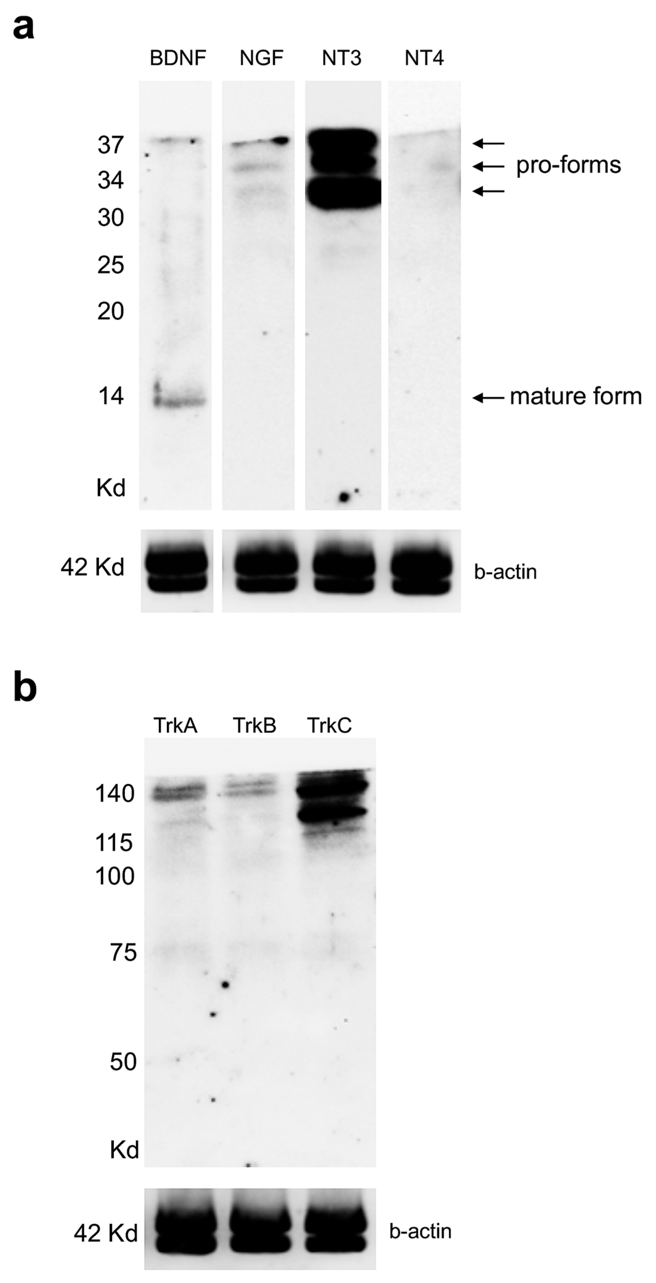
3.3. Expression Levels of Neurotrophin and Receptor Proteins in Adult Zebrafish Kidney

Next, to detect the levels of protein for each neurotrophin and receptor in the adult zebrafish kidney, we used Western blot. Interestingly, we found that BDNF antiserum recognized two lines, one at 14 kDa (mature form) and one at 37 kDa (immature form pro-BDNF). NGF antiserum recognized three bands of 30 kDa, 34 kDa, and 37 kDa (these forms are pro-NGF). NT-3 antiserum labeled three bands of 30 kDa, 34 kDa, and 37 kDa (these forms are pro-NT3). NT-4 antiserum was not detected (Figure 5a).
TrkA antiserum showed three bands of 75 kD (low intensity), 115 kD, and 140 kD; TrkB labeled a band of 140 kD and TrkC antiserum recognized two bands of 115 kD and 140 kD (Figure 5b).

3.4. Distribution of Neurotrophin and Receptor Proteins in Adult Zebrafish Kidney

Finally, by immunohistochemistry, we detected NGF protein in the distal and proximal tubules (Figure 6a). We found BDNF in a few hematopoietic cells and in the distal tubule region (Figure 6b).
The NT-3 distribution was very abundant in different regions of the kidney (interstitial hematopoietic cells, distal and proximal tubules) (Figure 7a). NT4 was not detected, confirming previous results obtained by Western blot (Figure 7b).
All receptors were expressed in the adult zebrafish kidney in different regions. TrkA was expressed in the collecting duct and proximal tubule (Figure 8a). TrkB was detected in hematopoietic cells (Figure 8b). TrkC was abundantly distributed in different regions. (Figure 8c). We have summarized all our results in the following Table 2.

4. Discussion

In the present study, we compared the expression profiles of both mRNA and protein of all neurotrophins/receptors in the kidney of the adult zebrafish.
In this organ, neurotrophin and receptor mRNAs are differently expressed. Our results, obtained by qPCR and ISH, show that nt3/trkC is the most expressed in different regions, including the hematopoietic cells, similarly to the mammalian kidney [62,63]. Previous observations in rats and humans highlighted that nt3 and trkC mRNA levels were elevated in the glomerular tissue, and this signaling could be involved in the regulation of renal tubule transport [64,65,66]. In detail, Lepa and colleagues, using a mammal animal model, confirmed that nephron-specific TrkC knockout (TrkC-KO) and TrkC-overexpressing (TrkC-OE) mice presented enlarged glomeruli with mesangial proliferation, podocyte loss during aging, basement membrane thickening, and albuminuria. They also showed that NT3-dependent TrkC activation in cultured podocytes and overexpression of TrkC in the nephron in mice lead to the phosphorylation of the activating tyrosine residues of the Igf1R [66].
Next, we observed that the ngf and trkA mRNAs were most widely distributed in the proximal tubule, in line with previous reports in the kidneys of humans, mice, and goldfish, where the colocalization of NGF and TrkA was described in the tubular cells [67,68]. Under normal physiological conditions, nerve growth factor and tyrosine kinase receptor-A in humans and mice present low serum levels in the kidney. However, the concentrations can increase during the progression of several inflammatory and autoimmune diseases, such as chronic kidney disease, glomerulonephritis, and after renal transplantation [27,69]. It has been observed that, differently from other tissue, such as the brain, where NGF promoted cell growth, differentiation, and survival, high levels of NGF in the renal tubular cells promote growth arrest and apoptosis [70,71,72,73,74].
More significant is the expression of bdnf compared to ngf. This result confirms the hypothesis that bdnf plays a protective role in the kidney; indeed, low levels of bdnf have been detected in an experimental model of chronic nephropathy [28]. Very interestingly, we observed low expression of trkB limited to the interstitial hematopoietic cells, in agreement with data reported in the mouse kidney. Indeed, TrkB immunoreactivity, on the other hand, was restricted to the differentiated vascular cells and extraglomerular mesangial cells [75]. nt4-mRNA displays a very low level of expression in the kidney of the zebrafish, as also demonstrated in the mouse [63].
With regard to the expression of neurotrophin/receptor proteins, Western blot analysis shows that the employed NGF antiserum recognized three bands, likely corresponding to proNGF proteins, differently from the kidneys of other teleosts, where the mature form of NGF [67] has been detected, and similar to the results obtained in the brain of the teleost in N. furzeri and zebrafish [61]. The BDNF antibody detected in two lines (or bands) matches the two isoforms, a first mature BDNF form, and a second immature form—very similar to the data shown in the kidneys of other teleost fish [67]. Pro-BDNF bands have been identified in the zebrafish ovary and testis [20]. NT-3 antiserum identified three bands, presumably pro-NT-3. Previous studies on other teleostean species documented that this antibody detected bands corresponding to the mature form in different tissues [61,67]. Concerning the receptors, the TrkA protein in the kidney is recognized in three bands, and these data have been described in different studies in Danio rerio and other teleost species [61,76]. The TrkB antibody detected one band (low level), similar to results reported in other tissue of the zebrafish [22]. The abundant presence of TrkC was recognized in two bands (high level), with a slight difference in the result reported in the brain of the zebrafish [61]. Finally, the immunohistochemical analysis confirms that NT-3 is abundant in the zebrafish kidney, in agreement with reports in the kidneys of goldfish and scorpionfish [67]. NGF and BDNF detection are restricted to the tubule and hematopoietic cells of the zebrafish kidney, differently from that reported in other teleost species [67], and remarkably similar to humans and mice [28,77,78]. NT-4 is not detected, confirming all our previous results obtained by qPCR, ISH, and Western blot. A similar result has been reported in mice [62,63]. In the case of Trk receptors, they were detected in the zebrafish kidney by immunohistochemistry: TrkA was detected in the collecting and proximal tubule, as reported in other teleost species; TrkB was detected only in hematopoietic cells; and TrkC was the most abundant receptor, distributed in different regions.

5. Conclusions

This work reports the anatomical distribution of neurotrophins and receptors in adult zebrafish kidneys and also compares the concentrations of mRNA and protein. It is interesting that BDNF and NGF in the zebrafish kidneys are present in tubule and hematopoietic cells, similar to humans and mice. This fact further reinforces the thesis that the zebrafish is an excellent animal model to be used in biomedical and veterinary research. At the same time, our analysis showed that NT3/TrkC is highly expressed in the zebrafish kidney, as also reported in humans and mice. We hope that our work will inspire further experimental studies that could lead to a new potential therapeutic role for neurotrophins and receptors in kidney diseases.

Supplementary Materials

The following supporting information can be downloaded at: https://www.mdpi.com/article/10.3390/vetsci9060296/s1, Figure S1. Adult zebrafish kidney section HE, available at https://bio-atlas.psu.edu/about.php, accessed on 11 June 2022. Figure S2. Full Western blot gels related to Figure 5a. Figure S3. Full Western blot gels related to Figure 5b.

Author Contributions

Conceptualization: P.C.; data acquisition and analysis: P.C. and C.L.; writing—original draft: P.C.; writing—reviewing and editing: P.C. and C.L.; funding acquisition: P.C. All authors have read and agreed to the published version of the manuscript.

Funding

This research was financially supported by the Gertrude Von Meissner Foundation (Dr. Pietro Cacialli, funding number ME11989).

Institutional Review Board Statement

All procedures were approved by the Institutional Committee of the University of Naples Federico II (CNR, n°2/2020-PR), and according to the Italian Decree 26/2014.

Informed Consent Statement

Not applicable.

Data Availability Statement

Not applicable.

Conflicts of Interest

The authors declare no conflict of interest.

References

  1. Chao, M.V. The p75 neurotrophin receptor. J. Neurobiol. 1994, 25, 1373–1385. [Google Scholar] [CrossRef] [PubMed]
  2. Yano, H.; Chao, M.V. Neurotrophin receptor structure and interactions. Pharm. Acta Helv. 2000, 74, 253–260. [Google Scholar] [CrossRef]
  3. Lee, F.S.; Rajagopal, R.; Chao, M.V. Distinctive features of Trk neurotrophin receptor transactivation by G protein-coupled receptors. Cytokine Growth Factor Rev. 2002, 13, 11–17. [Google Scholar] [CrossRef]
  4. Ibanez, C.F.; Hallbook, F.; Godeau, F.; Persson, H. Expression of neurotrophin-4 mRNA during oogenesis in Xenopus laevis. Int. J. Dev. Biol. 1992, 36, 239–245. [Google Scholar] [PubMed]
  5. Islam, N.; Gagnon, F.; Moss, T. Catalytic and non-catalytic forms of the neurotrophin receptor xTrkB mRNA are expressed in a pseudo-segmental manner within the early Xenopus central nervous system. Int. J. Dev. Biol. 1996, 40, 973–983. [Google Scholar]
  6. Rodriguez-Tebar, A.; Dechant, G.; Gotz, R.; Barde, Y.A. Binding of neurotrophin-3 to its neuronal receptors and interactions with nerve growth factor and brain-derived neurotrophic factor. EMBO J. 1992, 11, 917–922. [Google Scholar] [CrossRef]
  7. Philo, J.; Talvenheimo, J.; Wen, J.; Rosenfeld, R.; Welcher, A.; Arakawa, T. Interactions of neurotrophin-3 (NT-3), brain-derived neurotrophic factor (BDNF), and the NT-3.BDNF heterodimer with the extracellular domains of the TrkB and TrkC receptors. J. Biol. Chem. 1994, 269, 27840–27846. [Google Scholar] [CrossRef]
  8. Zhang, J.; Geula, C.; Lu, C.; Koziel, H.; Hatcher, L.M.; Roisen, F.J. Neurotrophins regulate proliferation and survival of two microglial cell lines in vitro. Exp. Neurol. 2003, 183, 469–481. [Google Scholar] [CrossRef]
  9. Hougland, M.T.; Harrison, B.J.; Magnuson, D.S.; Rouchka, E.C.; Petruska, J.C. The Transcriptional Response of Neurotrophins and Their Tyrosine Kinase Receptors in Lumbar Sensorimotor Circuits to Spinal Cord Contusion is Affected by Injury Severity and Survival Time. Front. Physiol. 2012, 3, 478. [Google Scholar] [CrossRef] [Green Version]
  10. Cacialli, P.; Lucini, C. Adult neurogenesis and regeneration in zebrafish brain: Are the neurotrophins involved in? Neural Regen. Res. 2019, 14, 2067–2068. [Google Scholar] [CrossRef]
  11. Mizuno, N.; Shiba, H.; Xu, W.P.; Inui, T.; Fujita, T.; Kajiya, M.; Takeda, K.; Hasegawa, N.; Kawaguchi, H.; Kurihara, H. Effect of neurotrophins on differentiation, calcification and proliferation in cultures of human pulp cells. Cell Biol. Int. 2007, 31, 1462–1469. [Google Scholar] [CrossRef] [PubMed]
  12. Cacialli, P. Neurotrophins Time Point Intervention after Traumatic Brain Injury: From Zebrafish to Human. Int. J. Mol. Sci. 2021, 22, 1585. [Google Scholar] [CrossRef] [PubMed]
  13. Cacialli, P.; Gatta, C.; D’Angelo, L.; Leggieri, A.; Palladino, A.; de Girolamo, P.; Pellegrini, E.; Lucini, C. Nerve growth factor is expressed and stored in central neurons of adult zebrafish. J. Anat. 2019, 235, 167–179. [Google Scholar] [CrossRef] [PubMed]
  14. Cacialli, P.; Gueguen, M.M.; Coumailleau, P.; D’Angelo, L.; Kah, O.; Lucini, C.; Pellegrini, E. BDNF Expression in Larval and Adult Zebrafish Brain: Distribution and Cell Identification. PLoS ONE 2016, 11, e0158057. [Google Scholar] [CrossRef] [Green Version]
  15. Chao, M.V. Neurotrophins and their receptors: A convergence point for many signalling pathways. Nat. Rev. Neurosci. 2003, 4, 299–309. [Google Scholar] [CrossRef]
  16. Lee, F.S.; Chao, M.V. Activation of Trk neurotrophin receptors in the absence of neurotrophins. Proc. Natl. Acad. Sci. USA 2001, 98, 3555–3560. [Google Scholar] [CrossRef] [Green Version]
  17. Johansson, M.; Jonsson, M.; Norrgard, O.; Forsgren, S. New aspects concerning ulcerative colitis and colonic carcinoma: Analysis of levels of neuropeptides, neurotrophins, and TNFalpha/TNF receptor in plasma and mucosa in parallel with histological evaluation of the intestine. Inflamm. Bowel Dis. 2008, 14, 1331–1340. [Google Scholar] [CrossRef]
  18. Serlin, H.K.; Fox, E.A. Neurotrophin-4 is essential for survival of the majority of vagal afferents to the mucosa of the small intestine, but not the stomach. Auton. Neurosci. 2021, 233, 102811. [Google Scholar] [CrossRef]
  19. Sariola, H. The neurotrophic factors in non-neuronal tissues. Cell Mol. Life Sci. 2001, 58, 1061–1066. [Google Scholar] [CrossRef]
  20. Cacialli, P.; D’Angelo, L.; de Girolamo, P.; Avallone, L.; Lucini, C.; Pellegrini, E.; Castaldo, L. Morpho-Functional Features of the Gonads of Danio rerio: The Role of Brain-Derived Neurotrophic Factor. Anat. Rec. 2018, 301, 140–147. [Google Scholar] [CrossRef] [Green Version]
  21. Gatta, C.; Castaldo, L.; Cellerino, A.; de Girolamo, P.; Lucini, C.; D’Angelo, L. Brain derived neurotrophic factor in the retina of the teleost N. furzeri. Ann. Anat. 2014, 196, 192–196. [Google Scholar] [CrossRef] [PubMed]
  22. Germana, A.; Guerrera, M.C.; Laura, R.; Levanti, M.; Aragona, M.; Mhalhel, K.; Germana, G.; Montalbano, G.; Abbate, F. Expression and Localization of BDNF/TrkB System in the Zebrafish Inner Ear. Int. J. Mol. Sci. 2020, 21, 5787. [Google Scholar] [CrossRef] [PubMed]
  23. Cacialli, P. Expression of Nerve Growth Factor and Its Receptor TrkA in the Reproductive System of Adult Zebrafish. Vet. Sci. 2022, 9, 225. [Google Scholar] [CrossRef] [PubMed]
  24. Karavanov, A.; Sainio, K.; Palgi, J.; Saarma, M.; Saxen, L.; Sariola, H. Neurotrophin 3 rescues neuronal precursors from apoptosis and promotes neuronal differentiation in the embryonic metanephric kidney. Proc. Natl. Acad. Sci. USA 1995, 92, 11279–11283. [Google Scholar] [CrossRef] [PubMed] [Green Version]
  25. Huber, L.J.; Hempstead, B.; Donovan, M.J. Neurotrophin and neurotrophin receptors in human fetal kidney. Dev. Biol. 1996, 179, 369–381. [Google Scholar] [CrossRef] [PubMed]
  26. De Girolamo, P.; Arcamone, N.; Lucini, C.; Simeoli, M.P.; Castaldo, L.; Gargiulo, G. TRK neurotrophin receptor-like proteins in the kidney of frog (Rana esculenta) and lizard (Podarcis sicula): An immunohistochemical study. Anat. Embryol. 2004, 207, 481–487. [Google Scholar] [CrossRef] [PubMed]
  27. Hahn, W.H.; Suh, J.S.; Cho, B.S. Linkage and association study of neurotrophins and their receptors as novel susceptibility genes for childhood IgA nephropathy. Pediatr. Res. 2011, 69, 299–305. [Google Scholar] [CrossRef] [Green Version]
  28. Tao, Y.S.; Piao, S.G.; Jin, Y.S.; Jin, J.Z.; Zheng, H.L.; Zhao, H.Y.; Lim, S.W.; Yang, C.W.; Li, C. Expression of brain-derived neurotrophic factor in kidneys from normal and cyclosporine-treated rats. BMC Nephrol. 2018, 19, 63. [Google Scholar] [CrossRef] [Green Version]
  29. Fedou, C.; Lescat, O.; Feuillet, G.; Buleon, M.; Neau, E.; Breuil, B.; Alves, M.; Batut, J.; Blader, P.; Decramer, S.; et al. The low affinity p75 neurotrophin receptor is down-regulated in congenital anomalies of the kidney and the urinary tract: Possible involvement in early nephrogenesis. Biochem. Biophys. Res. Commun. 2020, 533, 786–791. [Google Scholar] [CrossRef]
  30. D’Angelo, L.; Lossi, L.; Merighi, A.; de Girolamo, P. Anatomical features for the adequate choice of experimental animal models in biomedicine: I. Fishes. Ann. Anat. 2016, 205, 75–84. [Google Scholar] [CrossRef]
  31. van den Bos, R.; Mes, W.; Galligani, P.; Heil, A.; Zethof, J.; Flik, G.; Gorissen, M. Further characterisation of differences between TL and AB zebrafish (Danio rerio): Gene expression, physiology and behaviour at day 5 of the larval stage. PLoS ONE 2017, 12, e0175420. [Google Scholar] [CrossRef] [PubMed]
  32. Avendano-Herrera, R.; Benavides, I.; Espina, J.A.; Soto-Comte, D.; Poblete-Morales, M.; Valdes, J.A.; Feijoo, C.G.; Reyes, A.E. Zebrafish (Danio rerio) as an animal model for bath infection by Flavobacterium psychrophilum. J. Fish Dis. 2020, 43, 561–570. [Google Scholar] [CrossRef] [PubMed]
  33. Chahardehi, A.M.; Arsad, H.; Lim, V. Zebrafish as a Successful Animal Model for Screening Toxicity of Medicinal Plants. Plants 2020, 9, 1345. [Google Scholar] [CrossRef] [PubMed]
  34. Cassar, S.; Dunn, C.; Ramos, M.F. Zebrafish as an Animal Model for Ocular Toxicity Testing: A Review of Ocular Anatomy and Functional Assays. Toxicol. Pathol. 2021, 49, 438–454. [Google Scholar] [CrossRef] [PubMed]
  35. Saavedra, L.M.; Duchowicz, P.R. Predicting zebrafish (Danio rerio) embryo developmental toxicity through a non-conformational QSAR approach. Sci. Total Environ. 2021, 796, 148820. [Google Scholar] [CrossRef]
  36. Sales Cadena, M.R.; Cadena, P.G.; Watson, M.R.; Sarmah, S.; Boehm Ii, S.L.; Marrs, J.A. Zebrafish (Danio rerio) larvae show behavioral and embryonic development defects when exposed to opioids at embryo stage. Neurotoxicol. Teratol. 2021, 85, 106964. [Google Scholar] [CrossRef]
  37. Baran, A.; Yildirim, S.; Ghosigharehaghaji, A.; Bolat, I.; Sulukan, E.; Ceyhun, S.B. An approach to evaluating the potential teratogenic and neurotoxic mechanism of BHA based on apoptosis induced by oxidative stress in zebrafish embryo (Danio rerio). Hum. Exp. Toxicol. 2021, 40, 425–438. [Google Scholar] [CrossRef]
  38. Prakash, V.; Jain, V.; Chauhan, S.S.; Parthasarathi, R.; Roy, S.K.; Anbumani, S. Developmental toxicity assessment of 4-MBC in Danio rerio embryo-larval stages. Sci. Total Environ. 2022, 804, 149920. [Google Scholar] [CrossRef]
  39. Dooley, K.; Zon, L.I. Zebrafish: A model system for the study of human disease. Curr. Opin. Genet. Dev. 2000, 10, 252–256. [Google Scholar] [CrossRef]
  40. Kato, Y.; Tonomura, Y.; Hanafusa, H.; Nishimura, K.; Fukushima, T.; Ueno, M. Adult Zebrafish Model for Screening Drug-Induced Kidney Injury. Toxicol. Sci. 2020, 174, 241–253. [Google Scholar] [CrossRef]
  41. Jin, M.; Wang, D.; Xu, W.; Wang, H.; Cao, Y. Claudin-7b and Claudin-h are required for controlling cilia morphogenesis in the zebrafish kidney. Mech. Dev. 2020, 161, 103595. [Google Scholar] [CrossRef] [PubMed]
  42. Brilli Skvarca, L.; Han, H.I.; Espiritu, E.B.; Missinato, M.A.; Rochon, E.R.; McDaniels, M.D.; Bais, A.S.; Roman, B.L.; Waxman, J.S.; Watkins, S.C.; et al. Enhancing regeneration after acute kidney injury by promoting cellular dedifferentiation in zebrafish. Dis. Model. Mech. 2019, 12, dmm037390. [Google Scholar] [CrossRef] [PubMed] [Green Version]
  43. Bonatesta, F.; Emadi, C.; Price, E.R.; Wang, Y.; Greer, J.B.; Xu, E.G.; Schlenk, D.; Grosell, M.; Mager, E.M. The developing zebrafish kidney is impaired by Deepwater Horizon crude oil early-life stage exposure: A molecular to whole-organism perspective. Sci. Total Environ. 2022, 808, 151988. [Google Scholar] [CrossRef] [PubMed]
  44. Poureetezadi, S.J.; Wingert, R.A. Little fish, big catch: Zebrafish as a model for kidney disease. Kidney Int. 2016, 89, 1204–1210. [Google Scholar] [CrossRef] [Green Version]
  45. Drummond, I.A.; Davidson, A.J. Zebrafish kidney development. Methods Cell Biol. 2016, 134, 391–429. [Google Scholar] [CrossRef]
  46. Outtandy, P.; Russell, C.; Kleta, R.; Bockenhauer, D. Zebrafish as a model for kidney function and disease. Pediatr. Nephrol. 2019, 34, 751–762. [Google Scholar] [CrossRef] [Green Version]
  47. Menke, A.L.; Spitsbergen, J.M.; Wolterbeek, A.P.; Woutersen, R.A. Normal anatomy and histology of the adult zebrafish. Toxicol. Pathol. 2011, 39, 759–775. [Google Scholar] [CrossRef]
  48. Kobayashi, I.; Kondo, M.; Yamamori, S.; Kobayashi-Sun, J.; Taniguchi, M.; Kanemaru, K.; Katakura, F.; Traver, D. Enrichment of hematopoietic stem/progenitor cells in the zebrafish kidney. Sci. Rep. 2019, 9, 14205. [Google Scholar] [CrossRef] [Green Version]
  49. Drummond, B.E.; Wingert, R.A. Insights into kidney stem cell development and regeneration using zebrafish. World J. Stem Cells 2016, 8, 22–31. [Google Scholar] [CrossRef]
  50. Perner, B.; Schnerwitzki, D.; Graf, M.; Englert, C. Analysis of Zebrafish Kidney Development with Time-lapse Imaging Using a Dissecting Microscope Equipped for Optical Sectioning. J. Vis. Exp. 2016, 110, e53921. [Google Scholar] [CrossRef] [Green Version]
  51. Marra, A.N.; Wingert, R.A. Epithelial cell fate in the nephron tubule is mediated by the ETS transcription factors etv5a and etv4 during zebrafish kidney development. Dev. Biol. 2016, 411, 231–245. [Google Scholar] [CrossRef] [PubMed] [Green Version]
  52. Song, H.D.; Sun, X.J.; Deng, M.; Zhang, G.W.; Zhou, Y.; Wu, X.Y.; Sheng, Y.; Chen, Y.; Ruan, Z.; Jiang, C.L.; et al. Hematopoietic gene expression profile in zebrafish kidney marrow. Proc. Natl. Acad. Sci. USA 2004, 101, 16240–16245. [Google Scholar] [CrossRef] [PubMed] [Green Version]
  53. LeBlanc, J.; Bowman, T.V.; Zon, L. Transplantation of whole kidney marrow in adult zebrafish. J. Vis. Exp. 2007, 2, 159. [Google Scholar] [CrossRef] [PubMed]
  54. Ivanovski, O.; Kulkeaw, K.; Nakagawa, M.; Sasaki, T.; Mizuochi, C.; Horio, Y.; Ishitani, T.; Sugiyama, D. Characterization of kidney marrow in zebrafish (Danio rerio) by using a new surgical technique. Prilozi 2009, 30, 71–80. [Google Scholar] [PubMed]
  55. Cacialli, P.; Mahony, C.B.; Petzold, T.; Bordignon, P.; Rougemont, A.L.; Bertrand, J.Y. A connexin/ifi30 pathway bridges HSCs with their niche to dampen oxidative stress. Nat. Commun. 2021, 12, 4484. [Google Scholar] [CrossRef]
  56. Mahony, C.B.; Cacialli, P.; Pasche, C.; Monteiro, R.; Savvides, S.N.; Bertrand, J.Y. Hapln1b, a central organizer of the ECM, modulates kit signaling to control developmental hematopoiesis in zebrafish. Blood Adv. 2021, 5, 4935–4948. [Google Scholar] [CrossRef]
  57. Gasanov, E.V.; Rafieva, L.M.; Korzh, V.P. BDNF-TrkB axis regulates migration of the lateral line primordium and modulates the maintenance of mechanoreceptor progenitors. PLoS ONE 2015, 10, e0119711. [Google Scholar] [CrossRef]
  58. Cacialli, P.; D’Angelo, L.; Kah, O.; Coumailleau, P.; Gueguen, M.M.; Pellegrini, E.; Lucini, C. Neuronal expression of brain derived neurotrophic factor in the injured telencephalon of adult zebrafish. J. Comp. Neurol. 2018, 526, 569–582. [Google Scholar] [CrossRef]
  59. Sahu, M.P.; Pazos-Boubeta, Y.; Pajanoja, C.; Rozov, S.; Panula, P.; Castren, E. Neurotrophin receptor Ntrk2b function in the maintenance of dopamine and serotonin neurons in zebrafish. Sci. Rep. 2019, 9, 2036. [Google Scholar] [CrossRef] [Green Version]
  60. Nittoli, V.; Sepe, R.M.; Coppola, U.; D’Agostino, Y.; De Felice, E.; Palladino, A.; Vassalli, Q.A.; Locascio, A.; Ristoratore, F.; Spagnuolo, A.; et al. A comprehensive analysis of neurotrophins and neurotrophin tyrosine kinase receptors expression during development of zebrafish. J. Comp. Neurol. 2018, 526, 1057–1072. [Google Scholar] [CrossRef]
  61. Gatta, C.; Altamura, G.; Avallone, L.; Castaldo, L.; Corteggio, A.; D’Angelo, L.; de Girolamo, P.; Lucini, C. Neurotrophins and their Trk-receptors in the cerebellum of zebrafish. J. Morphol. 2016, 277, 725–736. [Google Scholar] [CrossRef] [PubMed]
  62. Maisonpierre, P.C.; Belluscio, L.; Squinto, S.; Ip, N.Y.; Furth, M.E.; Lindsay, R.M.; Yancopoulos, G.D. Neurotrophin-3: A neurotrophic factor related to NGF and BDNF. Science 1990, 247, 1446–1451. [Google Scholar] [CrossRef] [PubMed]
  63. Durbeej, M.; Soderstrom, S.; Ebendal, T.; Birchmeier, C.; Ekblom, P. Differential expression of neurotrophin receptors during renal development. Development 1993, 119, 977–989. [Google Scholar] [CrossRef] [PubMed]
  64. Good, D.W.; George, T. Neurotrophin-3 inhibits HCO absorption via a cAMP-dependent pathway in renal thick ascending limb. Am. J. Physiol. Cell Physiol. 2001, 281, C1804–C1811. [Google Scholar] [CrossRef]
  65. Gromnitza, S.; Lepa, C.; Weide, T.; Schwab, A.; Pavenstadt, H.; George, B. Tropomyosin-related kinase C (TrkC) enhances podocyte migration by ERK-mediated WAVE2 activation. FASEB J. 2018, 32, 1665–1676. [Google Scholar] [CrossRef] [Green Version]
  66. Lepa, C.; Hoppe, S.; Stober, A.; Skryabin, B.V.; Sievers, L.K.; Heitplatz, B.; Ciarimboli, G.; Neugebauer, U.; Lindenmeyer, M.T.; Cohen, C.D.; et al. TrkC Is Essential for Nephron Function and Trans-Activates Igf1R Signaling. J. Am. Soc. Nephrol. 2021, 32, 357–374. [Google Scholar] [CrossRef]
  67. Arcamone, N.; Lucini, C.; Borzacchiello, G.; Castaldo, L.; Gargiulo, G.; De Girolamo, P. Distribution of NGF and NT-3-like protein immunoreactivity in the teleost kidney. Microsc. Res. Tech. 2005, 66, 17–24. [Google Scholar] [CrossRef]
  68. Bonofiglio, R.; Antonucci, M.T.; Papalia, T.; Romeo, F.; Capocasale, G.; Caroleo, M.C.; Di Fausto, V.; Aloe, L. Nerve growth factor (NGF) and NGF-receptor expression in diseased human kidneys. J. Nephrol. 2007, 20, 186–195. [Google Scholar]
  69. Gigliotti, P.; Lofaro, D.; Leone, F.; Perri, A.; Vizza, D.; Papalia, T.; Bonofiglio, R. High nerve growth factor blood concentration in renal transplantation: A new prognostic marker? Transplant. Proc. 2013, 45, 2654–2656. [Google Scholar] [CrossRef]
  70. Vizza, D.; Perri, A.; Lofaro, D.; Toteda, G.; Lupinacci, S.; Leone, F.; Gigliotti, P.; Papalia, T.; Bonofiglio, R. Exposure to nerve growth factor worsens nephrotoxic effect induced by Cyclosporine A in HK-2 cells. PLoS ONE 2013, 8, e80113. [Google Scholar] [CrossRef] [Green Version]
  71. Hamano, Y.; Ito, F.; Suzuki, O.; Koura, M.; Matsuoka, S.; Kobayashi, T.; Sugitani, Y.; Wali, N.; Koyanagi, A.; Hino, O.; et al. Vasculitis and crescentic glomerulonephritis in a newly established congenic mouse strain derived from ANCA-associated vasculitis-prone SCG/Kj mice. Autoimmunity 2019, 52, 208–219. [Google Scholar] [CrossRef] [PubMed]
  72. Nanda, S.K.; Petrova, T.; Marchesi, F.; Gierlinski, M.; Razsolkov, M.; Lee, K.L.; Wright, S.W.; Rao, V.R.; Cohen, P.; Arthur, J.S.C. Distinct signals and immune cells drive liver pathology and glomerulonephritis in ABIN1[D485N] mice. Life Sci. Alliance 2019, 2, e201900533. [Google Scholar] [CrossRef] [PubMed] [Green Version]
  73. Moschovaki-Filippidou, F.; Steiger, S.; Lorenz, G.; Schmaderer, C.; Ribeiro, A.; von Rauchhaupt, E.; Cohen, C.D.; Anders, H.J.; Lindenmeyer, M.; Lech, M. Growth Differentiation Factor 15 Ameliorates Anti-Glomerular Basement Membrane Glomerulonephritis in Mice. Int. J. Mol. Sci. 2020, 21, 6978. [Google Scholar] [CrossRef] [PubMed]
  74. He, S.; Liu, X.; Lin, Z.; Liu, Y.; Gu, L.; Zhou, H.; Tang, W.; Zuo, J. Reversible SAHH inhibitor protects against glomerulonephritis in lupus-prone mice by downregulating renal alpha-actinin-4 expression and stabilizing integrin-cytoskeleton linkage. Arthritis Res. Ther. 2019, 21, 40. [Google Scholar] [CrossRef] [PubMed] [Green Version]
  75. Garcia-Suarez, O.; Gonzalez-Martinez, T.; Germana, A.; Monjil, D.F.; Torrecilla, J.R.; Laura, R.; Silos-Santiago, I.; Guate, J.L.; Vega, J.A. Expression of TrkB in the murine kidney. Microsc. Res. Tech. 2006, 69, 1014–1020. [Google Scholar] [CrossRef] [PubMed]
  76. Catania, S.; Germana, A.; Laura, R.; Gonzalez-Martinez, T.; Ciriaco, E.; Vega, J.A. The crypt neurons in the olfactory epithelium of the adult zebrafish express TrkA-like immunoreactivity. Neurosci. Lett. 2003, 350, 5–8. [Google Scholar] [CrossRef]
  77. Gezginci-Oktayoglu, S.; Coskun, E.; Ercin, M.; Bolkent, S. 4-Methylcatechol prevents streptozotocin-induced acute kidney injury through modulating NGF/TrkA and ROS-related Akt/GSK3beta/beta-catenin pathways. Int. Immunopharmacol. 2018, 64, 52–59. [Google Scholar] [CrossRef]
  78. Endlich, N.; Lange, T.; Kuhn, J.; Klemm, P.; Kotb, A.M.; Siegerist, F.; Kindt, F.; Lindenmeyer, M.T.; Cohen, C.D.; Kuss, A.W.; et al. BDNF: mRNA expression in urine cells of patients with chronic kidney disease and its role in kidney function. J. Cell. Mol. Med. 2018, 22, 5265–5277. [Google Scholar] [CrossRef]
Figure 1. Analysis of neurotrophin and receptor expression by qPCR. (a) qPCR analysis for neurotrophic factors: ngf; bdnf; nt3; nt4. ngf is more expressed than nt4 (not significatively expressed), but less than bdnf. nt3 is highly expressed compared with previous neurotrophins (* p < 0.018; ** p < 0.006; **** p < 0.0001). (b) qPCR analysis for receptors of tyrosine kinase: trkA; trkB; trkC. trkA is more expressed than trkB. However, trkC is highly expressed in the kidney compared with the expression of other receptors (* p < 0.020; ** p < 0.001; *** p < 0.0001).
Vetsci 09 00296 g001
Figure 2. Chromogenic and fluorescence in situ hybridization for ngf and bdnf on cryostat and paraffin sections of adult zebrafish kidney. (a) Chromogenic and (b) fluorescence ISH for ngf-mRNA in adult zebrafish kidney; it is expressed in proximal tubules (indicated with abbreviation PT). (c) Chromogenic and (d) fluorescence ISH for Bdnf-mRNA in adult zebrafish kidney; it is expressed in distal tubules (DT). Scale bars: 100 μm (a,c); 50 μm (b,d).
Vetsci 09 00296 g002
Figure 3. Chromogenic and fluorescence in situ hybridization for neurotrophins nt3 and nt4 on cryostat and paraffin sections of adult zebrafish kidney. (a) Chromogenic and (b) fluorescence ISH for nt3 in adult zebrafish kidney; it is highly expressed in different regions: proximal and distal tubules, interstitial hematopoietic cells (respective abbreviations: PT, DT, and Hc). (c) Chromogenic and (d) fluorescence ISH for nt4; it is not detected in adult zebrafish kidney. Scale bars: 100 μm (ad).
Vetsci 09 00296 g003
Figure 4. Chromogenic and fluorescence in situ hybridization for tyrosine kinase receptors trka, trkb, and trkc on cryostat and paraffin sections of adult zebrafish kidney. (a) Chromogenic and (b) fluorescence in situ hybridization of trkA-mRNA in adult zebrafish kidney; it is expressed in proximal tubules (PT). (c) Chromogenic and (d) fluorescence in situ hybridization of trKB-mRNA and cell nuclei marked with DAPI staining in adult zebrafish kidney; trkb is expressed in hematopoietic cells (Hc). (e) Chromogenic and (f) fluorescence in situ hybridization of trkC-mRNA; it is expressed in hematopoietic cells, and proximal and distal tubules (respectively, Hc, PT, and DT). Scale bars: 100 μm (af).
Vetsci 09 00296 g004
Figure 5. Western blotting analysis for neurotrophins and receptors in adult zebrafish kidney. (a) Western blot to detect BDNF, NGF, NT-3, and NT-4 in adult zebrafish kidney. Two bands for BDNF of 14 kD and 37 kD; 3 bands for NGF of 30 kD, 34 kD, and 37 kD; 3 bands for NT-3 of 30 kD, 34 kD, and 37 kD. NT4 is not detected. (b) Western blot to detect TrkA, TrkB, and TrkC in adult zebrafish kidney. Three bands for TrKA at 75 kD, 115 kD, and 140 kD. One band of 140 kD for TrkB and 2 bands of 115 kD and 140 kD for TrkC.
Vetsci 09 00296 g005
Figure 6. Immunohistochemistry to detect NGF and BDNF on paraffin sections of adult zebrafish kidney. (a) NGF protein in adult zebrafish kidney is detected in distal and proximal tubules (respectively, DT and PT). (b) BDNF protein is distributed in distal tubules and a few hematopoietic cells (DT and Hc). Scale bars: 200 μm and 50 μm (a,b).
Vetsci 09 00296 g006
Figure 7. Immunohistochemistry to detect NT-3 and NT-4 on paraffin sections of adult zebrafish kidney. (a) NT-3 protein in adult zebrafish kidney is detected in distal and proximal tubules, and hematopoietic cells (DT, PT, and Hc). (b) NT-4 protein is not detected. Scale bars: 200 μm and 50 μm (a,b).
Vetsci 09 00296 g007
Figure 8. Immunohistochemistry to detect TrkA, TrkB, and TrkC on paraffin sections of adult zebrafish kidney. (a) All receptors are expressed in adult zebrafish kidney in different regions. TrkA is expressed in the collecting duct and proximal tubule. (b) TrkB is detected in hematopoietic cells. (c) TrkC protein is abundantly distributed in different regions: hematopoietic cells; distal and proximal tubules (Hc, DT, and PT). Scale bars: 200 μm and 50 μm (ac).
Vetsci 09 00296 g008
Table 1. Antibodies used for immunohistochemistry and Western blotting.
NGFN-terminus of the mature chain of NGF of human origin1:100sc-548 and sc-548P
S. Cruz Biotechnology, CA
BDNFInternal region of BDNF of human origin aa. 100–1501:100sc-546 and sc-546P
S. Cruz Biotechnology, CA
NT-3Internal region of NT-3 of human origin1:100sc-547 and sc-547P
S. Cruz Biotechnology, CA
NT-4Internal region of NT-4 of human origin1:100sc-545 and sc-545P
S. Cruz Biotechnology, CA
TrkAHuman COO-domain 763–777 (intracytoplasmic region)1:100sc-118 and sc-118P
S. Cruz Biotechnology, CA
TrkBHuman COO-domain 794–808 (intracytoplasmic region)1:100sc-12 and sc-12P
S. Cruz Biotechnology, CA
TrkCHuman COO-domain 798–812 (intracytoplasmic region)1:100sc-117 and sc-117P
S. Cruz Biotechnology, CA
Table 2. Summary of all results obtained by in situ hybridization and immunohistochemistry.
NameProximal TubuleDistal TubuleCollecting DuctHematopoietic Cells
Ngf++
Bdnf++
Nt3++++
Nt4
Trka++
Trkb+
Trkc++++
Publisher’s Note: MDPI stays neutral with regard to jurisdictional claims in published maps and institutional affiliations.

