
Isolation, Serovar Identification, and Antimicrobial Susceptibility of Avibacterium paragallinarum from Chickens in China from 2019 to 2020


Abstract
:1. Introduction
2. Materials and Methods
2.1. Sampling
2.2. A. paragallinarum Isolation
2.3. Serotyping
2.4. Antimicrobial Sensitivity Test
3. Results
3.1. Isolation and Serotyping of A. paragallinarum
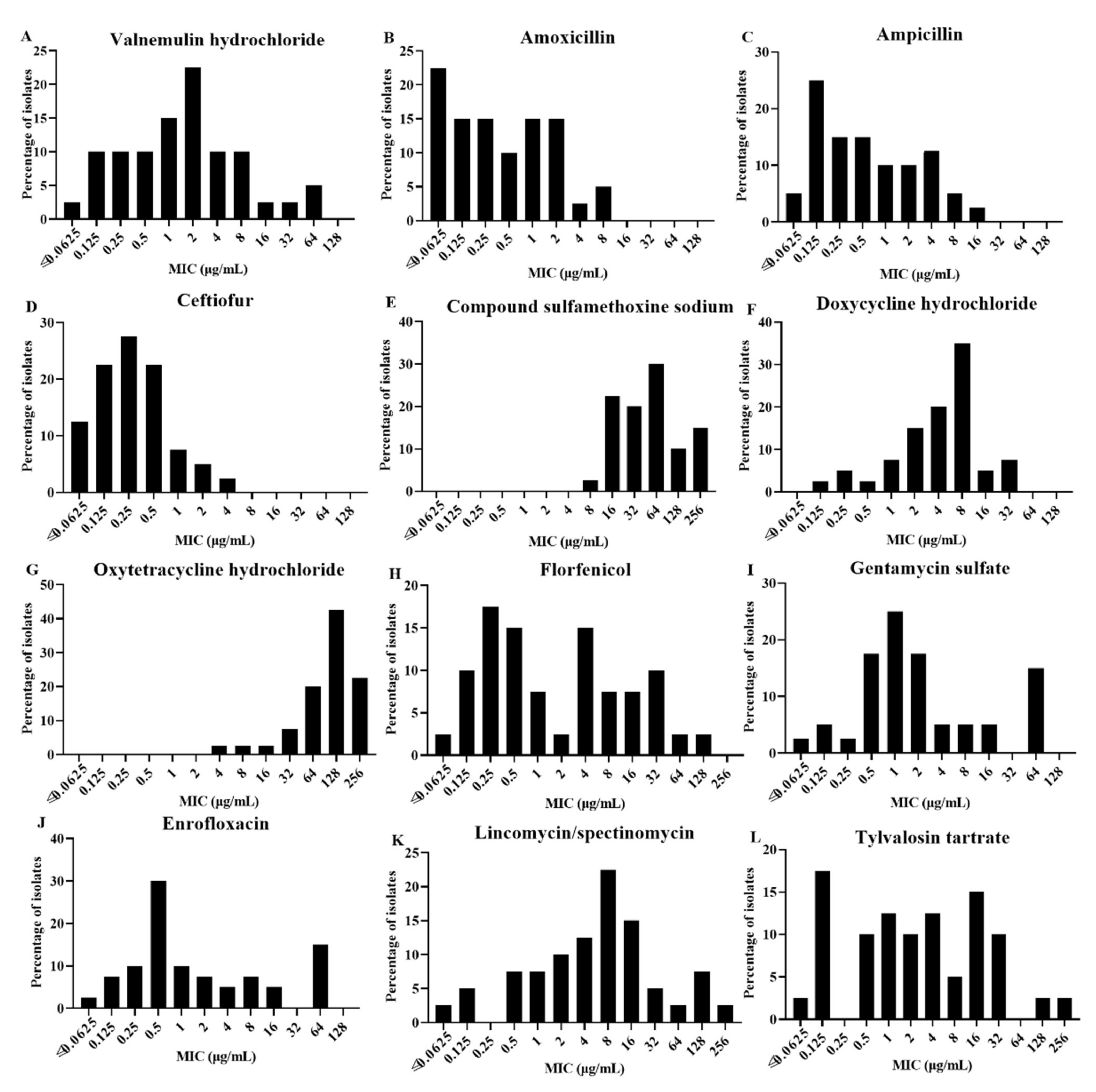
3.2. Antimicrobial Susceptibility Testing
4. Discussion
5. Conclusions
Author Contributions
Funding
Institutional Review Board Statement
Informed Consent Statement
Data Availability Statement
Acknowledgments
Conflicts of Interest
References
- Blackall, P.J.; Soriano-Vargas, E. Infectious coryza and related bacterial infections. Dis. Poult. 2020, 890–906. [Google Scholar] [CrossRef]
- Blackall, P.J.; Christensen, H.; Beckenham, T.; Blackall, L.L.; Bisgaard, M. Reclassification of Pasteurella gallinarum, [Haemophilus] paragallinarum, Pasteurella avium and Pasteurella volantium as Avibacterium gallinarum gen. nov., comb. nov., Avibacterium paragallinarum comb. nov., Avibacterium avium comb. nov. and Avibacterium volantium comb. nov. Int. J. Syst. Evol. Microbiol. 2005, 55, 353–362. [Google Scholar]
- Morales-Erasto, V.; Falconi-Agapito, F.; Luna-Galaz, G.A.; Saravia, L.E.; Montalvan-Avalos, A.; Soriano-Vargas, E.E.; Fernández-Díaz, M. Coinfection of Avibacterium paragallinarum and Ornithobacterium rhinotracheale in Chickens from Peru. Avian Dis. 2016, 60, 75–78. [Google Scholar] [CrossRef] [PubMed]
- Sandoval, V.E.; Terzolo, H.R.; Blackall, P.J. Complicated infectious coryza outbreaks in Argentina. Avian Dis. 1994, 38, 672–678. [Google Scholar] [CrossRef]
- Paudel, S.; Hess, M.; Hess, C. Coinfection of Avibacterium paragallinarum and Gallibacterium anatis in Specific-Pathogen-Free Chickens Complicates Clinical Signs of Infectious Coryza, Which Can Be Prevented by Vaccination. Avian Dis. 2017, 61, 55–63. [Google Scholar] [CrossRef] [PubMed]
- Gallardo, R.A.; Da Silva, A.P.; Egaña-Labrin, S.; Stoute, S.; Kern, C.; Zhou, H.; Cutler, G.; Corsiglia, C. Infectious Coryza: Persistence, Genotyping, and Vaccine Testing. Avian Dis. 2020, 64, 157–165. [Google Scholar] [CrossRef]
- Zhang, P.; Miao, M.; Gong, Y.; Sun, H.; Blackall, P. Infectious coryza due to Haemophilus paragallinarum serovar B in China. Aust. Vet. J. 2003, 81, 96–97. [Google Scholar] [CrossRef] [Green Version]
- Welchman Dde, B.; King, S.A.; Wragg, P.; Wood, A.M.; Irvine, R.M.; Pepper, W.J.; Dijkman, R.; de Wit, J.J. Infectious coryza in chickens in Great Britain. Vet. Rec. 2010, 167, 912–913. [Google Scholar] [CrossRef]
- Patil, V.V.; Mishra, D.; Mane, D.V. 16S ribosomal RNA sequencing and molecular serotyping of Avibacterium paragallinarum isolated from Indian field conditions. Vet. World 2017, 10, 1004–1007. [Google Scholar] [CrossRef] [PubMed] [Green Version]
- Crispo, M.; Sentíes-Cué, C.G.; Cooper, G.L.; Mountainspring, G.; Corsiglia, C.; Bickford, A.A.; Stoute, S. Otitis and meningoencephalitis associated with infectious coryza (Avibacterium paragallinarum) in commercial broiler chickens. J. Vet. Diagn. Investig. 2018, 30, 784–788. [Google Scholar] [CrossRef] [PubMed] [Green Version]
- Wahyuni, A.E.T.H.; Tabbu, C.R.; Artanto, S.; Setiawan, D.C.B.; Rajaguguk, S.I. Isolation, identification, and serotyping of Avibacterium paragallinarum from quails in Indonesia with typical infectious coryza disease symptoms. Vet. World 2018, 11, 519–524. [Google Scholar] [CrossRef] [Green Version]
- Page, L. Haemophilus infections in chickens. I. Characteristics of 12 Haemophilus isolates recovered from diseased chickens. Am. J. Vet. Res. 1962, 23, 85. [Google Scholar]
- Blackall, P.J.; Eaves, L.E.; Rogers, D.G. Proposal of a new serovar and altered nomenclature for Haemophilus paragallinarum in the Kume hemagglutinin scheme. J. Clin. Microbiol. 1990, 28, 1185–1187. [Google Scholar] [CrossRef] [PubMed] [Green Version]
- Kume, K.; Sawata, A.; Nakai, T.; Matsumoto, M. Serological classification of Haemophilus paragallinarum with a hemagglutinin system. J. Clin. Microbiol. 1983, 17, 958–964. [Google Scholar] [CrossRef] [PubMed] [Green Version]
- Xu, Y.; Cheng, J.; Huang, X.; Xu, M.; Feng, J.; Liu, C.; Zhang, G. Characterization of emergent Avibacterium paragallinarum strains and the protection conferred by infectious coryza vaccines against them in China. Poult. Sci. 2019, 98, 6463–6471. [Google Scholar] [CrossRef]
- Blackall, P.J. Infectious coryza: Overview of the disease and new diagnostic options. Clin. Microbiol. Rev. 1999, 12, 627–632. [Google Scholar] [CrossRef] [PubMed] [Green Version]
- Yamaguchi, T.; Blackall, P.J.; Takigami, S.; Iritani, Y.; Hayashi, Y. Immunogenicity of Haemophilus paragallinarum serovar B strains. Avian Dis. 1991, 965–968. [Google Scholar] [CrossRef]
- Soriano, E.V.; Garduño, M.L.; Téllez, G.; Rosas, P.F.; Suárez-Güemes, F.; Blackall, P.J. Cross-protection study of the nine serovars of Haemophilus paragallinarum in the Kume haemagglutinin scheme. Avian Pathol. 2004, 33, 506–511. [Google Scholar] [CrossRef] [PubMed]
- Heuvelink, A.; Wiegel, J.; Kehrenberg, C.; Dijkman, R.; Soriano-Vargas, E.; Feberwee, A. Antimicrobial susceptibility of Avibacterium paragallinarum isolates from outbreaks of infectious coryza in Dutch commercial poultry flocks, 2008–2017. Vet. Microbiol. 2018, 217, 135–143. [Google Scholar] [CrossRef] [PubMed]
- Chukiatsiri, K.; Sasipreeyajan, J.; Blackall, P.J.; Yuwatanichsampan, S.; Chansiripornchai, N. Serovar identification, antimicrobial sensitivity, and virulence of Avibacterium paragallinarum isolated from chickens in Thailand. Avian Dis. 2012, 56, 359–364. [Google Scholar] [CrossRef] [PubMed]
- Jeong, O.-M.; Kang, M.-S.; Jeon, B.-W.; Choi, B.-K.; Kwon, Y.-K.; Yoon, S.-Y.; Blackall, P.J.; Lee, H.-S.; Jung, S.-C.; Kim, J.-H. Isolation and characterization of Avibacterium paragallinarum with different nicotinamide adenine dinucleotide requirements. Vet. Microbiol. 2017, 205, 62–65. [Google Scholar] [CrossRef]
- Luna-Galaz, G.A.; Morales-Erasto, V.; Peñuelas-Rivas, C.G.; Soriano-Vargas, E.; Blackall, P.J. Antimicrobial sensitivity of Avibacterium paragallinarum isolates from four Latin American countries. Avian Dis. 2016, 60, 673–676. [Google Scholar] [CrossRef]
- Chen, X.; Miflin, J.K.; Zhang, P.; Blackall, P. Development and application of DNA probes and PCR tests for Haemophilus paragallinarum. Avian Dis. 1996, 40, 398–407. [Google Scholar] [CrossRef]
- Yamaguchi, T.; Blackall, P.J.; Takigami, S.; Iritani, Y.; Hayashi, Y. Pathogenicity and serovar-specific hemagglutinating antigens of Haemophilus paragallinarum serovar B strains. Avian Dis. 1990, 34, 964–968. [Google Scholar] [CrossRef]
- Han, M.-S.; Kim, J.-N.; Jeon, E.-O.; Lee, H.-R.; Koo, B.-S.; Min, K.-C.; Lee, S.-B.; Bae, Y.-J.; Mo, J.-S.; Cho, S.-H.; et al. The current epidemiological status of infectious coryza and efficacy of PoulShot Coryza in specific pathogen-free chickens. J. Vet. Sci. 2016, 17, 323–330. [Google Scholar] [CrossRef] [PubMed]
- Soriano, V.E.; Blackall, P.J.; Dabo, S.M.; Tellez, G.; Garcia-Delgado, G.A.; Fernandez, R.P. Serotyping of Haemophilus paragallinarum isolates from Mexico by the Kume hemagglutinin scheme. Avian Dis. 2001, 45, 680–683. [Google Scholar] [CrossRef] [PubMed]
- Jacobs, A.A.; van den Berg, K.; Malo, A. Efficacy of a new tetravalent coryza vaccine against emerging variant type B strains. Avian Pathol. 2003, 32, 265–269. [Google Scholar] [CrossRef] [PubMed] [Green Version]
- Sun, H.; Xie, S.; Li, X.; Xu, F.; Li, Y.; Boucher, C.E.; Chen, X. Selection of Avibacterium paragallinarum Page serovar B strains for an infectious coryza vaccine. Vet. Immunol. Immunopathol. 2018, 199, 77–80. [Google Scholar] [CrossRef] [PubMed]
- Tokunaga, E.; Sakaguchi, M.; Matsuo, K.; Hamada, F.; Tokiyoshi, S. Polypeptide for Haemophilus paragallinarum and Process for Preparing the Same; US6919080B2; Juridical Foundation The Chemo-Sero-Therapeutic Research Institute: Kumamoto-Ken, Japan, 2002. [Google Scholar]
- Wang, Y.-P.; Hsieh, M.-K.; Tan, D.-H.; Shien, J.-H.; Ou, S.-C.; Chen, C.-F.; Chang, P.-C. The haemagglutinin of Avibacterium paragallinarum is a trimeric autotransporter adhesin that confers haemagglutination, cell adherence and biofilm formation activities. Vet. Microbiol. 2014, 174, 474–482. [Google Scholar] [CrossRef]
- Sakamoto, R.; Baba, S.; Ushijima, T.; Kino, Y.; Honda, T.; Mizokami, H.; Sakaguchi, M. Development of a recombinant vaccine against infectious coryza in chickens. Res. Vet. Sci. 2013, 94, 504–509. [Google Scholar] [CrossRef] [PubMed]
- Wu, J.-R.; Wu, Y.-R.; Shien, J.-H.; Hsu, Y.-M.; Chen, C.-F.; Shieh, H.K.; Chang, P.-C. Recombinant proteins containing the hypervariable region of the haemagglutinin protect chickens against challenge with Avibacterium paragallinarum. Vaccine 2011, 29, 660–667. [Google Scholar] [CrossRef] [PubMed]
- Noro, T.; Yaguchi, K.; Amimoto, K.; Oishi, E. Identification and expression of a gene encoding an epitope that induces hemagglutination inhibition antibody to Avibacterium paragallinarum serovar A. Avian Dis. 2007, 51, 84–89. [Google Scholar] [CrossRef]
- Sakamoto, R.; Kino, Y.; Sakaguchi, M. Development of a multiplex PCR and PCR-RFLP method for serotyping of Avibacterium paragallinarum. J. Vet. Med. Sci. 2012, 74, 271–273. [Google Scholar] [CrossRef] [PubMed] [Green Version]
- Wang, H.; Sun, H.; Blackall, P.J.; Zhang, Z.; Zhou, H.; Xu, F.; Chen, X. Evaluation of a proposed molecular methodology for the serotyping of Avibacterium paragallinarum. J. Vet. Diagn. Investig. 2016, 28, 555–560. [Google Scholar] [CrossRef] [PubMed] [Green Version]
- Lublin, A.; Mechani, S.; Malkinson, M.; Weisman, Y. Efficacy of norfloxacin nicotinate treatment of broiler breeders against Haemophilus paragallinarum. Avian Dis. 1993, 37, 673–679. [Google Scholar] [CrossRef] [PubMed]
- Hanley, J.E.; Davis, R.B.; Sunka, E.M. An evaluation and comparison of spectinomycin and spectinomycin-erythromycin combinations for infectious coryza. Avian Dis. 1968, 12, 1–3. [Google Scholar] [CrossRef]
- Noonkhokhetkong, T.; Chukiatsiri, K.; Sasipreeyajan, J.; Chansiripornchai, N. Determination of Antimicrobial Susceptibility, Antimicrobial Resistance Genes and in vivo Testing of Antimicrobial Susceptibility of Avibacterium paragallinarum. Thai J. Vet. Med. 2013, 43, 525–531. [Google Scholar]
- Blackall, P.J.; Eaves, L.E.; Rogers, D.G. Biotyping of Haemophilus paragallinarum isolates using hemagglutinin serotyping, carbohydrate fermentation patterns, and antimicrobial drug resistance patterns. Avian Dis. 1989, 33, 491–496. [Google Scholar] [CrossRef] [PubMed]
- Hsu, Y.-M.; Shieh, H.K.; Chen, W.-H.; Sun, T.-Y.; Shiang, J.-H. Antimicrobial susceptibility, plasmid profiles and haemocin activities of Avibacterium paragallinarum strains. Vet. Microbiol. 2007, 124, 209–218. [Google Scholar] [CrossRef] [PubMed]
- Poernomo, S.; Sutarma, S.; Rafiee, M.; Blackall, P. Characterisation of isolates of Haemophilus paragallinarum from Indonesia. Aust. Vet. J. 2000, 78, 759–762. [Google Scholar] [CrossRef] [PubMed] [Green Version]

| Farm ID | Isolates | Origin | Production Type/Breed of Host | Size of Flocks | Husbandry Systems | Age | Time | Serovar |
|---|---|---|---|---|---|---|---|---|
| A | 2019/JS03 | Jiangsu | Hy-line brown, layer | 2,000,000 | H-type stacked-cage | 326 | 2019/04 | A |
| A | 2019/JS07 | Jiangsu | Hy-line brown, layer | 2,000,000 | H-type stacked-cage | 264 | 2019/04 | A |
| B | 2019/JS08 | Jiangsu | Hy-line brown, layer | 1,200,000 | H-type stacked-cage | 241 | 2019/04 | A |
| B | 2019/JS15 | Jiangsu | Hy-line brown, layer | 1,200,000 | H-type stacked-cage | 316 | 2019/04 | A |
| C | 2019/JS28 | Jiangsu | Hy-line brown, layer | 500,000 | H-type stacked-cage | 207 | 2019/04 | A |
| D | 2019/JS31 | Jiangsu | Hy-line brown, layer | 600,000 | H-type stacked-cage | 255 | 2019/07 | B |
| D | 2019/JS33 | Jiangsu | Hy-line brown, layer | 600,000 | H-type stacked-cage | 212 | 2019/07 | B |
| A | 2019/JS34 | Jiangsu | Hy-line brown, layer | 2,000,000 | H-type stacked-cage | 278 | 2019/07 | B |
| A | 2019/JS35 | Jiangsu | Hy-line brown, layer | 2,000,000 | H-type stacked-cage | 278 | 2019/07 | B |
| E | 2019/JS36 | Jiangsu | Hy-line brown, layer | 1,000,000 | H-type stacked-cage | 322 | 2019/07 | B |
| E | 2019/JS37 | Jiangsu | Hy-line brown, layer | 1,000,000 | H-type stacked-cage | 318 | 2019/07 | B |
| E | 2019/JS38 | Jiangsu | Hy-line brown, layer | 1,000,000 | H-type stacked-cage | 42 | 2019/07 | B |
| F | 2019/JS39 | Jiangsu | Jing Brown No. 1, layer | 300,000 | H-type stacked-cage | 78 | 2019/07 | B |
| F | 2019/JS40 | Jiangsu | Jing Brown No. 1, layer | 300,000 | H-type stacked-cage | 133 | 2019/07 | B |
| F | 2019/JS42 | Jiangsu | Jing Brown No. 1, layer | 300,000 | H-type stacked-cage | 311 | 2019/07 | A |
| F | 2019/JS44 | Jiangsu | Jing Brown No. 1, layer | 300,000 | H-type stacked-cage | 332 | 2019/07 | C |
| G | 2019/JS45 | Jiangsu | Nongda 3, layer | 600,000 | H-type stacked-cage | 276 | 2019/07 | A |
| G | 2019/JS46 | Jiangsu | Nongda 3, layer | 600,000 | H-type stacked-cage | 306 | 2019/07 | C |
| H | 2019/ NX56 | Ningxia | Hy-line brown, layer parent stock | 300,000 | Net-rearing | 333 | 2019/10 | C |
| H | 2019/ NX57 | Ningxia | Hy-line brown, layer parent stock | 300,000 | Net-rearing | 324 | 2019/10 | C |
| H | 2019/ NX58 | Ningxia | Hy-line brown, layer parent stock | 300,000 | Net-rearing | 312 | 2019/10 | C |
| I | 2019/ HB63 | Hebei | Hy-line brown, layer parent stock | 300,000 | Natural mating cage | 103 | 2019/11 | A |
| I | 2019/ HB64 | Hebei | Hy-line brown, layer parent stock | 300,000 | Natural mating cage | 128 | 2019/11 | A |
| I | 2019/ HB65 | Hebei | Hy-line brown, layer parent stock | 300,000 | Natural mating cage | 164 | 2019/11 | A |
| I | 2019/ HB68 | Hebei | Hy-line brown, layer parent stock | 300,000 | Natural mating cage | 164 | 2019/11 | B |
| J | 2020/JS69 | Jiangsu | Hy-line brown, layer | 100,000 | H-type stacked-cage | 273 | 2020/10 | A |
| J | 2020/JS70 | Jiangsu | Hy-line brown, layer | 100,000 | H-type stacked-cage | 266 | 2020/10 | C |
| F | 2020/JS71 | Jiangsu | Jing Brown No. 1, layer | 300,000 | H-type stacked-cage | 213 | 2020/10 | C |
| F | 2020/JS72 | Jiangsu | Jing Brown No. 1, layer | 300,000 | H-type stacked-cage | 298 | 2020/10 | C |
| F | 2020/JS73 | Jiangsu | Jing Brown No. 1, layer | 300,000 | H-type stacked-cage | 246 | 2020/10 | C |
| B | 2020/JS74 | Jiangsu | Hy-line brown, layer | 1,200,000 | H-type stacked-cage | 323 | 2020/10 | C |
| B | 2020/JS75 | Jiangsu | Hy-line brown, layer | 1,200,000 | H-type stacked-cage | 302 | 2020/10 | C |
| B | 2020/JS76 | Jiangsu | Hy-line brown, layer | 1,200,000 | H-type stacked-cage | 195 | 2020/10 | C |
| G | 2020/JS77 | Jiangsu | Nongda 3, layer | 600,000 | H-type stacked-cage | 311 | 2020/11 | C |
| F | 2020/JS78 | Jiangsu | Hy-line brown, layer | 300,000 | H-type stacked-cage | 204 | 2020/11 | C |
| G | 2020/JS79 | Jiangsu | Nongda 3, layer | 600,000 | H-type stacked-cage | 362 | 2020/11 | C |
| G | 2020/JS80 | Jiangsu | Nongda 3, layer | 600,000 | H-type stacked-cage | 188 | 2020/11 | C |
| K | 2020/JS81 | Jiangsu | Hy-line brown, layer | 200,000 | H-type stacked-cage | 76 | 2020/11 | C |
| K | 2020/JS82 | Jiangsu | Hy-line brown, layer | 200,000 | H-type stacked-cage | 34 | 2020/11 | C |
| L | 2020/JS83 | Jiangsu | Hy-line brown, layer | 300,000 | H-type stacked-cage | 57 | 2020/11 | C |
| Strains | MIC (μg/mL) | |||||||||||
|---|---|---|---|---|---|---|---|---|---|---|---|---|
| VA | AMX | CTF | AMP | COSMMS | DO | OT | FFC | CN | ENR | LS | TAT | |
| 221 | 0.125 | 0.125 | 0.25 | ≤0.0625 | ≤0.0625 | 2 | 1 | 1 | 0.125 | 0.5 | 32 | 0.25 |
| Spross | ≤0.0625 | ≤0.0625 | 0.25 | 0.5 | ≤0.0625 | ≤0.0625 | 0.125 | 2 | 0.25 | 0.125 | 0.5 | 0.5 |
| H-18 | 0.25 | 0.25 | 0.25 | 0.25 | ≤0.0625 | 2 | 16 | 0.5 | 0.125 | 1 | 16 | 0.5 |
| 2019/JS03 | 1 | ≤0.0625 | 0.25 | 0.25 | 16 | 2 | 32 | 8 | 0.5 | 0.25 | 4 | 2 |
| 2019/JS07 | 2 | 0.25 | 0.5 | 2 | 32 | 4 | 64 | 8 | 2 | 1 | 16 | 16 |
| 2019/JS08 | 1 | 0.5 | ≤0.0625 | 0.25 | 32 | 4 | 32 | 4 | 0.5 | 0.25 | 16 | 8 |
| 2019/JS15 | 1 | 0.25 | 0.25 | 0.5 | 32 | 8 | 64 | 1 | 1 | 2 | 8 | 16 |
| 2019/JS28 | 32 | 8 | 2 | 4 | 64 | 8 | 32 | 16 | 16 | 64 | 16 | 16 |
| 2019/JS42 | 64 | 1 | 0.5 | 2 | 256 | 32 | 256 | 64 | 64 | 64 | 256 | 256 |
| 2019/JS45 | 64 | 1 | 0.5 | 4 | 256 | 32 | 128 | 128 | 64 | 64 | 128 | 128 |
| 2019/HB63 | 4 | 4 | 0.25 | 8 | 256 | 4 | 64 | 32 | 64 | 8 | 32 | 1 |
| 2019/HB64 | 2 | 1 | 0.5 | 1 | 128 | 16 | 256 | 0.5 | 64 | 64 | 16 | 32 |
| 2019/HB65 | 2 | 0.25 | 0.25 | 1 | 64 | 32 | 256 | 4 | 64 | 64 | 16 | 32 |
| 2020/JS69 | 0.25 | 0.125 | 0.125 | 8 | 32 | 1 | 64 | 0.25 | 0.125 | 0.125 | 0.125 | 0.125 |
| 2019/JS31 | 2 | 0.5 | 1 | 0.5 | 128 | 8 | 128 | 4 | 1 | 8 | 128 | 32 |
| 2019/JS33 | 8 | 2 | 1 | 0.5 | 256 | 4 | 128 | 8 | 1 | 8 | 128 | 16 |
| 2019/JS34 | 16 | 0.5 | 0.25 | 0.5 | 256 | 8 | 256 | 4 | 1 | 1 | 64 | 32 |
| 2019/JS35 | 0.5 | 1 | 0.125 | 0.125 | 32 | 8 | 128 | 1 | 1 | 4 | 4 | 2 |
| 2019/JS36 | 0.0625 | 0.0625 | 0.125 | 0.125 | 128 | 16 | 128 | 32 | 1 | 0.5 | 8 | 2 |
| 2019/JS37 | 2 | 2 | 2 | 4 | 256 | 8 | 256 | 32 | 8 | 16 | 32 | 8 |
| 2019/JS38 | 1 | 1 | 0.5 | 1 | 128 | 8 | 128 | 16 | 1 | 2 | 8 | 4 |
| 2019/JS39 | 4 | 2 | 0.5 | 0.25 | 64 | 8 | 128 | 32 | 1 | 4 | 2 | 4 |
| 2019/JS40 | 8 | 0.25 | 1 | 16 | 64 | 8 | 64 | 16 | 1 | 1 | 16 | 4 |
| 2019/HB68 | 2 | 0.5 | 0.125 | 0.125 | 64 | 4 | 64 | 0.5 | 1 | 0.5 | 8 | 4 |
| 2019/JS44 | 0.125 | ≤0.0625 | 0.25 | 0.0625 | 32 | 2 | 64 | 0.25 | 0.5 | 0.5 | 2 | 0.5 |
| 2019/JS46 | 0.125 | 0.125 | 4 | 0.0625 | 16 | 1 | 64 | 0.125 | 64 | 64 | 4 | 1 |
| 2019/NX56 | 2 | 2 | 0.5 | 2 | 8 | 1 | 4 | 0.25 | 0.5 | 1 | 0.125 | 0.5 |
| 2019/NX57 | 0.5 | 0.25 | 0.125 | 0.125 | 16 | 0.5 | 8 | 0.5 | 0.25 | 16 | 4 | 1 |
| 2019/NX58 | 0.5 | 0.0625 | 0.0625 | 0.5 | 16 | 4 | 16 | 4 | 0.0625 | 0.125 | 2 | 0.0625 |
| 2020/JS70 | 2 | 1 | 0.25 | 0.5 | 32 | 8 | 128 | 2 | 0.125 | 0.0625 | 0.0625 | 0.5 |
| 2020/JS71 | 8 | 2 | 0.125 | 0.25 | 64 | 8 | 128 | 0.5 | 2 | 0.5 | 8 | 16 |
| 2020/JS72 | 1 | 0.125 | 0.5 | 2 | 16 | 0.125 | 128 | 0.25 | 0.5 | 0.5 | 1 | 0.125 |
| 2020/JS73 | 8 | 0.0625 | 0.0625 | 0.25 | 64 | 8 | 128 | 1 | 2 | 0.5 | 8 | 16 |
| 2020/JS74 | 4 | 2 | 0.25 | 4 | 16 | 2 | 256 | 0.25 | 4 | 0.5 | 8 | 1 |
| 2020/JS75 | 0.125 | 0.0625 | 0.5 | 0.125 | 16 | 0.25 | 256 | 4 | 0.5 | 0.5 | 0.5 | 0.5 |
| 2020/JS76 | 2 | 0.25 | 0.25 | 4 | 32 | 4 | 128 | 0.5 | 4 | 0.5 | 8 | 4 |
| 2020/JS77 | 1 | 0.0625 | 0.0625 | 0.125 | 16 | 0.25 | 128 | 0.25 | 2 | 0.5 | 0.5 | 1 |
| 2020/JS78 | 0.25 | 0.0625 | 0.25 | 0.25 | 64 | 8 | 128 | 0.125 | 0.5 | 0.5 | 4 | 0.125 |
| 2020/JS79 | 0.125 | 0.125 | 0.25 | 1 | 64 | 2 | 128 | 0.125 | 8 | 0.125 | 2 | 0.125 |
| 2020/JS80 | 0.5 | 0.125 | 0.125 | 0.125 | 64 | 4 | 256 | 0.0625 | 2 | 0.25 | 1 | 2 |
| 2020/JS81 | 0.25 | 0.0625 | 0.0625 | 0.125 | 16 | 2 | 256 | 0.5 | 2 | 0.25 | 0.5 | 0.125 |
| 2020/JS82 | 0.25 | 0.125 | 0.125 | 0.125 | 64 | 8 | 128 | 0.25 | 2 | 0.5 | 8 | 0.125 |
| 2020/JS83 | 4 | 8 | 0.125 | 0.125 | 64 | 2 | 128 | 0.125 | 16 | 2 | 1 | 0.125 |
Publisher’s Note: MDPI stays neutral with regard to jurisdictional claims in published maps and institutional affiliations. |
© 2022 by the authors. Licensee MDPI, Basel, Switzerland. This article is an open access article distributed under the terms and conditions of the Creative Commons Attribution (CC BY) license (https://creativecommons.org/licenses/by/4.0/).
Share and Cite
Guo, M.; Chen, X.; Zhang, H.; Liu, D.; Wu, Y.; Zhang, X. Isolation, Serovar Identification, and Antimicrobial Susceptibility of Avibacterium paragallinarum from Chickens in China from 2019 to 2020. Vet. Sci. 2022, 9, 27. https://doi.org/10.3390/vetsci9010027
Guo M, Chen X, Zhang H, Liu D, Wu Y, Zhang X. Isolation, Serovar Identification, and Antimicrobial Susceptibility of Avibacterium paragallinarum from Chickens in China from 2019 to 2020. Veterinary Sciences. 2022; 9(1):27. https://doi.org/10.3390/vetsci9010027
Chicago/Turabian StyleGuo, Mengjiao, Xiufang Chen, Hao Zhang, Donghui Liu, Yantao Wu, and Xiaorong Zhang. 2022. "Isolation, Serovar Identification, and Antimicrobial Susceptibility of Avibacterium paragallinarum from Chickens in China from 2019 to 2020" Veterinary Sciences 9, no. 1: 27. https://doi.org/10.3390/vetsci9010027
APA StyleGuo, M., Chen, X., Zhang, H., Liu, D., Wu, Y., & Zhang, X. (2022). Isolation, Serovar Identification, and Antimicrobial Susceptibility of Avibacterium paragallinarum from Chickens in China from 2019 to 2020. Veterinary Sciences, 9(1), 27. https://doi.org/10.3390/vetsci9010027