Article

First Isolation and Multilocus Sequence Typing of Brucella canis from a Subclinically Infected Pet Dog in China

1
College of Animal Science, Xichang University, Xichang 615000, China
2
Key Laboratory of Animal Disease and Human Health of Sichuan, College of Veterinary Medicine, Sichuan Agricultural University, Chengdu 611130, China
*
Author to whom correspondence should be addressed.
These authors have contributed equally to this work.
Vet. Sci. 2022, 9(1), 22; https://doi.org/10.3390/vetsci9010022
Submission received: 24 November 2021 / Revised: 22 December 2021 / Accepted: 7 January 2022 / Published: 10 January 2022
(This article belongs to the Special Issue Bacterial Infectious Diseases of Companion Animals)

Abstract

:
Canine brucellosis, a worldwide zoonotic disease, is mainly caused by Brucella canis. In the present study, we isolated a Brucella strain (CD3) from a subclinically infected pet dog in Sichuan Province, Southwestern China. Classical biotyping methods and molecular biological tests (BCSP31 and BcSS PCR) proved that the strain belonged to B. canis. Furthermore, B. canis CD3 and another two B. canis strains (WJ5 and YA4), which were all isolated from pet dogs in Sichuan, were genotyped using multilocus sequence typing (MLST). Our results showed that the three B. canis strains were identified as the same sequence type (ST21). The present study is the first to report B. canis strain from a subclinically infected pet dog in China, indicating a potential threat to public health posed by subclinical infections in pet dogs. We suggest that screening for B. canis should be incorporated into routine medical examination of pet dogs and other companion animals in areas with a history of animal or human brucellosis.

1. Introduction

Brucellosis, caused by Gram-negative, aerobic and facultative intracellular bacteria of the genus Brucella, is regarded as one of the most common zoonotic diseases worldwide [1,2]. Brucella canis is generally recognized as the pathogen of canine brucellosis, which primarily causes reproductive failure in dogs, including infertility and abortion [3]. B. canis infections in dogs have been increasing in China and other countries in recent years [3,4,5]. B. canis can also infect humans and cause severe diseases [5]. Human cases have been reported in many countries, such as China, the United States, Argentina, and Japan [6,7,8,9,10,11,12]. In some of these cases, pet dogs without clinical symptoms that had close contact with human brucellosis patients have been regarded as the source of infection [8]. As the number of pet dogs rises, the potential risk of human brucellosis due to the direct or indirect transfer from dogs to people is increased [13]. China is the most populous country in the world, and has raised about 50.08 million pet dogs as of 2018 [14]. The occurrence of B. canis infection in pet dogs in China was reported by our lab in 2015, in which the infected dog had clinical symptoms such as fever, enlarged lymph nodes and enlarged testicle [5]. However, subclinically infected pet dogs have not been reported in China.
Multilocus sequence typing (MLST) is a DNA sequence-based typing method that has been widely applied to microbial typing and epidemiological studies [15,16,17]. It was first used to genotype Brucella in 2007 and the result showed high differentiation among different species of Brucella strains [15]. Recent MLST studies on Brucella in China mainly focused on B. abortus, B. melitensis, and B. suis. The information of MLST of B. canis in China is relatively few. In 2019, Liu et al. had genotyped seven B. canis strains in Guangxi Province as ST21, enlarging our understanding of the genetic structure and the genetic diversity of B. canis population [18]. However, as far as we know, no report existed on MLST of B. canis in Sichuan Province.
In this study, bacteriological and molecular biological tests (BCSP31 and BcSS PCR) were used to identify a B. canis isolation (strain CD3) from a subclinically infected pet dog, and MLST method was then used to genotype the strain CD3 and another two B. canis strains (WJ5 and YA4), which were also isolated from pet dogs in Sichuan Province, China. For the first time, we isolated a B. canis strain from a subclinically infected pet dog in China, revealing a potential threat of subclinical infections in pet dogs to public health.

2. Materials and Methods

2.1. Clinical Sample

In August 2019, one male pet dog (golden retriever), aged three years and seven months, was tested for Brucella infection in Veterinary Medical Teaching Hospital of Sichuan Agricultural University at the request of the pregnant owner. Physical examination of the pet dog showed no clinical symptoms (medical history of reproductive ability was unknown). Blood samples were aseptically collected from a cephalic vein of the pet dog for serological tests and strain isolation.
In December 2015, one male dog (golden retriever), aged two years and five months, presented with low fever, enlarged lymph nodes, and unilateral testis. In October 2016, a male poodle, aged three years, presented with obviously enlarged testicle and undulating fever. Both pet dogs were diagnosed with canine brucellosis by Veterinary Medical Teaching Hospital of Sichuan Agricultural University. Two B. canis strains (WJ5, 2015 and YA4, 2016) were successfully isolated from the two cases and were stored in our lab [5].

2.2. Serological Tests

The Rose Bengal plate test (RBPT) and rapid slide agglutination test (RSAT) were performed with rough antigen to detect antibodies against Brucella in blood samples, as described previously [19].

2.3. Bacteriological Studies

Blood samples were plated and streaked on tryptic soy agar (Beijing Selarbio Science and Technology Co., Ltd., Beijing, China) for strain isolation. Plates were inoculated at 37 °C over 5 days [5]. Classical biotyping methods (including CO2 requirement, H2S production, hydrolysis of urea, agglutination in sera, growth on dyes and phage lysis tests) [20,21] were used to identify the isolated strain (named as CD3), and B. canis RM6/66 was used as a reference strain. All studies involving living bacteria isolation were performed under biosafety level 3 (BSL-3) laboratory (DYY-2019303075).

2.4. DNA Extraction

Before DNA extraction, specimens were boiled for 15 min. Brucella DNA was extracted using TIANamp Bacteria DNA extraction kit (TIANGEN Biotech Corporation, Beijing, China) according to the manufacturer’s instructions [5].

2.5. Polymerase Chain Reaction Tests

The isolated strain (CD3) was identified by two different PCR protocols using Brucella genus-specific (BCSP31) and B. canis species-specific (BcSS) primers, respectively. PCR amplification was conducted as described previously [13,22] and primers are listed in Table 1. B. abortus 544A, B. melitensis 16M, B. suis S2, and B. canis RM6/66 were used as reference strains. The PCR products (6 µL from each reaction mixture) were loaded for electrophoresis using a 1.5% agarose gel, after which the gel was stained with ethidium bromide and photographed [5].

2.6. MLST Genotyping

MLST genotyping was performed on the isolated B. canis strain (CD3). Two B. canis strains (WJ5 and YA4) from clinical cases of canine brucellosis in Sichuan were also analyzed [5]. Nine distinct genomic loci were analyzed, including seven housekeeping genes (gap, aroA, glk, dnaK, gyrB, trpE, and cobQ), one outer membrane protein (omp25), and one intergenic fragment (int_hyp) [15]. PCR amplification of the nine loci was performed, as described previously [15]. Primers are shown in Table 1. Sequences obtained from purified PCR products were aligned using MEGA X according to published MLST sequences in GenBank (accession numbers AM694191–AM695630) [17]. Distinct alleles identified at the nine selected loci were each given a numerical designation using a web-based MLST service (Brucella Base, https://pubmlst.org/Brucella, accessed on 9 August 2021) [17], and each unique allelic profile for the nine loci was identified as a sequence type (ST).

2.7. Analysis of MLST Data

Sequences of the nine loci were concatenated to produce a 4396 bp sequence for each genotype (ST). Sequence data of 3 B. canis strains (CD3, WJ5 and YA4) from pet dogs and 66 Brucella isolates in MLST database (https://pubmlst.org/Brucella, accessed on 9 August 2021), including four most common species (B. abortus, B. melitensis, B. suis, and B. canis) and two emerging species from marine mammals (B. ceti and B. pinnipedialis), were chosen for phylogenetic analysis using MEGA X. Neighbor joining trees were constructed using the Jukes–Cantor model and the percentage bootstrap confidence levels of internal branches were calculated from 1000 resamplings of the original data [15].

3. Results

3.1. Serological and Bacteriological Tests

RBPT and RSAT assays showed positive results for the tested blood samples. A suspected Brucella strain (CD3) was successfully isolated from the blood sample by tryptic soy agar. Further bacteriological tests showed that the results of CO2 requirement, H2S production and phage lysis tests were negative, while hydrolysis of urea, growth on thionin and agglutination in sera R were positive. The biotyping characteristics of the strain (CD3) were in accord with that of B. canis RM6/66 (Table 2).

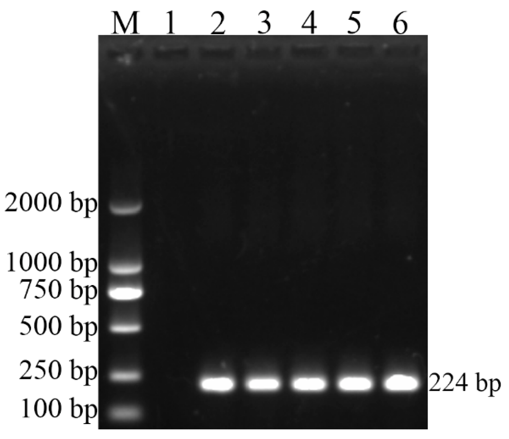
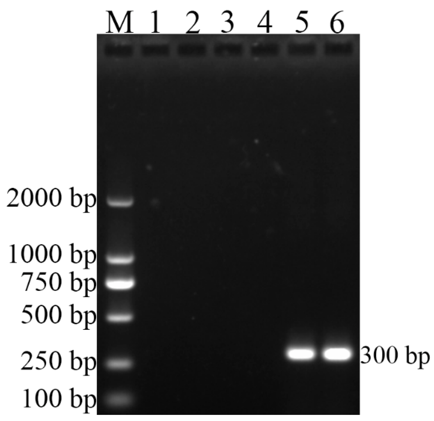
3.2. Polymerase Chain Reaction Tests

As Figure 1 shows, the Brucella genus-specific PCR (BCSP31-PCR), using all five strains (544A, 16M, S2, RM6/66, and CD3), successfully produced 224-bp PCR amplicon, indicating that CD3 strain belongs to the genus Brucella. Further B. canis species-specific PCR (BcSS-PCR) amplified a specific 300-bp amplicon (Figure 2), suggesting that the CD3 isolate is B. canis. All amplification results of PCR are listed in Table 3.

3.3. MLST Genotyping

Details of allelic profile of each Brucella strain are listed in Table 4. The MLST results showed that the isolated B. canis strain (CD3) and two B. canis strains stored in our lab (WJ5 and YA4) have the same allelic profile, and all three B. canis strains from pet dogs in Sichuan belong to genotype ST21.
In order to better understand the genetic relationship of the three B. canis strains (CD3, WJ5 and YA4) isolated from pet dogs, sequences of the nine loci were concatenated to conduct phylogenetic analysis with 66 Brucella isolates in the MLST database. As the dendrogram shows (Figure 3), four major clusters (A–D) were formed. The three B. canis strains (CD3, WJ5 and YA4) were clustered together with B. canis and B. suis strains. B. ceti and B. pinnipedialis were all grouped into cluster B together. B. abortus were grouped into cluster C, which split into two subclusters (cluster C1 and C2). B. melitensis were clustered into cluster D.

4. Discussion

In this study, we diagnosed a pet dog without clinical symptoms as B. canis infection in Sichuan Province, China, and a B. canis strain (CD3) was successfully isolated. Canine brucellosis is a zoonotic disease that has been underestimated worldwide, posing a public health risk to dogs and humans [3,13,23,24]. In China, serological data on brucellosis in pet dogs are relatively limited. An epidemiological study on brucellosis among 415 pet dogs between 2006–2007 in Beijing showed the seroprevalence was 0.24% (1/415) [25]. In 2012–2013, another study for pet dogs showed the incidence rate was 47.37% (18/38) [26]. These data reveal that the seroprevalence of brucellosis in pet dogs in China has increased in recent years. B. canis strains have been identified from dogs after the first isolation in 1984 [4]. Reported infected pet dogs usually showed clinical symptoms, including abortion, low fever, enlarged lymph nodes, obviously enlarged testicle and undulating fever [5,26,27]. In the present study, a pet dog without any clinical symptoms was diagnosed as subclinical infection with B. canis. The dog showed healthy on physical examination, while B. canis infection was revealed by serological screening and confirmed by subsequent bacteriological and molecular biological tests. This hidden case of B. canis infection in pet dog was reported in China for the first time, which will raise public health concerns about contracting B. canis. According to other reports, B. canis can be transferred from pet dogs to humans through direct contact with infected dogs or their blood or reproductive samples (aborted material, seminal fluid, vaginal discharge, and so forth), and even the spayed or neutered dogs can still shed bacteria in their secretions and urine [3,23]. However, due to the lack of indication of corresponding clinical symptoms, subclinically infected pet dogs are not easy to be detected by owners or veterinarians and may become a risky source of human and animal brucellosis. Transmission of B. canis from asymptomatically infected pet dog to human has been reported previously [8]. In New York, an eight-week-old male Yorkshire Terrier, which appeared healthy upon physical exam, was considered as the source of a three-year-old child’s B. canis infection because of the genetic similarity between B. canis isolates from the child and the pet dog [8]. In addition, human cases of B. canis infection have been reported in many countries, including China, the United States, Argentina, and Japan [6,7,8,9,10,11,12]. Considering the convenience and covertness of transmission of B. canis from pet dogs to humans, we suggest incorporating B. canis screening into the routine medical examination of pet dogs. Reproductive failure (including infertility and abortion) is the main problem in dogs infected with B. canis [3]. Reproductive ability of the pet dog in this study has not been tested and needs to be explored.
MLST and phylogenetic analysis was further performed to understand the molecular characteristics and genetic relationship of the three B. canis strains (CD3, WJ5 and YA4) isolated from pet dogs in Sichuan Province. Our results showed that the allelic profile of the three B. canis strains were identical to each other and all three B. canis strains were identified as existing sequence type ST21, which has been identified in B. canis strains in Guangxi, China [18], and the United States, Africa, and Europe [15,28]. All three B. canis isolates (CD3, WJ5 and YA4) were clustered together with other B. canis strains and B. suis strains. The result indicates the close relationship between B. canis and B. suis, which is consistent with findings of Whatmore et al. [15]. Meanwhile, STs of B. abortus and B. melitensis were grouped into individual clusters, respectively, and strains from marine mammals (B. ceti and B. pinnipedialis) were grouped into a cluster together. These findings show high resolution of MLST method for phylogenetic genotyping of Brucella. In addition, the three B. canis strains in our study were isolated in 2015, 2016 and 2019, respectively, and they were from three different cities in Sichuan Province (Wenjiang, Yaan and Chengdu). Therefore, we speculated that ST21 may be the dominant genotype of B. canis infecting pet dogs in Sichuan Province in recent years. However, due to the small number of cases, surveys of B. canis infection on a larger scale in epidemic areas are needed to better understand the genotype of B. canis.

5. Conclusions

We first isolated a B. canis strain from a subclinically infected pet dog in China, indicating a potential threat to public health posed by subclinical infections in pet dogs. We suggest that screening for B. canis should be incorporated into routine medical examination of pet dogs and other companion animals in areas with a history of animal or human brucellosis.

Author Contributions

Investigation, G.Y., Z.P. and Y.H.; project administration, Z.Z. (Zhijun Zhong); resources, Z.Z. (Zhijun Zhong); supervision, G.Y., Z.P. and Z.Z. (Zhijun Zhong); validation, Y.H., X.S. and G.P.; visualization, Y.H., Z.Z. (Ziyao Zhou), Z.R. and X.M.; writing—original draft, Z.P. and Z.Z. (Zhijun Zhong); software, H.L., Y.L., S.C., L.S. and Y.W.; formal analysis, L.G., D.C., Y.Z. (Yanqiu Zhu), Y.Z. (Yalin Zhong) and W.L. All authors have read and agreed to the published version of the manuscript.

Funding

This research was funded by the National Key Research and Development Program of China (No. 2018YFD0500900, 2016YFD0501009), the Applied Basic Research Program of Science and Technology Department of Sichuan Province (No. 2020YJ0399) and the Liangshan Science and Technology Bureau (No. 20ZDYF0070).

Institutional Review Board Statement

The study protocol was reviewed and approved by the Research Ethics Committee and the Animal Ethics Committee of Sichuan Agricultural University (identification code: SAU-2019303075 for sample CD3, DYY-S20157034 for samples WJ5 and YA4). Appropriate permissions were obtained from separate dog owners before sample collection.

Informed Consent Statement

Informed consent was obtained from the dog’s owner that was involved in the study.

Data Availability Statement

Publicly available datasets were analyzed in this study. These data can be found here: https://pubmlst.org/Brucella, accessed on 9 August 2021.

Acknowledgments

We thank Jiaming Dan for kindly reviewing the manuscript. We thank Junai Gan for the revision of English language.

Conflicts of Interest

The authors declare no conflict of interest.

References

  1. Zhong, Z.; Yu, S.; Wang, X.; Dong, S.; Xu, J.; Wang, Y.; Chen, Z.; Ren, Z.; Peng, G. Human brucellosis in the People’s Republic of China during 2005–2010. Int. J. Infect. Dis. 2013, 17, e289–e292. [Google Scholar] [CrossRef] [PubMed] [Green Version]
  2. Guerra, H. The Brucellae and Their Success as Pathogens. Crit. Rev. Microbiol. 2007, 33, 325–331. [Google Scholar] [CrossRef] [PubMed]
  3. Hensel, M.E.; Negron, M.; Arenas-Gamboa, A.M. Brucellosis in Dogs and Public Health Risk. Emerg. Infect. Dis. 2018, 24, 1401–1406. [Google Scholar] [CrossRef] [Green Version]
  4. Deqiu, S.; Donglou, X.; Jiming, Y. Epidemiology and control of brucellosis in China. Veter-Microbiology 2002, 90, 165–182. [Google Scholar] [CrossRef]
  5. Zhong, Z.; Tu, R.; Wang, X.; Geng, Y.; Xiao, Q.; Tian, Y.; Wei, B.; Dan, J.; Wang, Y.; Peng, G. First Isolation of Brucella canis from Pet Dogs in Sichuan Province, China: Molecular Characterization, Pathogenicity and Antigen Location Analysis. Pak. J. Zool. 2019, 52, 283–291. [Google Scholar] [CrossRef]
  6. Piao, D.; Wang, H.; Di, D.; Tian, G.; Luo, J.; Gao, W.; Zhao, H.; Xu, W.; Fan, W.; Jiang, H. MLVA and LPS Characteristics of Brucella canis Isolated from Humans and Dogs in Zhejiang, China. Front. Veter-Sci. 2017, 4, 223. [Google Scholar] [CrossRef] [Green Version]
  7. Munford, R.S.; E Weaver, R.; Patton, C.; Feeley, J.C.; A Feldman, R. Human disease caused by Brucella canis. A clinical and epidemiologic study of two cases. JAMA 1975, 231, 1267–1269. [Google Scholar] [CrossRef]
  8. Dentinger, C.M.; Jacob, K.; Lee, L.V.; Mendez, H.A.; Chotikanatis, K.; McDonough, P.L.; Chico, D.M.; De, B.K.; Tiller, R.V.; Traxler, R.M.; et al. Human Brucella canis Infection and Subsequent Laboratory Exposures Associated with a Puppy, New York City, 2012. Zoonoses Public Health 2014, 62, 407–414. [Google Scholar] [CrossRef]
  9. Swenson, R.M.; Carmichael, L.E.; Cundy, K.R. Human Infection with Brucella canis. Ann. Intern. Med. 1972, 76, 435–438. [Google Scholar] [CrossRef]
  10. Lucero, N.E.; Corazza, R.; Almuzara, M.N.; Reynes, E.; Escobar, G.I.; Boeri, E.; Ayala, S.M. Human Brucella canis outbreak linked to infection in dogs. Epidemiol. Infect. 2009, 138, 280–285. [Google Scholar] [CrossRef] [Green Version]
  11. Nomura, A.; Imaoka, K.; Imanishi, H.; Shimizu, H.; Nagura, F.; Maeda, K.; Tomino, T.; Fujita, Y.; Kimura, M.; Stein, G.H. Human Brucella canis Infections Diagnosed by Blood Culture. Emerg. Infect. Dis. 2010, 16, 1183–1185. [Google Scholar] [CrossRef]
  12. Tosi, M.F.; Nelson, T.J. Brucella canis infection in a 17-month-old child successfully treated with moxalactam. J. Pediatr. 1982, 101, 725–727. [Google Scholar] [CrossRef]
  13. Kang, S.-I.; Lee, S.-E.; Kim, J.-Y.; Lee, K.; Kim, J.-W.; Lee, H.-K.; Sung, S.-R.; Heo, Y.-R.; Jung, S.C.; Her, M. A new Brucella canis species-specific PCR assay for the diagnosis of canine brucellosis. Comp. Immunol. Microbiol. Infect. Dis. 2014, 37, 237–241. [Google Scholar] [CrossRef] [PubMed]
  14. Wang, S.; Hua, X.; Cui, L. Characterization of microbiota diversity of engorged ticks collected from dogs in China. J. Veter-Sci. 2021, 22, e37. [Google Scholar] [CrossRef] [PubMed]
  15. Whatmore, A.M.; Perrett, L.L.; Macmillan, A.P.; Whatmore, A.M.; Perrett, L.L.; Macmillan, A.P. Characterisation of the genetic diversity of Brucella by multilocus sequencing. BMC Microbiol. 2007, 7, 34. [Google Scholar] [CrossRef] [PubMed] [Green Version]
  16. Chen, Y.; Ke, Y.; Wang, Y.; Yuan, X.; Zhou, X.; Jiang, H.; Wang, Z.; Zhen, Q.; Yu, Y.; Huang, L.; et al. Changes of predominant species/biovars and sequence types of Brucella isolates, Inner Mongolia, China. BMC Infect. Dis. 2013, 13, 514. [Google Scholar] [CrossRef] [Green Version]
  17. Cao, X.; Shang, Y.; Liu, Y.; Li, Z.; Jing, Z. Genetic Characterization of Animal Brucella Isolates from Northwest Region in China. BioMed Res. Int. 2018, 2018, 1–5. [Google Scholar] [CrossRef] [Green Version]
  18. Liu, Z.-G.; Wang, M.; Zhao, H.-Y.; Piao, D.-R.; Jiang, H.; Li, Z.-J. Investigation of the molecular characteristics of Brucella isolates from Guangxi Province, China. BMC Microbiol. 2019, 19, 1–10. [Google Scholar] [CrossRef] [Green Version]
  19. Ali, S.; Neubauer, H.; Melzer, F.; Khan, I.; Akhter, S.; Jamil, T.; Umar, S. Molecular Identification of Bovine Brucellosis Causing Organisms at Selected Private Farms in Pothohar Plateau, Pakistan. Pak. J. Zool. 2017, 49, 1111–1114. [Google Scholar] [CrossRef]
  20. Alton, G.G.; Jones, L.M.; Pietz, D.E. Laboratory Techniques in Brucellosis. Monogr. Ser. World Health Organ 1975, 55, 1–163. [Google Scholar]
  21. Al Dahouk, S.; Tomaso, H.; Nöckler, K.; Neubauer, H.; Frangoulidis, D. Laboratory-based diagnosis of brucellosis--a review of the literature. Part I: Techniques for direct detection and identification of Brucella spp. Clin. Lab. 2003, 49, 487–505. [Google Scholar]
  22. Imaoka, K.; Kimura, M.; Suzuki, M.; Kamiyama, T.; Yamada, A. Simultaneous detection of the genus Brucella by combinatorial PCR. Jpn. J. Infect. Dis. 2007, 60, 137–139. [Google Scholar] [PubMed]
  23. Kauffman, L.K.; Petersen, C.A. Canine Brucellosis. Veter-Clin. N. Am. Small Anim. Pract. 2019, 49, 763–779. [Google Scholar] [CrossRef] [PubMed]
  24. Liu, Z.; Wang, H.; Wang, M.; Li, Z. Investigation of the molecular epizootiological characteristics and tracking of the geographical origins of Brucella canis strains in China. Transbound. Emerg. Dis. 2019, 67, 834–843. [Google Scholar] [CrossRef] [PubMed]
  25. Dan, H.; Wei, H.; Zhao, J.; Zhou, G.; Mao, K.; Cheng, J.; Shi, Z. Seroprevalence of Canine Brucellosis in Beijing, China. Chin. J. Vet. Med. 2009, 45, 64–65. (In Chinese) [Google Scholar]
  26. Wang, N.; Han, J.; Lv, Y.; Wu, Q. Isolation and Identification of Brucella from Pet Dogs in Beijing, China. Chin. J. Pre. Vet. Med. 2014, 36, 490–492. (In Chinese) [Google Scholar]
  27. Zhou, G.; I, X.L.; Wu, Q.; Cheng, J.; Wang, N.; Ding, J.; Mao, K.; Wei, H.; Xue, S.; Zhu, J. Isolation and Identification of a Brucella Canis Strain. Chin. J. Vet. Drug 2013, 47, 11–13. (In Chinese) [Google Scholar]
  28. Whatmore, A.M.; Koylass, M.S.; Muchowski, J.; Edwards-Smallbone, J.; Gopaul, K.K.; Perrett, L.L. Extended Multilocus Sequence Analysis to Describe the Global Population Structure of the Genus Brucella: Phylogeography and Relationship to Biovars. Front. Microbiol. 2016, 7, 2049. [Google Scholar] [CrossRef] [PubMed]
Figure 1. BCSP31-PCR results of the four reference strains and the isolate in this study (CD3). Lane M, molecular weight marker; Lane 1, negative control with no DNA added; Lane 2, DNA extracted from B. abortus (544A); Lane 3, DNA extracted from B. melitensis (16M); Lane 4, DNA extracted from B. suis (S2); Lane 5, DNA extracted from B. canis (RM6/66); Lane 6, DNA extracted from the isolated strain (CD3).
Vetsci 09 00022 g001
Figure 2. BcSS-PCR results of the four reference strains and the isolate in this study (CD3). Lane M, molecular weight marker; Lane 1, negative control with no DNA added; Lane 2, DNA extracted from B. abortus (544A); Lane 3, DNA extracted from B. melitensis (16M); Lane 4, DNA extracted from B. suis (S2); Lane 5, DNA extracted from B. canis (RM6/66); Lane 6, DNA extracted from the isolated strain (CD3).
Vetsci 09 00022 g002
Figure 3. Dendrogram based on the MLST genotyping assay showing relationship of 3 B. canis isolates and 66 Brucella isolates in MLST database. ● The B. canis strains from pet dogs in Sichuan, China.
Vetsci 09 00022 g003
Table 1. Primers used in PCR tests and MLST genotyping.
PrimerSequence (5′-3′)Amplicon Size (bp)
PCR tests
BCSP31F:TGGCTCGGTTGCCAATATCAA224
R:CGCGCTTGCCTTTCAGGTCTG
BcSSF:CCAGATAGACCTCTCTGGA300
R:TGGCCTTTTCTGATCTGTTCTT
MLST genotyping
gapF:YGCCAAGCGCGTCATCGT589
R:GCGGYTGGAGAAGCCCCA
aroAF:GACCATCGACGTGCCGGG565
R:YCATCAKGCCCATGAATTC
glkF:TATGGAAMAGATCGGCGG475
R:GGGCCTTGTCCTCGAAGG
dnaKF:CGTCTGGTCGAATATCTGG470
R:GCGTTTCAATGCCGAGCGA
gyrBF:ATGATTTCATCCGATCAGGT469
R:CTGTGCCGTTGCATTGTC
trpEF:GCGCGCMTGGTATGGCG486
R:CKCSCCGCCATAGGCTTC
cobQF:GCGGGTTTCAAATGCTTGGA422
R:GGCGTCAATCATGCCAGC
int_hypF:CAACTACTCTGTTGACCCGA430
R:GCAGCATCATAGCGACGGA
omp25F:ATGCGCACTCTTAAGTCTC490
R:GCCSAGGATGTTGTCCGT
Table 2. Biotyping characteristics of B. canis RM6/66 and the isolate CD3 from the subclinically infected pet dog in Sichuan, China.
IsolateCO2 RequirementH2S ProductionHydrolysis of Urea Agglutination in SeraGrowth on DyesPhage Lysis Tests
AMRThioninBasic FuchsinTbBK2Wb
CD3- 1-+ 2--++----
RM6/66--+--++----
1 negative result. 2 positive result.
Table 3. Results of two PCR assays of all five strains used in this study.
SpeciesStrainsPCR Results
BCSP31-PCRBcSS-PCR
B. abortus544A+ 1- 2
B. melitensis16M+-
B. suisS2+-
B. canisRM6/66++
B. canisCD3++
1 amplicon by PCR. 2 no amplicon by PCR.
Table 4. Allelic profiles of three B. canis strains isolated from pet dogs in Sichuan, China.
StrainsHostYearGaparoAglkdnaKgyrBtrpEcobQint_hypomp25ST
WJ5dog (golden retriever)201516415354521
YA4dog (poodle)201616415354521
CD3dog (golden retriever)201916415354521
Publisher’s Note: MDPI stays neutral with regard to jurisdictional claims in published maps and institutional affiliations.

