Cloning, Bioinformatics Analysis, and Preparation of Polyclonal Antibody for Duck Cellular Inhibitor of Apoptosis Protein 1
Simple Summary
Abstract
1. Introduction
2. Materials and Methods
2.1. Experimental Animals
2.2. Cloning of CDS Region of the DcIAP1
2.3. Bioinformatics Analysis of DcIAP1
2.4. Construction of Prokaryotic Expression Vector pET28a-DcIAP1
2.5. Optimization of Induction Conditions and Purification of DcIAP1 Recombinant Protein
2.6. Preparation of Mouse Anti-Duck cIAP1 Protein Polyclonal Antibody
2.7. Titer Detection of DcIAP1 Polyclonal Antibody by ELISA
2.8. The Application of DcIAP1 Polyclonal Antibody
3. Results
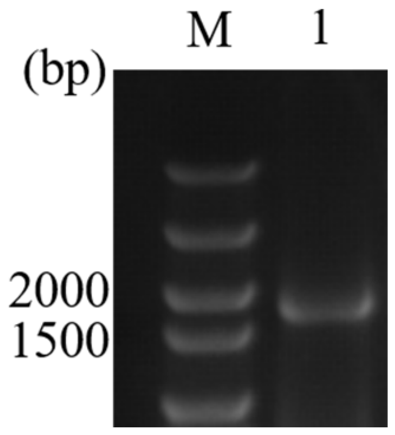
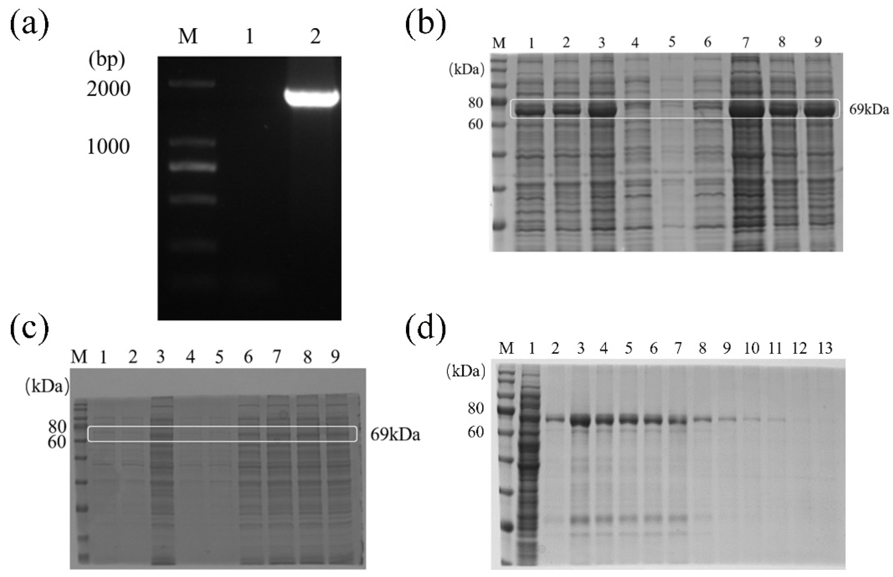
3.1. Cloning and Sequencing of CDS Region of the DcIAP1
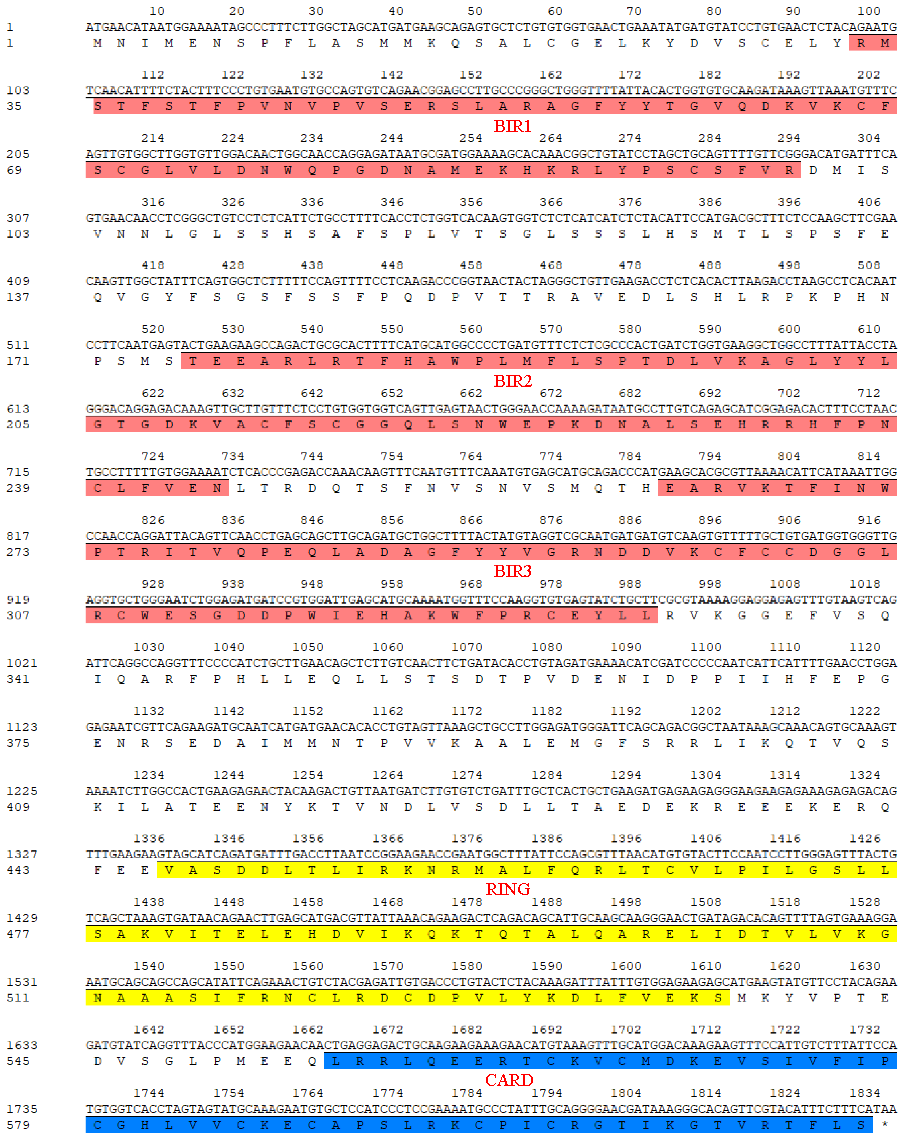
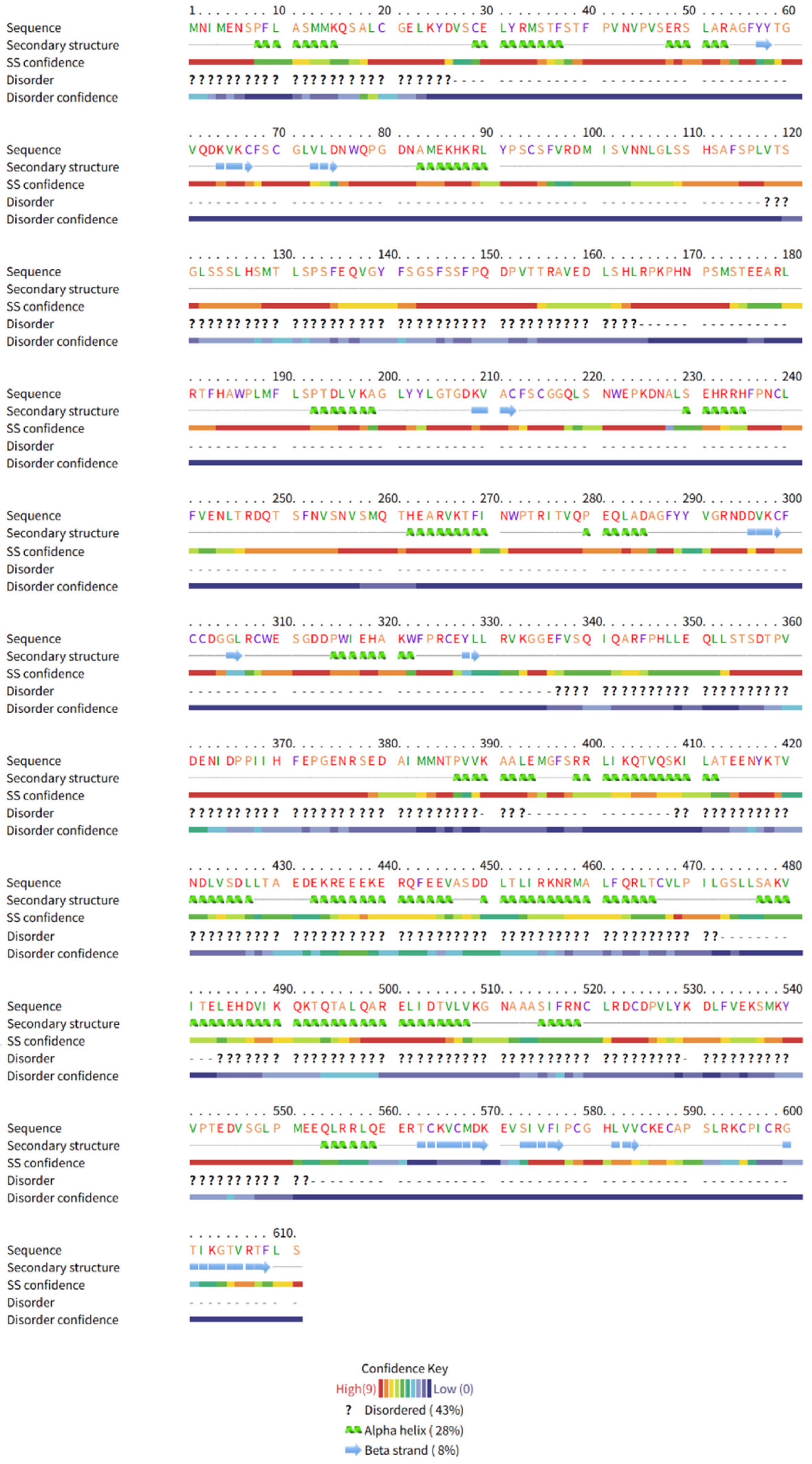
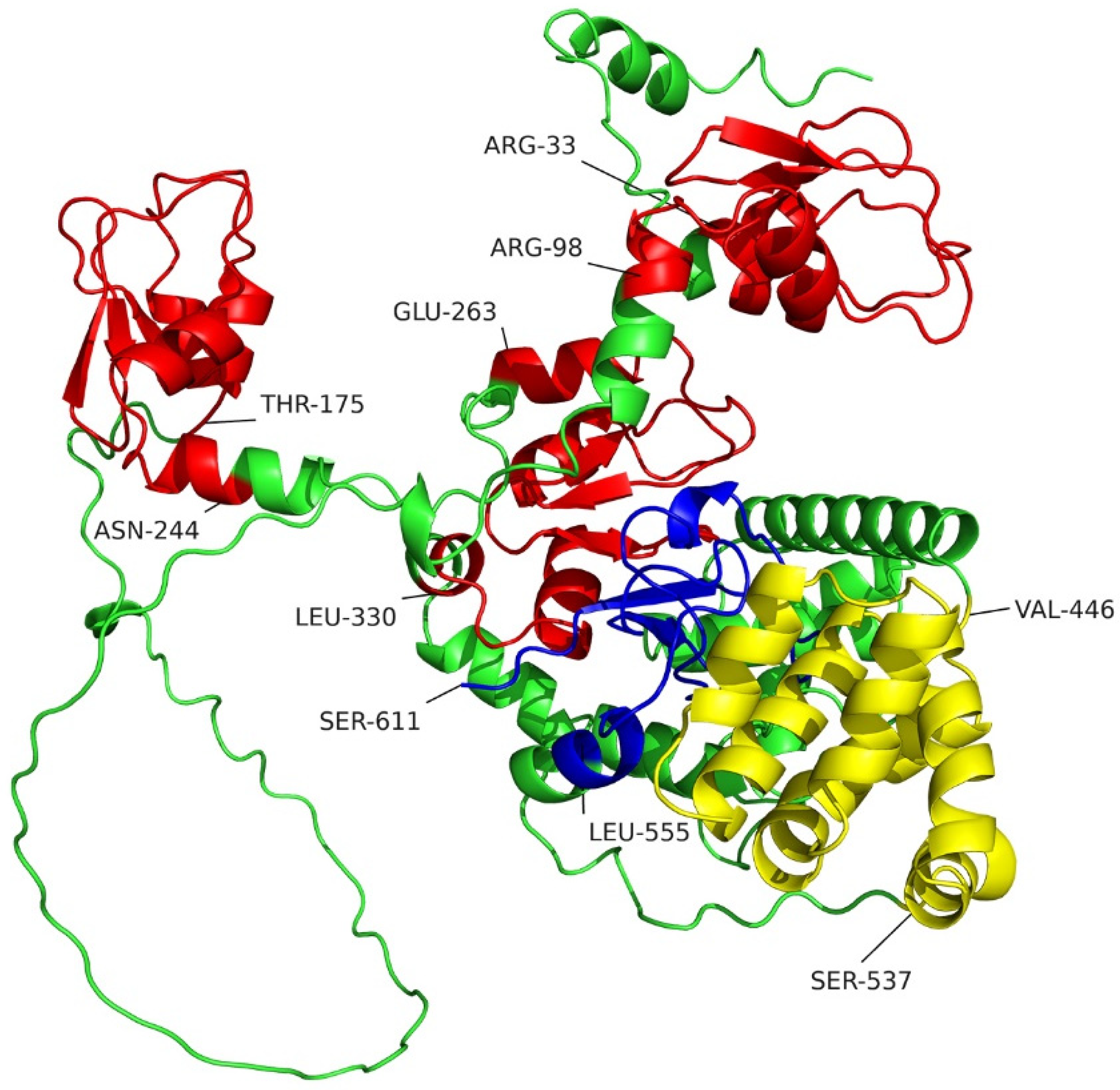
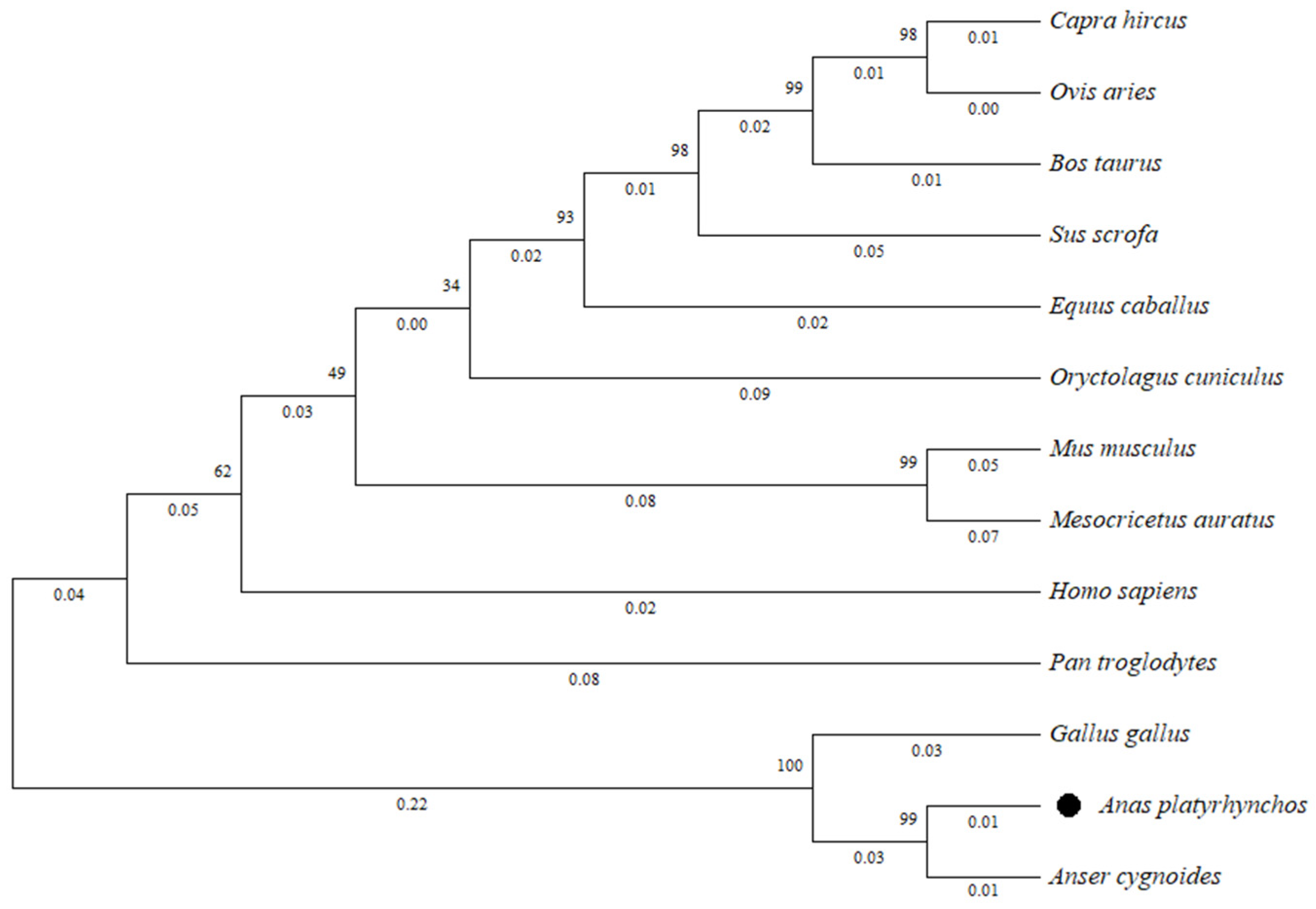
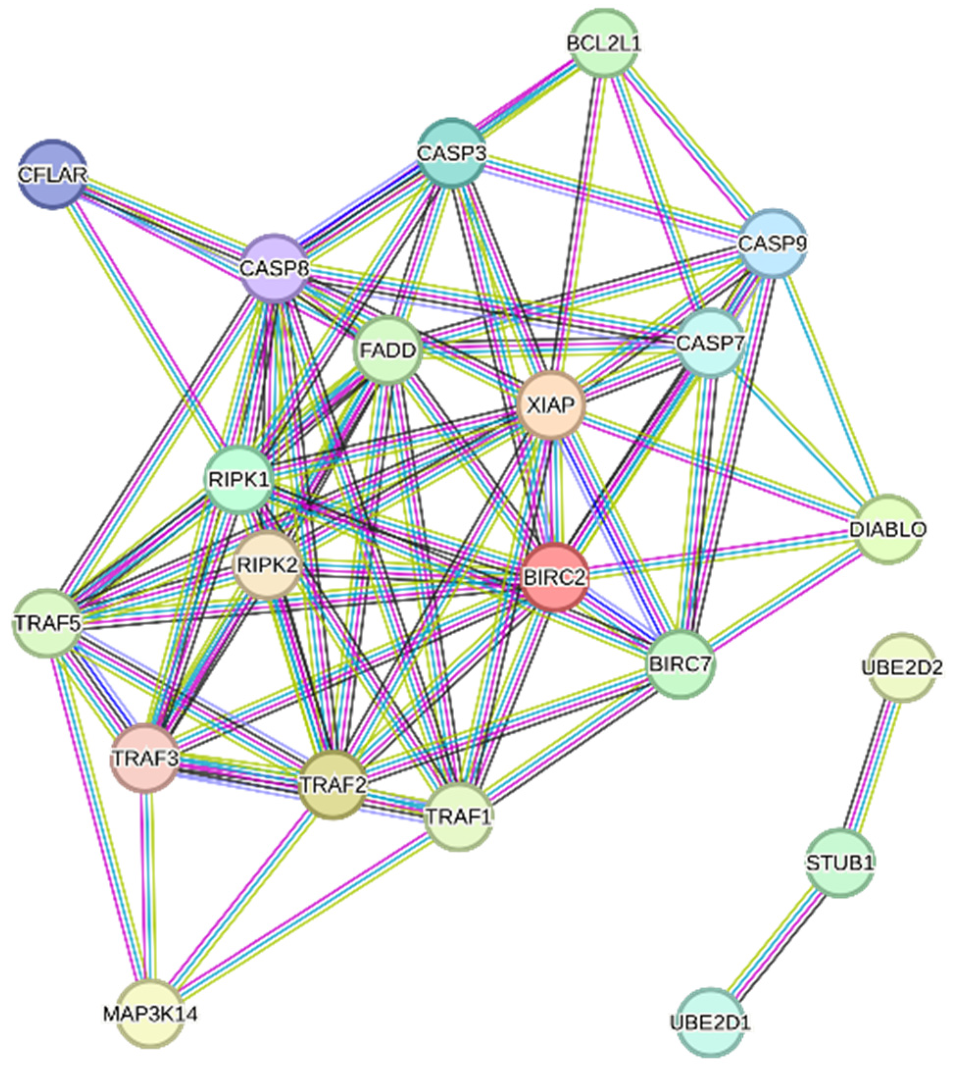
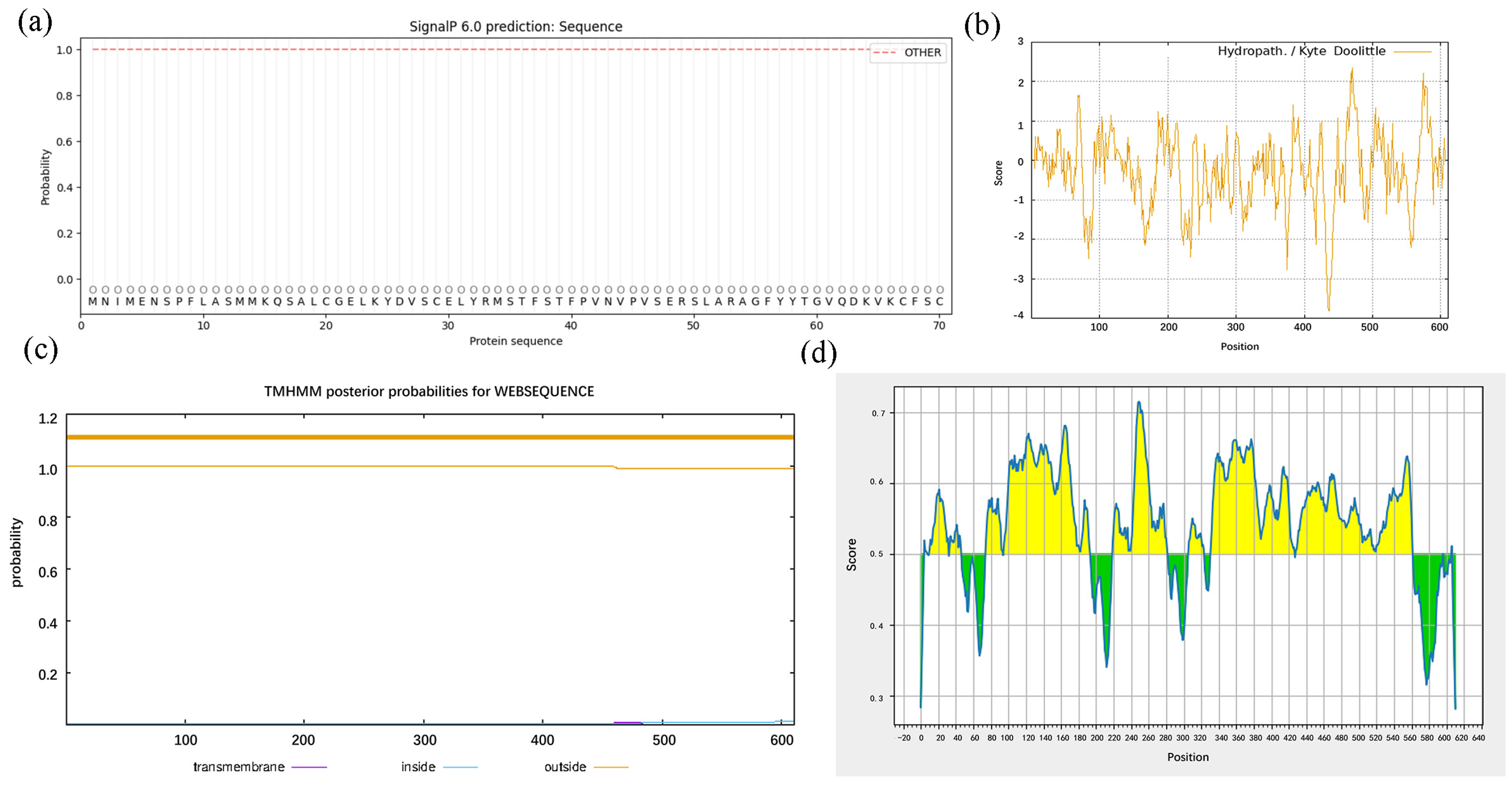
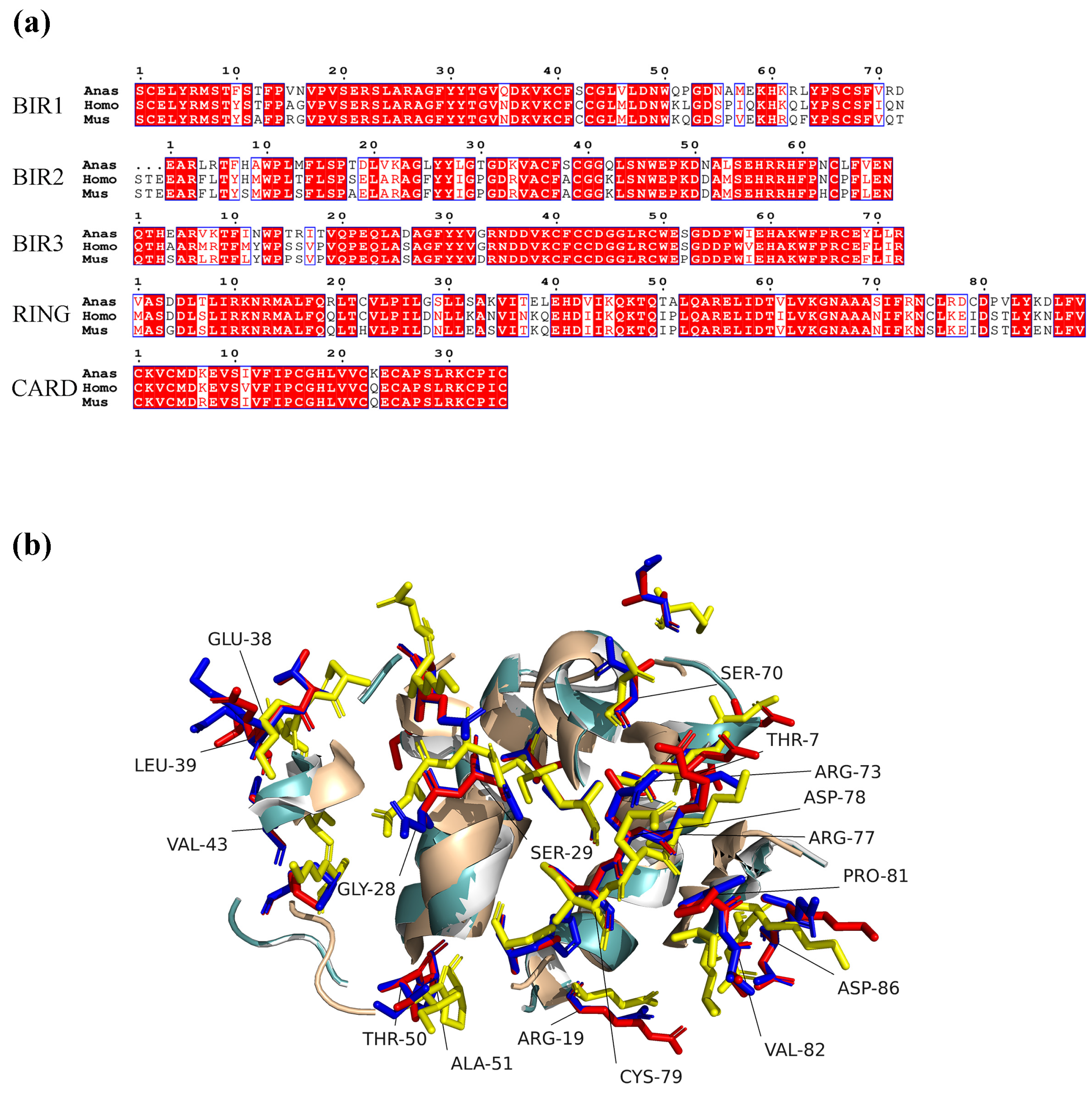
3.2. Bioinformatics Analysis of DcIAP1 Protein
3.3. Construction, Expression, and Purification of Recombinant DcIAP1 Protein
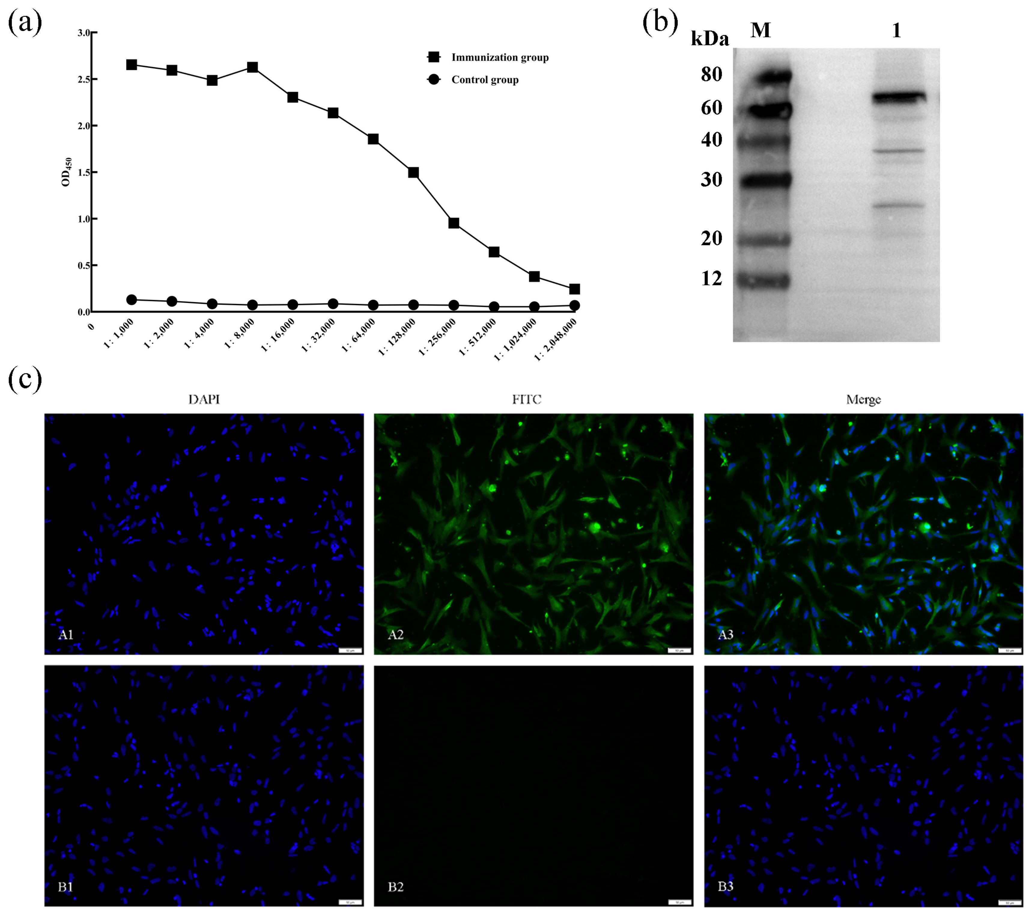
3.4. Determination of Serum Titer and Application of DcIAP1 Polyclonal Antibody
4. Discussion
5. Conclusions
Supplementary Materials
Author Contributions
Funding
Institutional Review Board Statement
Informed Consent Statement
Data Availability Statement
Conflicts of Interest
References
- Sun, Y.; Liu, K. Mechanistic insights into Influenza A virus-induced cell death and emerging treatment strategies. Vet. Sci. 2024, 11, 555. [Google Scholar] [CrossRef] [Scilit] [PubMed]
- Zhang, Y.; Wang, X.; Hu, A.; Wu, Y.; Zhang, P.; Yang, X.; Wen, Z.; Wen, M. Duck enteritis virus infection suppresses viability and induces apoptosis and endoplasmic reticulum stress in duck embryo fibroblast cells via the regulation of Ca(2). J. Vet. Med. Sci. 2021, 83, 549–557. [Google Scholar] [CrossRef] [Scilit] [PubMed]
- Sheng, X.D.; Zhang, W.P.; Zhang, Q.R.; Gu, C.Q.; Hu, X.Y.; Cheng, G.F. Apoptosis induction in duck tissues during duck hepatitis A virus type 1 infection. Poult. Sci. 2014, 93, 527–534. [Google Scholar] [CrossRef] [Scilit] [PubMed]
- Huang, Y.; Zhang, Y.; Yang, S.; Shi, Y.; Chu, X.; Ahmed, N.; Wu, J.; Chen, Q. Tembusu virus induced apoptosis in vacuolate spermatogenic cells is mediated by Cytc-mediated mitochondrial apoptotic signaling pathway. Theriogenology 2024, 215, 312–320. [Google Scholar] [CrossRef] [Scilit] [PubMed]
- Pan, Y.; Cheng, A.; Zhang, X.; Wang, M.; Chen, S.; Zhu, D.; Liu, M.; Zhao, X.; Yang, Q.; Wu, Y.; et al. Transcriptome analysis of duck embryo fibroblasts for the dynamic response to duck tembusu virus infection and dual regulation of apoptosis genes. Aging 2020, 12, 17503–17527. [Google Scholar] [CrossRef] [Scilit] [PubMed]
- Elsässer, A.; Suzuki, K.; Lorenz-Meyer, S.; Bode, C.; Schaper, J. The role of apoptosis in myocardial ischemia: A critical appraisal. Basic Res. Cardiol. 2001, 96, 219–226. [Google Scholar] [CrossRef] [Scilit] [PubMed]
- Miller, S.A.; Briglin, A. Apoptosis removes chick embryo tail gut and remnant of the primitive streak. Dev. Dyn. 1996, 206, 212–218. [Google Scholar] [CrossRef] [Scilit]
- Liu, X.; Chen, J.; Li, Z.; Gao, N.; Zhang, G. CIAP1/2 can regulate the inflammatory response and lung injury induced by apoptosis in septic rats. J. Investig. Med. 2024, 72, 100–111. [Google Scholar] [CrossRef] [Scilit] [PubMed]
- Schorn, F.; Werthenbach, J.P.; Hoffmann, M.; Daoud, M.; Stachelscheid, J.; Schiffmann, L.M.; Hildebrandt, X.; Lyu, S.I.; Peltzer, N.; Quaas, A.; et al. cIAPs control RIPK1 kinase activity-dependent and -independent cell death and tissue inflammation. EMBO J. 2023, 42, e113614. [Google Scholar] [CrossRef] [Scilit] [PubMed]
- Zadoroznyj, A.; Dubrez, L. Cytoplasmic and nuclear functions of cIAP1. Biomolecules 2022, 12, 322. [Google Scholar] [CrossRef] [Scilit] [PubMed]
- Akizuki, Y.; Morita, M.; Mori, Y.; Kaiho-Soma, A.; Dixit, S.; Endo, A.; Shimogawa, M.; Hayashi, G.; Naito, M.; Okamoto, A.; et al. cIAP1-based degraders induce degradation via branched ubiquitin architectures. Nat. Chem. Biol. 2023, 19, 311–322. [Google Scholar] [CrossRef] [Scilit] [PubMed]
- Bai, L.; Smith, D.C.; Wang, S. Small-molecule SMAC mimetics as new cancer therapeutics. Pharmacol. Ther. 2014, 144, 82–95. [Google Scholar] [CrossRef] [Scilit] [PubMed]
- Zhang, J.; Webster, J.D.; Dugger, D.L.; Goncharov, T.; Roose-Girma, M.; Hung, J.; Kwon, Y.C.; Vucic, D.; Newton, K.; Dixit, V.M. Ubiquitin ligases cIAP1 and cIAP2 limit cell death to prevent inflammation. Cell Rep. 2019, 27, 2679–2689.E3. [Google Scholar] [CrossRef] [Scilit] [PubMed]
- Smolewski, P.; Robak, T. Inhibitors of apoptosis proteins (IAPs) as potential molecular targets for therapy of hematological malignancies. Curr. Mol. Med. 2011, 11, 633–649. [Google Scholar] [CrossRef] [Scilit] [PubMed]
- Gao, B.; Qiao, Y.; Zhu, S.; Yang, N.; Zou, S.-S.; Liu, Y.-J.; Chen, J. USP36 inhibits apoptosis by deubiquitinating cIAP1 and survivin in colorectal cancer cells. J. Biol. Chem. 2024, 300, 107463. [Google Scholar] [CrossRef] [Scilit] [PubMed]
- Lin, S.-C.; Huang, Y.; Lo, Y.-C.; Lu, M.; Wu, H. Crystal structure of the BIR1 domain of XIAP in two crystal forms. J. Mol. Biol. 2007, 372, 847–854. [Google Scholar] [CrossRef] [Scilit] [PubMed]
- Mahoney, D.J.; Cheung, H.H.; Mrad, R.L.; Plenchette, S.; Simard, C.; Enwere, E.; Arora, V.; Mak, T.W.; Lacasse, E.C.; Waring, J.; et al. Both cIAP1 and cIAP2 regulate TNFalpha-mediated NF-kappaB activation. Proc. Natl. Acad. Sci. USA 2008, 105, 11778–11783. [Google Scholar] [CrossRef] [Scilit] [PubMed]
- Dueber, E.C.; Schoeffler, A.J.; Lingel, A.; Elliott, J.M.; Fedorova, A.V.; Giannetti, A.M.; Zobel, K.; Maurer, B.; Varfolomeev, E.; Wu, P.; et al. Antagonists induce a conformational change in cIAP1 that promotes autoubiquitination. Science 2011, 334, 376–380. [Google Scholar] [CrossRef] [Scilit] [PubMed]
- Hennessy, E.J.; Adam, A.; Aquila, B.M.; Castriotta, L.M.; Cook, D.; Hattersley, M.; Hird, A.W.; Huntington, C.; Kamhi, V.M.; Laing, N.M.; et al. Discovery of a novel class of dimeric Smac mimetics as potent IAP antagonists resulting in a clinical candidate for the treatment of cancer (AZD5582). J. Med. Chem. 2013, 56, 9897–9919. [Google Scholar] [CrossRef] [Scilit] [PubMed]
- Oetjen, K.A.; Duckett, C.S. Identifying the trigger of c-IAPs: Structural and functional characterization of CARD-mediated modulation of ubiquitin ligase activity. Mol. Cell 2011, 42, 553–554. [Google Scholar] [CrossRef] [Scilit] [PubMed]
- Cheung, H.H.; Plenchette, S.; Kern, C.J.; Mahoney, D.J.; Korneluk, R.G. The RING domain of cIAP1 mediates the degradation of RING-bearing inhibitor of apoptosis proteins by distinct pathways. Mol. Biol. Cell 2008, 19, 2729–2740. [Google Scholar] [CrossRef] [Scilit] [PubMed]
- Verhagen, A.M.; Coulson, E.J.; Vaux, D.L. Inhibitor of apoptosis proteins and their relatives: IAPs and other BIRPs. Genome Biol. 2001, 2, Reviews3009. [Google Scholar] [CrossRef] [Scilit] [PubMed]
- Lopez, J.; John, S.W.; Tenev, T.; Rautureau, G.J.; Hinds, M.G.; Francalanci, F.; Wilson, R.; Broemer, M.; Santoro, M.M.; Day, C.L.; et al. CARD-mediated autoinhibition of cIAP1’s E3 ligase activity suppresses cell proliferation and migration. Mol. Cell 2011, 42, 569–583. [Google Scholar] [CrossRef] [Scilit] [PubMed]




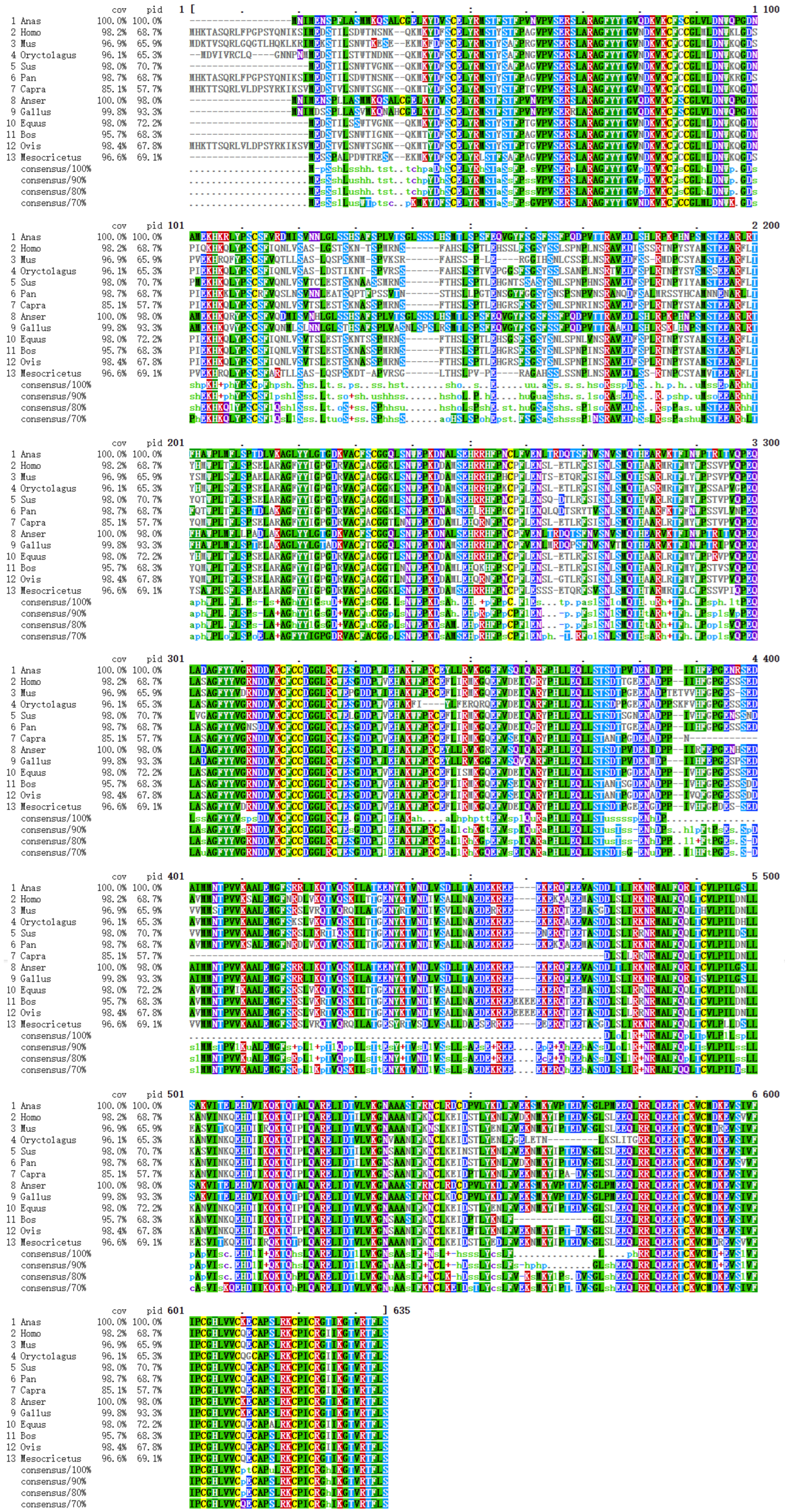




| Species Name | Identity/% | Coverage/% |
|---|---|---|
| Anser cygnoides | 98.02 | 100.0 |
| Gallus gallus | 93.22 | 99.8 |
| Equus caballus | 68.86 | 98.0 |
| Sus scrofa | 66.79 | 98.0 |
| Pan troglodytes | 66.59 | 98.7 |
| Homo sapiens | 66.39 | 98.2 |
| Bos taurus | 66.32 | 95.7 |
| Ovis aries | 65.99 | 98.4 |
| Mesocricetus auratus | 65.71 | 96.6 |
| Mus musculus | 64.48 | 96.9 |
| Capra hircus | 64.06 | 85.1 |
| Oryctolagus cuniculus | 63.13 | 96.1 |
Disclaimer/Publisher’s Note: The statements, opinions and data contained in all publications are solely those of the individual author(s) and contributor(s) and not of MDPI and/or the editor(s). MDPI and/or the editor(s) disclaim responsibility for any injury to people or property resulting from any ideas, methods, instructions or products referred to in the content. |
© 2026 by the authors. Licensee MDPI, Basel, Switzerland. This article is an open access article distributed under the terms and conditions of the Creative Commons Attribution (CC BY) license.
Share and Cite
Yang, S.; Zhu, Y.; Wang, X.; Huang, Y. Cloning, Bioinformatics Analysis, and Preparation of Polyclonal Antibody for Duck Cellular Inhibitor of Apoptosis Protein 1. Vet. Sci. 2026, 13, 830. https://doi.org/10.3390/vetsci13080830
Yang S, Zhu Y, Wang X, Huang Y. Cloning, Bioinformatics Analysis, and Preparation of Polyclonal Antibody for Duck Cellular Inhibitor of Apoptosis Protein 1. Veterinary Sciences. 2026; 13(8):830. https://doi.org/10.3390/vetsci13080830
Chicago/Turabian StyleYang, Sheng, Yikun Zhu, Xin Wang, and Yufei Huang. 2026. "Cloning, Bioinformatics Analysis, and Preparation of Polyclonal Antibody for Duck Cellular Inhibitor of Apoptosis Protein 1" Veterinary Sciences 13, no. 8: 830. https://doi.org/10.3390/vetsci13080830
APA StyleYang, S., Zhu, Y., Wang, X., & Huang, Y. (2026). Cloning, Bioinformatics Analysis, and Preparation of Polyclonal Antibody for Duck Cellular Inhibitor of Apoptosis Protein 1. Veterinary Sciences, 13(8), 830. https://doi.org/10.3390/vetsci13080830