Article

Exploring the Immunohistochemical Expression of Iron-Related Proteins in Non-Metastatic and Metastatic Feline Mammary Carcinomas

1
Department of Biology, University of Naples Federico II, 80126 Naples, Italy
2
Department of Veterinary Medicine and Animal Productions, University of Naples Federico II, 80126 Naples, Italy
*
Author to whom correspondence should be addressed.
Vet. Sci. 2026, 13(8), 810; https://doi.org/10.3390/vetsci13080810
Submission received: 10 July 2026 / Revised: 8 August 2026 / Accepted: 12 August 2026 / Published: 15 August 2026
(This article belongs to the Special Issue Advanced Therapy in Companion Animals—3rd Edition)

Simple Summary

Feline mammary tumors are frequent tumors in female cats and are characterized by high aggressiveness and poor prognosis. Given the pivotal role of iron in human breast cancer development, in this study we preliminarily explore the immunohistochemical expression of proteins involved in iron uptake, storage and efflux, namely Transferrin Receptor 1 (TfR1), Transferrin Receptor 2 (TfR2), ferritin (FTH1), and ferroportin (SLC40A1) in non-metastatic and metastatic feline mammary carcinomas and their tributary lymph node. Our results showed an increased expression of uptake proteins in relation to tumor progression, with TfR2 labeling detected mainly in tumoral samples. FTH1 immunolabeling was observed in cancer cells delimiting necrotic areas and in lymph node metastasis, indicating greater iron storage possibly associated with hypoxic environments, as suggested by increased expression of the hypoxic-inducible factor 1α. An increase in SLC40A1 labeling in tumoral cells suggested greater iron efflux. Although preliminary, our results underline interesting differences between feline normal and tumoral mammary tissues, which could pave the way to further in vitro studies to better understand the role of iron in the progression of feline mammary tumors.

Abstract

Feline mammary tumors are the third most common neoplasms in female cats and they are characterized by high aggressiveness and high metastatic rates. Given the pivotal role of iron in human breast cancer development, in this study, we preliminarily explore the immunohistochemical expression of proteins involved in iron uptake, storage and efflux [Transferrin Receptor 1 (TfR1), Transferrin Receptor 2 (TfR2), ferritin (FTH1), and ferroportin (SLC40A1)] in non-metastatic and metastatic feline mammary carcinomas and their tributary lymph node and the possible correlation with hypoxia-inducible factor-1 (HIF-1). Our results showed an increased expression of TfR1 and TfR2 in relation to tumor progression. FTH1 immunolabeling was mainly observed in cancer cells delimiting necrotic areas and in lymph node metastasis, indicating greater iron storage possibly associated with hypoxic environments, as suggested by increased expression of HIF-1α. Also, an increase in SLC40A1 labeling in tumoral cells suggested greater iron efflux. Although preliminary, our results underline interesting differences between feline normal and tumoral mammary tissues, which could pave the way to further in vitro studies to better understand the role of iron in the progression of feline mammary tumors.

1. Introduction

Mammary tumors are the third most common neoplasm in female cats [1], mainly affecting pure-bred middle-aged to older non-neutered females [2,3]. An amount of 80–96% of feline mammary tumors are malignant [4,5,6,7], showing high aggressiveness, ulceration and invasion of lymphatic vessels with distant metastasis localized mainly in the lungs, pleura, liver and lymph nodes [6,8]. High metastatic rates and late diagnosis determine the poor prognosis of the disease even after surgical resection [9]. Mean overall survival time after diagnosis is 8–12 months [10,11,12]; however, survival time is influenced by different parameters such as tumor size (234 days) and lymph node invasion (232 days), which worsen overall survival [6,13]. Surgical resection is the therapeutic method of choice in managing feline mammary cancer (FMC), with chain mastectomy reducing local tumor recurrence compared to conservative surgery; however, survival rates and time remain low [14,15,16].
Feline mammary cancers have been proposed as effective translational models for studying tumorigenesis and for evaluating the therapeutic efficiency of new anti-neoplastic molecules, overcoming the limits of rodent models and accelerating translation of results for human and household pets [17,18]. Specifically, FMCs share similar epidemiological features, like age incidence and hormonal dependency, as well as key risk factors and genes involved in the onset and development of mammary gland cancer in humans [19,20]. Moreover, research suggests a similar mechanism of tumorigenesis based on the overexpression and amplification of cyclin A [21] and overexpression of p53 [22]. Interestingly, De Maria et al. (2005) [23] demonstrated great similarities between FMC and the HER2-type breast cancer in humans, both exhibiting poor responses to standard therapies.
In the quest for new therapeutic approaches, the role of iron and its metabolism has sparked the interest of researchers due to its role in cancer onset and progression [24]. Iron is an essential metal and cofactor of numerous enzymes and proteins involved in basic biological mechanisms like DNA synthesis, cell division, electron transport and respiration [25,26,27], and it has been reported that tumor cells can modify their iron metabolism pathways, increasing the cytoplasmic labile iron pool (cLIP) to satisfy their increased metabolic demands [28,29,30].
Recent studies in women have described the phenomenon of iron addiction and the role of iron-related proteins in the onset, growth and progression of breast cancer. The overexpression of iron uptake proteins (Transferrin Receptor 1 (TfR1) and Transferrin Receptor 2 (TfR2)) in breast cancer has been previously associated with cell proliferation and progression of the disease, as they increase the cLIP and sustain DNA synthesis and cell division [31,32,33]. Similarly, downregulation of the iron-exporter protein ferroportin, also known as solute carrier family 40 member 1 (SLC40A1), and consequent increased cLIP have also been associated with increased malignancy of breast cancer cells [34]. Moreover, the altered expression of ferritin, particularly of ferritin heavy chain (FTH1), has been linked to further alteration in intracellular iron availability and the progression and aggressiveness of breast cancer [35]. The relevance of iron homeostasis in breast cancer is not only connected to its role in cell proliferation and in Reactive Oxygen Species (ROS) production [36], but it is further highlighted by the interplay of iron and estrogen signaling. Estrogen metabolites can promote the reduction in ferritin-bound Fe3+ to Fe2+, enhancing the cLIP and ROS generation, thereby accelerating carcinogenesis [37,38,39]. Moreover, recent evidence has shown that hypoxic environments occurring in tumors can regulate iron-related proteins via the hypoxia-inducible factor-1 (HIF-1), increasing iron uptake and challenging iron homeostasis [40,41,42].
In veterinary medicine, despite the growing literature describing the alterations of iron metabolism in tumors and linking iron and cancer [43,44,45,46,47], the topic remains underexplored and largely uncharacterized.
Given their pivotal role in breast cancer, we here explore the expression of TfR1, TfR2, ferritin heavy chain (FTH1), and SLC40A1, investigating iron uptake, storage and efflux, and the possible relation with HIF-1α, in non-metastatic and metastatic FMCs and their lymph node metastasis. Results provide first insights into the possible role of iron-related proteins in FMC progression, paving the way to future more in-depth research.

2. Materials and Methods

2.1. Case Selection

Fifty-two samples were selected from the histological archives (years 2020–2025) of the Laboratory of Veterinary Pathology of the Department of Veterinary Medicine and Animal Productions of the University of Naples Federico II. Screening, case availability and selection were made using the informatics system MyClinical from the Laboratory of Veterinary Pathology. The histological reports were collected to obtain information about age, breed, histological diagnosis and the presence or absence of lymph node metastasis. Where metastasis was present in the draining lymph node, this was also analyzed. All information is reported in Table S1. Ethical committee approval and authorization for animal testing were not required, as all tissue specimens analyzed in this study were sourced from archived materials.

2.2. Histology

Collected samples had been fixed in 10% neutral buffered formalin before undergoing standard histopathological processing. Thin sections of 3 μm were obtained from paraffin-embedded tissue blocks and subsequently stained with hematoxylin and eosin (H&E) for microscopic analysis [48]. Histopathological classification of neoplastic lesions was performed based on the recently updated World Health Organization (WHO) classification [49]: healthy mammary gland tissue (HT), feline mammary carcinoma without lymph node metastasis (N0 FMCs), feline mammary carcinoma with lymph node metastasis (N+ FMCs), and the tributary lymph node that had the metastasis of the primary mammary carcinoma (LM). FMCs were further stratified by histological subtype and by grade.

2.3. Immunohistochemistry

Additional 3 μm sections were prepared for immunohistochemical (IHC) analysis to assess the expression of iron-related proteins TfR1 [50], TfR2, FTH1, SLC40A1 and HIF-1α following previously established protocols [45,51]. Feline liver tissues were used as positive controls, while negative controls were obtained by omitting primary Ab. Details regarding the antibodies used, including their dilutions, are provided in Table 1. Immunolabeling was visualized using diaminobenzidine tetrahydrochloride (DAB), followed by hematoxylin counterstaining. The specimens were examined and documented using a light microscope (AXIO SCOPE.A1, Carl Zeiss S.p.A., Oberkochen, Germany) equipped with a digital microphotography camera (Axiocam 105 color, Carl Zeiss S.p.A., Oberkochen, Germany) [52]. A semiquantitative immunostaining evaluation was performed by an experienced pathologist (M.M.) to ensure consistency following previously established protocols [50,53] and a range score (H-score) from 0 to 300 was obtained for each sample for each antibody. Using Prism 9.0.0. (GraphPad Software), statistical analysis was performed to verify differences among groups. Prior to one-way ANOVA, data were tested for normality using the Shapiro–Wilk test and for homogeneity of variances using Levene’s test. Since the assumptions were met, one-way ANOVA with Tukey’s multiple comparison test was used. A p-value of less than 0.05 was considered statistically significant.

2.4. Western Blotting

To evaluate the specificity of the immunohistochemical signals, Western blot analysis was conducted on frozen non-neoplastic feline mammary gland samples retrieved from the archives. Tissues were homogenized on ice using a Potter homogenizer in 5 mL of RIPA buffer composed of 50 mM Tris-HCl (pH 7.4), 150 mM NaCl, 1 mM EDTA, 10% NP-40, and a protease inhibitor cocktail (3 mM aprotinin, 1 mM leupeptin, 1 mM E-64, 130 μM bestatin, 1 mM EDTA, and 2 mM AEBSF) [54]. Homogenates were then centrifuged at 7000× g for 10 min at 4 °C. Protein concentration in the supernatants was quantified using a NanoDrop 1000 spectrophotometer (ThermoFisher Scientific, Carlsbad, CA, USA). Subsequently, 60 µg of total protein per sample was separated by 10% SDS–PAGE and transferred onto PVDF membranes (1620177, Bio-Rad, Hercules, CA, USA). Membranes were blocked for 1 h in TBS-T containing 5% non-fat dry milk (70166, Merck Millipore, Darmstadt, Germany), followed by overnight incubation at 4 °C with primary antibodies, as listed in Table 2. After washing in 0.1% TBS, membranes were incubated for 1 h at room temperature with horseradish peroxidase-conjugated secondary antibodies: anti-rabbit IgG (Santa Cruz, Cat. sc-2004) diluted 1:5000 in TBS-T with 2.5% BSA, and anti-mouse IgG (Santa Cruz, Cat. sc-2005) also diluted 1:5000 in TBS-T with 2.5% BSA.
Chemiluminescent detection was carried out using Clarity Western ECL substrate (1705061, Bio-Rad, Hercules, CA, USA) for 5 min at room temperature, and signals were visualized using the Chemidoc Touch Imaging System (Bio-Rad, Hercules, CA, USA). Each experiment was performed in biological triplicates to ensure reproducibility.
Since frozen neoplastic tissue samples were not available, antibody validation by Western blot was performed using non-neoplastic feline mammary tissues. Therefore, although the antibodies recognized proteins at the expected molecular weights, their performance in neoplastic tissues was not directly assessed.

3. Results

3.1. Histological Results

Histological evaluation of the FMC allowed diagnosis of seven normal mammary tissue samples (N1–N7; 7/52), fifteen feline mammary carcinomas without lymph node metastasis (N0 FMCs) (S1–S15; 15/52), fifteen feline mammary carcinomas with lymph node metastasis (N+ FMCs) (S16–S30; 15/52), and fifteen tributary lymph nodes that had the metastasis of the primary mammary carcinomas (LM) (S31–S45; 15/52) (Figure 1). Among the N0 and N+ groups, tubular carcinoma was the most frequent, followed by comedocarcinoma and solid carcinoma, whereas for both groups ductal and invasive micropapillary carcinoma occurred less frequently. No tubulopapillary carcinomas were observed in the N+ groups, while it was diagnosed in N0 samples. Detailed diagnosis can be found in Table S1.

3.2. Immunohistochemical Results

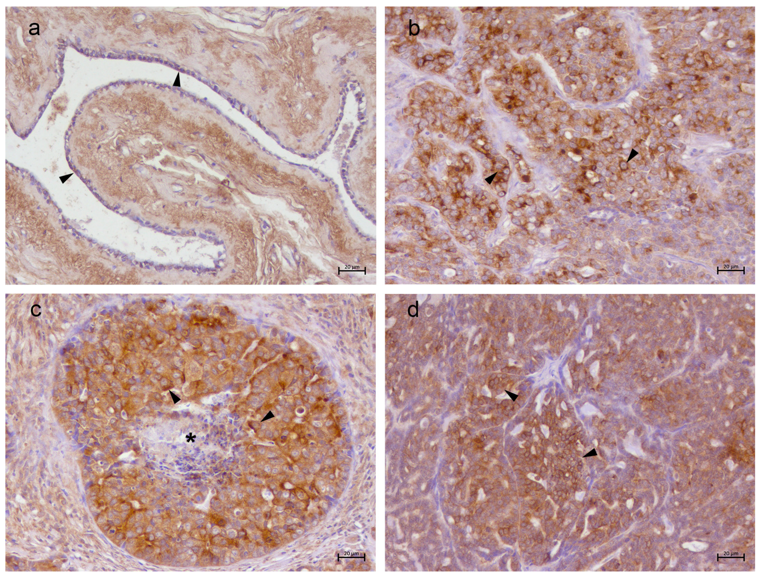
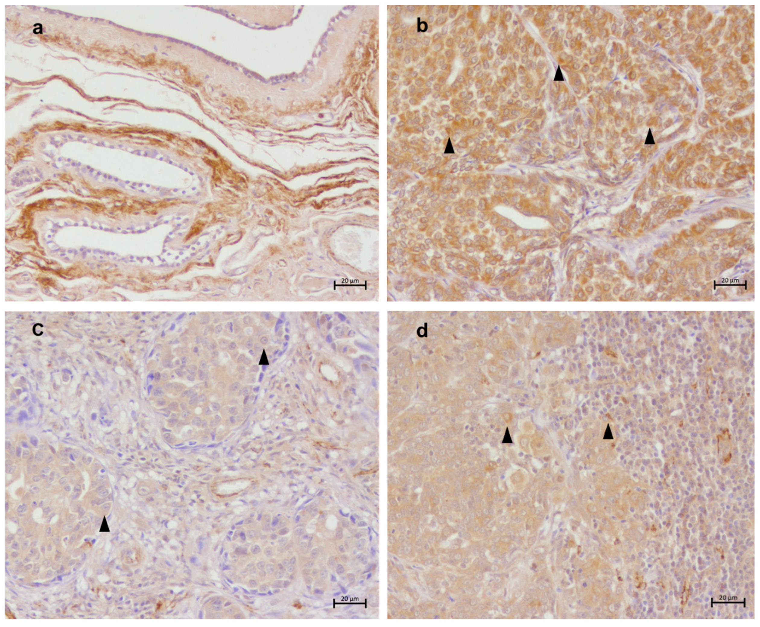
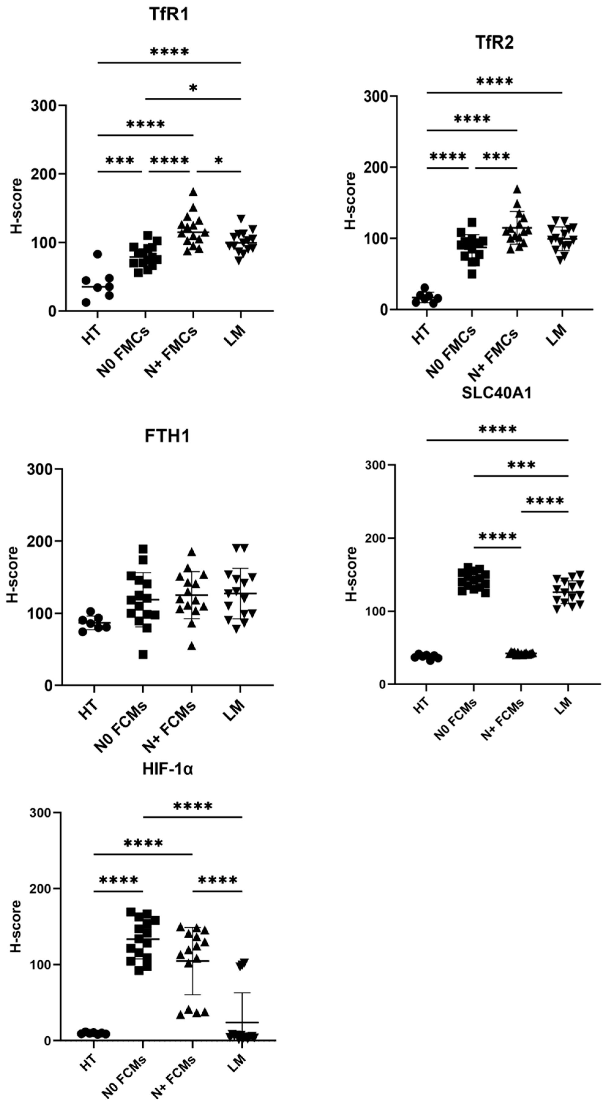
Immunohistochemistry of normal mammary gland tissue revealed membrane and cytoplasmic TfR1 immunolabeling in all mammary gland tissues (Figure 2a–d; Figure S6). The H-score showed an increased protein level of TfR1 in N0 FMCs (80.70 ± 14.90) versus healthy mammary tissue (mean 40.02 ± 20.86) (p < 0.01). Moreover, a marked upregulation of TfR1 expression was also observed in both the N+ FMCs (mean 119.34 ± 22.56) and LM (mean 100.47 ± 14.86) groups compared to the HT baseline (p < 0.0001 for both comparisons) and to the N0 FMCs group (Figure 7). These findings suggest a strong association between TfR1 overexpression and the progression of the tumor.
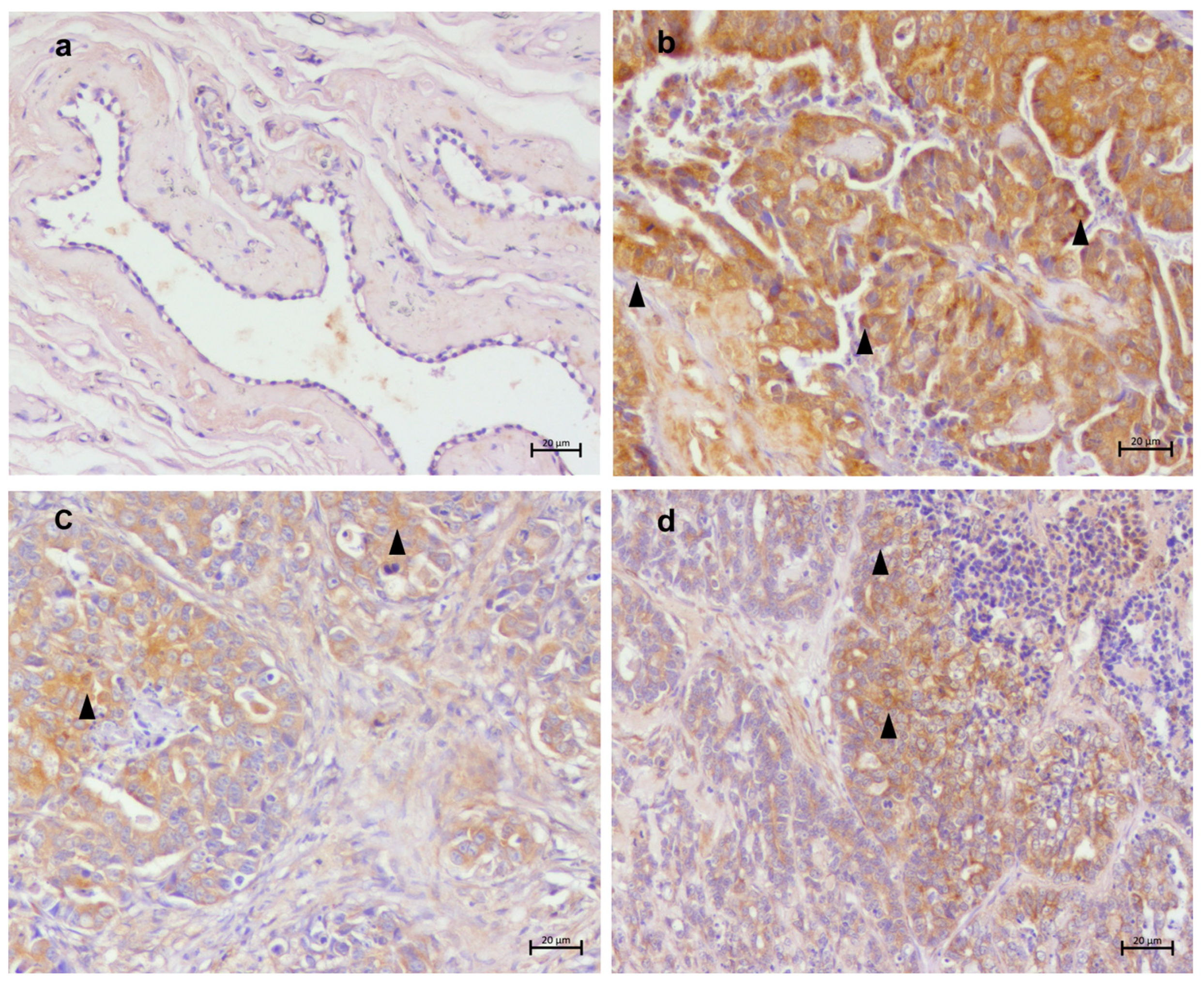
TfR2 was poorly detected in normal tissues of the feline mammary gland (mean 16.94 ± 6.77), but TfR2 labeling was detected in all tumoral samples (Figure 3a–d; Figure S7) with an increase in H-score following tumoral progression. In particular, TfR2 expression was significantly increased in the N0 FMCs group (mean 87.30 ± 17.37) compared with the HT group (p < 0.0001) and further increased in the N+ FMCs (114.77 ± 22.24), which showed significantly higher H-scores than both HT (p < 0.0001) and N0 FMCs (p = 0.0009). The LM group (99.35 ± 16.04) also exhibited significantly higher TfR2 expression compared to HT (p < 0.0001), although no significant differences were detected between LM and either N0 or N+ FMCs (Figure 7).
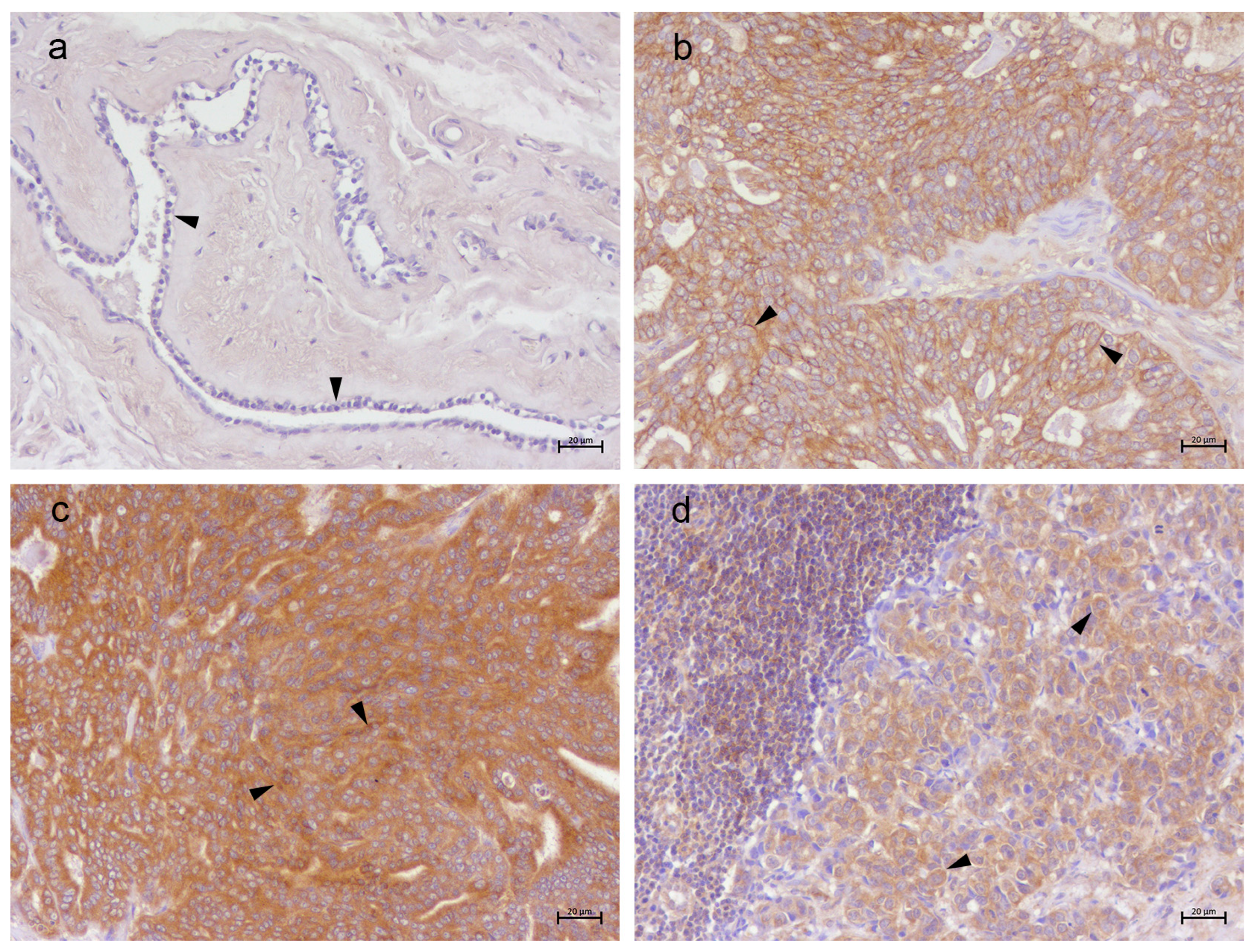
FTH1 was expressed in normal feline mammary gland tissues with a relatively moderate H-score (86.75 ± 8.80). In contrast, all feline mammary carcinoma groups showed increased FTH1 expression. Specifically, FTH1 immunoreactivity was significantly higher in the N0 FMCs (118.91 ± 36.32), N+ FMCs (125.26 ± 31.51), and LM (127.37 ± 33.79) groups compared with HT, whereas no significant differences were detected among the tumoral groups (Figure 7). Interestingly, FTH1 was highly detected in tumoral cells surrounding comedonic areas (Figure 4a–d; Figure S8).
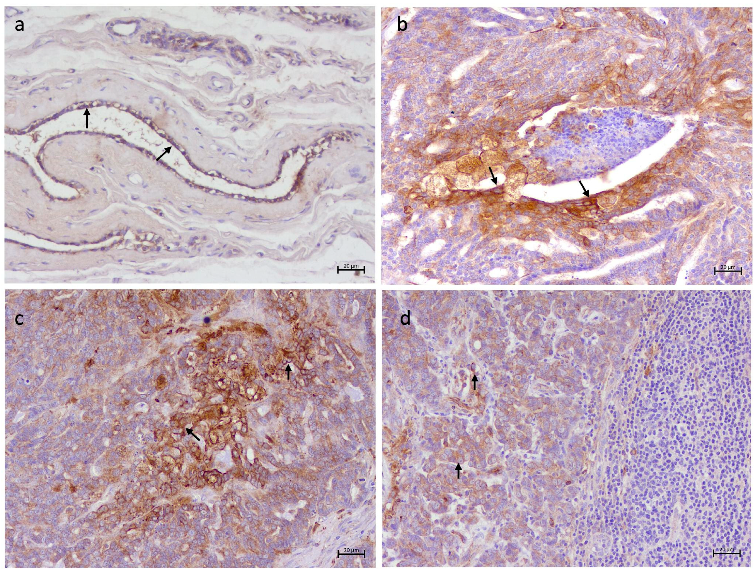
SLC40A1 immunoreactivity was low in both normal and N+FMCs with comparable H-scores (respectively 37.76 ± 3.08 and 42.30 ± 1.55). While, SLC40A1 expression increased markedly in N0 FMCs and in LM (Figure 5a–d; Figure S9). Specifically, both the N0 FMCs and LM groups showed significantly higher H-scores (respectively, 142.5 ± 11.18 and 125.95 ± 15.48) and immunolabeling than the N+ FMCs group (p < 0.0001 for both comparisons), whereas no significant differences were observed between LM and N0 FMCs (Figure 7).
HIF-1α was poorly detected in normal tissues of the feline mammary gland with the H-score of 9.01 ± 1.04. Whereas HIF-1α labeling was observed in all tumoral groups (Figure 6a–d; Figure S10), in particular it was higher in the N0 FMCs group (p < 0.0001) with a H-score of 133.62 ± 25.03. Moreover, the N+ FMCs group maintained high HIF-1α immunoreactivity, with a high H-score (104.86 ± 42.69). Conversely, the LM group exhibited a significant reduction in HIF-1α expression compared with the N0 FMCs group (p < 0.0001), with an H-score of 23.95 ± 37.87 (Figure 7).

3.3. Western Blotting Results

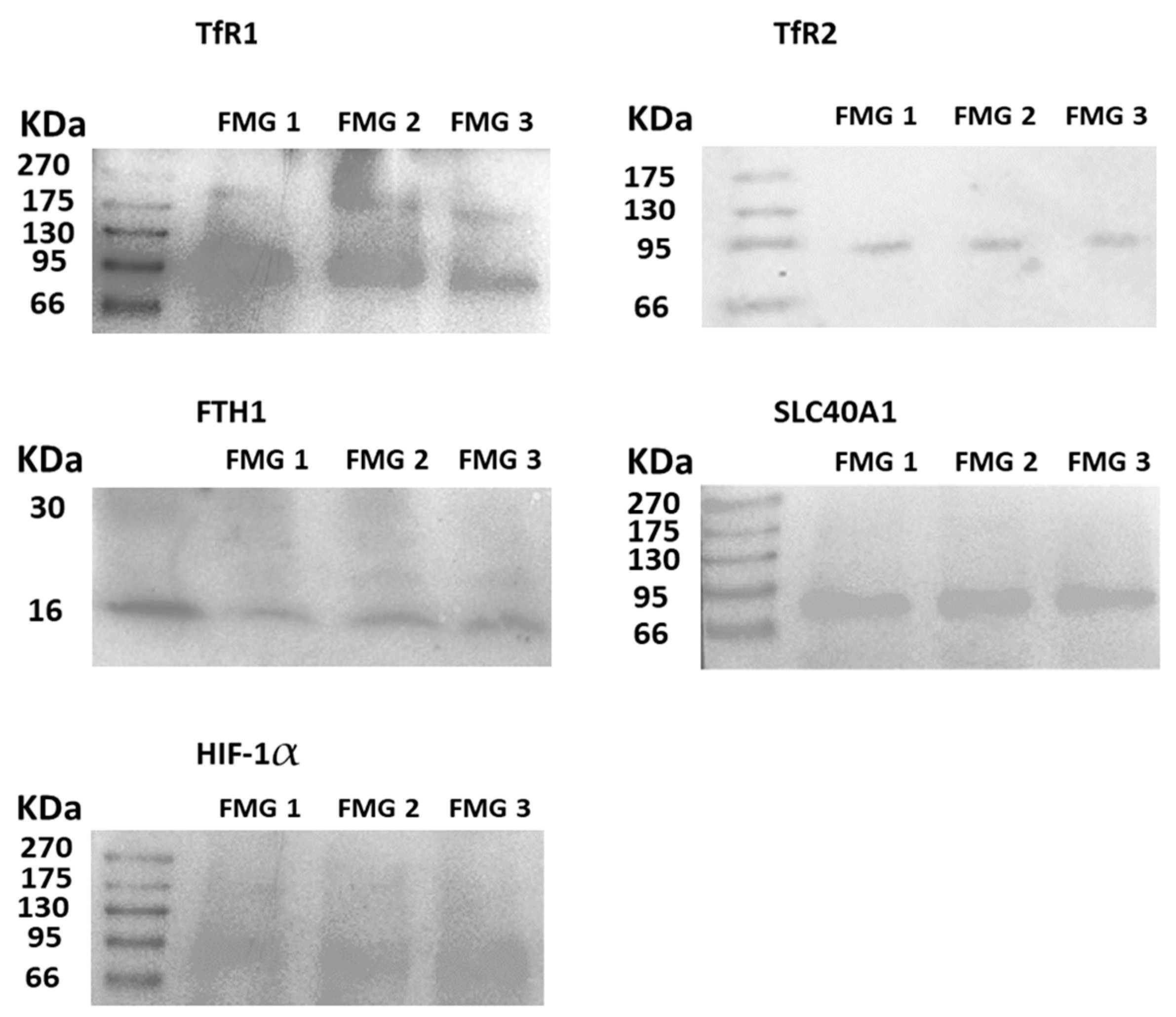
The specificity of the antibodies against TfR1, TfR2, FTH1, SLC40A1 and HIF-1α was confirmed via Western blot analysis on total protein lysates derived from non-neoplastic feline mammary gland tissues. As shown in Figure 8, the anti-TfR1 antibody detected an immunoreactive band at approximately 90 kDa while the anti-TfR2 antibody detected a single immunoreactive band at approximately 100 kDa. Also, an immunoreactive band was observed at 21 kDa. Following anti-FTH1F or SLC40A1, two immunoreactive bands were observed between approximately 62 and 70 kDa. Similar findings have been previously described for ferroportin and are thought to reflect different forms of the protein. Moreover, an immunoreactive band was also observed at 93 kDa following anti-HIF-1α binding. These bands correspond to the expected molecular weights of the respective feline protein isoforms, supporting the specificity of the antibodies.
Figure 7. H-score of TfR1, TfR2, FTH1, SLC40A1 and HIF-1α expression in feline mammary healthy tissue (HT (n = 7)), feline mammary carcinoma without lymph node metastasis (N0 FMCs) (n = 15), feline mammary carcinoma with lymph node metastasis (N+ FMCs) (n = 15) and lymph node metastasis (LM) (n = 15). Data are presented as mean ± SD. TfR1: HT vs. N0 FMCs, p = 0.0001; HT vs. N+ FMCs, p < 0.0001; HT vs. LM, p < 0.0001; N0 FMCs vs. N+ FMCs, p < 0.0001; N0 FMCs vs. LM, p = 0.0315; N+ FMCs vs. LM, p = 0.0436; TfR2: HT vs. N0 FMCs, p < 0.0001; HT vs. N+ FMCs, p < 0.0001; HT vs. LM, p < 0.0001; N0 FMCs vs. N+ FMCs, p = 0.0009; N0 FMCs vs. LM, p = 0.2860; N+ FMCs vs. LM, p = 0.1114; FTH1: HT vs. N0 FMCs, p = 0.1591; HT vs. N+ FMCs, p = 0.0653; HT vs. LM, p = 0.0471; N0 FMCs vs. N+ FMCs, p = 0.9524; N0 FMCs vs. LM, p = 0.8962; N+ FMCs vs. LM, p = 0.9981; SLC40A1: HT vs. N0 FMCs, p < 0.0001; HT vs. N+ FMCs, p = 0.7752; HT vs. LM, p < 0.0001; N0 FMCs vs. N+ FMCs, p < 0.0001; N0 FMCs vs. LM, p = 0.0004; N+ FMCs vs. LM, p < 0.0001; HIF-1α: HT vs. N0 FMCs, p < 0.0001; HT vs. N+ FMCs, p < 0.0001; HT vs. LM, p = 0.8132; N0 FMCs vs. N+ FMCs, p = 0.1219; N0 FMCs vs. LM, p < 0.0001; N+ FMCs vs. LM, p < 0.0001. (* p < 0.1), (** p < 0.01), (*** p < 0.001), (**** p < 0.0001).
Vetsci 13 00810 g007
Figure 8. Representative TfR1 (~90 kDa), TfR2 (~100 kDa), FTH1 (~20 kDa), SLC40A1 (~62–70 kDa) and HIF-1α (~93 kDa) immunoblot analysis in 3 non-neoplastic feline mammary glands (FMGs) (See Figures S1–S5).
Vetsci 13 00810 g008

4. Discussion

Iron is an essential trace element involved in fundamental biological processes, including DNA synthesis, cellular respiration, and cell proliferation [25,26,27]. However, the tight regulation of iron homeostasis is critical, especially in tissues with high proliferative rates, such as neoplastic ones [55]. Indeed, if, on one hand, iron sustains DNA synthesis and cell proliferation, on the other hand, its redox-active nature renders it potentially cytotoxic when present in excess, promoting oxidative stress through the Fenton reaction and leading to ferroptosis [56,57,58,59].
In breast cancer, upregulation of the expression of iron uptake proteins such as TfR1 and TfR2 was reported [60]. These findings overlap with those observed in our study, further supporting the possibility of using natural FMC as a model for human breast cancer also in the study of iron-related tumorigenesis and progression [61]. Our findings indicate that iron uptake is dynamically remodeled during FMC progression rather than being uniformly dysregulated. The expression patterns of the investigated proteins differed between non-metastatic primary tumors (N0 FMCs), metastatic primary tumors (N+ FMCs), and lymph node metastases (LM), suggesting that distinct mechanisms characterize different stages of disease progression. A previous study by Marques et al. (2017) [61] evaluated the expression of iron-related proteins in benign and malignant feline mammary tumors, but no significant differences were observed according to malignancy, while a study by Rensi et al. (2021) [50] described a correlation between tumor progression and expression of TfR1. Overexpression of TfR1 is a hallmark of several cancers, including mammary carcinoma, and it is associated with increased iron uptake, enhanced proliferation, and worse clinical outcomes [62,63,64,65,66,67,68], as iron overload can be associated with the progression of mammary cancer toward a more malignant phenotype [69]. Similarly, although less studied in veterinary medicine, TfR2 may contribute to tumorigenesis through iron-mediated mechanisms [51,70,71,72,73] and hormone signaling crosstalk, including estrogen responsiveness [74]. In human oncology, the role of TfR2 in Triple-Negative Breast Cancer (TNBC) was investigated through in vitro experiments [33]. The overexpression of TfR2 in TNBC cells compared with that of healthy breast epithelial cells was highlighted and its oncogenic effects were demonstrated. Moreover, it was described that TfR2 knockdown could inhibit TNBC cell proliferation by activating ferroptosis, an iron-dependent non-apoptotic cell death [75]. Although TfR2 has been found upregulated in several tumors, its connection to prognosis is still debated [76]. In our study, both TfR1 and TfR2 were poorly detected in normal mammary tissues, while they presented progressive expression according to malignancy, suggesting a possible use of iron uptake proteins as markers for FMCs progression.
On the one hand, in our study, we observed a possible increase in the uptake of iron; the expression of SL40A1 appears controversial, as it was less expressed in N+FMCs and more expressed in N0 FMCs and LM. SL40A1 is the only mechanism for exporting intracellular non-heme iron and has a central role in iron homeostasis and cLIP amounts. To date, SL40A1 has not been extensively studied in the context of cancer and its role in cancerogenesis is not fully understood. Nevertheless, it appears to be decreased in human breast cancer epithelial cells when compared to normal breast cells, with the lowest ferroportin expression associated with the highest severity and highest amounts of cLIP [34]. Consequently, our results appear slightly in contrast with those reported in breast cancer and the role of SL40A1 needs to be further elucidated. Nevertheless, the downregulation of the iron efflux mechanism in N+ FMCs could lead to increased amounts of cLIP, which in turn could be used to sustain tumor growth and cell metastasis, as previously described [77]. The upregulation of SL40A1 in N0 FMCs and LM is controversial and, currently, we can only hypothesize possible underlying mechanisms. On one hand, efflux increase could be read as a possible cell protective mechanism. Indeed, it has been demonstrated that increased iron efflux impeded tumor growth and metastasis, and inhibited epithelial–mesenchymal transition [77]. On the other hand, we cannot exclude that, by increasing iron efflux, tumoral cells are attempting to reduce the amount of cLIP and fight ferroptosis, which would benefit cancer cell survival. More likely, the differential expression of SL40A1 between N0 FMCs, N+FMCs and LM could be a consequence of other factors such as biological heterogeneity and microenvironmental differences, or even technical variation.
Ferritin, particularly ferritin heavy chain 1 (FTH1), can play a double role in cancer onset. On one hand, it sequesters excess intracellular iron, limiting oxidative damage [78]; on the other hand, high levels of ferritin are associated with immunosuppression and enhanced tumor cell survival [79,80]. In breast cancer, ferritin appears upregulated [60], and elevated ferritin levels in tissue have been correlated with tumor grade, epithelial proliferation, recurrence risk [35,81] and hypoxic conditions. The first results provided by our study show increased detection of ferritin in all tumoral samples. In N0 and N+ FMCs, FTH1 overexpression could be related to the hypoxic tumoral environment and the increased expression of HIF-1α. Indeed, iron metabolism is highly affected by hypoxia and the mechanisms of intake, storage, use and efflux appear modified. Particularly, in hypoxic contexts, TfR1 and FTH1 are usually up-regulated to sustain the higher iron requirements and to prevent ferroptosis through iron storage. On the contrary, as LM presented a very diverse expression of HIF-1α, increased iron storage cannot be justified solely by a hypoxic environment, but it is possibly due to a specific tumoral microenvironment different from that of primary tumors and resulting from the specific biological behavior of a particular cancer type. Particularly, the absence of necrosis in the analyzed LM samples might play a role in HIF-1α downregulation, given that HIF-1α is typically upregulated in necrotic areas. A greater number of samples and in vitro studies will help shed light on the uncertainties of the present study.

5. Conclusions

In many cancers, iron metabolism pathways are often altered, suggesting that reprogramming of iron metabolism is a key feature of tumorigenesis. The promotion of the activity of iron-related proteins in tumors can create a new balance able to feed the “iron addiction”, sustain progression and prevent damage caused by iron overload. Since the number of samples is limited, with a low number of healthy mammary tissues, the present findings should be interpreted with caution. Also, the limited sample size prevented any meaningful subtype-specific analyses and no correlation with clinicopathological data was therefore performed. Future studies should correlate the expression of iron-related proteins while also considering parameters like tumor size and grade and survival time. Nevertheless, our study confirms the alteration in the expression of iron-related proteins under the immunohistochemical profile in FMCs, providing a first glance at possible alterations in iron metabolism. If iron uptake appears to follow cancer progression also in FMCs, iron storage and efflux do not seem to follow this rule, possibly due to the different biological characteristics of different cancer types. Unfortunately, many of the proposed mechanisms underlying our results have not been fully investigated, due to the inherent constraints of a retrospective study that lacks standardized collection protocols; nevertheless, starting from this “static picture”, more dynamic in vitro approaches could better elucidate the hypothesis here provided.

Supplementary Materials

The following supporting information can be downloaded at https://www.mdpi.com/article/10.3390/vetsci13080810/s1, Table S1: breeds, age and histopathological diagnosis of 52 feline mammary tissue samples; Figures S1–S5: whole Western blots; Figures S6–S10: high-magnification of immunohistochemical images.

Author Contributions

Conceptualization, R.L. and K.P.; methodology, R.L. and G.R.; validation, T.C. and E.D.N.; investigation, R.L., G.R., T.C. and E.D.N.; data curation, R.L., T.C. and M.M.; writing—original draft preparation, R.L. and K.P.; writing—review and editing, M.M.; supervision, K.P. All authors have read and agreed to the published version of the manuscript.

Funding

This research received no external funding.

Institutional Review Board Statement

It was not required to obtain an ethics committee’s approval or to submit an animal testing request, since all animal tissue samples examined in this study were retrieved from the archives of the Department of Veterinary Medicine and Animal Productions, University of Naples Federico II-Naples.

Informed Consent Statement

Not applicable.

Data Availability Statement

The original contributions presented in this study are included in the article/Supplementary Materials. Further inquiries can be directed to the corresponding author.

Acknowledgments

The authors acknowledge the Histopathology and Diagnostics Core at the Department of Biology of the University of Naples “Federico II” for their technical and scientific support.

Conflicts of Interest

The authors declare no conflicts of interest.

References

  1. Zappulli, V.; De Zan, G.; Cardazzo, B.; Bargelloni, L.; Castagnaro, M. Feline Mammary Tumours in Comparative Oncology. J. Dairy Res. 2005, 72, 98–106. [Google Scholar] [CrossRef] [Scilit] [PubMed]
  2. Giugliano, R.; Dell’Anno, F.; De Paolis, L.; Crescio, M.I.; Ciccotelli, V.; Vivaldi, B.; Razzuoli, E. Mammary Gland, Skin and Soft Tissue Tumors in Pet Cats: Findings of the Feline Tumors Collected from 2002 to 2022. Front. Vet. Sci. 2024, 11, 1320696. [Google Scholar] [CrossRef] [Scilit] [PubMed]
  3. Souza, F.R.; Moreira, I.S.; Dariva, A.A.; Nakagaki, K.Y.R.; Abreu, C.C.; Balabram, D.; Cassali, G.D. Epidemiologic and Clinicopathological Characterization of Feline Mammary Lesions. Vet. Sci. 2024, 11, 549. [Google Scholar] [CrossRef] [Scilit] [PubMed]
  4. Lana, S. Tumors of the Mammary Gland. In Withrow & MacEwen’s Small Animal Clinical Oncology; Elsevier: Amsterdam, The Netherlands, 2007; pp. 619–636. [Google Scholar] [CrossRef] [Scilit]
  5. Salas, Y.; Márquez, A.; Diaz, D.; Romero, L. Epidemiological Study of Mammary Tumors in Female Dogs Diagnosed during the Period 2002–2012: A Growing Animal Health Problem. PLoS ONE 2015, 10, e0127381. [Google Scholar] [CrossRef] [Scilit] [PubMed]
  6. Zappulli, V.; Rasotto, R.; Caliari, D.; Mainenti, M.; Peña, L.; Goldschmidt, M.H.; Kiupel, M. Prognostic Evaluation of Feline Mammary Carcinomas: A Review of the Literature. Vet. Pathol. 2015, 52, 46–60. [Google Scholar] [CrossRef] [Scilit] [PubMed]
  7. Vascellari, M.; Capello, K.; Carminato, A.; Zanardello, C.; Baioni, E.; Mutinelli, F. Incidence of Mammary Tumors in the Canine Population Living in the Veneto Region (Northeastern Italy): Risk Factors and Similarities to Human Breast Cancer. Prev. Vet. Med. 2016, 126, 183–189. [Google Scholar] [CrossRef] [Scilit] [PubMed]
  8. Dagher, E.; Abadie, J.; Loussouarn, D.; Campone, M.; Nguyen, F. Feline Invasive Mammary Carcinomas: Prognostic Value of Histological Grading. Vet. Pathol. 2019, 56, 660–670. [Google Scholar] [CrossRef] [Scilit] [PubMed]
  9. Chocteau, F.; Mordelet, V.; Dagher, E.; Loussouarn, D.; Abadie, J.; Nguyen, F. One-Year Conditional Survival of Dogs and Cats with Invasive Mammary Carcinomas: A Concept Inspired from Human Breast Cancer. Vet. Comp. Oncol. 2021, 19, 140–151. [Google Scholar] [CrossRef] [Scilit] [PubMed]
  10. Castagnaro, M.; Casalone, C.; Bozzetta, E.; De Maria, R.; Biolatti, B.; Caramelli, M. Tumour Grading and the One-Year Post-Surgical Prognosis in Feline Mammary Carcinomas. J. Comp. Pathol. 1998, 119, 263–275. [Google Scholar] [CrossRef] [Scilit] [PubMed]
  11. Millanta, F.; Calandrella, M.; Citi, S.; Della Santa, D.; Poli, A. Overexpression of HER-2 in Feline Invasive Mammary Carcinomas: An Immunohistochemical Survey and Evaluation of Its Prognostic Potential. Vet. Pathol. 2005, 42, 30–34. [Google Scholar] [CrossRef] [Scilit] [PubMed]
  12. Seixas, F.; Palmeira, C.; Pires, M.A.; Bento, M.J.; Lopes, C. Grade Is an Independent Prognostic Factor for Feline Mammary Carcinomas: A Clinicopathological and Survival Analysis. Vet. J. 2011, 187, 65–71. [Google Scholar] [CrossRef] [Scilit] [PubMed]
  13. Preziosi, R.; Sarli, G.; Benazzi, C.; Mandrioli, L.; Marcato, P.S. Multiparametric Survival Analysis of Histological Stage and Proliferative Activity in Feline Mammary Carcinomas. Res. Vet. Sci. 2002, 73, 53–60. [Google Scholar] [CrossRef] [Scilit] [PubMed]
  14. MacEwen, E.G.; Hayes, A.A.; Harvey, H.J.; Patnaik, A.K.; Mooney, S.; Passe, S. Prognostic Factors for Feline Mammary Tumors. J. Am. Vet. Med. Assoc. 1984, 185, 201–204. [Google Scholar] [CrossRef] [Scilit]
  15. Hayes, A.A.; Mooney, S. Feline Mammary Tumors. Vet. Clin. N. Am. Small Anim. Pract. 1985, 15, 513–520. [Google Scholar] [CrossRef] [Scilit] [PubMed]
  16. Novosad, C.A. Principles of Treatment for Mammary Gland Tumors. Clin. Tech Small Anim. Pract. 2003, 18, 107–109. [Google Scholar] [CrossRef] [Scilit] [PubMed]
  17. Lyons, L.A. The Feline Genome and Clinical Implications. In The Cat; Elsevier: Amsterdam, The Netherlands, 2012; pp. 1263–1269. [Google Scholar] [CrossRef] [Scilit]
  18. Cannon, C.M. Cats, Cancer and Comparative Oncology. Vet. Sci. 2015, 2, 111–126. [Google Scholar] [CrossRef] [Scilit] [PubMed]
  19. Weijer, K.; Head, K.W.; Misdorp, W.; Hampe, J.F. Feline Malignant Mammary Tumors. I. Morphology and Biology: Some Comparisons with Human and Canine Mammary Carcinomas. J. Natl. Cancer Inst. 1972, 49, 1697–1704. [Google Scholar] [CrossRef] [Scilit] [PubMed]
  20. MacEwen, E.G. Spontaneous Tumors in Dogs and Cats: Models for the Study of Cancer Biology and Treatment. Cancer Metast Rev. 1990, 9, 125–136. [Google Scholar] [CrossRef] [Scilit] [PubMed]
  21. Murakami, Y.; Tateyama, S.; Rungsipipat, A.; Uchida, K.; Yamaguchi, R. Amplification of the Cyclin A Gene in Canine and Feline Mammary Tumors. J. Vet. Med. Sci. 2000, 62, 783–787. [Google Scholar] [CrossRef] [Scilit] [PubMed]
  22. Murakami, Y.; Tateyama, S.; Rungsipipat, A.; Uchida, K.; Yamaguchi, R. Immunohistochemical Analysis of Cyclin A, Cyclin D1 and P53 in Mammary Tumors, Squamous Cell Carcinomas and Basal Cell Tumors of Dogs and Cats. J. Vet. Med. Sci. 2000, 62, 743–750. [Google Scholar] [CrossRef] [Scilit] [PubMed]
  23. De Maria, R.; Olivero, M.; Iussich, S.; Nakaichi, M.; Murata, T.; Biolatti, B.; Di Renzo, M.F. Spontaneous Feline Mammary Carcinoma Is a Model of HER2 Overexpressing Poor Prognosis Human Breast Cancer. Cancer Res. 2005, 65, 907–912. [Google Scholar] [CrossRef] [Scilit]
  24. Torti, S.V.; Torti, F.M. Iron and Cancer: 2020 Vision. Cancer Res. 2020, 80, 5435–5448. [Google Scholar] [CrossRef] [Scilit] [PubMed]
  25. Osborne, C.; Lowe, K.; Shane, B.; Cichowicz, D.J.; Sussman, D.; Milman, G. Regulation of Folate Homeostasis. In 8 Montreal, Canada, June 15–20, 1986; De Gruyter: Berlin, Germany, 1986; pp. 925–928. [Google Scholar]
  26. Ganz, T.; Nemeth, E. Iron Homeostasis in Host Defence and Inflammation. Nat. Rev. Immunol. 2015, 15, 500–510. [Google Scholar] [CrossRef] [Scilit] [PubMed]
  27. Jiang, Y.; Li, C.; Wu, Q.; An, P.; Huang, L.; Wang, J.; Chen, C.; Chen, X.; Zhang, F.; Ma, L.; et al. Iron-Dependent Histone 3 Lysine 9 Demethylation Controls B Cell Proliferation and Humoral Immune Responses. Nat. Commun. 2019, 10, 2935. [Google Scholar] [CrossRef] [Scilit] [PubMed]
  28. Ionescu, J.G.; Novotny, J.; Stejskal, V.; Lätsch, A.; Blaurock-Busch, E.; Eisenmann-Klein, M. Increased Levels of Transition Metals in Breast Cancer Tissue. Neuro Endocrinol. Lett. 2006, 27, 36–39. [Google Scholar] [PubMed]
  29. Orlandi, R.; De Bortoli, M.; Ciniselli, C.M.; Vaghi, E.; Caccia, D.; Garrisi, V.; Pizzamiglio, S.; Veneroni, S.; Bonini, C.; Agresti, R.; et al. Hepcidin and Ferritin Blood Level as Noninvasive Tools for Predicting Breast Cancer. Ann. Oncol. 2014, 25, 352–357. [Google Scholar] [CrossRef] [Scilit] [PubMed]
  30. Pavithra, V.; Sathisha, T.G.; Kasturi, K.; Mallika, D.S.; Amos, S.J.; Ragunatha, S. Serum Levels of Metal Ions in Female Patients with Breast Cancer. J. Clin. Diagn. Res. 2015, 9, BC25–BC27. [Google Scholar] [CrossRef] [Scilit] [PubMed]
  31. Faulk, W.P.; Hsi, B.L.; Stevens, P.J. Transferrin and Transferrin Receptors in Carcinoma of the Breast. Lancet 1980, 2, 390–392. [Google Scholar] [CrossRef] [Scilit] [PubMed]
  32. Fontana, F.; Esser, A.K.; Egbulefu, C.; Karmakar, P.; Su, X.; Allen, J.S.; Xu, Y.; Davis, J.L.; Gabay, A.; Xiang, J.; et al. Transferrin Receptor in Primary and Metastatic Breast Cancer: Evaluation of Expression and Experimental Modulation to Improve Molecular Targeting. PLoS ONE 2023, 18, e0293700. [Google Scholar] [CrossRef] [Scilit] [PubMed]
  33. Yang, Y.; Du, J.; Huang, Y.-F.; He, W.; Liu, L.; Li, D.; Chen, R. Identification of TFR2 as a Novel Ferroptosis-related Gene That Serves an Important Role in Prognosis and Progression of Triple-negative Breast Cancer. Oncol. Lett. 2024, 27, 43. [Google Scholar] [CrossRef] [Scilit] [PubMed]
  34. Pinnix, Z.K.; Miller, L.D.; Wang, W.; D’Agostino, R.; Kute, T.; Willingham, M.C.; Hatcher, H.; Tesfay, L.; Sui, G.; Di, X.; et al. Ferroportin and Iron Regulation in Breast Cancer Progression and Prognosis. Sci. Transl. Med. 2010, 2, 43ra56. [Google Scholar] [CrossRef] [PubMed]
  35. Yang, D.C.; Wang, F.; Elliott, R.L.; Head, J.F. Expression of Transferrin Receptor and Ferritin H-Chain mRNA Are Associated with Clinical and Histopathological Prognostic Indicators in Breast Cancer. Anticancer Res. 2001, 21, 541–549. [Google Scholar] [PubMed]
  36. Torti, S.V.; Torti, F.M. Cellular Iron Metabolism in Prognosis and Therapy of Breast Cancer. Crit. Rev. Oncog. 2013, 18, 435–448. [Google Scholar] [CrossRef] [Scilit] [PubMed]
  37. Cavalieri, E.; Chakravarti, D.; Guttenplan, J.; Hart, E.; Ingle, J.; Jankowiak, R.; Muti, P.; Rogan, E.; Russo, J.; Santen, R.; et al. Catechol Estrogen Quinones as Initiators of Breast and Other Human Cancers: Implications for Biomarkers of Susceptibility and Cancer Prevention. Biochim Biophys. Acta 2006, 1766, 63–78. [Google Scholar] [CrossRef] [Scilit] [PubMed]
  38. Jian, J.; Yang, Q.; Dai, J.; Eckard, J.; Axelrod, D.; Smith, J.; Huang, X. Effects of Iron Deficiency and Iron Overload on Angiogenesis and Oxidative Stress-a Potential Dual Role for Iron in Breast Cancer. Free Radic. Biol. Med. 2011, 50, 841–847. [Google Scholar] [CrossRef] [Scilit] [PubMed]
  39. Bajbouj, K.; Shafarin, J.; Abdalla, M.Y.; Ahmad, I.M.; Hamad, M. Estrogen-Induced Disruption of Intracellular Iron Metabolism Leads to Oxidative Stress, Membrane Damage, and Cell Cycle Arrest in MCF-7 Cells. Tumour Biol. 2017, 39, 1010428317726184. [Google Scholar] [CrossRef] [Scilit] [PubMed]
  40. Eales, K.L.; Hollinshead, K.E.R.; Tennant, D.A. Hypoxia and Metabolic Adaptation of Cancer Cells. Oncogenesis 2016, 5, e190. [Google Scholar] [CrossRef] [Scilit] [PubMed]
  41. Liu, S.; Cao, X.; Wang, D.; Zhu, H. Iron Metabolism: State of the Art in Hypoxic Cancer Cell Biology. Arch. Biochem Biophys. 2022, 723, 109199. [Google Scholar] [CrossRef] [Scilit] [PubMed]
  42. Gao, X.; Hu, W.; Qian, D.; Bai, X.; He, H.; Li, L.; Sun, S. The Mechanisms of Ferroptosis Under Hypoxia. Cell Mol. Neurobiol. 2023, 43, 3329–3341. [Google Scholar] [CrossRef] [Scilit] [PubMed]
  43. Hann, H.W.; Stahlhut, M.W.; Menduke, H. Iron Enhances Tumor Growth. Observation on Spontaneous Mammary Tumors in Mice. Cancer 1991, 68, 2407–2410. [Google Scholar] [CrossRef] [Scilit]
  44. Humann-Ziehank, E. Selenium, Copper and Iron in Veterinary Medicine—From Clinical Implications to Scientific Models. J. Trace Elem. Med. Biol. 2016, 37, 96–103. [Google Scholar] [CrossRef] [Scilit] [PubMed]
  45. Leandri, R.; Power, K.; Buonocore, S.; De Vico, G. Preliminary Evidence of the Possible Roles of the Ferritinophagy-Iron Uptake Axis in Canine Testicular Cancer. Animals 2024, 14, 2619. [Google Scholar] [CrossRef] [Scilit] [PubMed]
  46. Yilmaz, D.; Tharehalli, U.; Paganoni, R.; Knoop, P.; Gruber, A.; Chen, Y.; Dong, R.; Leithäuser, F.; Seufferlein, T.; Leopold, K.; et al. Iron Metabolism in a Mouse Model of Hepatocellular Carcinoma. Sci. Rep. 2025, 15, 2180. [Google Scholar] [CrossRef] [Scilit] [PubMed]
  47. Power, K.; Leandri, R.; Federico, G.; De Vico, G.; Leonardi, L. Ferritinophagy: A Possible New Iron-Related Metabolic Target in Canine Osteoblastic Osteosarcoma. Front. Vet. Sci. 2025, 12, 1546872. [Google Scholar] [CrossRef] [Scilit] [PubMed]
  48. Marinaro, C.; Marino, A.; Bianchi, A.R.; Berman, B.; Trifuoggi, M.; Marano, A.; Palumbo, G.; Chianese, T.; Scudiero, R.; Rosati, L.; et al. Molecular and Toxicological Mechanisms behind the Effects of Chromium (VI) on the Male Reproductive System of Mytilus Galloprovincialis: First Evidence for Poly-ADP-Ribosylation of Protamine-like II. Chem.-Biol. Interact. 2024, 401, 111186. [Google Scholar] [CrossRef] [Scilit] [PubMed]
  49. Kiupel, M. Surgical Pathology of Tumors of Domestic Animals Volume 2, Mammary Tumors, 3rd ed.; Davis-Thompson Foundation: Gurnee, IL, USA, 2018; Volume 2. [Google Scholar]
  50. Rensi, N.; Sammarco, A.; Moccia, V.; Calore, A.; Torrigiani, F.; Prosperi, D.; Rizzuto, M.A.; Bellini, M.; De Maria, R.; Bonsembiante, F.; et al. Evaluation of TFR-1 Expression in Feline Mammary Cancer and In Vitro Antitumor Efficacy Study of Doxorubicin-Loaded H-Ferritin Nanocages. Cancers 2021, 13, 1248. [Google Scholar] [CrossRef] [Scilit] [PubMed]
  51. Leandri, R.; Buonocore, S.; Power, K. Transferrin Receptor 2 in Canine Testicular Tumors: An Emerging Key Role in Seminomas. Animals 2025, 15, 264. [Google Scholar] [CrossRef] [Scilit] [PubMed]
  52. Spampinato, M.; Siciliano, A.; Travaglione, A.; Chianese, T.; Mileo, A.; Libralato, G.; Guida, M.; Trifuoggi, M.; De Gregorio, V.; Rosati, L. Unravelling the Ecotoxicological Impacts of Gadolinium (Gd) on Mytilus galloprovincialis Embryos and Sperm in Seawater: A Preliminary Study. Heliyon 2024, 10, e31087. [Google Scholar] [CrossRef] [Scilit] [PubMed]
  53. Peña, L.; Gama, A.; Goldschmidt, M.H.; Abadie, J.; Benazzi, C.; Castagnaro, M.; Díez, L.; Gärtner, F.; Hellmén, E.; Kiupel, M.; et al. Canine Mammary Tumors: A Review and Consensus of Standard Guidelines on Epithelial and Myoepithelial Phenotype Markers, HER2, and Hormone Receptor Assessment Using Immunohistochemistry. Vet. Pathol. 2014, 51, 127–145. [Google Scholar] [CrossRef] [Scilit] [PubMed]
  54. Mileo, A.; Chianese, T.; Fasciolo, G.; Venditti, P.; Capaldo, A.; Rosati, L.; De Falco, M. Effects of Dibutylphthalate and Steroid Hormone Mixture on Human Prostate Cells. Int. J. Mol. Sci. 2023, 24, 14341. [Google Scholar] [CrossRef] [Scilit] [PubMed]
  55. Bu, X.; Wang, L. Iron Metabolism and the Tumor Microenvironment: A New Perspective on Cancer Intervention and Therapy (Review). Int. J. Mol. Med. 2025, 55, 39. [Google Scholar] [CrossRef] [Scilit] [PubMed]
  56. Reizenstein, P. Iron, Free Radicals and Cancer. Med. Oncol. Tumor Pharmacother. 1991, 8, 229–233. [Google Scholar] [CrossRef] [Scilit] [PubMed]
  57. Toyokuni, S. Iron-Induced Carcinogenesis: The Role of Redox Regulation. Free Radic. Biol. Med. 1996, 20, 553–566. [Google Scholar] [CrossRef] [Scilit] [PubMed]
  58. Huang, X. Iron Overload and Its Association with Cancer Risk in Humans: Evidence for Iron as a Carcinogenic Metal. Mutat. Res. 2003, 533, 153–171. [Google Scholar] [CrossRef] [Scilit] [PubMed]
  59. McCord, J.M. Iron, Free Radicals, and Oxidative Injury. J. Nutr. 2004, 134, 3171S–3172S. [Google Scholar] [CrossRef] [Scilit] [PubMed]
  60. Islam, S.; Hoque, N.; Nasrin, N.; Hossain, M.; Rizwan, F.; Biswas, K.; Asaduzzaman, M.; Rahman, S.; Hoskin, D.W.; Sultana, S.; et al. Iron Overload and Breast Cancer: Iron Chelation as a Potential Therapeutic Approach. Life 2022, 12, 963. [Google Scholar] [CrossRef] [Scilit] [PubMed]
  61. Marques, O.; Canadas, A.; Faria, F.; Oliveira, E.; Amorim, I.; Seixas, F.; Gama, A.; Lobo-da-Cunha, A.; da Silva, B.M.; Porto, G.; et al. Expression of Iron-Related Proteins in Feline and Canine Mammary Gland Reveals Unexpected Accumulation of Iron. Biotech. Histochem 2017, 92, 584–594. [Google Scholar] [CrossRef] [Scilit] [PubMed]
  62. Larrick, J.W.; Cresswell, P. Modulation of Cell Surface Iron Transferrin Receptors by Cellular Density and State of Activation. J. Supramol. Struct. 1979, 11, 579–586. [Google Scholar] [CrossRef] [Scilit] [PubMed]
  63. Sutherland, R.; Delia, D.; Schneider, C.; Newman, R.; Kemshead, J.; Greaves, M. Ubiquitous Cell-Surface Glycoprotein on Tumor Cells Is Proliferation-Associated Receptor for Transferrin. Proc. Natl. Acad. Sci. USA 1981, 78, 4515–4519. [Google Scholar] [CrossRef] [Scilit] [PubMed]
  64. Trowbridge, I.S.; Lopez, F. Monoclonal Antibody to Transferrin Receptor Blocks Transferrin Binding and Inhibits Human Tumor Cell Growth in Vitro. Proc. Natl. Acad. Sci. USA 1982, 79, 1175–1179. [Google Scholar] [CrossRef] [Scilit] [PubMed]
  65. Chitambar, C.R.; Massey, E.J.; Seligman, P.A. Regulation of Transferrin Receptor Expression on Human Leukemic Cells during Proliferation and Induction of Differentiation. Effects of Gallium and Dimethylsulfoxide. J. Clin. Investig. 1983, 72, 1314–1325. [Google Scholar] [CrossRef] [Scilit] [PubMed]
  66. Taetle, R.; Honeysett, J.M. Effects of Monoclonal Anti-Transferrin Receptor Antibodies on in Vitro Growth of Human Solid Tumor Cells. Cancer Res. 1987, 47, 2040–2044. [Google Scholar] [PubMed]
  67. Richardson, D.R.; Baker, E. The Uptake of Iron and Transferrin by the Human Malignant Melanoma Cell. Biochim. Biophys. Acta 1990, 1053, 1–12. [Google Scholar] [CrossRef] [Scilit] [PubMed]
  68. Trinder, D.; Zak, O.; Aisen, P. Transferrin Receptor-Independent Uptake of Differic Transferrin by Human Hepatoma Cells with Antisense Inhibition of Receptor Expression. Hepatology 1996, 23, 1512–1520. [Google Scholar] [CrossRef] [Scilit] [PubMed]
  69. Rehman, S.; Husnain, S.M. A Probable Risk Factor of Female Breast Cancer: Study on Benign and Malignant Breast Tissue Samples. Biol. Trace Elem. Res. 2014, 157, 24–29. [Google Scholar] [CrossRef] [Scilit] [PubMed]
  70. Kawabata, H.; Germain, R.S.; Vuong, P.T.; Nakamaki, T.; Said, J.W.; Koeffler, H.P. Transferrin Receptor 2-Alpha Supports Cell Growth Both in Iron-Chelated Cultured Cells and in Vivo. J. Biol. Chem. 2000, 275, 16618–16625. [Google Scholar] [CrossRef] [Scilit] [PubMed]
  71. Nakamaki, T.; Kawabata, H.; Saito, B.; Matsunawa, M.; Suzuki, J.; Adachi, D.; Tomoyasu, S.; Phillip Koeffler, H. Elevated Levels of Transferrin Receptor 2 mRNA, Not Transferrin Receptor 1 mRNA, Are Associated with Increased Survival in Acute Myeloid Leukaemia. Br. J. Haematol. 2004, 125, 42–49. [Google Scholar] [CrossRef] [Scilit] [PubMed]
  72. Smilevska, T.; Stamatopoulos, K.; Samara, M.; Belessi, C.; Tsompanakou, A.; Paterakis, G.; Stavroyianni, N.; Athanasiadou, I.; Chiotoglou, I.; Hadzidimitriou, A.; et al. Transferrin Receptor-1 and 2 Expression in Chronic Lymphocytic Leukemia. Leuk. Res. 2006, 30, 183–189. [Google Scholar] [CrossRef] [Scilit] [PubMed]
  73. Calzolari, A.; Oliviero, I.; Deaglio, S.; Mariani, G.; Biffoni, M.; Sposi, N.M.; Malavasi, F.; Peschle, C.; Testa, U. Transferrin Receptor 2 Is Frequently Expressed in Human Cancer Cell Lines. Blood Cells Mol. Dis. 2007, 39, 82–91. [Google Scholar] [CrossRef] [Scilit] [PubMed]
  74. Muhammad, J.S.; Bajbouj, K.; Shafarin, J.; Hamad, M. Estrogen-Induced Epigenetic Silencing of FTH1 and TFRC Genes Reduces Liver Cancer Cell Growth and Survival. Epigenetics 2020, 15, 1302–1318. [Google Scholar] [CrossRef] [Scilit] [PubMed]
  75. Dixon, S.J.; Lemberg, K.M.; Lamprecht, M.R.; Skouta, R.; Zaitsev, E.M.; Gleason, C.E.; Patel, D.N.; Bauer, A.J.; Cantley, A.M.; Yang, W.S.; et al. Ferroptosis: An Iron-Dependent Form of Nonapoptotic Cell Death. Cell 2012, 149, 1060–1072. [Google Scholar] [CrossRef] [Scilit] [PubMed]
  76. Guo, Q.; Li, L.; Hou, S.; Yuan, Z.; Li, C.; Zhang, W.; Zheng, L.; Li, X. The Role of Iron in Cancer Progression. Front. Oncol. 2021, 11, 778492. [Google Scholar] [CrossRef] [Scilit] [PubMed]
  77. Guo, W.; Zhang, S.; Chen, Y.; Zhang, D.; Yuan, L.; Cong, H.; Liu, S. An Important Role of the Hepcidin-Ferroportin Signaling in Affecting Tumor Growth and Metastasis. Acta Biochim. Biophys. Sin. 2015, 47, 703–715. [Google Scholar] [CrossRef] [Scilit] [PubMed]
  78. Wu, K.J.; Polack, A.; Dalla-Favera, R. Coordinated Regulation of Iron-Controlling Genes, H-Ferritin and IRP2, by c-MYC. Science 1999, 283, 676–679. [Google Scholar] [CrossRef] [Scilit] [PubMed]
  79. Broxmeyer, H.E.; Bognacki, J.; Dorner, M.H.; de Sousa, M. Identification of Leukemia-Associated Inhibitory Activity as Acidic Isoferritins. A Regulatory Role for Acidic Isoferritins in the Production of Granulocytes and Macrophages. J. Exp. Med. 1981, 153, 1426–1444. [Google Scholar] [CrossRef] [Scilit] [PubMed]
  80. Broxmeyer, H.E.; Cooper, S.; Levi, S.; Arosio, P. Mutated Recombinant Human Heavy-Chain Ferritins and Myelosuppression in Vitro and in Vivo: A Link between Ferritin Ferroxidase Activity and Biological Function. Proc. Natl. Acad. Sci. USA 1991, 88, 770–774. [Google Scholar] [CrossRef] [Scilit] [PubMed]
  81. Mannello, F.; Tonti, G.A.; Simone, P.; Ligi, D.; Medda, V. Iron-Binding Proteins and C-Reactive Protein in Nipple Aspirate Fluids: Role of Iron-Driven Inflammation in Breast Cancer Microenvironment? Am. J. Transl. Res. 2011, 3, 100–113. [Google Scholar]
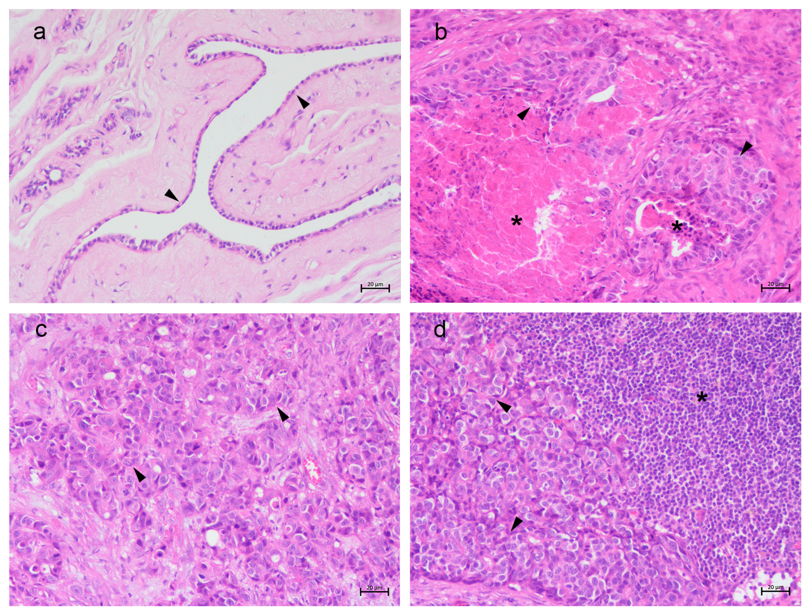
Figure 1. Feline mammary tissue. (a) Normal mammary gland with normal epithelial cells (arrow heads); (b) non-metastatic mammary carcinoma with tumoral cells (arrow heads) and necrosis (asterisks); (c) metastatic mammary carcinoma with tumoral cells (arrow heads); (d) tributary lymph node presenting lymphocytes (asterisk) and metastatic tumoral cells (arrow heads). Hematoxylin and eosin staining, 20×, scale bar 20 μm.
Vetsci 13 00810 g001
Figure 2. Feline mammary tissue. TfR1 immunostaining. (a) Normal mammary gland showing scattered membranous labeling in epithelial cells (arrow heads); (b) non-metastatic mammary carcinoma showing strong cytoplasmic labeling in tumoral cells (arrow heads); (c) metastatic mammary carcinoma showing strong cytoplasmic labeling in tumoral cells (arrow heads) and necrosis (asterisk); (d) tributary lymph node with metastatic cells showing cytoplasmic labeling (arrow heads). 20×, scale bar 20 μm.
Vetsci 13 00810 g002
Figure 3. Feline mammary tissue. TfR2 immunostaining. (a) Normal mammary gland showing few labeled epithelial cells (arrow heads); (b) non-metastatic mammary carcinoma showing strong membranal labeling in tumoral cells (arrow heads); (c) metastatic mammary carcinoma showing very strong cytoplasmic labeling in tumoral cells (arrow heads); (d) tributary lymph node with metastatic cells showing cytoplasmic labeling in tumoral cells (arrow heads), 20×, scale bar 20 μm.
Vetsci 13 00810 g003
Figure 4. Feline mammary tissue. FTH1 immunostaining. (a) Normal mammary gland showing few labeled epithelial cells (arrow); (b) non-metastatic mammary carcinoma showing strong labeling in tumoral cells (arrow); (c) metastatic mammary carcinoma showing strong cytoplasmic labeling in tumoral cells (arrow); (d) tributary lymph node with metastatic cells showing cytoplasmic labeling in tumoral cells (arrow), 20×, scale bar 20 μm.
Vetsci 13 00810 g004
Figure 5. Feline mammary tissue. SLC40A1 immunostaining. (a) Normal mammary gland not showing labeled epithelial cells; (b) non-metastatic mammary carcinoma showing strong cytoplasmic labeling in tumoral cells (arrow heads); (c) metastatic mammary carcinoma showing weak cytoplasmic labeling in a few tumoral cells (arrow heads); (d) tributary lymph node with metastatic cells showing cytoplasmic labeling in tumoral cells (arrow heads), 20×, Scale bar 20 μm.
Vetsci 13 00810 g005
Figure 6. Feline mammary tissue. HIF-1α immunostaining. (a) Normal mammary gland not showing labeled cells; (b) non-metastatic mammary carcinoma showing strong cytoplasmic labeling in tumoral cells (arrow heads); (c) metastatic mammary carcinoma showing cytoplasmic labeling in a few tumoral cells (arrow heads); (d) tributary lymph node with metastatic cells showing moderate cytoplasmic labeling in tumoral cells (arrow heads), 20×, scale bar 20 μm.
Vetsci 13 00810 g006
Table 1. Primary antibodies used for immunohistochemistry analysis.
AntibodyManufacturer/CloneHost SpeciesDilution
TfR1ThermoFisher 13-6800Mouse1:100
TfR2Antibodies ABIN2782221/PolyclonalRabbit1:100
FTH1Antibodies ABIN2785803/PolyclonalRabbit1:100
SLC40A1Elabscience E-AB-19866/PolyclonalRabbit1:100
HIF-1αAbcam ab1/PolyclonalRabbit1:100
Table 2. Details about primary antibodies used in Western blot analysis.
AntibodyManufacturer/CloneHost SpeciesDilution
TfR1ThermoFisher, 13.6800Mouse1:2000
TfR2Antibodies ABIN2782221/PolyclonalRabbit1:1000
FTH1Antibodies ABIN2785803/PolyclonalRabbit1:500
SLC40A1Elabscience E-AB-19866/PolyclonalRabbit1:1000
HIF-1αAbcam ab1/PolyclonalRabbit1:1000
Disclaimer/Publisher’s Note: The statements, opinions and data contained in all publications are solely those of the individual author(s) and contributor(s) and not of MDPI and/or the editor(s). MDPI and/or the editor(s) disclaim responsibility for any injury to people or property resulting from any ideas, methods, instructions or products referred to in the content.