Shiga Toxin-Producing Escherichia coli in Aquaculture: A Decade (2015–2025)-Long Global Retrospective Outlook


Simple Summary
Abstract
1. Introduction
2. Materials and Methods
2.1. Inclusion Criteria
2.2. Exclusion Criteria
2.3. Systemic Literature Screening
2.4. Statistical Analysis
3. Results
3.1. Distribution of STEC Isolates
3.1.1. Continent/Country-Wise
3.1.2. Aquaculture Sample Type
3.2. Distribution of VAGs Among Aquaculture and the Associated Environment
3.3. Shiga Toxins and Intimin Genes
3.4. Detection of ARGs in Aquaculture and Associated Environments
3.5. Serogroups
4. Discussion
Supplementary Materials
Author Contributions
Funding
Institutional Review Board Statement
Informed Consent Statement
Data Availability Statement
Conflicts of Interest
Abbreviations
| STEC | Shiga toxin-producing E. coli |
| ARGs | Antibiotic resistance genes |
| VAGs | Virulence-associated genes |
| MDR | Multidrug Resistance |
| HGT | Horizontal gene transfer |
| AMR | Antimicrobial resistance |
| EPS | Extracellular polymeric substance |
| HUS | Hemolytic uremic syndrome |
References
- Tacon, A.G.J.; Metian, M.; Parsons, G.J.; Shumway, S.E. A global aquaculture and aquafeed production update: 2010 to 2023. Rev. Fish. Sci. Aquac. 2025, 34, 1–10. [Google Scholar] [CrossRef] [Scilit]
- Mansour, A.M.; Nossair, M.A.; Ibrahim, E.S.E.; Abd-Elaziz, R.A.; Althobaiti, F.; Alotaibi, K.S.; Albattal, S.B.; Marzouk, M.A.; Soliman, M.M.; El-Maksoud, E.M.A. Exploring the toxicity of thiamethoxam on aquatic ecosystems: An overarching assessment of biochemical parameters, genotoxicity, fish flesh residues and histological alterations in Nile tilapia. Environ. Pollut. Bioavailab. 2025, 37, 2493050. [Google Scholar] [CrossRef] [Scilit]
- Reverter, M.; Sarter, S.; Caruso, D.; Avarre, J.-C.; Combe, M.; Pepey, E.; Pouyaud, L.; Vega-Heredía, S.; de Verdal, H.; Gozlan, R.E. Aquaculture at the crossroads of global warming and antimicrobial resistance. Nat. Commun. 2020, 11, 1870. [Google Scholar] [CrossRef] [Scilit] [PubMed]
- Santos, R.F.B.; Godoy, A.C.; Fantini-Hoag, L.; Gomes, S.F.; De Moraes, L.B.S.; Cahú, T.B.; Silva, J.F.; Galvez, A.O.; Honorato, C.A.; Bezerra, R.D.S. Artemia biomass protein hydrolysate as a new feed for aquaculture: Litopenaeus vannamei (Mysis) performance. Aquac. Int. 2025, 33, 250. [Google Scholar] [CrossRef] [Scilit]
- Fatima, M.; Sadiq, S.; Hudda, N.; Chaudhry, M.; Tanvir, R.; Mushtaq, H. Antimicrobial resistance in Salmonella spp. from cultured Rohu (Labeo rohita), Tilapia (Oreochromis niloticus), and Carp (Cyprinus carpio): A possible threat to public health. Iran. J. Fish. Sci. 2025, 24, 665–677. [Google Scholar]
- Dushayeva, L.Z. Antimicrobial resistance in foodborne Escherichia coli and Salmonella spp. from animal-origin foods: Transmission pathways, global surveillance gaps, and alternative therapeutic strategies. Vet. World 2025, 18, 3288. [Google Scholar] [CrossRef] [Scilit] [PubMed]
- de Paula, C.C.; Porto, Y.D.; Castro, V.S.; do Carmo Dias Matos, É.; Cunha-Neto, A.; Carvalho, R.C.T.; Savay-da-Silva, L.K.; de Souza Figueiredo, E.E.; Motheo, T.F. Antimicrobial Resistance in Salmonella Across Aquaculture, Seafood and Related Aquatic Environments: A Scoping Review With Exploratory Textual Analysis. Rev. Aquac. 2026, 18, e70136. [Google Scholar] [CrossRef] [Scilit]
- Mahmud, M.N.; Jony, M.Z.R.; Sakidar, S.; Hasan, N.A.; Alam, M.M.; Haque, M.M. Breaking Antibiotic Dependency in Aquaculture: Evaluating Alternative Disease Management Strategies for Sustainable Aquaculture in Bangladesh. Preprint. 2026. [CrossRef] [Scilit] [PubMed]
- Schar, D.; Zhao, C.; Wang, Y.; Larsson, D.G.J.; Gilbert, M.; Van Boeckel, T.P. Twenty-year trends in antimicrobial resistance from aquaculture and fisheries in Asia. Nat. Commun. 2021, 12, 5384. [Google Scholar] [CrossRef] [Scilit] [PubMed]
- Lulijwa, R.; Alfaro, A.C.; Venter, L.; Young, T.; Decker, P.; Merien, F.; Meyer, J. Haematological and metabolic profiles of brooder giant kokopu, Galaxias argenteus (Gmelin 1789) based on sex and temperature. Aquac. Res. 2022, 53, 1542–1552. [Google Scholar]
- Rodriguez-Mozaz, S.; Jaén-Gil, A. Evaluation of Antibiotic Transformation During Water Treatment Using Non-target Screening Combined with Effect-Based Strategies. In Screening of Pollutants in the Environment: Non-Target Strategies and Latest Trends; Springer: Berlin/Heidelberg, Germany, 2025; pp. 41–58. [Google Scholar]
- Nobili, G.; Cocomazzi, A.; Basanisi, M.G.; Damato, A.M.; Coppola, R.; Cariglia, M.G.; Franconieri, I.; Stallone, A.; Notarangelo, M.; Scirocco, T.; et al. High Occurrence of Diverse ESBL-, AmpC- and Carbapenemase-Producing Escherichia coli and Klebsiella pneumoniae in Surface Waters, Southern Italy, 2023–2024. Microorganisms 2026, 14, 508. [Google Scholar] [CrossRef] [Scilit] [PubMed]
- Ntshonga, P.; Ntereke, T.D.; Zankere, T.; Morse, D.P.; Koto, G.; Gobe, I.; Paganotti, G.M. Lack of Association Between qacE and qacE∆ 1 Gene Variants and Sodium Hypochlorite Resistance in Clinical Isolates of ESBL-and Carbapenemase-Producing Klebsiella spp. and Enterobacter spp., from Gaborone, Botswana. Antibiotics 2025, 14, 662. [Google Scholar] [CrossRef] [Scilit] [PubMed]
- Lynch, F.; Lim, T.J.Y.; Steele, M.; Stevens, M.; Bata, L.; Lynch, G.P.; Dewi, D.A.P.R.; Sharp, S.; Orr, N.; Ward, A.; et al. Enhanced detection of animal-derived microbial hazards in forested catchments using high-volume ultrafiltration and amplicon-based microbial source tracking. Front. Water 2025, 7, 1694489. [Google Scholar] [CrossRef] [Scilit]
- Moračanin, S.V.; Milijašević, M.; Milijašević, J.B.; Danilović, B.; Zdolec, N. Major foodborne zoonotic bacteria and their role in antimicrobial resistance. Sci. J. Meat Technol. 2025, 66, 428–434. [Google Scholar] [CrossRef] [Scilit]
- Nahar, K.; Sunny, M.; Hassan, R.; Alam, J.Q.; Nupur, S.S.; Rahman, S.H.; Yasmin, F. Antibiotic susceptibility and MDR (Multi drug resistance) pattern of common bacteria isolated from wound infection in a tertiary care hospital, Gazipur, Bangladesh. Pac. J. Med. Res. 2025, 2, 55–62. [Google Scholar] [CrossRef] [Scilit]
- Crona, B.; Käll, S.; Van Holt, T. Fishery Improvement Projects as a governance tool for fisheries sustainability: A global comparative analysis. PLoS ONE 2019, 14, e0223054. [Google Scholar] [CrossRef] [Scilit] [PubMed]
- Morgado, K.R. Escherichia coli Produtoras de AmpC Plasmidiais Em Águas de Diferentes Contextos Ambientais No Rio de Janeiro: Ocorrência e Fenótipos de Resistência Associados. Master’s Thesis, Universidade Federal do Rio de Janeiro, Rio de Janeiro, Brazil, 2025. [Google Scholar]
- Li, Y.; Ye, Y.; Tang, Z.; Zhou, Z.; Yao, Z.; Gao, P.; Zhao, Y.; Lai, Q. Effects of high pH on molting and calcium absorption of Pacific white shrimp (Litopenaeus vannamei). Aquac. Rep. 2026, 46, 103274. [Google Scholar] [CrossRef] [Scilit]
- El Badawy, A.; Hamza, D.; Ahmed, Z.; Sabry, M.A. Seasonal and environmental drivers of antibiotic resistance and virulence in Escherichia coli from aquaculture and their public health implications. Sci. Rep. 2025, 15, 16100. [Google Scholar] [CrossRef] [Scilit] [PubMed]
- Ebied, N.; Abdou, M.; Abass, M.; Kassab, A. Heavy Metal Residues and Microbial Status of Farmed and Channeled Cat Fish. J. Hell. Vet. Med. Soc. 2024, 75, 6897–6906. [Google Scholar]
- Mohammed, A.; Kareem, M.N.; Azawi, N.A.; Al-Juhaishi, O.A. Comparison of the Histological Structure of Gill Tissue in Local Common Carp and Catfish and Their Relationship to Oxygen Requirements. Egypt. J. Aquat. Biol. Fish. 2025, 29, 523–529. [Google Scholar] [CrossRef] [Scilit]
- Al Qabili, D.M.A.; Aboueisha, A.-K.M.; Youssef, A.I.; El-Mahallawy, H.S. Antimicrobial resistance and virulence genes of Shiga toxin-producing Escherichia coli (STEC) isolated from tilapia and mullet and its public health significance. Suez Canal Vet. Med. 2023, 28, 55–72. [Google Scholar] [CrossRef] [Scilit]
- Elsharawy, N.T.; Ramadan, M.A.A.; Elsayed, A.M. Prevalence of Escherichia coli Resistance Genes in Mullet Fish. J. Adv. Vet. Res. 2023, 13, 3488. [Google Scholar] [CrossRef] [Scilit] [PubMed]
- Ahmed, W.; Haque, R.; Siddique, A.B.; Akbor, A.; Hasan, M.; Rahman, M. Is Biofloc fish a safe alternative to conventionally cultivated fish regarding metal bioaccumulation in Bangladesh? Environ. Chall. 2023, 11, 100704. [Google Scholar] [CrossRef] [Scilit]
- Alexandre, L.A.; da Silva, A.C.; Nascimento, F.L.d.A.; de Melo, A.P.Z.; Verruck, S. Antimicrobial resistance of Escherichia coli isolated from fish and aquaculture water: An emerging concern for consumers. Lett. Appl. Microbiol. 2025, 78, ovaf099. [Google Scholar] [CrossRef] [Scilit] [PubMed]
- Al Qabili, D.M.A.; Aboueisha, A.-K.M.; Ibrahim, G.A.; Youssef, A.I.; El-Mahallawy, H.S. Virulence and antimicrobial-resistance of shiga toxin-producing E. coli (STEC) Isolated from edible shellfish and its public health significance. Arch. Microbiol. 2022, 204, 510. [Google Scholar] [CrossRef] [Scilit] [PubMed]
- Ramadan, R.S.; Dawood, Y.H.; Yehia, M.M.; Gad, A. Environmental and health impact of current uranium mining activities in southwestern Sinai, Egypt. Environ. Earth Sci. 2022, 81, 213. [Google Scholar] [CrossRef] [Scilit]
- Seleem, A.; Sabry, M.A.; Abdel-Moein, K.A. Migratory birds as a potential overseas transmitter of Shiga toxin-producing Escherichia coli. Int. J. Vet. Sci. Med. 2021, 9, 52–58. [Google Scholar] [CrossRef] [Scilit] [PubMed]
- Hussein, M.A.; Merwad, A.M.; Elabbasy, M.T.; Suelam, I.I.; Abdelwahab, A.M.; Taha, M.A. Prevalence of Enterotoxigenic Staphylococcus aureus and Shiga Toxin Producing Escherichia coli in fish in Egypt: Quality parameters and public health hazard. Vector-Borne Zoonotic Dis. 2019, 19, 255–264. [Google Scholar] [CrossRef] [Scilit] [PubMed]
- Shokr, N.M.; Mahmoud, A.; Ibrahim, M. Detection of E. coli in Nile Tilapia from retail in Gharbiya Governorate. Alex. J. Vet. Sci. 2018, 59, 76. [Google Scholar] [CrossRef] [Scilit]
- Abdel-Latif, H.M.; Sedeek, E.K. Diversity of Enterobacteriaceae retrieved from diseased cultured Oreochromis niloticus. Int. J. Fish. Aquat. Stud. 2017, 5, 29–34. [Google Scholar] [CrossRef] [Scilit] [PubMed]
- Zaki, V.; El-Gohary, A.H.; Nabih, M.M. Molecular identification of E. coli and Salmonella species isolated from fresh water fish by Polymerase Chain Reactions (PCR). Mansoura Vet. Med. J. 2017, 18, 34. [Google Scholar] [CrossRef] [Scilit]
- Saqr, S.; Khaliel, R.; Ibrahim, M.S. Antibiotic Resistance and Virulence Genes of E. coli Isolated From Fresh Nile Tilapia (Oreochromis niloticus) in El-Behera Governorate, Egypt. Alex. J. Vet. Sci. 2016, 48, 83–90. [Google Scholar] [CrossRef] [Scilit]
- Bedane, D.T.; Asfaw, S.L. Performance evaluation of a two-phase anaerobic reactor coupled with microalgae photobioreactors for slaughterhouse wastewater treatment in Ethiopia. Biomass Convers. Biorefinery 2025, 15, 5659–5671. [Google Scholar]
- Abdi, M.; Mirkalantari, S.; Amirmozafari, N. Bacterial resistance to antimicrobial peptides. J. Pept. Sci. 2019, 25, e3210. [Google Scholar] [CrossRef] [Scilit] [PubMed]
- Ayalew, T.; Teshome, M.; Aemro, D. Food and Feeding habits of the introduced common carp (Cyprinus carpio L. 1758) in Lake Ardibo, South Wollo, Ethiopia. Heliyon 2025, 11, e42596. [Google Scholar] [CrossRef] [Scilit] [PubMed]
- Mitiku, B.A.; Mitiku, M.A.; Ayalew, G.G.; Alemu, H.Y.; Geremew, U.M.; Wubayehu, M.T. Microbiological quality assessment of fish origin food along the production chain in upper Blue Nile watershed, Ethiopia. Food Sci. Nutr. 2023, 11, 1096–1103. [Google Scholar] [CrossRef] [Scilit]
- Kambire, O.; Adingra, A.A.; Yao, K.M.; Koffi-Nevry, R. Prevalence of virulence genes associated with diarrheagenic pathotypes of Escherichia coli isolates from water, sediment, fish, and crab in Aby Lagoon, Côte d’Ivoire. Int. J. Microbiol. 2017, 2017, 9532170. [Google Scholar] [CrossRef] [Scilit] [PubMed]
- Aliyu, R.M.; Abubakar, M.B.; Yakubu, Y.; Shuaibu, A.B. Prevalence of Escherichia coli O157: H7 in some animal products sold within Sokoto Metropolis, Nigeria. Afr. J. Bacteriol. Res. 2021, 13, 1–6. [Google Scholar] [CrossRef] [Scilit] [PubMed]
- Aleru, C.P. Detection of resistance genes in Escherichia coli isolated from fishes and shellfishes in Creek Road/Bonny Estuary, Port Harcourt, Nigeria. Microbiol. Res. J. Int. 2022, 32, 11–22. [Google Scholar] [CrossRef] [Scilit]
- Odumosu, B.T.; Obeten, H.I.; Bamidele, T.A. Incidence of Multidrug-Resistant Escherichia coli Harbouring bla TEM and tet A Genes Isolated from Seafoods in Lagos Nigeria. Curr. Microbiol. 2021, 78, 2414–2419. [Google Scholar] [CrossRef] [Scilit] [PubMed]
- Aliyu, A. Bacteriological and Physicochemical Qualities of Water and Fish Samples From River Lavun, Bida, Niger State, Nigeria. Master’s Thesis, Ahmadu Bello University, Zaria, Nigeria, 2016. [Google Scholar]
- Mwanza, F.; Komba, E.V.G.; Kambarage, D.M. Occurrence and determination of antimicrobial resistant Escherichia coli isolates in fish and vegetables as indicator organism of faecal contamination in Dar es Salaam, Tanzania. Int. J. Microbiol. 2021, 2021, 6633488. [Google Scholar] [CrossRef] [Scilit] [PubMed]
- Ethimonnier, D.; Bentz, B.; Mikolasek, O.; Oswald, M. Case study of innovations in commercial West African family fish farming that led to an ecological intensification. Aquat. Living Resour. 2022, 35, 6. [Google Scholar] [CrossRef] [Scilit]
- Mumbo, M.T.; Nyaboga, E.N.; Kinyua, J.K.; Muge, E.K.; Mathenge, S.G.K.; Rotich, H.; Muriira, G.; Njiraini, B.; Njiru, J.M. Antimicrobial resistance profiles of Salmonella spp. and Escherichia coli isolated from fresh Nile tilapia (Oreochromis niloticus) fish marketed for human consumption. BMC Microbiol. 2023, 23, 306. [Google Scholar] [CrossRef] [Scilit] [PubMed]
- Songe, M.M.; Hang’ombe, B.M.; Knight-Jones, T.J.D.; Grace, D. Antimicrobial resistant enteropathogenic Escherichia coli and Salmonella spp. in houseflies infesting fish in food markets in Zambia. Int. J. Environ. Res. Public Health 2017, 14, 21. [Google Scholar]
- Gufe, C.; Kapomba, K.; Chari, T.A.; Jambwa, P.; Muteveri, T.; Machakwa, J.; Jackson, B.; Mbonjani, B.; Katsande, P.; Musari, S. Comparative analysis of antimicrobial resistance in Nile tilapia from polluted natural waters versus aquaculture systems in Zimbabwe: Public health and sustainable aquaculture implications. Cogent Food Agric. 2025, 11, 2548929. [Google Scholar] [CrossRef] [Scilit]
- Hove, H. Monitoring Program for Pharmaceuticals, Illegal Substances, and Contaminants in Farmed Fish-Annual Report for 2017; Institute of Marine Research: Bergen, Norway, 2018. [Google Scholar]
- Dewi, R.R.; Hassan, L.; Daud, H.M.; Matori, M.F.; Zakaria, Z.; Ahmad, N.I.; Aziz, S.A.; Jajere, S.M. On-farm practices associated with multi-drug-resistant Escherichia coli and Vibrio parahaemolyticus derived from cultured fish. Microorganisms 2022, 10, 1520. [Google Scholar] [CrossRef] [Scilit] [PubMed]
- Faridullah, M.; Roy, V.C.; Lithi, U.J. Prevalence of Salmonella and Escherichia coli contamination in shrimp (Penaeus monodon) farms, depots and processing plants in different areas of Bangladesh. Asian J. Med. Biol. Res. 2016, 2, 171–176. [Google Scholar] [CrossRef] [Scilit]
- Moorthy, G.; Arumugam, U. Antibiotic Resistance and Molecular Epidemiology of ESBL-Producing E. coli in Shrimp Aquaculture: A Study from Tiruvallur District, Tamil Nadu, India: G. Moorthy, U. Arumugam. Curr. Microbiol. 2025, 82, 401. [Google Scholar] [CrossRef] [Scilit] [PubMed]
- Dwivedi, A.; Kumar, C.B.; Kumar, A.; Soni, M.; Sahu, V.; Awasthi, A.; Rathore, G. Molecular characterization of carbapenem resistant E. coli of fish origin reveals the dissemination of NDM-5 in freshwater aquaculture environment by the high risk clone ST167 and ST361. Environ. Sci. Pollut. Res. 2023, 30, 49314–49326. [Google Scholar] [CrossRef] [Scilit] [PubMed]
- Kavinesan, K.; Sugumar, G.; Chrisolite, B.; Muthiahsethupathy, A.; Sudarshan, S.; Parthiban, F.; Mansoor, M. Phenotypic and genotypic characterization of pathogenic Escherichia coli identified in resistance mapping of β-lactam drug-resistant isolates from seafood along Tuticorin coast. Environ. Sci. Pollut. Res. 2023, 30, 68111–68128. [Google Scholar] [CrossRef] [Scilit] [PubMed]
- Roos, D.; Taconet, J.; Gentil, C.; Brisset, B.; Evano, H.; Aumond, Y.; Huet, J.; Lepetit, C.; Boymond-Morales, R.; Rungassamy, T. Variation of the relationships between lengths and weights applied to 123 fish species observed at Réunion Island (Indian Ocean). Afr. J. Mar. Sci. 2022, 44, 171–180. [Google Scholar] [CrossRef] [Scilit]
- Abraham, T.J.; Qureshi, Q.A.; Bardhan, A. Enteric pathogenic and multiple antibiotic-resistant Escherichia coli in farmed Indian major carps and their environments in Peri-Urban Kolkata, India. J. Aquat. Food Product. Technol. 2022, 31, 1092–1108. [Google Scholar] [CrossRef] [Scilit]
- Francis, B.; Antony, A.; Sukumaran, D.; Hatha, A. Prevalence, antimicrobial resistance, and molecular characterization of Escherichia coli isolated from food contact surfaces in seafood pre-processing plants (India). J. Food Qual. Hazards Control 2022, 9, 3–13. [Google Scholar]
- Safeena, M.; Preenanka, R.; Sreedudha, N.V.; Kishore, P.; Blossom, K.L. Molecular characterisation and antibiotic resistance profile of Escherichia coli. Indian J. Fish. 2022, 69, 93–99. [Google Scholar] [CrossRef] [Scilit]
- Kusunur, A.B.; Kuraganti, G.K.; Mogilipuri, S.S.; Vaiyapuri, M.; Narayanan, S.V.; Badireddy, M.R. Multidrug resistance of Escherichia coli in fish supply chain: A preliminary investigation. J. Food Saf. 2022, 42, e12972. [Google Scholar] [CrossRef] [Scilit]
- Singh, A.S.; Nayak, B.B.; Kumar, S.H. High prevalence of multiple antibiotic-resistant, extendedspectrum β-lactamase (ESBL)-producing Escherichia coli in fresh seafood sold inretail markets of Mumbai, India. Vet. Sci. 2020, 7, 46. [Google Scholar] [CrossRef] [Scilit] [PubMed]
- Francis, B.; Antony, J.; Aravind, R.; Anand, P.S.; Balasubramanian, C.P.; Rajamanickam, S.; Veerachamy, P.; Ambasankar, K.; Gopal, C.; Vijayan, K.K. Reproductive performance, salinity tolerance, growth and production performance of a cryptic species Penaeus (Marsupenaeus) japonicus. Aquac. Res. 2021, 52, 5506–5516. [Google Scholar] [CrossRef] [Scilit]
- Bajpai, V.; Pragyan, D.; Mohanty, J.; Sahoo, P.K. Detection and Phylogenetic Analysis of Fish Viruses Inhabiting Freshwater Ornamental and Food Fish Species in India: A Survey-Based Study. Genet. Aquat. Org. 2021, 5, 9–18. [Google Scholar]
- Sivaraman, G.; Muneeb, K.H.; Cole, J.; Shome, B.; Holmes, M. Prevalence of extended spectrum beta lactamase (ESBL) E. coli in fishes from the retail markets of Guwahati, Assam. FishTech Rep. 2020, 6, 10–11. [Google Scholar]
- Ayobami, O.; Brinkwirth, S.; Eckmanns, T.; Markwart, R. Antibiotic resistance in hospital-acquired ESKAPE-E infections in low-and lower-middle-income countries: A systematic review and meta-analysis. Emerg. Microbes Infect. 2022, 11, 443–451. [Google Scholar] [CrossRef] [Scilit] [PubMed]
- Divya, P.S.; Thajudeen, J.; Yousuf, J.; Madavan, A.; Abdulla, M.H. Genetic relatedness, phylogenetic groups, antibiotic resistance, and virulence genes associated with ExPEC in Escherichia coli isolates from finfish and shellfish. J. Food Process. Preserv. 2020, 44, e14837. [Google Scholar] [CrossRef] [Scilit]
- Khan, M.R. Microbiological and Physicochemical Analyses of Supplied Water in Selected Dairy Farms of Dhaka City. Master’s Thesis, Sher-e-Bangla Agricultural University, Dhaka, Bangladesh, 2020. [Google Scholar]
- Siddhnath, K.; Majumdar, R.; Parhi, J.; Sharma, S.; Mehta, N.; Laishram, M. Detection and characterization of Shiga toxin-producing Escherichia coli from carps from integrated aquaculture system. Aquaculture 2018, 487, 97–101. [Google Scholar] [CrossRef] [Scilit]
- Cojocaru, A.L.; Liu, Y.; Smith, M.D.; Akpalu, W.; Chávez, C.; Dey, M.M.; Dresdner, J.; Kahui, V.; Pincinato, R.B.M.; Tran, N. The “seafood” system: Aquatic foods, food security, and the Global South. Rev. Environ. Econ. Policy 2022, 16, 306–326. [Google Scholar] [CrossRef] [Scilit]
- Kumar, M.; Acharya, A.P.; Kumar, S.; Thakuria, J.; Basumatary, G.; Chaturvedi, C.S. A review on Microsatellite markers and their application in Fisheries and Aquaculture. J. Exp. Zool. India 2019, 22, 1311–1319. [Google Scholar]
- Khalifa, A.; Gammal, G.; Fadl, S.; Gohary, M.; Barakat, M. Laboratory Microbiological and Chemical Analysis for Detection of Water Pollution in Fresh Water Fish Farms. Alex. J. Vet. Sci. 2020, 66, 76. [Google Scholar] [CrossRef] [Scilit]
- Sivaraman, G.; Prasad, M.; Jha, A.; Visnuvinayagam, S.; Renuka, V.; Remya, S.; Kriplani, Y.; Vanik, D. Prevalence of extended-spectrum β-lactamase producing Escherichia coli in seafood from the retail fishery outlets of Veraval, Gujarat, India. J. Environ. Biol. 2017, 38, 523. [Google Scholar] [CrossRef] [Scilit]
- Sekhar, M.S.; Sharif, N.M.; Rao, T.S. Serotypes of sorbitol-positive shiga toxigenic Escherichia coli (SP-STEC) isolated from freshwater fish. Int. J. Fish. Aquat. Sci. 2017, 5, 503–505. [Google Scholar]
- Roschanski, N.; Guenther, S.; Vu, T.T.T.; Fischer, J.; Semmler, T.; Huehn, S.; Alter, T.; Roesler, U. VIM-1 carbapenemase-producing Escherichia coli isolated from retail seafood, Germany 2016. Eurosurveillance 2017, 22, 17-00032. [Google Scholar] [CrossRef] [Scilit] [PubMed]
- Sivaraman, G.K.; Rajan, V.; Vijayan, A.; Elangovan, R.; Prendiville, A.; Bachmann, T.T. Antibiotic resistance profiles and molecular characteristics of extended-spectrum beta-lactamase (ESBL)-producing Escherichia coli and Klebsiella pneumoniae isolated from shrimp aquaculture farms in Kerala, India. Front. Microbiol. 2021, 12, 622891. [Google Scholar] [CrossRef] [Scilit] [PubMed]
- Mohseni, P.; Ghanbarpour, R.; Jajarmi, M.; Bagheri, M. Molecular and phenotypic characteristics of isolated Escherichia coli from the skin, gills, and intestine of rainbow trout in retail stores of Kerman, Iran. Iran. J. Vet. Res. 2025, 26, 17. [Google Scholar] [CrossRef] [Scilit] [PubMed]
- Ullah, M.; Rasool, F.; Khan, N.; Ali, S.; Sheikh, A.A. Antibiotic resistance and its gene profile in Escherichia coli isolated from diseased farmraised carps in Punjab, Pakistan. Pak. Vet. J. 2023, 41, 1–7. [Google Scholar]
- Ranjbar, R.; Pezeshknejad, P.; Khamesipour, F.; Amini, K.; Kheiri, R. Genomic fingerprints of Escherichia coli strains isolated from surface water in Alborz province, Iran. BMC Res. Notes 2017, 10, 295. [Google Scholar] [CrossRef] [Scilit] [PubMed]
- Tajbakhsh, E.; Ahmadi, P.; Abedpour-Dehkordi, E.; Arbab-Soleimani, N.; Khamesipour, F. Biofilm formation, antimicrobial susceptibility, serogroups and virulence genes of uropathogenic E. coli isolated from clinical samples in Iran. Antimicrob. Resist. Infect. Control 2016, 5, 11. [Google Scholar] [CrossRef] [Scilit] [PubMed]
- Bonyadian, M. Identifying verotoxigenic Escherishia. coli isolated from cold water fishes by Multiplex PCR in Chaharmahal Va Bakhtiary province. J. Microb. Biol. 2015, 3, 53–58. [Google Scholar]
- Mohamed, A.-R.M.; Hassan, K.A.; Abed, J.M. Fishery, Growth and Reproductive Biology of Metapenaeus affinis (Decapoda, Penaeidae) in the Iraqi waters. Int. J. Environ. Eng. Dev. 2025, 3, 100–110. [Google Scholar] [CrossRef] [Scilit]
- Sharef, N.A.; Khidhir, Z.K.; Arif, E.D. Evaluating the Hygienic Quality of Fish Meat and Environmental Contamination with Escherichia coli. Egypt. J. Aquat. Biol. Fish. 2025, 29, 149. [Google Scholar] [CrossRef] [Scilit]
- Altan, H.N.; Budak, M.M. Almanya’nin Göç Geçmişinin Genel Değerlendirmesi General Assessment Of Germany’s Migration History. In Proceedings of the ACADEMY 1st International Conference on Migration Studies, Barcelona, Spain, 25–28 January 2023. [Google Scholar]
- Nairoukh, Y.R.; Mahafzah, A.M.; Irshaid, A.; Shehabi, A.A. Molecular characterization of multidrug resistant uropathogenic E. coli isolates from Jordanian patients. Open Microbiol. J. 2018, 12, 1–7. [Google Scholar] [CrossRef] [Scilit] [PubMed]
- Hassan, J.; Eddine, R.Z.; Mann, D.; Li, S.; Deng, X.; Saoud, I.P.; Kassem, I.I. The mobile colistin resistance gene, mcr-1.1, is carried on IncX4 plasmids in multidrug resistant E. coli isolated from rainbow trout aquaculture. Microorganisms 2020, 8, 1636. [Google Scholar] [CrossRef] [Scilit] [PubMed]
- Hong, T.K.; George, B.; Norizan, N.; Francis, M. First detection of Shiga toxin producing Escherichia coli O157:H7 (HlyA gene) and enumeration of most probable number (MPN) of fecal coliforms and Escherichia coli in cage cultured oysters (Crassostrea iredalei) and water from southern Malaysia. Malays. J. Microbiol. 2021, 17, 384–392. [Google Scholar] [CrossRef] [Scilit]
- Ghaderpour, A.; Ho, W.S.; Chew, L.-L.; Bong, C.W.; Chong, V.C.; Thong, K.-L.; Chai, L.C. Diverse and abundant multi-drug resistant E. coli in Matang mangrove estuaries, Malaysia. Front. Microbiol. 2015, 6, 977. [Google Scholar] [CrossRef] [Scilit] [PubMed]
- Rasheed, S.; Shah, S.Q.A.; Naz, H.; Ahmad, A.S. Prevalence and Antibiotic Resistance Profiles of Quinolone-Resistant Bacteria from Freshwater Fish Farms in Southern Punjab, Pakistan. Pak. Vet. J. 2025, 45, 1262–1270. [Google Scholar]
- Hussain, N.; Xia, Y.; Han, J.; Saeed, M.; Arain, M.A.; Hassan, M.F.; Chen, H. Lactoferrin in Aquaculture: A Holistic Review of its Health Benefits and Functional Feed Application. Pak. Vet. J. 2024, 44, 581–591. [Google Scholar] [CrossRef] [Scilit]
- Sarwar, A.; Aslam, B.; Rasool, M.H.; Bekhit, M.M.S.; Sasanya, J. A health threat from farm to fork: Shiga toxin-producing Escherichia coli Co-Harboring bla NDM-1 and MCR-1 in various sources of the food supply chain. Pathogens 2024, 13, 659. [Google Scholar] [CrossRef] [Scilit] [PubMed]
- Uddin, M.; Hossain, M.T.; Proshad, R.; Kormoker, T.; Chandra, K.; Rimi, T.A. Identification of Pathogenic Escherichia coli Strain from River and Sewage Water in Bangladesh. Arch. Agric. Environ. Sci. 2019, 4, 39–44. [Google Scholar] [CrossRef] [Scilit]
- Ture, M.; Altinok, I.; Alp, H. Effects of cage farming on antimicrobial and heavy metal resistance of Escherichia coli, Enterococcus faecium, and Lactococcus garvieae. Microb. Drug Resist. 2018, 24, 1422–1430. [Google Scholar] [CrossRef] [Scilit] [PubMed]
- Jang, H.M.; Kim, Y.B.; Choi, S.; Lee, Y.; Shin, S.G.; Unno, T.; Kim, Y.M. Prevalence of antibiotic resistance genes from effluent of coastal aquaculture, South Korea. Environ. Pollut. 2018, 233, 1049–1057. [Google Scholar] [CrossRef] [Scilit] [PubMed]
- Cheng, S.S.; Senoo, S.; Siddiquee, S.; Rodrigues, K.F. Genetic variation in the mitochondrial genome of the giant grouper Epinephelus lanceolatus (Bloch, 1790) and its application for the identification of broodstock. Aquac. Rep. 2015, 2, 139–143. [Google Scholar] [CrossRef] [Scilit]
- Hinthong, W.; Thaotumpitak, V.; Sripradite, J.; Indrawattana, N.; Srisook, T.; Kongngoen, T.; Atwill, E.R.; Jeamsripong, S. Antimicrobial resistance, virulence profile, and genetic analysis of ESBL-producing Escherichia coli isolated from Nile tilapia in fresh markets and supermarkets in Thailand. PLoS ONE 2024, 19, e0296857. [Google Scholar] [CrossRef] [Scilit] [PubMed]
- Jeamsripong, S.; Kuldee, M.; Thaaotumpitak, V.; Chuanchuen, R. Antimicrobial resistance,Extended-spectrum beta lactamases production and virulence genes in Salmonella enterica and Escherchia coli isolates from esturaine enviornment. PLoS ONE 2023, 18, e0283359. [Google Scholar] [PubMed]
- Kuldee, M. Detection of Antimicrobial Resistance in Escherichia coli and Salmonella spp. Originated from Cultivated Oysters and Estuarine Waters. Master’s Thesis, Chulalongkorn University, Bangkok, Thailand, 2021. [Google Scholar]
- Jeamsripong, S.; Thaotumpitak, V.; Anuntawirun, S.; Roongrojmongkhon, N.; Atwill, E.R.; Hinthong, W. Molecular Epidemiology of Antimicrobial Resistance and Virulence Profiles of Escherichia coli, Salmonella spp., and Vibrio spp. Isolated from Coastal Seawater for Aquaculture. Antibiotics 2022, 11, 1688. [Google Scholar] [CrossRef] [Scilit] [PubMed]
- Nimnoi, P.; Pongsilp, N. Distribution and expression of virulence genes in potentially pathogenic bacteria isolated from seafood in Thailand. CyTA-J. Food 2020, 18, 753–763. [Google Scholar] [CrossRef] [Scilit]
- Xue, L.; Luo, X.; Xing, J.-H.; Wang, D.; Zhang, D.-X. Isolation and pathogenicity evaluation of Escherichia coli O157: H7 from common carp, Cyprinus carpio. Microb. Pathog. 2023, 182, 106250. [Google Scholar] [CrossRef] [Scilit] [PubMed]
- Liang, P.; Wu, S.; Zhang, C.; Zhang, J.; Wong, M. Environmental geochemistry of Hg in intensive fish farming sites: Implications of Hg speciation change related to its health perspectives. Curr. Opin. Environ. Sci. Health 2021, 20, 100242. [Google Scholar] [CrossRef] [Scilit]
- Stein, R.W.; Mull, C.G.; Kuhn, T.S.; Aschliman, N.C.; Davidson, L.N.K.; Joy, J.B.; Smith, G.J.; Dulvy, N.K.; Mooers, A.O. Global priorities for conserving the evolutionary history of sharks, rays and chimaeras. Nat. Ecol. Evol. 2018, 2, 288–298. [Google Scholar] [CrossRef] [Scilit] [PubMed]
- Zhang, H.; Zhou, Y.; Guo, S.; Chang, W. Prevalence and characteristics of extended-spectrum beta-lactamase (ESBL)-producing Enterobacteriaceae isolated from rural well water in Taian, China, 2014. Environ. Sci. Pollut. Res. 2015, 22, 11488–11492. [Google Scholar] [CrossRef] [Scilit] [PubMed]
- Bölükbaş, A.; Gücükoğlu, A.; Uyanık, T.; Küllük, E.; Çadırcı, Ö. Determination of virulence factors and antibiotic resistance profile of E. coli O157: H7 in some marine and freshwater fish from Samsun, Türkiye. Turk. J. Fish. Aquat. Sci. 2024, 24, TRJFAS25057. [Google Scholar] [CrossRef] [Scilit]
- Yibar, A.; Saticioglu, I.B.; Ajmi, N.; Duman, M. Molecular characterization and antibacterial resistance determination of Escherichia coli isolated from fresh raw mussels and ready-to-eat stuffed mussels: A major public health concern. Pathogens 2024, 13, 532. [Google Scholar] [CrossRef] [Scilit] [PubMed]
- Onmaz, N.E.; Yildirim, Y.; Karadal, F.; Hizlisoy, H.; Al, S.; Gungor, C.; Disli, H.B.; Barel, M.; Dishan, A.; Tegin, R.A.A.; et al. Escherichia coli O157 in fish: Prevalence, antimicrobial resistance, biofilm formation capacity, and molecular characterization. LWT 2020, 133, 109940. [Google Scholar] [CrossRef] [Scilit]
- Baran, A.; Adigüzel, M.C.; Yüksel, M. Prevalence of antibiotic-resistant and extended-spectrum beta-lactamase-producing Escherichia coli in chicken meat from eastern Turkey. Pak. Vet. J. 2020, 40, 355–359. [Google Scholar] [CrossRef] [Scilit] [PubMed]
- De Vleeschouwer, J. Blockchain en Smart Contracts in de Verzekeringssector: Juridische Aspecten. Master’s Thesis, KU Leuven, Leuven, Belgium, 2019. [Google Scholar]
- Ellis-Iversen, J.; Seyfarth, A.M.; Korsgaard, H.; Bortolaia, V.; Munck, N.; Dalsgaard, A. Antimicrobial resistant E. coli and enterococci in pangasius fillets and prawns in Danish retail imported from Asia. Food Control 2020, 114, 106958. [Google Scholar] [CrossRef] [Scilit]
- Gross, S.; Müller, A.; Seinige, D.; Wohlsein, P.; Oliveira, M.; Steinhagen, D.; Kehrenberg, C.; Siebert, U. Occurrence of antimicrobial-resistant Escherichia coli in marine mammals of the North and Baltic Seas: Sentinels for human health. Antibiotics 2022, 11, 1248. [Google Scholar] [CrossRef] [Scilit] [PubMed]
- Parlapani, F.F.; Boziaris, I.S. Fish, Antibiotic Resistance, and Food Safety at the Global Level. In Handbook of Public Health Nutrition: International, National, and Regional Perspectives; Springer: Berlin/Heidelberg, Germany, 2025; pp. 1–21. [Google Scholar]
- Marceddu, M.; Lamon, S.; Consolati, S.G.; Ciulli, S.; Mazza, R.; Mureddu, A.; Meloni, D. Determination of Salmonella spp., E. coli VTEC, Vibrio spp., and Norovirus GI-GII in bivalve molluscs collected from growing natural beds in Sardinia (Italy). Foods 2017, 6, 88. [Google Scholar] [CrossRef] [Scilit] [PubMed]
- Rrahman, F.; Puto, K.; Maxhuni, Q.; Hulaj, B.; Bislimi, K. Analyses of Microbiological Data Indicates From Physic-Chemcal Parameters, In Fish Species From Sitnica, Lepenci And Lumbardhi I Prizrenit Rivers (Kosova). Int. J. Ecosyst. Ecol. Sci. 2015, 5, 207–212. [Google Scholar]
- Grevskott, D.H.; Svanevik, C.S.; Sunde, M.; Wester, A.L.; Lunestad, B.T. Marine bivalve mollusks as possible indicators of multidrug-resistant Escherichia coli and other species of the Enterobacteriaceae family. Front. Microbiol. 2017, 8, 24. [Google Scholar] [CrossRef] [Scilit] [PubMed]
- Boss, R.; Overesch, G.; Baumgartner, A. Antimicrobial resistance of Escherichia coli, Enterococci, Pseudomonas aeruginosa, and Staphylococcus aureus from raw fish and seafood imported into Switzerland. J. Food Prot. 2016, 79, 1240–1246. [Google Scholar] [CrossRef] [Scilit] [PubMed]
- Luo, J.; McDonald, R.R.; Wringe, B.F.; Heyer, C.E.D.; Smith, B.; Yan, Y.; Flemming, J.M. A spatial analysis of longline survey data for improved indices of Atlantic halibut abundance. ICES J. Mar. Sci. 2022, 79, 1954–1964. [Google Scholar] [CrossRef] [Scilit]
- Ofori, K.F.; Parsaeimehr, A.; Ozbay, G. Investigation of the presence and persistence of bacteria in seawater and oysters from an aquaculture farm in Rehoboth Bay, Delaware. Microbiol. Spectr. 2025, 13, e0305424. [Google Scholar] [CrossRef] [Scilit] [PubMed]
- Gomez-Gomez, A.; Malpica-Cruz, L.; Montaño-Moctezuma, C.; Cisneros-Montemayor, A.; Salomon, A.; Seingier, G. Diversity, challenges, and opportunities of Fishery Improvement Projects in the Latin America and the Caribbean region. Mar. Policy 2024, 163, 106116. [Google Scholar] [CrossRef] [Scilit]
- da Silva, A.C.; Alexandre, L.A.; Nascimento, F.L.A.; Verruck, S. Multidrug-resistant and colistin-resistant Escherichia coli harboring mcr-1 and ESBL genes in Brazilian mariculture and estuarine ecosystems. Int. J. Food Microbiol. 2025, 412, 110543. [Google Scholar]
- Tanyag, B.E.; Leyritana, K.M.; Quiambao, J.J.; Almagro, A.R.; Benitez, K.C.; Perelonia, K.B.; Cambia, F.; Montojo, U. Fecal Coliforms as Accurate Predictors of Health Risk: Current Status and Presence of Escherichia coli O157: H7 in Aquaculture Farms Surrounding Manila Bay. Philipp. J. Sci. 2025, 154, 1003. [Google Scholar] [CrossRef] [Scilit]
- Gonzalez, G.A.; Cerqueira, M.A. Shiga toxin-producing Escherichia coli in the animal reservoir and food in Brazil. J. Appl. Microbiol. 2020, 128, 1568–1582. [Google Scholar] [CrossRef] [Scilit] [PubMed]
- Barbosa, C.; Conceição, T.A.; Baliza, M.D.; Camilo, V.M.A.; Juiz, P.J.L.; Silva, I.M.M. Virulence genes in Escherichia coli isolates from commercialized saltwater mussels Mytella guyanensis (Lamarck, 1819). Braz. J. Biol. 2019, 79, 625–628. [Google Scholar] [CrossRef] [Scilit] [PubMed]
- Vásquez-García, A.; de Oliveira, A.P.S.C.; Mejia-Ballesteros, J.E.; de Godoy, S.H.S.; Barbieri, E.; de Sousa, R.L.M.; Fernandes, A.M. Escherichia coli detection and identification in shellfish from southeastern Brazil. Aquaculture 2019, 504, 158–163. [Google Scholar] [CrossRef] [Scilit]
- Ngu, R.C.; Choukem, S.-P.; Dimala, C.A.; Ngu, J.N.; Monekosso, G.L. Prevalence and determinants of selected cardio-metabolic risk factors among people living with HIV/AIDS and receiving care in the South West Regional Hospitals of Cameroon: A cross-sectional study. BMC Res. Notes 2018, 11, 305. [Google Scholar] [CrossRef] [Scilit] [PubMed]
- Ribeiro, L.; Barbosa, M.; Pinto, F.d.R.; Guariz, C.; Maluta, R.; Rossi, J.; Rossi, G.; Lemos, M.; Amaral, L.D. Shiga toxigenic and enteropathogenic Escherichia coli in water and fish from pay-to-fish ponds. Lett. Appl. Microbiol. 2016, 62, 216–220. [Google Scholar] [CrossRef] [Scilit] [PubMed]
- Brandão, M.A.D.R.; Lopes, A.I.S.T.; Neta, M.R.D.S.T.; de Oliveira, R.B.F.; Rezende, R.P.; Albuquerque, G.R.; Gonçalves, V.D.; Rodrigues, D.D.P.; Boehs, G.; Maciel, B.M. Microbiological quality and prevalence of β-lactam antibiotic resistance genes in oysters (Crassostrea rhizophorae). J. Food Prot. 2017, 80, 488–496. [Google Scholar] [CrossRef] [Scilit] [PubMed]
- Sánchez, F.; Fuenzalida, V.; Ramos, R.; Escobar, B.; Neira, V.; Borie, C.; Lapierre, L.; López, P.; Venegas, L.; Dettleff, P.; et al. Genomic features and antimicrobial resistance patterns of Shiga toxin-producing Escherichia coli strains isolated from food in Chile. Zoonoses Public Health 2021, 68, 226–238. [Google Scholar] [CrossRef] [Scilit] [PubMed]
- Tafur Zevallos, L.R.; Álvarez Janampa, C.; Turpo Calcina, J.; Robles Huaynate, R.A. Presence of Zoonotic Pathogenic Bacteria in the Digestive Tract of Fish of Marine Origin Commercialized in the Tingo Maria Supply Market; Universidad Nacional Agraria de la Selva: Tingo María, Peru, 2019. [Google Scholar]
- Nemati, M.; Shahosseini, S.R.; Ariaii, P. Review of fish protein hydrolysates: Production methods, antioxidant and antimicrobial activity and nanoencapsulation. Food Sci. Biotechnol. 2024, 33, 1789. [Google Scholar] [CrossRef] [Scilit] [PubMed]
- James, L.; Webster, T.M.U.; Thomas, G.; Middelboe, M.; Consuegra, S. A Review of Phage Therapy for Aquaculture Applications: Efficacy, Health Ramifications and Research Challenges. Fish. Fish. 2026, 27, 300–312. [Google Scholar] [CrossRef] [Scilit]
- Kanwal, S.; Abdullah, M.; Kumar, S.; Arshad, S.; Shahroz, M.; Zhang, D.; Kumar, D. An optimal internet of things-driven intelligent decision-making system for real-time fishpond water quality monitoring and species survival. Sensors 2024, 24, 7842. [Google Scholar] [CrossRef] [Scilit] [PubMed]
- Au-Yeung, C.; Tsui, Y.-L.; Choi, M.-H.; Chan, K.-W.; Wong, S.-N.; Ling, Y.-K.; Lam, C.-M.; Lam, K.-L.; Mo, W.-Y. Antibiotic abuse in ornamental fish: An overlooked reservoir for antibiotic resistance. Microorganisms 2025, 13, 937. [Google Scholar] [CrossRef] [Scilit] [PubMed]
- Ortega, S.; Lutfi, E.; Horn, S.S.; Durland, E. Quantification of fat content in the liver of different aquaculture fish species using hyperspectral image analysis. Aquaculture 2025, 597, 741924. [Google Scholar] [CrossRef] [Scilit]
- Ramatla, T.; Ramaili, T.; Lekota, K.E.; Ndou, R.; Mphuti, N.; Bezuidenhout, C.; Thekisoe, O. A systematic review and meta-analysis on prevalence and antimicrobial resistance profile of Escherichia coli isolated from water in africa (2000–2021). Heliyon 2023, 9, e16123. [Google Scholar] [CrossRef] [Scilit] [PubMed]
- Liao, C.-Y.; Balasubramanian, B.; Peng, J.-J.; Tao, S.-R.; Liu, W.-C.; Ma, Y. Antimicrobial resistance of Escherichia coli from aquaculture farms and their environment in Zhanjiang, China. Front. Vet. Sci. 2021, 8, 806653. [Google Scholar] [CrossRef] [Scilit] [PubMed]
- Li, P.; Han, H.; Zhang, S.; Fang, H.; Fan, W.; Zhao, F.; Xu, C. Reviews on the development of digital intelligent fisheries technology in aquaculture. Aquac. Int. 2025, 33, 191. [Google Scholar] [CrossRef] [Scilit]
- Zhuzzhassarova, G.; Azarbayjani, F.; Zamaratskaia, G. Fish and seafood safety: Human exposure to toxic metals from the aquatic environment and fish in Central Asia. Int. J. Mol. Sci. 2024, 25, 1590. [Google Scholar] [CrossRef] [Scilit] [PubMed]
- Alhabib, I.; Elhadi, N. Antimicrobial resistance pattern of Escherichia coli isolated from imported frozen shrimp in Saudi Arabia. PeerJ 2024, 12, e18689. [Google Scholar] [CrossRef] [Scilit] [PubMed]
- Page, M.J.; McKenzie, J.E.; Bossuyt, P.M.; Boutron, I.; Hoffmann, T.C.; Mulrow, C.D.; Shamseer, L.; Tetzlaff, J.M.; Akl, E.A.; Brennan, S.E.; et al. The PRISMA 2020 statement: An updated guideline for reporting systematic reviews. BMJ 2021, 372, n71. [Google Scholar] [CrossRef] [Scilit]


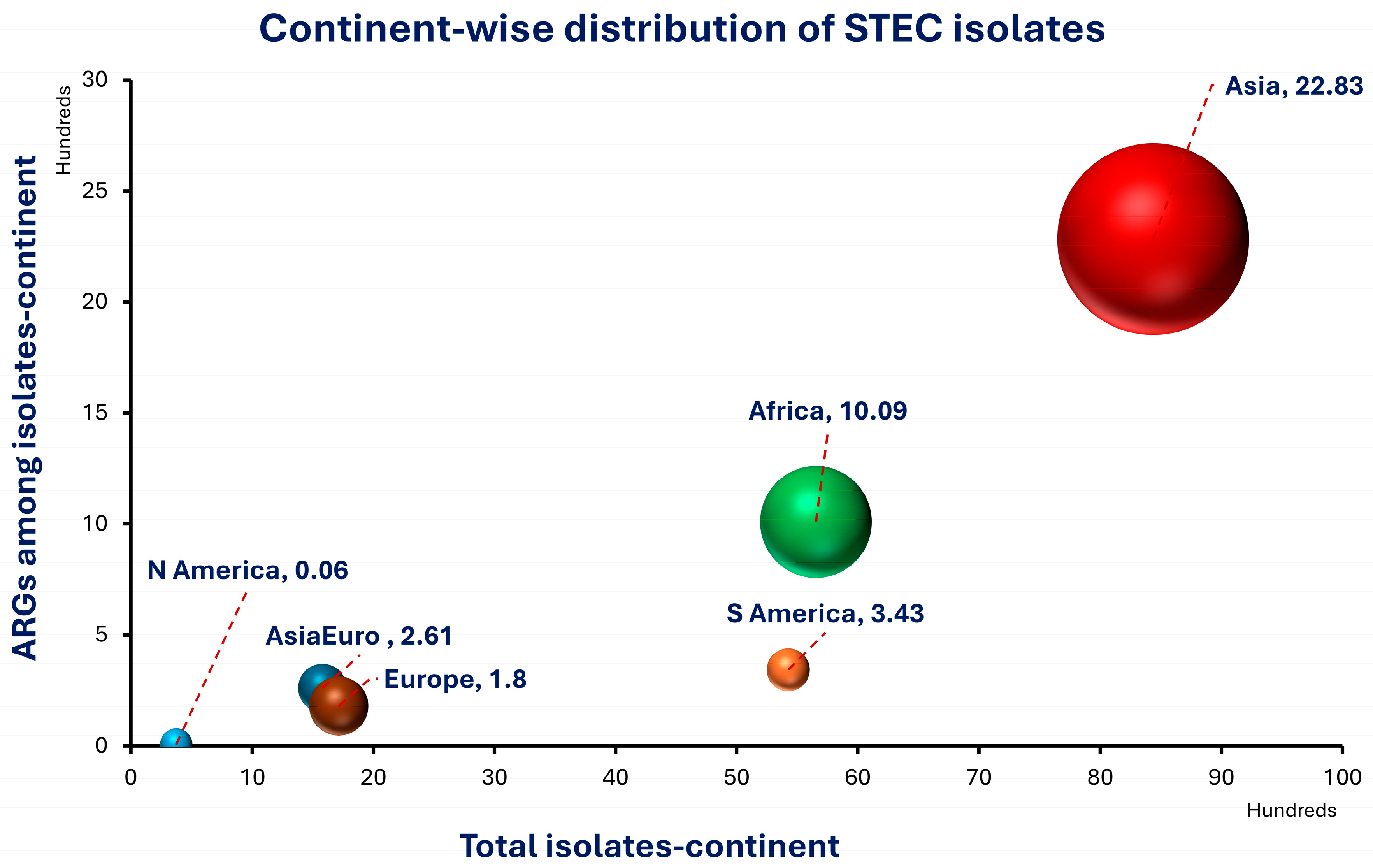

| Continent | Total No. of Isolates | STEC Isolates | VAGs | ARGs | ||
|---|---|---|---|---|---|---|
| Africa | 5655 | 1009 | 270 | 657 | ||
| Asia | 8437 | 2283 | 926 | 1938 | ||
| Asia/Europe | 1579 | 261 | 7 | 124 | ||
| Europe | 1714 | 180 | 93 | 184 | ||
| North America | 375 | 6 | ND | 55 | ||
| South America | 5426 | 343 | 123 | 97 | ||
| 23,186 | 4082 | 1419 | 3055 | |||
| ANOVA | ||||||
| Source | SS | Df | MS | F | p-value | F crit |
| Between Groups | “52,285,211” | 3 | “17,428,403.6” | 6.273698673 | 0.003551 * | 3.098391 |
| Within Groups | “55,560,219” | 20 | “2,778,010.95” | |||
| Continent | Country | Source | Total No. of Isolates | STEC Isolates | VAGs | ARGs | Reference |
|---|---|---|---|---|---|---|---|
| Africa | Egypt | Fish farm water (fish, O. niloticus, S. pilchardus | 328 | 76 | stx1, stx2, eaeA, st, lt, bfpA | blaTEM, blaCTX-M, blaOXA-48 | [20] |
| Clarias gariepinus catfish | 50 | 10 | stx1, stx2, | ND | [21] | ||
| Oysters | 33 | 27 | sfa, exhA, eaeA, estA, aer, stx1, stx2, rfc, hlyA, etrA, cnf1, sepA, | blaTEM, blaSHV, blaCTX−M, blaOXA−1 | [22] | ||
| Finfish (Tilapia, mullet) | 150 | 79 | stx1, stx2 | MDR gene | [23] | ||
| Mullet meat | 100 | 20 | SLT, LT | aaC2 gene | [24] | ||
| Gandolfi, Ruditapes decussatus | 20 | 20 | stx1, phoA, stx2 | ND | [25] | ||
| Shrimp (L. vannamei) | 82 | 11 | ND | blaTEM, tetA, blaOXA, sul1, aadA, ermB | [26] | ||
| Seafood | 135 | 57 | stx1, stx2, eaeA, | ND | [27] | ||
| Seafood samples | 192 | 10 | stx1, stx2 and eaeA | blaTEM, tetA | [28] | ||
| Migratory birds | 349 | 4 | stx1, stx2 and eaeA | MDR gene | [29] | ||
| Fish | 150 | 10 | stx1, stx2, and eaeA | ND | [30] | ||
| Fish samples from gills, musculature | 200 | 9 | stx1, stx2 and eaeA | ND | [31] | ||
| Earthen pond-cultured O. niloticus | 144 | 11 | ND | blaTEM, blaCTX-M, blaSHV, blaAMP blaOXA-2 | [32] | ||
| Tilapia (O. niloticus), Clarias gariepine | 400 | 171 | stx1, stx2 and eaeA | tetA, AmpC | [33] | ||
| Fresh Nile Tilapia fish | 218 | 24 | stx2, eaeA | tetA, blaTEM, sul1, Sul 2, aadA2 | [34] | ||
| Ethiopia | Fish | 450 | 8 | stx1, stx2, eaeA, | blaTEM-1B, tetA, fos7, qnrS, gyrA | [35] | |
| Fish | 410 | 1 | stx1, stx2 | ND | [36] | ||
| Fish | 121 | 41 | eae, hlyA1 | [37] | |||
| Fish | 328 | 62 | Nil | ND | [38] | ||
| Ivory Coast | Fish, crab, water sediment | 62 | 9 | stx1, stx2, eae, AggR | elt, est | [39] | |
| Nigeria | Miscellaneous retail fish | 256 | 28 | ND | ND | [40] | |
| Fish, shellfish | 72 | 11 | stx2, hlyA | blaSHV, blaTEM, mcr genes | [41] | ||
| Nile Tilapia, fish farms | 50 | - | ND | ND | [42] | ||
| Clarias gariepinus (catfish and aquaculture) | 47 | 5 | ND | ND | [43] | ||
| Tanzania | Manure, vegetables, and fish | 36 | 2 | Stx2 | ND | [44] | |
| West Africa | Fish farm | 156 | 99 | ND | ND | [45] | |
| Kenya | Nile Tilapia fish | 68 | 24 | ND | tetA, blaTEM, sul1, Sul 2, aadA2 | [46] | |
| Zambia | Fish | 418 | 56 | stx1, stx2 and eaeA | blaSHV, blaTEM, blaCTX-M | [47] | |
| Zimbabwe | Water sample (natural lake) | 600 | 120 | ND | ND | [48] | |
| Fish | 30 | 4 | ND | ND | [49] | ||
| Asia | Bangladesh | Fish | 312 | 139 | ND | ND | [50] |
| Shrimps (water, basket, mat) | 58 | 29 | ND | ND | [51] | ||
| India | Shrimp farms (pond water) | 102 | 79 | stx2 | blaTEM, blaCTX-M, blaSHV, AmpC, sul1 | [52] | |
| Fish (freshwater pond sites, retail) | 243 | 18 | ND | blaTEM, blaCTX-M-15, blaOXA-1, tetA, sul1, qnrS, cat1, cat2 | [53] | ||
| Seafood samples | 63 | 34 | ND | ND | [54] | ||
| Seafood samples | 130 | 60 | ND | blaCTX-M, blaSHV, blaTEM, AmpC | [55] | ||
| Carp, aquaculture | 232 | 49 | stx1, stx2 | MDR gene, enteric genes | [56] | ||
| Seafood pre-processing plants | 144 | 110 | stx1, stx2, eaeA | uid A gene | [57] | ||
| Seafood | 43 | 21 | ND | Rfb | [58] | ||
| Fish supply chain | 91 | 21 | ND | Sul1, blaCTX-M, tetD, dfrA, AmpC | [59] | ||
| Fish | 70 | 28 | ND | ND | [60] | ||
| Shrimps | 144 | 18 | stx1, stx2, eaeA, hlyA | uid A gene | [61] | ||
| Salmon sashimi (fish) | 30 | 15 | ND | ND | [62] | ||
| Food fish | 79 | 5 | ND | blaTEM, blaSHV, blaOXA, tetA, StrA, Sul1, Sul2, oqxA, qnrB | [63] | ||
| Fish | 100 | 6 | ND | ND | [64] | ||
| Finfish, shellfish | 134 | 2 | papAH, papC, sfa/focDE, iutA, kpsMT-II | blaTEM, blaSHV, blaCTXM, blaNDM, tetA, tetB, aphA2, strA, sul1, sul2, dhfrIa | [65] | ||
| Fish | 200 | 76 | ND | blaTEM, blaSHV, blaCTX-M | [66] | ||
| Carp from integrated aquaculture ponds | 30 | 1 | stx1, stx2, eae, ehlyA, rfbO157 | Rfb | [67] | ||
| Fresh seafood | 100 | 9 | stx1d, stx2, eaeA, hlyA | ND | [68] | ||
| Carps | 33 | 9 | stx1, stx2, eae, ehlyA, rfbO157 | ND | [69] | ||
| Fish market waste | 35 | 1 | ND | ND | [70] | ||
| Fish | 238 | 28 | ND | ND | [71] | ||
| Freshwater fish (Catla catla) | 104 | 18 | stx1, stx2, eaeA, hlyA | ND | [72] | ||
| Clams and shrimps | 160 | 12 | ND | blaVIM | [73] | ||
| Shrimp farms | 261 | 9 | viz., iutA, entB, mrkD | blaTEM, tetA, tetB, Sul1, Sul2, catA, CmlA, qnrS | [74] | ||
| Iran | Fish (retail store) | 166 | 65 | stx1a, stx1c, stx1d, stx2c, stx2d, stx2e stx2f | blaTEM, qnrA, tetB, sul2, blaSHV, sul, blaCTX-M, blaOXA | [75] | |
| River and sewage water | 70 | 14 | stx1, eae | ND | [76] | ||
| Cold-smoked salted fish | 200 | 5 | ND | ND | [77] | ||
| Aquaculture water | 150 | 27 | ND | tetA, Cmla, acc-IV, qnrA, sul1, sul2 | [78] | ||
| Cold water fish | 100 | 14 | stx1, stx2, eae | MDR genes | [79] | ||
| Iraq | Cultured fish (Labeo rohita, Cyprinus) | 120 | 11 | ND | blaTEM, blaCTX-M | [80] | |
| Water, sediment, feed, tools | 230 | 40 | ND | ESBL producing isolates | [81] | ||
| Fish farms, fish markets | 150 | 40 | ND | MDR gene | [82] | ||
| Jordan | Recreational lake water | 262 | 130 | ND | MDR genes | [83] | |
| Lebanon | Fish, rainbow trout, aquaculture | 135 | 106 | ND | mcr-1 | [84] | |
| Malaysia | Oyster (Crassostrea iredalei) | 120 | 104 | HlyA | ND | [85] | |
| Downstream of a fishing village | 175 | 148 | ND | MDR gene | [86] | ||
| Pakistan | Freshwater fish-farming sites | 101 | 55 | ND | qnrA, qnrB, qnrS | [87] | |
| Fish (Channa marulius) | 480 | 29 | ND | tetA, sul3, gyrB, blaTEM | [88] | ||
| Fish, shrimp, aquaculture water | 225 | 65 | stx1, stx2, eaeA | blaNDM-1, mcr-1 | [89] | ||
| Cerus (Labeo rohita, Catla catla) | 216 | 25 | ND | blaSHV, blaTEM, blaCTX-M, tetA, tetB, ermA, ermB, ermC, vgaA, strA, strB, aadA1, aac3-I | [90] | ||
| Singapore | Aquaculture farms | 31 | 23 | stx, intL | blaSHV, blaOXA, blaCTX-M, qnrA | [91] | |
| South Korea | Coastal aquaculture | 22 | 22 | ND | strA, strB, aadA1, aac3-I | [92] | |
| Fish (gizzard shad, halibut, rockfish) | 206 | 49 | ND | ND | [93] | ||
| Thailand | Nile Tilapia | 828 | 158 | stx, eaeA, | blaTEM, tetA, qnrS | [94] | |
| Oysters and estuarine water | 409 | 8 | stx1, stx2 | blaTEM, tetA | [95] | ||
| Oysters, estuarine water | 109 | 59 | stx1, stx2, eae | blaTEM, tet A, blaSHV, stn, sul2 | [96] | ||
| Aquatic environment, coastal waters | 384 | 84 | ND | blaTEM, blaCTX-M-15, blaOXA-1, tetA, sul1, qnrS | [97] | ||
| Fish | 42 | 22 | scnf2s, LTIs or vt2e | ND | [98] | ||
| China | Fish (carp) | 20 | 8 | ND | ND | [99] | |
| Aquaculture farms, soil sediment | 90 | 28 | ND | blaTEM, blaNDM, floR, OptrA, cmlA, aphA1, Sul2, oqxA, qnrS | [100] | ||
| Duck fish | 250 | 143 | ND | mcr-1 | [101] | ||
| Grass carp fish | 10 | 9 | ND | blaCTX-M 27, blaCTX-M15 | [102] | ||
| Asia/Europe | Türkiye | Fish (Engraulis encrasicolus, Merlangu) | 300 | 157 | stx1, stx2 | ND | [103] |
| Raw mussels | 71 | 2 | Sul3, tetB, qnrA, qnrB, sul1, fimH, floR | [104] | |||
| Fish | 140 | 2 | stx1, stx2 | ND | [105] | ||
| Mussels, sea bass | 823 | 100 | ND | blaTEM, tetA | [106] | ||
| Fish | 245 | - | ND | ND | [107] | ||
| Europe | Denmark | Zebra fish | 300 | 67 | ND | blaTEM, Sul2, qnrS, mcr-1 | [108] |
| Germany | Fish in Baltic sea | 116 | 41 | ND | ND | [109] | |
| Italy | Clams and shrimps | 620 | 14 | stx1, stx2, eaeA, hlyA | blaVIM, acc, blaSHV | [110] | |
| Carpet shells (Cerastoderma spp.) | 40 | 20 | stx1, stx2 | ND | [111] | ||
| Kosovo | Salmon fish | 54 | 2 | ND | ND | [112] | |
| Norway | Marine bivalve mollusks | 540 | 10 | ND | blaCTX-M | [113] | |
| Switzerland | Shellfish (oysters, shrimps) | 44 | 26 | ND | ND | [114] | |
| North America | Canada | Seafood | 277 | 2 | ND | blaKPC, blaOXA, blaNDM | [115] |
| USA | Seawater, shellfish (oysters) | 8 | 4 | ND | ND | [116] | |
| Nile Tilapia | 90 | - | ND | blaTEM, blaCTX-M, blaSHV, ampC, sul1 | [117] | ||
| South America | Brazil | Fish, shrimp, oysters, aquaculture water | 225 | 65 | ND | blaTEM, blaCTX-M, blaCMY-2, tetA, tetB, sul1, sul2, qnrA, qnrB, qnrS, aadA, floR, mcr-1 | [118] |
| Culture farms (fish, shrimp, oysters) | 69 | 29 | ND | ND | [119] | ||
| Aquaculture farms (fish, shrimp, oysters, seafood) | 6 | 5 | ND | blaTEM, blaCTX-M, blaCMY-2, tetA, sul1, qnrS | [120] | ||
| Mussels | 21 | 21 | stx1, elT, bfpA | ND | [121] | ||
| Shellfish | 150 | 1 | phoA, eae | ND | [122] | ||
| Frozen Tilapia filets (processing units) | 100 | 50 | ND | ND | [123] | ||
| Fish, fish ponds | 115 | 13 | eaeA, stx1, stx2 | ND | [124] | ||
| Oysters | 940 | 23 | eaeA, stx1, stx2, Ial, STa, STb, LT | blaTEM, blaSHV, blaKPC | [125] | ||
| Chile | Fresh seafood | 330 | 18 | hylA, stx2, ipfA | pmrB, qnrB | [126] | |
| Peru | Fish | 500 | 118 | ND | ND | [127] |
| Continent | No. of Studies | Representative Sources Sampled | Human Risk Categories Detected | Animal Fecal/Tissue Contamination Markers | Primary Aquaculture Pathogens (Strains) | Methodologies & Techniques Employed |
|---|---|---|---|---|---|---|
| Asia | 53 | Cultured fish Labeo rohita, Cyprinus carpio, fish, freshwater fish-farming sites | Potential public health risk, hand swabs | Fecal contamination, skin, gills, intestines | STEC, E. coli O157:H7, STEC, EPEC | PCR, culture, genotyping, MALDI-TOF |
| Africa | 30 | Nile Tilapia O. niloticus, shrimp L. vannamei, dish farm water | N/A, zoonotic risk identified | Yes—92.68%, tissue | E. coli O157:H7, STEC O157, STEC | Culture, 16S rRNA PCR, PCR, pathogenicity trial |
| Europe | 12 | Fish Engraulis encrasicolus, Merlangius euxmus, Trachurus mediterraneus, Mullus barbutus, Tinca tinca, Cyprinus carpio and Oncorchynhus mykiss, raw mussels, fish | N/A | Beef, meat | E. coli O157:H7, E. coli, STEC, APEC | mPCR, E-test method, Electrophoresis, RT-PCR |
| South America | 10 | Fish, shrimp, oysters, aquaculture water, aquaculture farm fish, shrimp, oysters, aquaculture pond water, seafood, meat, vegetables and ready-to-eat street-vended food | Yes | Fecal contamination | STEC, EPEC, ETEC, STEC, E. coli O157:H7, STEC | PCR, AMR profiling, RT-PCR, qPCR |
| North America | 3 | Seawater, shellfish, oysters, seafood, fish feces | Fecal contamination | Fecal contamination | STEC O157, E. coli, CRE, E. coli O157:H7 | PCR, qPCR, Multiplex PCR |
| Total Studies | 108 |
Disclaimer/Publisher’s Note: The statements, opinions and data contained in all publications are solely those of the individual author(s) and contributor(s) and not of MDPI and/or the editor(s). MDPI and/or the editor(s) disclaim responsibility for any injury to people or property resulting from any ideas, methods, instructions or products referred to in the content. |
© 2026 by the authors. Licensee MDPI, Basel, Switzerland. This article is an open access article distributed under the terms and conditions of the Creative Commons Attribution (CC BY) license.
Share and Cite
Sarwar, A.; Aslam, B.; Aljasir, S.F. Shiga Toxin-Producing Escherichia coli in Aquaculture: A Decade (2015–2025)-Long Global Retrospective Outlook. Vet. Sci. 2026, 13, 779. https://doi.org/10.3390/vetsci13080779
Sarwar A, Aslam B, Aljasir SF. Shiga Toxin-Producing Escherichia coli in Aquaculture: A Decade (2015–2025)-Long Global Retrospective Outlook. Veterinary Sciences. 2026; 13(8):779. https://doi.org/10.3390/vetsci13080779
Chicago/Turabian StyleSarwar, Ayesha, Bilal Aslam, and Sulaiman F. Aljasir. 2026. "Shiga Toxin-Producing Escherichia coli in Aquaculture: A Decade (2015–2025)-Long Global Retrospective Outlook" Veterinary Sciences 13, no. 8: 779. https://doi.org/10.3390/vetsci13080779
APA StyleSarwar, A., Aslam, B., & Aljasir, S. F. (2026). Shiga Toxin-Producing Escherichia coli in Aquaculture: A Decade (2015–2025)-Long Global Retrospective Outlook. Veterinary Sciences, 13(8), 779. https://doi.org/10.3390/vetsci13080779