Article

Molecular Characterization and Comparative Genomics of Two Staphylococcus pseudintermedius Strains from Humans in Egypt

1
Department of Veterinary Biomedical Sciences, Rowan University, Glassboro, NJ 08062, USA
2
Department of Animal Hygiene and Zoonoses, Faculty of Veterinary Medicine, Alexandria University, Alexandria 22758, Egypt
3
Department of Infectious Diseases and Epidemics, Faculty of Veterinary Medicine, Damanhour University, Damanhour 22511, Egypt
4
Department of Medical Microbiology and Immunology, Faculty of Medicine, Alexandria University, Alexandria 21131, Egypt
5
Department of Biomedical and Diagnostic Sciences, University of Tennessee, Knoxville, TN 37996, USA
6
Research and Development Department, Middle East for Vaccines (MEVAC), El Sharqia 44813, Egypt
*
Author to whom correspondence should be addressed.
Vet. Sci. 2026, 13(5), 424; https://doi.org/10.3390/vetsci13050424
Submission received: 26 February 2026 / Revised: 10 April 2026 / Accepted: 22 April 2026 / Published: 27 April 2026

Simple Summary

Staphylococcus pseudintermedius is a bacterial species commonly found in dogs, but has recently been detected in human infections, raising zoonotic concerns. In our study, we isolated two strains of Staphylococcus pseudintermedius from clinical samples obtained from human patients in Egypt and sequenced their complete genomes for the first time. We then compared these samples with those from different countries to explore their relationships and identify genes that may confer antibiotic resistance or enhance pathogenicity. The findings highlight the need for expanded genomic surveillance of S. pseudintermedius in North Africa and at the animal–human interface. Future studies should integrate genomic, epidemiological, and phenotypic data for a comprehensive understanding.

Abstract

Staphylococcus pseudintermedius is an opportunistic bacterium previously associated with dogs but has recently been found in human infections, raising zoonotic concerns. Genomic characterization of human S. pseudintermedius isolates can provide preliminary information on antibiotic resistance, pathogenicity, and genomic features relevant to host range. Two S. pseudintermedius isolates (hereafter referred to as S. pseudintermedius EGH1 and S. pseudintermedius EGH2) from human clinical samples in Egypt were sequenced using the Illumina NovaSeq X Plus platform. To assess genetic relatedness to human S. pseudintermedius isolates worldwide, multilocus sequence typing (MLST), pangenome analysis, and antimicrobial resistance gene profiling were performed. The sequencing produced a total of 9,499,989 reads for S. pseudintermedius EGH1 and 9,567,531 reads for S. pseudintermedius EGH2. Sequences were assembled with Geneious Prime® 2025 and annotated using NCBI Prokaryotic Genome Annotation Pipeline v6.10. Pangenome analysis identified 9574 genes, comprising 1681 core genes (17.56%), 180 soft-core genes (1.88%), 837 shell genes (8.74%), and 6876 cloud genes (71.82%). MLST was conducted on human S. pseudintermedius genome assemblies using MLST v2.23.0. The analysis revealed both isolates as novel sequence types: S. pseudintermedius EGH1 was assigned ST-3037 with a new allele (purA-107), and S. pseudintermedius EGH2 was assigned ST-2874. Clonal relationships among S. pseudintermedius isolates were evaluated using the eBURST algorithm. This study presents the first next-generation genome sequencing and comparative genomic analysis of S. pseudintermedius isolates from humans in Egypt. Future studies integrating genomic, epidemiological, and phenotypic data are required.

1. Introduction

Species of the Staphylococcus genus are commensal bacteria that live on the skin surface and the mucosa of the upper respiratory tract of animals and humans; however, they can cause opportunistic infections in both hosts [1,2]. S. pseudintermedius can cause a variety of infections in companion animals, including pyoderma, otitis externa, and post-surgical wound infections [3].
The prevalence of human colonization by S. pseudintermedius is unknown, as it can often be misidentified as Staphylococcus aureus [4,5,6]. A study found nasal colonization in 4.1% of humans with pets compared to 27.7% for S. aureus, and a lack of handwashing after handling pets was significantly associated with this colonization [7]. Additionally, 3.9% of small-animal dermatologists were colonized with MRSP [8]. Owners of dogs with pyoderma were more likely to test positive for S. pseudintermedius [9].
A key challenge in treating infections caused by S. pseudintermedius is its methicillin (β-lactam) resistance, caused by the penicillin-binding protein 2a (PBP2a) encoded by the mecA gene [10]. Since the first phenotypic characterization of Methicillin-resistant Staphylococcus pseudintermedius (MRSP) in the 1980s, MRSP prevalence has surged, rising from below 5% in 2001 to about 30% in 2008 at one U.S. veterinary center. The prevalence of MRSP in humans remains largely unknown, primarily due to frequent misidentification as S. aureus. Furthermore, the established breakpoints for mecA-mediated resistance are not applicable to S. pseudintermedius, leading to inaccurate susceptibility test results [6].
Comparative genomic analyses have provided valuable insights into the genetic makeup, virulence, and antimicrobial resistance mechanisms of S. pseudintermedius [11,12,13,14]. Whole-genome sequencing (WGS) has revealed a high level of genetic diversity within the species, with distinct clonal lineages exhibiting different host-adaptation and antimicrobial-resistance profiles [15,16]. Despite these advances, information on the genomic characteristics of S. pseudintermedius isolates from Egypt remains limited, especially regarding strains isolated from human hosts. Regional differences in the distribution of sequence types (STs) suggest that local factors, including antibiotic usage patterns, pet ownership trends, and veterinary infection control practices, play a role in the emergence of new or regionally restricted lineages [17]. Understanding the genomic landscape of S. pseudintermedius in underrepresented regions, such as North Africa, is therefore essential for clarifying global transmission dynamics and potential zoonotic exchange pathways between companion animals and humans. Comparative genomics offers a powerful approach for identifying and analyzing the genetic factors underlying host adaptation, antimicrobial resistance, and virulence. Pangenome analysis, which separates the core and accessory genomes, provides a framework for examining the evolutionary forces that shape bacterial populations [11]. The accessory genome often contains genes acquired through horizontal gene transfer that give bacteria adaptive benefits under selective pressures, such as antibiotic exposure or host immune responses. In this study, we performed whole-genome sequencing and comparative genomic analysis on two S. pseudintermedius strains (hereafter called S. pseudintermedius EGH1 and S. pseudintermedius EGH2) isolated from human clinical samples in Egypt. Using a combination of multilocus sequence typing (MLST), pangenome analysis (Roary), and antimicrobial resistance gene profiling (AMRFinderPlus), we assessed the genetic relationship of these isolates compared to a global collection of human S. pseudintermedius genomes. Our objectives were to (i) describe the genomic features of these two Egyptian human isolates in the context of global lineages and (ii) examine the distribution of antimicrobial resistance genes. Although limited to two isolates, this study provides initial genomic data from an underrepresented region and underscores the need for broader surveillance at the interface between veterinary and human infections.

2. Materials and Methods

2.1. Bacterial Strains, Media, and Growth Conditions

A cross-sectional study was conducted from March 2022 to November 2022 in the Alexandria governorate of Egypt. During this period, a total of 174 pus swabs were collected from septic wounds of human patients at Alexandria University Hospital. Bacteria were propagated in this study as previously described [18]. Samples were individually inoculated by streaking on defibrinated 5% sheep blood agar and incubated aerobically overnight at 37 °C to identify beta-hemolytic activity, following laboratory procedures at the Faculty of Medicine, Alexandria University, Egypt. Catalase-positive, Gram-positive cocci were subcultured directly into mannitol salt agar for selective isolation of Staphylococci.

2.2. DNA Extraction, Library Preparation, and Whole-Genome Sequencing

DNA was extracted as previously described and purified using the MagMAX Viral/Pathogen Nucleic Acid Isolation Kit (Thermo Fisher Scientific, Waltham, MA, USA) [18]. The DNA quantity and quality were evaluated using a NanoDrop 2000 (Thermo Fisher Scientific, USA) and Qubit fluorometer (Fisher, Waltham, MA, USA). Illumina sequencing libraries and sequencing were performed by SeqCenter in Pittsburgh, PA, USA. Illumina sequencing libraries were prepared using the tagmentation-based and PCR-based Illumina DNA Prep kit (Illumina, San Diego, CA, USA) and custom IDT 10 bp unique dual indices (UDI) with a target insert size of 280 bp (Integrated DNA Technologies, Coralville, IA, USA). No additional DNA fragmentation or size selection steps were performed. Illumina sequencing was performed on an Illumina NovaSeq X Plus sequencer (SeqCenter in Pittsburgh, PA) in a single multiplexed, shared-flow-cell run, producing 2 × 151 bp paired-end reads. Demultiplexing, quality control, and adapter trimming were performed with bcl-convert (v4.2.4). Sequences were de novo assembled using Geneious Prime® 2025 [19]. The quality of the assembled genomes was determined using the quality assessment tool for genomic assemblies (QUAST v5.3.0) [20]. The annotation of each isolate was performed using the NCBI Prokaryotic Genome Annotation Pipeline v6.10.

2.3. Multi-Locus Sequence Typing

Multi-locus sequence typing (MLST) was performed on S. pseudintermedius EGH1 and S. pseudintermedius EGH2 genome assemblies using MLST v2.23.0 (https://github.com/tseemann/mlst, accessed on 1 October 2025) within a Docker container (staphb/mlst:latest, platform linux/amd64) [21]. The analysis employed the S. pseudintermedius MLST scheme, which comprises seven housekeeping genes: ack, cpn60, fdh, pta, purA, sar, and tuf. Allelic profiles generated by MLST were matched against the complete PubMLST S. pseudintermedius sequence definitions database (accessed 1 October 2025, containing 3037 sequence types) to assign sequence types (STs) to each isolate. The database was exported from the PubMLST platform (https://pubmlst.org/organisms/staphylococcus-pseudintermedius, accessed on 1 October 2025) and contained all allelic profiles and ST designations available at the time of analysis [22]. Profile matching was performed using exact allele–number comparisons across all seven loci, with isolates receiving ST assignments only when their complete seven-locus profiles matched existing database entries.
We queried the publicly accessible BIGSdb database (https://pubmlst.org/bigsdb?db=pubmlst_spseudintermedius_isolates&l=1&page=query, accessed on 12 October 2025) for S. pseudintermedius human isolates and retrieved all matching records [22].
Clonal relationships among typed isolates were analyzed using the eBURST (Based Upon Related Sequence Types) algorithm (https://www.mlst.net/eburst/, accessed on 15 October 2025).

2.4. Pangenome Analysis

Genome sequences of S. pseudintermedius were retrieved from the NCBI Pathogen Detection database (https://www.ncbi.nlm.nih.gov/pathogens, accessed on 16 October 2025) using the search term “Staphylococcus pseudintermedius”. The search results were filtered by organism group and host to include only isolates associated with Homo sapiens. A total of 310 publicly available human-source S. pseudintermedius genome assemblies were retrieved and used as the comparative dataset Table S1. Then, genome assemblies were downloaded using the “dehydrated download” feature available through the NCBI Datasets tool (https://www.ncbi.nlm.nih.gov/pathogens/docs/datasets_assemblies/). The downloaded datasets were subsequently rehydrated and extracted to FASTA format for downstream comparative genomic and phylogenetic analyses.
Genome assemblies were annotated using Prokka v1.14.5 annotation (https://github.com/tseemann/prokka) with default parameters to generate GFF3 files for downstream analysis [23]. The annotated files were then analyzed using the Roary v3.13.0 pangenome pipeline (https://github.com/sanger-pathogens/Roary) with default settings [24]. The output included gene classification into core, soft-core, shell, and cloud categories based on their frequency across all genomes. The pangenome matrix and core gene alignment generated by Roary were subsequently used for phylogenetic reconstruction and visualization.

2.5. Antimicrobial Resistance Genes

Antimicrobial resistance genes were identified in S. pseudintermedius EGH1 and EGH2 using AMRFinderPlus v4.0.23 in combined nucleotide and protein modes (https://github.com/ncbi/amr/releases) [25].

3. Results

3.1. Genomic Features of S. pseudintermedius EGH1 and EGH2

S. pseudintermedius EGH1 and S. pseudintermedius EGH2 were sequenced on the NovaSeq X Plus platform, producing 2 × 151 bp paired-end reads. Sequences were assembled with Geneious Prime® 2025 and annotated using NCBI Prokaryotic Genome Annotation Pipeline v6.10.
The number of reads, genome length, N50 values, number of contigs, GC percent, genome coverage, and number of genes of the genome sequences are listed in Table 1.
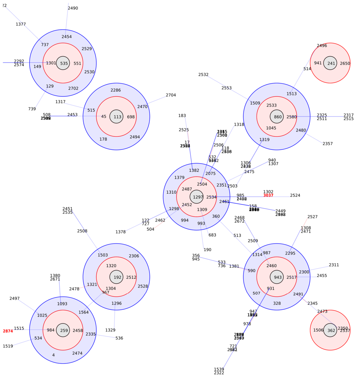
MLST was conducted on human S. pseudintermedius genome assemblies using MLST v2.23.0. The analysis identified both isolates as novel sequence types: S. pseudintermedius EGH1 was assigned ST-3037, which carries a new allele (purA-107), while S. pseudintermedius EGH2 was assigned ST-2874 Table 2. Population structure and clonal relationships among typed isolates were examined using the eBURST (Based Upon Related Sequence Types) algorithm. Single-locus variants (SLVs), defined as isolates differing at exactly one of the seven MLST loci, were identified to construct a minimum spanning tree of clonal relationships. Clonal complexes (CCs) were defined as groups of three or more related STs connected through SLV relationships, with the most common ST designated as the founder of each complex Figure 1.

3.2. Pangenome Analysis

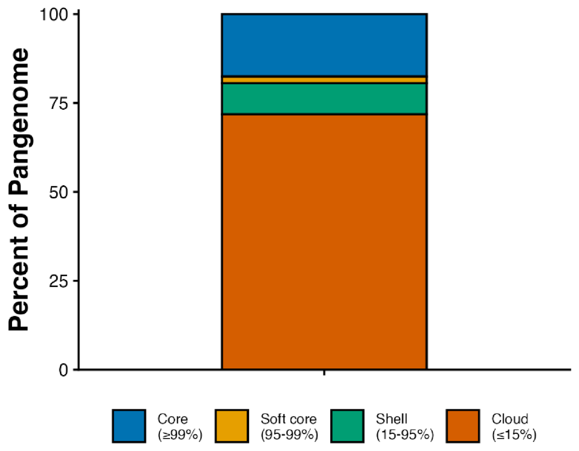
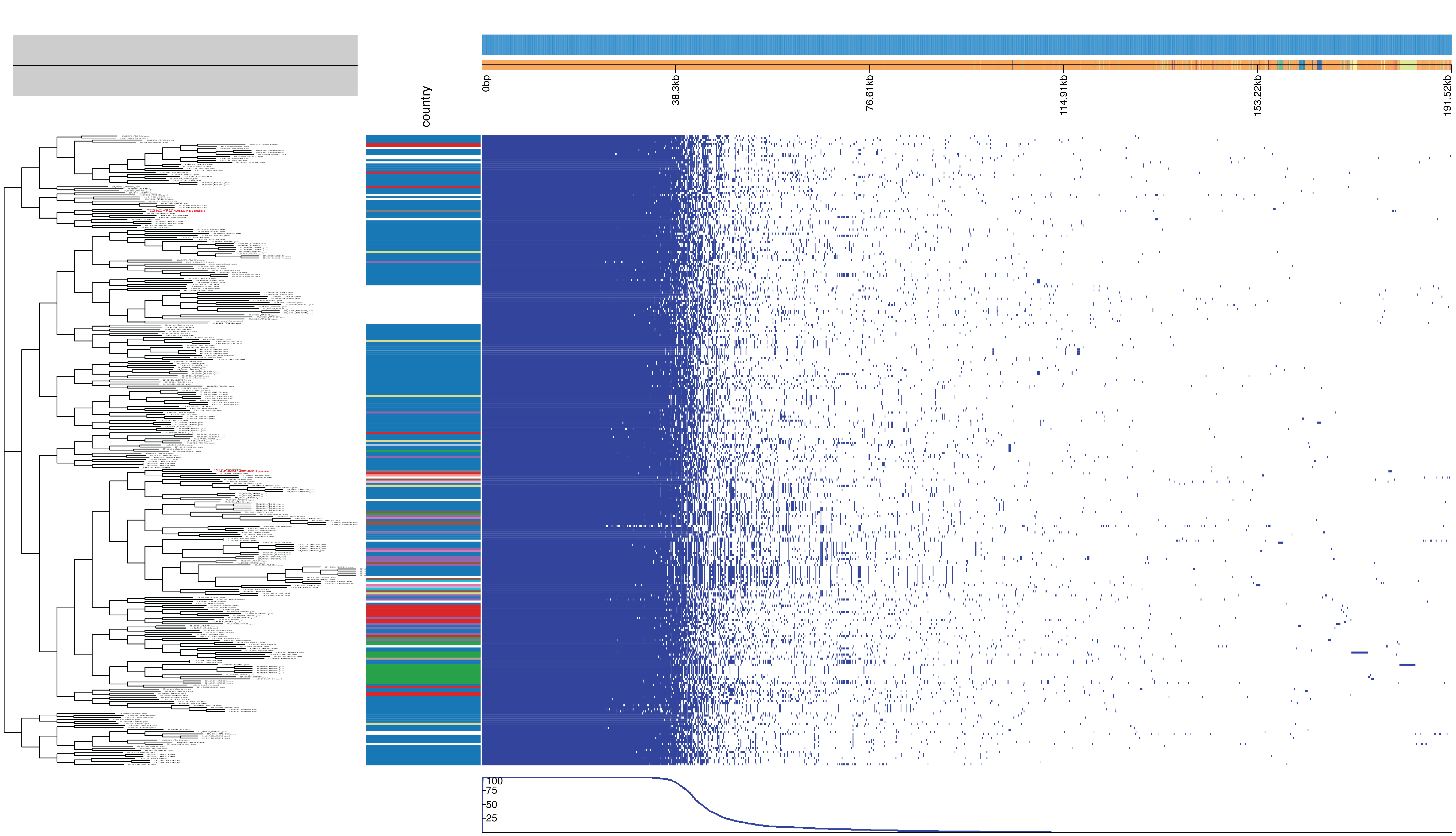
A total of 310 publicly available human-source S. pseudintermedius genome assemblies were retrieved from the NCBI Pathogen Detection database (accessed 16 October 2025) and used as the comparative dataset Table S1. These isolates originated from different countries, with the majority from the USA, and were associated with diverse clinical presentations; see Figure 2: Pangenome analysis of human S. pseudintermedius genomes using Roary. Pangenome analysis revealed a total of 9574 genes, comprising 1681 core (17.56%), 180 soft-core (1.88%), 837 shell (8.74%), and 6876 cloud genes (71.82%); see Figure 3. Eighty-five genes were found to be unique to the Egyptian isolates, with a significant number identified as hypothetical proteins, alongside the putative ATP-dependent DNA helicase yoaA.
A total of 85 genes were identified as unique to both isolates, including those coding for the putative ATP-dependent DNA helicase yoaA, several metabolic and cell-surface-associated proteins, and 62 hypothetical proteins. Conversely, 6890 genes found in other isolates were absent from EGH1 and EGH2. Additionally, 2599 genes were shared between the two isolates and at least some of the other genomes.

3.3. Antimicrobial Resistance Genes

AMRFinderPlus analysis identified multiple antimicrobial resistance (AMR) genes in both isolates. In S. pseudintermedius EGH1, genes conferring aminoglycoside resistance (ant(6)-Ia, aph(3′)-IIIa), β-lactam resistance (blaI, blaPC1, blaR1), chloramphenicol resistance (catA), macrolide-lincosamide-streptogramin B resistance (erm(B)), fusidic acid resistance (fusC), streptothricin resistance (sat4), and tetracycline resistance (tet(M)) were detected Table 3.
In S. pseudintermedius EGH2, genes associated with β-lactam resistance (blaI, blaPC1, blaR1), trimethoprim resistance (dfrG), fusidic acid resistance (fusC), and tetracycline resistance (tet(K)) were identified and are shown in Table 3.

4. Discussion

S. pseudintermedius is a coagulase-positive bacterium frequently found as a commensal organism on the skin and mucous membranes of dogs and, less often, cats. However, in recent years, S. pseudintermedius has emerged as an opportunistic pathogen responsible for a wide range of infections in companion animals. Its increasing detection in humans, especially among pet owners, veterinarians, and immunocompromised individuals, underscores its growing significance as a zoonotic agent of clinical importance [26,27,28].
Whole-genome sequencing (WGS) and comparative genomics have revealed a high level of genetic diversity within the species, with distinct clonal lineages exhibiting different host-adaptation and antimicrobial-resistance profiles [15,16]. Despite these advances, information on the genomic characteristics of S. pseudintermedius isolates from Egypt remains limited, especially regarding strains isolated from human hosts. Understanding the genomic landscape of S. pseudintermedius in underrepresented regions, such as North Africa, is therefore essential for clarifying global transmission dynamics and potential zoonotic exchange pathways between companion animals and humans. In this study, we performed whole-genome sequencing of two human S. pseudintermedius isolates from Egypt. Using a combination of multilocus sequence typing (MLST), pangenome analysis (Roary), and antimicrobial resistance gene profiling (AMRFinderPlus), we assessed the genetic relationship of these isolates to a global collection of human S. pseudintermedius genomes. Although limited to two isolates, this study provides initial genomic data from an underrepresented region and underscores the need for broader surveillance at the interface between veterinary and human infections.
AMRFinderPlus analysis revealed the presence of AMR genes in both isolates. In S. pseudintermedius EGH1, genes associated with resistance to aminoglycosides, β-lactams, chloramphenicol, macrolide-lincosamide-streptogramin B, fusidic acid, streptothricin, and tetracycline were detected. Meanwhile, S. pseudintermedius EGH2 harbored genes conferring resistance to β-lactams, trimethoprim, fusidic acid, and tetracycline. These findings, consistent with previous studies, highlight the urgent need for robust strategies to tackle AMR and for integrated One Health surveillance to curb its spread at the animal–human interface [29,30,31].
The pangenome analysis revealed a highly dynamic accessory genome, with 9574 total gene clusters spread across a relatively small core genome (1681 genes, 17.56%) and a large cloud genome (6876 genes, 71.82%). Both EGH1 and EGH2 shared core genomic features typical of S. pseudintermedius. This pangenome structure, characterized by a large accessory gene pool relative to the conserved core, has been observed in bacterial species occupying diverse niches and undergoing frequent horizontal gene transfer [32]. It should be noted, however, that including only two Egyptian isolates limits the conclusions that can be drawn about regional genomic diversity, and the patterns described here should be considered preliminary.
Among the 85 genes identified as uniquely present in EGH1 and EGH2 were genes involved in capsule biosynthesis (capC), teichoic acid biosynthesis (tarL and tarJ), and peptidoglycan synthesis. In other staphylococcal species, variation in teichoic acid structure has been associated with altered immune recognition and reduced antimicrobial susceptibility [33,34]. Whether these genes confer a similar functional advantage in the two Egyptian isolates remains to be determined through phenotypic and larger-scale comparative studies.
Several limitations in this study warrant consideration. Most notably, this study is based solely on two human isolates of S. pseudintermedius from Egypt. As a result, all findings regarding genomic content, resistance profiles, and host-associated characteristics should be considered preliminary and cannot be generalized to the broader S. pseudintermedius population in Egypt or North Africa. Furthermore, the comparative dataset is predominantly composed of isolates from the USA, which may introduce bias into population-structure analyses and restrict the applicability of the findings to global populations. Clinical metadata—including infection type, patient risk factors, and treatment outcomes—were not available for the isolates examined in this study, precluding genotype–phenotype associations. Future studies that collect standardized clinical data alongside whole-genome sequencing would enable such comparisons.

5. Conclusions

This study presents the first next-generation genome sequencing and comparative genomic analysis of S. pseudintermedius isolates from humans in Egypt. S. pseudintermedius EGH1 and EGH2 harbor unique genetic elements, multiple hypothetical proteins, and a diverse profile of antimicrobial resistance genes. Expanding genomic surveillance in North Africa and beyond will be important for building a more representative picture of S. pseudintermedius diversity in this region. Future studies with larger sample sizes, integrating genomic, epidemiological, and phenotypic data, will be needed to clarify the clinical significance and population dynamics of S. pseudintermedius across human and veterinary settings.

Supplementary Materials

The following supporting information can be downloaded at: https://www.mdpi.com/article/10.3390/vetsci13050424/s1.

Author Contributions

Conceptualization, M.A.A., H.E. and Y.B.; methodology, O.K.E., H.E. and M.R.; software, M.A.A.; validation, M.A.A. and H.E.; formal analysis, O.K.E.; investigation, H.E., Y.B. and M.R.; resources, M.A.A., H.E. and M.R.; data curation, O.K.E.; writing—original draft preparation, O.K.E.; writing—review and editing, M.A.A., S.A.K., H.E., Y.B., M.R. and H.A.; visualization, O.K.E.; supervision, M.A.A.; project administration, M.A.A.; funding acquisition, M.A.A. All authors have read and agreed to the published version of the manuscript.

Funding

Open access funding provided by Rowan University.

Institutional Review Board Statement

All the study procedures were conducted with the approval of the Ethics Committee of Faculty of Medicine, Alexandria University (Serial Number: 0305470) (IRB NO: 00012098) (FWO NO: 00018699) on 23 February 2023, Research Ethics Review Committee of Faculty of Veterinary Medicine, Alexandria University (AU130102024070) and Institutional Animal Care and Use Committee, Alexandria University (ALEXU-IACUC) (No: 309).

Informed Consent Statement

Not applicable.

Data Availability Statement

The data presented in this study are openly available in the NCBI BioProject under accession number: PRJNA1285324. S. pseudintermedius isolates EGH1 and EGH2 complete genome sequences were deposited in NCBI under accession numbers SRR34336090 and SRR34336089, respectively. Genome assemblies were deposited under accession numbers GCA_051374085.1 and GCA_051374445.1.

Conflicts of Interest

All authors state that they have no conflicts of interest or any other competing financial interest to disclose.

References

  1. Arefin, K.S.A.; Rana, E.A.; Islam, M.S.; Hossain, B.; Rahman, M.A.; Barua, H. Molecular Prevalence and Antimicrobial Resistance Profile of Staphylococcus aureus and Staphylococcus pseudintermedius Isolated From Hospital-Visited Cats. Vet. Med. Int. 2025, 2025, 4879266. [Google Scholar] [CrossRef] [PubMed]
  2. Dewulf, S.; Boyen, F.; Paepe, D.; Clercx, C.; Tilman, N.; Dewulf, J.; Boland, C. Antimicrobial Resistance Characterization of Methicillin-Resistant Staphylococcus aureus and Staphylococcus pseudintermedius Isolates from Clinical Cases in Dogs and Cats in Belgium. Antibiotics 2025, 14, 631. [Google Scholar] [CrossRef] [PubMed]
  3. Popa, I.; Iancu, I.; Iorgoni, V.; Degi, J.; Gligor, A.; Imre, K.; Tirziu, E.; Bochis, T.; Pop, C.; Plotuna, A.M.; et al. Antimicrobial Resistance Profile of Staphylococcus pseudintermedius Isolated from Dogs with Otitis Externa and Healthy Dogs: Veterinary and Zoonotic Implications. Antibiotics 2025, 14, 1027. [Google Scholar] [CrossRef] [PubMed]
  4. Borjesson, S.; Gomez-Sanz, E.; Ekstrom, K.; Torres, C.; Gronlund, U. Staphylococcus pseudintermedius can be misdiagnosed as Staphylococcus aureus in humans with dog bite wounds. Eur. J. Clin. Microbiol. Infect. Dis. 2015, 34, 839–844. [Google Scholar] [CrossRef]
  5. Yarbrough, M.L.; Lainhart, W.; Burnham, C.A. Epidemiology, Clinical Characteristics, and Antimicrobial Susceptibility Profiles of Human Clinical Isolates of Staphylococcus intermedius Group. J. Clin. Microbiol. 2018, 56, e01788-17. [Google Scholar] [CrossRef]
  6. Carroll, K.C.; Burnham, C.D.; Westblade, L.F. From canines to humans: Clinical importance of Staphylococcus pseudintermedius. PLoS Pathog. 2021, 17, e1009961. [Google Scholar] [CrossRef]
  7. Hanselman, B.A.; Kruth, S.A.; Rousseau, J.; Weese, J.S. Coagulase positive staphylococcal colonization of humans and their household pets. Can. Vet. J. 2009, 50, 954–958. [Google Scholar]
  8. Paul, N.C.; Moodley, A.; Ghibaudo, G.; Guardabassi, L. Carriage of methicillin-resistant Staphylococcus pseudintermedius in small animal veterinarians: Indirect evidence of zoonotic transmission. Zoonoses Public Health 2011, 58, 533–539. [Google Scholar] [CrossRef]
  9. Guardabassi, L.; Loeber, M.E.; Jacobson, A. Transmission of multiple antimicrobial-resistant Staphylococcus intermedius between dogs affected by deep pyoderma and their owners. Vet. Microbiol. 2004, 98, 23–27. [Google Scholar] [CrossRef]
  10. Peacock, S.J.; Paterson, G.K. Mechanisms of Methicillin Resistance in Staphylococcus aureus. Annu. Rev. Biochem. 2015, 84, 577–601. [Google Scholar] [CrossRef]
  11. Zehr, J.D.; Sun, Q.; Ceres, K.; Merrill, A.; Tyson, G.H.; Ceric, O.; Guag, J.; Pauley, S.; McQueary, H.C.; Sams, K.; et al. Population and pan-genomic analyses of Staphylococcus pseudintermedius identify geographic distinctions in accessory gene content and novel loci associated with AMR. Appl. Environ. Microbiol. 2025, 91, e0001025. [Google Scholar] [CrossRef] [PubMed]
  12. Abouelkhair, M.A.; Bemis, D.A.; Giannone, R.J.; Frank, L.A.; Kania, S.A. Characterization of a leukocidin identified in Staphylococcus pseudintermedius. PLoS ONE 2018, 13, e0204450. [Google Scholar] [CrossRef] [PubMed]
  13. Abouelkhair, M.A.; Bemis, D.A.; Giannone, R.J.; Frank, L.A.; Kania, S.A. Identification, cloning and characterization of SpEX exotoxin produced by Staphylococcus pseudintermedius. PLoS ONE 2019, 14, e0220301. [Google Scholar] [CrossRef] [PubMed]
  14. Ferrer, L.; Garcia-Fonticoba, R.; Perez, D.; Vines, J.; Fabregas, N.; Madronero, S.; Meroni, G.; Martino, P.A.; Martinez, S.; Mate, M.L.; et al. Whole genome sequencing and de novo assembly of Staphylococcus pseudintermedius: A pangenome approach to unravelling pathogenesis of canine pyoderma. Vet. Dermatol. 2021, 32, 654–663. [Google Scholar] [CrossRef]
  15. Saengsawang, P.; Tanonkaew, R.; Kimseng, R.; Nissapatorn, V.; Wintachai, P.; Rodriguez-Ortega, M.J.; Mitsuwan, W. Whole Genome Sequence Analysis of Multidrug-Resistant Staphylococcus aureus and Staphylococcus pseudintermedius Isolated from Superficial Pyoderma in Dogs and Cats. Antibiotics 2025, 14, 643. [Google Scholar] [CrossRef]
  16. Phophi, L.; Abouelkhair, M.; Jones, R.; Henton, M.; Qekwana, D.N.; Kania, S.A. The molecular epidemiology and antimicrobial resistance of Staphylococcus pseudintermedius canine clinical isolates submitted to a veterinary diagnostic laboratory in South Africa. PLoS ONE 2023, 18, e0290645. [Google Scholar] [CrossRef]
  17. Phophi, L.; Abouelkhair, M.A.; Jones, R.; Zehr, J.; Kania, S.A. Temporal changes in antibiotic resistance and population structure of methicillin-resistant Staphylococcus pseudintermedius between 2010 and 2021 in the United States. Comp. Immunol. Microbiol. Infect. Dis. 2023, 100, 102028. [Google Scholar] [CrossRef]
  18. Elaadli, H.; Badr, Y.; Raouf, M.; Kania, S.A.; Elsakhawy, O.K.; Altaib, H.; Abouelkhair, M.A. Isolation and Molecular Characterization of Three Staphylococcus pseudintermedius Strains from Dogs and Humans in Egypt. Curr. Microbiol. 2025, 82, 493. [Google Scholar] [CrossRef]
  19. Kearse, M.; Moir, R.; Wilson, A.; Stones-Havas, S.; Cheung, M.; Sturrock, S.; Buxton, S.; Cooper, A.; Markowitz, S.; Duran, C.; et al. Geneious Basic: An integrated and extendable desktop software platform for the organization and analysis of sequence data. Bioinformatics 2012, 28, 1647–1649. [Google Scholar] [CrossRef]
  20. Gurevich, A.; Saveliev, V.; Vyahhi, N.; Tesler, G. QUAST: Quality assessment tool for genome assemblies. Bioinformatics 2013, 29, 1072–1075. [Google Scholar] [CrossRef]
  21. Merkel, D. Docker: Lightweight Linux containers for consistent development and deployment. Linux J. 2014, 2014, 2. [Google Scholar]
  22. Jolley, K.A.; Bray, J.E.; Maiden, M.C.J. Open-access bacterial population genomics: BIGSdb software, the PubMLST.org website and their applications. Wellcome Open Res. 2018, 3, 124. [Google Scholar] [CrossRef] [PubMed]
  23. Seemann, T. Prokka: Rapid prokaryotic genome annotation. Bioinformatics 2014, 30, 2068–2069. [Google Scholar] [CrossRef]
  24. Page, A.J.; Cummins, C.A.; Hunt, M.; Wong, V.K.; Reuter, S.; Holden, M.T.; Fookes, M.; Falush, D.; Keane, J.A.; Parkhill, J. Roary: Rapid large-scale prokaryote pan genome analysis. Bioinformatics 2015, 31, 3691–3693. [Google Scholar] [CrossRef] [PubMed]
  25. Feldgarden, M.; Brover, V.; Gonzalez-Escalona, N.; Frye, J.G.; Haendiges, J.; Haft, D.H.; Hoffmann, M.; Pettengill, J.B.; Prasad, A.B.; Tillman, G.E.; et al. AMRFinderPlus and the Reference Gene Catalog facilitate examination of the genomic links among antimicrobial resistance, stress response, and virulence. Sci. Rep. 2021, 11, 12728. [Google Scholar] [CrossRef]
  26. Gomez-Sanz, E.; Torres, C.; Lozano, C.; Zarazaga, M. High diversity of Staphylococcus aureus and Staphylococcus pseudintermedius lineages and toxigenic traits in healthy pet-owning household members. Underestimating normal household contact? Comp. Immunol. Microbiol. Infect. Dis. 2013, 36, 83–94. [Google Scholar] [CrossRef]
  27. Kronbichler, A.; Blane, B.; Holmes, M.A.; Wagner, J.; Parkhill, J.; Peacock, S.J.; Jayne, D.R.W.; Harrison, E.M. Nasal carriage of Staphylococcus pseudintermedius in patients with granulomatosis with polyangiitis. Rheumatology 2019, 58, 548–550. [Google Scholar] [CrossRef]
  28. Kuan, E.C.; Yoon, A.J.; Vijayan, T.; Humphries, R.M.; Suh, J.D. Canine Staphylococcus pseudintermedius sinonasal infection in human hosts. Int. Forum Allergy Rhinol. 2016, 6, 710–715. [Google Scholar] [CrossRef]
  29. Alhadz, G.G.; Salasia, S.I.O.; Lestari, F.B.; Yosyana, A.R.P.; Wasissa, M.; Setianingrum, Y.R.; Widayanti, R. Occurrence, molecular confirmation, and multidrug resistance of methicillin-resistant Staphylococcus aureus and Staphylococcus pseudintermedius in companion animals in Indonesia. Vet. World 2026, 19, 324–338. [Google Scholar] [CrossRef]
  30. Afema, J.A.; Mastromonaco, C.; Davis, M.A.; Stone, D.M.; Jones, L.P.; Paterson, T.E.; Perea, M.L.; Butler, B.P. Emergence of methicillin resistant Staphylococcus pseudintermedius in dogs sampled in 2018 in the island nation of Grenada, West Indies. Front. Vet. Sci. 2026, 13, 1761713. [Google Scholar] [CrossRef]
  31. Douan, J.; Kohler, C.; Haralambiev, L.; Idelevich, E.A.; Becker, K. Comparative phylogenetic, antimicrobial resistance, and clinical characterization of human spondylodiscitis-associated Staphylococcus pseudintermedius. Front. Microbiol. 2026, 17, 1735075. [Google Scholar] [CrossRef]
  32. Wiedenbeck, J.; Cohan, F.M. Origins of bacterial diversity through horizontal genetic transfer and adaptation to new ecological niches. FEMS Microbiol. Rev. 2011, 35, 957–976. [Google Scholar] [CrossRef]
  33. Di Carluccio, C.; Soriano-Maldonado, P.; Berni, F.; de Haas, C.J.C.; Temming, A.R.; Hendriks, A.; Ali, S.; Molinaro, A.; Silipo, A.; van Sorge, N.M.; et al. Antibody Recognition of Different Staphylococcus aureus Wall Teichoic Acid Glycoforms. ACS Cent. Sci. 2022, 8, 1383–1392. [Google Scholar] [CrossRef]
  34. Atilano, M.L.; Yates, J.; Glittenberg, M.; Filipe, S.R.; Ligoxygakis, P. Wall teichoic acids of Staphylococcus aureus limit recognition by the drosophila peptidoglycan recognition protein-SA to promote pathogenicity. PLoS Pathog. 2011, 7, e1002421. [Google Scholar] [CrossRef]
Figure 1. Analysis of clonal complexes of human Staphylococcus pseudintermedius isolates using the eBURST algorithm. Single locus variant (SLV) profiles that match the central profile are indicated within a red circle, while double locus variant (DLV) profiles are marked within a blue circle. More distant profiles, referred to as triple locus variants, are connected by a line. Sequence types of S. pseudintermedius EGH1 (ST 3037) and EGH2 (ST 2874) are highlighted in red.
Vetsci 13 00424 g001
Figure 2. Pangenome analysis of human S. pseudintermedius genomes using Roary. The left panel shows a Bayesian phylogenetic tree constructed from single-nucleotide polymorphisms (SNPs) in the core genome. The right panel presents a Roary gene presence/absence matrix, where each column represents an isolate and each row corresponds to a gene cluster. Blue shading indicates the presence of a gene, while white denotes its absence. Isolates from different countries are color-coded as follows: USA (#1f77b4), Trinidad and Tobago (#ff7f0e), Brazil (#2ca02c), United Kingdom (#d62728), South Africa (#9467bd), Japan (#8c564b), Thailand (#e377c2), Egypt (#7f7f7f), Australia (#bcbd22), Switzerland (#17becf), Ireland (#aec7e8), New Zealand (#ffbb78), France (#98df8a), Hungary (#ff9896), South Korea (#c5b0d5), Argentina (#c49c94), Germany (#f7b6d2), Sweden (#c7c7c7), not collected (#dbdb8d).
Vetsci 13 00424 g002
Figure 3. Pangenome composition of human Staphylococcus pseudintermedius isolates analyzed using Roary. The figure illustrates the proportion of genes classified as core (present in all genomes, blue: #0072B2), soft-core (present in 95–99% of genomes, orange: #E69F00), shell (present in 15–95% of genomes, green: #009E73), and cloud (present in <15% of genomes, red#D55E00).
Vetsci 13 00424 g003
Table 1. Characteristics of the genome sequences of S. pseudintermedius EGH1 and S. pseudintermedius EGH2.
EGH1EGH2
Number of reads9,499,9899,567,531
Genome length2,536,812 bp2,636,008 bp
N50141,100 bp139,200 bp
Number of contigs4199
GC percent37.5%37.5%
Genome coverage9.22×9.29×
Genes24242596
Table 2. MLST profile of the two human S. pseudintermedius isolates.
Isolateackcpn60fdhptapuraSartufSequence Type (ST)
S. pseudintermdius EGH16211107123037
S. pseudintermdius EGH2118223112874
Table 3. Antimicrobial resistance genes identified in S. pseudintermedius EGH1 and EGH2.
Antimicrobial ClassGeneEGH1EGH2
Aminoglycosideant(6)-Ia+
aph(3′)-IIIa+
β-LactamblaI++
blaPC1++
blaR1++
ChloramphenicolcatA+
Fusidic acidfusC++
Macrolide–lincosamide–streptogramin Berm(B)+
Streptothricinsat4+
Tetracyclinetet(M)+
tet(K)+
TrimethoprimdfrG+
Disclaimer/Publisher’s Note: The statements, opinions and data contained in all publications are solely those of the individual author(s) and contributor(s) and not of MDPI and/or the editor(s). MDPI and/or the editor(s) disclaim responsibility for any injury to people or property resulting from any ideas, methods, instructions or products referred to in the content.